

The DOI matches its Crossref record; 19 of the 19 references it cites were found in a registry or an index.
Primary exposure to SARS-CoV-2 protects against reinfection in rhesus macaques
Open accessAbstract
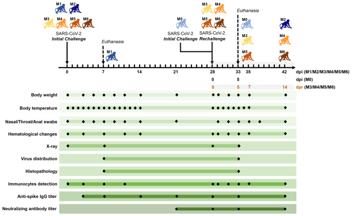
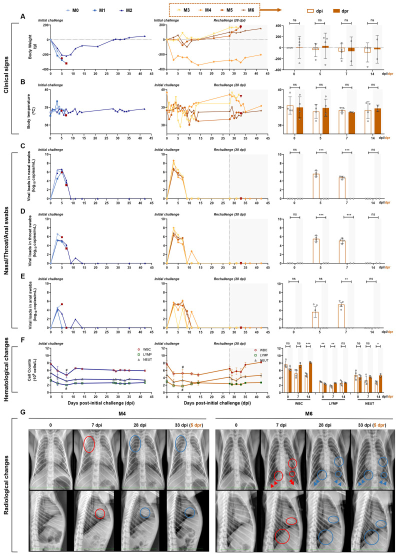
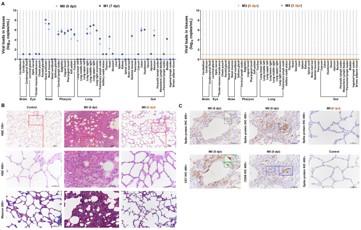
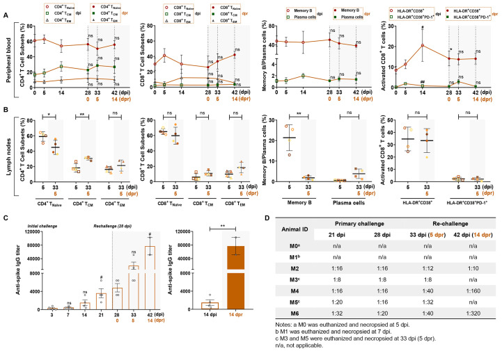
Immunity from reinfection: One of the many open questions about severe acute respiratory syndrome coronavirus 2 (SARS-CoV-2) infection is whether an individual who has cleared the virus can be infected a second time and get sick. Chandrashekar et al. and Deng et al. generated rhesus macaque models of SARS-CoV-2 infection and tested whether natural SARS-CoV-2 infection could result in immunity to viral rechallenge. They found that animals indeed developed immune responses that protected against a second infection. Although there are differences between SARS-CoV-2 infection in macaques and in humans, these findings have key implications for public health and economic initiatives if validated in human studies. Science , this issue p. 812 , p. 818
Overview
The claims and statistics of this paper haven’t been checked yet.
Run the check — free, no account needed. It reads the open-access text and checks the sources it cites.
Integrity signals
Registry
Cites
Statistics
Figures
Verification
Claims the paper makes
The claims and statistics of this paper haven’t been checked yet.
Run the check — free, no account needed. It reads the open-access text and checks the sources it cites.
Claims other papers make about this one
Each is checked against this paper’s own text: does it say what the citing paper says it does?
In the citing paper
“Initial studies in macaques showed that a primary infection with SARS-CoV-2 protects against subsequent viral challenge for at least 28–35 days, with re-exposure driving memory immune responses that included boosting of binding and neutralizing antibody levels as well as of IFNγ secretion by T cells46,47.”
From Prospects for durable immune control of SARS-CoV-2 and prevention of reinfection (2021) · cites this paper as [46]
In the citing paper
“Preexisting immunity to SARS-CoV-2 is associated with protection against reinfection in humans (9–11) and in nonhuman primates (12, 13).”
From mRNA vaccination boosts cross-variant neutralizing antibodies elicited by SARS-CoV-2 infection (2021) · cites this paper as [13]
In the citing paper
“Animals infected with SARS-CoV-2 were protected from a high dose re-challenge with SARS-CoV-2 1 month later, and protection was associated with neutralizing antibodies”
From Adaptive immunity to SARS-CoV-2 and COVID-19 (2021) · cites this paper as Deng et al., 2020
Self-check
Percentages, sums and reported differences re-computed from the paper’s own numbers.
The self-check needs the paper’s full text.
Citations
Cites
A novel coronavirus from patients with pneumonia in China, 2019
Zhu, N., Zhang, D., Wang, W., Li, X., Yang, B., Song, J., Zhao, X., Huang, B., Shi, W., Lu, R., et al. (2020) · New England Journal of Medicine · doi:10.1056/nejmoa2001017 · Cited by 28,244
To, K. K., Tsang, O. T.-Y., Leung, W.-S., Tam, A. R., Wu, T.-C., Lung, D. C., Yip, C. C., Cai, J., Chan, J. M., Chik, T. S., et al. (2020) · The Lancet Infectious Diseases · doi:10.1016/s1473-3099(20)30196-1 · Cited by 3,045
Okba, N. M. A., Müller, M. A., Li, W., Wang, C., GeurtsvanKessel, C. H., Corman, V. M., Lamers, M. M., Sikkema, R. S., de Bruin, E., Chandler, F. D., et al. (2020) · Emerging Infectious Diseases · doi:10.3201/eid2607.200841 · Cited by 1,614
Positive RT-PCR test results in patients recovered from COVID-19
Lan, L., Xu, D., Ye, G., Xia, C., Wang, S., Li, Y., & Xu, H. (2020) · JAMA · doi:10.1001/jama.2020.2783 · Cited by 1,251
Self-citationThe pathogenicity of SARS-CoV-2 in hACE2 transgenic mice
Bao, L., Deng, W., Huang, B., Gao, H., Liu, J., Ren, L., Wei, Q., Yu, P., Xu, Y., Qi, F., et al. (2020) · Nature · doi:10.1038/s41586-020-2312-y · Cited by 1,126
Self-citationComparative pathogenesis of COVID-19, MERS, and SARS in a nonhuman primate model
Rockx, B., Kuiken, T., Herfst, S., Bestebroer, T., Lamers, M. M., Oude Munnink, B. B., de Meulder, D., van Amerongen, G., van den Brand, J., Okba, N. M. A., et al. (2020) · Science · doi:10.1126/science.abb7314 · Cited by 901
SARS-CoV-2 infection protects against rechallenge in rhesus macaques
Chandrashekar, A., Liu, J., Martinot, A. J., McMahan, K., Mercado, N. B., Peter, L., Tostanoski, L. H., Yu, J., Maliga, Z., Nekorchuk, M., et al. (2020) · Science · doi:10.1126/science.abc4776 · Cited by 816
Self-citationWu, F., Wang, A., Liu, M., Wang, Q., Chen, J., Xia, S., Ling, Y., Zhang, Y., Xun, J., Lu, L., et al. (2020) · doi:10.1101/2020.03.30.20047365 · Cited by 754
Self-citationRespiratory disease in rhesus macaques inoculated with SARS-CoV-2
Munster, V. J., Feldmann, F., Williamson, B. N., van Doremalen, N., Pérez-Pérez, L., Schulz, J., Meade-White, K., Okumura, A., Callison, J., Brumbaugh, B., et al. (2020) · Nature · doi:10.1038/s41586-020-2324-7 · Cited by 720
Clinical benefit of remdesivir in rhesus macaques infected with SARS-CoV-2
Williamson, B. N., Feldmann, F., Schwarz, B. H., Meade-White, K. D., Porter, D., Schulz, J. E., van Doremalen, N., Leighton, I. A., Yinda, C. K., Pérez-Pérez, L., et al. (2020) · Nature · doi:10.1038/s41586-020-2423-5 · Cited by 696
Cited by
Sorted by citations
Immunological memory to SARS-CoV-2 assessed for up to 8 months after infection.
Dan, J. M., Mateus, J., Kato, Y., Hastie, K. M., Yu, E. D., Faliti, C. E., Grifoni, A., Ramirez, S. I., Haupt, S., Frazier, A., et al. (2021) · Science · doi:10.1126/science.abf4063 · Cited by 2,656
Folegatti, P. M., Ewer, K. J., Aley, P. K., Angus, B., Becker, S., Belij-Rammerstorfer, S., Bellamy, D., Bibi, S., Bittaye, M., Clutterbuck, E. A., et al. (2020) · The Lancet · doi:10.1016/s0140-6736(20)31604-4 · Cited by 2,419
SARS-CoV-2 vaccines in development
Krammer, F. (2020) · Nature · doi:10.1038/s41586-020-2798-3 · Cited by 2,098
Adaptive immunity to SARS-CoV-2 and COVID-19
Sette, A., & Crotty, S. (2021) · Cell · doi:10.1016/j.cell.2021.01.007 · Cited by 1,860
Seow, J., Graham, C., Merrick, B., Acors, S., Pickering, S., Steel, K. J. A., Hemmings, O., O’Byrne, A., Kouphou, N., Galao, R. P., et al. (2020) · Nature Microbiology · doi:10.1038/s41564-020-00813-8 · Cited by 1,419
Robust neutralizing antibodies to SARS-CoV-2 infection persist for months
Wajnberg, A., Amanat, F., Firpo, A., Altman, D. R., Bailey, M. J., Mansour, M., McMahon, M., Meade, P., Mendu, D. R., Muellers, K., et al. (2020) · Science · doi:10.1126/science.abd7728 · Cited by 1,195
Humoral Immune Response to SARS-CoV-2 in Iceland
Gudbjartsson, D. F., Norddahl, G. L., Melsted, P., Gunnarsdottir, K., Holm, H., Eythorsson, E., Arnthorsson, A. O., Helgason, D., Bjarnadottir, K., Ingvarsson, R. F., et al. (2020) · New England Journal of Medicine · doi:10.1056/nejmoa2026116 · Cited by 903
Adaptation of SARS-CoV-2 in BALB/c mice for testing vaccine efficacy
Gu, H., Chen, Q., Yang, G., He, L., Fan, H., Deng, Y.-Q., Wang, Y., Teng, Y., Zhao, Z., Cui, Y., et al. (2020) · Science · doi:10.1126/science.abc4730 · Cited by 762
Self-citationEffect of an Inactivated Vaccine Against SARS-CoV-2 on Safety and Immunogenicity Outcomes
Xia, S., Duan, K., Zhang, Y., Zhao, D., Zhang, H., Xie, Z., Li, X., Peng, C., Zhang, Y., Zhang, W., et al. (2020) · JAMA · doi:10.1001/jama.2020.15543 · Cited by 768
COVID-19 Neutralizing Antibodies Predict Disease Severity and Survival
García-Beltrán, W. F., Lam, E. C., Astudillo, M. G., Yang, D., Miller, T. E., Feldman, J. E., Hauser, B. M., Caradonna, T. M., Clayton, K., Nitido, A. D., et al. (2020) · Cell · doi:10.1016/j.cell.2020.12.015 · Cited by 708
Munro, A. P. S., Janani, L., Cornelius, V. R., Aley, P. K., Babbage, G., Baxter, D. N., Bula, M., Cathie, K., Chatterjee, K., Dodd, K., et al. (2021) · The Lancet · doi:10.1016/s0140-6736(21)02717-3 · Cited by 656
Self-citationImmunity, endothelial injury and complement-induced coagulopathy in COVID-19
Perico, L., Benigni, A., Casiraghi, F., Ng, L. F. P., Rénia, L., & Remuzzi, G. (2020) · Nature Reviews Nephrology · doi:10.1038/s41581-020-00357-4 · Cited by 616
SARS-CoV-2 immunity: review and applications to phase 3 vaccine candidates
Poland, G. A., Ovsyannikova, I. G., & Kennedy, R. B. (2020) · The Lancet · doi:10.1016/s0140-6736(20)32137-1 · Cited by 572
mRNA vaccination boosts cross-variant neutralizing antibodies elicited by SARS-CoV-2 infection
Stamatatos, L., Czartoski, J., Wan, Y.-H., Homad, L. J., Rubin, V., Glantz, H., Neradilek, M., Seydoux, E., Jennewein, M. F., MacCamy, A. J., et al. (2021) · Science · doi:10.1126/science.abg9175 · Cited by 557
Addetia, A., Crawford, K. H. D., Dingens, A., Zhu, H., Roychoudhury, P., Huang, M.-L., Jerome, K. R., Bloom, J. D., & Greninger, A. L. (2020) · Journal of Clinical Microbiology · doi:10.1128/jcm.02107-20 · Cited by 525
REGN-COV2 antibodies prevent and treat SARS-CoV-2 infection in rhesus macaques and hamsters
Baum, A., Ajithdoss, D., Copin, R., Zhou, A., Lanza, K., Negron, N., Ni, M., Wei, Y., Mohammadi, K., Musser, B., et al. (2020) · Science · doi:10.1126/science.abe2402 · Cited by 517
COVID-19: Discovery, diagnostics and drug development
Asselah, T., Durantel, D., Pasmant, É., Lau, G. K. K., & Schinazi, R. F. (2020) · Journal of Hepatology · doi:10.1016/j.jhep.2020.09.031 · Cited by 478
The first 12 months of COVID-19: a timeline of immunological insights
Carvalho, T., Krammer, F., & Iwasaki, A. (2021) · Nature reviews. Immunology · doi:10.1038/s41577-021-00522-1 · Cited by 483
Multi-organ proteomic landscape of COVID-19 autopsies
Nie, X., Qian, L., Sun, R., Huang, B., Dong, X., Xiao, Q., Zhang, Q., Lu, T., Yue, L., Chen, S., et al. (2021) · Cell · doi:10.1016/j.cell.2021.01.004 · Cited by 403
Self-citationEvolution of immune responses to SARS-CoV-2 in mild-moderate COVID-19
Wheatley, A. K., Juno, J. A., Wang, J. J., Selva, K. J., Reynaldi, A., Tan, H.-X., Lee, W. S., Wragg, K. M., Kelly, H. G., Esterbauer, R., et al. (2021) · Nature Communications · doi:10.1038/s41467-021-21444-5 · Cited by 351
Potential intestinal infection and faecal–oral transmission of SARS-CoV-2
Guo, M., Tao, W., Flavell, R. A., & Zhu, S. (2021) · Nature Reviews Gastroenterology & Hepatology · doi:10.1038/s41575-021-00416-6 · Cited by 314
Correlates of protection against SARS ‐ CoV ‐2 infection and COVID‐19 disease
Goldblatt, D., Alter, G., Crotty, S., & Plotkin, S. A. (2022) · Immunological Reviews · doi:10.1111/imr.13091 · Cited by 301
Prendecki, M., Clarke, C., Brown, J., Cox, A., Gleeson, S., Guckian, M., Randell, P., Pria, A. D., Lightstone, L., Xu, X.-N., et al. (2021) · The Lancet · doi:10.1016/s0140-6736(21)00502-x · Cited by 271
A therapeutic neutralizing antibody targeting receptor binding domain of SARS-CoV-2 spike protein
Kim, C.-M., Ryu, D.-K., Lee, J., Kim, Y., Seo, J. M., Kim, Y., Jeong, J.-H., Kim, M., Kim, J.-I., Kim, P., et al. (2021) · Nature Communications · doi:10.1038/s41467-020-20602-5 · Cited by 283
Hoepel, W., Chen, H., Geyer, C. E., Allahverdiyeva, S., Manz, X. D., de Taeye, S. W., Aman, J., Mes, L., Steenhuis, M., Griffith, G. R., et al. (2021) · Science Translational Medicine · doi:10.1126/scitranslmed.abf8654 · Cited by 282
Prospects for durable immune control of SARS-CoV-2 and prevention of reinfection
Cromer, D., Juno, J. A., Khoury, D., Reynaldi, A., Wheatley, A. K., Kent, S. J., & Davenport, M. P. (2021) · Nature Reviews Immunology · doi:10.1038/s41577-021-00550-x · Cited by 265
A smartphone-read ultrasensitive and quantitative saliva test for COVID-19
Ning, B., Yu, T., Zhang, S., Huang, Z. J., Tian, D., Lin, Z. N., Niu, A., Golden, N., Hensley, K., Threeton, B., et al. (2021) · Science Advances · doi:10.1126/sciadv.abe3703 · Cited by 255
Self-citationHumoral Responses and Serological Assays in SARS-CoV-2 Infections
Galipeau, Y., Greig, M., Liu, C., Driedger, M., & Langlois, M. (2020) · Frontiers in Immunology · doi:10.3389/fimmu.2020.610688 · Cited by 247
Du, S., Cao, Y., Zhu, Q., Yü, P., Qi, F., Wang, G., Du, X., Bao, L., Deng, W., Zhu, H., et al. (2020) · Cell · doi:10.1016/j.cell.2020.09.035 · Cited by 238
Self-citationZhang, M., Wang, P., Luo, R., Wang, Y., Li, Z., Guo, Y., Yao, Y., Li, M., Tao, T., Chen, W., et al. (2021) · Advanced Science · doi:10.1002/advs.202002928 · Cited by 236
Establishment of an African green monkey model for COVID-19 and protection against re-infection
Woolsey, C. B., Borisevich, V., Prasad, A. N., Agans, K. N., Deer, D. J., Dobias, N. S., Heymann, J. C., Foster, S. L., Levine, C. B., Medina, L. O., et al. (2020) · Nature Immunology · doi:10.1038/s41590-020-00835-8 · Cited by 232
Feng, L.-Q., Wang, Q., Chao, S., Yang, C., Feng, Y., Wu, J., Liu, X., Zhou, Y., Jiang, R.-D., Hu, P., et al. (2020) · Nature Communications · doi:10.1038/s41467-020-18077-5 · Cited by 223
Self-citationSARS-CoV-2 Epitopes Are Recognized by a Public and Diverse Repertoire of Human T Cell Receptors
Shomuradova, A. S., Vagida, M. S., Sheetikov, S. A., Zornikova, K. V., Kiryukhin, D. O., Titov, A., Peshkova, I. O., Khmelevskaya, A. A., Dianov, D. V., Malasheva, M., et al. (2020) · Immunity · doi:10.1016/j.immuni.2020.11.004 · Cited by 230
COVID-19 and the Path to Immunity
Stephens, D. S., & McElrath, M. J. (2020) · JAMA · doi:10.1001/jama.2020.16656 · Cited by 217
Rhee, C., Kanjilal, S., Baker, M. A., & Klompas, M. (2020) · Clinical Infectious Diseases · doi:10.1093/cid/ciaa1249 · Cited by 222
Responses to acute infection with SARS-CoV-2 in the lungs of rhesus macaques, baboons and marmosets
Singh, D. K., Singh, B., Ganatra, S. R., Gazi, M., Cole, J., Thippeshappa, R., Alfson, K. J., Clemmons, E., Gonzalez, O., Escobedo, R., et al. (2020) · Nature Microbiology · doi:10.1038/s41564-020-00841-4 · Cited by 209
Decline of Humoral Responses against SARS-CoV-2 Spike in Convalescent Individuals
Beaudoin-Bussières, G., Laumaea, A., Anand, S. P., Prévost, J., Gasser, R., Goyette, G., Medjahed, H., Perreault, J., Tremblay, T., Lewin, A., et al. (2020) · mBio · doi:10.1128/mbio.02590-20 · Cited by 189
Ryan, K. A., Bewley, K. R., Fotheringham, S. A., Slack, G. S., Brown, P. J., Hall, Y., Wand, N. I. V., Marriott, A. C., Cavell, B. E., Tree, J. A., et al. (2021) · Nature Communications · doi:10.1038/s41467-020-20439-y · Cited by 191
Public mental health problems during COVID-19 pandemic: a large-scale meta-analysis of the evidence
Liu, X., Zhu, M., Zhang, R., Zhang, J., Zhang, C., Liu, P., Feng, Z., & Chen, Z. (2021) · Translational Psychiatry · doi:10.1038/s41398-021-01501-9 · Cited by 195
Self-citationComparative ACE2 variation and primate COVID-19 risk
Melin, A. D., Janiak, M. C., Marrone, F., Arora, P. S., & Higham, J. P. (2020) · Communications Biology · doi:10.1038/s42003-020-01370-w · Cited by 185
Lu, J., Peng, J.-J., Xiong, Q.-L., Liu, Z., Lin, H.-F., Tan, X.-H., Kang, M., Yuan, R.-Y., Zeng, L., Zhou, P., et al. (2020) · medRxiv · doi:10.1016/j.ebiom.2020.102960 · Cited by 167
Nambulli, S., Xiang, Y., Tilston‐Lunel, N. L., Rennick, L. J., Sang, Z., Klimstra, W. B., Reed, D. S., Crossland, N. A., Shi, Y., & Duprex, W. P. (2021) · Science Advances · doi:10.1126/sciadv.abh0319 · Cited by 160
An Overview of SARS-CoV-2 and Animal Infection
Mahdy, M. A. A., Younis, W., & Ewaida, Z. M. (2020) · Frontiers in Veterinary Science · doi:10.3389/fvets.2020.596391 · Cited by 158
Schwarzkopf, S., Krawczyk, A., Knop, D., Klump, H., Heinold, A., Heinemann, F. M., Thümmler, L., Temme, C., Breyer, M., Witzke, O., et al. (2020) · Emerging infectious diseases · doi:10.3201/2701.203772 · Cited by 138
Persistent cellular immunity to SARS-CoV-2 infection
Breton, G., Mendoza, P., Hägglöf, T., Oliveira, T. Y., Schaefer-Babajew, D., Gaebler, C., Turroja, M., Hurley, A., Caskey, M., & Nussenzweig, M. C. (2021) · Journal of Experimental Medicine · doi:10.1084/jem.20202515 · Cited by 129
Blockchain-Based Solution for COVID-19 Digital Medical Passports and Immunity Certificates
Hasan, H. R., Salah, K., Jayaraman, R., Arshad, J., Yaqoob, I., Omar, M., & Ellahham, S. H. (2020) · IEEE Access · doi:10.1109/access.2020.3043350 · Cited by 128
COVID-19 vaccines for patients with cancer: benefits likely outweigh risks
Hwang, J. K., Zhang, T., Wang, A. Z., & Li, Z. (2021) · Journal of Hematology & Oncology · doi:10.1186/s13045-021-01046-w · Cited by 127
Immunological memory to SARS-CoV-2 assessed for up to eight months after infection.
Dan, J. M., Mateus, J., Kato, Y., Hastie, K. M., Yu, E. D., Faliti, C. E., Grifoni, A., Ramirez, S. I., Haupt, S., Frazier, A., et al. (2020) · eScholarship (California Digital Library) · Cited by 121
Protective humoral and cellular immune responses to SARS-CoV-2 persist up to 1 year after recovery
Feng, C., Shi, J., Fan, Q., Wang, Y., Huang, H., Chen, F., Tang, G., Li, Y., Li, P., Li, J. J., et al. (2021) · Nature Communications · doi:10.1038/s41467-021-25312-0 · Cited by 120
Self-citationWang, S., Li, W., Wang, Z., Yang, W., Li, E., Xia, X., Yan, F., & Chiu, S. (2024) · Signal Transduction and Targeted Therapy · doi:10.1038/s41392-024-01917-x · Cited by 124
Self-citationYou, M., Chen, L., Zhang, D., Zhao, P., Chen, Z., Qin, E., Gao, Y., Davis, M. M., & Yang, P. (2021) · Nature Cell Biology · doi:10.1038/s41556-021-00690-1 · Cited by 124
Measuring immunity to SARS-CoV-2 infection: comparing assays and animal models
Khoury, D. S., Wheatley, A. K., Ramuta, M. D., Reynaldi, A., Cromer, D., Subbarao, K., O’Connor, D. H., Kent, S. J., & Davenport, M. P. (2020) · Nature Reviews Immunology · doi:10.1038/s41577-020-00471-1 · Cited by 115
Carrillo, J., Izquierdo‐Useros, N., Ávila‐Nieto, C., Pradenas, E., Clotet, B., & Blanco, J. (2020) · Biochemical and Biophysical Research Communications · doi:10.1016/j.bbrc.2020.10.108 · Cited by 120
Risk of human‐to‐wildlife transmission of SARS‐CoV‐2
Gryseels, S., Bruyn, L. D., Gyselings, R., Calvignac‐Spencer, S., Leendertz, F. H., & Leirs, H. (2020) · Mammal Review · doi:10.1111/mam.12225 · Cited by 114
Yang, Y., Yang, M., Peng, Y., Liang, Y., Wei, J., Xing, L., Guo, L., Li, X., Li, J., Wang, J., et al. (2022) · Nature Microbiology · doi:10.1038/s41564-021-01051-2 · Cited by 111
Self-citationLiu, Z., Zhou, J., Xu, W., Deng, W., Wang, Y., Wang, M., Wang, Q., Hsieh, M. K., Dong, J., Wang, X., et al. (2022) · Cell Research · doi:10.1038/s41422-022-00612-2 · Cited by 113
Self-citationWang, G., Yang, M., Duan, Z., Liu, F., Jin, L., Long, C., Zhang, M., Tang, X., Xu, L., Li, Y.-C., et al. (2020) · Cell Research · doi:10.1038/s41422-020-00450-0 · Cited by 108
Self-citationImpact of virus genetic variability and host immunity for the success of COVID-19 vaccines
dos Santos, W. G. (2021) · Biomedicine & Pharmacotherapy · doi:10.1016/j.biopha.2021.111272 · Cited by 108
Reinfection in patients with COVID-19: a systematic review
Ren, X., Zhou, J., Guo, J., Hao, C., Zheng, M., Zhang, R., Huang, Q. Y., Yao, X., Li, R., & Jin, Y. (2022) · Global Health Research and Policy · doi:10.1186/s41256-022-00245-3 · Cited by 104
Animal models in SARS-CoV-2 research
Chu, H., Chan, J. F.-W., & Yuen, K.-Y. (2022) · Nature Methods · doi:10.1038/s41592-022-01447-w · Cited by 103
Prior SARS-CoV-2 infection is associated with protection against symptomatic reinfection
Hanrath, A. T., Payne, B. A. I., & Duncan, C. J. A. (2020) · Journal of Infection · doi:10.1016/j.jinf.2020.12.023 · Cited by 103
The immunology of SARS-CoV-2 infections and vaccines
Grigoryan, L., & Pulendran, B. (2020) · Seminars in Immunology · doi:10.1016/j.smim.2020.101422 · Cited by 102
Trinité, B., Tarrés-Freixas, F., Rodon, J., Pradenas, E., Urrea, V., Marfil, S., de la Concepción, M. L. R., Ávila‐Nieto, C., Aguilar‐Gurrieri, C., Barajas, A., et al. (2021) · Scientific Reports · doi:10.1038/s41598-021-81862-9 · Cited by 104
Sokol, H., Contreras, V., Maisonnasse, P., Desmons, A., Delache, B., Sencio, V., Machelart, A., Brisebarre, A., Humbert, L., Deryuter, L., et al. (2021) · Gut Microbes · doi:10.1080/19490976.2021.1893113 · Cited by 103
Natural and Experimental SARS-CoV-2 Infection in Domestic and Wild Animals
Meekins, D. A., Gaudreault, N. N., & Richt, J. A. (2021) · Viruses · doi:10.3390/v13101993 · Cited by 105
Stamatatos, L., Czartoski, J., Wan, Y.-H., Homad, L. J., Rubin, V., Glantz, H., Neradilek, M., Seydoux, E., Jennewein, M. F., MacCamy, A. J., et al. (2021) · doi:10.1101/2021.02.05.21251182 · Cited by 559
Advances in gene-based vaccine platforms to address the COVID-19 pandemic
Pushparajah, D., Jimenez, S., Wong, S. L., Alattas, H., Nafissi, N., & Slavcev, R. A. (2021) · Advanced Drug Delivery Reviews · doi:10.1016/j.addr.2021.01.003 · Cited by 99
A statistical analysis of the novel coronavirus (COVID-19) in Italy and Spain
Chu, J. (2021) · PLoS ONE · doi:10.1371/journal.pone.0249037 · Cited by 96
Replication, pathogenicity, and transmission of SARS-CoV-2 in minks
Shuai, L., Zhong, G., Yuan, Q., Wen, Z., Wang, C., He, X., Liu, R., Wang, J., Zhao, Q., Liu, Y., et al. (2020) · National Science Review · doi:10.1093/nsr/nwaa291 · Cited by 98
Self-citationCOVID‐19: Current knowledge in clinical features, immunological responses, and vaccine development
Singh, R., Kang, A., Luo, X., Jeyanathan, M., Gillgrass, A. E., Afkhami, S., & Xing, Z. (2021) · The FASEB Journal · doi:10.1096/fj.202002662r · Cited by 101
Correlates of protection from SARS-CoV-2 infection
Krammer, F. (2021) · The Lancet · doi:10.1016/s0140-6736(21)00782-0 · Cited by 89
Durable SARS-CoV-2 B cell immunity after mild or severe disease
Ogega, C. O., Skinner, N., Blair, P. W., Park, H.-S., Littlefield, K. E., Ganesan, A., Dhakal, S., Ladiwala, P., Antar, A. A. R., Ray, S. C., et al. (2021) · Journal of Clinical Investigation · doi:10.1172/jci145516 · Cited by 91
Algaissi, A. A., Alfaleh, M. A., Hala, S. M., Abujamel, T. S., Alamri, S. S., Almahboub, S. A., Alluhaybi, K. A., Hobani, H. I., Alsulaiman, R. M., Alharbi, R. H., et al. (2020) · Scientific Reports · doi:10.1038/s41598-020-73491-5 · Cited by 93
Zucman, N., Uhel, F., Descamps, D., Roux, D., & Ricard, J.-D. (2021) · Clinical Infectious Diseases · doi:10.1093/cid/ciab129 · Cited by 87
Rosa, R. B., Dantas, W. M., do Nascimento, J. C. F., da Silva, M. V., de Oliveira, R. N., & Pena, L. (2021) · Viruses · doi:10.3390/v13030379 · Cited by 89
COVID-19 makes B cells forget, but T cells remember
Cañete, P. F., & Vinuesa, C. G. (2020) · Cell · doi:10.1016/j.cell.2020.09.013 · Cited by 83
Varghese, P. M., Tsolaki, A. G., Yasmin, H., Shastri, A., Ferluga, J., Vatish, M., Madan, T., & Kishore, U. (2020) · Immunobiology · doi:10.1016/j.imbio.2020.152008 · Cited by 83
Kim, S. I., Noh, J., Kim, S., Choi, Y. G., Yoo, D. K., Lee, Y., Lee, H., Jung, J., Kang, C. K., Song, K., et al. (2021) · Science Translational Medicine · doi:10.1126/scitranslmed.abd6990 · Cited by 82
Jin, P., Li, J., Pan, H., Wu, Y., & Zhu, F. (2021) · Signal Transduction and Targeted Therapy · doi:10.1038/s41392-021-00481-y · Cited by 80
Animal models for SARS‐CoV‐2 research: A comprehensive literature review
Pandey, K., Acharya, A., Mohan, M., Ng, C. L., Reid, S. P., & Byrareddy, S. N. (2020) · Transboundary and Emerging Diseases · doi:10.1111/tbed.13907 · Cited by 82
Development and deployment of COVID-19 vaccines for those most vulnerable
Koff, W. C., Schenkelberg, T., Williams, T. M., Baric, R. S., McDermott, A. B., Cameron, C., Cameron, M. J., Frieman, M. B., Neumann, G., Kawaoka, Y., et al. (2021) · Science Translational Medicine · doi:10.1126/scitranslmed.abd1525 · Cited by 80
Vaccine-Associated Enhanced Disease and Pathogenic Human Coronaviruses
Gartlan, C., Tipton, T., Salguero, F. J., Sattentau, Q. J., GORRINGE, A. R., & Carroll, M. W. (2022) · Frontiers in Immunology · doi:10.3389/fimmu.2022.882972 · Cited by 79
Tan, H., Juno, J. A., Lee, W. S., Barber-Axthelm, I. M., Kelly, H. G., Wragg, K. M., Esterbauer, R., Amarasena, T. H., Mordant, F. L., Subbarao, K., et al. (2021) · Nature Communications · doi:10.1038/s41467-021-21665-8 · Cited by 79
COVID-19 Vaccines: Adenoviral Vectors
Jacob-Dolan, C., & Barouch, D. H. (2021) · Annual Review of Medicine · doi:10.1146/annurev-med-012621-102252 · Cited by 76
Dynamics of CD4 T Cell and Antibody Responses in COVID-19 Patients With Different Disease Severity
Koblischke, M., Traugott, M. T., Medits, I., Spitzer, F. S., Zoufaly, A., Weseslindtner, L., Simonitsch, C., Seitz, T., Hoepler, W., Puchhammer‐Stöckl, E., et al. (2020) · Frontiers in Medicine · doi:10.3389/fmed.2020.592629 · Cited by 73
Li, C., Yu, D., Wu, X. J., Hong, L., Zhou, Z., Xie, Y., Li, T., Wu, J., Lu, F., Feng, L. Z., et al. (2021) · Nature Communications · doi:10.1038/s41467-021-24230-5 · Cited by 75
Oguntuyo, K. Y., Stevens, C. S., Hung, C.-T., Ikegame, S., Acklin, J. A., Kowdle, S., Carmichael, J. C., Chiu, H.-P., Azarm, K. D., Haas, G. D., et al. (2021) · mBio · doi:10.1128/mbio.02492-20 · Cited by 73
A defective viral genome strategy elicits broad protective immunity against respiratory viruses
Xiao, Y., Lidsky, P. V., Shirogane, Y., Aviner, R., Wu, C.-T., Li, W., Zheng, W., Talbot, D., Catching, A., Doitsh, G., et al. (2021) · Cell · doi:10.1016/j.cell.2021.11.023 · Cited by 75
Development of a coronavirus disease 2019 nonhuman primate model using airborne exposure
Johnston, S. C., Ricks, K. M., Jay, A. N., Raymond, J. L., Rossi, F., Zeng, X., Scruggs, J. L., Dyer, D. N., Frick, O. M., Koehler, J. W., et al. (2021) · PLoS ONE · doi:10.1371/journal.pone.0246366 · Cited by 70
Microfluidic Organs-on-a-Chip for Modeling Human Infectious Diseases
Wang, Y., Wang, P., & Qin, J. (2021) · Accounts of Chemical Research · doi:10.1021/acs.accounts.1c00411 · Cited by 69
Waning antibody responses in COVID-19: what can we learn from the analysis of other coronaviruses?
Hamady, A., Lee, J., & Loboda, Z. (2021) · Infection · doi:10.1007/s15010-021-01664-z · Cited by 66
Structural and functional modelling of SARS-CoV-2 entry in animal models
Brooke, G. N., & Prischi, F. (2020) · Scientific Reports · doi:10.1038/s41598-020-72528-z · Cited by 66
Zheng, J., Deng, Y., Zhao, Z., Mao, B., Lu, M., Lin, Y., & Huang, A. (2021) · Cellular and Molecular Immunology · doi:10.1038/s41423-021-00774-w · Cited by 65
Zoonotic and Reverse Zoonotic Transmissibility of SARS-CoV-2
Goraichuk, I. V., Arefiev, V. L., Stegniy, B. Т., & Gerilovych, A. (2021) · Virus Research · doi:10.1016/j.virusres.2021.198473 · Cited by 66
Chen, A., Wang, C., Zhu, W.-L., & Chen, W. (2022) · Aging and Disease · doi:10.14336/ad.2021.0704 · Cited by 60
SARS-CoV-2: vaccines in the pandemic era
Li, D., & Li, Q. (2021) · Military Medical Research · doi:10.1186/s40779-020-00296-y · Cited by 61
Almendro-Vázquez, P., Laguna‐Goya, R., Ruiz‐Ruigómez, M., Utrero‐Rico, A., Lalueza, A., de la Calle, G. M., Delgado, P., Ordoño, L. P., Muro, E., Vila, J. J., et al. (2021) · PLoS Pathogens · doi:10.1371/journal.ppat.1010211 · Cited by 60
SARS-CoV-2 Causes a Systemically Multiple Organs Damages and Dissemination in Hamsters
Song, Z., Bao, L., Yü, P., Qi, F., Gong, S., Wang, J., Zhao, B., Liu, M., Han, Y., Deng, W., et al. (2021) · Frontiers in Microbiology · doi:10.3389/fmicb.2020.618891 · Cited by 59
Self-citationde Morais, H. A., dos Santos, A. P., do Nascimento, N. C., Kmetiuk, L. B., Barbosa, D. S., Brandão, P. E., Guimarães, A. M. S., Pettan-Brewer, C., & Biondo, A. W. (2020) · Frontiers in Veterinary Science · doi:10.3389/fvets.2020.591216 · Cited by 61
Metadata
- Authors
- Deng, W., Bao, L., Liu, J., Xiao, C., Liu, J., Xue, J., Lv, Q., Qi, F., Gao, H., Yu, P., Xu, Y., Qu, Y., Li, F., Xiang, Z., Yu, H., Gong, S., Liu, M., Wang, G., Wang, S., Song, Z., Liu, Y., Zhao, W., Han, Y., Zhao, L., Liu, X., Wei, Q., & Qin, C.
- Affiliations
- Chinese Academy of Medical Sciences & Peking Union Medical College (CN)
- Capital Medical University (CN)
- Beijing Key Laboratory for Animal Models of Emerging and Remerging Infectious Diseases, NHC Key Laboratory of Human Disease Comparative Medicine, Institute of Laboratory Animal Science, Chinese Academy of Medical Sciences and Comparative Medicine Center, Peking Union Medical College, Beijing, China.
- Corresponding author
- Deng, Wei
- Bao, Linlin
- Liu, Jiangning
- Xiao, Chong
- Liu, Jiayi
- Xue, Jing
- Lv, Qi
- Qin, Chuan
- ORCID
- Venue
- Science
- Published
- 2020-08-14
- Volume
- 369
- Issue
- 6505
- Pages
- 818-823
- ISSN
- 0036-8075 (ISSN-L)
- 1095-9203
- PMC
- PMC7402625
- OpenAlex
- W3038698073
- Cited by
- 440
- References
- 16
- Citations by year
- 2026: 6 · 2025: 11 · 2024: 18 · 2023: 41 · 2022: 84 · 2021: 176 · 2020: 104
- FWCI
- 13.33
- Type
- journal article
- Language
- en
- Open access
- hybrid
- doi.org
- Licence
- Locations
- Science (publishedVersion, cc-by)
- PubMed Central (publishedVersion, cc-by)
- PubMed (publishedVersion)
- Funders
- National key research and development project of China (2016YFD0500304)
- National Mega projects of China for Major Infectious Diseases (2017ZX10304402)
- CAMS Initiative for Innovative Medicine of China (2016-I2M-2-006)
- Topics
- Health Sciences › Medicine › Infectious Diseases › SARS-CoV-2 and COVID-19 Research
- Life Sciences › Agricultural and Biological Sciences › Animal Science and Zoology › Animal Virus Infections Studies
- Health Sciences › Medicine › Infectious Diseases › COVID-19 Clinical Research Studies
- Keywords
- SARS-CoV-2, COVID-19 pandemic, reinfection, rhesus monkeys, primary infection, drug rechallenge, neutralizing antibodies, humoral immunity, cell-mediated immunity, interstitial lung disease, viral dissemination, convalescence
- MeSH terms
- Betacoronavirus, Host Microbial Interactions, COVID-19, SARS-CoV-2, Animals, Antibodies, Viral, Anal Canal, Disease Models, Animal, Immunity, Cellular, Lung, Macaca mulatta, Nasopharynx, Pneumonia, Viral, Recurrence, Virus Replication, B-Lymphocyte Subsets, T-Lymphocyte Subsets, Lung Diseases, Interstitial, Coronavirus Infections, Viral Load, Immunity, Humoral, Antibodies, Neutralizing, Pandemics
About these checks
Registry, retraction, Cites and statistics results are computed, not judged, with one exception: a printed reference that no index lists is read once by a model before it counts as a citation error. Claims are extracted by a model and kept only when their quote appears verbatim in the text; each cited source is then checked, with a model’s help, for whether it says what the claim relies on. Those verdicts can be wrong — read the claim’s quote and the cited source before relying on one.
Statistics and arithmetic are recomputed, not judged.
Confidence bands are shown only for fields calibrated on our annotated test set. None is calibrated yet, so no band is shown.
What counts as a claim ›How we measure confidence ›How we check this ›
Are you an author, or do you see a problem with this page? Request a review
