

The DOI matches its Crossref record; 302 of the 303 references it cites were found in a registry or an index.
Fluid Biomarkers for Synaptic Dysfunction and Loss
Open accessAbstract
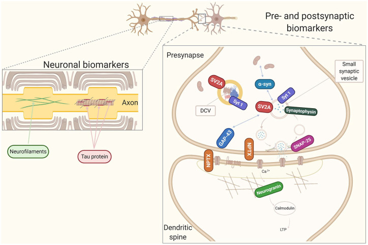
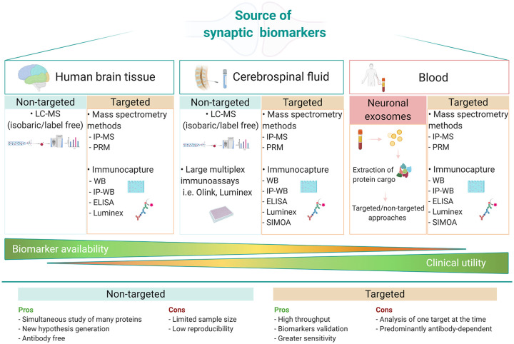
Synapses are the site for brain communication where information is transmitted between neurons and stored for memory formation. Synaptic degeneration is a global and early pathogenic event in neurodegenerative disorders with reduced levels of pre- and postsynaptic proteins being recognized as a core feature of Alzheimer's disease (AD) pathophysiology. Together with AD, other neurodegenerative and neurodevelopmental disorders show altered synaptic homeostasis as an important pathogenic event, and due to that, they are commonly referred to as synaptopathies. The exact mechanisms of synapse dysfunction in the different diseases are not well understood and their study would help understanding the pathogenic role of synaptic degeneration, as well as differences and commonalities among them and highlight candidate synaptic biomarkers for specific disorders. The assessment of synaptic proteins in cerebrospinal fluid (CSF), which can reflect synaptic dysfunction in patients with cognitive disorders, is a keen area of interest. Substantial research efforts are now directed toward the investigation of CSF synaptic pathology to improve the diagnosis of neurodegenerative disorders at an early stage as well as to monitor clinical progression. In this review, we will first summarize the pathological events that lead to synapse loss and then discuss the available data on established (eg, neurogranin, SNAP-25, synaptotagmin-1, GAP-43, and α-syn) and emerging (eg, synaptic vesicle glycoprotein 2A and neuronal pentraxins) CSF biomarkers for synapse dysfunction, while highlighting possible utilities, disease specificity, and technical challenges for their detection.
Overview
The claims and statistics of this paper haven’t been checked yet.
Run the check — free, no account needed. It reads the open-access text and checks the sources it cites.
Integrity signals
Registry
Cites
Statistics
Figures
Verification
Claims the paper makes
The claims and statistics of this paper haven’t been checked yet.
Run the check — free, no account needed. It reads the open-access text and checks the sources it cites.
Claims other papers make about this one
Each is checked against this paper’s own text: does it say what the citing paper says it does?
In the citing paper
“Furthermore, synapses may promote the spread of aberrant proteins via prion-like mechanisms, thus enhancing neurodegeneration (Camporesi et al. 2020; Gong and Lippa 2010; Swanson et al. 2018).”
From Neuronal pentraxins as biomarkers of synaptic activity: from physiological functions to pathological changes in neurodegeneration (2022) · cites this paper as Camporesi et al. 2020
Self-check
Percentages, sums and reported differences re-computed from the paper’s own numbers.
The self-check needs the paper’s full text.
Citations
Cites
NIA‐AA Research Framework: Toward a biological definition of Alzheimer's disease
Jack, C. R., Bennett, D. A., Blennow, K., Carrillo, M. C., Dunn, B., Haeberlein, S. B., Holtzman, D. M., Jagust, W., Jessen, F., Karlawish, J., et al. (2018) · Alzheimer's & Dementia · doi:10.1016/j.jalz.2018.02.018 · Cited by 10,537
Self-citationAmyloid oligomers exacerbate tau pathology in a mouse model of tauopathy
Selenica, M.-L. B., Brownlow, M., Jimenez, J. P., Lee, D. C., Pena, G., Dickey, C. A., Gordon, M. N., & Morgan, D. (2013) · Neurodegenerative Diseases · doi:10.1159/000337230 · Cited by 10,359
Alzheimer's Disease Is a Synaptic Failure
Selkoe, D. J. (2002) · Science · doi:10.1126/science.1074069 · Cited by 4,420
Terry, R. D., Masliah, E., Salmon, D. P., Butters, N., DeTeresa, R., Hill, R., Hansen, L. A., & Katzman, R. (1991) · Annals of Neurology · doi:10.1002/ana.410300410 · Cited by 4,397
Walsh, D. M., Klyubin, I., Fadeeva, J. V., Cullen, W. K., Anwyl, R., Wolfe, M. S., Rowan, M. J., & Selkoe, D. J. (2002) · Nature · doi:10.1038/416535a · Cited by 4,291
Diffusible, nonfibrillar ligands derived from Aβ 1–42 are potent central nervous system neurotoxins
Lambert, M. P., Barlow, A. K., Chromy, B. A., Edwards, C., Freed, R., Liosatos, M., Morgan, T. E., Rozovsky, I., Trommer, B., Viola, K. L., et al. (1998) · Proceedings of the National Academy of Sciences · doi:10.1073/pnas.95.11.6448 · Cited by 3,506
Advancing research diagnostic criteria for Alzheimer's disease: the IWG-2 criteria
Dubois, B., Feldman, H. H., Jacova, C., Hampel, H., Molinuevo, J. L., Blennow, K., DeKosky, S. T., Gauthier, S., Selkoe, D., Bateman, R., et al. (2014) · The Lancet Neurology · doi:10.1016/s1474-4422(14)70090-0 · Cited by 3,321
Self-citationAdult Neurogenesis in the Mammalian Brain: Significant Answers and Significant Questions
Ming, G.-L., & Song, H. (2011) · Neuron · doi:10.1016/j.neuron.2011.05.001 · Cited by 2,651
Südhof, T. C. (2004) · Annual Review of Neuroscience · doi:10.1146/annurev.neuro.26.041002.131412 · Cited by 2,491
Synapse Loss and Microglial Activation Precede Tangles in a P301S Tauopathy Mouse Model
Yoshiyama, Y., Higuchi, M., Zhang, B., Huang, S.-M., Iwata, N., Saido, T. C., Maeda, J., Suhara, T., Trojanowski, J. Q., & Lee, V. M.-Y. (2007) · Neuron · doi:10.1016/j.neuron.2007.01.010 · Cited by 2,208
Bloom (2014) · doi:10.1001/jamaneurol.2013.5847 · Cited by 2,194
α-Synuclein is phosphorylated in synucleinopathy lesions
Fujiwara, H., Hasegawa, M., Dohmae, N., Kawashima, A., Masliah, E., Goldberg, M. S., Shen, J., Takio, K., & Iwatsubo, T. (2002) · Nature Cell Biology · doi:10.1038/ncb748 · Cited by 2,174
The clinical use of structural MRI in Alzheimer disease
Frisoni, G. B., Fox, N. C., Jack, C. R., Scheltens, P., & Thompson, P. M. (2010) · Nature Reviews Neurology · doi:10.1038/nrneurol.2009.215 · Cited by 2,113
Olsson, B., Lautner, R., Andreasson, U., Öhrfelt, A., Portelius, E., Bjerke, M., Hölttä, M., Rosén, C., Olsson, C., Strobel, G., et al. (2016) · The Lancet Neurology · doi:10.1016/s1474-4422(16)00070-3 · Cited by 2,049
Self-citationNeurofilaments as biomarkers in neurological disorders
Khalil, M., Teunissen, C. E., Otto, M., Piehl, F., Sormani, M. P., Gattringer, T., Barro, C., Kappos, L., Comabella, M., Fazekas, F., et al. (2018) · Nature Reviews Neurology · doi:10.1038/s41582-018-0058-z · Cited by 1,988
Self-citationα-Synuclein Promotes SNARE-Complex Assembly in Vivo and in Vitro
Burré, J., Sharma, M., Tsetsenis, T., Buchman, V. L., Etherton, M. R., & Südhof, T. C. (2010) · Science · doi:10.1126/science.1195227 · Cited by 1,907
A/T/N: An unbiased descriptive classification scheme for Alzheimer disease biomarkers
Jack, C. R., Bennett, D. A., Blennow, K., Carrillo, M. C., Feldman, H. H., Frisoni, G. B., Hampel, H., Jagust, W. J., Johnson, K. A., Knopman, D. S., et al. (2016) · Neurology · doi:10.1212/wnl.0000000000002923 · Cited by 1,864
Self-citationLiang, H., Liang, P., Xu, Y., Wu, J., Liang, T., & Xu, X. (2009) · Journal of Neurotrauma · doi:10.1089/neu.2008.0850 · Cited by 1,749
High performance plasma amyloid-β biomarkers for Alzheimer's disease
Nakamura, A., Kaneko, N., Villemagne, V. L., Kato, T., Doecke, J., Doré, V., Fowler, C., Li, Q.-X., Martins, R., Rowe, C., et al. (2018) · Nature · doi:10.1038/nature25456 · Cited by 1,681
(1998) · Europe PMC (PubMed Central) · Cited by 1,594
Shankar, G. M., Bloodgood, B. L., Townsend, M., Walsh, D. M., Selkoe, D. J., & Sabatini, B. L. (2007) · The Journal of Neuroscience · doi:10.1523/jneurosci.4970-06.2007 · Cited by 1,592
Molecular cloning of cDNA encoding an unrecognized component of amyloid in Alzheimer disease.
Uéda, K., Fukushima, H., Masliah, E., Xia, Y., Iwai, A., Yoshimoto, M., Otero, D. A., Kondo, J. D. M., Ihara, Y., & Saitoh, T. (1993) · Proceedings of the National Academy of Sciences · doi:10.1073/pnas.90.23.11282 · Cited by 1,502
Propagation of Tau Pathology in a Model of Early Alzheimer's Disease
de Calignon, A., Polydoro, M., Suárez-Calvet, M., William, C., Adamowicz, D. H., Kopeikina, K. J., Pitstick, R., Sahara, N., Ashe, K. H., Carlson, G. A., et al. (2012) · Neuron · doi:10.1016/j.neuron.2011.11.033 · Cited by 1,427
α-Synuclein Blocks ER-Golgi Traffic and Rab1 Rescues Neuron Loss in Parkinson's Models
Cooper, A. A., Gitler, A. D., Cashikar, A. G., Haynes, C. M., Hill, K. J., Bhullar, B., Liu, K., Xu, K., Strathearn, K. E., Liu, F., et al. (2006) · Science · doi:10.1126/science.1129462 · Cited by 1,411
Synaptic plasticity and addiction
Kauer, J. A., & Malenka, R. C. (2007) · Nature Reviews Neuroscience · doi:10.1038/nrn2234 · Cited by 1,397
GAP-43: an intrinsic determinant of neuronal development and plasticity
Benowitz, L. I., & Routtenberg, A. (1997) · Trends in Neurosciences · doi:10.1016/s0166-2236(96)10072-2 · Cited by 1,329
Karikari, T. K., Pascoal, T. A., Ashton, N. J., Janelidze, S., Benedet, A. L., Rodriguez, J. L., Chamoun, M., Savard, M., Kang, M. S., Therriault, J., et al. (2020) · The Lancet Neurology · doi:10.1016/s1474-4422(20)30071-5 · Cited by 1,316
Self-citationSynaptic plasticity, memory and the hippocampus: a neural network approach to causality
Neves, G., Cooke, S. F., & Bliss, T. (2007) · Nature reviews. Neuroscience · doi:10.1038/nrn2303 · Cited by 1,287
Plasticity in the human central nervous system
Cooke, S. F. (2006) · Brain · doi:10.1093/brain/awl082 · Cited by 1,260
Janelidze, S., Mattsson, N., Palmqvist, S., Smith, R., Beach, T. G., Serrano, G. E., Chai, X., Proctor, N. K., Eichenlaub, U., Zetterberg, H., et al. (2020) · Nature Medicine · doi:10.1038/s41591-020-0755-1 · Cited by 1,247
Self-citationNeurotransmitter Release: The Last Millisecond in the Life of a Synaptic Vesicle
Südhof, T. C. (2013) · Neuron · doi:10.1016/j.neuron.2013.10.022 · Cited by 1,197
Beach, T. G., Monsell, S. E., Phillips, L. E., & Kukull, W. (2012) · Journal of Neuropathology & Experimental Neurology · doi:10.1097/nen.0b013e31824b211b · Cited by 1,103
Association of plasma neurofilament light with neurodegeneration in patients with Alzheimer disease
Mattsson, N., Andreasson, U., Zetterberg, H., Blennow, K., & Initiative, F. T. A. D. N. (2017) · JAMA Neurology · doi:10.1001/jamaneurol.2016.6117 · Cited by 1,070
Self-citationHippocampal synaptic loss in early Alzheimer's disease and mild cognitive impairment
Scheff, S. W., Price, D. A., Schmitt, F. A., & Mufson, E. J. (2006) · Neurobiology of Aging · doi:10.1016/j.neurobiolaging.2005.09.012 · Cited by 1,017
Dentritic spines : structure, dynamics and regulation
Hering, H., & Sheng, M. (2001) · Nature Reviews Neuroscience · doi:10.1038/35104061 · Cited by 961
Dominantly Inherited Alzheimer Network, Preische, O., Schultz, S. A., Apel, A., Kuhle, J., Kaeser, S. A., Barro, C., Gräber, S., Kuder-Buletta, E., LaFougere, C., et al. (2019) · Nature Medicine · doi:10.1038/s41591-018-0304-3 · Cited by 927
Murphy, D. D., Rueter, S. M., Trojanowski, J. Q., & Lee, V. M. ‐. (2000) · Journal of Neuroscience · doi:10.1523/jneurosci.20-09-03214.2000 · Cited by 927
High-precision plasma β-amyloid 42/40 predicts current and future brain amyloidosis
Schindler, S. E., Bollinger, J. G., Ovod, V., Mawuenyega, K. G., Li, Y., Gordon, B. A., Holtzman, D. M., Morris, J. C., Benzinger, T. L. S., Xiong, C., et al. (2019) · Neurology · doi:10.1212/wnl.0000000000008081 · Cited by 885
The Amyloid-β Oligomer Hypothesis: Beginning of the Third Decade
Cline, E. N., Bicca, M. A., Viola, K. L., & Klein, W. L. (2018) · Journal of Alzheimer's Disease · doi:10.3233/jad-179941 · Cited by 870
Synaptic alterations in CA1 in mild Alzheimer disease and mild cognitive impairment
Scheff, S. W., Price, D. A., Schmitt, F. A., DeKosky, S. T., & Mufson, E. J. (2007) · Neurology · doi:10.1212/01.wnl.0000260698.46517.8f · Cited by 824
Structures of α-synuclein filaments from multiple system atrophy
Schweighauser, M., Shi, Y., Tarutani, A., Kametani, F., Murzin, A. G., Ghetti, B. F., Matsubara, T., Tomita, T., Ando, T., Hasegawa, K., et al. (2020) · Nature · doi:10.1038/s41586-020-2317-6 · Cited by 803
Brain homogenates from human tauopathies induce tau inclusions in mouse brain
Clavaguera, F., Akatsu, H., Fraser, G. M., Crowther, R. A., Frank, S., Hench, J., Probst, A., Winkler, D. T., Reichwald, J., Staufenbiel, M., et al. (2013) · Proceedings of the National Academy of Sciences · doi:10.1073/pnas.1301175110 · Cited by 792
Advancing Research and Treatment for Frontotemporal Lobar Degeneration (ARTFL) investigators, Thijssen, E. H., La Joie, R., Wolf, A., Strom, A., Wang, P., Iaccarino, L., Bourakova, V., Cobigo, Y., Heuer, H., et al. (2020) · Nature Medicine · doi:10.1038/s41591-020-0762-2 · Cited by 761
Self-citationHamilton, R. L. (2000) · Brain Pathology · doi:10.1111/j.1750-3639.2000.tb00269.x · Cited by 754
Mattsson, N., Cullen, N. C., Andreasson, U., Zetterberg, H., & Blennow, K. (2019) · JAMA Neurology · doi:10.1001/jamaneurol.2019.0765 · Cited by 726
Self-citationAltered expression of synaptic proteins occurs early during progression of Alzheimer's disease
Masliah, E., Mallory, M., Alford, M., DeTeresa, R., Hansen, L. A., McKeel, D. W., & Morris, J. C. (2001) · Neurology · doi:10.1212/wnl.56.1.127 · Cited by 720
El‐Agnaf, O. M. A., Salem, S. A., Paleologou, K. E., Curran, M. D., Gibson, M. J., Court, J. A., Schlossmacher, M. G., & Allsop, D. (2006) · The FASEB Journal · doi:10.1096/fj.03-1449com · Cited by 715
Towards an Understanding of Synapse Formation
Südhof, T. C. (2018) · Neuron · doi:10.1016/j.neuron.2018.09.040 · Cited by 698
Plasma exosomal α-synuclein is likely CNS-derived and increased in Parkinson’s disease
Shi, M., Liu, C., Cook, T. J., Bullock, K. M., Zhao, Y., Ginghina, C., Li, Y., Aro, P., Dator, R., He, C., et al. (2014) · Acta Neuropathologica · doi:10.1007/s00401-014-1314-y · Cited by 676
Cell-to-cell transmission of pathogenic proteins in neurodegenerative diseases
Guo, J. L., & Lee, V. M. Y. (2014) · Nature Medicine · doi:10.1038/nm.3457 · Cited by 641
Structural and Molecular Remodeling of Dendritic Spine Substructures during Long-Term Potentiation
Bosch, M., Castro, J., Saneyoshi, T., Matsuno, H., Sur, M., & Hayashi, Y. (2014) · Neuron · doi:10.1016/j.neuron.2014.03.021 · Cited by 637
Mollenhauer, B., Locascio, J. J., Schulz‐Schaeffer, W., Sixel‐Döring, F., Trenkwalder, C., & Schlossmacher, M. G. (2011) · The Lancet Neurology · doi:10.1016/s1474-4422(11)70014-x · Cited by 636
Plasma β-amyloid in Alzheimer's disease and vascular disease
Janelidze, S., Stomrud, E., Palmqvist, S., Zetterberg, H., van Westen, D., Jeromin, A., Song, L., Hanlon, D., Tan Hehir, C. A., Baker, D., et al. (2016) · Scientific Reports · doi:10.1038/srep26801 · Cited by 626
Self-citationMielke, M. M., Hagen, C. E., Xu, J., Chai, X., Vemuri, P., Lowe, V. J., Airey, D. C., Knopman, D. S., Roberts, R. O., Machulda, M. M., et al. (2018) · Alzheimer's & Dementia · doi:10.1016/j.jalz.2018.02.013 · Cited by 621
Oligomeric Aβ-induced synaptic dysfunction in Alzheimer’s disease
Tu, S., Okamoto, S., Lipton, S. A., & Xu, H. (2014) · Molecular Neurodegeneration · doi:10.1186/1750-1326-9-48 · Cited by 613
Alpha‐synuclein RT ‐Qu IC in the CSF of patients with alpha‐synucleinopathies
Fairfoul, G., McGuire, L. I., Pal, S., Ironside, J. W., Neumann, J., Christie, S., Joachim, C., Esiri, M., Evetts, S. G., Rolinski, M., et al. (2016) · Annals of Clinical and Translational Neurology · doi:10.1002/acn3.338 · Cited by 608
Cellular milieu imparts distinct pathological α-synuclein strains in α-synucleinopathies
Peng, C., Gathagan, R. J., Covell, D. J., Medellin, C., Stieber, A., Robinson, J. L., Zhang, B., Pitkin, R. M., Olufemi, M. F., Luk, K. C., et al. (2018) · Nature · doi:10.1038/s41586-018-0104-4 · Cited by 605
The Synucleinopathies: Twenty Years On
Goedert, M., Jakes, R., & Spillantini, M. G. (2017) · Journal of Parkinson s Disease · doi:10.3233/jpd-179005 · Cited by 590
Imaging synaptic density in the living human brain
Finnema, S. J., Nabulsi, N. B., Eid, T., Detyniecki, K., Lin, S.-F., Chen, M.-K., Dhaher, R., Matuskey, D., Baum, E., Holden, D., et al. (2016) · Science Translational Medicine · doi:10.1126/scitranslmed.aaf6667 · Cited by 575
SZE, C.-I., TRONCOSO, J. C., KAWAS, C., MOUTION, P., PRICE, D. L., & MARTIN, L. J. (1997) · Journal of Neuropathology and Experimental Neurology · doi:10.1097/00005072-199708000-00011 · Cited by 562
Red Blood Cells Are the Major Source of Alpha-Synuclein in Blood
Barbour, R., Kling, K., Anderson, J. P., Banducci, K., Cole, T. A., Diep, L., Fox, M., Goldstein, J. M., Soriano, F., Seubert, P. A., et al. (2008) · Neurodegenerative Diseases · doi:10.1159/000112832 · Cited by 548
A Broken α-Helix in Folded α-Synuclein
Chandra, S. S., Chen, X., Rizo, J., Jahn, R., & Südhof, T. C. (2003) · Journal of Biological Chemistry · doi:10.1074/jbc.m213128200 · Cited by 540
Wang, H.-W., Pasternak, J. F., Kuo, H., Ristic, H., Lambert, M. P., Chromy, B., Viola, K. L., Klein, W. L., Stine, W. B., Krafft, G. A., et al. (2002) · Brain Research · doi:10.1016/s0006-8993(01)03058-x · Cited by 527
Tau stabilizes microtubules by binding at the interface between tubulin heterodimers
Kadavath, H., Hofele, R. V., Biernat, J., Kumar, S., Tepper, K., Urlaub, H., Mandelkow, E., & Zweckstetter, M. (2015) · Proceedings of the National Academy of Sciences · doi:10.1073/pnas.1504081112 · Cited by 524
Dejanovic, B., Huntley, M. A., De Mazière, A., Meilandt, W. J., Wu, T., Srinivasan, K., Jiang, Z., Gandham, V., Friedman, B. A., Ngu, H., et al. (2018) · Neuron · doi:10.1016/j.neuron.2018.10.014 · Cited by 517
Blood-based NfL: A biomarker for differential diagnosis of parkinsonian disorder
Hansson, O., Janelidze, S., Hall, S., Magdalinou, N., Lees, A. J., Andreasson, U., Norgren, N., Linder, J., Forsgren, L., Constantinescu, R., et al. (2017) · Neurology · doi:10.1212/wnl.0000000000003680 · Cited by 506
Self-citationPlasma tau in Alzheimer disease
Mattsson, N., Zetterberg, H., Janelidze, S., Insel, P. S., Andreasson, U., Stomrud, E., Palmqvist, S., Baker, D., Tan Hehir, C. A., Jeromin, A., et al. (2016) · Neurology · doi:10.1212/wnl.0000000000003246 · Cited by 502
Self-citationZetterberg, H., Skillbäck, T., Mattsson, N., Trojanowski, J. Q., Portelius, E., Shaw, L. M., Weiner, M. W., Blennow, K., & Initiative, F. T. A. D. N. (2016) · JAMA Neurology · doi:10.1001/jamaneurol.2015.3037 · Cited by 487
Self-citationShahnawaz, M., Tokuda, T., Waragai, M., Méndez, N., Ishii, R., Trenkwalder, C., Mollenhauer, B., & Soto, C. A. (2016) · JAMA Neurology · doi:10.1001/jamaneurol.2016.4547 · Cited by 485
Hall, S., Öhrfelt, A., Constantinescu, R., Andreasson, U., Surova, Y., Bostrom, F., Nilsson, C., Widner, H., Decraemer, H., Nägga, K., et al. (2012) · Archives of Neurology · doi:10.1001/archneurol.2012.1654 · Cited by 473
Self-citationDifferential expression of synaptic vesicle protein 2 (SV2) isoforms
Bajjalieh, S., Frantz, G., Weimann, J., McConnell, S., & Scheller, R. (1994) · The Journal of Neuroscience · doi:10.1523/jneurosci.14-09-05223.1994 · Cited by 440
Palmqvist, S., Janelidze, S., Stomrud, E., Zetterberg, H., Karl, J., Zink, K., Bittner, T., Mattsson, N., Eichenlaub, U., Blennow, K., et al. (2019) · JAMA Neurology · doi:10.1001/jamaneurol.2019.1632 · Cited by 435
Self-citationReversing EphB2 depletion rescues cognitive functions in Alzheimer model
Cissé, M., Halabisky, B., Harris, J., Devidze, N., Dubal, D. B., Sun, B., Orr, A., Lotz, G., Kim, D. H., Hamto, P., et al. (2011) · Nature · doi:10.1038/nature09635 · Cited by 433
Chen, M.-K., Mecca, A. P., Naganawa, M., Finnema, S. J., Toyonaga, T., Lin, S.-F., Najafzadeh, S., Ropchan, J., Lu, Y., McDonald, J. W., et al. (2018) · JAMA Neurology · doi:10.1001/jamaneurol.2018.1836 · Cited by 431
Galvin, J. E., Uryu, K., Lee, V. M. ‐., & Trojanowski, J. Q. (1999) · Proceedings of the National Academy of Sciences · doi:10.1073/pnas.96.23.13450 · Cited by 430
Tokuda, T., Salem, S. A., Allsop, D., Mizuno, T., Nakagawa, M., Qureshi, M. M., Locascio, J. J., Schlossmacher, M. G., & El-Agnaf, O. M. A. (2006) · Biochemical and Biophysical Research Communications · doi:10.1016/j.bbrc.2006.08.024 · Cited by 425
Abnormal neurotransmission in mice lacking synaptic vesicle protein 2A (SV2A)
Crowder, K. M., Gunther, J. M., Jones, T. A., Hale, B. D., Zhang, H. Z., Peterson, M. R., Scheller, R. H., Chavkin, C., & Bajjalieh, S. M. (1999) · Proceedings of the National Academy of Sciences · doi:10.1073/pnas.96.26.15268 · Cited by 415
Groveman, B. R., Orru, C. D., Hughson, A. G., Raymond, L. D., Zanusso, G., Ghetti, B. F., Campbell, K. J., Safar, J., Galasko, D. R., & Caughey, B. W. (2018) · Acta Neuropathologica Communications · doi:10.1186/s40478-018-0508-2 · Cited by 414
Exosomes: Vehicles for the Transfer of Toxic Proteins Associated with Neurodegenerative Diseases?
Bellingham, S. A., Guo, B. B., Coleman, B. M., & Hill, A. F. (2012) · Frontiers in Physiology · doi:10.3389/fphys.2012.00124 · Cited by 412
Decreased synaptic proteins in neuronal exosomes of frontotemporal dementia and Alzheimer's disease
Goetzl, E. J., Kapogiannis, D., Schwartz, J. B., Lobach, I. V., Goetzl, L., Abner, E. L., Jicha, G. A., Karydas, A. M., Boxer, A., & Miller, B. L. (2016) · The FASEB Journal · doi:10.1096/fj.201600816r · Cited by 398
Beta-amyloid accumulation in APP mutant neurons reduces PSD-95 and GluR1 in synapses
Almeida, C. G., Tampellini, D., Takahashi, R. H., Greengard, P., Lin, M. T., Snyder, E. M., & Gouras, G. K. (2005) · Neurobiology of Disease · doi:10.1016/j.nbd.2005.02.008 · Cited by 397
Synaptopathies: synaptic dysfunction in neurological disorders – A review from students to students
Lepeta, K., Lourenco, M. V., Schweitzer, B. C., Martino Adami, P. V., Banerjee, P., Catuara‐Solarz, S., de La Fuente Revenga, M., Guillem, A. M., Haidar, M., Ijomone, O. M., et al. (2016) · Journal of Neurochemistry · doi:10.1111/jnc.13713 · Cited by 386
Differential loss of synaptic proteins in Alzheimer's disease: Implications for synaptic dysfunction
Reddy, P. H., Mani, G., Park, B. S., Jacques, J., MURDOCH, G. H., Whetsell, W. O., Kaye, J. A., & Mańczak, M. (2005) · Journal of Alzheimer s Disease · doi:10.3233/jad-2005-7203 · Cited by 384
Mollenhauer, B., Cullen, V., Kahn, I., Krastins, B., Outeiro, T. F., Pepivani, I., Ng, J., Schulz-Schaeffer, W., Kretzschmar, H. A., McLean, P. J., et al. (2008) · Experimental Neurology · doi:10.1016/j.expneurol.2008.06.004 · Cited by 382
Kovács, G. G., Milenković, I., Wöehrer, A., Höftberger, R., Gelpí, E., Haberler, C., Hönigschnabl, S., Reiner‐Concin, A., Heinzl, H., Jungwirth, S., et al. (2013) · Acta Neuropathologica · doi:10.1007/s00401-013-1157-y · Cited by 380
Alzheimer’s pathogenesis: is there neuron-to-neuron propagation?
Braak, H., & Tredici, K. D. (2011) · Acta Neuropathologica · doi:10.1007/s00401-011-0825-z · Cited by 375
Amyloid beta from axons and dendrites reduces local spine number and plasticity
Wei, W., Nguyen, L. N., Kessels, H. W., Hagiwara, H., Sisodia, S., & Malinow, R. (2010) · Nature Neuroscience · doi:10.1038/nn.2476 · Cited by 357
Kvartsberg, H., Duits, F. H., Ingelsson, M., Andreasen, N., Öhrfelt, A., Andersson, K., Brinkmalm, G., Lannfelt, L., Minthon, L., Hansson, O., et al. (2014) · Alzheimer s & Dementia · doi:10.1016/j.jalz.2014.10.009 · Cited by 349
Self-citationKovács, G. G. (2016) · International Journal of Molecular Sciences · doi:10.3390/ijms17020189 · Cited by 346
Deficits in dopaminergic transmission precede neuron loss and dysfunction in a new Parkinson model
Janezic, S., Threlfell, S., Dodson, P. D., Dowie, M. J., Taylor, T. N., Potgieter, D., Parkkinen, L., Senior, S. L., Anwar, S., Ryan, B., et al. (2013) · Proceedings of the National Academy of Sciences · doi:10.1073/pnas.1309143110 · Cited by 344
A systematic review of prevalence and incidence studies of dementia with Lewy bodies
ZACCAI, J. H., McCracken, C., & Brayne, C. (2005) · Age and Ageing · doi:10.1093/ageing/afi190 · Cited by 342
Elevated levels of tau-protein in cerebrospinal fluid of patients with Creutzfeldt–Jakob disease
Otto, M., Wiltfang, J., Tumani, H., Zerr, I., Lantsch, M., Kornhuber, J., Weber, T., Kretzschmar, H. A., & Poser, S. (1997) · Neuroscience Letters · doi:10.1016/s0304-3940(97)00215-2 · Cited by 339
Encyclopedia of Child Behavior and Development
Goldstein, S., & Naglieri, J. A. (2011) · Springer eBooks · doi:10.1007/978-0-387-79061-9 · Cited by 338
The clinical promise of biomarkers of synapse damage or loss in Alzheimer’s disease
the Synaptic Health Endpoints Working Group, Colom-Cadena, M., Spires-Jones, T., Zetterberg, H., Blennow, K., Caggiano, A., DeKosky, S. T., Fillit, H., Harrison, J. E., Schneider, L. S., et al. (2020) · Alzheimer's Research & Therapy · doi:10.1186/s13195-020-00588-4 · Cited by 328
Self-citationThe Synuclein FamilyPreprint
Lavedan, C. N. (1998) · Genome Research · doi:10.1101/gr.8.9.871 · Cited by 323
NPTX2 and cognitive dysfunction in Alzheimer’s Disease
Xiao, M.-F., Xu, D., Craig, M. T., Pelkey, K. A., Chien, C.-C., Shi, Y., Zhang, J., Resnick, S., Pletnikova, O., Salmon, D., et al. (2017) · eLife · doi:10.7554/elife.23798 · Cited by 316
Alpha-synuclein and tau: teammates in neurodegeneration?
Moussaud, S., Jones, D. R., Moussaud-Lamodière, E. L., Delenclos, M., Ross, O. A., & McLean, P. J. (2014) · Molecular Neurodegeneration · doi:10.1186/1750-1326-9-43 · Cited by 301
A critical appraisal of the pathogenic protein spread hypothesis of neurodegeneration
Walsh, D. M., & Selkoe, D. J. (2016) · Nature reviews. Neuroscience · doi:10.1038/nrn.2016.13 · Cited by 301
Brinkmalm, A., Brinkmalm, G., Honer, W. G., Frölich, L., Hausner, L., Minthon, L., Hansson, O., Wallin, A., Zetterberg, H., Blennow, K., et al. (2014) · Molecular Neurodegeneration · doi:10.1186/1750-1326-9-53 · Cited by 293
Self-citationIn vivo measurement of widespread synaptic loss in Alzheimer’s disease with SV2A PET
Mecca, A. P., Chen, M., O'Dell, R. S., Naganawa, M., Toyonaga, T., Godek, T. A., Harris, J. E., Bartlett, H. H., Zhao, W., Nabulsi, N. B., et al. (2020) · Alzheimer's & Dementia · doi:10.1002/alz.12097 · Cited by 288
de Wilde, M. C., Overk, C., Sijben, J., & Masliah, E. (2016) · Alzheimer s & Dementia · doi:10.1016/j.jalz.2015.12.005 · Cited by 287
Oligomeric and phosphorylated alpha-synuclein as potential CSF biomarkers for Parkinson’s disease
Majbour, N. K., Vaikath, N. N., van Dijk, K. D., Ardah, M. T., Varghese, S., Vesterager, L. B., Montezinho, L. P., Poole, S., Safieh‐Garabedian, B., Tokuda, T., et al. (2016) · Molecular Neurodegeneration · doi:10.1186/s13024-016-0072-9 · Cited by 287
Roselli, F., Tirard, M., Lu, J., Hutzler, P., Lamberti, P., Livrea, P., Morabito, M. A., & Almeida, O. F. X. (2005) · Journal of Neuroscience · doi:10.1523/jneurosci.3034-05.2005 · Cited by 284
Cerebrospinal fluid neurogranin: relation to cognition and neurodegeneration in Alzheimer’s disease
Portelius, E., Zetterberg, H., Skillbäck, T., Törnqvist, U., Andreasson, U., Trojanowski, J. Q., Weiner, M. W., Shaw, L. M., & Mattsson, N. (2015) · Brain · doi:10.1093/brain/awv267 · Cited by 279
Self-citationCerebrospinal fluid lysosomal enzymes and alpha‐synuclein in Parkinson's disease
Parnetti, L., Chiasserini, D., Persichetti, E., Eusebi, P., Varghese, S., Qureshi, M. M., Dardis, A., Deganuto, M., Carlo, C. D., Castrioto, A., et al. (2014) · Movement Disorders · doi:10.1002/mds.25772 · Cited by 273
Neurogranin: immunocytochemical localization of a brain-specific protein kinase C substrate
Represa, A., Deloulme, J., Sensenbrenner, M., Ben‐Ari, Y., & Baudier, J. (1990) · Journal of Neuroscience · doi:10.1523/jneurosci.10-12-03782.1990 · Cited by 273
Synapse loss in the temporal lobe in Alzheimer's disease
Scheff, S. W., & PRICE, D. A. (1993) · Annals of Neurology · doi:10.1002/ana.410330209 · Cited by 273
Osimo, E. F., Beck, K., Marques, T. R., & Howes, O. D. (2018) · Molecular Psychiatry · doi:10.1038/s41380-018-0041-5 · Cited by 273
Microglial activation correlates in vivo with both tau and amyloid in Alzheimer’s disease
Dani, M., Wood, M., Mizoguchi, R., Fan, Z., Walker, Z., Morgan, R., Hinz, R., Biju, M., Kuruvilla, T., Brooks, D. J., et al. (2018) · Brain · doi:10.1093/brain/awy188 · Cited by 270
Proteomic analysis of cerebrospinal fluid extracellular vesicles: A comprehensive dataset
Chiasserini, D., van Weering, J. R. T., Piersma, S. R., Pham, T. V., Malekzadeh, A., Teunissen, C. E., de Wit, H., & Jiménez, C. R. (2014) · Journal of Proteomics · doi:10.1016/j.jprot.2014.04.028 · Cited by 269
Xu, D., Hopf, C., Reddy, R., Cho, R. W., Guo, L., Lanahan, A., Petralia, R. S., Wenthold, R. J., O'Brien, R. J., & Worley, P. (2003) · Neuron · doi:10.1016/s0896-6273(03)00463-x · Cited by 265
Prevention and management of dementia: a priority for public health
Frankish, H. M., & Horton, R. C. (2017) · The Lancet · doi:10.1016/s0140-6736(17)31756-7 · Cited by 258
Synaptopathies: diseases of the synaptome
Grant, S. G. (2012) · Current Opinion in Neurobiology · doi:10.1016/j.conb.2012.02.002 · Cited by 248
Schöll, M., Maaß, A., Mattsson, N., Ashton, N. J., Blennow, K., Zetterberg, H., & Jagust, W. J. (2018) · Molecular and Cellular Neuroscience · doi:10.1016/j.mcn.2018.12.001 · Cited by 248
Self-citationNeurogranin as a Cerebrospinal Fluid Biomarker for Synaptic Loss in Symptomatic Alzheimer Disease
Kester, M. I., Teunissen, C. E., Crimmins, D. L., Herries, E. M., Ladenson, J. H., Scheltens, P., van der Flier, W. M., Morris, J. C., Holtzman, D. M., & Fagan, A. M. (2015) · JAMA Neurology · doi:10.1001/jamaneurol.2015.1867 · Cited by 246
Serum neurofilament light in familial Alzheimer disease: A marker of early neurodegeneration
Weston, P. S. J., Poole, T., Ryan, N. S., Nair, A., Liang, Y., Macpherson, K., Druyeh, R., Malone, I. B., Ahsan, R. L., Pemberton, H., et al. (2017) · Neurology · doi:10.1212/wnl.0000000000004667 · Cited by 245
Self-citationSpreading of Neurodegenerative Pathology via Neuron-to-Neuron Transmission of -Amyloid
Nath, S., Agholme, L., Kurudenkandy, F. R., Granseth, B., Marcusson, J., & Hallbeck, M. (2012) · Journal of Neuroscience · doi:10.1523/jneurosci.0615-12.2012 · Cited by 244
A Brief Overview of Tauopathy: Causes, Consequences, and Therapeutic Strategies
Orr, M. E., Sullivan, A. C., & Frost, B. E. (2017) · Trends in Pharmacological Sciences · doi:10.1016/j.tips.2017.03.011 · Cited by 242
Sjögren, M. J. C., Minthon, L., Davidsson, P., Granérus, A., Clarberg, A., Vanderstichele, H., Vanmechelen, E., Wallin, A., & Blennow, K. (2000) · Journal of Neural Transmission · doi:10.1007/s007020070079 · Cited by 238
Self-citationRiemenschneider, M., Wagenpfeil, S., Vanderstichele, H., Otto, M., Wiltfang, J., Kretzschmar, H., Vanmechelen, E., Förstl, H., & Kurz, A. (2003) · Molecular Psychiatry · doi:10.1038/sj.mp.4001220 · Cited by 238
Exosomes and other extracellular vesicles in neural cells and neurodegenerative diseases
Janas, A., Sapoń, K., Janas, T., Stowell, M. H. B., & Janas, T. (2016) · Biochimica et Biophysica Acta (BBA) - Biomembranes · doi:10.1016/j.bbamem.2016.02.011 · Cited by 235
Increased CSF neurogranin concentration is specific to Alzheimer disease
Wellington, H., Paterson, R. W., Portelius, E., Törnqvist, U., Magdalinou, N., Fox, N. C., Blennow, K., Schott, J. M., & Zetterberg, H. (2016) · Neurology · doi:10.1212/wnl.0000000000002423 · Cited by 232
Self-citationDiagnostic and Prognostic Utility of the Synaptic Marker Neurogranin in Alzheimer Disease
Tarawneh, R. M., D’Angelo, G., Crimmins, D. L., Herries, E. M., Griest, T. A., Fagan, A. M., Zipfel, G. J., Ladenson, J. H., Morris, J. C., & Holtzman, D. M. (2016) · JAMA Neurology · doi:10.1001/jamaneurol.2016.0086 · Cited by 228
Fan, Z., Pan, Y.-T., Zhang, Z.-Y., Yang, H., Yu, S.-Y., Zheng, Y., Ma, J.-H., & Wang, X.-M. (2020) · Journal of Neuroinflammation · doi:10.1186/s12974-019-1670-6 · Cited by 228
Eusebi, P., Giannandrea, D., Biscetti, L., Abraha, I., Chiasserini, D., Orso, M., Calabresi, P., & Parnetti, L. (2017) · Movement Disorders · doi:10.1002/mds.27110 · Cited by 226
Prots, I., Grosch, J., Brazdis, R.-M., Simmnacher, K., Veber, V., Havlicek, S., Hannappel, C., Krach, F., Krumbiegel, M., Schütz, O., et al. (2018) · Proceedings of the National Academy of Sciences · doi:10.1073/pnas.1713129115 · Cited by 223
Salivary α-synuclein and DJ-1: potential biomarkers for Parkinson's disease
Devic, I., Hwang, H., Edgar, J. S., Izutsu, K. T., Presland, R. B., Pan, C., Goodlett, D. R., Wang, Y., Armaly, J., Tumas, V., et al. (2011) · Brain · doi:10.1093/brain/awr015 · Cited by 222
Wang, X., Becker, K., Levine, N. D., Zhang, M. H., Lieberman, A. P., Moore, D. J., & Ma, J. (2019) · Acta Neuropathologica Communications · doi:10.1186/s40478-019-0696-4 · Cited by 221
Goetzl, E. J., Abner, E. L., Jicha, G. A., Kapogiannis, D., & Schwartz, J. B. (2018) · The FASEB Journal · doi:10.1096/fj.201700731r · Cited by 219
Lleó, A., Núñez-Llaves, R., Alcolea, D., Chiva, C., Balateu-Paños, D., Colom-Cadena, M., Gomez-Giro, G., Muñoz, L., Querol-Vilaseca, M., Pegueroles, J., et al. (2019) · Molecular & Cellular Proteomics · doi:10.1074/mcp.ra118.001290 · Cited by 219
Co-Localization of Amyloid Beta and Tau Pathology in Alzheimer's Disease Synaptosomes
Fein, J. A., Sokolow, S., Miller, C. A., Vinters, H. V., Yang, F., Cole, G. M., & Gylys, K. H. (2008) · American Journal Of Pathology · doi:10.2353/ajpath.2008.070829 · Cited by 218
Blennow, K., Bogdanović, N., Alafuzoff, I. I., Ekman, R., & Davidsson, P. (1996) · Journal of Neural Transmission · doi:10.1007/bf01273157 · Cited by 217
Self-citationHansson, O., Hall, S., Öhrfelt, A., Zetterberg, H., Blennow, K., Minthon, L., Nägga, K., Londos, E., Varghese, S., Majbour, N. K., et al. (2014) · Alzheimer s Research & Therapy · doi:10.1186/alzrt255 · Cited by 214
Self-citationTaoufik, E., Kouroupi, G., Zygogianni, O., & Matsas, R. (2018) · Open Biology · doi:10.1098/rsob.180138 · Cited by 213
Gao, L., Tang, H., Nie, K., Wang, L., Zhao, J., Gan, R., Huang, J., Zhu, R., Feng, S., Duan, Z., et al. (2014) · International Journal of Neuroscience · doi:10.3109/00207454.2014.961454 · Cited by 210
Foulds, P. G., Mitchell, J. D., Parker, A., Turner, R., Green, G., Diggle, P. J., Hasegawa, M., Taylor, M., Mann, D. M. A., & Allsop, D. (2011) · The FASEB Journal · doi:10.1096/fj.10-179192 · Cited by 210
Neurofilament protein in cerebrospinal fluid: A marker of white matter changes
Sjögren, M., Blomberg, M., Jonsson, M., Wahlund, L. ‐., Edman, Å., Lind, K., Rosengren, L., Blennow, K., & Wallin, A. (2001) · Journal of Neuroscience Research · doi:10.1002/jnr.1242 · Cited by 209
Self-citationReview: Parkinson's disease: from synaptic loss to connectome dysfunction
Bellucci, A., Mercuri, N. B., Venneri, A., Faustini, G., Longhena, F., Pizzi, M., Missale, C., & Spano, P. (2015) · Neuropathology and Applied Neurobiology · doi:10.1111/nan.12297 · Cited by 208
Tao, C.-L., Liu, Y.-T., Sun, R., Zhang, B., Qi, L., Shivakoti, S., Tian, C.-L., Zhang, P., Lau, P.-M., Zhou, Z. H., et al. (2018) · The Journal of Neuroscience · doi:10.1523/jneurosci.1548-17.2017 · Cited by 207
RC3/neurogranin, a postsynaptic calpacitin for setting the response threshold to calcium influxes
Gerendasy, D. D., & Sutcliffe, J. G. (1997) · Molecular Neurobiology · doi:10.1007/bf02740632 · Cited by 206
Neurogranin in cerebrospinal fluid as a marker of synaptic degeneration in Alzheimer's disease
Thorsell, A., Bjerke, M., Gobom, J., Brunhage, E., Vanmechelen, E., Andreasen, N., Hansson, O., Minthon, L., Zetterberg, H., & Blennow, K. (2010) · Brain Research · doi:10.1016/j.brainres.2010.09.073 · Cited by 204
Self-citationSNAP-25, a Known Presynaptic Protein with Emerging Postsynaptic Functions
Antonucci, F., Corradini, I., Fossati, G., Tomasoni, R., Menna, E., & Matteoli, M. (2016) · Frontiers in Synaptic Neuroscience · doi:10.3389/fnsyn.2016.00007 · Cited by 202
Cerebrospinal fluid α-synuclein in neurodegenerative disorders—A marker of synapse loss?
Öhrfelt, A., Grognet, P., Andreasen, N., Wallin, A., Vanmechelen, E., Blennow, K., & Zetterberg, H. (2008) · Neuroscience Letters · doi:10.1016/j.neulet.2008.11.015 · Cited by 201
Self-citationHolahan, M. R. (2017) · Frontiers in Cellular Neuroscience · doi:10.3389/fncel.2017.00266 · Cited by 200
Park, M., Cheon, S., Bae, H.-R., Kim, S.-H., & Kim, J. W. (2011) · Journal of Clinical Neurology · doi:10.3988/jcn.2011.7.4.215 · Cited by 199
Alpha-Synuclein Physiology and Pathology: A Perspective on Cellular Structures and Organelles
Bernal-Conde, L. D., Ramos-Acevedo, R., Reyes-Hernández, M. A., Balbuena-Olvera, A. J., Morales-Moreno, I. D., Argüero‐Sánchez, R., Schüle, B., & Guerra‐Crespo, M. (2020) · Frontiers in Neuroscience · doi:10.3389/fnins.2019.01399 · Cited by 197
Intersection of pathological tau and microglia at the synapse
Vogels, T., Murgoci, A.-N., & Hromádka, T. (2019) · Acta Neuropathologica Communications · doi:10.1186/s40478-019-0754-y · Cited by 197
Neurogranin/RC3 Enhances Long-Term Potentiation and Learning by Promoting Calcium-Mediated Signaling
Huang, K., Huang, F. L., Jäger, T., Li, J., Reymann, K. G., & Balschun, D. (2004) · Journal of Neuroscience · doi:10.1523/jneurosci.2213-04.2004 · Cited by 195
Elevated CSF GAP‐43 is Alzheimer's disease specific and associated with tau and amyloid pathology
Sandelius, Å., Portelius, E., Källén, Å., Zetterberg, H., Rot, U., Olsson, B., Toledo, J. B., Shaw, L. M. J., Lee, V. M. ‐., Irwin, D. J., et al. (2018) · Alzheimer s & Dementia · doi:10.1016/j.jalz.2018.08.006 · Cited by 194
Self-citationA longitudinal study on α-synuclein in blood plasma as a biomarker for Parkinson's disease
Foulds, P. G., Diggle, P. J., Mitchell, J. D., Parker, A., Hasegawa, M., Masuda‐Suzukake, M., Mann, D. M. A., & Allsop, D. (2013) · Scientific Reports · doi:10.1038/srep02540 · Cited by 191
Synaptic Changes in Parkinson Disease Assessed with in vivo Imaging
Matuskey, D., Tinaz, S., Wilcox, K. C., Naganawa, M., Toyonaga, T., Dias, M., Henry, S. E., Pittman, B. P., Ropchan, J. R., Nabulsi, N., et al. (2020) · Annals of Neurology · doi:10.1002/ana.25682 · Cited by 186
An update on blood-based biomarkers for non-Alzheimer neurodegenerative disorders
Ashton, N. J., Hye, A., Rajkumar, A. P., Leuzy, A., Snowden, S. G., Suárez‐Calvet, M., Karikari, T. K., Schöll, M., Joie, R. L., Rabinovici, G. D., et al. (2020) · Nature Reviews Neurology · doi:10.1038/s41582-020-0348-0 · Cited by 185
Self-citationThe pre-synaptic vesicle protein synaptotagmin is a novel biomarker for Alzheimer’s disease
Öhrfelt, A., Brinkmalm, A., Dumurgier, J., Brinkmalm, G., Hansson, O., Zetterberg, H., Bouaziz-Amar, E., Hugon, J., Paquet, C., & Blennow, K. (2016) · Alzheimer's Research & Therapy · doi:10.1186/s13195-016-0208-8 · Cited by 184
Self-citationMitochondrial dynamics in Parkinson's disease: a role for α-synuclein?
Devoto, V. M. P., & Falzone, T. L. (2017) · Disease Models & Mechanisms · doi:10.1242/dmm.026294 · Cited by 184
Cytoskeleton proteins in CSF distinguish frontotemporal dementia from AD
Sjögren, M., Rosengren, L., Minthon, L., Davidsson, P., Blennow, K., & Wallin, A. (2000) · Neurology · doi:10.1212/wnl.54.10.1960 · Cited by 181
Self-citationNeurochemical Dissection of Synaptic Pathology in Alzheimer's Disease
Davidsson, P., & Blennow, K. (1998) · International Psychogeriatrics · doi:10.1017/s1041610298005110 · Cited by 180
Self-citationHamelin, L., Lagarde, J., Dorothée, G., Potier, M. C., Corlier, F., Kuhnast, B., Caillé, F., Dubois, B., Fillon, L., Chupin, M., et al. (2018) · Brain · doi:10.1093/brain/awy079 · Cited by 177
LTP and memory impairment caused by extracellular Aβ and Tau oligomers is APP-dependent
Puzzo, D., Piacentini, R., Fá, M., Gulisano, W., Li Puma, D. D., Staniszewski, A., Zhang, H., Tropea, M. R., Cocco, S., Palmeri, A., et al. (2017) · eLife · doi:10.7554/elife.26991 · Cited by 172
Ashton, N. J., Leuzy, A., Lim, Y. M., Troakes, C., Hortobágyi, T., Höglund, K., Aarsland, D., Lovestone, S., Schöll, M., Blennow, K., et al. (2019) · Acta Neuropathologica Communications · doi:10.1186/s40478-018-0649-3 · Cited by 172
Self-citationPortelius, E., Olsson, B., Höglund, K., Cullen, N. C., Kvartsberg, H., Andréasson, U., Zetterberg, H., Sandelius, Å., Shaw, L. M. J., Lee, V. M. ‐., et al. (2018) · Acta Neuropathologica · doi:10.1007/s00401-018-1851-x · Cited by 171
Self-citationTau Oligomers: The Toxic Player at Synapses in Alzheimer’s Disease
Guerrero-Muñoz, M. J., Gerson, J., & Castillo-Carranza, D. L. (2015) · Frontiers in Cellular Neuroscience · doi:10.3389/fncel.2015.00464 · Cited by 168
Cotrafficking of SV2 and synaptotagmin at the synapse
Yao, J., Nowack, A., Kensel-Hammes, P., Gardner, R. G., & Bajjalieh, S. M. (2010) · The Journal of Neuroscience · doi:10.1523/jneurosci.4781-09.2010 · Cited by 164
Clare, R., King, V. G., Wirenfeldt, M., & Vinters, H. V. (2010) · Journal of Neuroscience Research · doi:10.1002/jnr.22392 · Cited by 163
Sutphen, C. L., McCue, L., Herries, E. M., Xiong, C., Ladenson, J. H., Holtzman, D. M., Fagan, A. M., & On behalf of, A. D. N. I. (2018) · Alzheimer's & Dementia · doi:10.1016/j.jalz.2018.01.012 · Cited by 162
Spellman, D. S., Wildsmith, K. R., Honigberg, L. A., Tuefferd, M., Baker, D., Raghavan, N., Nairn, A. C., Croteau, P., Schirm, M., Allard, R., et al. (2015) · PROTEOMICS – Clinical Applications · doi:10.1002/prca.201400178 · Cited by 161
Vos, A. D., Jacobs, D., Struyfs, H., Fransén, E., Andersson, K., Portelius, E., Andréasson, U., Surgeloose, D. D., Hernalsteen, D., Sleegers, K., et al. (2015) · Alzheimer s & Dementia · doi:10.1016/j.jalz.2015.05.012 · Cited by 158
Self-citationWang, Z., Jackson, R. J., Hong, W., Taylor, W. M., Corbett, G. T., Moreno, A. J., Liu, W., Li, S., Frosch, M. P., Slutsky, I., et al. (2017) · Journal of Neuroscience · doi:10.1523/jneurosci.2009-17.2017 · Cited by 157
Robinson, J. L., Molina-Porcel, L., Corrada, M. M., Raible, K., Lee, E. B., Lee, V. M.-Y., Kawas, C. H., & Trojanowski, J. Q. (2014) · Brain · doi:10.1093/brain/awu190 · Cited by 157
A comparative study of CSF neurofilament light and heavy chain protein in MS
Kuhle, J., Plattner, K., Bestwick, J. P., Lindberg, R. L., Ramagopalan, S. V., Norgren, N., Nissim, A., Malaspina, A., Leppert, D., Giovannoni, G., et al. (2013) · Multiple Sclerosis Journal · doi:10.1177/1352458513482374 · Cited by 155
Papp–Lantos inclusions and the pathogenesis of multiple system atrophy: an update
Jellinger, K. A., & Lantos, P. L. (2010) · Acta Neuropathologica · doi:10.1007/s00401-010-0672-3 · Cited by 152
Manek, R., Moghieb, A. M., Yang, Z., Kumar, D., Kobessiy, F., Sarkis, G. A., Raghavan, V., & Wang, K. (2017) · Molecular Neurobiology · doi:10.1007/s12035-017-0821-y · Cited by 152
Ferretti, M. T., Martínková, J., Biskup, E. M., Benke, T., Gialdini, G., Nedelská, Z., Rauen, K., Mantua, V., Religa, D., Hort, J., et al. (2020) · European Journal of Neurology · doi:10.1111/ene.14174 · Cited by 152
Wang, X., Yu, S., Li, F., & Feng, T. (2015) · Neuroscience Letters · doi:10.1016/j.neulet.2015.05.030 · Cited by 151
Co-occurrence of Alzheimer's disease β-amyloid and tau pathologies at synapses
Takahashi, R. H., Capetillo-Zarate, E., Lin, M. T., Milner, T. A., & Gouras, G. K. (2010) · Neurobiology of Aging · doi:10.1016/j.neurobiolaging.2008.07.021 · Cited by 150
Presynaptic neuronal pentraxin receptor organizes excitatory and inhibitory synapses
Lee, S.-J., Wei, M., Zhang, C., Maxeiner, S., Pak, C., Calado Botelho, S., Trotter, J., Sterky, F. H., & Südhof, T. C. (2017) · The Journal of Neuroscience · doi:10.1523/jneurosci.2768-16.2016 · Cited by 146
Kvartsberg, H., Portelius, E., Andréasson, U., Brinkmalm, G., Hellwig, K., Lelental, N., Kornhuber, J., Hansson, O., Minthon, L., Spitzer, P., et al. (2015) · Alzheimer s Research & Therapy · doi:10.1186/s13195-015-0124-3 · Cited by 145
Self-citationGauss, C., Kalkum, M., Löwe, M., Lehrach, H., & Klose, J. (1999) · Electrophoresis · doi:10.1002/(sici)1522-2683(19990301)20:3 · Cited by 142
Self-citationWennström, M., Surova, Y., Hall, S., Nilsson, C., Minthon, L., Boström, F., Hansson, O., & Nielsen, H. M. (2013) · PLoS ONE · doi:10.1371/journal.pone.0053250 · Cited by 141
14-3-3 proteins in neurological disorders.
Foote, M. M., & Zhou, Y. (2012) · PubMed · Cited by 140
Wildsmith, K. R., Schauer, S. P., Smith, A. M., Arnott, D. P., Zhu, Y., Haznedar, J., Kaur, S., Mathews, W. R., & Honigberg, L. A. (2014) · Molecular Neurodegeneration · doi:10.1186/1750-1326-9-22 · Cited by 140
Oeckl, P., Metzger, F., Nagl, M., Arnim, C. A. F. V., Halbgebauer, S., Steinacker, P., Ludolph, A. C., & Otto, M. (2016) · Molecular & Cellular Proteomics · doi:10.1074/mcp.m116.059915 · Cited by 140
SV2 regulates neurotransmitter release via multiple mechanisms
Nowack, A., Yao, J., Custer, K. L., & Bajjalieh, S. M. (2010) · American Journal of Physiology-Cell Physiology · doi:10.1152/ajpcell.00259.2010 · Cited by 138
Homeostatic synaptic plasticity: from single synapses to neural circuits
Vitureira, N., Letellier, M., & Goda, Y. (2011) · Current Opinion in Neurobiology · doi:10.1016/j.conb.2011.09.006 · Cited by 137
Abnormal Salivary Total and Oligomeric Alpha-Synuclein in Parkinson’s Disease
Vivacqua, G., Latorre, A., Suppa, A., Nardi, M., Pietracupa, S., Mancinelli, R., Fabbrini, G., Colosimo, C., Gaudio, E., & Berardelli, A. (2016) · PLoS ONE · doi:10.1371/journal.pone.0151156 · Cited by 137
Prion-Like Mechanisms in Parkinson’s Disease
Ma, J., Gao, J., Wang, J., & Xie, A. (2019) · Frontiers in Neuroscience · doi:10.3389/fnins.2019.00552 · Cited by 134
Galasko, D., Xiao, M., Xu, D., Smirnov, D., Salmon, D. P., Dewit, N., Vanbrabant, J., Jacobs, D., Vanderstichele, H., Vanmechelen, E., et al. (2019) · Alzheimer's & Dementia: Translational Research & Clinical Interventions · doi:10.1016/j.trci.2019.11.002 · Cited by 133
Maiti, P., Manna, J., Ilavazhagan, G., Rossignol, J., & Dunbar, G. L. (2015) · Neuroscience & Biobehavioral Reviews · doi:10.1016/j.neubiorev.2015.09.020 · Cited by 128
Cho, R. W., Park, J. M., Wolff, S. B. E., Xu, D., Hopf, C., Kim, J.-A., Reddy, R. C., Petralia, R. S., Perin, M. S., Linden, D. J., et al. (2008) · Neuron · doi:10.1016/j.neuron.2008.01.010 · Cited by 125
Autism, epilepsy, and synaptopathies: a not rare association
Keller, R., Basta, R., Salerno, L., & Elia, M. (2017) · Neurological Sciences · doi:10.1007/s10072-017-2974-x · Cited by 124
Synapses and Dendritic Spines as Pathogenic Targets in Alzheimer’s Disease
Yu, W., & Lu, B. (2012) · Neural Plasticity · doi:10.1155/2012/247150 · Cited by 123
Bertoni–Freddari, C., Fattoretti, P., Casoli, T., Meier-Ruge, W. A., & ULRICH, J. (1990) · Brain Research · doi:10.1016/0006-8993(90)91009-6 · Cited by 121
Tateno, F., Sakakibara, R., Kawai, T., Kishi, M., & Murano, T. (2012) · Alzheimer Disease & Associated Disorders · doi:10.1097/wad.0b013e31823899cc · Cited by 121
LTD-like molecular pathways in developmental synaptic pruning
Piochon, C., Kano, M., & Hansel, C. (2016) · Nature Neuroscience · doi:10.1038/nn.4389 · Cited by 118
Is synaptic loss a unique hallmark of Alzheimer’s disease?
Scheff, S. W., Neltner, J. H., & Nelson, P. T. (2014) · Biochemical Pharmacology · doi:10.1016/j.bcp.2013.12.028 · Cited by 118
Sjögren, M. J. C., Davidsson, P., Gottfries, J., Vanderstichele, H., Edman, Å., Vanmechelen, E., Wallin, A., & Blennow, K. (2001) · Dementia and Geriatric Cognitive Disorders · doi:10.1159/000051268 · Cited by 116
Self-citationOligomeropathies and pathogenesis of Alzheimer and Parkinson's diseases
Forloni, G., Artuso, V., Vitola, P. L., & Balducci, C. (2016) · Movement Disorders · doi:10.1002/mds.26624 · Cited by 116
Assembly of Excitatory Synapses in the Absence of Glutamatergic Neurotransmission
Sando, R. C., Bushong, E. A., Zhu, Y., Huang, M., Considine, C., Phan, S., Ju, S., Uytiepo, M., Ellisman, M. H., & Maximov, A. (2017) · Neuron · doi:10.1016/j.neuron.2017.03.047 · Cited by 116
Synaptic Elimination in Neurological Disorders
Cardozo, P. L., de Lima, I. B. Q., Maciel, E. M. A., Silva, N. C., Dobránsky, T., & Ribeiro, F. M. (2019) · Current Neuropharmacology · doi:10.2174/1570159x17666190603170511 · Cited by 115
Brinkmalm, G., Sjödin, S., Simonsen, A. H., Hasselbalch, S. G., Zetterberg, H., Brinkmalm, A., & Blennow, K. (2018) · PROTEOMICS – Clinical Applications · doi:10.1002/prca.201700131 · Cited by 111
Self-citationBinding Sites for Amyloid-β Oligomers and Synaptic Toxicity
Smith, L. M., & Strittmatter, S. M. (2017) · Cold Spring Harbor Perspectives in Medicine · doi:10.1101/cshperspect.a024075 · Cited by 109
Research works printed but not linked: 7
- Dani M, 2018, Brain, 141, 2740
- 10.1212/WNL.56.1.127 · DOI · Paper page
- Baba M, 1998, Am J Pathol, 152, 879
- Filippi L, 2018, Am J Nucl Med Mol Imaging, 8, 268
- Zhou B, 2015, Parkinsons Dis, 2015, 567386
- Blennow K, 1995, Dementia, 6, 306
- Foote M, 2012, Int J Biochem Mol Biol, 3, 152
Cited by
Sorted by citations
Blood-based biomarkers for Alzheimer's disease: towards clinical implementation
Teunissen, C. E., Verberk, I. M. W., Thijssen, E. H., Vermunt, L., Hansson, O., Zetterberg, H., van der Flier, W. M., Mielke, M. M., & del Campo, M. (2022) · The Lancet Neurology · doi:10.1016/s1474-4422(21)00361-6 · Cited by 1,045
Self-citationPhosphorylated Tau in Alzheimer’s Disease and Other Tauopathies
Rawat, P., Sehar, U., Bisht, J., Selman, A., Culberson, J., & Reddy, P. H. (2022) · International Journal of Molecular Sciences · doi:10.3390/ijms232112841 · Cited by 509
Omics sciences for systems biology in Alzheimer’s disease: State-of-the-art of the evidence
Hampel, H. J., Nisticò, R., Seyfried, N. T., Levey, A. I., Modeste, E. S., Lemercier, P., Baldacci, F., Toschi, N., Garaci, F., Perry, G. L., et al. (2021) · Ageing Research Reviews · doi:10.1016/j.arr.2021.101346 · Cited by 147
Milà‐Alomà, M., Brinkmalm, A., Ashton, N. J., Kvartsberg, H., Shekari, M., Operto, G., Salvadó, G., Falcón, C., Gispert, J. D., Vilor‐Tejedor, N., et al. (2021) · Neurology · doi:10.1212/wnl.0000000000012853 · Cited by 118
Self-citationGómez de San José, N., Massa, F., Halbgebauer, S., Oeckl, P., Steinacker, P., & Otto, M. (2022) · Journal of Neural Transmission · doi:10.1007/s00702-021-02411-2 · Cited by 112
Metadata
- Authors
- Camporesi, E., Nilsson, J., Brinkmalm, A., Becker, B., Ashton, N. J., Blennow, K., & Zetterberg, H.
- Affiliations
- University of Gothenburg (SE)
- Sahlgrenska University Hospital (SE)
- King's College London (GB)
- Corresponding author
- Camporesi, Elena
- ORCID
- Venue
- Biomarker Insights
- Publisher
- SAGE Publications
- Published
- 2020-01
- Volume
- 15
- Pages
- 117727192095031
- ISSN
- 1177-2719 (ISSN-L)
- PMC
- PMC7444114
- OpenAlex
- W3080191672
- Cited by
- 165
- References
- 303
- Citations by year
- 2026: 19 · 2025: 30 · 2024: 33 · 2023: 23 · 2022: 35 · 2021: 23 · 2020: 2
- FWCI
- 2.64
- Type
- journal article
- Language
- en
- Open access
- gold
- doi.org
- Licence
- Locations
- Biomarker Insights (publishedVersion, cc-by-nc)
- PubMed Central (publishedVersion, cc-by-nc)
- PubMed (publishedVersion)
- DOAJ (DOAJ: Directory of Open Access Journals) (submittedVersion, cc-by-sa)
- UCL Discovery (University College London) (submittedVersion)
- Topics
- Life Sciences › Neuroscience › Cellular and Molecular Neuroscience › Alzheimer's disease research and treatments
- Life Sciences › Neuroscience › Cellular and Molecular Neuroscience › Neuroscience and Neuropharmacology Research
- Health Sciences › Medicine › Psychiatry and Mental health › Dementia and Cognitive Impairment Research
- Keywords
- synaptic dysfunction, synaptic loss, cerebrospinal fluid biomarkers, synaptopathy, neurodegenerative diseases, Alzheimer's disease, neurogranin, SNAP-25, synaptotagmin-1, GAP-43, alpha-synuclein, SV2A, synaptic proteins, early diagnosis
About these checks
Registry, retraction, Cites and statistics results are computed, not judged, with one exception: a printed reference that no index lists is read once by a model before it counts as a citation error. Claims are extracted by a model and kept only when their quote appears verbatim in the text; each cited source is then checked, with a model’s help, for whether it says what the claim relies on. Those verdicts can be wrong — read the claim’s quote and the cited source before relying on one.
Statistics and arithmetic are recomputed, not judged.
Confidence bands are shown only for fields calibrated on our annotated test set. None is calibrated yet, so no band is shown.
What counts as a claim ›How we measure confidence ›How we check this ›
Are you an author, or do you see a problem with this page? Request a review
