

The DOI matches its Crossref record; 22 of the 22 references it cites were found in a registry or an index.
Genetic and epigenetic control of gene expression by CRISPR–Cas systems
Abstract
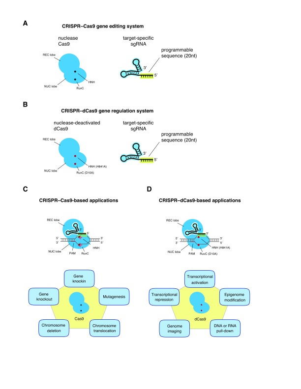
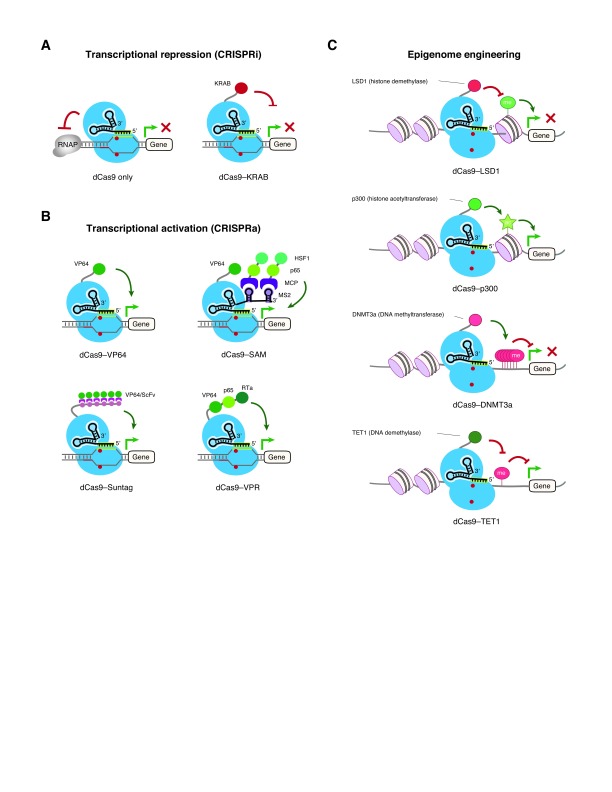
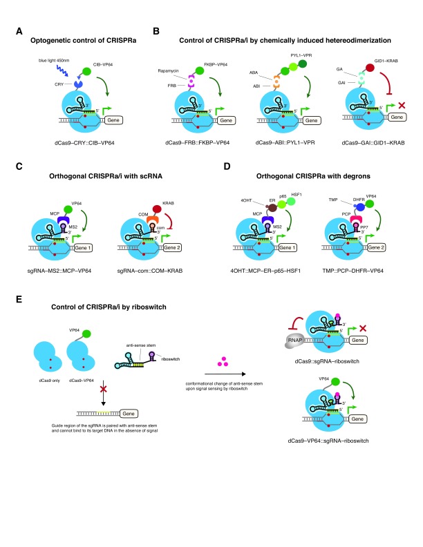
The discovery and adaption of bacterial clustered regularly interspaced short palindromic repeats (CRISPR)–CRISPR-associated (Cas) systems has revolutionized the way researchers edit genomes. Engineering of catalytically inactivated Cas variants (nuclease-deficient or nuclease-deactivated [dCas]) combined with transcriptional repressors, activators, or epigenetic modifiers enable sequence-specific regulation of gene expression and chromatin state. These CRISPR–Cas-based technologies have contributed to the rapid development of disease models and functional genomics screening approaches, which can facilitate genetic target identification and drug discovery. In this short review, we will cover recent advances of CRISPR–dCas9 systems and their use for transcriptional repression and activation, epigenome editing, and engineered synthetic circuits for complex control of the mammalian genome.
Overview
The claims and statistics of this paper haven’t been checked yet.
Run the check — free, no account needed. It reads the open-access text and checks the sources it cites.
Integrity signals
Registry
Cites
Statistics
Figures
Verification
Claims the paper makes
The claims and statistics of this paper haven’t been checked yet.
Run the check — free, no account needed. It reads the open-access text and checks the sources it cites.
Claims other papers make about this one
Each is checked against this paper’s own text: does it say what the citing paper says it does?
The cited source says what the claim attributes to it.
In the citing paper
“However, persistent modifications can be achieved with dCas9 by fusing it to acetyltransferase, histone/DNA demethylase or methyltransferase, altering histone acetylation/methylation or DNA methylation marks and inducing potentially inheritable epigenetic expression modulation when dividing cells are targeted [78,79,80].”
From CRISPR-Cas: Converting A Bacterial Defence Mechanism into A State-of-the-Art Genetic Manipulation Tool (2019) · cites this paper as [78]
The cited source says what the claim attributes to it.
In the citing paper
“However, specific and long-lasting modifications to genetic expression are possible through the fusion of epigenetic modifiers to dCas9 [78].”
From CRISPR-Cas: Converting A Bacterial Defence Mechanism into A State-of-the-Art Genetic Manipulation Tool (2019) · cites this paper as [78]
Self-check
Percentages, sums and reported differences re-computed from the paper’s own numbers.
The self-check needs the paper’s full text.
Citations
Cites
Regulation of chromatin by histone modifications
Bannister, A. J., & Kouzarides, T. (2011) · Cell Research · doi:10.1038/cr.2011.22 · Cited by 6,012
Regulation of microRNA biogenesis
Ha, M., & Kim, V. N. (2014) · Nature Reviews Molecular Cell Biology · doi:10.1038/nrm3838 · Cited by 5,462
Repurposing CRISPR as an RNA-Guided Platform for Sequence-Specific Control of Gene Expression
Qi, L. S., Larson, M. H., Gilbert, L. A., Doudna, J. A., Weissman, J. S., Arkin, A. P., & Lim, W. A. (2013) · Cell · doi:10.1016/j.cell.2013.02.022 · Cited by 5,016
Self-citationSystematic Localization of Common Disease-Associated Variation in Regulatory DNA
Maurano, M. T., Humbert, R., Rynes, E., Thurman, R. E., Haugen, E., Wang, H., Reynolds, A. P., Sandstrom, R., Qu, H., Brody, J., et al. (2012) · Science · doi:10.1126/science.1222794 · Cited by 3,716
CRISPR-Mediated Modular RNA-Guided Regulation of Transcription in Eukaryotes
Gilbert, L. A., Larson, M. H., Morsut, L., Liu, Z., Brar, G. A., Torres, S. E., Stern-Ginossar, N., Brandman, O., Whitehead, E. H., Doudna, J. A., et al. (2013) · Cell · doi:10.1016/j.cell.2013.06.044 · Cited by 3,696
Self-citationWang, H., Yang, H., Shivalila, C. S., Dawlaty, M. M., Cheng, A. W., Zhang, F., & Jaenisch, R. (2013) · Cell · doi:10.1016/j.cell.2013.04.025 · Cited by 3,495
Central Dogma of Molecular Biology
CRICK, F. (1970) · Nature · doi:10.1038/227561a0 · Cited by 3,444
Efficient genome editing in zebrafish using a CRISPR-Cas system
Hwang, W. Y., Fu, Y., Reyon, D., Maeder, M. L., Tsai, S. Q., Sander, J. D., Peterson, R. T., Yeh, J.-R. J., & Joung, J. K. (2013) · Nature Biotechnology · doi:10.1038/nbt.2501 · Cited by 2,937
Genome-scale transcriptional activation by an engineered CRISPR-Cas9 complex
Konermann, S., Brigham, M. D., Trevino, A. E., Joung, J., Abudayyeh, O. O., Barcena, C., Hsu, P. D., Habib, N., Gootenberg, J. S., Nishimasu, H., et al. (2015) · Nature · doi:10.1038/nature14136 · Cited by 2,825 · cited 3×
“This method is referred to as the synergistic activation mediator (SAM) system (Figure 2B) 75 .”
BackgroundMethodGenome-Scale CRISPR-Mediated Control of Gene Repression and Activation
Gilbert, L. A., Horlbeck, M. A., Adamson, B., Villalta, J. E., Chen, Y., Whitehead, E. H., Guimaraes, C., Panning, B., Ploegh, H. L., Bassik, M. C., et al. (2014) · Cell · doi:10.1016/j.cell.2014.09.029 · Cited by 2,728
Self-citationCRISPR RNA Maturation by Trans-Encoded Small RNA and Host Factor RNase III
Deltcheva, E., Chylinski, K., Sharma, C. M., Gonzales, K., Chao, Y., Pirzada, Z. A., Eckert, M. R., Vogel, J., & Charpentier, E. (2011) · Nature · doi:10.1038/nature09886 · Cited by 2,563
RNA-guided editing of bacterial genomes using CRISPR-Cas systems
Jiang, W., Bikard, D., Cox, D., Zhang, F., & Marraffini, L. A. (2013) · Nature Biotechnology · doi:10.1038/nbt.2508 · Cited by 2,429
Linking DNA methylation and histone modification: patterns and paradigms
Cedar, H., & Bergman, Y. (2009) · Nature Reviews Genetics · doi:10.1038/nrg2540 · Cited by 2,267
Kim, S., Kim, D., Cho, S. W., Kim, J., & Kim, J.-S. (2014) · Genome Research · doi:10.1101/gr.171322.113 · Cited by 1,856
Mali, P., Aach, J., Stranges, P. B., Esvelt, K. M., Moosburner, M., Kosuri, S., Yang, L., & Church, G. M. (2013) · Nature Biotechnology · doi:10.1038/nbt.2675 · Cited by 1,752 · cited 1×
“While these simple fusion proteins are able to activate the expression of reporters or endogenous genes, the potency of these constructs in transcriptional activation is quite modest, about 2-fold to 5-fold on average when using a single sgRNA 63,67,69–71 .”
BackgroundGenetics of gene expression and its effect on disease
Emilsson, V., Thorleifsson, G., Zhang, B., Leonardson, A. S., Zink, F., Zhu, J., Carlson, S., Helgason, A., Walters, G. B., Gunnarsdottir, S., et al. (2008) · Nature · doi:10.1038/nature06758 · Cited by 1,296
Editing DNA Methylation in the Mammalian Genome
Liu, X. S., Wu, H., Ji, X., Stelzer, Y., Wu, X., Czauderna, S., Shu, J., Dadon, D., Young, R. A., & Jaenisch, R. (2016) · Cell · doi:10.1016/j.cell.2016.08.056 · Cited by 1,224 · cited 3×
“The dCas9–TET1 fusion can demethylate 30 to 60% of the CpG islands at tested promoters, which drives the transcriptional activation of target genes in various cell types including embryonic stem cells, cancer cell lines, fibroblasts, and primary neurons 86–89 .”
BackgroundCharting histone modifications and the functional organization of mammalian genomes
Zhou, V. W., Goren, A., & Bernstein, B. E. (2011) · Nature Reviews Genetics · doi:10.1038/nrg2905 · Cited by 1,147
Thakore, P. I., D'Ippolito, A. M., Song, L., Safi, A., Shivakumar, N. K., Kabadi, A. M., Reddy, T. E., Crawford, G. E., & Gersbach, C. A. (2015) · Nature Methods · doi:10.1038/nmeth.3630 · Cited by 1,012
Beyond editing: repurposing CRISPR-Cas9 for precision genome regulation and interrogation
Dominguez, A. A., Lim, W. A., & Qi, L. S. (2016) · Nature Reviews Molecular Cell Biology · doi:10.1038/nrm.2015.2 · Cited by 832
Self-citationChimeric nucleases stimulate gene targeting in human cells
Porteus, M. H., & Baltimore, D. (2003) · Science · doi:10.1126/science.1078395 · Cited by 741
Gene disruption by cell-penetrating peptide-mediated delivery of Cas9 protein and guide RNA
Ramakrishna, S., Kwaku Dad, A.-B., Beloor, J., Gopalappa, R., Lee, S.-K., & Kim, H. (2014) · Genome Research · doi:10.1101/gr.171264.113 · Cited by 655
Research works printed but not linked: 121
- A Feinberg, 2016, Epigenetic modulators, modifiers and mediators in cancer aetiology and progression., Nat Rev Genet., 17, 284-99, 10.1038/nrg.2016.13 · DOI · Paper page
- G Gasiunas, 2012, Cas9-crRNA ribonucleoprotein complex mediates specific DNA cleavage for adaptive immunity in bacteria., Proc Natl Acad Sci U S A., 109, E2579-86, 10.1073/pnas.1208507109 · DOI · Paper page
- M Jinek, 2012, A programmable dual-RNA-guided DNA endonuclease in adaptive bacterial immunity., Science., 337, 816-21, 10.1126/science.1225829 · DOI · Paper page
- R Sapranauskas, 2011, The Streptococcus thermophilus CRISPR/Cas system provides immunity in Escherichia coli., Nucleic Acids Res., 39, 9275-82, 10.1093/nar/gkr606 · DOI
- J Garneau, 2010, The CRISPR/Cas bacterial immune system cleaves bacteriophage and plasmid DNA., Nature., 468, 67-71, 10.1038/nature09523 · DOI
- R Barrangou, 2007, CRISPR provides acquired resistance against viruses in prokaryotes., Science., 315, 1709-12, 10.1126/science.1138140 · DOI · Paper page
- S Brouns, 2008, Small CRISPR RNAs guide antiviral defense in prokaryotes., Science., 321, 960-4, 10.1126/science.1159689 · DOI · Paper page
- C Pourcel, 2005, CRISPR elements in Yersinia pestis acquire new repeats by preferential uptake of bacteriophage DNA, and provide additional tools for evolutionary studies., Microbiology., 151, 653-63, 10.1099/mic.0.27437-0 · DOI · Paper page
- S Shmakov, 2015, Discovery and Functional Characterization of Diverse Class 2 CRISPR-Cas Systems., Mol Cell., 60, 385-97, 10.1016/j.molcel.2015.10.008 · DOI · Paper page
- D Rath, 2015, The CRISPR-Cas immune system: biology, mechanisms and applications., Biochimie., 117, 119-28, 10.1016/j.biochi.2015.03.025 · DOI · Paper page
- F Mojica, 2005, Intervening sequences of regularly spaced prokaryotic repeats derive from foreign genetic elements., J Mol Evol., 60, 174-82, 10.1007/s00239-004-0046-3 · DOI · Paper page
- A Bolotin, 2005, Clustered regularly interspaced short palindrome repeats (CRISPRs) have spacers of extrachromosomal origin., Microbiology., 151, 2551-61, 10.1099/mic.0.28048-0 · DOI · Paper page
- L Marraffini, 2008, CRISPR interference limits horizontal gene transfer in staphylococci by targeting DNA., Science., 322, 1843-5, 10.1126/science.1165771 · DOI · Paper page
- K Makarova, 2015, An updated evolutionary classification of CRISPR-Cas systems., Nat Rev Microbiol., 13, 722-36, 10.1038/nrmicro3569 · DOI · Paper page
- S Shmakov, 2017, Diversity and evolution of class 2 CRISPR-Cas systems., Nat Rev Microbiol., 15, 169-82, 10.1038/nrmicro.2016.184 · DOI · Paper page
- K Makarova, 2017, SnapShot: Class 2 CRISPR-Cas Systems., Cell., 168, 328-328.e1, 10.1016/j.cell.2016.12.038 · DOI
- B Zetsche, 2015, Cpf1 is a single RNA-guided endonuclease of a class 2 CRISPR-Cas system., Cell., 163, 759-71, 10.1016/j.cell.2015.09.038 · DOI · Paper page
- M Lim, 2016, UDP-galactose and acetyl-CoA transporters as Plasmodium multidrug resistance genes., Nat Microbiol., 10.1038/nmicrobiol.2016.166 · DOI
- C Blondel, 2016, CRISPR/Cas9 Screens Reveal Requirements for Host Cell Sulfation and Fucosylation in Bacterial Type III Secretion System-Mediated Cytotoxicity., Cell Host Microbe., 20, 226-37, 10.1016/j.chom.2016.06.010 · DOI
- Y Wang, 2016, High-Efficiency Genome Editing of Streptomyces Species by an Engineered CRISPR/Cas System., Methods Enzymol., 575, 271-84, 10.1016/bs.mie.2016.03.014 · DOI
- J DiCarlo, 2013, Genome engineering in Saccharomyces cerevisiae using CRISPR-Cas systems., Nucleic Acids Res., 41, 4336-43, 10.1093/nar/gkt135 · DOI · Paper page
- A Reider Apel, 2017, A Cas9-based toolkit to program gene expression in Saccharomyces cerevisiae., Nucleic Acids Res., 45, 496-508, 10.1093/nar/gkw1023 · DOI
- M Laughery, 2015, New vectors for simple and streamlined CRISPR-Cas9 genome editing in Saccharomyces cerevisiae., Yeast., 32, 711-20, 10.1002/yea.3098 · DOI
- Z Bao, 2015, Homology-integrated CRISPR-Cas (HI-CRISPR) system for one-step multigene disruption in Saccharomyces cerevisiae., ACS Synth Biol., 4, 585-94, 10.1021/sb500255k · DOI · Paper page
- Y Miyanari, 2013, Live visualization of chromatin dynamics with fluorescent TALEs., Nat Struct Mol Biol., 20, 1321-4, 10.1038/nsmb.2680 · DOI
- V Nekrasov, 2013, Targeted mutagenesis in the model plant Nicotiana benthamiana using Cas9 RNA-guided endonuclease., Nat Biotechnol., 31, 691-3, 10.1038/nbt.2655 · DOI
- Z Feng, 2013, Efficient genome editing in plants using a CRISPR/Cas system., Cell Res., 23, 1229-32, 10.1038/cr.2013.114 · DOI
- K Xie, 2013, RNA-guided genome editing in plants using a CRISPR-Cas system., Mol Plant., 6, 1975-83, 10.1093/mp/sst119 · DOI
- T Čermák, 2015, High-frequency, precise modification of the tomato genome., Genome Biol., 16, 232, 10.1186/s13059-015-0796-9 · DOI
- Z Feng, 2014, Multigeneration analysis reveals the inheritance, specificity, and patterns of CRISPR/Cas-induced gene modifications in Arabidopsis., Proc Natl Acad Sci U S A., 111, 4632-7, 10.1073/pnas.1400822111 · DOI
- Q Shan, 2014, Genome editing in rice and wheat using the CRISPR/Cas system., Nat Protoc., 9, 2395-410, 10.1038/nprot.2014.157 · DOI
- S Svitashev, 2016, Genome editing in maize directed by CRISPR-Cas9 ribonucleoprotein complexes., Nat Commun., 7, 10.1038/ncomms13274 · DOI
- J Woo, 2015, DNA-free genome editing in plants with preassembled CRISPR-Cas9 ribonucleoproteins., Nat Biotechnol., 33, 1162-4, 10.1038/nbt.3389 · DOI · Paper page
- Y Zhang, 2016, Efficient and transgene-free genome editing in wheat through transient expression of CRISPR/Cas9 DNA or RNA., Nat Commun., 7, 10.1038/ncomms12617 · DOI
- H Zhou, 2014, Large chromosomal deletions and heritable small genetic changes induced by CRISPR/Cas9 in rice., Nucleic Acids Res., 42, 10903-14, 10.1093/nar/gku806 · DOI
- Q Lv, 2016, Efficient Generation of Myostatin Gene Mutated Rabbit by CRISPR/Cas9., Sci Rep., 6, 10.1038/srep25029 · DOI
- X Wang, 2015, Generation of gene-modified goats targeting MSTN and FGF5 via zygote injection of CRISPR/Cas9 system., Sci Rep., 5, 13878, 10.1038/srep13878 · DOI
- W Li, 2013, Simultaneous generation and germline transmission of multiple gene mutations in rat using CRISPR-Cas systems., Nat Biotechnol., 31, 684-6, 10.1038/nbt.2652 · DOI
- K Wang, 2015, Efficient Generation of Myostatin Mutations in Pigs Using the CRISPR/Cas9 System., Sci Rep., 5, 10.1038/srep16623 · DOI
- F Mojica, 2009, Short motif sequences determine the targets of the prokaryotic CRISPR defence system., Microbiology., 155, 733-40, 10.1099/mic.0.023960-0 · DOI · Paper page
- L Cong, 2013, Multiplex genome engineering using CRISPR/Cas systems., Science., 339, 819-23, 10.1126/science.1231143 · DOI · Paper page
- P Mali, 2013, RNA-guided human genome engineering via Cas9., Science., 339, 823-6, 10.1126/science.1232033 · DOI · Paper page
- S Cho, 2013, Targeted genome engineering in human cells with the Cas9 RNA-guided endonuclease., Nat Biotechnol., 31, 230-2, 10.1038/nbt.2507 · DOI · Paper page
- M Jinek, 2013, RNA-programmed genome editing in human cells., eLife., 2, e00471, 10.7554/eLife.00471 · DOI · Paper page
- A Wright, 2016, Biology and Applications of CRISPR Systems: Harnessing Nature's Toolbox for Genome Engineering., Cell., 164, 29-44, 10.1016/j.cell.2015.12.035 · DOI · Paper page
- H Wang, 2016, CRISPR/Cas9 in Genome Editing and Beyond., Annu Rev Biochem., 85, 227-64, 10.1146/annurev-biochem-060815-014607 · DOI
- A Komor, 2017, CRISPR-Based Technologies for the Manipulation of Eukaryotic Genomes., Cell., 168, 20-36, 10.1016/j.cell.2016.10.044 · DOI
- M La Russa, 2015, The New State of the Art: Cas9 for Gene Activation and Repression., Mol Cell Biol., 35, 3800-9, 10.1128/MCB.00512-15 · DOI
- F Jiang, 2017, CRISPR-Cas9 Structures and Mechanisms., Annu Rev Biophys., 10.1146/annurev-biophys-062215-010822 · DOI · Paper page
- D Bikard, 2013, Programmable repression and activation of bacterial gene expression using an engineered CRISPR-Cas system., Nucleic Acids Res., 41, 7429-37, 10.1093/nar/gkt520 · DOI · Paper page
- M Larson, 2013, CRISPR interference (CRISPRi) for sequence-specific control of gene expression., Nat Protoc., 8, 2180-96, 10.1038/nprot.2013.132 · DOI
- W Ji, 2014, Specific gene repression by CRISPRi system transferred through bacterial conjugation., ACS Synth Biol., 3, 929-31, 10.1021/sb500036q · DOI
- J Peters, 2016, A Comprehensive, CRISPR-based Functional Analysis of Essential Genes in Bacteria., Cell., 165, 1493-506, 10.1016/j.cell.2016.05.003 · DOI
- I Lawhorn, 2014, Evaluation of sgRNA target sites for CRISPR-mediated repression of TP53., PLoS One., 9, e113232, 10.1371/journal.pone.0113232 · DOI
- S Konermann, 2013, Optical control of mammalian endogenous transcription and epigenetic states., Nature., 500, 472-6, 10.1038/nature12466 · DOI · Paper page
- A Cheng, 2013, Multiplexed activation of endogenous genes by CRISPR-on, an RNA-guided transcriptional activator system., Cell Res., 23, 1163-71, 10.1038/cr.2013.122 · DOI
- X Gao, 2014, Comparison of TALE designer transcription factors and the CRISPR/dCas9 in regulation of gene expression by targeting enhancers., Nucleic Acids Res., 42, e155, 10.1093/nar/gku836 · DOI
- M Maeder, 2013, CRISPR RNA-guided activation of endogenous human genes., Nat Methods., 10, 977-9, 10.1038/nmeth.2598 · DOI · Paper page
- P Perez-Pinera, 2013, RNA-guided gene activation by CRISPR-Cas9-based transcription factors., Nat Methods., 10, 973-6, 10.1038/nmeth.2600 · DOI · Paper page
- M Tanenbaum, 2014, A protein-tagging system for signal amplification in gene expression and fluorescence imaging., Cell., 159, 635-46, 10.1016/j.cell.2014.09.039 · DOI
- J Zalatan, 2015, Engineering complex synthetic transcriptional programs with CRISPR RNA scaffolds., Cell., 160, 339-50, 10.1016/j.cell.2014.11.052 · DOI · Paper page
- A Chavez, 2015, Highly efficient Cas9-mediated transcriptional programming., Nat Methods., 12, 326-8, 10.1038/nmeth.3312 · DOI · Paper page
- A Chavez, 2016, Comparison of Cas9 activators in multiple species., Nat Methods., 13, 563-7, 10.1038/nmeth.3871 · DOI
- E Consortium, 2012, An integrated encyclopedia of DNA elements in the human genome., Nature., 489, 57-74, 10.1038/nature11247 · DOI · Paper page
- A Kundaje, 2015, Integrative analysis of 111 reference human epigenomes., Nature., 518, 317-30, 10.1038/nature14248 · DOI · Paper page
- N Kearns, 2015, Functional annotation of native enhancers with a Cas9-histone demethylase fusion., Nat Methods., 12, 401-3, 10.1038/nmeth.3325 · DOI · Paper page
- I Hilton, 2015, Epigenome editing by a CRISPR-Cas9-based acetyltransferase activates genes from promoters and enhancers., Nat Biotechnol., 33, 510-7, 10.1038/nbt.3199 · DOI
- D Cano-Rodriguez, 2016, Writing of H3K4Me3 overcomes epigenetic silencing in a sustained but context-dependent manner., Nat Commun., 7, 12284, 10.1038/ncomms12284 · DOI
- X Xu, 2016, A CRISPR-based approach for targeted DNA demethylation., Cell Discov., 2, 16009, 10.1038/celldisc.2016.9 · DOI
- S Morita, 2016, Targeted DNA demethylation in vivo using dCas9-peptide repeat and scFv-TET1 catalytic domain fusions., Nat Biotechnol., 34, 1060-5, 10.1038/nbt.3658 · DOI
- S Choudhury, 2016, CRISPR-dCas9 mediated TET1 targeting for selective DNA demethylation at BRCA1 promoter., Oncotarget., 7, 46545-56, 10.18632/oncotarget.10234 · DOI
- J McDonald, 2016, Reprogrammable CRISPR/Cas9-based system for inducing site-specific DNA methylation., Biol Open., 5, 866-74, 10.1242/bio.019067 · DOI
- A Vojta, 2016, Repurposing the CRISPR-Cas9 system for targeted DNA methylation., Nucleic Acids Res., 44, 5615-28, 10.1093/nar/gkw159 · DOI
- A Amabile, 2016, Inheritable Silencing of Endogenous Genes by Hit-and-Run Targeted Epigenetic Editing., Cell., 167, 219-232.e14, 10.1016/j.cell.2016.09.006 · DOI · Paper page
- Y Nihongaki, 2015, CRISPR-Cas9-based photoactivatable transcription system., Chem Biol., 22, 169-74, 10.1016/j.chembiol.2014.12.011 · DOI
- L Polstein, 2015, A light-inducible CRISPR-Cas9 system for control of endogenous gene activation., Nat Chem Biol., 11, 198-200, 10.1038/nchembio.1753 · DOI
- B Zetsche, 2015, A split-Cas9 architecture for inducible genome editing and transcription modulation., Nat Biotechnol., 33, 139-42, 10.1038/nbt.3149 · DOI
- B Oakes, 2016, Profiling of engineering hotspots identifies an allosteric CRISPR-Cas9 switch., Nat Biotechnol., 34, 646-51, 10.1038/nbt.3528 · DOI
- K Esvelt, 2013, Orthogonal Cas9 proteins for RNA-guided gene regulation and editing., Nat Methods., 10, 1116-21, 10.1038/nmeth.2681 · DOI · Paper page
- I Fonfara, 2014, Phylogeny of Cas9 determines functional exchangeability of dual-RNA and Cas9 among orthologous type II CRISPR-Cas systems., Nucleic Acids Res., 42, 2577-90, 10.1093/nar/gkt1074 · DOI · Paper page
- A Briner, 2014, Guide RNA functional modules direct Cas9 activity and orthogonality., Mol Cell., 56, 333-9, 10.1016/j.molcel.2014.09.019 · DOI
- Y Gao, 2016, Complex transcriptional modulation with orthogonal and inducible dCas9 regulators., Nat Methods., 13, 1043-9, 10.1038/nmeth.4042 · DOI
- Z Bao, 2017, Orthogonal Genetic Regulation in Human Cells Using Chemically Induced CRISPR/Cas9 Activators., ACS Synth Biol., 6, 686-693, 10.1021/acssynbio.6b00313 · DOI
- D Shechner, 2015, Multiplexable, locus-specific targeting of long RNAs with CRISPR-Display., Nat Methods., 12, 664-70, 10.1038/nmeth.3433 · DOI
- B Maji, 2017, Multidimensional chemical control of CRISPR-Cas9., Nat Chem Biol., 13, 9-11, 10.1038/nchembio.2224 · DOI
- Y Liu, 2016, Directing cellular information flow via CRISPR signal conductors., Nat Methods., 13, 938-44, 10.1038/nmeth.3994 · DOI
- S Kiani, 2014, CRISPR transcriptional repression devices and layered circuits in mammalian cells., Nat Methods., 11, 723-6, 10.1038/nmeth.2969 · DOI
- A Wright, 2015, Rational design of a split-Cas9 enzyme complex., Proc Natl Acad Sci U S A., 112, 2984-9, 10.1073/pnas.1501698112 · DOI
- D Truong, 2015, Development of an intein-mediated split-Cas9 system for gene therapy., Nucleic Acids Res., 43, 6450-8, 10.1093/nar/gkv601 · DOI · Paper page
- H Nishimasu, 2015, Crystal Structure of Staphylococcus aureus Cas9., Cell., 162, 1113-26, 10.1016/j.cell.2015.08.007 · DOI
- J Zuris, 2015, Cationic lipid-mediated delivery of proteins enables efficient protein-based genome editing in vitro and in vivo., Nat Biotechnol., 33, 73-80, 10.1038/nbt.3081 · DOI · Paper page
- Y Nihongaki, 2015, Photoactivatable CRISPR-Cas9 for optogenetic genome editing., Nat Biotechnol., 33, 755-60, 10.1038/nbt.3245 · DOI
- L Dow, 2015, Inducible in vivo genome editing with CRISPR-Cas9., Nat Biotechnol., 33, 390-4, 10.1038/nbt.3155 · DOI
- F González, 2014, An iCRISPR platform for rapid, multiplexable, and inducible genome editing in human pluripotent stem cells., Cell Stem Cell., 15, 215-26, 10.1016/j.stem.2014.05.018 · DOI
- J Hemphill, 2015, Optical Control of CRISPR/Cas9 Gene Editing., J Am Chem Soc., 137, 5642-5, 10.1021/ja512664v · DOI
- P Jain, 2016, Development of Light-Activated CRISPR Using Guide RNAs with Photocleavable Protectors., Angew Chem Int Ed Engl., 55, 12440-4, 10.1002/anie.201606123 · DOI
- B Aubrey, 2015, An inducible lentiviral guide RNA platform enables the identification of tumor-essential genes and tumor-promoting mutations in vivo., Cell Rep., 10, 1422-32, 10.1016/j.celrep.2015.02.002 · DOI
- K Davis, 2015, Small molecule-triggered Cas9 protein with improved genome-editing specificity., Nat Chem Biol., 11, 316-8, 10.1038/nchembio.1793 · DOI
- T Wang, 2014, Genetic screens in human cells using the CRISPR-Cas9 system., Science., 343, 80-4, 10.1126/science.1246981 · DOI · Paper page
- R Carthew, 2009, Origins and Mechanisms of miRNAs and siRNAs., Cell., 136, 642-55, 10.1016/j.cell.2009.01.035 · DOI
- C Mello, 2004, Revealing the world of RNA interference., Nature., 431, 338-42, 10.1038/nature02872 · DOI
- G Hutvagner, 2008, Argonaute proteins: key players in RNA silencing., Nat Rev Mol Cell Biol., 9, 22-32, 10.1038/nrm2321 · DOI
- G Prelich, 2012, Gene overexpression: uses, mechanisms, and interpretation., Genetics., 190, 841-54, 10.1534/genetics.111.136911 · DOI
- J Miller, 2007, An improved zinc-finger nuclease architecture for highly specific genome editing., Nat Biotechnol., 25, 778-85, 10.1038/nbt1319 · DOI · Paper page
- J Sander, 2011, Selection-free zinc-finger-nuclease engineering by context-dependent assembly (CoDA)., Nat Methods., 8, 67-9, 10.1038/nmeth.1542 · DOI
- A Wood, 2011, Targeted genome editing across species using ZFNs and TALENs., Science., 333, 307, 10.1126/science.1207773 · DOI
- M Christian, 2010, Targeting DNA double-strand breaks with TAL effector nucleases., Genetics., 186, 757-61, 10.1534/genetics.110.120717 · DOI · Paper page
- F Zhang, 2011, Efficient construction of sequence-specific TAL effectors for modulating mammalian transcription., Nat Biotechnol., 29, 149-53, 10.1038/nbt.1775 · DOI · Paper page
- D Hockemeyer, 2011, Genetic engineering of human pluripotent cells using TALE nucleases., Nat Biotechnol., 29, 731-4, 10.1038/nbt.1927 · DOI
- D Reyon, 2012, FLASH assembly of TALENs for high-throughput genome editing., Nat Biotechnol., 30, 460-5, 10.1038/nbt.2170 · DOI
- J Boch, 2009, Breaking the code of DNA binding specificity of TAL-type III effectors., Science., 326, 1509-12, 10.1126/science.1178811 · DOI · Paper page
- M Moscou, 2009, A simple cipher governs DNA recognition by TAL effectors., Science., 326, 1501, 10.1126/science.1178817 · DOI
- N Sanjana, 2012, A transcription activator-like effector toolbox for genome engineering., Nat Protoc., 7, 171-92, 10.1038/nprot.2011.431 · DOI
- Y Zhao, 2014, Sequence-specific inhibition of microRNA via CRISPR/CRISPRi system., Sci Rep., 4, 3943, 10.1038/srep03943 · DOI
- Y Choo, 2000, Advances in zinc finger engineering., Curr Opin Struct Biol., 10, 411-6, 10.1016/S0959-440X(00)00107-X · DOI
- T Sera, 2009, Zinc-finger-based artificial transcription factors and their applications., Adv Drug Deliv Rev., 61, 513-26, 10.1016/j.addr.2009.03.012 · DOI
- L Cong, 2012, Comprehensive interrogation of natural TALE DNA-binding modules and transcriptional repressor domains., Nat Commun., 3, 968, 10.1038/ncomms1962 · DOI
- A Garg, 2012, Engineering synthetic TAL effectors with orthogonal target sites., Nucleic Acids Res., 40, 7584-95, 10.1093/nar/gks404 · DOI
- M Mahfouz, 2012, Targeted transcriptional repression using a chimeric TALE-SRDX repressor protein., Plant Mol Biol., 78, 311-21, 10.1007/s11103-011-9866-x · DOI
- M Maeder, 2013, Robust, synergistic regulation of human gene expression using TALE activators., Nat Methods., 10, 243-5, 10.1038/nmeth.2366 · DOI
- L Polstein, 2015, Genome-wide specificity of DNA binding, gene regulation, and chromatin remodeling by TALE- and CRISPR/Cas9-based transcriptional activators., Genome Res., 25, 1158-69, 10.1101/gr.179044.114 · DOI
Cited by
Sorted by citations
Loureiro, A., & da Silva, G. J. (2019) · Antibiotics · doi:10.3390/antibiotics8010018 · Cited by 129
Current Bioinformatics Tools to Optimize CRISPR/Cas9 Experiments to Reduce Off-Target Effects
Naeem, M., & Alkhnbashi, O. S. (2023) · International Journal of Molecular Sciences · doi:10.3390/ijms24076261 · Cited by 70
Metadata
- Authors
- Lo, A., & Qi, L.
- ORCID
- Qi, Lei
- Venue
- F1000Research
- Publisher
- F1000 Research Ltd
- Published
- 2017-05-25
- Volume
- 6
- Pages
- 747
- ISSN
- 2046-1402
- PMC
- PMC5464239
- Cited by
- 72
- References
- 22
- Language
- en
- Funders
- Alfred P. Sloan Foundation
- NIH Office of the Director (DP5 OD017887)
- The Pew Charitable Trusts
About these checks
Registry, retraction, Cites and statistics results are computed, not judged, with one exception: a printed reference that no index lists is read once by a model before it counts as a citation error. Claims are extracted by a model and kept only when their quote appears verbatim in the text; each cited source is then checked, with a model’s help, for whether it says what the claim relies on. Those verdicts can be wrong — read the claim’s quote and the cited source before relying on one.
Statistics and arithmetic are recomputed, not judged.
Confidence bands are shown only for fields calibrated on our annotated test set. None is calibrated yet, so no band is shown.
What counts as a claim ›How we measure confidence ›How we check this ›
Are you an author, or do you see a problem with this page? Request a review
