

The DOI matches its Crossref record; 114 of the 115 references it cites were found in a registry or an index; 39 of the 50 sources its claims cite were checked for support.
Diffusion MRI Indices and Their Relation to Cognitive Impairment in Brain Aging: The Updated Multi-protocol Approach in ADNI3
Open accessPublished version of a preprint
Abstract
Abstract Brain imaging with diffusion-weighted MRI (dMRI) is sensitive to microstructural white matter changes associated with brain aging and neurodegeneration. In its third phase, the Alzheimer’s Disease Neuroimaging Initiative (ADNI3) is collecting data across multiple sites and scanners using different dMRI acquisition protocols, to better understand disease effects. It is vital to understand when data can be pooled across scanners, and how the choice of dMRI protocol affects the sensitivity of extracted measures to differences in clinical impairment. Here, we analyzed ADNI3 data from 317 participants (mean age: 75.4±7.9 years; 143 men/174 women), who were each scanned at one of 47 sites with one of six dMRI protocols using scanners from three different manufacturers. We computed four standard diffusion tensor imaging (DTI) indices including fractional anisotropy (FA DTI ) and mean, radial, and axial diffusivity, and one FA index based on the tensor distribution function (FA TDF ), in 24 bilaterally averaged white matter regions of interest. We found that protocol differences significantly affected dMRI indices, in particular FA DTI . We ranked the diffusion indices for their strength of association with four clinical assessments. In addition to diagnosis, we evaluated cognitive impairment as indexed by three commonly used screening tools for detecting dementia and Alzheimer’s disease: the Alzheimer’s Disease Assessment Scale (ADAS-cog), the Mini-Mental State Examination (MMSE), and the Clinical Dementia Rating scale sum-of-boxes (CDR-sob). Using a nested random-effects model to account for protocol and site, we found that across all dMRI indices and clinical measures, the hippocampal-cingulum and fornix ( crus ) / stria terminalis regions most consistently showed strong associations with clinical impairment. Overall, the greatest effect sizes were detected in the hippocampal-cingulum and uncinate fasciculus for associations between axial or mean diffusivity and CDR-sob. FA TDF detected robust widespread associations with clinical measures, while FA DTI was the weakest of the five indices for detecting associations. Ultimately, we were able to successfully pool dMRI data from multiple acquisition protocols from ADNI3 and detect consistent and robust associations with clinical impairment and age.
Overview
AI snapshot
Written from the paper’s full text.
TL;DR
In ADNI3, diffusion MRI indices pooled across six acquisition protocols and 47 sites showed robust associations with age and cognitive impairment, despite significant protocol effects. The TDF-derived FA index (FA_TDF) was more sensitive than standard DTI FA, and axial/mean diffusivity in temporal WM tracts showed the strongest clinical associations.
Key findings
- Across 317 participants (mean age 75.4 ± 7.9 years; 143 men/174 women), protocol differences significantly affected all five dMRI indices; FA_DTI was least stable (significant protocol differences in 81.9% of pairwise tests), while AxD_DTI was most stable (20.6%).
- The hippocampal-cingulum (CGH) and fornix (crus)/stria terminalis (Fx/ST) most consistently showed strong associations with clinical impairment; the largest effects were in CGH and uncinate fasciculus (UNC) for axial/mean diffusivity with CDR-sob (AxD_DTI UNC: d = 1.344, p = 3.13 × 10⁻²³; MD_DTI UNC: d = 1.342, p = 3.47 × 10⁻²³).
- FA_TDF detected more widespread clinical associations than FA_DTI: 69/72 tests (94.4%) vs 55/72 (76.4%) for cognitive measures, and 20/24 ROIs vs 3/24 ROIs for CN vs MCI diagnosis.
- ComBat harmonization reduced significant pairwise protocol differences by 93.8%, with only the corticospinal tract still showing protocol differences; age associations remained significant after harmonization.
Methods
- Baseline ADNI3 data available as of April 30, 2018: 381 scanned participants, 55 excluded after quality assurance, leaving 317 from 47 sites scanned with six dMRI protocols from GE, Philips, and Siemens scanners.
- Four DTI indices (FA, mean, radial, axial diffusivity) and one TDF-based FA index were computed in 24 bilaterally averaged JHU atlas WM ROIs; analyses used nested random-effects regressions with protocol and site as nested random variables and FDR correction (q = 0.05).
- Clinical associations were tested with ADAS-cog, MMSE, CDR-sob, and CN vs MCI diagnosis; AD participants (N = 22) were not compared across diagnostic groups due to limited and unevenly distributed sample.
Limitations
- The paper states that sample sizes and demographics varied by protocol, complicating direct protocol comparisons, and that separating protocol differences from scanner-manufacturer differences is difficult; few AD participants were scanned, limiting diagnostic-group comparisons.
- The paper notes that the cognitive assessments are proxy measures and acknowledges their limitations for indexing specific cognitive abilities.
- Evident from the text: analyses are cross-sectional baseline data, so longitudinal predictive utility is not established.
Model-written summary. Check it against the paper before relying on it.
Integrity signals
Registry
Cites
Statistics
Figures
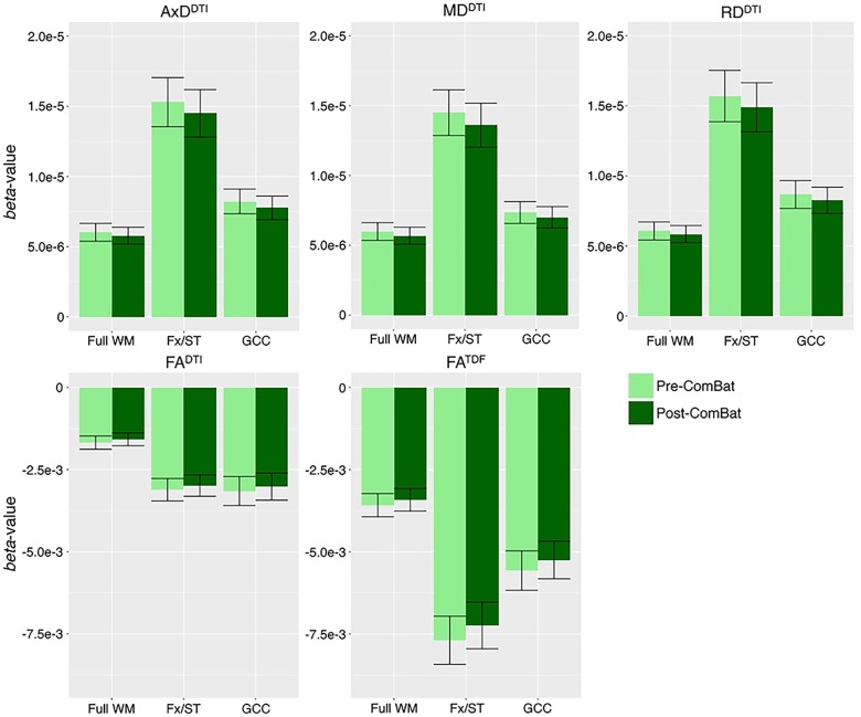
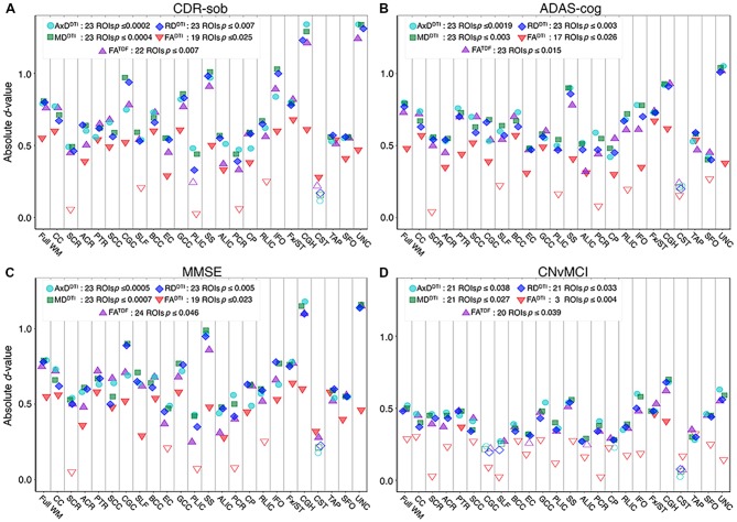
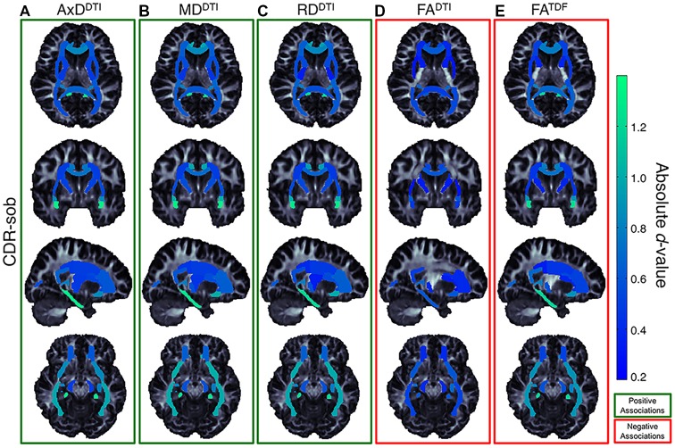
Verification
Claims the paper makes
65 Own result · 15 Partly supported · 13 Own method · 9 Supported · 4 Own interpretation · 3 No full text of the sources · 2 No basis given · 1 Supported so far · 1 Not supported · 1 Couldn’t confirm · accuracy not yet measured
At least one cited source doesn’t support this claim or points to a different paper, and none confirmed it — see the verdicts below.
In the paper
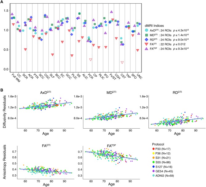
“Due to differences in age and sample size between protocols and phases, effect sizes could not be directly compared (Button et al., 2013), but the directions of associations with age were largely consistent for ADNI2 and ADNI3 phases separately, and each ADNI3 protocol (Figures 1, 2).”
§ Diffusion MRI Indices and Their Relation to Cognitive Impairment in Brain Aging: The Updated Multi-protocol Approach in ADNI3 > Results > ADNI2 and ADNI3 Protocols in Cognitively Normal Participants
What the sources say
- Disputedcites Button et al., 2013 sourceExperimental — accuracy not yet measured
The “Effect size” section (page unavailable) says standardized effect sizes allow estimates from different studies to be compared directly, contrary to the cited rationale that differing sample sizes prevent direct comparison. The paper does not address the particular age differences, protocols, or phases in the citing sentence. (LLM, conf=0.78)
Checked by a reasoning model — may be wrong.
plain- Asserted: passes — The authors put it forward as true.
- Checkable: passes — It can be checked against evidence.
- Load-bearing: passes — The paper’s conclusions depend on it.
- Based: passes — The paper gives a reason why it holds.
- Disputed
The cited sources support only part of this claim — read each source’s verdict below.
In the paper
“differences in dMRI acquisition parameters, including vendor, voxel size, and angular resolution, are known to affect derived dMRI measures (Alexander et al., 2001; Cercignani et al., 2003; Zhan et al., 2010; Zhu et al., 2011).”
§ Diffusion MRI Indices and Their Relation to Cognitive Impairment in Brain Aging: The Updated Multi-protocol Approach in ADNI3 > Introduction
What the sources say
- Partly supportedcites Alexander et al., 2001 sourceExperimental — accuracy not yet measured
The Imaging Experiments and Discussion (page not provided) show that voxel size and the orientation of diffusion-encoding axes affect derived trace and fractional-anisotropy measurements, particularly where partial-volume effects are significant. The paper uses one scanner vendor and does not establish an effect of vendor differences or angular resolution; rotating a fixed set of directions is not a test of angular resolution. (LLM, conf=0.88)
- Not checked — no full text of the sourcecites Cercignani et al., 2003
- Partly supportedcites Zhan et al., 2010 sourceExperimental — accuracy not yet measured
The Conclusions report that the number of diffusion gradients affects the signal-to-noise ratio of derived measures, supporting the angular-resolution part of the claim. They also say achievable SNR depends on the scanner and spatial resolution, but do not establish that vendor or voxel-size differences affect derived dMRI measures. No page numbers are available in our copy. (LLM, conf=0.86)
boosteddirect- Asserted: passes — The authors put it forward as true.
- Checkable: passes — It can be checked against evidence.
- Load-bearing: passes — The paper’s conclusions depend on it.
- Based: passes — The paper gives a reason why it holds.
- Partly supported
Every source the paper cites for this claim was checked and supports it.
In the paper
“we also evaluated a modified measure of FA, derived from the tensor distribution function (FATDF; Leow et al., 2009) which can be more sensitive to neurodegenerative disease-related WM abnormalities than FADTI across high- and low-angular resolution dMRI (Nir et al., 2017).”
§ Diffusion MRI Indices and Their Relation to Cognitive Impairment in Brain Aging: The Updated Multi-protocol Approach in ADNI3 > Introduction
What the sources say
- Supportedcites Leow et al., 2009 source
The Leow et al. citation identifies the tensor distribution function underlying the measure, while the separate Nir et al. citation is attached to the sensitivity comparison. Leow et al. introduce the TDF in the Abstract and describe deriving anisotropy measures from it; their Discussion also describes an inverse anisotropy measure. This confirms the methodological identification attributed to Leow et al., not the disease-sensitivity comparison. (LLM, conf=0.88)
- Supportedcites Nir et al., 2017 source
The Results (Clinical Associations and Effect Sizes; page not provided) report that FATDF was more sensitive than FADTI to Alzheimer’s disease-related white-matter deficits, including at 7 gradient directions. The Discussion and Conclusions report larger FATDF effect sizes at the full 41-direction resolution and when subsampled to 30, 15, or 7 directions. Alzheimer’s disease is the neurodegenerative condition tested here. (LLM, conf=0.93)
plaincorrelational- Asserted: passes — The authors put it forward as true.
- Checkable: passes — It can be checked against evidence.
- Load-bearing: passes — The paper’s conclusions depend on it.
- Based: passes — The paper gives a reason why it holds.
- Supported
The cited sources support only part of this claim — read each source’s verdict below.
In the paper
“suggesting that tensor limitations have likely confounded previous diffusion studies of cognitive decline that have found little or no effects with FA (Acosta-Cabronero et al., 2010).”
§ Diffusion MRI Indices and Their Relation to Cognitive Impairment in Brain Aging: The Updated Multi-protocol Approach in ADNI3 > Discussion
What the sources say
- Partly supportedcites Acosta-Cabronero et al., 2010 sourceExperimental — accuracy not yet measured
The Discussion (page not available in our copy) explains that FA can miss proportional changes in diffusion and reports that FA was less sensitive than absolute diffusivity measures in this Alzheimer’s disease study. It does not establish that this limitation confounded previous studies of cognitive decline that found little or no FA effect; the authors caution that its relevance beyond Alzheimer’s disease needs to be demonstrated. (LLM, conf=0.84)
hedgedconditional- Asserted: passes — The authors put it forward as true.
- Checkable: passes — It can be checked against evidence.
- Load-bearing: passes — The paper’s conclusions depend on it.
- Based: passes — The paper gives a reason why it holds.
- Partly supported
Every source the paper cites for this claim was checked and supports it.
In the paper
“As in our prior analysis of ADNI2 (Nir et al., 2013), FADTI was the least sensitive DTI measure.”
§ Diffusion MRI Indices and Their Relation to Cognitive Impairment in Brain Aging: The Updated Multi-protocol Approach in ADNI3 > Discussion
What the sources say
- Supportedcites Nir et al., 2013 source
The Results (Voxel-based analyses; page not available) report that FA had the smallest effect size in associations with clinical scores and that diffusivity measures showed larger, more diffuse differences between AD patients and controls. The Discussion identifies the study’s MCI cohort as ADNI2, supporting the reference to a prior ADNI2 analysis. (LLM, conf=0.91)
plain- Asserted: passes — The authors put it forward as true.
- Checkable: passes — It can be checked against evidence.
- Load-bearing: unclear — It is unclear whether the paper’s conclusions depend on it.
- Based: passes — The paper gives a reason why it holds.
- Supported
The cited sources support only part of this claim — read each source’s verdict below.
In the paper
“Similarly, in ADNI2, AxDDTI was the most sensitive to differences between CN and MCI diagnosis (Nir et al., 2013).”
§ Diffusion MRI Indices and Their Relation to Cognitive Impairment in Brain Aging: The Updated Multi-protocol Approach in ADNI3 > Discussion
What the sources say
- Partly supportedcites Nir et al., 2013 sourceExperimental — accuracy not yet measured
The Discussion (page unavailable) reports slightly larger effect sizes for axial diffusivity (AxD) when comparing controls with MCI in full-ROI analyses, supporting the sensitivity comparison. The claim omits that analysis-specific qualifier: the Results, under Voxel-based analyses (page unavailable), found no significant difference between controls and MCI as a whole for any DTI measure. (LLM, conf=0.83)
plaincorrelational- Asserted: passes — The authors put it forward as true.
- Checkable: passes — It can be checked against evidence.
- Load-bearing: unclear — It is unclear whether the paper’s conclusions depend on it.
- Based: passes — The paper gives a reason why it holds.
- Partly supported
Every source the paper cites for this claim was checked and supports it.
In the paper
“In Fortin et al. (2017), compared to other harmonization methods, ComBat increased the number of voxels where significant associations between age and FADTI or MDDTI were detected.”
§ Diffusion MRI Indices and Their Relation to Cognitive Impairment in Brain Aging: The Updated Multi-protocol Approach in ADNI3 > Discussion
What the sources say
- Supportedcites Fortin et al. (2017) source
Section 3.3 (page not available in our copy) compares harmonization methods and reports the number of white-matter voxels significantly associated with age in FA and MD maps. It says ComBat produced the most substantial gain for both measures. (LLM, conf=0.94)
plaindirect- Asserted: passes — The authors put it forward as true.
- Checkable: passes — It can be checked against evidence.
- Load-bearing: unclear — It is unclear whether the paper’s conclusions depend on it.
- Based: passes — The paper gives a reason why it holds.
- Supported
The cited sources support only part of this claim — read each source’s verdict below.
In the paper
“These are all regions that show early degenerative changes in MCI and AD (Mielke et al., 2009; Nir et al., 2013; Maggipinto et al., 2017; Powell et al., 2018).”
§ Diffusion MRI Indices and Their Relation to Cognitive Impairment in Brain Aging: The Updated Multi-protocol Approach in ADNI3 > Discussion
What the sources say
- Partly supportedcites Mielke et al., 2009 sourceExperimental — accuracy not yet measured
The Discussion reports reduced fiber-tract integrity in the fornix and anterior cingulum in AD, and describes the fornix and anterior cingulum as potential early markers. The MCI findings are narrower: a cross-sectional fornix difference appeared only in the most impaired subgroup, while anterior-cingulum FA declined relative to controls over three months. The paper therefore supports early changes in some studied regions, but not a blanket claim that all the unspecified regions show early degenerative changes in both MCI and AD. (LLM, conf=0.82)
- Supportedcites Nir et al., 2013 source
The Discussion reports white-matter changes in both MCI and AD, particularly in medial temporal structures, which it describes as regions showing the earliest pathological changes. It also identifies degeneration in temporal and posterior temporo-parietal regions in studies of MCI and AD. Page numbers were not available in our copy. (LLM, conf=0.86)
- Not checked — no full text of the sourcecites Maggipinto et al., 2017 source
plaincorrelational- Asserted: passes — The authors put it forward as true.
- Checkable: passes — It can be checked against evidence.
- Load-bearing: unclear — It is unclear whether the paper’s conclusions depend on it.
- Based: passes — The paper gives a reason why it holds.
- Partly supported
Every source the paper cites for this claim was checked and supports it.
In the paper
“This is consistent with the notion that DTI measures vary with voxel size due to partial voluming (Zhan et al., 2013).”
§ Diffusion MRI Indices and Their Relation to Cognitive Impairment in Brain Aging: The Updated Multi-protocol Approach in ADNI3 > Discussion
What the sources say
- Supportedcites Zhan et al., 2013 source
The Introduction says larger voxels can bias DTI-derived measures through partial volume averaging. Section 3.4 (Longitudinal stability) specifically reports that partial volume averaging can incorrectly reduce estimated fractional anisotropy, supporting the claimed relationship between voxel size and DTI measures. (LLM, conf=0.94)
plaindirect- Asserted: passes — The authors put it forward as true.
- Checkable: passes — It can be checked against evidence.
- Load-bearing: unclear — It is unclear whether the paper’s conclusions depend on it.
- Based: passes — The paper gives a reason why it holds.
- Supported
The cited sources support only part of this claim — read each source’s verdict below.
In the paper
“The current study is limited in that the sample sizes and sample demographics available for each protocol vary, complicating direct comparison of the protocols (Button et al., 2013).”
§ Diffusion MRI Indices and Their Relation to Cognitive Impairment in Brain Aging: The Updated Multi-protocol Approach in ADNI3 > Discussion
What the sources say
- Partly supportedcites Button et al., 2013 sourceExperimental — accuracy not yet measured
The Results and ‘Low power in the presence of other biases’ sections (page unavailable) explain why small sample sizes can undermine reliability. They do not address variation in sample demographics or establish that differences between the citing study’s protocols complicate direct comparison. (LLM, conf=0.78)
plaincorrelational- Asserted: passes — The authors put it forward as true.
- Checkable: passes — It can be checked against evidence.
- Load-bearing: unclear — It is unclear whether the paper’s conclusions depend on it.
- Based: passes — The paper gives a reason why it holds.
- Partly supported
- It may state two separate claims as one. 36
- Probably not a substantial claim. 22
The cited sources support only part of this claim — read each source’s verdict below.
In the paper
“The TDF model addresses well-established limitations of the standard single-tensor diffusion model—which cannot resolve complex profiles of WM architecture such as crossing or mixing fibers, present in up to 90% of WM voxels (Tournier et al., 2004; Descoteaux et al., 2007, 2009; Jeurissen et al., 2013).”
§ Diffusion MRI Indices and Their Relation to Cognitive Impairment in Brain Aging: The Updated Multi-protocol Approach in ADNI3 > Introduction
What the sources say
- Not checked — no full text of the sourcecites Tournier et al., 2004 source
- Partly supportedcites Descoteaux et al., 2007 sourceExperimental — accuracy not yet measured
The Introduction (page not available) describes DTI’s well-known limitations in regions with multiple fiber crossings, and Human Brain Data shows crossings recovered by Q-ball ODFs where DTI profiles are limited. This paper does not establish that crossing or mixing fibers occur in up to 90% of white-matter voxels, and it studies Q-ball imaging rather than the TDF model. (LLM, conf=0.88)
- Supportedcites Jeurissen et al., 2013 source
The Introduction says the single-tensor model is inadequate in voxels with multiple fiber orientations. The Conclusion reports multiple orientations in approximately 90% of white-matter voxels using CSD; this supports the limitation and prevalence attributed to Jeurissen et al., rather than making a claim about TDF itself. (LLM, conf=0.92)
boosteddirect- Asserted: passes — The authors put it forward as true.
- Checkable: passes — It can be checked against evidence.
- Load-bearing: passes — The paper’s conclusions depend on it.
- Based: passes — The paper gives a reason why it holds.
It may state two separate claims as one.
The sources with a verdict support this claim; the rest weren’t checked yet, have no full text we could read, or their verdicts aren’t rated yet.
In the paper
“ComBat—originally developed to model and remove batch effects from genomic microarray data (Johnson et al., 2007)—was one of the most effective methods for harmonizing DTI measures in a recent comparison of such techniques (Fortin et al., 2017).”
§ Diffusion MRI Indices and Their Relation to Cognitive Impairment in Brain Aging: The Updated Multi-protocol Approach in ADNI3 > Introduction
What the sources say
- Not checked — no full text of the sourcecites Johnson et al., 2007 source
- Supportedcites Fortin et al., 2017 source
The Discussion (section 4) describes a comparison of harmonization methods and states that ComBat performed best at harmonizing FA and MD maps, both DTI measures. This directly supports the claim that it was one of the most effective methods. (LLM, conf=0.95)
plaincorrelational- Asserted: passes — The authors put it forward as true.
- Checkable: passes — It can be checked against evidence.
- Load-bearing: fails — The paper’s conclusions may not depend on it.
- Based: passes — The paper gives a reason why it holds.
Probably not a substantial claim. The paper’s conclusions may not depend on it.
The cited sources support only part of this claim — read each source’s verdict below.
In the paper
“Although many large-scale multi-site DTI studies have obtained consistent results even when acquisition protocols across sites are not harmonized in advance (Jahanshad et al., 2013; Kochunov et al., 2014; Acheson et al., 2017; Kelly et al., 2018), differences in dMRI acquisition parameters, including vendor, voxel size, and angular resolution, are known to affect derived dMRI measures (Alexander”
§ Diffusion MRI Indices and Their Relation to Cognitive Impairment in Brain Aging: The Updated Multi-protocol Approach in ADNI3 > Introduction
What the sources say
- Supportedcites Jahanshad et al., 2013 source
The Discussion describes datasets with different imaging protocols that were harmonized after collection and reports moderate correlations in heritability estimates across two datasets differing in image acquisition. The Results quantify that correlation as ρ = 0.50. This paper supports the cited sentence as one example, rather than establishing by itself that many studies obtained consistent results. (LLM, conf=0.85)
- Supportedcites Kochunov et al., 2014 source
The Discussion describes a post hoc harmonization protocol for data collected at five sites with diverse imaging protocols, rather than acquisition protocols harmonized in advance. The Results report significant whole-brain FA heritability in all five cohorts, with no significant difference between their estimates. This paper supports the cited sentence as one example of a large multi-site DTI study, not as evidence about how many such studies exist. (LLM, conf=0.88)
- Partly supportedcites Acheson et al., 2017 sourceExperimental — accuracy not yet measured
The Discussion (page not specified in our copy) says ENIGMA-DTI was developed to homogenize measurements across diverse imaging protocols and reports a consistent heritability pattern in several large population studies. But this paper’s own reproducibility study used one scanner and the same acquisition protocol (Methods; Conclusions). It does not establish that many multi-site studies obtained consistent results without harmonizing acquisition protocols in advance. (LLM, conf=0.82)
plain- Asserted: passes — The authors put it forward as true.
- Checkable: passes — It can be checked against evidence.
- Load-bearing: passes — The paper’s conclusions depend on it.
- Based: passes — The paper gives a reason why it holds.
Probably not a substantial claim.
- Supported
Every source the paper cites for this claim was checked and supports it.
In the paper
“Effect size was not correlated with ROI size (Supplementary Figure S13), consistent with prior studies of other disorders (Kelly et al., 2018).”
§ Diffusion MRI Indices and Their Relation to Cognitive Impairment in Brain Aging: The Updated Multi-protocol Approach in ADNI3 > Results > Cognitive Measure Associations With Pooled ADNI3 dMRI Indices
What the sources say
- Supportedcites Kelly et al., 2018 source
The Study limitations passage reports a nonsignificant correlation between effect size and ROI size in Kelly et al.’s schizophrenia study, supporting the cited comparison with another disorder. It also notes a medium-sized trend and generally larger effects in larger ROIs, so the finding is best understood as no statistically significant correlation, not proof of no association. (LLM, conf=0.86)
plain- Asserted: passes — The authors put it forward as true.
- Checkable: passes — It can be checked against evidence.
- Load-bearing: fails — The paper’s conclusions may not depend on it.
- Based: passes — The paper gives a reason why it holds.
Probably not a substantial claim. The paper’s conclusions may not depend on it. It may state two separate claims as one.
- Supported
Every source the paper cites for this claim was checked and supports it.
In the paper
“ComBat uses an empirical Bayes framework to reduce unwanted variation in multi-site data due to differences in acquisition protocol, while preserving the desired biological variation in the data (Fortin et al., 2017).”
§ Diffusion MRI Indices and Their Relation to Cognitive Impairment in Brain Aging: The Updated Multi-protocol Approach in ADNI3 > Materials and Methods > Comparing the ADNI2 and ADNI3 Protocols in Cognitively Normal Participants
What the sources say
- Supportedcites Fortin et al., 2017 source
Methods §2.3 describes ComBat’s empirical Bayes framework for estimating site effects. The Introduction says it removes unwanted site-induced variation while preserving between-subject biological variability, and Results §3.6 reports that it removed site effects without removing the age-related signal. The paper studies data acquired on different scanners, consistent with the claim’s reference to acquisition differences. (LLM, conf=0.93)
plaindirect- Asserted: passes — The authors put it forward as true.
- Checkable: passes — It can be checked against evidence.
- Load-bearing: passes — The paper’s conclusions depend on it.
- Based: passes — The paper gives a reason why it holds.
Probably not a substantial claim. It may state two separate claims as one.
- Supported
Every source the paper cites for this claim was checked and supports it.
In the paper
“ADNI’s second phase (ADNI2) introduced to the initiative the use of diffusion-weighted MRI (dMRI) as an additional approach for tracking AD progression (Jack et al., 2015).”
§ Diffusion MRI Indices and Their Relation to Cognitive Impairment in Brain Aging: The Updated Multi-protocol Approach in ADNI3 > Introduction
What the sources say
- Supportedcites Jack et al., 2015 source
The Introduction says ADNI GO/2 added diffusion MRI as an experimental sequence. The DTI section says an ADNI2 goal was to assess diffusion-weighted MRI’s added value for monitoring brain changes in aging, MCI, and AD, supporting the claim’s description of it as an additional approach for tracking AD progression. (LLM, conf=0.93)
plaindirect- Asserted: passes — The authors put it forward as true.
- Checkable: passes — It can be checked against evidence.
- Load-bearing: fails — The paper’s conclusions may not depend on it.
- Based: passes — The paper gives a reason why it holds.
Probably not a substantial claim. The paper’s conclusions may not depend on it.
- Supported
The cited sources support only part of this claim — read each source’s verdict below.
In the paper
“Lower FADTI and higher MDDTI are most frequently reported in studies of AD (Kavcic et al., 2008; Clerx et al., 2012; Nir et al., 2013; Maggipinto et al., 2017; Mayo et al., 2017)”
§ Diffusion MRI Indices and Their Relation to Cognitive Impairment in Brain Aging: The Updated Multi-protocol Approach in ADNI3 > Discussion
What the sources say
- Partly supportedcites Kavcic et al., 2008 sourceExperimental — accuracy not yet measured
The Results (Linking Performance and DTI; page unavailable) identify decreased FA in posterior regions as distinguishing the AD and older-adult groups. But the DTI Analyses report no statistically significant group difference in either FA or mean diffusivity after correction. The paper does not establish that higher mean diffusivity is a frequent finding across AD studies. (LLM, conf=0.82)
- Not checked — no full text of the sourcecites Clerx et al., 2012 source
- Supportedcites Nir et al., 2013 source
The Introduction (page not available) says numerous DTI studies of AD and MCI associate greater impairment with lower fractional anisotropy (FA). The Results (page not available) report higher mean diffusivity (MD) in AD patients than in controls. Together, these support the cited sentence’s description of commonly reported AD findings. (LLM, conf=0.88)
plaincorrelational- Asserted: passes — The authors put it forward as true.
- Checkable: passes — It can be checked against evidence.
- Load-bearing: unclear — It is unclear whether the paper’s conclusions depend on it.
- Based: passes — The paper gives a reason why it holds.
It may state two separate claims as one.
- Partly supported
The cited sources support only part of this claim — read each source’s verdict below.
In the paper
“dMRI has since been used in numerous studies to understand the effects of AD on white matter (WM) microstructure and brain connectivity (Daianu et al., 2013a,b; Nir et al., 2013; Prasad et al., 2013).”
§ Diffusion MRI Indices and Their Relation to Cognitive Impairment in Brain Aging: The Updated Multi-protocol Approach in ADNI3 > Introduction
What the sources say
- Not matchedcites Daianu et al., 2013a,b
Couldn’t be matched to a paper in Crossref, so it can’t be checked.
- Supportedcites Nir et al., 2013 source
The Discussion describes growing diffusion-imaging evidence of AD-related white matter changes, cites numerous studies, and explicitly lists tract or connectivity analyses among the DTI methods used to assess AD. This supports the broad background claim; the supplied passages do not provide page numbers. (LLM, conf=0.91)
- Supportedcites Prasad et al., 2013 source
The Introduction says diffusion imaging was added to several large-scale AD studies to examine brain integrity and connectivity. This paper is also an example: it uses diffusion MRI to study white-matter networks in people with AD and comparison groups. (LLM, conf=0.88)
plain- Asserted: passes — The authors put it forward as true.
- Checkable: passes — It can be checked against evidence.
- Load-bearing: fails — The paper’s conclusions may not depend on it.
- Based: passes — The paper gives a reason why it holds.
Probably not a substantial claim. The paper’s conclusions may not depend on it.
- Not matched
The cited sources that could be checked couldn’t confirm or dispute this claim.
In the paper
“The participants recruited for ADNI3 tend to be younger and healthier, on average, than those in ADNI2, as they were recruited with the intention of studying the transition from CN to AD (Jack et al., 2015).”
§ Diffusion MRI Indices and Their Relation to Cognitive Impairment in Brain Aging: The Updated Multi-protocol Approach in ADNI3 > Discussion
What the sources say
- Could not verifycites Jack et al., 2015 sourceExperimental — accuracy not yet measured
The ADNI 3 MRI section (page not available in our copy) describes planned scans and imaging aims, but does not compare ADNI 3 recruits with ADNI 2 recruits. Searches for younger or healthier participants and recruitment to study the transition from cognitively normal to AD found no passage supporting those claims. (LLM, conf=0.77)
hedgeddirect- Asserted: passes — The authors put it forward as true.
- Checkable: passes — It can be checked against evidence.
- Load-bearing: passes — The paper’s conclusions depend on it.
- Based: passes — The paper gives a reason why it holds.
It may state two separate claims as one.
- Could not verify
The cited sources support only part of this claim — read each source’s verdict below.
In the paper
“but AxDDTI may be more sensitive to unspecific microscopic cellular loss earlier in the disease (O’Dwyer et al., 2011), perhaps making it more sensitive in the healthier participants of the ADNI3 dataset.”
§ Diffusion MRI Indices and Their Relation to Cognitive Impairment in Brain Aging: The Updated Multi-protocol Approach in ADNI3 > Discussion
What the sources say
- Partly supportedcites O’Dwyer et al., 2011 sourceExperimental — accuracy not yet measured
The Results (Early detection of diffusional changes in MCI) report axial diffusivity (DA) differences in MCI, supporting sensitivity to early white-matter changes. The Introduction and Discussion relate DA changes to white-matter damage and possible axonal degeneration, but do not establish that DA is specifically sensitive to nonspecific microscopic cellular loss. The ADNI3 inference is the citing authors’ own extrapolation. (LLM, conf=0.84)
hedgedconditional- Asserted: passes — The authors put it forward as true.
- Checkable: passes — It can be checked against evidence.
- Load-bearing: unclear — It is unclear whether the paper’s conclusions depend on it.
- Based: passes — The paper gives a reason why it holds.
It may state two separate claims as one.
- Partly supported
The cited sources support only part of this claim — read each source’s verdict below.
In the paper
“As a result, improved harmonization of multi-site diffusion data is of great interest (Grech-Sollars et al., 2015; Pohl et al., 2016; Palacios et al., 2017).”
§ Diffusion MRI Indices and Their Relation to Cognitive Impairment in Brain Aging: The Updated Multi-protocol Approach in ADNI3 > Introduction
What the sources say
- Partly supportedcites Grech-Sollars et al., 2015 sourceExperimental — accuracy not yet measured
The Introduction identifies comparability of diffusion data from different scanners as an important question and discusses harmonization across sites. But the Discussion (> Volunteers; page unavailable) concludes that large multi-centre studies may be feasible without extensive protocol harmonization, rather than advocating improved harmonization. (LLM, conf=0.78)
- Supportedcites Pohl et al., 2016 source
The Discussion’s “Limitations, considerations, and conclusions” section calls harmonization essential for making data collected across multiple sites and systems comparable. The “Data harmonization across MR system manufacturers” section also reports that scanner differences prevented the authors from combining DTI data without a correction factor. (LLM, conf=0.94)
- Supportedcites Palacios et al., 2017 source
The Introduction says imaging biomarkers must be standardized for precise, reproducible measurements across different instruments and procedures. The Discussion adds that compensating for inter-site differences may further reduce variability in multisite diffusion MRI studies, supporting the cited interest in improved harmonization. Page numbers are not provided in our copy. (LLM, conf=0.92)
plain- Asserted: passes — The authors put it forward as true.
- Checkable: fails — It may be too vague to check against evidence.
- Load-bearing: passes — The paper’s conclusions depend on it.
- Based: passes — The paper gives a reason why it holds.
Probably not a substantial claim. It may be too vague to check against evidence.
- Partly supported
The cited sources support only part of this claim — read each source’s verdict below.
In the paper
“The DTI model cannot differentiate these components or even crossing fibers (Tuch et al., 2002; Jbabdi et al., 2010), which are estimated to occur in up to 90% of WM voxels at the typical dMRI resolution (Descoteaux et al., 2009; Jeurissen et al., 2013).”
§ Diffusion MRI Indices and Their Relation to Cognitive Impairment in Brain Aging: The Updated Multi-protocol Approach in ADNI3 > Discussion
What the sources say
- Not checked — no full text of the sourcecites Tuch et al., 2002 source
- Not checked — no full text of the sourcecites Jbabdi et al., 2010 source
- Partly supportedcites Descoteaux et al., 2009 sourceExperimental — accuracy not yet measured
The Introduction says that many voxels contain multiple crossing or otherwise complex fiber populations at DTI acquisition resolution. Section 4.1 also reports voxels with two- and three-fiber crossings. Neither passage gives the cited estimate of up to 90% of white-matter voxels, so the numerical part attributed to this source is unsupported. (LLM, conf=0.86)
plain- Asserted: passes — The authors put it forward as true.
- Checkable: passes — It can be checked against evidence.
- Load-bearing: passes — The paper’s conclusions depend on it.
- Based: passes — The paper gives a reason why it holds.
It may state two separate claims as one.
The cited sources support only part of this claim — read each source’s verdict below.
In the paper
“Among the three cognitive assessments, CDR-sob showed the strongest correlations with dMRI indices, in line with prior ADNI brain imaging studies (Hua et al., 2009; Nir et al., 2013).”
§ Diffusion MRI Indices and Their Relation to Cognitive Impairment in Brain Aging: The Updated Multi-protocol Approach in ADNI3 > Discussion
What the sources say
- Partly supportedcites Hua et al., 2009 sourceExperimental — accuracy not yet measured
The Results section (“Minimal sample size estimates and power analyses”; page unavailable) identifies CDR-SB as the best of the three clinical assessments, but ranks them by the sample sizes needed to detect change. It does not show that CDR-SB had the strongest correlations with dMRI indices; the imaging measure studied was tensor-based morphometry of structural MRI. (LLM, conf=0.83)
- Partly supportedcites Nir et al., 2013 sourceExperimental — accuracy not yet measured
The Discussion reports that CDR-sob was the most widely correlated of the three assessments with diffusivity in the MCI group, in both voxel-based and ROI analyses. It does not establish that CDR-sob had the strongest correlations with dMRI indices across the full cohort or across all dMRI measures, as the unqualified claim implies. Page numbers are unavailable in our copy. (LLM, conf=0.86)
plaincorrelational- Asserted: passes — The authors put it forward as true.
- Checkable: passes — It can be checked against evidence.
- Load-bearing: passes — The paper’s conclusions depend on it.
- Based: passes — The paper gives a reason why it holds.
It may state two separate claims as one.
- Partly supported
The cited sources support only part of this claim — read each source’s verdict below.
In the paper
“However, the CST ROI from the JHU WMPM atlas is limited to a small region in the inferior portion of the brain and has been shown to be the least reliable and reproducible ROI (Jahanshad et al., 2013; Acheson et al., 2017)”
§ Diffusion MRI Indices and Their Relation to Cognitive Impairment in Brain Aging: The Updated Multi-protocol Approach in ADNI3 > Discussion
What the sources say
- Partly supportedcites Jahanshad et al., 2013 sourceExperimental — accuracy not yet measured
The Methods (Region of interest extraction; page unavailable) identify the JHU white matter parcellation atlas, and the Results (Meta and mega analyses; page unavailable) describe the CST as small and, with the fornix, among the least heritable regions and difficult to register. The Discussion cautions that CST may not be a reliable endophenotype in multisite studies. These passages do not establish that its atlas ROI is confined to an inferior brain region or that it is the least reliable and reproducible ROI. (LLM, conf=0.86)
- Partly supportedcites Acheson et al., 2017 sourceExperimental — accuracy not yet measured
The Methods (page not provided) say the tract ROIs were derived from the JHU white matter parcellation atlas, and the Results report relatively poor CST reproducibility. But the paper does not describe the CST ROI as a small region confined to the inferior brain, and it does not rank CST as the least reproducible: in the adolescent results, the fornix had higher variance and a lower ICC. (LLM, conf=0.90)
plain- Asserted: passes — The authors put it forward as true.
- Checkable: passes — It can be checked against evidence.
- Load-bearing: passes — The paper’s conclusions depend on it.
- Based: passes — The paper gives a reason why it holds.
It may state two separate claims as one.
- Partly supported
Every source the paper cites for this claim was checked and supports it.
In the paper
“In healthy tissue with crossing fibers, the DTI model may show low FA. FADTI may paradoxically appear to increase in regions where crossing fibers deteriorate in neurodegenerative diseases such as AD (Douaud et al., 2011).”
§ Diffusion MRI Indices and Their Relation to Cognitive Impairment in Brain Aging: The Updated Multi-protocol Approach in ADNI3 > Discussion
What the sources say
- Supportedcites Douaud et al., 2011 source
The Image processing section says crossing fibres can cause a substantial drop in FA. The Results and Discussion report higher FA in AD patients than controls in a crossing-fibre region, associated with relative sparing of one pathway compared with the affected pathway. Page numbers were not supplied in our copy. (LLM, conf=0.93)
hedgedconditional- Asserted: passes — The authors put it forward as true.
- Checkable: passes — It can be checked against evidence.
- Load-bearing: passes — The paper’s conclusions depend on it.
- Based: passes — The paper gives a reason why it holds.
It may state two separate claims as one.
- Supported
The cited sources support only part of this claim — read each source’s verdict below.
In the paper
“This discrepancy between our findings and that of Fortin et al. (2017), may be due to several differences between studies: (1) ADNI3 includes more sites and protocols; (2) in contrast to the number of voxels, the number of ROIs is far less than the number of participants; and (3) the age effects in the elderly populations tested here are stronger than the effects tested in adolescents in Fortin et”
§ Diffusion MRI Indices and Their Relation to Cognitive Impairment in Brain Aging: The Updated Multi-protocol Approach in ADNI3 > Discussion
What the sources say
- Partly supportedcites Fortin et al. (2017) sourceExperimental — accuracy not yet measured
Split into 2 atoms — 1 confirmed, 1 partially supported, 0 disputed, 0 misattributed, 0 unverifiable. Expand the claim to see per-atom verdicts.
hedgedconditional- Asserted: passes — The authors put it forward as true.
- Checkable: passes — It can be checked against evidence.
- Load-bearing: fails — The paper’s conclusions may not depend on it.
- Based: passes — The paper gives a reason why it holds.
Probably not a substantial claim. The paper’s conclusions may not depend on it. It may state two separate claims as one.
- Partly supported
A result the paper reports from its own data — it cites no other source for it, so there is none to check. The quote shows it is in the paper.
In the paper
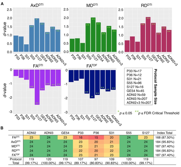
“Each ADNI protocol showed directionally consistent associations in more than 89% of tests (24 ROIs * 5 dMRI indices), except for P36 which was consistent in 81%, but had the smallest sample size (N = 12; Figure 2B; Supplementary Tables S5–S11).”
§ Diffusion MRI Indices and Their Relation to Cognitive Impairment in Brain Aging: The Updated Multi-protocol Approach in ADNI3 > Results > ADNI2 and ADNI3 Protocols in Cognitively Normal Participants
plaincorrelationaln = 12- Asserted: passes — The authors put it forward as true.
- Checkable: passes — It can be checked against evidence.
- Load-bearing: passes — The paper’s conclusions depend on it.
- Based: passes — The paper gives a reason why it holds.
It may state two separate claims as one.
A result the paper reports from its own data — it cites no other source for it, so there is none to check. The quote shows it is in the paper.
In the paper
“but the directions of associations with age were largely consistent for ADNI2 and ADNI3 phases separately, and each ADNI3 protocol (Figures 1, 2).”
§ Diffusion MRI Indices and Their Relation to Cognitive Impairment in Brain Aging: The Updated Multi-protocol Approach in ADNI3 > Results > ADNI2 and ADNI3 Protocols in Cognitively Normal Participants
plaincorrelational- Asserted: passes — The authors put it forward as true.
- Checkable: passes — It can be checked against evidence.
- Load-bearing: passes — The paper’s conclusions depend on it.
- Based: passes — The paper gives a reason why it holds.
It may state two separate claims as one.
A result the paper reports from its own data — it cites no other source for it, so there is none to check. The quote shows it is in the paper.
In the paper
“FATDF and all three diffusivity indices were consistent in ≥96% of tests (24 ROIs * 8 protocols/phases), while FADTI was only consistent in 88% of tests.”
§ Diffusion MRI Indices and Their Relation to Cognitive Impairment in Brain Aging: The Updated Multi-protocol Approach in ADNI3 > Results > ADNI2 and ADNI3 Protocols in Cognitively Normal Participants
plain- Asserted: passes — The authors put it forward as true.
- Checkable: passes — It can be checked against evidence.
- Load-bearing: passes — The paper’s conclusions depend on it.
- Based: passes — The paper gives a reason why it holds.
It may state two separate claims as one.
A result the paper reports from its own data — it cites no other source for it, so there is none to check. The quote shows it is in the paper.
In the paper
“None of the associations in the unexpected direction were significant after multiple comparisons correction, and only two had a p ≤ 0.05.”
§ Diffusion MRI Indices and Their Relation to Cognitive Impairment in Brain Aging: The Updated Multi-protocol Approach in ADNI3 > Results > ADNI2 and ADNI3 Protocols in Cognitively Normal Participants
plain- Asserted: passes — The authors put it forward as true.
- Checkable: passes — It can be checked against evidence.
- Load-bearing: unclear — It is unclear whether the paper’s conclusions depend on it.
- Based: passes — The paper gives a reason why it holds.
It may state two separate claims as one.
A result the paper reports from its own data — it cites no other source for it, so there is none to check. The quote shows it is in the paper.
In the paper
“FATDF detected robust widespread associations with clinical measures, while FADTI was the weakest of the five indices for detecting associations.”
§ Diffusion MRI Indices and Their Relation to Cognitive Impairment in Brain Aging: The Updated Multi-protocol Approach in ADNI3 > Abstract
boostedcorrelational- Asserted: passes — The authors put it forward as true.
- Checkable: passes — It can be checked against evidence.
- Load-bearing: passes — The paper’s conclusions depend on it.
- Based: passes — The paper gives a reason why it holds.
It may state two separate claims as one.
A result the paper reports from its own data — it cites no other source for it, so there is none to check. The quote shows it is in the paper.
In the paper
“Pairwise tests comparing the mean age of CN participants scanned with each protocol also showed significant differences between those scanned with S31 and two other protocols: GE54 and S31 (p = 0.026); P33 and S31 (p = 0.0037).”
§ Diffusion MRI Indices and Their Relation to Cognitive Impairment in Brain Aging: The Updated Multi-protocol Approach in ADNI3 > Results > ADNI2 and ADNI3 Protocols in Cognitively Normal Participants
plaincorrelational- Asserted: passes — The authors put it forward as true.
- Checkable: passes — It can be checked against evidence.
- Load-bearing: unclear — It is unclear whether the paper’s conclusions depend on it.
- Based: passes — The paper gives a reason why it holds.
It may state two separate claims as one.
A result the paper reports from its own data — it cites no other source for it, so there is none to check. The quote shows it is in the paper.
In the paper
“Of the three cognitive measures, CDR-sob associations showed the largest effect sizes across dMRI indices (in the UNC followed by the CGH for all indices except FADTI)”
§ Diffusion MRI Indices and Their Relation to Cognitive Impairment in Brain Aging: The Updated Multi-protocol Approach in ADNI3 > Results > Cognitive Measure Associations With Pooled ADNI3 dMRI Indices
plaincorrelational- Asserted: passes — The authors put it forward as true.
- Checkable: passes — It can be checked against evidence.
- Load-bearing: unclear — It is unclear whether the paper’s conclusions depend on it.
- Based: passes — The paper gives a reason why it holds.
It may state two separate claims as one.
A result the paper reports from its own data — it cites no other source for it, so there is none to check. The quote shows it is in the paper.
In the paper
“Compared to FADTI, FATDF showed larger effect sizes; across cognitive tests, the strongest FATDF associations were detected in the UNC with CDR-sob (d = −1.244; p = 1.39 × 10⁻²⁰), followed by the CGH (d = −1.213; p = 8.86 × 10⁻²⁰).”
§ Diffusion MRI Indices and Their Relation to Cognitive Impairment in Brain Aging: The Updated Multi-protocol Approach in ADNI3 > Results > Cognitive Measure Associations With Pooled ADNI3 dMRI Indices
plaincorrelational- Asserted: passes — The authors put it forward as true.
- Checkable: passes — It can be checked against evidence.
- Load-bearing: passes — The paper’s conclusions depend on it.
- Based: passes — The paper gives a reason why it holds.
It may state two separate claims as one.
A result the paper reports from its own data — it cites no other source for it, so there is none to check. The quote shows it is in the paper.
In the paper
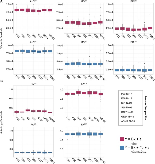
“After using ComBat to harmonize dMRI indices across protocols, ANCOVAs revealed that significant protocol differences in dMRI indices were all but eliminated across ROIs (Supplementary Figure S2A); significant protocol differences were detected only in the CST, for each of the dMRI indices.”
§ Diffusion MRI Indices and Their Relation to Cognitive Impairment in Brain Aging: The Updated Multi-protocol Approach in ADNI3 > Results > ADNI2 and ADNI3 Protocols in Cognitively Normal Participants
plain- Asserted: passes — The authors put it forward as true.
- Checkable: passes — It can be checked against evidence.
- Load-bearing: passes — The paper’s conclusions depend on it.
- Based: passes — The paper gives a reason why it holds.
It may state two separate claims as one.
A result the paper reports from its own data — it cites no other source for it, so there is none to check. The quote shows it is in the paper.
In the paper
“The UNC showed either the first or second largest effect size (alternating with CGH) across diffusivity indices and FATDF tests, but was significant only for cognitive measure associations with FADTI (i.e., three of four clinical tests).”
§ Diffusion MRI Indices and Their Relation to Cognitive Impairment in Brain Aging: The Updated Multi-protocol Approach in ADNI3 > Results > CN vs. MCI Diagnosis Associations With Pooled ADNI3 dMRI Indices
plaincorrelational- Asserted: passes — The authors put it forward as true.
- Checkable: passes — It can be checked against evidence.
- Load-bearing: unclear — It is unclear whether the paper’s conclusions depend on it.
- Based: passes — The paper gives a reason why it holds.
It may state two separate claims as one.
A result the paper reports from its own data — it cites no other source for it, so there is none to check. The quote shows it is in the paper.
In the paper
“Greater cognitive impairment was associated with lower anisotropy and higher diffusivity.”
- Also stated in: § Cognitive Measure Associations With Pooled ADNI3 dMRI Indices · plain
“higher diffusivity was associated with greater cognitive impairment. Lower (D) FADTI (FDR critical threshold p = 0.025) and (E) FATDF (FDR critical threshold p = 7.73 × 10⁻³) were also associated with greater impairment”
§ Diffusion MRI Indices and Their Relation to Cognitive Impairment in Brain Aging: The Updated Multi-protocol Approach in ADNI3 > Results > Cognitive Measure Associations With Pooled ADNI3 dMRI Indices
plaincorrelational- Asserted: passes — The authors put it forward as true.
- Checkable: passes — It can be checked against evidence.
- Load-bearing: passes — The paper’s conclusions depend on it.
- Based: passes — The paper gives a reason why it holds.
It may state two separate claims as one.
- Also stated in: § Cognitive Measure Associations With Pooled ADNI3 dMRI Indices · plain
A result the paper reports from its own data — it cites no other source for it, so there is none to check. The quote shows it is in the paper.
In the paper
“On the other hand, FATDF was significant in 20 out of 24 ROIs, similar to diffusivity indices.”
§ Diffusion MRI Indices and Their Relation to Cognitive Impairment in Brain Aging: The Updated Multi-protocol Approach in ADNI3 > Results > CN vs. MCI Diagnosis Associations With Pooled ADNI3 dMRI Indices
plaincorrelational- Asserted: passes — The authors put it forward as true.
- Checkable: passes — It can be checked against evidence.
- Load-bearing: passes — The paper’s conclusions depend on it.
- Based: passes — The paper gives a reason why it holds.
It may state two separate claims as one.
A result the paper reports from its own data — it cites no other source for it, so there is none to check. The quote shows it is in the paper.
In the paper
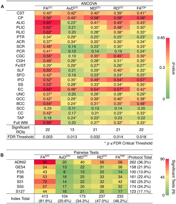
“ANCOVAs revealed significant protocol differences for 22 ROIs for FADTI and FATDF, with the highest overall effect size detected in the anterior limb of the internal capsule (ALIC) and the external capsule (EC) for FADTI (ALIC: d = 0.648; EC: d = 0.652).”
§ Diffusion MRI Indices and Their Relation to Cognitive Impairment in Brain Aging: The Updated Multi-protocol Approach in ADNI3 > Results > ADNI2 and ADNI3 Protocols in Cognitively Normal Participants
plain- Asserted: passes — The authors put it forward as true.
- Checkable: passes — It can be checked against evidence.
- Load-bearing: passes — The paper’s conclusions depend on it.
- Based: passes — The paper gives a reason why it holds.
It may state two separate claims as one.
A result the paper reports from its own data — it cites no other source for it, so there is none to check. The quote shows it is in the paper.
In the paper
“AxDDTI had the smallest effect size, overall, in the splenium of the corpus callosum (SCC; d = 0.106), and only 13 ROIs showed significant AxDDTI differences between protocols.”
§ Diffusion MRI Indices and Their Relation to Cognitive Impairment in Brain Aging: The Updated Multi-protocol Approach in ADNI3 > Results > ADNI2 and ADNI3 Protocols in Cognitively Normal Participants
plain- Asserted: passes — The authors put it forward as true.
- Checkable: passes — It can be checked against evidence.
- Load-bearing: unclear — It is unclear whether the paper’s conclusions depend on it.
- Based: passes — The paper gives a reason why it holds.
It may state two separate claims as one.
A result the paper reports from its own data — it cites no other source for it, so there is none to check. The quote shows it is in the paper.
In the paper
“In 14 of 15 tests, the CGH consistently showed one of the top two largest effect sizes (CGH FADTI association with CDR-sob was the third largest), along with the uncinate fasciculus (UNC), which was top two in 12 of 15 tests (while significant, cognitive associations with UNC FADTI never showed one of the largest effect sizes).”
§ Diffusion MRI Indices and Their Relation to Cognitive Impairment in Brain Aging: The Updated Multi-protocol Approach in ADNI3 > Results > Cognitive Measure Associations With Pooled ADNI3 dMRI Indices
plaincorrelational- Asserted: passes — The authors put it forward as true.
- Checkable: passes — It can be checked against evidence.
- Load-bearing: unclear — It is unclear whether the paper’s conclusions depend on it.
- Based: passes — The paper gives a reason why it holds.
It may state two separate claims as one.
A result the paper reports from its own data — it cites no other source for it, so there is none to check. The quote shows it is in the paper.
In the paper
“In pairwise analyses, AxDDTI was the most stable index across protocols, as significant protocol differences were detected in only 20.6% of pairwise tests (24 ROIs * 21 pairwise tests), compared to FADTI, the most variable index, which showed significant protocol differences in 81.9% of tests (Figure 4B).”
§ Diffusion MRI Indices and Their Relation to Cognitive Impairment in Brain Aging: The Updated Multi-protocol Approach in ADNI3 > Results > ADNI2 and ADNI3 Protocols in Cognitively Normal Participants
plain- Asserted: passes — The authors put it forward as true.
- Checkable: passes — It can be checked against evidence.
- Load-bearing: passes — The paper’s conclusions depend on it.
- Based: passes — The paper gives a reason why it holds.
It may state two separate claims as one.
A result the paper reports from its own data — it cites no other source for it, so there is none to check. The quote shows it is in the paper.
In the paper
“FATDF was significant in 89 of the 96 tests (92.7%; 24 ROIs * 4 clinical tests), while FADTI was only significant in 58 (60.4%).”
§ Diffusion MRI Indices and Their Relation to Cognitive Impairment in Brain Aging: The Updated Multi-protocol Approach in ADNI3 > Discussion
plaincorrelational- Asserted: passes — The authors put it forward as true.
- Checkable: passes — It can be checked against evidence.
- Load-bearing: passes — The paper’s conclusions depend on it.
- Based: passes — The paper gives a reason why it holds.
It may state two separate claims as one.
A result the paper reports from its own data — it cites no other source for it, so there is none to check. The quote shows it is in the paper.
In the paper
“FADTI showed the greatest significant differences (largest d-values; dark red) between protocols and AxDDTI the fewest (dark green).”
- Also stated in: § ADNI2 and ADNI3 Protocols in Cognitively Normal Participants · plain
“AxDDTI was the most stable dMRI index across protocols, while FADTI was the least stable.”
§ Diffusion MRI Indices and Their Relation to Cognitive Impairment in Brain Aging: The Updated Multi-protocol Approach in ADNI3 > Results > ADNI2 and ADNI3 Protocols in Cognitively Normal Participants
plain- Asserted: passes — The authors put it forward as true.
- Checkable: passes — It can be checked against evidence.
- Load-bearing: unclear — It is unclear whether the paper’s conclusions depend on it.
- Based: passes — The paper gives a reason why it holds.
It may state two separate claims as one.
- Also stated in: § ADNI2 and ADNI3 Protocols in Cognitively Normal Participants · plain
A result the paper reports from its own data — it cites no other source for it, so there is none to check. The quote shows it is in the paper.
In the paper
“the largest effect sizes across all tests were detected with AxDDTI (UNC: d = 1.344) and MDDTI (UNC: d = 1.342, p = 3.47 × 10⁻²³).”
§ Diffusion MRI Indices and Their Relation to Cognitive Impairment in Brain Aging: The Updated Multi-protocol Approach in ADNI3 > Results > Cognitive Measure Associations With Pooled ADNI3 dMRI Indices
plaincorrelational- Asserted: passes — The authors put it forward as true.
- Checkable: passes — It can be checked against evidence.
- Load-bearing: unclear — It is unclear whether the paper’s conclusions depend on it.
- Based: passes — The paper gives a reason why it holds.
It may state two separate claims as one.
A result the paper reports from its own data — it cites no other source for it, so there is none to check. The quote shows it is in the paper.
In the paper
“In a preliminary analysis, we showed that one harmonization method performed on these regional measures, ComBat, reduced cross-site differences in dMRI indices, while preserving biological relationships with age in CN controls.”
§ Diffusion MRI Indices and Their Relation to Cognitive Impairment in Brain Aging: The Updated Multi-protocol Approach in ADNI3 > Discussion
plaindirect- Asserted: passes — The authors put it forward as true.
- Checkable: passes — It can be checked against evidence.
- Load-bearing: passes — The paper’s conclusions depend on it.
- Based: passes — The paper gives a reason why it holds.
It may state two separate claims as one.
A result the paper reports from its own data — it cites no other source for it, so there is none to check. The quote shows it is in the paper.
In the paper
“While associations with clinical impairment were detected throughout the WM, the region that most frequently showed the lowest effect sizes and was significant in only 3 of the 20 clinical tests, was the corticospinal tract (CST).”
§ Diffusion MRI Indices and Their Relation to Cognitive Impairment in Brain Aging: The Updated Multi-protocol Approach in ADNI3 > Discussion
plaincorrelational- Asserted: passes — The authors put it forward as true.
- Checkable: passes — It can be checked against evidence.
- Load-bearing: unclear — It is unclear whether the paper’s conclusions depend on it.
- Based: passes — The paper gives a reason why it holds.
It may state two separate claims as one.
A result the paper reports from its own data — it cites no other source for it, so there is none to check. The quote shows it is in the paper.
In the paper
“Here, the number of significant ROIs and the magnitude of effect sizes were comparable for ComBat and nested random-effects model approaches.”
§ Diffusion MRI Indices and Their Relation to Cognitive Impairment in Brain Aging: The Updated Multi-protocol Approach in ADNI3 > Discussion
plain- Asserted: passes — The authors put it forward as true.
- Checkable: passes — It can be checked against evidence.
- Load-bearing: passes — The paper’s conclusions depend on it.
- Based: passes — The paper gives a reason why it holds.
It may state two separate claims as one.
What the paper did — it cites no source for this, so there is none to check. The quote shows it is in the paper.
In the paper
“As before, we used FDR to account for multiple comparisons.”
§ Diffusion MRI Indices and Their Relation to Cognitive Impairment in Brain Aging: The Updated Multi-protocol Approach in ADNI3 > Materials and Methods > Comparing the ADNI2 and ADNI3 Protocols in Cognitively Normal Participants
plain- Asserted: passes — The authors put it forward as true.
- Checkable: passes — It can be checked against evidence.
- Load-bearing: fails — The paper’s conclusions may not depend on it.
- Based: passes — The paper gives a reason why it holds.
Probably not a substantial claim. The paper’s conclusions may not depend on it.
What the paper did — it cites no source for this, so there is none to check. The quote shows it is in the paper.
In the paper
“Due to the limited available sample size of AD participants (N = 22), and their uneven distribution across the acquisition protocols tested here, we compared only groups of people with CN and MCI diagnoses.”
§ Diffusion MRI Indices and Their Relation to Cognitive Impairment in Brain Aging: The Updated Multi-protocol Approach in ADNI3 > Materials and Methods > Clinical Assessments and Their Relation to Pooled ADNI3 Diffusion Indices
plaindirectn = 22- Asserted: passes — The authors put it forward as true.
- Checkable: passes — It can be checked against evidence.
- Load-bearing: passes — The paper’s conclusions depend on it.
- Based: passes — The paper gives a reason why it holds.
It may state two separate claims as one.
What the paper did — it cites no source for this, so there is none to check. The quote shows it is in the paper.
In the paper
“Age, sex, and age*sex interactions were controlled for as fixed effects, and the protocol and acquisition site were modeled as nested random variables.”
§ Diffusion MRI Indices and Their Relation to Cognitive Impairment in Brain Aging: The Updated Multi-protocol Approach in ADNI3 > Materials and Methods > Clinical Assessments and Their Relation to Pooled ADNI3 Diffusion Indices
plaincorrelationaln = 22- Asserted: passes — The authors put it forward as true.
- Checkable: passes — It can be checked against evidence.
- Load-bearing: passes — The paper’s conclusions depend on it.
- Based: passes — The paper gives a reason why it holds.
Probably not a substantial claim. It may state two separate claims as one.
What the paper did — it cites no source for this, so there is none to check. The quote shows it is in the paper.
In the paper
“For each dMRI index, we used FDR to correct for multiple comparisons across the 24 ROIs assessed.”
§ Diffusion MRI Indices and Their Relation to Cognitive Impairment in Brain Aging: The Updated Multi-protocol Approach in ADNI3 > Materials and Methods > Comparing the ADNI2 and ADNI3 Protocols in Cognitively Normal Participants
plain- Asserted: passes — The authors put it forward as true.
- Checkable: passes — It can be checked against evidence.
- Load-bearing: passes — The paper’s conclusions depend on it.
- Based: passes — The paper gives a reason why it holds.
Probably not a substantial claim.
What the paper did — it cites no source for this, so there is none to check. The quote shows it is in the paper.
In the paper
“All DWI and T1-weighted images were visually checked for quality assurance.”
§ Diffusion MRI Indices and Their Relation to Cognitive Impairment in Brain Aging: The Updated Multi-protocol Approach in ADNI3 > Materials and Methods > dMRI Preprocessing and Scalar Indices
plain- Asserted: passes — The authors put it forward as true.
- Checkable: passes — It can be checked against evidence.
- Load-bearing: fails — The paper’s conclusions may not depend on it.
- Based: passes — The paper gives a reason why it holds.
Probably not a substantial claim. The paper’s conclusions may not depend on it.
What the paper did — it cites no source for this, so there is none to check. The quote shows it is in the paper.
In the paper
“In the CN participants from ADNI2 and ADNI3, we ran ComBat on each of the dMRI indices, including age, sex, age*sex, and information from all 24 ROIs to inform the statistical properties of the protocol effects.”
§ Diffusion MRI Indices and Their Relation to Cognitive Impairment in Brain Aging: The Updated Multi-protocol Approach in ADNI3 > Materials and Methods > Comparing the ADNI2 and ADNI3 Protocols in Cognitively Normal Participants
plain- Asserted: passes — The authors put it forward as true.
- Checkable: passes — It can be checked against evidence.
- Load-bearing: passes — The paper’s conclusions depend on it.
- Based: passes — The paper gives a reason why it holds.
Probably not a substantial claim.
What the paper did — it cites no source for this, so there is none to check. The quote shows it is in the paper.
In the paper
“In CN participants, multivariate random-effects linear regressions were used to assess whether dMRI indices from each ADNI protocol individually were associated with age, controlling for sex and age*sex interactions as fixed variables, and acquisition site as a random variable.”
§ Diffusion MRI Indices and Their Relation to Cognitive Impairment in Brain Aging: The Updated Multi-protocol Approach in ADNI3 > Materials and Methods > Comparing the ADNI2 and ADNI3 Protocols in Cognitively Normal Participants
plaincorrelational- Asserted: passes — The authors put it forward as true.
- Checkable: passes — It can be checked against evidence.
- Load-bearing: passes — The paper’s conclusions depend on it.
- Based: passes — The paper gives a reason why it holds.
Probably not a substantial claim.
In the paper
“FDR was again used to correct for 24 ROI tests (q = 0.05; Benjamini and Hochberg, 1995).”
§ Diffusion MRI Indices and Their Relation to Cognitive Impairment in Brain Aging: The Updated Multi-protocol Approach in ADNI3 > Materials and Methods > Clinical Assessments and Their Relation to Pooled ADNI3 Diffusion Indices
What the sources say
- Not checked — no full text of the sourcecites Benjamini and Hochberg, 1995 source
plain- Asserted: passes — The authors put it forward as true.
- Checkable: passes — It can be checked against evidence.
- Load-bearing: passes — The paper’s conclusions depend on it.
- Based: passes — The paper gives a reason why it holds.
Probably not a substantial claim.
In the paper
“We used the false discovery rate (FDR) procedure to correct for multiple comparisons (q = 0.05; Benjamini and Hochberg, 1995) across the 24 ROIs assessed for each dMRI index.”
§ Diffusion MRI Indices and Their Relation to Cognitive Impairment in Brain Aging: The Updated Multi-protocol Approach in ADNI3 > Materials and Methods > Comparing the ADNI2 and ADNI3 Protocols in Cognitively Normal Participants
What the sources say
- Not checked — no full text of the sourcecites Benjamini and Hochberg, 1995 source
plain- Asserted: passes — The authors put it forward as true.
- Checkable: passes — It can be checked against evidence.
- Load-bearing: passes — The paper’s conclusions depend on it.
- Based: passes — The paper gives a reason why it holds.
Probably not a substantial claim.
What the paper did — it cites no source for this, so there is none to check. The quote shows it is in the paper.
In the paper
“In CN participants, we tested for significant differences in dMRI indices between the seven ADNI protocols using analyses of covariance (ANCOVAs), adjusting for age, sex, and age*sex interactions as fixed variables, and acquisition site as a random variable.”
§ Diffusion MRI Indices and Their Relation to Cognitive Impairment in Brain Aging: The Updated Multi-protocol Approach in ADNI3 > Materials and Methods > Comparing the ADNI2 and ADNI3 Protocols in Cognitively Normal Participants
plain- Asserted: passes — The authors put it forward as true.
- Checkable: passes — It can be checked against evidence.
- Load-bearing: passes — The paper’s conclusions depend on it.
- Based: passes — The paper gives a reason why it holds.
Probably not a substantial claim.
What the paper did — it cites no source for this, so there is none to check. The quote shows it is in the paper.
In the paper
“Of the 381 participants scanned to date, 55 were excluded after quality assurance: this included ensuring complete clinical and demographic information, and image-level quality control (removing scans with severe motion, missing volumes, or corrupt files).”
§ Diffusion MRI Indices and Their Relation to Cognitive Impairment in Brain Aging: The Updated Multi-protocol Approach in ADNI3 > Materials and Methods > ADNI Participants
plainn = 381- Asserted: passes — The authors put it forward as true.
- Checkable: passes — It can be checked against evidence.
- Load-bearing: passes — The paper’s conclusions depend on it.
- Based: passes — The paper gives a reason why it holds.
Probably not a substantial claim.
What the paper did — it cites no source for this, so there is none to check. The quote shows it is in the paper.
In the paper
“To ensure sufficient statistical power to assess differences in data collected with different protocols, we evaluated only those protocols with complete available data for at least 10 participants at the time of download; we did not assess protocol GE36, for which scans from 9 of 12 participants passed quality assurance.”
§ Diffusion MRI Indices and Their Relation to Cognitive Impairment in Brain Aging: The Updated Multi-protocol Approach in ADNI3 > Materials and Methods > ADNI Participants
plainn = 10- Asserted: passes — The authors put it forward as true.
- Checkable: passes — It can be checked against evidence.
- Load-bearing: passes — The paper’s conclusions depend on it.
- Based: passes — The paper gives a reason why it holds.
Probably not a substantial claim.
What the paper did — it cites no source for this, so there is none to check. The quote shows it is in the paper.
In the paper
“we did not assess protocol GE36, for which scans from 9 of 12 participants passed quality assurance.”
§ Diffusion MRI Indices and Their Relation to Cognitive Impairment in Brain Aging: The Updated Multi-protocol Approach in ADNI3 > Materials and Methods > ADNI Participants
plaindirectn = 12- Asserted: passes — The authors put it forward as true.
- Checkable: passes — It can be checked against evidence.
- Load-bearing: passes — The paper’s conclusions depend on it.
- Based: passes — The paper gives a reason why it holds.
Probably not a substantial claim. It may state two separate claims as one.
What the paper did — it cites no source for this, so there is none to check. The quote shows it is in the paper.
In the paper
“Even so, separating protocol differences from differences in scanner manufacturer is difficult.”
§ Diffusion MRI Indices and Their Relation to Cognitive Impairment in Brain Aging: The Updated Multi-protocol Approach in ADNI3 > Discussion
plain- Asserted: passes — The authors put it forward as true.
- Checkable: passes — It can be checked against evidence.
- Load-bearing: fails — The paper’s conclusions may not depend on it.
- Based: fails — The paper may give no reason why it holds.
Probably not a substantial claim. The paper’s conclusions may not depend on it. The paper may give no reason why it holds.
A conclusion the paper draws from its own results — it cites no other source for it, so there is none to check. The quote shows it is in the paper.
In the paper
“perhaps due to the larger voxel size in ADNI2 (2.7 mm3 vs. 2.0 mm3 isotropic voxels used in ADNI3).”
§ Diffusion MRI Indices and Their Relation to Cognitive Impairment in Brain Aging: The Updated Multi-protocol Approach in ADNI3 > Discussion
hedgedconditional- Asserted: passes — The authors put it forward as true.
- Checkable: passes — It can be checked against evidence.
- Load-bearing: fails — The paper’s conclusions may not depend on it.
- Based: passes — The paper gives a reason why it holds.
Probably not a substantial claim. The paper’s conclusions may not depend on it.
The paper relies on this without support.
The clinical assessments (ADAS-cog, MMSE, CDR-sob) are valid proxy measures of cognitive impairment.
“We refer to these tools as “cognitive measures”, but recognize the limitations of these assessments as proxy measures of specific cognitive abilities (Balsis et al., 2015).”
The paper relies on this without support.
At the spatial resolutions now used, a single voxel typically captures partial volumes of different tissue compartments (intra- and extra-cellular, vascular, CSF, and myelin), each affecting water diffusion and the MR signal.
“However, at the spatial resolutions now used, a single voxel typically captures partial volumes of different tissue compartments–e.g., the intra- and extra-cellular compartments, the vascular compartment, the CSF and myelin; each affects water diffusion and the MR signal.”
Self-check
Percentages, sums and reported differences re-computed from the paper’s own numbers.
Each contradiction between two passages listed here was confirmed by an independent check.
Rated minor: it would not touch a result — a value no result rests on, a p-value that stays on the same side of the significance level, or a conflict that is clerical, resolved elsewhere in the paper, or outside its own findings.
In the paper
“ADNI2 (2.7 mm3 vs. 2.0 mm3 isotropic voxels used in ADNI3)”
§ Diffusion MRI Indices and Their Relation to Cognitive Impairment in Brain Aging: The Updated Multi-protocol Approach in ADNI3 > Discussion
Reported 2.7 · recomputed 19.683
Rated minor: it would not touch a result — a value no result rests on, a p-value that stays on the same side of the significance level, or a conflict that is clerical, resolved elsewhere in the paper, or outside its own findings.
In the paper
§ Diffusion MRI Indices and Their Relation to Cognitive Impairment in Brain Aging: The Updated Multi-protocol Approach in ADNI3 > Results > ADNI2 and ADNI3 Protocols in Cognitively Normal Participants
“When data were pooled across ADNI2 and ADNI3, significant associations with age were detected throughout the WM. Figure 1A shows effect sizes for ROIs significantly associated with age after FDR multiple comparisons correction”
§ Diffusion MRI Indices and Their Relation to Cognitive Impairment in Brain Aging: The Updated Multi-protocol Approach in ADNI3 > Results > ADNI2 and ADNI3 Protocols in Cognitively Normal Participants
“Figure: Figure 1 (A) For each diffusion-weighted MRI (dMRI) index, the absolute values of effect sizes (d-value) are plotted for regional white matter (WM) microstructural associations with age when all ADNI3 dMRI data are pooled”
Rated minor: it would not touch a result — a value no result rests on, a p-value that stays on the same side of the significance level, or a conflict that is clerical, resolved elsewhere in the paper, or outside its own findings.
In the paper
§ Diffusion MRI Indices and Their Relation to Cognitive Impairment in Brain Aging: The Updated Multi-protocol Approach in ADNI3 > Materials and Methods > ADNI Participants
“| P33 | 24 | 3 | 78.1 ± 7.1 | 13 (54.2%) | 17 | 4 | 3 | 28.75 ± 2.03 | 1.31 ± 2.84 | 13.32 ± 6.76 |”
§ Diffusion MRI Indices and Their Relation to Cognitive Impairment in Brain Aging: The Updated Multi-protocol Approach in ADNI3 > Materials and Methods > Clinical Assessments
“Scores range from 0 (no dementia) to 3 (severe dementia; Berg, 1988).”
Citations
Cites
Controlling the False Discovery Rate: A Practical and Powerful Approach to Multiple Testing
Benjamini, Y., & Hochberg, Y. (1995) · Journal of the Royal Statistical Society Series B: Statistical Methodology · doi:10.1111/j.2517-6161.1995.tb02031.x · Cited by 109,482 · cited 2×
“…uses an empirical Bayes framework to reduce unwanted variation in multi-site data due to differences in acquisition protocol, while preserving the desired biological variation in the data (Fortin et al., 2017 FDR was again used to correct for 24 ROI tests ( q = 0.05; Benjamini and Hochberg, 1995).”
InfluentialFolstein, M. F., Folstein, S. E., & McHugh, P. R. (1975) · Journal of Psychiatric Research · doi:10.1016/0022-3956(75)90026-6 · Cited by 88,282
Hirono, N., Mori, E., Ikejiri, Y., Imamura, T., Shimomura, T., Hashimoto, M., Yamashita, H., & Ikeda, M. (1997) · PubMed · Cited by 21,012
Kok, J. S., Berg, I. J., & Scherder, E. J. A. (2013) · Dementia and Geriatric Cognitive Disorders Extra · doi:10.1159/000353441 · Cited by 20,589
Older Adults' Reasons for Using Technology while Aging in Place
Peek, S. T. M., Luijkx, K. G., Rijnaard, M. D., Nieboer, M. E., van der Voort, C. S., Aarts, S., van Hoof, J., Vrijhoef, H. J., & Wouters, E. (2015) · Gerontology · doi:10.1159/000430949 · Cited by 19,852
Sensitivity and False Alarm Rate of a Fall Sensor in Long-Term Fall Detection in the Elderly
Kangas, M., Korpelainen, R., Vikman, I., Nyberg, L., & Jämsä, T. J. (2014) · Gerontology · doi:10.1159/000362720 · Cited by 19,007
Neuropathological stageing of Alzheimer-related changes.
Braak, H., & Braak, E. (1991) · Acta Neuropathologica · doi:10.1007/bf00308809 · Cited by 16,123 · cited 1×
“Accumulation of amyloid plaques and neurofibrillary tangles (NFTs) in the brain (Braak and Braak, 1991, 1996; Frank et al., 2003; Shaw et al., 2007) can directly impact WM (Lee et al., 2004; Roth et al., 2005), promoting myelin degeneration and axonal loss (Braak and Braak, 1996; Kneynsberg et al.,…”
Adjusting batch effects in microarray expression data using empirical Bayes methods
Johnson, W. E., Li, C., & Rabinovic, A. (2007) · Biostatistics · doi:10.1093/biostatistics/kxj037 · Cited by 8,658 · cited 1×
“For example, ComBat—originally developed to model and remove batch effects from genomic microarray data (Johnson et al., 2007)—was one of the most effective methods for harmonizing DTI measures in a recent comparison of such techniques (Fortin et al., 2017).”
Power failure: why small sample size undermines the reliability of neuroscience
Button, K. S., Ioannidis, J. P. A., Mokrysz, C., Nosek, B. A., Flint, J., Robinson, E. S. J., & Munafò, M. R. (2013) · Nature Reviews Neuroscience · doi:10.1038/nrn3475 · Cited by 7,641
MR diffusion tensor spectroscopy and imaging
Basser, P. J., Mattiello, J., & LeBihan, D. (1994) · Biophysical Journal · doi:10.1016/s0006-3495(94)80775-1 · Cited by 5,970 · cited 1×
“Scalar dMRI indices were derived from two reconstruction models: the single tensor model (DTI; Basser et al., 1994) and the tensor distribution function (TDF; Leow et al.”
The basis of anisotropic water diffusion in the nervous system—A technical review.
Beaulieu, C. (2002) · NMR in Biomedicine · doi:10.1002/nbm.782 · Cited by 4,628
A new rating scale for Alzheimer's disease
(1984) · American Journal of Psychiatry · doi:10.1176/ajp.141.11.1356 · Cited by 4,175 · cited 2×
“The ADAS-cog is frequently used in pharmaceutical trials, with scores ranging from 0 to 70; higher scores represent more severe cognitive dysfunction (Rosen et al., 1984).”
Essentials of Behavioral Research: Methods and Data Analysis
Rosenthal, R., & Rosnow, R. L. (1984) · Cited by 3,925
NODDI: Practical in vivo neurite orientation dispersion and density imaging of the human brain
Zhang, H., Schneider, T., Wheeler-Kingshott, C. A., & Alexander, D. C. (2012) · NeuroImage · doi:10.1016/j.neuroimage.2012.03.072 · Cited by 3,288 · cited 1×
“…kurtosis imaging (DKI; Jensen et al., 2005; Chen et al., 2017; Cheng et al., 2018; Wang M.-L. et al., 2018), and multi-compartment models such as neurite orientation dispersion and density imaging (NODDI; Zhang et al., 2012; Colgan et al., 2016; Slattery et al., 2017; Parker et al., 2018).”
Jensen, J. H., Helpern, J. A., Ramani, A., Lu, H., & Kaczynski, K. (2005) · Magnetic Resonance in Medicine · doi:10.1002/mrm.20508 · Cited by 2,488 · cited 1×
“…multi-shell protocols to compute diffusion indices from models that do not assume mono-exponential decay, such as diffusion kurtosis imaging (DKI; Jensen et al., 2005; Chen et al., 2017; Cheng et al., 2018; Wang M.-L. et al., 2018), and multi-compartment models such as neurite orientation…”
2018 Alzheimer's disease facts and figures
Alzheimer's Association (2018) · Alzheimer's & Dementia · doi:10.1016/j.jalz.2018.02.001 · Cited by 1,912
Stereotaxic white matter atlas based on diffusion tensor imaging in an ICBM template
Mori, S., Oishi, K., Jiang, H., Jiang, L., Li, X., Akhter, K., Hua, K., Faria, A. V., Mahmood, A., Woods, R. P., et al. (2008) · NeuroImage · doi:10.1016/j.neuroimage.2007.12.035 · Cited by 1,832 · cited 3×
“The following 24 ROIs from the JHU atlas (Mori et al., 2008) were analyzed.”
MethodInfluentialSong, S.-K., Sun, S.-W., Ju, W.-K., Lin, S.-J., Cross, A. H., & Neufeld, A. H. (2003) · NeuroImage · doi:10.1016/j.neuroimage.2003.07.005 · Cited by 1,738 · cited 1×
“…more coherent intact axons, while lower anisotropy and higher diffusivity may reflect factors such as axonal injury and demyelination, among other factors (Beaulieu, 2002; Song et al., 2003, 2005; Harsan et al., 2006; Le Bihan and Johansen-Berg, 2012; Kantarci et al., 2017; Moore et al., 2018).”
Demyelination increases radial diffusivity in corpus callosum of mouse brain
Song, S.-K., Yoshino, J., Le, T. Q., Lin, S.-J., Sun, S.-W., Cross, A. H., & Armstrong, R. C. (2005) · NeuroImage · doi:10.1016/j.neuroimage.2005.01.028 · Cited by 1,658 · cited 1×
“…axons, while lower anisotropy and higher diffusivity may reflect factors such as axonal injury and demyelination, among other factors (Beaulieu et al., 2002; Song et al., 2003; Song et al., 2005; Harsan et al., 2006; Le Bihan and Johansen-Berg 2012; Kantarci et al., 2017; Moore et al., 2018).”
BackgroundTournier, J.-D., Calamante, F., Gadian, D. G., & Connelly, A. (2004) · NeuroImage · doi:10.1016/j.neuroimage.2004.07.037 · Cited by 1,635 · cited 1×
“…model addresses well-established limitations of the standard single-tensor diffusion model—which cannot resolve complex profiles of WM architecture such as crossing or mixing fibers, present in up to 90% of WM voxels (Tournier et al., 2004; Descoteaux et al., 2007, 2009; Jeurissen et al., 2013).”
High angular resolution diffusion imaging reveals intravoxel white matter fiber heterogeneity
Tuch, D. S., Reese, T. G., Wiegell, M. R., Makris, N., Belliveau, J. W., & Wedeen, V. J. (2002) · Magnetic Resonance in Medicine · doi:10.1002/mrm.10268 · Cited by 1,548 · cited 1×
“The DTI model cannot differentiate these components or even crossing fibers (Tuch et al., 2002; Jbabdi et al., 2010), which are estimated to occur in up to 90% of WM voxels at the typical dMRI resolution (Descoteaux et al., 2009; Jeurissen et al., 2013).”
Hua, K., Zhang, J., Wakana, S., Jiang, H., Li, X., Reich, D. S., Calabresi, P. A., Pekar, J. J., van Zijl, P. C. M., & Mori, S. (2007) · NeuroImage · doi:10.1016/j.neuroimage.2007.07.053 · Cited by 1,507 · cited 1×
“…shown to be the least reliable and reproducible ROI (Jahanshad et al., 2013; Acheson et al., 2017), suggesting alternate approaches, such as tractography-based evaluations (Jin et al., 2017), or the use of the probabilistic JHU atlas (Hua et al., 2008), may be more appropriate for studying the CST.”
Tractography-based connectomes are dominated by false-positive connections
Maier-Hein, K. H., Neher, P., Houde, J.-C., Côté, M.-A., Garyfallidis, E., Zhong, J., Chamberland, M., Yeh, F.-C., Lin, Y.-C., Ji, Q., et al. (2016) · doi:10.1101/084137 · Cited by 1,376
Self-citationHarmonization of multi-site diffusion tensor imaging data
Fortin, J.-P., Parker, D., Tunç, B., Watanabe, T., Elliott, M. A., Ruparel, K., Roalf, D. R., Satterthwaite, T. D., Gur, R. C., Gur, R. E., et al. (2017) · NeuroImage · doi:10.1016/j.neuroimage.2017.08.047 · Cited by 1,261 · cited 8×
“…uses an empirical Bayes framework to reduce unwanted variation in multi-site data due to differences in acquisition protocol, while preserving the desired biological variation in the data (Fortin et al., 2017 FDR was again used to correct for 24 ROI tests ( q = 0.05; Benjamini and Hochberg, 1995).”
InfluentialMueller, S. G., Weiner, M. W., Thal, L. J., Petersen, R. C., Jack, C. R., Jagust, W., Trojanowski, J. Q., Toga, A. W., & Beckett, L. (2005) · Alzheimer's & Dementia · doi:10.1016/j.jalz.2005.06.003 · Cited by 1,233 · cited 1×
“MCI is now the focus of intense research; it is essential to find ways to clinically categorize the transitional stages between normal aging and AD to evaluate targeted treatments, as pathophysiological mechanisms may differ or change throughout the course of AD (Mueller et al., 2005).”
Self-citationJeurissen, B., Leemans, A., Tournier, J., Jones, D. K., & Sijbers, J. (2013) · Human Brain Mapping · doi:10.1002/hbm.22099 · Cited by 1,088 · cited 2×
“The DTI model cannot differentiate these components or even crossing fibers (Tuch et al., 2002; Jbabdi et al., 2010), which are estimated to occur in up to 90% of WM voxels at the typical dMRI resolution (Descoteaux et al., 2009; Jeurissen et al., 2013).”
Regularized, fast, and robust analytical Q-ball imaging
Descoteaux, M., Angelino, E., Fitzgibbons, S., & Deriche, R. (2007) · Magnetic Resonance in Medicine · doi:10.1002/mrm.21277 · Cited by 850
The ENIGMA Consortium: large-scale collaborative analyses of neuroimaging and genetic data
the Alzheimer’s Disease Neuroimaging Initiative, E. C., Thompson, P. M., Stein, J. L., Medland, S. E., Hibar, D. P., Vasquez, A. A., Renteria, M. E., Toro, R., Jahanshad, N., Schumann, G., et al. (2014) · Brain Imaging and Behavior · doi:10.1007/s11682-013-9269-5 · Cited by 838 · cited 1×
“Site differences can be accounted for at the time of overall group inference, such as with the random-effects regression level correction used here, or by using a meta-analysis approach in lieu of pooling data (Thompson et al., 2014).”
Self-citationEvolution of the neuropathology of Alzheimer's disease
Braak, H., & Braak, E. (1996) · Acta Neurologica Scandinavica · doi:10.1111/j.1600-0404.1996.tb05866.x · Cited by 710 · cited 3×
“…of amyloid plaques and neurofibrillary tangles (NFTs) in the brain (Braak and Braak, 1991, 1996; Frank et al., 2003; Shaw et al., 2007) can directly impact WM (Lee et al., 2004; Roth et al., 2005), promoting myelin degeneration and axonal loss (Braak and Braak, 1996; Kneynsberg et al., 2017).”
Kelly, S., Jahanshad, N., Zalesky, A., Kochunov, P., Agartz, I., Alloza, C., Andreassen, O. A., Arango, C., Banaj, N., Bouix, S., et al. (2018) · Molecular Psychiatry · doi:10.1038/mp.2017.170 · Cited by 701 · cited 2×
“Effect size was not correlated with ROI size ( Supplementary Figure S13 ), consistent with prior studies of other disorders (Kelly et al., 2018).”
Self-citationDeterministic and probabilistic tractography based on complex fibre orientation distributions
Descoteaux, M., Deriche, R., Knosche, T. R., & Anwander, A. (2009) · IEEE Transactions on Medical Imaging · doi:10.1109/tmi.2008.2004424 · Cited by 698 · cited 2×
“The DTI model cannot differentiate these components or even crossing fibers (Tuch et al., 2002; Jbabdi et al., 2010), which are estimated to occur in up to 90% of WM voxels at the typical dMRI resolution (Descoteaux et al., 2009; Jeurissen et al., 2013).”
Clinical Dementia Rating (CDR)
Gauthier, S. G. (2001) · CRC Press eBooks · doi:10.3109/9780203213896-34 · Cited by 696
Analysis of partial volume effects in diffusion‐tensor MRI
Alexander, A. L., Hasan, K. M., Lazar, M., Tsuruda, J. S., & Parker, D. L. (2001) · Magnetic Resonance in Medicine · doi:10.1002/mrm.1105 · Cited by 695 · cited 2×
“…2013; Kochunov et al., 2014; Acheson et al., 2017; Kelly et al., 2018), differences in dMRI acquisition parameters, including vendor, voxel size, and angular resolution, are known to affect derived dMRI measures (Alexander et al., 2001; Cercignani et al., 2003; Zhan et al., 2010; Zhu et al., 2011).”
Douaud, G., Jbabdi, S., Behrens, T. E. J., Menke, R. A., Gass, A., Monsch, A. U., Rao, A., Whitcher, B., Kindlmann, G., Matthews, P. M., et al. (2011) · NeuroImage · doi:10.1016/j.neuroimage.2010.12.008 · Cited by 514
Diffusion MRI at 25: exploring brain tissue structure and function
Le Bihan, D., & Johansen-Berg, H. (2012) · NeuroImage · doi:10.1016/j.neuroimage.2011.11.006 · Cited by 498 · cited 1×
“…more coherent intact axons, while lower anisotropy and higher diffusivity may reflect factors such as axonal injury and demyelination, among other factors (Beaulieu, 2002; Song et al., 2003, 2005; Harsan et al., 2006; Le Bihan and Johansen-Berg, 2012; Kantarci et al., 2017; Moore et al., 2018).”
Veitch, D. P., Weiner, M. W., Aisen, P. S., Beckett, L. A., Cairns, N. J., Green, R. C., Harvey, D., Jack, C. R., Jagust, W., Morris, J. C., et al. (2019) · Alzheimer's & Dementia · doi:10.1016/j.jalz.2018.08.005 · Cited by 454 · cited 1×
“…in its third phase (ADNI3), ADNI continues to incorporate newer technologies as they become established (Jack et al., 2015); data from ADNI, collected at participating sites across the U.S. and Canada, is publicly available and has been used in a diverse range of publications (Veitch et al., 2018).”
BackgroundSelf-citationIn vivo staging of regional amyloid deposition
Grothe, M. J., Barthel, H., Sepulcre, J., Dyrba, M., Sabri, O., Teipel, S. J., Initiative, F. T. A. D. N., Initiative, A. D. N., Weiner, M., Aisen, P., et al. (2017) · Neurology · doi:10.1212/wnl.0000000000004643 · Cited by 440 · cited 2×
“…changes in aging and AD would include imaging metrics from other modalities, such as perfusion imaging, resting state functional MRI (Wang et al., 2017), and radiotracer methods such as FDG-PET (Popuri et al., 2018), or amyloid-and tau-sensitive PET (Grothe et al., 2017; Phillips et al., 2018).”
Self-citationAbsolute diffusivities define the landscape of white matter degeneration in Alzheimer’s disease
Acosta-Cabronero, J., Williams, G. B., Pengas, G., & Nestor, P. J. (2010) · Brain · doi:10.1093/brain/awp257 · Cited by 393
Jahanshad, N., Kochunov, P. V., Sprooten, E., Mandl, R. C., Nichols, T. E., Almasy, L., Blangero, J., Brouwer, R. M., Curran, J. E., de Zubicaray, G. I., et al. (2013) · NeuroImage · doi:10.1016/j.neuroimage.2013.04.061 · Cited by 393 · cited 4×
“Although many largescale multi-site DTI studies have obtained consistent results even when acquisition protocols across sites are not harmonized in advance (Jahanshad et al., 2013; Kochunov et al., 2014; Acheson et al., 2017; Kelly et al., 2018), differences in dMRI acquisition parameters, including vendor, voxel size…”
BackgroundInfluentialSelf-citationEffectiveness of regional DTI measures in distinguishing Alzheimer’s disease, MCI, and normal aging
Nir, T. M., Jahanshad, N., Villalon-Reina, J. E., Toga, A. W., Jack, C. R., Weiner, M. W., & Thompson, P. M. (2013) · NeuroImage: Clinical · doi:10.1016/j.nicl.2013.07.006 · Cited by 348 · cited 14×
“We hypothesized that the diffusion indices from ADNI2 (Nir et al., 2013, 2017) would still be associated with clinical measures of disease burden in ADNI3—despite the variation in protocols.”
InfluentialSelf-citationBiomarkers of neurodegeneration for diagnosis and monitoring therapeutics
Shaw, L. M. J., Korecka, M., Clark, C. M., Lee, V. M. ‐., & Trojanowski, J. Q. (2007) · Nature Reviews Drug Discovery · doi:10.1038/nrd2176 · Cited by 348 · cited 1×
“Accumulation of amyloid plaques and neurofibrillary tangles (NFTs) in the brain (Braak and Braak, 1991, 1996; Frank et al., 2003; Shaw et al., 2007) can directly impact WM (Lee et al., 2004; Roth et al., 2005), promoting myelin degeneration and axonal loss (Braak and Braak, 1996; Kneynsberg et al.,…”
Harsan, L. A., Poulet, P., Guignard, B., Steibel, J., Parizel, N., de Sousa, P. L., Boehm, N., Grucker, D., & Ghandour, M. S. (2006) · Journal of Neuroscience Research · doi:10.1002/jnr.20742 · Cited by 295 · cited 1×
“…more coherent intact axons, while lower anisotropy and higher diffusivity may reflect factors such as axonal injury and demyelination, among other factors (Beaulieu, 2002; Song et al., 2003, 2005; Harsan et al., 2006; Le Bihan and Johansen-Berg, 2012; Kantarci et al., 2017; Moore et al., 2018).”
In vivo staging of regional amyloid deposition
Grothe, M. J., Barthel, H., Sepulcre, J., Dyrba, M., Sabri, O., Teipel, S. J., Stern, Y., & Initiative, A. D. N. (2022) · DZNE Pub · doi:10.7916/0vj1-4c02 · Cited by 294
Lee, J.-T., Xu, J., Lee, J.-M., Ku, G., Han, X., Yang, D.-I., Chen, S., & Hsu, C. Y. (2004) · The Journal of Cell Biology · doi:10.1083/jcb.200307017 · Cited by 280
Biological markers for therapeutic trials in Alzheimer’s disease
Frank, R., Galasko, D., Hampel, H. J., Hardy, J., de Leon, M. J., Mehta, P. D., Rogers, J., Siemers, E., & Trojanowski, J. Q. (2003) · Neurobiology of Aging · doi:10.1016/s0197-4580(03)00002-2 · Cited by 254 · cited 1×
“Accumulation of amyloid plaques and neurofibrillary tangles (NFTs) in the brain (Braak and Braak, 1991, 1996; Frank et al., 2003; Shaw et al., 2007) can directly impact WM (Lee et al., 2004; Roth et al., 2005), promoting myelin degeneration and axonal loss (Braak and Braak, 1996; Kneynsberg et al.,…”
Regionally-specific diffusion tensor imaging in mild cognitive impairment and Alzheimer’s disease.
Mielke, M. M., Kozauer, N. A., Chan, K. C. G., George, M., Toroney, J., Zerrate, M., Bandeen-Roche, K., Wang, M.-C., vanZijl, P., & Pekar, J. J. (2009) · NeuroImage · doi:10.1016/j.neuroimage.2009.01.054 · Cited by 225 · cited 1×
“These are all regions that show early degenerative changes in MCI and AD (Mielke et al., 2009; Nir et al., 2013; Maggipinto et al., 2017; Powell et al., 2018).”
Magnetic resonance imaging in Alzheimer’s disease neuroimaging initiative 2
Jack, C. R., Barnes, J., Bernstein, M. A., Borowski, B. J., Brewer, J., Clegg, S., Dale, A. M., Carmichael, O., Ching, C., DeCarli, C., et al. (2015) · Alzheimer's & Dementia · doi:10.1016/j.jalz.2015.05.002 · Cited by 220 · cited 3×
“…second phase (ADNI2) introduced to the initiative the use of diffusion-weighted MRI (dMRI) as an additional approach for tracking AD progression (Jack et al., 2015). dMRI has since been used in numerous studies to understand the effects of AD on white matter (WM) microstructure and brain…”
Self-citationColgan, N., Siow, B., O'Callaghan, J. M., Harrison, I. F., Wells, J. A., Holmes, H. E., Ismail, O., Richardson, S., Alexander, D. C., Collins, E. C., et al. (2016) · NeuroImage · doi:10.1016/j.neuroimage.2015.10.043 · Cited by 218 · cited 1×
“…as diffusion kurtosis imaging (DKI; Jensen et al., 2005; Chen et al., 2017; Cheng et al., 2018; Wang et al., 2018), and multi-compartment models such as neurite orientation dispersion and density imaging (NODDI; Zhang et al., 2012; Colgan et al., 2016; Slattery et al., 2017; Parker et al., 2018).”
MethodDementia with Lewy bodies and Alzheimer disease
Kantarci, K., Avula, R. T. V., Senjem, M. L., Samikoglu, A. R., Zhang, B., Weigand, S. D., Przybelski, S. A., Edmonson, H. A., Vemuri, P., Knopman, D. S., et al. (2010) · Neurology · doi:10.1212/wnl.0b013e3181e0f7cf · Cited by 216
Multi-centre reproducibility of diffusion MRI parameters for clinical sequences in the brain
Grech‐Sollars, M., Hales, P. W., Miyazaki, K., Raschke, F., Rodriguez, D., Wilson, M., Gill, S. K., Banks, T., Saunders, D. E., Clayden, J. D., et al. (2015) · NMR in Biomedicine · doi:10.1002/nbm.3269 · Cited by 213
Leow, A., Yanovsky, I., Chiang, M.-C., Lee, A. D., Klunder, A. D., Lu, A. P., Becker, J. T., Davis, S. W., Toga, A. W., & Thompson, P. M. (2007) · IEEE Transactions on Medical Imaging · doi:10.1109/tmi.2007.892646 · Cited by 209 · cited 1×
“…Eve atlas (JHU-DTI-SS 4 ) was registered to each participant’s corrected FA image using an inverse consistent mutual information based registration (Leow et al., 2007); the transformation was then applied to the atlas WM parcellation map (WMPM) ROI labels (Mori et al., 2008) using nearest…”
Self-citationWhite-matter integrity on DTI and the pathologic staging of Alzheimer’s disease
Kantarci, K., Murray, M. E., Schwarz, C. G., Reid, R. I., Przybelski, S. A., Lesnick, T., Zuk, S. M., Raman, M. R., Senjem, M. L., Gunter, J. L., et al. (2017) · Neurobiology of Aging · doi:10.1016/j.neurobiolaging.2017.04.024 · Cited by 205 · cited 2×
“…recent study that compared NFT stages in ante-mortem MRI and post-mortem tissue; elevated MD DTI and lower FA DTI significantly correlated with higher postmortem NFT stage, particularly in the crus of the fornix, the ventral cingulum tracts, the precuneus, and entorhinal WM (Kantarci et al., 2017).”
Self-citationCrossing fibres in tract-based spatial statistics
Jbabdi, S., Behrens, T. E. J., & Smith, S. M. (2010) · NeuroImage · doi:10.1016/j.neuroimage.2009.08.039 · Cited by 187 · cited 1×
“The DTI model cannot differentiate these components or even crossing fibers (Tuch et al., 2002; Jbabdi et al., 2010), which are estimated to occur in up to 90% of WM voxels at the typical dMRI resolution (Descoteaux et al., 2009; Jeurissen et al., 2013).”
Daianu, M., Jahanshad, N., Nir, T. M., Toga, A. W., Jack, C. R., Weiner, M. W., & Thompson, F. T. A. D. (2013) · Brain Connectivity · doi:10.1089/brain.2012.0137 · Cited by 173 · cited 1×
“…MRI (dMRI) as an additional approach for tracking AD progression (Jack et al., 2015). dMRI has since been used in numerous studies to understand the effects of AD on white matter (WM) microstructure and brain connectivity (Daianu et al., 2013a,b; Nir et al., 2013; Prasad et al., 2013).”
BackgroundSelf-citationLongitudinal changes in microstructural white matter metrics in Alzheimer's disease
Mayo, C. D., Mazerolle, E. L., Ritchie, L. J., Fisk, J. D., & Gawryluk, J. R. (2016) · NeuroImage Clinical · doi:10.1016/j.nicl.2016.12.012 · Cited by 159 · cited 1×
“…and higher MD DTI are most frequently reported in studies of AD (Kavcic et al., 2008; Clerx et al., 2012; Nir et al., 2013; Maggipinto et al., 2017; Mayo et al., 2017), but AxD DTI may be more sensitive to unspecific microscopic cellular loss earlier in the disease (O’Dwyer et al., 2011), perhaps…”
Removing inter-subject technical variability in magnetic resonance imaging studies
Fortin, J., Sweeney, E. M., Muschelli, J., Crainiceanu, C. M., & Shinohara, R. T. (2016) · NeuroImage · doi:10.1016/j.neuroimage.2016.02.036 · Cited by 158 · cited 1×
“Some methods, such as ComBat and RAVEL, use the distribution of derived features, such as diffusivity and anisotropy measures (Fortin et al., 2016, 2017).”
Hua, X., Lee, S., Yanovsky, I., Leow, A. D., Chou, Y.-Y., Ho, A. J., Gutman, B., Toga, A. W., Jack, C. R., Bernstein, M. A., et al. (2009) · NeuroImage · doi:10.1016/j.neuroimage.2009.07.011 · Cited by 145 · cited 1×
“Among the three cognitive assessments, CDR-sob showed the strongest correlations with dMRI indices, in line with prior ADNI brain imaging studies (Hua et al., 2009; Nir et al., 2013).”
Self-citationIs Alzheimer’s a disease of the white matter?
Sachdev, P. S., Zhuang, L., Braidy, N., & Wen, W. (2013) · Current Opinion in Psychiatry · doi:10.1097/yco.0b013e32835ed6e8 · Cited by 137 · cited 1×
“In aggregate, these studies point to WM abnormalities in AD, which may play a key role in early pathogenesis and diagnosis (Sachdev et al., 2013).”
Oligodendrocytes damage in Alzheimer's disease: Beta amyloid toxicity and inflammation
Roth, A. D., Ramírez, G., Alarcón, R., & Rommy von Bernhardi, M. (2005) · Biological Research · doi:10.4067/s0716-97602005000400011 · Cited by 136 · cited 2×
“…of amyloid plaques and neurofibrillary tangles (NFTs) in the brain (Braak and Braak, 1991, 1996; Frank et al., 2003; Shaw et al., 2007) can directly impact WM (Lee et al., 2004; Roth et al., 2005), promoting myelin degeneration and axonal loss (Braak and Braak, 1996; Kneynsberg et al., 2017).”
DTI analyses and clinical applications in Alzheimer’s disease
Oishi, K., Mielke, M. M., Albert, M., Lyketsos, C. G., & Mori, S. (2011) · Journal of Alzheimer's Disease · doi:10.3233/jad-2011-0007 · Cited by 134 · cited 1×
“DTI is widely recognized as a useful tool for studying neurodegenerative disorders such as AD (Oishi et al., 2011; Müller and Kassubek, 2013; Abhinav et al., 2014; Acosta-Cabronero and Nestor, 2014; Maggipinto et al., 2017).”
Zhu, T., Hu, R. M., Qiu, X., Taylor, M. J., Tso, Y., Yiannoutsos, C. T., NAVIA, B. A., Mori, S., EKHOLM, S. E., Schifitto, G., et al. (2011) · NeuroImage · doi:10.1016/j.neuroimage.2011.02.010 · Cited by 134 · cited 2×
“Variability in dMRI studies is due in part to heterogeneity in acquisition protocols, scanning parameters, and scanner manufacturers (Zhu et al., 2009, 2011, 2018).”
Kochunov, P., Jahanshad, N., Sprooten, E., Nichols, T. E., Mandl, R. C., Almasy, L., Booth, T., Brouwer, R. M., Curran, J. E., de Zubicaray, G. I., et al. (2014) · NeuroImage · doi:10.1016/j.neuroimage.2014.03.033 · Cited by 133
Self-citationClerx, L., Visser, P. J., Verhey, F., & Aalten, P. (2012) · Journal of Alzheimer s Disease · doi:10.3233/jad-2011-110797 · Cited by 132
How Do Scores on the ADAS-Cog, MMSE, and CDR-SOB Correspond?
Balsis, S. M., Benge, J. F., Lowe, D. A., Geraci, L. D., & Doody, R. S. (2015) · The Clinical Neuropsychologist · doi:10.1080/13854046.2015.1119312 · Cited by 127
Parker, T. D., Slattery, C. F., Zhang, J., Nicholas, J. M., Paterson, R. W., Foulkes, A. J. M., Malone, I. B., Thomas, D. L., Modat, M., Cash, D. M., et al. (2018) · Human Brain Mapping · doi:10.1002/hbm.24056 · Cited by 122 · cited 1×
“…kurtosis imaging (DKI; Jensen et al., 2005; Chen et al., 2017; Cheng et al., 2018; Wang M.-L. et al., 2018), and multi-compartment models such as neurite orientation dispersion and density imaging (NODDI; Zhang et al., 2012; Colgan et al., 2016; Slattery et al., 2017; Parker et al., 2018).”
Abhinav, K., Yeh, F.-C. F., Pathak, S., Suski, V. R., Lacomis, D., Friedlander, R. M., & Fernandez-Miranda, J. C. (2014) · Biochimica et Biophysica Acta (BBA) - Molecular Basis of Disease · doi:10.1016/j.bbadis.2014.08.002 · Cited by 121 · cited 1×
“DTI is widely recognized as a useful tool for studying neurodegenerative disorders such as AD (Oishi et al., 2011; Müller and Kassubek, 2013; Abhinav et al., 2014; Acosta-Cabronero and Nestor, 2014; Maggipinto et al., 2017).”
O'Dwyer, L., Lamberton, F., Bokde, A. L. W., Ewers, M., Faluyi, Y. O., Tanner, C., Mazoyer, B., O'Neill, D., Bartley, M., Collins, D. R., et al. (2011) · PLoS ONE · doi:10.1371/journal.pone.0021745 · Cited by 119 · cited 1×
“…al., 2008; Clerx et al., 2012; Nir et al., 2013; Maggipinto et al., 2017; Mayo et al., 2017), but AxD DTI may be more sensitive to unspecific microscopic cellular loss earlier in the disease (O’Dwyer et al., 2011), perhaps making it more sensitive in the healthier participants of the ADNI3 dataset.”
Decreased Complexity in Alzheimer's Disease: Resting-State fMRI Evidence of Brain Entropy Mapping
Wang, B., Niu, Y., Miao, L., Cao, R., Yan, P., Guo, H., Li, D., Guo, Y., Yan, T., Wu, J., et al. (2017) · Frontiers in Aging Neuroscience · doi:10.3389/fnagi.2017.00378 · Cited by 117
Palacios, E. M., Martin, A. J., Boss, M. A., Ezekiel, F., Chang, Y. S., Yuh, E. L., Vassar, M. J., Schnyer, D. M., MacDonald, C. L., Crawford, K. L., et al. (2017) · American Journal of Neuroradiology · doi:10.3174/ajnr.a5025 · Cited by 110 · cited 1×
“As a result, improved harmonization of multi-site diffusion data is of great interest (Grech-Sollars et al., 2015; Pohl et al., 2016; Palacios et al., 2017).”
Robust and fast nonlinear optimization of diffusion MRI microstructure models
Harms, R., Fritz, F. J., Tobisch, A., Goebel, R., & Roebroeck, A. F. (2017) · NeuroImage · doi:10.1016/j.neuroimage.2017.04.064 · Cited by 110 · cited 1×
“Recently proposed biophysical models of brain tissue may help to relate diffusion signals directly to underlying microstructure and different tissue compartments (Harms et al., 2017).”
Acosta‐Cabronero, J., & Nestor, P. J. (2014) · Frontiers in Aging Neuroscience · doi:10.3389/fnagi.2014.00266 · Cited by 109 · cited 1×
“DTI is widely recognized as a useful tool for studying neurodegenerative disorders such as AD (Oishi et al., 2011; Müller and Kassubek, 2013; Abhinav et al., 2014; Acosta-Cabronero and Nestor, 2014; Maggipinto et al., 2017).”
ApoE influences regional white-matter axonal density loss in Alzheimer's disease
Slattery, C. F., Zhang, J., Paterson, R. W., Foulkes, A. J. M., Carton, A., Macpherson, K., Mancini, L., Thomas, D. L., Modat, M., Toussaint, N., et al. (2017) · Neurobiology of Aging · doi:10.1016/j.neurobiolaging.2017.04.021 · Cited by 102
Axonal degeneration in tauopathies: disease relevance and underlying mechanisms
Kneynsberg, A., Combs, B., Christensen, K., Morfini, G., & Kanaan, N. M. (2017) · Frontiers in Neuroscience · doi:10.3389/fnins.2017.00572 · Cited by 102 · cited 1×
“…of amyloid plaques and neurofibrillary tangles (NFTs) in the brain (Braak and Braak, 1991, 1996; Frank et al., 2003; Shaw et al., 2007) can directly impact WM (Lee et al., 2004; Roth et al., 2005), promoting myelin degeneration and axonal loss (Braak and Braak, 1996; Kneynsberg et al., 2017).”
The tensor distribution function
Leow, A. D., Zhu, S., Zhan, L., McMahon, K., de Zubicaray, G. I., Meredith, M., Wright, M. J., Toga, A. W., & Thompson, P. M. (2009) · Magnetic Resonance in Medicine · doi:10.1002/mrm.21852 · Cited by 101 · cited 2×
“…(RD DTI ), and axial diffusivity (AxD DTI )—we also evaluated a modified measure of FA, derived from the tensor distribution function (FA TDF ; Leow et al., 2009) which can be more sensitive to neurodegenerative disease-related WM abnormalities than FA DTI across high-and low-angular…”
Self-citationMulti-site harmonization of diffusion MRI data in a registration framework
Mirzaalian, H., Ning, L., Savadjiev, P., Pasternak, O., Sylvain Bouix, A., Michailovich, O., Karmacharya, S., Grant, G. A., Marx, C. E., Morey, R. A., et al. (2017) · Brain Imaging and Behavior · doi:10.1007/s11682-016-9670-y · Cited by 93 · cited 1×
“Mirzaalian et al. (2018) use voxel-wise spherical harmonic residual networks to derive local correction parameters.”
MethodPohl, K. M., Sullivan, E. V., Rohlfing, T., Chu, W., Kwon, D., Nichols, B. N., Zhang, Y., Brown, S. A., Tapert, S. F., Cummins, K., et al. (2016) · NeuroImage · doi:10.1016/j.neuroimage.2016.01.061 · Cited by 85 · cited 1×
“As a result, improved harmonization of multi-site diffusion data is of great interest (Grech-Sollars et al., 2015; Pohl et al., 2016; Palacios et al., 2017).”
BackgroundHow does angular resolution affect diffusion imaging measures?
Zhan, L., Leow, A. D., Jahanshad, N., Chiang, M.-C., Barysheva, M., Lee, A. D., Toga, A. W., McMahon, K. L., de Zubicaray, G. I., Wright, M. J., et al. (2010) · NeuroImage · doi:10.1016/j.neuroimage.2009.09.057 · Cited by 79 · cited 2×
“…2013; Kochunov et al., 2014; Acheson et al., 2017; Kelly et al., 2018), differences in dMRI acquisition parameters, including vendor, voxel size, and angular resolution, are known to affect derived dMRI measures (Alexander et al., 2001; Cercignani et al., 2003; Zhan et al., 2010; Zhu et al., 2011).”
Self-citation(2003) · PubMed Central · Cited by 76
Giannelli, M., Cosottini, M., Michelassi, M. C., et al. (2009) · Journal of Applied Clinical Medical Physics · doi:10.1120/jacmp.v11i1.2927 · Cited by 74
DTI measurements for Alzheimer’s classification
Maggipinto, T., Bellotti, R., Amoroso, N., Diacono, D., Donvito, G., Lella, E., Monaco, A., Antonella Scelsi, M., Tangaro, S., & Initiative, A. D. N. (2017) · Physics in Medicine & Biology · doi:10.1088/1361-6560/aa5dbe · Cited by 70 · cited 4×
“These are all regions that show early degenerative changes in MCI and AD (Mielke et al., 2009; Nir et al., 2013; Maggipinto et al., 2017; Powell et al., 2018).”
InfluentialTau PET imaging predicts cognition in atypical variants of Alzheimer's disease
Phillips, J. S., Das, S. R., McMillan, C. T., Irwin, D. J., Roll, E. E., Da Re, F., Nasrallah, I. M., Wolk, D. A., & Grossman, M. (2018) · Human Brain Mapping · doi:10.1002/hbm.23874 · Cited by 68 · cited 1×
“…changes in aging and AD would include imaging metrics from other modalities, such as perfusion imaging, resting state functional MRI (Wang et al., 2017), and radiotracer methods such as FDG-PET (Popuri et al., 2018), or amyloid- and tau-sensitive PET (Grothe et al., 2017; Phillips et al., 2018).”
MethodWhite matter integrity linked to functional impairments in aging and early Alzheimer’s disease
Kavcic, V., Ni, H., Zhu, T., Zhong, J., & Duffy, C. J. (2008) · Alzheimer's & Dementia · doi:10.1016/j.jalz.2008.07.001 · Cited by 65 · cited 1×
“Lower FA DTI and higher MD DTI are most frequently reported in studies of AD (Kavcic et al., 2008; Clerx et al., 2012; Nir et al., 2013; Maggipinto et al., 2017; Mayo et al., 2017), but AxD DTI may be more sensitive to unspecific microscopic cellular loss earlier in the disease (O’Dwyer et al.,…”
Neurofilament relates to white matter microstructure in older adults
Moore, E. E., Hohman, T. J., Badami, F. S., Pechman, K. R., Osborn, K. E., Acosta, L. M. Y., Bell, S. P., Babicz, M. A., Gifford, K. A., Anderson, A. W., et al. (2018) · Neurobiology of Aging · doi:10.1016/j.neurobiolaging.2018.06.023 · Cited by 65 · cited 1×
“…more coherent intact axons, while lower anisotropy and higher diffusivity may reflect factors such as axonal injury and demyelination, among other factors (Beaulieu, 2002; Song et al., 2003, 2005; Harsan et al., 2006; Le Bihan and Johansen-Berg, 2012; Kantarci et al., 2017; Moore et al., 2018).”
Kauppi, K., Fan, C. C., McEvoy, L. K., Holland, D., Tan, C. H., Chen, C.-H., Andreassen, O. A., Desikan, R. S., Dale, A. M., & Initiative, T. A. D. N. (2018) · Frontiers in Neuroscience · doi:10.3389/fnins.2018.00260 · Cited by 60 · cited 1×
“Genetic and other ‘‘omics’’ data could be analyzed as well, and may help to predict diagnostic classification and brain aging, when combined with other neuroimaging markers (Ding et al., 2018; Kauppi et al., 2018).”
Correia, M., Carpenter, T. A., & Williams, G. B. (2008) · Magnetic Resonance Imaging · doi:10.1016/j.mri.2008.06.011 · Cited by 57
Deep Multi-task Multi-channel Learning for Joint Classification and Regression of Brain Status
Liu, M., Zhang, J., Adeli, E., & Shen, D. G. (2017) · Lecture notes in computer science · doi:10.1007/978-3-319-66179-7_1 · Cited by 57
Angular versus spatial resolution trade-offs for diffusion imaging under time constraints
Zhan, L., Jahanshad, N., Ennis, D. B., Jin, Y., Bernstein, M. A., Borowski, B. J., Jack, C. R., Toga, A. W., Leow, A. D., & Thompson, P. M. (2013) · Human Brain Mapping · doi:10.1002/hbm.22094 · Cited by 52 · cited 1×
“This is consistent with the notion that DTI measures vary with voxel size due to partial voluming (Zhan et al., 2013).”
Self-citationAlzheimer's disease disrupts rich club organization in brain connectivity networks
Daianu, M., Dennis, E. L., Jahanshad, N., Nir, T. M., Toga, A. W., Jack, C. R., Weiner, M. W., & Thompson, P. M. (2013) · IEEE International Symposium on Biomedical Imaging (ISBI) · doi:10.1109/isbi.2013.6556463 · Cited by 50 · cited 1×
“…MRI (dMRI) as an additional approach for tracking AD progression (Jack et al., 2015). dMRI has since been used in numerous studies to understand the effects of AD on white matter (WM) microstructure and brain connectivity (Daianu et al., 2013a,b; Nir et al., 2013; Prasad et al., 2013).”
Self-citation3 D tract‐specific local and global analysis of white matter integrity in A lzheimer's disease
Jin, Y., Huang, C., Daianu, M., Zhan, L., Dennis, E. L., Reid, R. I., Jack, C. R., Hongtu Zhu, B., Thompson, P. M., & Initiative, A. D. N. (2016) · Human Brain Mapping · doi:10.1002/hbm.23448 · Cited by 50 · cited 1×
“…shown to be the least reliable and reproducible ROI (Jahanshad et al., 2013; Acheson et al., 2017), suggesting alternate approaches, such as tractography-based evaluations (Jin et al., 2017), or the use of the probabilistic JHU atlas (Hua et al., 2008), may be more appropriate for studying the CST.”
MethodSelf-citationSpatial resolution dependence of DTI tractography in human occipito-callosal region
Kim, M., Ronen, I., Uǧurbil, K., et al. (2006) · NeuroImage · doi:10.1016/j.neuroimage.2006.06.006 · Cited by 49 · cited 1×
“Anisotropy and diffusivity maps are affected by angular and spatial resolution (Alexander et al., 2001; Kim et al., 2006; Zhan et al., 2010), the number of DWI directions (Giannelli et al., 2009), and the number of acquired b -values (Correia et al., 2009).”
Diffusion Tensor Magnetic Resonance Imaging in the Analysis of Neurodegenerative Diseases
Müller, H., & Kassubek, J. (2013) · Journal of Visualized Experiments · doi:10.3791/50427 · Cited by 49 · cited 1×
“DTI is widely recognized as a useful tool for studying neurodegenerative disorders such as AD (Oishi et al., 2011; Müller and Kassubek, 2013; Abhinav et al., 2014; Acosta-Cabronero and Nestor, 2014; Maggipinto et al., 2017).”
Reproducibility of tract‐based white matter microstructural measures using the ENIGMA ‐ DTI protocol
Acheson, A., Wijtenburg, S. A., Rowland, L. M., Winkler, A., Mathias, C. W., Hong, L. E., Jahanshad, N., Patel, B., Thompson, P. M., McGuire, S. A., et al. (2017) · Brain and Behavior · doi:10.1002/brb3.615 · Cited by 43
Self-citationChen, Y., Sha, M., Zhao, X. W., Ma, J., Ni, H., Gao, W., & Ming, D. (2017) · Psychiatry Research Neuroimaging · doi:10.1016/j.pscychresns.2017.04.004 · Cited by 43
Popuri, K., Balachandar, R., Alpert, K. I., Lu, D., Bhalla, M., Mackenzie, I. R., Hsiung, G. R., Wang, L., & Beg, M. F. (2018) · NeuroImage Clinical · doi:10.1016/j.nicl.2018.03.007 · Cited by 42 · cited 2×
“…changes in aging and AD would include imaging metrics from other modalities, such as perfusion imaging, resting state functional MRI (Wang et al., 2017), and radiotracer methods such as FDG-PET (Popuri et al., 2018), or amyloid-and tau-sensitive PET (Grothe et al., 2017; Phillips et al., 2018).”
Giulietti, G., Torso, M., Serra, L., Spanò, B., Marra, C., Caltagirone, C., Cercignani, M., Bozzali, M., & (ADNI), T. A. D. N. I. (2018) · Journal of Magnetic Resonance Imaging · doi:10.1002/jmri.25947 · Cited by 36 · cited 2×
“Some of these approaches assess dMRI indices in normal appearing WM (Giulietti et al., 2018), while others use tractography and graph-theoretic analysis to study abnormalities in structural brain networks (Nir et al.”
Tractography density and network measures in Alzheimer’s disease
Prasad, G., Nir, T. M., Toga, A. W., & Thompson, P. M. (2013) · 2013 IEEE 10th International Symposium on Biomedical Imaging · doi:10.1109/isbi.2013.6556569 · Cited by 35 · cited 9×
“We hypothesized that the diffusion indices from ADNI2 (Nir et al., 2013, 2017) would still be associated with clinical measures of disease burden in ADNI3—despite the variation in protocols.”
InfluentialSelf-citationLocalizing Sources of Brain Disease Progression with Network Diffusion Model
Hu, C., Hua, X., Ying, J., Thompson, P. M., Fakhri, G. E., & Li, Q. (2016) · IEEE Journal of Selected Topics in Signal Processing · doi:10.1109/jstsp.2016.2601695 · Cited by 35 · cited 1×
“…T1-weighted images (Giulietti et al., 2018), while others use tractography and graph-theory analysis to study abnormalities in structural brain networks (Nir et al., 2015; Hu et al., 2016; Maggipinto et al., 2017; Sulaimany et al., 2017; Powell et al., 2018; Sanchez-Rodriguez et al., 2018).”
Self-citationConnectivity network measures predict volumetric atrophy in mild cognitive impairment
Nir, T. M., Jahanshad, N., Toga, A. W., Bernstein, M. A., Jack, C. R., Weiner, M. W., & Thompson, P. M. (2014) · Neurobiology of Aging · doi:10.1016/j.neurobiolaging.2014.04.038 · Cited by 34 · cited 1×
“…T1-weighted images (Giulietti et al., 2018), while others use tractography and graph-theory analysis to study abnormalities in structural brain networks (Nir et al., 2015; Hu et al., 2016; Maggipinto et al., 2017; Sulaimany et al., 2017; Powell et al., 2018; Sanchez-Rodriguez et al., 2018).”
Self-citationZhu, T., Liu, X., Gaugh, M. D., Connelly, P. R., Ni, H., EKHOLM, S. E., Schifitto, G., & Zhong, J. (2009) · Journal of Magnetic Resonance Imaging · doi:10.1002/jmri.21647 · Cited by 34 · cited 1×
“Variability in dMRI studies is due in part to heterogeneity in acquisition protocols, scanning parameters, and scanner manufacturers (Zhu et al., 2009, 2011, 2018).”
Nir, T. M., Jahanshad, N., Villalon‐Reina, J. E., Isaev, D., Zavaliangos‐Petropulu, A., Zhan, L., Leow, A. D., Jack, C. R., Weiner, M. W., Thompson, P. M., et al. (2017) · Magnetic Resonance in Medicine · doi:10.1002/mrm.26623 · Cited by 34 · cited 4×
“FA TDF addresses this limitation even in low angular resolution data (Nir et al., 2017).”
InfluentialSelf-citationBerg, L. (1988) · British Journal of Psychiatry · doi:10.1192/s000712500022268x · Cited by 33
Zhou, T., Thung, K., Zhu, X., & Shen, D. G. (2017) · Lecture notes in computer science · doi:10.1007/978-3-319-67389-9_16 · Cited by 31 · cited 1×
“Multivariate methods—such as machine learning (Zhou et al., 2017; Wang X. et al., 2018) and even deep learning (Liu et al., 2017)—may also help to extract and capitalize on features that predict clinical decline beyond those studied here.”
Challenges and Opportunities in dMRI Data Harmonization
Zhu, A. H., Moyer, D., Nir, T. M., Thompson, P. M., & Jahanshad, N. (2019) · Mathematics and visualization · doi:10.1007/978-3-030-05831-9_13 · Cited by 29
Self-citationPowell, F., Tosun, D., Sadeghi, R., Weiner, M., & Raj, A. (2018) · Journal of Alzheimer's Disease · doi:10.3233/jad-170798 · Cited by 27 · cited 2×
“These are all regions that show early degenerative changes in MCI and AD (Mielke et al., 2009; Nir et al., 2013; Maggipinto et al., 2017; Powell et al., 2018).”
Self-citationDivergent topological networks in Alzheimer’s disease: a diffusion kurtosis imaging analysis
Cheng, J.-X., Zhang, H., Peng, Z.-K., Xu, Y. Q., Tang, H., Wu, J., & Xu, J. L. (2018) · Translational Neurodegeneration · doi:10.1186/s40035-018-0115-y · Cited by 27
Design of optimal nonlinear network controllers for Alzheimer's disease
Sanchez-Rodriguez, L. M., Iturria‐Medina, Y., Baines, E. A., Mallo, S. C., Dousty, M., Sotero, R. C., & Initiative, O. B. O. T. A. D. N. (2018) · PLoS Computational Biology · doi:10.1371/journal.pcbi.1006136 · Cited by 26 · cited 1×
“…T1-weighted images (Giulietti et al., 2018), while others use tractography and graph-theory analysis to study abnormalities in structural brain networks (Nir et al., 2015; Hu et al., 2016; Maggipinto et al., 2017; Sulaimany et al., 2017; Powell et al., 2018; Sanchez-Rodriguez et al., 2018).”
Cognitive Assessment Prediction in Alzheimer’s Disease by Multi-Layer Multi-Target Regression
Wang, X., Zhen, X., Li, Q., et al. (2018) · Neuroinformatics · doi:10.1007/s12021-018-9381-1 · Cited by 25 · cited 2×
“…that do not assume mono-exponential decay, such as diffusion kurtosis imaging (DKI; Jensen et al., 2005; Chen et al., 2017; Cheng et al., 2018; Wang et al., 2018), and multi-compartment models such as neurite orientation dispersion and density imaging (NODDI; Zhang et al., 2012; Colgan et…”
MethodSubcortical nuclei in Alzheimer’s disease: a volumetric and diffusion kurtosis imaging study
Wang, M.-L., Wei, X., Fu, J., et al. (2018) · Acta Radiologica · doi:10.1177/0284185118758122 · Cited by 22 · cited 1×
“Several AD studies have already used multi-shell protocols to compute diffusion indices from models that do not assume monoexponential decay, such as diffusion kurtosis imaging (DKI; Jensen et al., 2005; Chen et al., 2017; Cheng et al., 2018; Wang et al., 2018), and multi-compartment models such as neurite orientation…”
Predicting brain network changes in Alzheimer's disease with link prediction algorithms
Sulaimany, S., Khansari, M., Zarrineh, P., Daianu, M., Jahanshad, N., Thompson, P. M., & Masoudi‐Nejad, A. (2017) · Molecular BioSystems · doi:10.1039/c6mb00815a · Cited by 21 · cited 1×
“…T1-weighted images (Giulietti et al., 2018), while others use tractography and graph-theory analysis to study abnormalities in structural brain networks (Nir et al., 2015; Hu et al., 2016; Maggipinto et al., 2017; Sulaimany et al., 2017; Powell et al., 2018; Sanchez-Rodriguez et al., 2018).”
Self-citationKochunov, P., Dickie, E. W., Viviano, J. D., Turner, J. A., Kingsley, P. B., Jahanshad, N., Thompson, P. M., Ryan, M. C., Fieremans, E., Novikov, D. S., et al. (2017) · Human Brain Mapping · doi:10.1002/hbm.23900 · Cited by 17 · cited 1×
“For example, Kochunov et al. (2018) calculated the signal to noise ratio for each protocol and include it in their regression models.”
Self-citationBonet-Carné, E., Grussu, F., Ning, L., Sepehrband, F., & Tax, C. M. W. (2019) · Mathematics and visualization · doi:10.1007/978-3-030-05831-9 · Cited by 13
Nir, T. M., Jahanshad, N., Toga, A. W., Jack, C. R., Weiner, M. W., & Thompson, P. M. (2012) · Lecture notes in computer science · doi:10.1007/978-3-642-33530-3_4 · Cited by 6
Self-citationTax, C. M. W., Grussu, F., Kaden, E., et al. (2018) · Research Bank (Australian Catholic University) · doi:10.13140/rg.2.2.13861.96480 · Cited by 5
Ranking diffusion tensor measures of brain aging and Alzheimer’s disease
Borowski, B., Jack, C. R., Weiner, M. W., Thompson, P. M., Zavaliangos‐Petropulu, A., Nir, T. M., Thomopoulos, S. I., Jahanshad, N., Reid, R. I., & Bernstein, M. A. (2018) · International Symposium on Medical Information Processing and Analysis (SIPAIM) · doi:10.1117/12.2506694 · Cited by 3 · cited 1×
“from the 14th International Symposium on Medical Information Processing and Analysis (SIPAIM; Zavaliangos-Petropulu et al., 2018).”
BackgroundSelf-citationDing, L., Haberg, A. K., Thompson, P. M., Jahanshad, N., Faskowitz, J., Zhu, A. H., & Saremi, A. (2018) · International Symposium on Medical Information Processing and Analysis (SIPAIM) · doi:10.1117/12.2506660 · Cited by 2
Self-citation
Research works printed but not linked: 1
- Cross-vendor and Cross-protocol harmonisation of diffusion MRI data: a comparative study TaxC. M. W. GrussuF. KadenE. NingL. RudrapatnaU. EvansJ. Proc. Jt. Annu. Meet.4712018
Cited by
Sorted by citations
White matter abnormalities across different epilepsy syndromes in adults: an ENIGMA-Epilepsy study
Hatton, S. N., Huynh, K. H., Bonilha, L., Abela, E., Alhusaini, S., Altmann, A., Alvim, M. K. M., Balachandra, A. R., Bartolini, E., Bender, B., et al. (2020) · Brain · doi:10.1093/brain/awaa200 · Cited by 238
Self-citationHu, F., Chen, A. A., Horng, H., Bashyam, V., Davatzikos, C. A., Alexander‐Bloch, A. F., Li, M., Shou, H., Satterthwaite, T. D., Yu, M., et al. (2023) · NeuroImage · doi:10.1016/j.neuroimage.2023.120125 · Cited by 163
“…to demonstrate improved predictive performance and model robustness in the prediction of a variety of outcomes, including malignancy, age, survival, neurodegenerative disease, and more ( Fortin et al., 2018 ; Tixier et al., 2021 ; H.M. Whit-ney et al., 2020 ; Zavaliangos-Petropulu et al., 2019 ).”
Harmonization of Brain Diffusion MRI: Concepts and Methods
Pinto, M. S., Paolella, R., Billiet, T., Dyck, P. V., Guns, P., Jeurissen, B., Ribbens, A., den Dekker, A. J., & Sijbers, J. (2020) · Frontiers in Neuroscience · doi:10.3389/fnins.2020.00396 · Cited by 133
Scanner invariant representations for diffusion MRI harmonization
Moyer, D., Steeg, G. V., Tax, C. M. W., & Thompson, P. M. (2020) · Magnetic Resonance in Medicine · doi:10.1002/mrm.28243 · Cited by 131
“(7) (x,s)[ log P(x�s)]− I(z, s)≥ (x,s)[−KL[q(z�x)‖p(z)] ⏟⏞⏞⏞⏞⏞⏞⏞⏟⏞⏞⏞⏞⏞⏞⏞⏟”
Self-citationCai, L. Y., Yang, Q., Hansen, C. B., Nath, V., Ramadass, K., Johnson, G. W., Conrad, B. N., Boyd, B. D., Begnoche, J. P., Beason‐Held, L. L., et al. (2021) · Magnetic Resonance in Medicine · doi:10.1002/mrm.28678 · Cited by 121
“It forms the basis for many studies including those investigating autism (2,3), aging (4,5), multiple sclerosis (6,7), schizophrenia (8,9), and the structural human connectome (10) and has been increasingly used in context of neurosurgical planning and outcomes (11–14).”
Background
Metadata
- Authors
- Zavaliangos-Petropulu, A., Nir, T. M., Thomopoulos, S. I., Reid, R. I., Bernstein, M. A., Borowski, B., Jack Jr., C. R., Weiner, M. W., Jahanshad, N., & Thompson, P. M.
- Affiliations
- University of Southern California (US)
- Mayo Clinic (US)
- University of California, San Francisco (US)
- Corresponding author
- Thompson, Paul M.
- ORCID
- Venue
- Frontiers in Neuroinformatics
- Publisher
- Frontiers Media SA
- Published
- 2019-02-19
- Volume
- 13
- Pages
- 2-2
- ISSN
- 1662-5196 (ISSN-L)
- PMC
- PMC6390411
- OpenAlex
- W2951488689
- Semantic Scholar
- 326a05bc8846dc5712fa669a1b0296e583214063
- Cited by
- 101
- References
- 113
- Citations by year
- 2026: 8 · 2025: 10 · 2024: 18 · 2023: 20 · 2022: 16 · 2021: 11 · 2020: 16 · 2019: 2
- FWCI
- 6.74
- Type
- journal article
- Language
- en
- Open access
- gold
- doi.org
- Licence
- Locations
- Frontiers in Neuroinformatics (publishedVersion, cc-by)
- bioRxiv (Cold Spring Harbor Laboratory) (acceptedVersion, cc-by-nd)
- PubMed (publishedVersion)
- eScholarship (California Digital Library) (submittedVersion, cc-by)
- Europe PMC (PubMed Central) (submittedVersion, cc-by)
- PubMed Central (submittedVersion, cc-by)
- DOAJ (DOAJ: Directory of Open Access Journals) (submittedVersion, cc-by-sa)
- Funders
- U.S. Department of Defense (U01 AG024904, W81XWH, W81XWH-12-2-, W81XWH-12, W81XWH1220012, AG024904)
- Foundation for the National Institutes of Health (W81XWH-12-2-0012, P41 EB015922, R56AG058854, U01AG024904, Grant U01 AG024904, AG024904)
- Alzheimer's Association (U01 AG024904, W81XWH-12-2-0012)
- Bristol-Myers Squibb (U01 AG024904, AG024904, W81XWH-12-2-0012)
- Eli Lilly and Company (U01 AG024904, AG024904, W81XWH-12-2-0012)
- Pfizer (AG024904, W81XWH-12-2-0012, U01 AG024904)
- Biogen (W81XWH-12-2-0012, U01 AG024904, AG024904)
- F. Hoffmann-La Roche (W81XWH-12-2-0012, AG024904, U01 AG024904)
- Meso Scale Diagnostics (U01 AG024904, AG024904, W81XWH-12-2-0012)
- BioClinica (U01 AG024904, W81XWH-12-2-0012, AG024904)
- Novartis Pharmaceuticals Corporation (W81XWH-12-2-0012, U01 AG024904, AG024904)
- Northern California Institute for Research and Education (U01 AG024904, W81XWH-12-2-0012)
- Eisai (W81XWH-12-2-0012, U01 AG024904, AG024904)
- Servier (AG024904, W81XWH-12-2-0012, U01 AG024904)
- H. Lundbeck A/S (U01 AG024904, AG024904, W81XWH-12-2-0012)
- IXICO (AG024904, W81XWH-12-2-0012, U01 AG024904)
- National Institutes of Health (3u01ag024904-10s2, W81XWH, W81XWH-12-2-0012, 1rf1ag051710-01, EB015922, U01-AG024904, P01AG026572, P41-EB015922, Grant U01 AG024904, R56 AG058854, AG024904, AG026572, 5u24ag021886-24)
- Genentech (W81XWH-12-2-0012, AG024904, U01 AG024904)
- Canadian Institutes of Health Research (U01 AG024904, R56AG058854, W81XWH-12-2-0012)
- National Institute on Aging (U01-AG-024904, AG026572, RF1 AG051710, U24‐AG021886, R56 AG058854, W81XWH-12-2–0012, AG 024904, P01-AG026572)
- National Institute of Biomedical Imaging and Bioengineering (W81XWH-12-2-0012, P41EB015922, U01 AG024904, AG024904)
- Johnson and Johnson Pharmaceutical Research and Development (W81XWH-12-2-0012, U01 AG024904)
- Janssen Alzheimer Immunotherapy Research And Development (AG024904, U01 AG024904, W81XWH-12-2-0012)
- Topics
- Health Sciences › Medicine › Radiology, Nuclear Medicine and Imaging › Advanced Neuroimaging Techniques and Applications
- Health Sciences › Medicine › Psychiatry and Mental health › Dementia and Cognitive Impairment Research
- Life Sciences › Neuroscience › Cognitive Neuroscience › Functional Brain Connectivity Studies
- Keywords
- diffusion MRI, brain aging, cognitive impairment, Alzheimer's disease, fractional anisotropy, mean diffusivity, axial diffusivity, radial diffusivity, white matter microstructure, fornix, uncinate fasciculus, dementia screening, Clinical Dementia Rating
About these checks
Registry, retraction, Cites and statistics results are computed, not judged, with one exception: a printed reference that no index lists is read once by a model before it counts as a citation error. Claims are extracted by a model and kept only when their quote appears verbatim in the text; each cited source is then checked, with a model’s help, for whether it says what the claim relies on. Those verdicts can be wrong — read the claim’s quote and the cited source before relying on one.
A language model drew the claims from the paper on 2026-10-18, under version 3 of the claim definition (model deepseek-v4.1-flash, prompt extract-v3).
A second model labelled each claim on 2026-10-18 (model jev-1.13-20260917, definition revision gold-guideline-v0.2).
Cited sources are checked with a language model’s help (gpt-6-sol).
Statistics and arithmetic are recomputed, not judged.
Confidence bands are shown only for fields calibrated on our annotated test set. None is calibrated yet, so no band is shown.
What counts as a claim ›How we measure confidence ›How we check this ›
Are you an author, or do you see a problem with this page? Request a review
