

The DOI matches its Crossref record; 27 of the 27 references it cites were found in a registry or an index.
Newly Identified Deficiencies in the Multiple Sclerosis Central Nervous System and Their Impact on the Remyelination Failure
Abstract
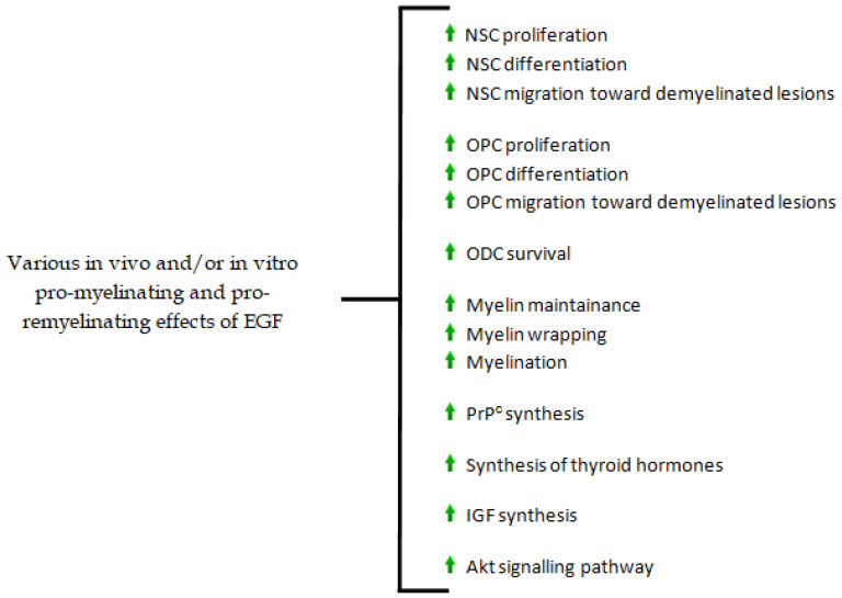
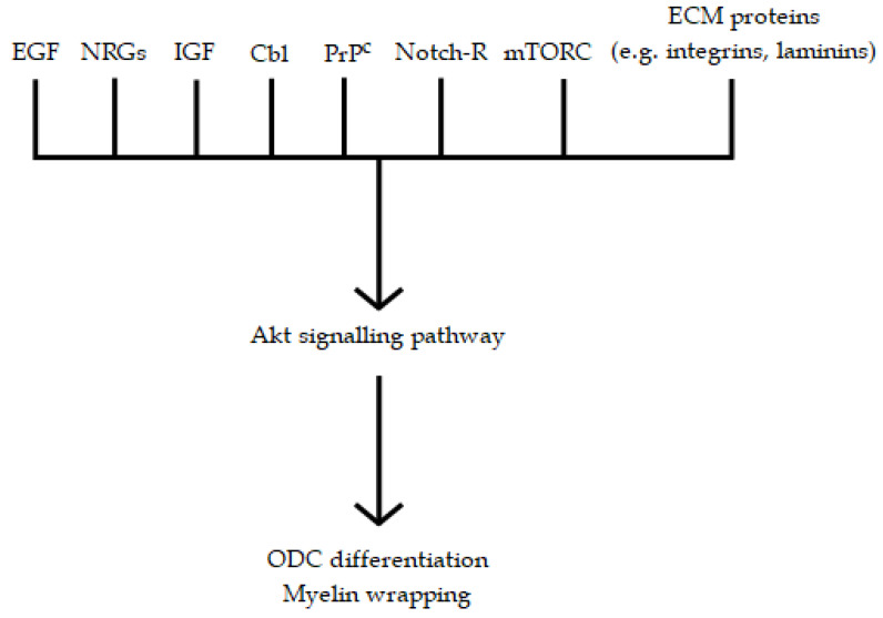
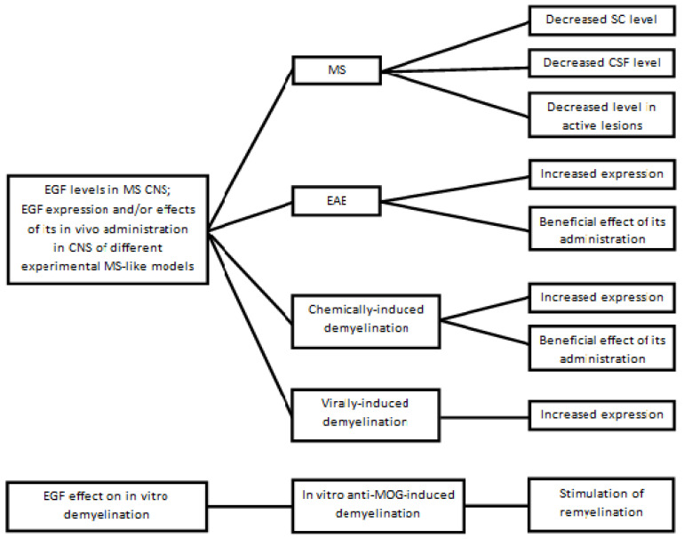
The pathogenesis of multiple sclerosis (MS) remains enigmatic and controversial. Myelin sheaths in the central nervous system (CNS) insulate axons and allow saltatory nerve conduction. MS brings about the destruction of myelin sheaths and the myelin-producing oligodendrocytes (ODCs). The conundrum of remyelination failure is, therefore, crucial in MS. In this review, the roles of epidermal growth factor (EGF), normal prions, and cobalamin in CNS myelinogenesis are briefly summarized. Thereafter, some findings of other authors and ourselves on MS and MS-like models are recapitulated, because they have shown that: (a) EGF is significantly decreased in the CNS of living or deceased MS patients; (b) its repeated administration to mice in various MS-models prevents demyelination and inflammatory reaction; (c) as was the case for EGF, normal prion levels are decreased in the MS CNS, with a strong correspondence between liquid and tissue levels; and (d) MS cobalamin levels are increased in the cerebrospinal fluid, but decreased in the spinal cord. In fact, no remyelination can occur in MS if these molecules (essential for any form of CNS myelination) are lacking. Lastly, other non-immunological MS abnormalities are reviewed. Together, these results have led to a critical reassessment of MS pathogenesis, partly because EGF has little or no role in immunology.
Overview
The claims and statistics of this paper haven’t been checked yet.
Run the check — free, no account needed. It reads the open-access text and checks the sources it cites.
Integrity signals
Registry
Cites
Statistics
Figures
Verification
Claims the paper makes
The claims and statistics of this paper haven’t been checked yet.
Run the check — free, no account needed. It reads the open-access text and checks the sources it cites.
Claims other papers make about this one
Each is checked against this paper’s own text: does it say what the citing paper says it does?
In the citing paper
“Specific miRNAs may also influence the basic mechanisms of tissue repair, including myelin repair, gliogenesis, and neurogenesis in multiple sclerosis.”
From MiR-124 and MiR-155 as Therapeutic Targets in Microglia-Mediated Inflammation in Multiple Sclerosis (2025) · cites this paper as Scalabrino 2022
Self-check
Percentages, sums and reported differences re-computed from the paper’s own numbers.
The self-check needs the paper’s full text.
Citations
Cites
Premyelinating Oligodendrocytes in Chronic Lesions of Multiple Sclerosis
Chang, A., Tourtellotte, W. W., Rudick, R., & Trapp, B. D. (2002) · New England Journal of Medicine · doi:10.1056/nejmoa010994 · Cited by 991
Oligodendrocytes: biology and pathology
Bradl, M., & Lassmann, H. (2010) · Acta Neuropathologica · doi:10.1007/s00401-009-0601-5 · Cited by 933
Oligodendrocyte Dynamics in the Healthy Adult CNS: Evidence for Myelin Remodeling
Young, K. M., Psachoulia, K., Tripathi, R. B., Dunn, S.-J., Cossell, L., Attwell, D., Tohyama, K., & Richardson, W. D. (2013) · Neuron · doi:10.1016/j.neuron.2013.01.006 · Cited by 919
Vitamin B12, folic acid, and the nervous system
Reynolds, E. (2006) · The Lancet Neurology · doi:10.1016/s1474-4422(06)70598-1 · Cited by 860
Myelin Impairs CNS Remyelination by Inhibiting Oligodendrocyte Precursor Cell Differentiation
Kotter, M. R., Li, W.-W., Zhao, C., & Franklin, R. J. M. (2006) · The Journal of Neuroscience · doi:10.1523/jneurosci.2615-05.2006 · Cited by 768
Regulation of oligodendrocyte differentiation and myelination
Emery, B. (2010) · Science · doi:10.1126/science.1190927 · Cited by 653
Regenerating CNS myelin — from mechanisms to experimental medicines
Franklin, R. J. M., & ffrench-Constant, C. (2017) · Nature Reviews Neuroscience · doi:10.1038/nrn.2017.136 · Cited by 647
Control of local protein synthesis and initial events in myelination by action potentials
Wake, H., Lee, P. R., & Fields, R. D. (2011) · Science · doi:10.1126/science.1206998 · Cited by 636
Oligodendrocytes in development, myelin generation and beyond
Kuhn, S., Gritti, L., Crooks, D., & Dombrowski, Y. (2019) · Cells · doi:10.3390/cells8111424 · Cited by 625
Pathophysiology of the brain extracellular matrix: a new target for remyelination
Lau, L. W., Cua, R., Keough, M. B., Haylock-Jacobs, S., & Yong, V. W. (2013) · Nature reviews. Neuroscience · doi:10.1038/nrn3550 · Cited by 549
Research works printed but not linked: 460
- Love, 2015, Demyelinating Diseases, Greenfield’s Neuropathology, Volume 2, 1297
- Wingerchuk, 2001, Multiple sclerosis: Current pathophysiological concepts, Lab. Investig., 81, 263, 10.1038/labinvest.3780235 · DOI
- Peterson, 2005, Neuropathobiology of multiple sclerosis, Neurol. Clin., 23, 107, 10.1016/j.ncl.2004.09.008 · DOI
- Wood, P.L. (2003). Neuroimmunologic Mechanisms in the Etiology of Multiple Sclerosis. Neuroinflammation. Mechanisms and Management, Humana Press. [2nd ed.].
- Lucchinetti, 2005, The pathology of multiple sclerosis, Neurol. Clin., 23, 77, 10.1016/j.ncl.2004.09.002 · DOI
- Levin, 2014, Neurodegeneration in multiple sclerosis involves multiple pathogenic mechanisms, Degen. Neurol. Neuromus. Dis., 4, 49
- Waxman, 2006, Ions, energy and axonal injury: Towards a molecular neurology of multiple sclerosis, Trends Mol. Med., 12, 192, 10.1016/j.molmed.2006.03.001 · DOI
- Stadelmann, 2005, The spectrum of multiple sclerosis: New lesson from pathology, Curr. Opin. Neurol., 18, 221, 10.1097/01.wco.0000169736.60922.20 · DOI
- Duncan, I.D., and Franklin, R.J.M. (2013). Demyelination and Remyelination in Multiple Sclerosis. Myelin Repair and Neuroprotection in Multiple Sclerosis, Springer. [1st ed.]. · DOI
- Groppa, S., and Meuth, S.G. (2021). Pathophysiological Basis of Autoimmune-Initiated/Mediated Neurodegeneration. Translational Methods for Multiple Sclerosis Research, Humana Press.
- Pérez-Pérez, S., Dominguez-Mozo, M.I., Garcia-Martínez, M.Á., Ballester-González, R., Gañán, I., Arroyo, R., and Alvarez-Lafuente, R. (2022). Epstein-Barr virus load correlates with multiple sclerosis-associated retrovirus envelope expression. Biomedicines, 10. · DOI
- Pimentel, 1994, Epidermal Growth Factor, Handbook of Growth Factors, Volume 2, 97
- Baserga, R. (1981). Epidermal Growth Factor. Tissue Growth Factors, Springer. · DOI
- Iwakura, 2013, Erb1-4-dependent EGF/neuregulin signals and their cross talk in the central nervous system: Pathological implications in schizophrenia and Parkinson’s disease, Front. Cell. Neurosci., 7, 4, 10.3389/fncel.2013.00004 · DOI
- Hirata, 1982, Presence of human epidermal growth factor in human cerebrospinal fluid, J. Clin. Endocrinol. Metab., 55, 1174, 10.1210/jcem-55-6-1174 · DOI
- Fallon, 1984, Epidermal growth factor immunoreactive material in the central nervous system: Location and development, Science, 224, 1107, 10.1126/science.6144184 · DOI
- Lazar, 1992, Regional distribution and developmental expression of epidermal growth factor and transforming growth factor-α mRNA in mouse brain by a quantitative nuclease protection assay, J. Neurosci., 12, 1688, 10.1523/JNEUROSCI.12-05-01688.1992 · DOI
- Piao, 2005, Differential distribution of peptides in the epidermal growth factor family and phosphorylation of ErbB1 receptor in adult rat brain, Neurosci. Lett., 390, 21, 10.1016/j.neulet.2005.07.048 · DOI
- Zeng, 2014, Epidermal growth factor, from gene organization to bedside, Sem. Cell. Dev. Biol., 28, 2, 10.1016/j.semcdb.2014.01.011 · DOI
- Chevalier, 2011, Enteric glia promote intestinal mucosal healing via activation of focal adhesion kinase and release of proEGF, Am. J. Physiol. Gastrointest. Liver Physiol., 300, G976, 10.1152/ajpgi.00427.2010 · DOI
- Neunlist, 2013, The digestive neuronal-glial-epithelial unit: A new actor in gut health and disease, Nat. Rev. Gastroenterol. Hepatol., 10, 90, 10.1038/nrgastro.2012.221 · DOI
- Scalabrino, 1995, Subacute combined degeneration in the spinal cords of totally gastrectomized rats. Ornithine decarboxylase induction, cobalamin status, and astroglial reaction, Lab. Investig., 72, 114
- Sharma, H.S., and Westman, J. (2004). Transport of Cytokines and Neurotrophins across the Blood-Brain Barrier and Their Regulation after Spinal Cord Injury. Blood-Spinal Cord and Brain Barriers in Health and Disease, Elsevier-Academic Press.
- Pan, 1999, Entry of EGF into brain is rapid and saturable, Peptides, 20, 1091, 10.1016/S0196-9781(99)00094-7 · DOI
- Nagano, 2007, In vivo administration of epidermal growth factor and its homologue attenuates developmental maturation of functional excitatory synapses in cortical GABAergic neurons, Eur. J. Neurosci., 25, 380, 10.1111/j.1460-9568.2007.05297.x · DOI
- Sharma, H.S., and Westman, J. (2004). Volume Transmission-Mediated Protective Impact of Choroid Plexus-Cerebrospinal Fluid Growth Factors on Forebrain Ischemic Injury. Blood-Spinal Cord and Brain Barriers in Health and Disease, Elsevier-Academic Press.
- Scalabrino, 2009, The multi-faceted basis of vitamin B12 (cobalamin) neurotrophism in adult central nervous system: Lessons learned from its deficiency, Prog. Neurobiol., 88, 203, 10.1016/j.pneurobio.2009.04.004 · DOI
- Fenech, 2012, Folate (vitamin B9) and vitamin B12 and their function in the maintenance of nuclear and mitochondrial genome integrity, Mut. Res., 733, 21, 10.1016/j.mrfmmm.2011.11.003 · DOI
- Scalabrino, 2009, Vitamin-regulated cytokines and growth factors in the central nervous system and elsewhere, J. Neurochem., 111, 1309, 10.1111/j.1471-4159.2009.06417.x · DOI
- Wolffenbuttel, 2019, The many faces of cobalamin (Vitamin B12) deficiency, Mayo Clin. Proc. Inn. Qual. Out., 3, 200, 10.1016/j.mayocpiqo.2019.03.002 · DOI
- Rai, S.N., Singh, P., Steinbusch, H.W.M., Vamanu, E., Ashraf, G., and Singh, M.P. (2021). The role of vitamins in neurodegenerative disease: An update. Biomedicines, 9. · DOI
- Linden, 2008, Physiology of the prion protein, Physiol. Rev., 88, 673, 10.1152/physrev.00007.2007 · DOI · Paper page
- Castle, 2017, Physiological functions of the cellular prion protein, Front. Mol. Biosci., 4, 19, 10.3389/fmolb.2017.00019 · DOI
- Linden, 2017, The biological function of the prion protein: A cell surface scaffold of signalling modules, Front. Mol. Neurosci., 10, 77, 10.3389/fnmol.2017.00077 · DOI
- Harris, 1999, Cellular biology of prion diseases, Clin. Microbiol. Rev., 12, 429, 10.1128/CMR.12.3.429 · DOI
- Mehrpour, 2010, Prion protein: From physiology to cancer biology, Cancer Lett., 290, 1, 10.1016/j.canlet.2009.07.009 · DOI
- Westergard, 2007, The cellular prio protein (PrPc): Its physiological function and role in disease, Biochim. Biophys. Acta, 1172, 629, 10.1016/j.bbadis.2007.02.011 · DOI
- Rutishauser, D., Mertz, K.D., Moos, R., Brunner, E., Rülicke, T., Calella, A.M., and Aguzzi, A. (2009). The comprehensive native interactome of a fully functional tagged prion protein. PLoS ONE, 4. · DOI
- Chiesa, R. (2015). The elusive role of the prion protein and the mechanism of toxicity in prion disease. PLoS Pathog., 11. · DOI
- Scalabrino, 2013, Cobalamin and normal prions: A new horizon for cobalamin neurotrophism, Biochemie, 95, 1041, 10.1016/j.biochi.2013.01.002 · DOI
- Baumann, 2007, Lethal recessive myelin toxicity of prion protein lacking its central domain, EMBO J., 26, 538, 10.1038/sj.emboj.7601510 · DOI
- Mitteregger, 2007, The role of octapeptide region in neuroprotective function of the cellular prion protein, Brain Pathol., 17, 174, 10.1111/j.1750-3639.2007.00061.x · DOI
- Li, 2007, Prion protein with an octapeptide insertion has impaired neuroprotective activity in transgenic mice, EMBO J., 26, 2777, 10.1038/sj.emboj.7601726 · DOI
- Scalabrino, 2014, Myelin damage due to local quantitative abnormalities in normal prion levels: Evidence from subacute combined degeneration and multiple sclerosis, J. Neurol., 261, 1451, 10.1007/s00415-013-7152-3 · DOI
- Veber, 2015, Are PrPcs involved in some human myelin diseases? Relating experimental studies to human pathology, J. Neurol. Sci., 359, 396, 10.1016/j.jns.2015.09.365 · DOI
- Watts, 2018, The function of the cellular prion protein in health and disease, Acta Neuropathol., 135, 159, 10.1007/s00401-017-1790-y · DOI
- Scalabrino, 2014, Relationship between cobalamin, epidermal growth factor, and normal prions in the myelin maintenance of central nervous system, Int. J. Biochem. Cell Biol., 55, 232, 10.1016/j.biocel.2014.09.011 · DOI
- Scalabrino, 2020, Epidermal growth factor in the CNS: A beguiling journey from integrated cell biology to multiple sclerosis, An extensive translational overview, Cell. Mol. Neurobiol., 42, 891, 10.1007/s10571-020-00989-x · DOI
- Stadelmann, 2019, Myelin in the central nervous system: Structure, function, and pathology, Physiol. Rev., 99, 1381, 10.1152/physrev.00031.2018 · DOI
- Lazzarini, 2004, Structure of the Myelinated Axon, Myelin Biology and Disorders, Volume 1, 3
- Aggarwal, 2011, Central nervous system myelin: Structure, synthesis, and assembly, Trends Cell Biol., 21, 585, 10.1016/j.tcb.2011.06.004 · DOI · Paper page
- Lazzarini, 2004, Cell Biology of Myelin Assembly, Myelin Biology and Disorders, Volume 1, 29
- Sherman, 2005, Mechanisms of axon ensheathment and myelin growth, Nat. Rev. Neurosci., 6, 683, 10.1038/nrn1743 · DOI · Paper page
- Lazzarini, 2004, The Transport, Assembly, and Function of Myelin Lipids, Myelin Biology and Disorders, Volume 1, 57
- Hughes, 2016, The cell biology of CNS myelination, Curr. Opin. Neurobiol., 39, 93, 10.1016/j.conb.2016.04.013 · DOI
- Simons, 2016, Oligodendrocytes: Myelination and Axonal Support, Cold Sping Harbor Persp. Biol., 8, a020479, 10.1101/cshperspect.a020479 · DOI
- Miron, 2011, Cells of the oligodendroglial lineage, myelination, and remyelination, Biochim. Biophys. Acta, 1812, 184, 10.1016/j.bbadis.2010.09.010 · DOI
- Tognatta, 2020, Astrocytes are required for oligodendrocyte survival and maintenance of myelin compaction and integrity, Front. Cell. Neurosci., 14, 74, 10.3389/fncel.2020.00074 · DOI
- Nutma, E., van Gent, D., Amor, S., and Peferoen, L.A.N. (2020). Astrocyte and oligodendrocyte cross-talk in the central nervous system. Cells, 9. · DOI
- Barnett, 2012, Myelination: Do astrocytes play a role?, Neuroscientist, 20, 1
- Miron, 2019, Astrocytes in myelination and remyelination, Neurosci. Lett., 713, 134532, 10.1016/j.neulet.2019.134532 · DOI
- Gibson, 2014, Neuronal activity promotes oligodendrogenesis and adaptive myelination in the mammalian brain, Science, 344, 1252304, 10.1126/science.1252304 · DOI · Paper page
- Saab, 2013, The role of myelin and oligodendrocytes in axonal energy metabolism, Curr. Opin. Neurobiol., 23, 1065, 10.1016/j.conb.2013.09.008 · DOI · Paper page
- Lee, 2012, Oligodendroglia metabolically support axons and contribute to neurodegeneration, Nature, 487, 443, 10.1038/nature11314 · DOI · Paper page
- Lazzarini, 2004, Functional Organization of the Nodes of Ranvier, Myelin Biology and Disorders, Volume 1, 89
- Nave, 2010, Myelination and the trophic support of long axons, Nat. Rev. Neurosci., 11, 275, 10.1038/nrn2797 · DOI · Paper page
- Lazzarini, 2004, Electrophysiological Consequences of Myelination, Myelin Biology and Disorders, Volume 1, 117
- Simons, 2006, Neuron-glia communication in the control of oligodendrocyte function and myelin biogenesis, J. Cell Sci., 119, 4381, 10.1242/jcs.03242 · DOI
- Bercury, 2015, Dynamics and mechanism of CNS myelination, Dev. Cell., 32, 447, 10.1016/j.devcel.2015.01.016 · DOI · Paper page
- Tomassy, 2016, Diversity matters: A revised guide to myelination, Trends Cell Biol., 26, 135, 10.1016/j.tcb.2015.09.002 · DOI · Paper page
- Chang, 2016, Remodeling myelination: Implications for mechanisms of neural plasticity, Nat. Neurosci., 19, 190, 10.1038/nn.4200 · DOI
- Alizadeh, 2015, Myelin damage and repair on pathologic CNS: Challenges and prospects, Front. Mol. Neurosci., 8, 35, 10.3389/fnmol.2015.00035 · DOI
- Nave, 2014, Myelination of the nervous system: Mechanisms and functions, Annu. Rev. Cell Dev. Biol., 30, 503, 10.1146/annurev-cellbio-100913-013101 · DOI · Paper page
- Cayre, 2021, Myelin repair: From animal model to humans, Front. Cell. Neurosci., 15, 604865, 10.3389/fncel.2021.604865 · DOI
- Chamberlain, 2016, Oligodendrocyte regeneration: Its significance in myelin replacement and neuroprotection in multiple sclerosis, Neuropharmacology, 110, 633, 10.1016/j.neuropharm.2015.10.010 · DOI
- Piaton, 2010, Axon-oligodendrocyte interactions during developmental myelination, demyelination and repair, J. Neurochem., 114, 1243, 10.1111/j.1471-4159.2010.06831.x · DOI
- Trapp, 2008, Multiple sclerosis: An immune or neurodegenerative disorder?, Annu. Rev. Neurosci., 31, 247, 10.1146/annurev.neuro.30.051606.094313 · DOI · Paper page
- Kornek, 2000, Multiple sclerosis and chronic autoimmune encephalomyelitis. A comparative quantitative study of axonal injury in active, inactive, and remyelinated lesions, Am. J. Pathol., 157, 267, 10.1016/S0002-9440(10)64537-3 · DOI
- Irvine, 2008, Remyelination protects axons from demyelination-associated axon degeneration, Brain, 131, 1464, 10.1093/brain/awn080 · DOI · Paper page
- Duncan, I.D., and Franklin, R.J.M. (2013). Endogenous Remyelination in the CNS. Myelin Repair and Neuroprotection in Multiple Sclerosis, Springer. · DOI
- Campbell, 2012, Mitochondrial changes within axons in multiple sclerosis: An update, Curr. Opin. Neurol., 25, 221, 10.1097/WCO.0b013e3283533a25 · DOI
- Hinks, 1999, Distinctive patterns of PDGF-A, FGF-2, IGF-1 and TGF-β1 gene expression during remyelination of experimentally-induced spinal cord demyelination, Mol. Cell. Neurosci., 14, 153, 10.1006/mcne.1999.0771 · DOI
- Fancy, 2011, Myelin regeneration: A recapitulation of development?, Annu. Rev. Neurosci., 34, 21, 10.1146/annurev-neuro-061010-113629 · DOI
- Hanafy, 2011, Regulation of remyelination in multiple sclerosis, FEBS Lett., 585, 3821, 10.1016/j.febslet.2011.03.048 · DOI
- Werkman, 2021, Macroglial diversity: White and grey areas and relevance to remyelination, Cell. Mol. Life Sci., 78, 143, 10.1007/s00018-020-03586-9 · DOI
- Clemente, 2013, The effect of glia-glia interactions on oligodendrocyte precursor cell biology during development and in demyelinating diseases, Front. Cell. Neurosci., 7, 268, 10.3389/fncel.2013.00268 · DOI
- Kovacs, G.G., and Alafuzoff, I. (2018). Inflammatory demyelinating diseases of the central nervous system. Handbook of Clinical Neurology, Neuropathology, Elsevier.
- Frohman, 2006, Multiple sclerosis—The plaque and its pathogenesis, N. Engl. J. Med., 354, 942, 10.1056/NEJMra052130 · DOI
- Frischer, 2015, Clinical and pathological insights into the dynamic nature of the white matter multiple sclerosis plaque, Ann. Neurol., 78, 710, 10.1002/ana.24497 · DOI · Paper page
- Lucchinetti, 2000, Heterogeneity of multiple sclerosis lesions: Implications for the pathogenesis of demyelination, Ann. Neurol., 47, 707, 10.1002/1531-8249(200006)47:6<707::AID-ANA3>3.0.CO;2-Q · DOI
- Petropoulos, 1983, Alpha-Fetoprotein and albumin mRNA levels in liver regeneration and carcinogenesis, J. Biol. Chem., 258, 4901, 10.1016/S0021-9258(18)32512-2 · DOI
- Gao, 2021, All routes lead to Rome: Multifaceted origin of hepatocytes during liver regeneration, Cell Regen., 10, 2, 10.1186/s13619-020-00063-3 · DOI
- Ritter, 2003, The molecular basis of myocardial hypertrophy and heart failure, Trends Mol. Med., 9, 313, 10.1016/S1471-4914(03)00114-X · DOI
- Lyon, 2015, Mechanotransduction in cardiac hypertrophy and failure, Circ. Res., 116, 1462, 10.1161/CIRCRESAHA.116.304937 · DOI
- Kuhlmann, 2003, Remyelination in multiple sclerosis, J. Neurol. Sci., 206, 181, 10.1016/S0022-510X(02)00191-0 · DOI
- Macchi, 2014, Oligodendrogenesis in the normal and pathological central nervous system, Front. Neurosci., 8, 145
- Domingues, 2016, Oligodendrocyte, astrocyte, and microglia crosstalk in myelin development, damage, and repair, Front. Cell Dev. Biol., 4, 71
- Patel, 2011, Mediators of oligodendrocyte differentiation during remyelination, FEBS Lett., 585, 3730, 10.1016/j.febslet.2011.04.037 · DOI
- Parras, 2020, Chromatin remodelers in oligodendroglia, Glia, 68, 1604, 10.1002/glia.23837 · DOI
- Tiane, A., Schepers, M., Rombaut, B., Hupperts, R., Prickaerts, J., Hellings, N., van den Hove, D., and Vanmierlo, T. (2019). From OPC to oligodendrocyte: An epigenetic journey. Cells, 8. · DOI
- Elbaz, 2019, Molecular control of oligodendrocyte development, Trends Neurosci., 42, 263, 10.1016/j.tins.2019.01.002 · DOI
- Verhaagen, 2009, Remyelination in multiple sclerosis, Progress in Brain Research, Volume 175, 453, 10.1016/S0079-6123(09)17530-1 · DOI
- Wegner, 2008, A matter of identity: Transcriptional control in oligodendrocytes, J. Mol. Neurosci., 35, 3, 10.1007/s12031-007-9008-8 · DOI
- Copray, 2009, Epigenetic mechanisms facilitating oligodendrocyte development, maturation, and aging, Glia, 57, 1579, 10.1002/glia.20881 · DOI
- Liu, 2010, Epigenetic regulation of oligodendrocyte identity, Trends Neurosci., 33, 193, 10.1016/j.tins.2010.01.007 · DOI
- Liu, 2016, Epigenetic control of oligodendrocyte development: Adding new players to old keepers, Curr. Opin. Neurobiol., 39, 133, 10.1016/j.conb.2016.06.002 · DOI
- Patrikios, 2006, Remyelination is extensive in a subset of multiple sclerosis patients, Brain, 129, 3165, 10.1093/brain/awl217 · DOI · Paper page
- Patani, 2007, Remyelination can be extensive in multiple sclerosis despite a long disease course, Neuropathol. Appl. Neurobiol., 33, 277, 10.1111/j.1365-2990.2007.00805.x · DOI
- Bramow, 2010, Demyelination versus remyelination in progressive multiple sclerosis, Brain, 133, 2983, 10.1093/brain/awq250 · DOI
- Reynolds, 1992, Generation of neurons and astrocytes from isolated cells of the adult mammalian central nervous system, Science, 255, 1707, 10.1126/science.1553558 · DOI · Paper page
- Black, 1990, Neuroblast mitosis in dissociated culture: Regulation and relationship to differentiation, J. Cell Biol., 110, 2073, 10.1083/jcb.110.6.2073 · DOI
- Armstrong, 1992, Pre-oligodendrocytes from adult human CNS, J. Neurosci., 12, 1538, 10.1523/JNEUROSCI.12-04-01538.1992 · DOI
- Los, 1993, Proliferating subventricular zone cells in the adult mammalian forebrain can differentiate into neurons and glia, Proc. Natl. Acad. Sci. USA, 90, 2074, 10.1073/pnas.90.5.2074 · DOI
- Zhang, 1999, Adult brain retains the potential to generate oligodendroglial progenitors with extensive myelination capacity, Proc. Natl. Acad. Sci. USA, 96, 4089, 10.1073/pnas.96.7.4089 · DOI
- Decker, 1999, Progenitor cells of the adult mouse subventricular zone proliferate, migrate and differentiate into oligodendrocytes after demyelination, Eur. J. Neurosci., 11, 4357, 10.1046/j.1460-9568.1999.00873.x · DOI · Paper page
- Horner, 2000, Proliferation and differentiation of progenitor cells throughout the intact adult rat spinal cord, J. Neurosci., 20, 2218, 10.1523/JNEUROSCI.20-06-02218.2000 · DOI · Paper page
- Chang, 2000, NG-2 positive oligodendrocyte progenitor cells in adult human brain and multiple sclerosis lesions, J. Neurosci., 20, 6404, 10.1523/JNEUROSCI.20-17-06404.2000 · DOI
- Doetsch, 2003, A niche for adult neural stem cells, Curr. Opin. Genet. Dev., 13, 543, 10.1016/j.gde.2003.08.012 · DOI
- Xing, 2014, Adult neural precursor cells from the subventricular zone contribute significantly to oligodendrocyte regeneration and remyelination, J. Neurosci., 34, 14128, 10.1523/JNEUROSCI.3491-13.2014 · DOI · Paper page
- Fancy, 2004, Increased expression of Nkx2.2 and Olig2 identifies reactive oligodendrocyte progenitor cells responding to demyelination in the adult CNS, Mol. Cell. Neurosci., 27, 247, 10.1016/j.mcn.2004.06.015 · DOI
- Zawadzka, 2010, CNS-Resident glial progenitor/stem cells produce Schwann cells as well as oligodendrocytes during repair of CNS demyelination, Cell Stem Cell., 6, 578, 10.1016/j.stem.2010.04.002 · DOI · Paper page
- Marmur, 1998, Differentiation of oligodendroglial progenitors derived from cortical multipotent cells requires extrinsic signals including activation of gp130/LIFβ receptors, J. Neurosci., 18, 9800, 10.1523/JNEUROSCI.18-23-09800.1998 · DOI
- Gago, 2003, Control of cell survival and proliferation of postnatal PSA-NCAM+ progenitors, Mol. Cell. Neurosci., 22, 162, 10.1016/S1044-7431(02)00030-1 · DOI
- Nunes, 2003, Identification and isolation of multipotent neural progenitor cells from the subcortical white matter of the adult human brain, Nat. Med., 9, 439, 10.1038/nm837 · DOI
- Reynolds, 1996, Clonal and population analyses demonstrate that an EGF-responsive mammalian embryonic CNS precursor is a stem cell, Dev. Biol., 175, 1, 10.1006/dbio.1996.0090 · DOI
- 2013, The role of EGFR and ErbB family related proteins in the oligodendrocyte specification in germinal niches of the adult mammalian brain, Front. Cell. Neurosci., 7, 258
- Doetsch, 1999, Subventricular zone astrocytes are neural stem cells in the adult mammalian brain, Cell, 97, 703, 10.1016/S0092-8674(00)80783-7 · DOI · Paper page
- Laywell, 2000, Identification of a multipotent astrocytic stem cell in the immature and adult mouse brain, Proc. Natl. Acad. Sci. USA, 97, 13883, 10.1073/pnas.250471697 · DOI
- Akkermann, 2017, Heterogenous populations of neural stem cells contribute to myelin repair, Neural Regen. Res., 12, 509, 10.4103/1673-5374.204999 · DOI
- Foerster, 2019, Diversity in the oligodendrocyte lineage: Plasticity or heterogeneity?, Glia, 67, 1797, 10.1002/glia.23607 · DOI
- 2017, White matter plasticity in the adult brain, Neuron, 96, 1239, 10.1016/j.neuron.2017.11.026 · DOI
- Covarrubias, 1995, Epidermal growth factor (EGF), transforming growth factor-α (TGF-α), and basic fibroblast growth factor (bFGF) differentially influence neural precursor cells of mouse embryonic mesencephalon, J. Neurosci. Res., 42, 172, 10.1002/jnr.490420204 · DOI
- Craig, 1996, In vivo growth factor expansion of endogenous subependymal neural precursor cell populations in the adulte mouse brain, J. Neurosci., 16, 2649, 10.1523/JNEUROSCI.16-08-02649.1996 · DOI
- Martens, 2000, Separate proliferation kinetics of fibroblast growth factor-responsive and epidermal growth factor-responsive neural stem cells within the embryonic forebrain germinal zone, J. Neurosci., 20, 1085, 10.1523/JNEUROSCI.20-03-01085.2000 · DOI
- Kerninon, 2007, Activation of the subventricular zone in multiple sclerosis: Evidence for early glial progenitors, Proc. Natl. Acad. Sci. USA, 104, 4694, 10.1073/pnas.0606835104 · DOI
- Brousse, 2015, Region and dynamic specificities of adult neural stem cells and oligodendrocyte precursors in myelin regeneration in the mouse brain, Biol. Open, 4, 980, 10.1242/bio.012773 · DOI · Paper page
- Tripathi, 2010, NG2 Glia generate new oligodendrocytes but few astrocytes in a murine experimental autoimmune encephalomyelitis model of demyelinating disease, J. Neurosci., 30, 16383, 10.1523/JNEUROSCI.3411-10.2010 · DOI · Paper page
- Lazzarini, 2004, Remyelination by Endogenous Glia, Myelin Biology and Disorders, Volume 1, 173
- Cantarella, 2008, Intranasal HB-EGF administration favors adult SVZ cell mobilization to demyelinated lesions in mouse corpus callosum, Devel. Neurobiol., 68, 223, 10.1002/dneu.20588 · DOI
- Decker, 2002, Experimental autoimmune encephalomyelitis mobilizes neural progenitors from the subventricular zone to undergo oligodendrogenesis in adult mice, Proc. Natl. Acad. Sci. USA, 99, 13211, 10.1073/pnas.192314199 · DOI
- Cayre, 2006, Migrating and myelinating potential of subventricular zone neural progenitor cells in white matter tracts of the adult rodent brain, Mol. Cell. Neurosci., 31, 748, 10.1016/j.mcn.2006.01.004 · DOI
- Mecha, 2013, Mobilization of progenitors in the subventricular zone to undergo oligodendrogenesis in the Theiler’s virus model of multiple sclerosis: Implications for remyelination at lesions sites, Exp. Neurol., 250, 348, 10.1016/j.expneurol.2013.10.011 · DOI
- 2010, Dose-dependent of EGF on migration and differentiation of adult subventricular zone astrocytes, Glia, 58, 975, 10.1002/glia.20979 · DOI
- 2011, Oligodendrogenesis in the subventricular zone and the role of epidermal growth factor, Brain Res. Rev., 67, 147, 10.1016/j.brainresrev.2011.01.001 · DOI
- Solanky, 2001, Proliferating oligodendrocytes are present in both active and chronic inactive multiple sclerosis plaques, J. Neurosci. Res., 65, 308, 10.1002/jnr.1155 · DOI
- Colognato, 2011, Glia unglued: How signals from the extracellular matrix regulate the development of myelinating glia, Dev. Neurobiol., 71, 924, 10.1002/dneu.20966 · DOI
- Mitew, 2014, Mechanisms regulating the development of oligodendrocytes and central nervous system, Neuroscience, 276, 29, 10.1016/j.neuroscience.2013.11.029 · DOI
- Boggs, 2006, Myelin basic protein: A multifunctional protein, Cell. Mol. Life Sci., 63, 1945, 10.1007/s00018-006-6094-7 · DOI · Paper page
- Emery, 2015, Transcriptional and epigenetic regulation of oligodendrocyte development and myelination in the central nervous system, Cold Spring Harb. Perspect. Biol., 7, a020461, 10.1101/cshperspect.a020461 · DOI
- Menn, 2006, Origin of oligodendrocytes in the subventricular zone of the adult brain, J. Neurosci., 26, 7907, 10.1523/JNEUROSCI.1299-06.2006 · DOI · Paper page
- 2009, EGF induces the progeny of subventricular zone type B cells to migrate and differentiate into oligodendrocytes, Stem Cells, 27, 2032, 10.1002/stem.119 · DOI
- Aguirre, 2005, Overexpression of the epidermal growth factor receptor confers migratory properties to nonmigratory postnatal neural progenitors, J. Neurosci., 25, 11092, 10.1523/JNEUROSCI.2981-05.2005 · DOI
- Grinspan, 2002, Cells and signalling in oligodendrocyte development, J. Neuropath. Exp. Neurol., 61, 297, 10.1093/jnen/61.4.297 · DOI
- Bankston, 2013, Oligodendroglia and neurotrophic factors in neurodegeneration, Neurosci. Bull., 29, 216, 10.1007/s12264-013-1321-3 · DOI
- He, 2016, Oligodendrocyte progenitor programming and reprogramming: Toward myelin regeneration, Brain Res., 1638, 209, 10.1016/j.brainres.2015.10.051 · DOI
- Temple, 1999, Stem cells in the adult mammalian central nervous system, Curr. Opin. Neurobiol., 9, 135, 10.1016/S0959-4388(99)80017-8 · DOI
- Akkermann, R., Jadasz, J.J., Azim, K., and Küry, P. (2016). Taking advantage of nature’s gift: Can endogenous neural stem cells improve myelin regeneration?. Int. J. Mol. Sci., 17. · DOI
- Raabe, 2004, Localization of neuregulin isoforms and erbB receptors in myelinating glial cells, Glia, 45, 197, 10.1002/glia.10311 · DOI
- Deadwyler, 2000, Neuregulins and erbB receptor expression in adult human oligodendrocytes, Glia, 32, 304, 10.1002/1098-1136(200012)32:3<304::AID-GLIA90>3.0.CO;2-Z · DOI
- Mayoral, 2016, The environment rules: Spatiotemporal regulation of oligodendrocyte differentiation, Curr. Opin. Neurobiol., 39, 47, 10.1016/j.conb.2016.04.002 · DOI
- Li, 2009, Two-tier transcriptional control of oligodendrocyte differentiation, Curr. Opin. Neurobiol., 19, 479, 10.1016/j.conb.2009.08.004 · DOI
- Moore, 2001, How factors secreted from astrocytes impact myelin repair, J. Neurosci. Res., 89, 13, 10.1002/jnr.22482 · DOI
- Kiray, 2016, The multifaceted role of astrocytes in regulating myelination, Exp. Neurol., 283, 541, 10.1016/j.expneurol.2016.03.009 · DOI
- Connor, 1995, Cellular management of iron in the brain, J. Neurol. Sci., 134, 33, 10.1016/0022-510X(95)00206-H · DOI
- Li, 2019, Astrocyte-to-astrocyte contact and a positive feedback loop of growth factor signaling regulate astrocyte maturation, Glia, 67, 1571, 10.1002/glia.23630 · DOI
- Hughes, 2021, Glial cells promote myelin formation and elimination, Front. Cell Dev. Biol., 9, 661486, 10.3389/fcell.2021.661486 · DOI
- Grove, 1996, Plasticity of astrocytes derived from aged mouse cerebral hemispheres: Changes with cell passage and immortalization, Brain Res. Bull., 39, 211, 10.1016/0361-9230(95)02130-2 · DOI
- Anlauf, 2013, Glutamine synthetase as an astrocytic marker: Its cell type and vescicle localization, Front. Endocrinol., 4, 144, 10.3389/fendo.2013.00144 · DOI
- Kettenmann, H., and Ranson, B.R. (2013). Amino Acid Neurotransmitter Synthesis and Removal. Neuroglia, Oxford University Press. [3rd ed.].
- Eng, 1990, Glutamine synthetase immunoreactivity is present in oligodendroglia of various regions of the central nervous system, Glia, 3, 335, 10.1002/glia.440030504 · DOI · Paper page
- Zhou, 2019, Selective deletion of glutamine synthetase in the mouse cerebral cortex induces glial dysfunction and vascular impairment that precede epilepsy and neurodegeneration, Neurochem. Int., 123, 22, 10.1016/j.neuint.2018.07.009 · DOI
- Zhou, 2020, Novel aspects of glutamine synthetase in ammonia homeostasis, Neurochem. Int., 140, 104809, 10.1016/j.neuint.2020.104809 · DOI
- Haim, 2021, Evidence for glutamine synthetase function in mouse spinal cord, Glia, 69, 2812, 10.1002/glia.24071 · DOI
- Gautier, 2015, Neuronal activity regulates remyelination via glutamate signalling to oligodendrocyte progenitors, Nat. Commun., 6, 8518, 10.1038/ncomms9518 · DOI
- Habermacher, 2019, Glutamate versus GABA in neuron-oligodendroglia communication, Glia, 67, 2092, 10.1002/glia.23618 · DOI
- Han, 1992, Mitogenic activity of epidermal growth factor on newborn rat astroglia: Interaction with insulin-like growth factors, Endocrinology, 131, 1134, 10.1210/endo.131.3.1380434 · DOI
- Chernausek, 1993, Insulin-like growth factor-I (IGF-I) production by astroglial cells: Regulation and importance for epidermal growth factor-induced cell replication, J. Neurosci. Res., 34, 189, 10.1002/jnr.490340206 · DOI
- Saneto, 1988, Insulin/insulin-like growth factor I and other epigenetic modulators of myelin basic protein expression in isolated oligodendrocyte progenitor cells, J. Neurosci. Res., 21, 210, 10.1002/jnr.490210213 · DOI
- Lazzarini, R.A. (2004). Oligodendrocyte lineage. Myelin Biology and Disorders, Elsevier-Academic Press.
- Hsieh, 2004, IGF-I instructs multipotent adult neural progenitor cells to become oligodendrocytes, J. Cell. Biol., 164, 111, 10.1083/jcb.200308101 · DOI
- Ortiz, 2019, Neuronal activity in vivo enhances functional myelin repair, JCI Insight, 4, e123434, 10.1172/jci.insight.123434 · DOI
- Canoll, 1996, GGF/Neuregulin is a neuronal signal that promotes the proliferation and survival and inhibits the differentiation of oligodendrocyte progenitors, Neuron, 17, 229, 10.1016/S0896-6273(00)80155-5 · DOI
- Roger, 1985, Antagonistic effect of thyrotropin and epidermal growth factor on thyroglobulin mRNA level in cultured thyroid cells, Eur. J. Biochem., 152, 239, 10.1111/j.1432-1033.1985.tb09189.x · DOI
- Johe, 1996, Single factors direct the differentiation of stem cells from the fetal and adult central nervous system, Genes Dev., 10, 3129, 10.1101/gad.10.24.3129 · DOI
- Nicolay, 2007, Transcriptional control of oligodendrogenesis, Glia, 55, 1287, 10.1002/glia.20540 · DOI
- Lee, 2016, Thyroid hormone signalling in oligodendrocytes: From extracellular transport to intracellular signal, Mol. Neurobiol., 53, 6568, 10.1007/s12035-016-0013-1 · DOI
- Vancamp, 2020, Thyroid hormone and neural stem cells: Repair potential following brain and spinal cord injury, Front. Neurosci., 14, 875, 10.3389/fnins.2020.00875 · DOI
- Pagnin, 2021, Role of thyroid hormones in normal and abnormal central nervous system myelination in humans and rodents, Front. Neuroendocrinol., 61, 100901, 10.1016/j.yfrne.2021.100901 · DOI
- Grade, 2013, Oligodendrogenesis from neural stem cells: Perspectives for remyelinating strategies, Int. J. Dev. Neurosci., 31, 692, 10.1016/j.ijdevneu.2013.01.004 · DOI
- Rivera, 2010, Deciphering the oligodendrocyte program of neural progenitors: Cell intrinsic and extrinsic regulators, Stem Cells Dev., 19, 595, 10.1089/scd.2009.0293 · DOI
- Long, K.L.P., Breton, J.M., Barraza, M.K., Perloff, O.S., and Kaufer, D. (2021). Hormonal regulation of oligodendrogenesis I: Effects across the lifespan. Biomolecules, 11. · DOI
- Breton, J.M., Long, K.L.P., Barraza, M.K., Perloff, O.S., and Kaufer, D. (2021). Hormonal regulation of oligodendrogenesis II: Implications for myelin repair. Biomolecules, 11. · DOI
- Webster, 1997, Growth factors and myelin regeneration in multiple sclerosis, Mult. Scler., 3, 113, 10.1177/135245859700300210 · DOI
- Franco, 2008, Thyroid hormones promote differentiation of oligodendrocyte progenitor cells and improve remyelination after cuprizone-induced demyelination, Exp. Neurol., 212, 458, 10.1016/j.expneurol.2008.04.039 · DOI
- Zhang, 2015, Thyroid hormone alleviates demyelination induced by cuprizone through its role in remyelination during the remission period, Exp. Biol. Med., 240, 1183, 10.1177/1535370214565975 · DOI
- Zhang, 2016, Thyroid hormone potentially benefits multiple sclerosis via facilitating remyelination, Mol. Neurobiol., 53, 4406, 10.1007/s12035-015-9375-z · DOI
- Gallo, 2014, Glial development: The crossroads of regeneration and repair in the CNS, Neuron, 83, 283, 10.1016/j.neuron.2014.06.010 · DOI
- Yao, 1995, Insulin-like growth factor I treatment reduces demyelination and up-regulates gene expression of myelin-related proteins in experimental autoimmune encephalomyelitis, Proc. Natl. Acad. Sci. USA, 92, 6190, 10.1073/pnas.92.13.6190 · DOI
- Liu, 1995, Insulin-like growth factor I treatment reduces clinical effects and lesion severity in acute experimental autoimmune encephalomyelitis, Mult. Scler. J., 1, 2, 10.1177/135245859500100102 · DOI
- Romano, R., and Bucci, C. (2020). Role of EGFR in the nervous system. Cells, 9. · DOI
200 of 460 shown
Cited by
Sorted by citations
MiR-124 and MiR-155 as Therapeutic Targets in Microglia-Mediated Inflammation in Multiple Sclerosis
Asl, E. R., Hosseini, S. E., Tahmasebi, F., Bolandi, N., & Barati, S. (2025) · Cellular and Molecular Neurobiology · doi:10.1007/s10571-025-01578-6 · Cited by 19
Metadata
- Authors
- Scalabrino, G.
- Venue
- Biomedicines
- Publisher
- MDPI AG
- Published
- 2022-03-30
- Volume
- 10
- Issue
- 4
- Pages
- 815
- ISSN
- 2227-9059
- PMC
- PMC9026986
- Cited by
- 10
- References
- 27
- Language
- en
About these checks
Registry, retraction, Cites and statistics results are computed, not judged, with one exception: a printed reference that no index lists is read once by a model before it counts as a citation error. Claims are extracted by a model and kept only when their quote appears verbatim in the text; each cited source is then checked, with a model’s help, for whether it says what the claim relies on. Those verdicts can be wrong — read the claim’s quote and the cited source before relying on one.
Statistics and arithmetic are recomputed, not judged.
Confidence bands are shown only for fields calibrated on our annotated test set. None is calibrated yet, so no band is shown.
What counts as a claim ›How we measure confidence ›How we check this ›
Are you an author, or do you see a problem with this page? Request a review
