

The DOI matches its Crossref record; 231 of the 233 references it cites were found in a registry or an index; 165 of the 248 sources its claims cite were checked for support.
RhoA Signaling in Neurodegenerative Diseases
Open accessAbstract
Ras homolog gene family member A (RhoA) is a small GTPase of the Rho family involved in regulating multiple signal transduction pathways that influence a diverse range of cellular functions. RhoA and many of its downstream effector proteins are highly expressed in the nervous system, implying an important role for RhoA signaling in neurons and glial cells. Indeed, emerging evidence points toward a role of aberrant RhoA signaling in neurodegenerative diseases such as Parkinson’s disease, Alzheimer’s disease, Huntington’s disease, and amyotrophic lateral sclerosis. In this review, we summarize the current knowledge of RhoA regulation and downstream cellular functions with an emphasis on the role of RhoA signaling in neurodegenerative diseases and the therapeutic potential of RhoA inhibition in neurodegeneration.
Overview
AI snapshot
Written from the paper’s full text.
TL;DR
This review summarizes RhoA regulation and downstream functions, emphasizing aberrant RhoA/ROCK signaling in Parkinson’s disease, Alzheimer’s disease, Huntington’s disease, and amyotrophic lateral sclerosis. It concludes that inhibiting RhoA signaling has disease-modifying potential in preclinical models but requires further mechanistic investigation.
Key findings
- RhoA is one of 20 human Rho GTPase family members and is controlled by GDP/GTP cycling, GEFs, GAPs, GDIs, GDFs, post-translational modifications including prenylation at Cys190, and miRNAs of 17–24 nucleotides; only three RhoGDIs have been described in mammals.
- RhoA/ROCK signaling regulates cytoskeletal dynamics, cell death, mitochondrial fission/mitophagy, autophagy, neuroinflammation, and gene transcription, with many RhoA effectors highly expressed in the nervous system.
- In disease models, increased RhoA/ROCK signaling is implicated in PD, AD, HD, and ALS; RhoA/ROCK inhibition protects dopaminergic neurons, reduces α-synuclein/tau/Aβ/Htt pathology, improves motor and cognitive outcomes, and extends survival in SOD1G93A mice.
- A high-throughput screen of ~3000 compounds identified ROCK inhibitors that increased Parkin recruitment to damaged mitochondria, with SR3677 being the most efficient and neuroprotective in a fly PD model.
Methods
- Narrative review; no systematic review protocol, search dates, databases, or inclusion/exclusion criteria are stated.
- Synthesizes published literature on RhoA structure/regulation, downstream targets, and studies in PD, AD, HD, and ALS, including human postmortem tissue, patient blood, iPSC-derived neurons, primary neuronal cultures, cell lines, and rodent/Drosophila models.
- Main analysis is qualitative synthesis; Table 3 summarizes the studies discussed and their RhoA inhibitory strategies.
Limitations
- Stated: the exact downstream targets of RhoA/ROCK responsible for autophagy effects are not fully elucidated, and more studies are needed; mechanisms underlying the disease-modifying potential in HD need further investigation; future research should investigate aberrant downstream pathways.
- Stated: some ALS studies applied ROCK inhibition at the presymptomatic stage, and a more advanced-stage SOD1G93A study did not show increased motor neuron survival or reduced microglial infiltration, although motor function improved in male mice.
- Evident: as a narrative review, it lacks systematic search and quality assessment and reports no primary data or effect sizes, so selection bias is possible.
Model-written summary. Check it against the paper before relying on it.
Integrity signals
Registry
Cites
Statistics
Figures
Verification
Claims the paper makes
66 Supported · 53 Partly supported · 45 No full text of the sources · 12 Own interpretation · 8 Not checked yet · 3 Supported so far · 3 Not supported · 3 Couldn’t confirm · 1 Mixed support · 1 No basis given · accuracy not yet measured
At least one cited source doesn’t support this claim or points to a different paper, and none confirmed it — see the verdicts below.
In the paper
“Furthermore, miR-142 promoted neurogenic differentiation of adipose-derived stem cells by targeting RhoA [114,115].”
§ RhoA Signaling in Neurodegenerative Diseases > 2. RhoA Structure and Regulation of RhoA Activity and Expression > 2.4. Post-Transcriptional Regulation of RhoA by miRNAs
What the sources say
- Not checked — no full text of the sourcecites [114] Yang, 2018, miR-142-5p Improves Neural Differentiation and Proliferation of Adipose-Derived Stem Cells, Cell. Physiol. Biochem. source
- Disputedcites [115] Wang, Y., Wang, D., and Guo, D. (2016). MiR-124 Promote Neurogenic Transdifferentiation of Adipose Derived Mesenchymal Stromal Cells Partly through RhoA/ROCK1, but Not ROCK2 Signaling Pathway. PLoS ONE, 11. sourceExperimental — accuracy not yet measured
The Results section identifies miR-124—not miR-142—as the microRNA that directly targets RhoA. The Abstract’s Results also attribute the increased neurogenic transdifferentiation of adipose-derived mesenchymal stromal cells to miR-124. (LLM, conf=0.90)
Checked by a reasoning model — may be wrong.
plaindirect- Asserted: passes — The authors put it forward as true.
- Checkable: passes — It can be checked against evidence.
- Load-bearing: unclear — It is unclear whether the paper’s conclusions depend on it.
- Based: passes — The paper gives a reason why it holds.
The cited sources support only part of this claim — read each source’s verdict below.
In the paper
“Hence, RhoA and Rac/Cdc42 typically exhibit an antagonistic relationship to regulate neuronal morphology [1,7].”
§ RhoA Signaling in Neurodegenerative Diseases > 3. Downstream Targets of RhoA > 3.1. Cytoskeletal Dynamics
What the sources say
- Supportedcites [1] Stankiewicz, 2014, Rho family GTPases: Key players in neuronal development, neuronal survival, and neurodegeneration, Front. Cell. Neurosci. source
The Introduction explicitly says that Rac/Cdc42 and Rho typically have an antagonistic relationship in determining neuronal morphology. The section on neuronal development likewise says Rho generally opposes the effects of Rac. The paper notes exceptions, but the claim’s word “typically” preserves that qualification. (LLM, conf=0.94)
- Partly supportedcites [7] Mulherkar, S., and Tolias, K.F. (2020). RhoA-ROCK Signaling as a Therapeutic Target in Traumatic Brain Injury. Cells, 9. sourceExperimental — accuracy not yet measured
Section 3 says Rac1 promotes axon and dendrite growth and branching and dendritic-spine maintenance, whereas RhoA typically inhibits those processes. This supports opposing effects on neuronal morphology, but the paper does not establish the same relationship for Cdc42; Section 2 only identifies Cdc42 as a Rho-family GTPase involved in morphogenesis. No page number is available in our copy. (LLM, conf=0.84)
plaindirect- Asserted: passes — The authors put it forward as true.
- Checkable: passes — It can be checked against evidence.
- Load-bearing: unclear — It is unclear whether the paper’s conclusions depend on it.
- Based: passes — The paper gives a reason why it holds.
- Supported
The cited sources support only part of this claim — read each source’s verdict below.
In the paper
“RhoA nitration on Tyr34 has been reported in response to LPS-induced NO increase, leading to increased RhoA activity.”
§ RhoA Signaling in Neurodegenerative Diseases > 2. RhoA Structure and Regulation of RhoA Activity and Expression > 2.2. Post-Translational Modifications of RhoA
What the sources say
- Partly supportedcites [72] Rafikov, 2014, Lipopolysaccharide-induced lung injury involves the nitration-mediated activation of RhoA, J. Biol. Chem. sourceExperimental — accuracy not yet measured
The ‘LPS Increases RhoA Nitration and Activity’ section (page not available) reports that LPS increased RhoA nitration and activity, identifies Tyr34 as the nitration site, and concludes that nitration there increases activity. It attributes the nitration to peroxynitrite produced during LPS exposure; the cited passages do not establish the claimed LPS-induced increase in NO. (LLM, conf=0.85)
plaindirect- Asserted: passes — The authors put it forward as true.
- Checkable: passes — It can be checked against evidence.
- Load-bearing: unclear — It is unclear whether the paper’s conclusions depend on it.
- Based: passes — The paper gives a reason why it holds.
- Partly supported
The cited sources support only part of this claim — read each source’s verdict below.
In the paper
“Finally, various types of transglutamination of the Gln63 residue have been found to abolish the intrinsic and GAP-stimulated GTPase activity of RhoA, leading to its permanent activation [74,75,76].”
§ RhoA Signaling in Neurodegenerative Diseases > 2. RhoA Structure and Regulation of RhoA Activity and Expression > 2.2. Post-Translational Modifications of RhoA
What the sources say
- Not checked — no full text of the sourcecites [74] Schmidt, 1997, Gln 63 of Rho is deamidated by Escherichia coli cytotoxic necrotizing factor-1, Nature source
- Partly supportedcites [75] Singh, 2001, Role of transglutaminase II in retinoic acid-induced activation of RhoA-associated kinase-2, EMBO J. sourceExperimental — accuracy not yet measured
The Discussion identifies Gln63 as a critical site for RhoA transamidation, and the paper describes transamidated RhoA as constitutively active. However, the Introduction attributes abolition of intrinsic and GAP-stimulated GTP hydrolysis specifically to deamidation of Gln63, not transamidation. The claimed mechanism for transamidation is therefore not established by this paper. (LLM, conf=0.88)
- Partly supportedcites [76] Walther, 2003, Serotonylation of small GTPases is a signal transduction pathway that triggers platelet α-granule release, Cell sourceExperimental — accuracy not yet measured
The Abstract says transamidation blocks intrinsic GTP-hydrolyzing activity and renders small GTPases constitutively active. The Results show that transamidated RhoA binds an effector strongly even when GDP-bound. Our copy does not establish that the modified residue is Gln63 or that GAP-stimulated GTPase activity is abolished. (LLM, conf=0.85)
plaindirect- Asserted: passes — The authors put it forward as true.
- Checkable: passes — It can be checked against evidence.
- Load-bearing: unclear — It is unclear whether the paper’s conclusions depend on it.
- Based: passes — The paper gives a reason why it holds.
Every source the paper cites for this claim was checked and supports it.
In the paper
“Within the central nervous system, Rho GTPases have been implicated in nearly all steps of brain development [9].”
§ RhoA Signaling in Neurodegenerative Diseases > 1. Introduction
What the sources say
- Supportedcites [9] Zamboni, V., Jones, R., Umbach, A., Ammoni, A., Passafaro, M., Hirsch, E., and Merlo, G.R. (2018). Rho GTPases in Intellectual Disability: From Genetics to Therapeutic Opportunities. Int. J. Mol. Sci., 19. source
Section 2 describes Rho GTPases acting at early stages of neurogenesis and later stages of neuronal migration, axon and dendrite development, and network construction. The same section also describes their role in synaptic plasticity in adult neurons, supporting the claim's broad developmental scope. (LLM, conf=0.93)
plaincorrelational- Asserted: passes — The authors put it forward as true.
- Checkable: passes — It can be checked against evidence.
- Load-bearing: unclear — It is unclear whether the paper’s conclusions depend on it.
- Based: passes — The paper gives a reason why it holds.
- Supported
The cited sources support only part of this claim — read each source’s verdict below.
In the paper
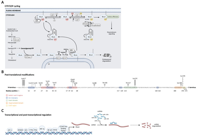
“RhoA is ubiquitylated by three different E3 ubiquitin ligase complexes: the SMAD-specific E3 ubiquitin protein ligase 1 (SMURF1), SKP1-CUL1-F-box FBXL19 (SCFFBXL19), and the BTB/POZ domain-containing adaptor for CUL3-mediated RhoA degradation (CUL3BACURD) ligase complex.”
§ RhoA Signaling in Neurodegenerative Diseases > 2. RhoA Structure and Regulation of RhoA Activity and Expression > 2.2. Post-Translational Modifications of RhoA
What the sources say
- Not checked — no full text of the sourcecites [66] Wang, 2003, Regulation of cell polarity and protrusion formation by targeting RhoA for degradation, Science source
- Not checked — no full text of the sourcecites [67] Ozdamar, 2005, Regulation of the polarity protein Par6 by TGFβ receptors controls epithelial cell plasticity, Science source
- Partly supportedcites [68] Chen, 2009, Cullin mediates degradation of RhoA through evolutionarily conserved BTB adaptors to control actin cytoskeleton structure and cell movement, Mol. Cell sourceExperimental — accuracy not yet measured
The Introduction reports that Cul3/BACURD complexes ubiquitinate RhoA and identifies Smurf1 as a previously known RhoA-targeting ubiquitin ligase. Our copy does not identify SCF–FBXL19 as a RhoA ubiquitin ligase, so it does not support the claim that all three listed complexes ubiquitinate RhoA. No page numbers are provided for these passages. (LLM, conf=0.88)
- Partly supportedcites [69] Ibeawuchi, 2015, Hypertension-causing Mutations in Cullin3 Protein Impair RhoA Protein Ubiquitination and Augment the Association with Substrate Adaptors, J. Biol. Chem. sourceExperimental — accuracy not yet measured
The “Defects in Cullin3 Decrease RhoA Ubiquitination” section reports RhoA ubiquitination by the Cul3/Bacurd1 complex (Fig. 4A). The passages found do not address RhoA ubiquitination by SMURF1 or SCF–FBXL19, so this source supports one of the three named complexes, not the full list. (LLM, conf=0.88)
- Supportedcites [62] Wei, 2013, A new mechanism of RhoA ubiquitination and degradation: Roles of SCF(FBXL19) E3 ligase and Erk2, Biochim. Biophys. Acta. source
The Introduction (section 1.1) says Smurf1 targets activated RhoA, Cullin-3/BACURD targets GDP-bound RhoA for ubiquitination and degradation, and SCF-FBXL19 targets both forms. The Discussion (section 4) also identifies Smurf1 and Cullin-3/BACURD as the other two known E3 ligases alongside SCF-FBXL19. (LLM, conf=0.94)
plaindirect- Asserted: passes — The authors put it forward as true.
- Checkable: passes — It can be checked against evidence.
- Load-bearing: unclear — It is unclear whether the paper’s conclusions depend on it.
- Based: passes — The paper gives a reason why it holds.
The cited sources support only part of this claim — read each source’s verdict below.
In the paper
“Indeed, emerging evidence points toward a role of aberrant RhoA signaling in neurodegenerative diseases such as Parkinson’s disease, Alzheimer’s disease, Huntington’s disease, and amyotrophic lateral sclerosis.”
- Also stated in: § 1. Introduction · hedged
“Dysregulation of Rho GTPases has been studied in relation to cancer progression for a number of years [3], but accumulating evidence supports a role in traumatic brain injury and neurodegenerative diseases as well [4,5,6,7].”
§ RhoA Signaling in Neurodegenerative Diseases > Abstract
What the sources say
- Not checked — no full text of the sourcecites [4] DeGeer, 2013, Rho GTPases in neurodegeneration diseases, Exp. Cell Res. source
- Partly supportedcites [5] Arrazola Sastre, A., Luque Montoro, M., Galvez-Martin, P., Lacerda, H.M., Lucia, A.M., Llavero, F., and Zugaza, J.L. (2020). Small GTPases of the Ras and Rho Families Switch on/off Signaling Pathways in Neurodegenerative Diseases. Int. J. Mol. Sci., 21. sourceExperimental — accuracy not yet measured
Section 3.1 (RhoA; page not supplied) links RhoA and RhoA/ROCK signaling to Alzheimer’s and Parkinson’s disease. The review’s Introduction says it focuses on those two diseases; the passages found do not support extending the claim to Huntington’s disease or amyotrophic lateral sclerosis. (LLM, conf=0.89)
- Partly supportedcites [6] Kimura, T., Horikoshi, Y., Kuriyagawa, C., and Niiyama, Y. (2021). Rho/ROCK Pathway and Noncoding RNAs: Implications in Ischemic Stroke and Spinal Cord Injury. Int. J. Mol. Sci., 22. sourceExperimental — accuracy not yet measured
The Introduction (page not provided) says excessive Rho/ROCK activity contributes to Parkinson’s and Alzheimer’s diseases. The cited passages do not extend that statement to Huntington’s disease or amyotrophic lateral sclerosis, so the full list is not supported. (LLM, conf=0.88)
- Partly supportedcites [7] Mulherkar, S., and Tolias, K.F. (2020). RhoA-ROCK Signaling as a Therapeutic Target in Traumatic Brain Injury. Cells, 9. sourceExperimental — accuracy not yet measured
Section 3 implicates dysregulated Rho GTPase signaling in Parkinson’s disease, Alzheimer’s disease, and amyotrophic lateral sclerosis. It does not name Huntington’s disease, and its disease-specific statement concerns Rho GTPases generally rather than RhoA specifically; the following RhoA discussion concerns CNS injury. (LLM, conf=0.86)
hedgedcorrelational- Asserted: passes — The authors put it forward as true.
- Checkable: passes — It can be checked against evidence.
- Load-bearing: passes — The paper’s conclusions depend on it.
- Based: passes — The paper gives a reason why it holds.
- Also stated in: § 1. Introduction · hedged
The cited sources that could be checked couldn’t confirm or dispute this claim.
In the paper
“The antiapoptotic effects of Rac are antagonized by RhoA/ROCK signaling through a number of downstream ROCK targets involved in both mitochondrial-dependent intrinsic apoptotic pathways and death receptor-dependent extrinsic apoptotic pathways.”
§ RhoA Signaling in Neurodegenerative Diseases > 3. Downstream Targets of RhoA > 3.2. Cell Death
What the sources say
- Could not verifycites [129] Lai, 2014, Excitotoxicity and stroke: Identifying novel targets for neuroprotection, Prog. Neurobio.l sourceExperimental — accuracy not yet measured
Section 3.1 discusses Akt-mediated neuronal survival and uses “rac” as an alternative name for Akt, not as the Rac in the claimed Rac–RhoA/ROCK interaction. Searches for RhoA, ROCK, and death receptor-dependent extrinsic apoptosis found no passage in our copy addressing that interaction or the claimed downstream pathways. The available passages therefore do not verify the claim. (LLM, conf=0.75)
plaindirect- Asserted: passes — The authors put it forward as true.
- Checkable: passes — It can be checked against evidence.
- Load-bearing: passes — The paper’s conclusions depend on it.
- Based: passes — The paper gives a reason why it holds.
- Could not verify
The cited sources support only part of this claim — read each source’s verdict below.
In the paper
“While SMURF1 ubiquitinates active GTP-bound RhoA on Lys6 and Lys7 targeting RhoA for proteasomal degradation [66,67], CUL3BACURD ubiquitinates only GDP-bound inactive RhoA [68,69].”
§ RhoA Signaling in Neurodegenerative Diseases > 2. RhoA Structure and Regulation of RhoA Activity and Expression > 2.2. Post-Translational Modifications of RhoA
What the sources say
- Partly supportedcites [68] Chen, 2009, Cullin mediates degradation of RhoA through evolutionarily conserved BTB adaptors to control actin cytoskeleton structure and cell movement, Mol. Cell sourceExperimental — accuracy not yet measured
In the passage headed “Targeting RhoA for Polyubiquitination by the Cul3/BACURD Ubiquitin Ligase Complex,” the paper reports robust ubiquitination of GDP-loaded RhoA. It describes GTP-loaded and Mg2+-free RhoA as much poorer substrates, not as substrates that cannot be ubiquitinated; therefore, “only” overstates the finding. (LLM, conf=0.90)
- Partly supportedcites [69] Ibeawuchi, 2015, Hypertension-causing Mutations in Cullin3 Protein Impair RhoA Protein Ubiquitination and Augment the Association with Substrate Adaptors, J. Biol. Chem. sourceExperimental — accuracy not yet measured
In “Defects in Cullin3 Decrease RhoA Ubiquitination” (page not available), the paper reports ubiquitination of RhoA-T19N, a mimic of inactive GDP-bound RhoA. It says Bacurd1 preferentially binds GDP-bound over GTP-bound RhoA, but does not establish that the Cul3–Bacurd1 complex ubiquitinates only GDP-bound RhoA. (LLM, conf=0.87)
plaindirect- Asserted: passes — The authors put it forward as true.
- Checkable: passes — It can be checked against evidence.
- Load-bearing: unclear — It is unclear whether the paper’s conclusions depend on it.
- Based: passes — The paper gives a reason why it holds.
- Partly supported
The cited sources support only part of this claim — read each source’s verdict below.
In the paper
“mDia is a formin molecule that catalyzes actin nucleation and polymerization through its interaction with the actin-binding protein profilin [121].”
§ RhoA Signaling in Neurodegenerative Diseases > 3. Downstream Targets of RhoA > 3.1. Cytoskeletal Dynamics
What the sources say
- Partly supportedcites [121] Watanabe, 1997, p140mDia, a mammalian homolog of Drosophila diaphanous, is a target protein for Rho small GTPase and is a ligand for profilin, EMBO J. sourceExperimental — accuracy not yet measured
In the section on activation-dependent clustering (page unavailable), the paper identifies p140mDia as a formin-related protein and proposes that it recruits profilin to sites where actin polymerization occurs. It reports enhanced actin filament assembly with p140mDia overexpression, but says whether p140mDia influences profilin-catalyzed polymerization remains to be investigated. It does not establish that mDia catalyzes actin nucleation or polymerization through profilin. (LLM, conf=0.90)
plaindirect- Asserted: passes — The authors put it forward as true.
- Checkable: passes — It can be checked against evidence.
- Load-bearing: unclear — It is unclear whether the paper’s conclusions depend on it.
- Based: passes — The paper gives a reason why it holds.
- Partly supported
- Probably not a substantial claim. 29
- It may state two separate claims as one. 73
At least one cited source doesn’t support this claim or points to a different paper, and none confirmed it — see the verdicts below.
In the paper
“In contrast, HIF-1α transcriptional activation of RHOA has consistently been reported across a number of different cell types, including breast cancer cells [87], cardiac myocytes [88], neuroblastoma cells [89], mesenchymal stem cells [90], and chondrocytes [91].”
§ RhoA Signaling in Neurodegenerative Diseases > 2. RhoA Structure and Regulation of RhoA Activity and Expression > 2.3. Transcriptional Regulation of RhoA
What the sources say
- Not checked — no full text of the sourcecites [87] Gilkes, 2014, Hypoxia-inducible factors mediate coordinated RhoA-ROCK1 expression and signaling in breast cancer cells, Proc. Natl. Acad. Sci. USA source
- Disputedcites [88] Falcon, 2015, Hypoxic induction of T-type Ca2+ channels in rat cardiac myocytes: Role of HIF-1α and RhoA/ROCK signalling, J. Physiol. sourceExperimental — accuracy not yet measured
For the cardiac-myocyte citation, the Results (page not specified) report that constitutively active HIF-1α did not affect RhoA activity. The authors instead conclude that RhoA acts upstream of HIF-1α, contrary to the attributed direction of regulation. Their hypoxia-induced increase in RhoA does not establish HIF-1α transcriptional activation of RHOA. (LLM, conf=0.90)
Checked by a reasoning model — may be wrong.
- Disputedcites [89] Yin, 2015, Upregulation of HIF-1α protects neuroblastoma cells from hypoxia-induced apoptosis in a RhoA-dependent manner, Mol. Med. Rep. sourceExperimental — accuracy not yet measured
The cited paper’s ‘Hypoxia induces RhoA in PC12 cells’ section reports that hypoxia increased RhoA expression in neuroblastoma cells. Its RhoA-knockdown experiment found that reducing RhoA reduced hypoxia-induced HIF-1α expression—the opposite regulatory direction from the claimed HIF-1α activation of RHOA. The paper does not establish HIF-1α transcriptional activation of RHOA. (LLM, conf=0.85)
Checked by a reasoning model — may be wrong.
boosteddirect- Asserted: passes — The authors put it forward as true.
- Checkable: passes — It can be checked against evidence.
- Load-bearing: fails — The paper’s conclusions may not depend on it.
- Based: passes — The paper gives a reason why it holds.
Probably not a substantial claim. The paper’s conclusions may not depend on it.
At least one cited source doesn’t support this claim or points to a different paper, and none confirmed it — see the verdicts below.
In the paper
“In 6-hydroxydopamine lesioned rats with dyskinesia, RhoA and ROCK were increased in the substantia nigra and striatum, and ROCK inhibition using Fasudil was found to reduce the development of L-DOPA-induced dyskinesia and to inhibit already established dyskinesia affecting the therapeutic effect of L-DOPA [179].”
§ RhoA Signaling in Neurodegenerative Diseases > 4. RhoA Signaling in Neurodegenerative Diseases > 4.1. Parkinson’s Disease
What the sources say
- Disputedcites [179] Labandeira, 2020, Rho kinase inhibitor fasudil reduces l-DOPA-induced dyskinesia in a rat model of Parkinson’s disease, Br. J. Pharmacol. sourceExperimental — accuracy not yet measured
The Discussion (section 4) supports the claim that fasudil reduced both the development of dyskinesia and already established dyskinesia. But it explicitly says this occurred without affecting the therapeutic effect of l-DOPA, contrary to the citing sentence’s statement that it affected that effect. The Results (section 3.2) likewise report that the motor benefit of l-DOPA was preserved. (LLM, conf=0.88)
Checked by a reasoning model — may be wrong.
plaindirect- Asserted: passes — The authors put it forward as true.
- Checkable: passes — It can be checked against evidence.
- Load-bearing: passes — The paper’s conclusions depend on it.
- Based: passes — The paper gives a reason why it holds.
It may state two separate claims as one.
- Disputed
Some cited sources support this claim and some don’t — read each source’s verdict below.
In the paper
“p75NTR was increased in HD mouse models and in postmortem samples from HD patients [203,204].”
§ RhoA Signaling in Neurodegenerative Diseases > 4. RhoA Signaling in Neurodegenerative Diseases > 4.3. Huntington’s Disease
What the sources say
- Supportedcites [203] Brito, 2014, Neurotrophin receptor p75(NTR) mediates Huntington’s disease-associated synaptic and memory dysfunction, J. Clin. Investig. source
The Results report increased hippocampal p75NTR in two HD mouse models and in postmortem samples from HD patients. The increase was hippocampal; the paper reports no corresponding cortical increase. (LLM, conf=0.94)
- Disputedcites [204] Plotkin, 2014, Impaired TrkB receptor signaling underlies corticostriatal dysfunction in Huntington’s disease, Neuron sourceExperimental — accuracy not yet measured
The Results section reports that p75NTR mRNA expression was not measurably different in the HD mouse neurons studied; the Discussion attributes increased p75NTR signaling to elevated PTEN, not increased p75NTR itself. The cited passages do not report p75NTR expression in postmortem samples from HD patients. (LLM, conf=0.88)
Checked by a reasoning model — may be wrong.
plaincorrelational- Asserted: passes — The authors put it forward as true.
- Checkable: passes — It can be checked against evidence.
- Load-bearing: passes — The paper’s conclusions depend on it.
- Based: passes — The paper gives a reason why it holds.
It may state two separate claims as one.
- Supported
Every source the paper cites for this claim was checked and supports it.
In the paper
“Inactivation is further facilitated by guanine nucleotide dissociation inhibitors (GDIs) that prevent dissociation of GDP from the GTPase and thus retain the inactive GDP-bound GTPases in the cytosol.”
§ RhoA Signaling in Neurodegenerative Diseases > 2. RhoA Structure and Regulation of RhoA Activity and Expression > 2.1. GDP/GTP Cycling
What the sources say
- Supportedcites [15] Boulter, 2011, The ‘invisible hand’: Regulation of RHO GTPases by RHOGDIs, Nat. Rev. Mol. Cell Biol. source
In ‘RHOGDIs as negative regulators of RHO GTPases’ (page not provided), the review says RHOGDIs inhibit GDP release and can extract RHO proteins from membranes, holding them in the cytosol. It describes this inhibitory role as acting mostly on inactive RHO GTPases. (LLM, conf=0.92)
plaindirect- Asserted: passes — The authors put it forward as true.
- Checkable: passes — It can be checked against evidence.
- Load-bearing: fails — The paper’s conclusions may not depend on it.
- Based: passes — The paper gives a reason why it holds.
Probably not a substantial claim. The paper’s conclusions may not depend on it. It may state two separate claims as one.
- Supported
Every source the paper cites for this claim was checked and supports it.
In the paper
“Conversely, GTPase-activating proteins (GAPs) enhance the relatively slow intrinsic GTPase activity of the small GTPases, resulting in GTP hydrolysis to GDP and inorganic phosphate (Pi) and inactivation of the GTPases [5,14].”
§ RhoA Signaling in Neurodegenerative Diseases > 2. RhoA Structure and Regulation of RhoA Activity and Expression > 2.1. GDP/GTP Cycling
What the sources say
- Supportedcites [5] Arrazola Sastre, A., Luque Montoro, M., Galvez-Martin, P., Lacerda, H.M., Lucia, A.M., Llavero, F., and Zugaza, J.L. (2020). Small GTPases of the Ras and Rho Families Switch on/off Signaling Pathways in Neurodegenerative Diseases. Int. J. Mol. Sci., 21. source
The Introduction (page not provided) states that GAPs activate the intrinsic GTPase activity of small GTPases, causing GTP hydrolysis to GDP and inorganic phosphate and inactivating the GTPase. (LLM, conf=0.94)
- Supportedcites [14] Toma-Fukai, S., and Shimizu, T. (2019). Structural Insights into the Regulation Mechanism of Small GTPases by GEFs. Molecules, 24. source
Section 1.2 says small GTPases have intrinsically low GTPase activity, GAPs stimulate GTP hydrolysis, and the GDP-bound form is generally inactive. This supports the cited account of GAP-driven hydrolysis and inactivation; inorganic phosphate is the implied product of that hydrolysis. (LLM, conf=0.91)
plaindirect- Asserted: passes — The authors put it forward as true.
- Checkable: passes — It can be checked against evidence.
- Load-bearing: fails — The paper’s conclusions may not depend on it.
- Based: passes — The paper gives a reason why it holds.
Probably not a substantial claim. The paper’s conclusions may not depend on it.
- Supported
Every source the paper cites for this claim was checked and supports it.
In the paper
“A novel RhoA effector protein FAM65A is also associated with the Golgi complex.”
§ RhoA Signaling in Neurodegenerative Diseases > 3. Downstream Targets of RhoA > 3.1. Cytoskeletal Dynamics
What the sources say
- Supportedcites [128] Mardakheh, 2016, RHO binding to FAM65A regulates Golgi reorientation during cell migration, J. Cell Sci. source
The Results section identifies FAM65A as a new RHO effector and shows that it binds active RHOA. The Discussion describes FAM65A as localising to the Golgi and cytoplasmic punctae. Page numbers are not provided in our copy. (LLM, conf=0.94)
plaincorrelational- Asserted: passes — The authors put it forward as true.
- Checkable: passes — It can be checked against evidence.
- Load-bearing: unclear — It is unclear whether the paper’s conclusions depend on it.
- Based: passes — The paper gives a reason why it holds.
It may state two separate claims as one.
- Supported
Every source the paper cites for this claim was checked and supports it.
In the paper
“Extracellular signal-related kinase (ERK) phosphorylates RhoA on Ser88 and Thr100, which has been reported to upregulate RhoA activity as evaluated by its binding capacity to the effector protein Rhotekin [61].”
§ RhoA Signaling in Neurodegenerative Diseases > 2. RhoA Structure and Regulation of RhoA Activity and Expression > 2.2. Post-Translational Modifications of RhoA
What the sources say
- Supportedcites [61] Tong, J., Li, L., Ballermann, B., and Wang, Z. (2016). Phosphorylation and Activation of RhoA by ERK in Response to Epidermal Growth Factor Stimulation. PLoS ONE, 11. source
The Results section on RhoA activity states that activation was assessed by binding to Rhotekin’s Rho-binding domain and that the S88E/T100E phosphomimetic bound more strongly than wild-type RhoA. The same section concludes that ERK phosphorylation of S88 and T100 enhances RhoA activity in response to EGF. (LLM, conf=0.94)
hedgeddirect- Asserted: passes — The authors put it forward as true.
- Checkable: passes — It can be checked against evidence.
- Load-bearing: fails — The paper’s conclusions may not depend on it.
- Based: passes — The paper gives a reason why it holds.
Probably not a substantial claim. The paper’s conclusions may not depend on it. It may state two separate claims as one.
- Supported
Every source the paper cites for this claim was checked and supports it.
In the paper
“Mammalian Ste20-like kinase 3 (Mst3) phosphorylates RhoA on Ser26 [60].”
§ RhoA Signaling in Neurodegenerative Diseases > 2. RhoA Structure and Regulation of RhoA Activity and Expression > 2.2. Post-Translational Modifications of RhoA
What the sources say
- Supportedcites [60] Tang, 2014, Cdk5-dependent Mst3 phosphorylation and activity regulate neuronal migration through RhoA inhibition, J. Neurosci. source
The section on negative regulation of RhoA by Mst3 phosphorylation states that an in vitro assay confirmed Mst3 phosphorylates RhoA at Ser26 (Fig. 6B). The Discussion also states this finding directly. (LLM, conf=0.95)
plaindirect- Asserted: passes — The authors put it forward as true.
- Checkable: passes — It can be checked against evidence.
- Load-bearing: fails — The paper’s conclusions may not depend on it.
- Based: passes — The paper gives a reason why it holds.
Probably not a substantial claim. The paper’s conclusions may not depend on it.
- Supported
The cited sources support only part of this claim — read each source’s verdict below.
In the paper
“While citron kinase is ubiquitously expressed in proliferating cells, the isoform citron N, which is also a scaffold protein for RhoA, is expressed only in postmitotic and fully differentiated neurons [124].”
§ RhoA Signaling in Neurodegenerative Diseases > 3. Downstream Targets of RhoA > 3.1. Cytoskeletal Dynamics
What the sources say
- Partly supportedcites [124] Bianchi, 2020, Of rings and spines: The multiple facets of Citron proteins in neural development, Small GTPases sourceExperimental — accuracy not yet measured
The “Expression and localization of CIT proteins” section says CIT-N expression begins when cells become post-mitotic and reaches its highest levels in fully differentiated neurons. It does not say CIT-N is expressed only in neurons or only after full differentiation: the same section describes CIT-N in astrocytes and in early post-mitotic neurons. (LLM, conf=0.90)
plain- Asserted: passes — The authors put it forward as true.
- Checkable: passes — It can be checked against evidence.
- Load-bearing: fails — The paper’s conclusions may not depend on it.
- Based: passes — The paper gives a reason why it holds.
Probably not a substantial claim. The paper’s conclusions may not depend on it.
- Partly supported
Every source the paper cites for this claim was checked and supports it.
In the paper
“As mDia activation leads to the formation of actin filaments and ROCK activates myosin to crosslink them, ROCK and mDia cooperate for actomyosin assembly and contractility [122].”
§ RhoA Signaling in Neurodegenerative Diseases > 3. Downstream Targets of RhoA > 3.1. Cytoskeletal Dynamics
What the sources say
- Supportedcites [122] Narumiya, 2009, Rho signaling, ROCK and mDia1, in transformation, metastasis and invasion, Cancer Metastasis Rev. source
The Abstract states that mDia produces actin filaments and ROCK activates myosin to cross-link them, inducing actomyosin bundles and contractility. The Fig. 2 caption in section 2 describes the same complementary mechanisms. Page numbers are not provided in our copy. (LLM, conf=0.95)
plaindirect- Asserted: passes — The authors put it forward as true.
- Checkable: passes — It can be checked against evidence.
- Load-bearing: unclear — It is unclear whether the paper’s conclusions depend on it.
- Based: passes — The paper gives a reason why it holds.
It may state two separate claims as one.
- Supported
The cited sources support only part of this claim — read each source’s verdict below.
In the paper
“The prenylation step takes place in the cytosol and is followed by proteolytic removal of the last three C-terminal (AAX) residues by the protease RAS-converting enzyme 1 (RCE1) and methylation of the prenylated Cys190 by isoprenylcysteine carboxyl methyltransferase (ICMT) at the cytoplasmic surface of the endoplasmic reticulum (ER) [47,48].”
§ RhoA Signaling in Neurodegenerative Diseases > 2. RhoA Structure and Regulation of RhoA Activity and Expression > 2.2. Post-Translational Modifications of RhoA
What the sources say
- Partly supportedcites [47] Roberts, 2008, Rho Family GTPase modification and dependence on CAAX motif-signaled posttranslational modification, J. Biol. Chem. sourceExperimental — accuracy not yet measured
The paper’s introductory description of CAAX processing says that prenylation is followed by Rce1 removal of the three AAX residues and Icmt methylation of the prenylated cysteine; its Rce1/Icmt processing section discusses these steps in relation to RhoA. The passages do not identify that cysteine as Cys190 or place the reactions at the cytoplasmic surface of the ER, so those specifics are not supported by this copy. (LLM, conf=0.84)
- Not checked — no full text of the sourcecites [48] Casey, 2005, Post-prenylation-processing enzymes as new targets in oncogenesis, Nat. Rev. Cancer source
plaindirect- Asserted: passes — The authors put it forward as true.
- Checkable: passes — It can be checked against evidence.
- Load-bearing: fails — The paper’s conclusions may not depend on it.
- Based: passes — The paper gives a reason why it holds.
Probably not a substantial claim. The paper’s conclusions may not depend on it. It may state two separate claims as one.
- Partly supported
Every source the paper cites for this claim was checked and supports it.
In the paper
“In contrast to the vast number of known RhoGEFs and RhoGAPs, only three RhoGDIs have been described in mammals [15].”
§ RhoA Signaling in Neurodegenerative Diseases > 2. RhoA Structure and Regulation of RhoA Activity and Expression > 2.1. GDP/GTP Cycling
What the sources say
- Supportedcites [15] Boulter, 2011, The ‘invisible hand’: Regulation of RHO GTPases by RHOGDIs, Nat. Rev. Mol. Cell Biol. source
The Introduction states that mammals have only three genes encoding RHOGDIs and names RHOGDI1, RHOGDI2 and RHOGDI3. The section on regulation of RHOGDI–RHO GTPase interactions also refers to “the three mammalian RHOGDIs.” (LLM, conf=0.95)
plain- Asserted: passes — The authors put it forward as true.
- Checkable: passes — It can be checked against evidence.
- Load-bearing: fails — The paper’s conclusions may not depend on it.
- Based: passes — The paper gives a reason why it holds.
Probably not a substantial claim. The paper’s conclusions may not depend on it.
- Supported
The cited sources support only part of this claim — read each source’s verdict below.
In the paper
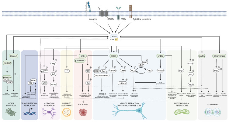
“ROCK also phosphorylates the myosin light chain (MLC) subunit of the actin-based motor protein myosin IIb, leading to actomyosin assembly and contractility. This occurs both through a direct interaction and indirectly by ROCK-mediated inhibition of myosin light chain phosphatase (MLCP) through phosphorylation of its myosin phosphatase-targeting subunit 1 (MYPT1) [7].”
§ RhoA Signaling in Neurodegenerative Diseases > 3. Downstream Targets of RhoA > 3.1. Cytoskeletal Dynamics
What the sources say
- Partly supportedcites [7] Mulherkar, S., and Tolias, K.F. (2020). RhoA-ROCK Signaling as a Therapeutic Target in Traumatic Brain Injury. Cells, 9. sourceExperimental — accuracy not yet measured
Section 2, “Rho GTPase Signaling” (page not provided), says ROCK increases MLC phosphorylation and actomyosin contractile force, both directly and through phosphorylation and inhibition of myosin phosphatase. It identifies the motor protein as myosin II, not specifically myosin IIb, and does not identify MYPT1 as the phosphorylated phosphatase subunit or explicitly describe actomyosin assembly. (LLM, conf=0.88)
plaindirect- Asserted: passes — The authors put it forward as true.
- Checkable: passes — It can be checked against evidence.
- Load-bearing: unclear — It is unclear whether the paper’s conclusions depend on it.
- Based: passes — The paper gives a reason why it holds.
It may state two separate claims as one.
- Partly supported
Every source the paper cites for this claim was checked and supports it.
In the paper
“Interestingly, the RhoA scaffold effector protein Rhotekin binds GTP-bound RhoA and inhibits both the intrinsic and GAP-enhanced GTPase activity of RhoA; this is why it is widely used in pull-down assays for activated RhoA [31].”
§ RhoA Signaling in Neurodegenerative Diseases > 2. RhoA Structure and Regulation of RhoA Activity and Expression > 2.1. GDP/GTP Cycling
What the sources say
- Supportedcites [31] Ito, H., Morishita, R., and Nagata, K.I. (2018). Functions of Rhotekin, an Effector of Rho GTPase, and Its Binding Partners in Mammals. Int. J. Mol. Sci., 19. source
The Introduction (page not available in our copy) states that Rhotekin’s Rho-binding domain selectively interacts with GTP-bound active Rho and inhibits both its intrinsic and GAP-enhanced GTPase activity. It also says the domain has been used to monitor activated Rho, supporting the citing sentence’s assay context. (LLM, conf=0.90)
plaindirect- Asserted: passes — The authors put it forward as true.
- Checkable: passes — It can be checked against evidence.
- Load-bearing: fails — The paper’s conclusions may not depend on it.
- Based: passes — The paper gives a reason why it holds.
Probably not a substantial claim. The paper’s conclusions may not depend on it. It may state two separate claims as one.
- Supported
The cited sources support only part of this claim — read each source’s verdict below.
In the paper
“Dysregulation of Rho GTPases has been studied in relation to cancer progression for a number of years [3], but accumulating evidence supports a role in traumatic brain injury and neurodegenerative diseases as well [4,5,6,7].”
§ RhoA Signaling in Neurodegenerative Diseases > 1. Introduction
What the sources say
- Not checked — no full text of the sourcecites [4] DeGeer, 2013, Rho GTPases in neurodegeneration diseases, Exp. Cell Res. source
- Partly supportedcites [5] Arrazola Sastre, A., Luque Montoro, M., Galvez-Martin, P., Lacerda, H.M., Lucia, A.M., Llavero, F., and Zugaza, J.L. (2020). Small GTPases of the Ras and Rho Families Switch on/off Signaling Pathways in Neurodegenerative Diseases. Int. J. Mol. Sci., 21. sourceExperimental — accuracy not yet measured
Section 3, “Small GTPases of the Rho Family” (page not provided), says Rho GTPases play an important role in neurodegenerative processes, including those associated with Alzheimer’s and Parkinson’s diseases. Our copy does not address traumatic brain injury, so it supports only the neurodegenerative-disease part of the claim. The cancer-progression clause is attached to a different citation [3]. (LLM, conf=0.86)
- Partly supportedcites [6] Kimura, T., Horikoshi, Y., Kuriyagawa, C., and Niiyama, Y. (2021). Rho/ROCK Pathway and Noncoding RNAs: Implications in Ischemic Stroke and Spinal Cord Injury. Int. J. Mol. Sci., 22. sourceExperimental — accuracy not yet measured
The Introduction links excessive Rho/ROCK activity to Parkinson’s and Alzheimer’s diseases, supporting the neurodegenerative-disease part of the claim. The paper discusses ischemic stroke and spinal cord injury, but the passages found do not address traumatic brain injury. The cancer clause is cited to a different source [3], so it is not part of this verdict. (LLM, conf=0.86)
- Supportedcites [7] Mulherkar, S., and Tolias, K.F. (2020). RhoA-ROCK Signaling as a Therapeutic Target in Traumatic Brain Injury. Cells, 9. source
Section 3, “Functions of Rho GTPases in the Central Nervous System” (page not provided), links dysregulated Rho GTPase signaling to neurodegenerative disorders and describes RhoA activation after traumatic brain injury. The cancer-progression clause is attached to a different citation and is not attributed to this source. (LLM, conf=0.94)
plaincorrelational- Asserted: passes — The authors put it forward as true.
- Checkable: passes — It can be checked against evidence.
- Load-bearing: unclear — It is unclear whether the paper’s conclusions depend on it.
- Based: passes — The paper gives a reason why it holds.
It may state two separate claims as one.
The cited sources support only part of this claim — read each source’s verdict below.
In the paper
“This is essential for both membrane trafficking from the Golgi and the orientation of the Golgi toward the leading edge of the cell [128].”
§ RhoA Signaling in Neurodegenerative Diseases > 3. Downstream Targets of RhoA > 3.1. Cytoskeletal Dynamics
What the sources say
- Partly supportedcites [128] Mardakheh, 2016, RHO binding to FAM65A regulates Golgi reorientation during cell migration, J. Cell Sci. sourceExperimental — accuracy not yet measured
The Introduction and Discussion state that RHO-dependent relocation of the FAM65A–CCM3–MST complex is crucial for Golgi reorientation toward the leading edge. The Introduction discusses secretory traffic from the Golgi, but the paper does not establish that this complex’s relocation is essential for membrane trafficking from the Golgi. Page numbers are unavailable in our copy. (LLM, conf=0.86)
plaindirect- Asserted: passes — The authors put it forward as true.
- Checkable: passes — It can be checked against evidence.
- Load-bearing: unclear — It is unclear whether the paper’s conclusions depend on it.
- Based: passes — The paper gives a reason why it holds.
It may state two separate claims as one.
- Partly supported
Every source the paper cites for this claim was checked and supports it.
In the paper
“In colon cancer cells, the transcription factors SMAD family member 4 (SMAD4) and specific protein 1 (SP1) activated RHOA expression, while c-Myc was reported to interfere with the binding of SP1 to the RHOA promoter, leading to downregulation of RHOA expression [85].”
§ RhoA Signaling in Neurodegenerative Diseases > 2. RhoA Structure and Regulation of RhoA Activity and Expression > 2.3. Transcriptional Regulation of RhoA
What the sources say
- Supportedcites [85] Dopeso, 2018, Mechanisms of inactivation of the tumour suppressor gene RHOA in colorectal cancer, Br. J. Cancer source
The Results section on TGF-β signalling reports that SMAD4 inactivation reduced RHOA mRNA in colon cancer cells and concludes that RHOA is a transcriptional target of SMAD4. The Results section on c-MYC and SP1 shows that SP1 sustains RHOA expression and that reduced c-MYC increases SP1 binding to the RHOA promoter; the Discussion explicitly states that c-MYC interferes with that binding and downregulates RHOA. (LLM, conf=0.94)
plaindirect- Asserted: passes — The authors put it forward as true.
- Checkable: passes — It can be checked against evidence.
- Load-bearing: unclear — It is unclear whether the paper’s conclusions depend on it.
- Based: passes — The paper gives a reason why it holds.
It may state two separate claims as one.
- Supported
The cited sources support only part of this claim — read each source’s verdict below.
In the paper
“In response to extracellular stimuli emanating from cell surface receptors, guanine nucleotide exchange factors (GEFs) catalyze the exchange of GDP for GTP, leading to the conformational changes in the switch I and switch II regions required for activation of the small GTPases.”
§ RhoA Signaling in Neurodegenerative Diseases > 2. RhoA Structure and Regulation of RhoA Activity and Expression > 2.1. GDP/GTP Cycling
What the sources say
- Partly supportedcites [5] Arrazola Sastre, A., Luque Montoro, M., Galvez-Martin, P., Lacerda, H.M., Lucia, A.M., Llavero, F., and Zugaza, J.L. (2020). Small GTPases of the Ras and Rho Families Switch on/off Signaling Pathways in Neurodegenerative Diseases. Int. J. Mol. Sci., 21. sourceExperimental — accuracy not yet measured
The Introduction states that GEFs promote GDP release and GTP binding, shifting small GTPases from an inactive to an active state. It also places small GTPases in signaling responses to extracellular stimuli, but does not identify conformational changes in switch I and switch II as the mechanism of activation. (LLM, conf=0.87)
- Supportedcites [14] Toma-Fukai, S., and Shimizu, T. (2019). Structural Insights into the Regulation Mechanism of Small GTPases by GEFs. Molecules, 24. source
Section 2.2 describes GEF-stimulated nucleotide exchange that produces GTP-bound small GTPases. Section 1.4 states that nucleotide exchange changes switch I and switch II, with the GTP-bound form being active. These passages support the attributed claim. (LLM, conf=0.93)
plaindirect- Asserted: passes — The authors put it forward as true.
- Checkable: passes — It can be checked against evidence.
- Load-bearing: fails — The paper’s conclusions may not depend on it.
- Based: passes — The paper gives a reason why it holds.
Probably not a substantial claim. The paper’s conclusions may not depend on it.
- Partly supported
The cited sources support only part of this claim — read each source’s verdict below.
In the paper
“Several transcription factors have been shown to regulate RHOA expression, including c-Myc, Max, hypoxia-inducible factor 1 alpha (HIF-1α), nuclear factor-κB (NF-κB), and signal transducer and activator of transcription 6 (STAT6), together with several reported miRNAs (Figure 2C) [82,83].”
§ RhoA Signaling in Neurodegenerative Diseases > 2. RhoA Structure and Regulation of RhoA Activity and Expression > 2.3. Transcriptional Regulation of RhoA
What the sources say
- Partly supportedcites [82] Kim, 2018, Regulation of RhoA GTPase and various transcription factors in the RhoA pathway, J. Cell. Physiol. sourceExperimental — accuracy not yet measured
The Abstract and sections 2.3–2.4 support regulation of RhoA expression by c-Myc, HIF-1α, NF-κB, STAT6, and several miRNAs. The passages on transcriptional regulation describe a Myc–Skp2–Miz1–p300 complex, but do not identify Max as a regulator of RhoA expression. (LLM, conf=0.88)
- Partly supportedcites [83] Nomikou, 2018, Transcriptional and post-transcriptional regulation of the genes encoding the small GTPases RhoA, RhoB, and RhoC: Implications for the pathogenesis of human diseases, Cell. Mol. Life Sci. sourceExperimental — accuracy not yet measured
The sections on transcriptional regulation of RhoA describe Myc, NF-κB, and STAT6 as regulators, and the miRNA section identifies several miRNAs that regulate RhoA expression (page numbers unavailable). The copy does not establish Max or HIF-1α as RhoA regulators; its hypoxia passage instead concerns HIF-3α regulation of RhoC. (LLM, conf=0.87)
plaindirect- Asserted: passes — The authors put it forward as true.
- Checkable: passes — It can be checked against evidence.
- Load-bearing: fails — The paper’s conclusions may not depend on it.
- Based: passes — The paper gives a reason why it holds.
Probably not a substantial claim. The paper’s conclusions may not depend on it.
- Partly supported
The cited sources support only part of this claim — read each source’s verdict below.
In the paper
“RhoA and many of its downstream effector proteins are highly expressed in the nervous system, implying an important role for RhoA signaling in neurons and glial cells.”
- Also stated in: § 3. Downstream Targets of RhoA · plain
“RhoA and many of its effector proteins are highly expressed in the nervous system, implying an important role for RhoA signaling in neurons and glial cells [1,117].”
§ RhoA Signaling in Neurodegenerative Diseases > Abstract
What the sources say
- Partly supportedcites [1] Stankiewicz, 2014, Rho family GTPases: Key players in neuronal development, neuronal survival, and neurodegeneration, Front. Cell. Neurosci. sourceExperimental — accuracy not yet measured
The “Rho in Neuronal Development” section (page unavailable) describes roles for RhoA in neurons and radial glia, supporting the inference that its signaling matters in both. Neither that section nor the passages retrieved on expression state that RhoA and many downstream effectors are highly expressed throughout the nervous system; the expression findings concern specific injury conditions. (LLM, conf=0.84)
- Partly supportedcites [117] Guiler, 2021, Pharmacological Modulators of Small GTPases of Rho Family in Neurodegenerative Diseases, Front. Cell. Neurosci. sourceExperimental — accuracy not yet measured
The Introduction’s “Rho GTPase Subfamily” section describes RhoA’s effects on neuronal processes and reports that loss of RhoA in microglia led to synapse loss in mice. The “Small Molecules” section says the RhoA effector ROCK2 is mainly expressed in the brain and spinal cord. Neither passage establishes that RhoA and many of its downstream effectors are highly expressed throughout the nervous system; no page numbers are available in our copy. (LLM, conf=0.86)
plaincorrelational- Asserted: passes — The authors put it forward as true.
- Checkable: passes — It can be checked against evidence.
- Load-bearing: passes — The paper’s conclusions depend on it.
- Based: passes — The paper gives a reason why it holds.
It may state two separate claims as one.
- Also stated in: § 3. Downstream Targets of RhoA · plain
The cited sources support only part of this claim — read each source’s verdict below.
In the paper
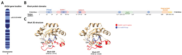
“The most well-studied phosphorylation event of RhoA is protein kinase A (PKA) and cyclic GMP-dependent protein kinase (PKG)-mediated phosphorylation on Ser188, close to the prenylated Cys190 in the C-terminus [49,50,56]. This phosphorylation inhibits RhoA activity by favoring interaction with RhoGDI, leading to its extraction from the plasma membrane [50,51,55], and protects GTP-bound RhoA from ub”
§ RhoA Signaling in Neurodegenerative Diseases > 2. RhoA Structure and Regulation of RhoA Activity and Expression > 2.2. Post-Translational Modifications of RhoA
What the sources say
- Partly supportedcites [49] Lang, 1996, Protein kinase A phosphorylation of RhoA mediates the morphological and functional effects of cyclic AMP in cytotoxic lymphocytes, EMBO J. sourceExperimental — accuracy not yet measured
The Results passages show that PKA phosphorylates RhoA at Ser188, increases RhoGDI binding to phosphorylated RhoA-GTP, and enables GDI to extract RhoA from membranes. The Discussion says this may terminate RhoA signalling, but explicitly states that the fate of phosphorylated RhoA-GTP was not investigated. Our copy does not establish PKG-mediated phosphorylation or protection from ubiquitin-mediated proteasomal degradation. Page numbers are unavailable in the indexed passages. (LLM, conf=0.89)
- Not checked — no full text of the sourcecites [50] Ellerbroek, 2003, Serine phosphorylation negatively regulates RhoA in vivo, J. Biol. Chem. source
- Partly supportedcites [56] Sauzeau, 2000, Cyclic GMP-dependent protein kinase signaling pathway inhibits RhoA-induced Ca2+ sensitization of contraction in vascular smooth muscle, J. Biol. Chem. sourceExperimental — accuracy not yet measured
The ‘cGK Phosphorylates RhoA in Vitro’ passage identifies Ser188 as the PKG phosphorylation site. The Discussion reports that PKG phosphorylation moves activated RhoA from the membrane to the cytosol and recounts earlier work linking PKA phosphorylation to increased RhoGDI interaction. Our copy does not address protection of GTP-bound RhoA from ubiquitin-mediated proteasomal degradation. (LLM, conf=0.87)
- Partly supportedcites [51] Tkachenko, 2011, Protein kinase A governs a RhoA-RhoGDI protrusion-retraction pacemaker in migrating cells, Nat. Cell Biol. sourceExperimental — accuracy not yet measured
The Results section ('PKA Phosphorylation Regulates the Dynamics of RhoA Activation'; page unavailable) states that PKA phosphorylation of RhoA at Ser188 increases RhoGDI binding and reduces RhoA activity in part through sequestration from the membrane. The passages found do not establish PKG-mediated phosphorylation or protection of GTP-bound RhoA from ubiquitin-mediated proteasomal degradation. (LLM, conf=0.87)
- Not checked — no full text of the sourcecites [55] Sawada, 2001, cGMP-dependent protein kinase phosphorylates and inactivates RhoA, Biochem. Biophys. Res. Commun. source
- Not checked — no full text of the sourcecites [52] Sauzeau, 2005, Phosphorylation of serine 188 protects RhoA from ubiquitin/proteasome-mediated degradation in vascular smooth muscle cells, Circ. Res. source
boosteddirect- Asserted: passes — The authors put it forward as true.
- Checkable: passes — It can be checked against evidence.
- Load-bearing: unclear — It is unclear whether the paper’s conclusions depend on it.
- Based: passes — The paper gives a reason why it holds.
It may state two separate claims as one.
- Partly supported
Every source the paper cites for this claim was checked and supports it.
In the paper
“Another RhoA-targeting miRNA linked to dendrite formation is miR-340, which has been shown to promote dendrite extension through suppressing RhoA expression in melanocytes [113].”
§ RhoA Signaling in Neurodegenerative Diseases > 2. RhoA Structure and Regulation of RhoA Activity and Expression > 2.4. Post-Transcriptional Regulation of RhoA by miRNAs
What the sources say
- Supportedcites [113] Jian, 2014, MicroRNA 340 is involved in UVB-induced dendrite formation through the regulation of RhoA expression in melanocytes, Mol. Cell. Biol. source
The Results report that miR-340 mimics significantly elongated melanocyte dendrites and negatively regulated RhoA expression. The paper also reports direct targeting of RhoA by miR-340, supporting the cited mechanism. (LLM, conf=0.94)
plaindirect- Asserted: passes — The authors put it forward as true.
- Checkable: passes — It can be checked against evidence.
- Load-bearing: fails — The paper’s conclusions may not depend on it.
- Based: passes — The paper gives a reason why it holds.
Probably not a substantial claim. The paper’s conclusions may not depend on it.
- Supported
Every source the paper cites for this claim was checked and supports it.
In the paper
“Another example is SmgGDS, which specifically activates RhoA and RhoC but not RhoB [22].”
§ RhoA Signaling in Neurodegenerative Diseases > 2. RhoA Structure and Regulation of RhoA Activity and Expression > 2.1. GDP/GTP Cycling
What the sources say
- Supportedcites [22] Hamel, 2011, SmgGDS is a guanine nucleotide exchange factor that specifically activates RhoA and RhoC, J. Biol. Chem. source
The Results section, “SmgGDS Specifically Activates RhoA and RhoC,” reports nucleotide exchange for RhoA and RhoC but not other tested GTPases. The cell-assay passage likewise states that SmgGDS-558 activated RhoA and RhoC, but not RhoB. (LLM, conf=0.95)
plaindirect- Asserted: passes — The authors put it forward as true.
- Checkable: passes — It can be checked against evidence.
- Load-bearing: fails — The paper’s conclusions may not depend on it.
- Based: passes — The paper gives a reason why it holds.
Probably not a substantial claim. The paper’s conclusions may not depend on it.
- Supported
The cited sources support only part of this claim — read each source’s verdict below.
In the paper
“Many neuronal studies support the general model that Rac and Cdc42 stimulate these processes, while RhoA inhibits them and causes axonal and dendritic retraction and spine and synapse loss.”
§ RhoA Signaling in Neurodegenerative Diseases > 3. Downstream Targets of RhoA > 3.1. Cytoskeletal Dynamics
What the sources say
- Partly supportedcites [1] Stankiewicz, 2014, Rho family GTPases: Key players in neuronal development, neuronal survival, and neurodegeneration, Front. Cell. Neurosci. sourceExperimental — accuracy not yet measured
Split into 2 atoms — 1 confirmed, 1 partially supported, 0 disputed, 0 misattributed, 0 unverifiable. Expand the claim to see per-atom verdicts.
- Partly supportedcites [7] Mulherkar, S., and Tolias, K.F. (2020). RhoA-ROCK Signaling as a Therapeutic Target in Traumatic Brain Injury. Cells, 9. sourceExperimental — accuracy not yet measured
Split into 2 atoms — 1 confirmed, 1 partially supported, 0 disputed, 0 misattributed, 0 unverifiable. Expand the claim to see per-atom verdicts.
plaindirect- Asserted: passes — The authors put it forward as true.
- Checkable: passes — It can be checked against evidence.
- Load-bearing: unclear — It is unclear whether the paper’s conclusions depend on it.
- Based: passes — The paper gives a reason why it holds.
It may state two separate claims as one.
- Partly supported
The cited sources support only part of this claim — read each source’s verdict below.
In the paper
“Upon binding to RhoA, the FAM65A/CCM3/MST complex relocates away from the Golgi complex to the cytoplasmic membrane, relieving the inhibitory effect of MST4 on Golgi reorientation.”
§ RhoA Signaling in Neurodegenerative Diseases > 3. Downstream Targets of RhoA > 3.1. Cytoskeletal Dynamics
What the sources say
- Partly supportedcites [128] Mardakheh, 2016, RHO binding to FAM65A regulates Golgi reorientation during cell migration, J. Cell Sci. sourceExperimental — accuracy not yet measured
The Discussion (page unavailable) proposes that RHO–GTP binding to FAM65A relocates the FAM65A–CCM3–MST complex from the Golgi, relieving MST-mediated inhibition of Golgi reorientation. The Results identify the destination as cytoplasmic punctae, likely vesicular compartments, not the ‘cytoplasmic membrane’ specified in the claim. (LLM, conf=0.87)
plaindirect- Asserted: passes — The authors put it forward as true.
- Checkable: passes — It can be checked against evidence.
- Load-bearing: unclear — It is unclear whether the paper’s conclusions depend on it.
- Based: passes — The paper gives a reason why it holds.
It may state two separate claims as one.
- Partly supported
Every source the paper cites for this claim was checked and supports it.
In the paper
“Activated RhoA binds FAM65A, which in turn binds cerebral cavernous malformation-3 protein (CCM3) and the interacting mammalian STE20-like protein kinase (MST).”
§ RhoA Signaling in Neurodegenerative Diseases > 3. Downstream Targets of RhoA > 3.1. Cytoskeletal Dynamics
What the sources say
- Supportedcites [128] Mardakheh, 2016, RHO binding to FAM65A regulates Golgi reorientation during cell migration, J. Cell Sci. source
The Introduction states that FAM65A associates with GTP-bound RHOA and with CCM3, MST4, and MST3. The Results further describe CCM3 as an adaptor connecting FAM65A to MST proteins, supporting the claimed binding pathway without requiring direct FAM65A–MST binding. (LLM, conf=0.92)
plaincorrelational- Asserted: passes — The authors put it forward as true.
- Checkable: passes — It can be checked against evidence.
- Load-bearing: unclear — It is unclear whether the paper’s conclusions depend on it.
- Based: passes — The paper gives a reason why it holds.
It may state two separate claims as one.
- Supported
The cited sources support only part of this claim — read each source’s verdict below.
In the paper
“For example, p190-RhoGAP inactivates RhoA and not Cdc42 and Rac1 [27], but it does not differentiate among RhoA, RhoB, and RhoC [28].”
§ RhoA Signaling in Neurodegenerative Diseases > 2. RhoA Structure and Regulation of RhoA Activity and Expression > 2.1. GDP/GTP Cycling
What the sources say
- Partly supportedcites [27] Ridley, 1993, rho family GTPase activating proteins p190, bcr and rhoGAP show distinct specificities in vitro and in vivo, EMBO J. sourceExperimental — accuracy not yet measured
The in vitro results report that p190GAP is ninefold more effective against rho than against rac or G25K, supporting a preference for rho but not the claim that it does not act on Rac1. The same section says p190GAP has some capacity to down-regulate all three tested proteins. The RhoA/RhoB/RhoC comparison is attached to a different citation [28], so it is not assessed here. (LLM, conf=0.91)
- Partly supportedcites [28] Thomas, 2011, Src and caveolin-1 reciprocally regulate metastasis via a common downstream signaling pathway in bladder cancer, Cancer Res. sourceExperimental — accuracy not yet measured
Citation [28] is attached to the claim that p190-RhoGAP does not differentiate among RhoA, RhoB, and RhoC. The Results section reports that p190RhoGAP knockdown increased activation of both RhoA and RhoC in RT4 cells, but it does not assess RhoB. The separate claim about Cdc42 and Rac1 is attached to [27], not this source. No page number is available in our copy. (LLM, conf=0.84)
plaindirect- Asserted: passes — The authors put it forward as true.
- Checkable: passes — It can be checked against evidence.
- Load-bearing: fails — The paper’s conclusions may not depend on it.
- Based: passes — The paper gives a reason why it holds.
Probably not a substantial claim. The paper’s conclusions may not depend on it. It may state two separate claims as one.
- Partly supported
The sources with a verdict support this claim; the rest weren’t checked yet, have no full text we could read, or their verdicts aren’t rated yet.
In the paper
“Citron N is enriched in dendritic spines and is associated with the endomembrane system, in particular the Golgi complex, where it maintains Golgi architecture by modulating local actin polymerization together with ROCK [126,127].”
§ RhoA Signaling in Neurodegenerative Diseases > 3. Downstream Targets of RhoA > 3.1. Cytoskeletal Dynamics
What the sources say
- Not checked — no full text of the sourcecites [126] Camera, 2003, Citron-N is a neuronal Rho-associated protein involved in Golgi organization through actin cytoskeleton regulation, Nat. Cell Biol. source
- Supportedcites [127] Camera, 2008, The RhoA-associated protein Citron-N controls dendritic spine maintenance by interacting with spine-associated Golgi compartments, EMBO Rep. source
In “Cit-N associates with Golgi at spines,” the paper reports that Cit-N accumulates at dendritic spines and is abundantly associated with TGN38-positive membranes. It also reports colocalization with Golgi markers, supporting the attributed association with the Golgi complex. (LLM, conf=0.94)
plaincorrelational- Asserted: passes — The authors put it forward as true.
- Checkable: passes — It can be checked against evidence.
- Load-bearing: unclear — It is unclear whether the paper’s conclusions depend on it.
- Based: passes — The paper gives a reason why it holds.
It may state two separate claims as one.
Every source the paper cites for this claim was checked and supports it.
In the paper
“One example is XPLN, which shows activity toward RhoA and RhoB but not RhoC due to an amino acid difference (Ile43 vs. Val43) at the N-terminus of the Rho isoforms [20,21].”
§ RhoA Signaling in Neurodegenerative Diseases > 2. RhoA Structure and Regulation of RhoA Activity and Expression > 2.1. GDP/GTP Cycling
What the sources say
- Supportedcites [20] Arthur, 2002, XPLN, a guanine nucleotide exchange factor for RhoA and RhoB, but not RhoC, J. Biol. Chem. source
The Results section reports that XPLN stimulates exchange on RhoA and RhoB but not RhoC, and that changing RhoC’s isoleucine 43 to valine makes it a substrate. The Discussion identifies valine 43 in RhoA and RhoB versus isoleucine 43 in RhoC as an important determinant, while noting that other residues also contribute. (LLM, conf=0.93)
- Supportedcites [21] Sloan, 2012, Divergence of Rho residue 43 impacts GEF activity, Small GTPases source
The Introduction reports that XPLN catalyzed nucleotide exchange for RhoA and RhoB in vitro but showed little activity toward RhoC. The Introduction and Results identify Val43 in RhoA/B versus Ile43 in RhoC and show that swapping residue 43 changes XPLN activity, supporting the stated explanation. (LLM, conf=0.90)
plaindirect- Asserted: passes — The authors put it forward as true.
- Checkable: passes — It can be checked against evidence.
- Load-bearing: fails — The paper’s conclusions may not depend on it.
- Based: passes — The paper gives a reason why it holds.
Probably not a substantial claim. The paper’s conclusions may not depend on it.
- Supported
The cited sources support only part of this claim — read each source’s verdict below.
In the paper
“This step is catalyzed by the geranylgeranyltransferase enzyme I (GGTase-I), which transfers the geranylgeranyl moiety from geranylgeranyl pyrophosphate, produced as a product of the mevalonate pathway involved in cholesterol synthesis, to RhoA.”
§ RhoA Signaling in Neurodegenerative Diseases > 2. RhoA Structure and Regulation of RhoA Activity and Expression > 2.2. Post-Translational Modifications of RhoA
What the sources say
- Partly supportedcites [47] Roberts, 2008, Rho Family GTPase modification and dependence on CAAX motif-signaled posttranslational modification, J. Biol. Chem. sourceExperimental — accuracy not yet measured
The paper identifies RhoA as a GGTase-I substrate in the section on prenyltransferase specificity. It also describes GGTase-I adding a geranylgeranyl group and uses GGPP in its GGTase-I assays. The passages do not state that GGPP is produced by the mevalonate pathway involved in cholesterol synthesis; that part of the attributed claim is unsupported. (LLM, conf=0.86)
- Not checked — no full text of the sourcecites [48] Casey, 2005, Post-prenylation-processing enzymes as new targets in oncogenesis, Nat. Rev. Cancer source
plaindirect- Asserted: passes — The authors put it forward as true.
- Checkable: passes — It can be checked against evidence.
- Load-bearing: fails — The paper’s conclusions may not depend on it.
- Based: passes — The paper gives a reason why it holds.
Probably not a substantial claim. The paper’s conclusions may not depend on it.
- Partly supported
Every source the paper cites for this claim was checked and supports it.
In the paper
“The reduced miR-185 expression caused deficits in dendritic complexity and spine development in hippocampal neurons [108], possibly mediated by increased RhoA expression as the two validated miR-185 targets, RhoA and Cdc42 [109], have altered expression levels in schizophrenia [110,111,112].”
§ RhoA Signaling in Neurodegenerative Diseases > 2. RhoA Structure and Regulation of RhoA Activity and Expression > 2.4. Post-Transcriptional Regulation of RhoA by miRNAs
What the sources say
- Supportedcites [108] Xu, 2013, Derepression of a neuronal inhibitor due to miRNA dysregulation in a schizophrenia-related microdeletion, Cell source
The Results section, “Altered miR-185 levels contribute to structural alterations of Df(16)A+/− neurons” (Figures 6A–F; page not provided), reports that reducing miR-185 in primary hippocampal neurons decreased dendritic complexity and spine density. Citation [108] is attached to that finding; the proposed RhoA mechanism is associated with subsequent citations, not attributed to this source. (LLM, conf=0.94)
hedgedconditional- Asserted: passes — The authors put it forward as true.
- Checkable: passes — It can be checked against evidence.
- Load-bearing: unclear — It is unclear whether the paper’s conclusions depend on it.
- Based: passes — The paper gives a reason why it holds.
It may state two separate claims as one.
- Supported
Every source the paper cites for this claim was checked and supports it.
In the paper
“In contrast, SCFFBXL19 targets both the active and inactive forms of RhoA on Lys135, but the process is dependent on prior Erk2-mediated phosphorylation of RhoA [62].”
§ RhoA Signaling in Neurodegenerative Diseases > 2. RhoA Structure and Regulation of RhoA Activity and Expression > 2.2. Post-Translational Modifications of RhoA
What the sources say
- Supportedcites [62] Wei, 2013, A new mechanism of RhoA ubiquitination and degradation: Roles of SCF(FBXL19) E3 ligase and Erk2, Biochim. Biophys. Acta. source
The Introduction states that SCF-FBXL19 targets both active and inactive RhoA. Results §3.3 identifies Lys135 as the ubiquitin-acceptor site, and §3.4 shows that Erk2 promotes RhoA phosphorylation and that inhibiting the Erk pathway attenuates FBXL19-mediated ubiquitination and degradation. Our copy provides section labels but no page numbers for these passages. (LLM, conf=0.90)
plaindirect- Asserted: passes — The authors put it forward as true.
- Checkable: passes — It can be checked against evidence.
- Load-bearing: unclear — It is unclear whether the paper’s conclusions depend on it.
- Based: passes — The paper gives a reason why it holds.
It may state two separate claims as one.
- Supported
Every source the paper cites for this claim was checked and supports it.
In the paper
“Before the GTPase can be activated by GEFs, it needs to be dissociated from the GDI as GEFs cannot directly act on the GTPase–GDI complex. This step is facilitated by GDI displacement factors (GDFs) (Figure 2A) [16].”
§ RhoA Signaling in Neurodegenerative Diseases > 2. RhoA Structure and Regulation of RhoA Activity and Expression > 2.1. GDP/GTP Cycling
What the sources say
- Supportedcites [16] Choi, 2020, Regulation of RhoA GTPase and novel target proteins for ROCK, Small GTPases source
The Introduction states that Rho GTPases must dissociate from RhoGDI before activation because GEFs cannot act directly on the complex, and identifies GDI displacement factors as enabling that step. The section on factors affecting dissociation repeats the same sequence for RhoA. (LLM, conf=0.95)
plaindirect- Asserted: passes — The authors put it forward as true.
- Checkable: passes — It can be checked against evidence.
- Load-bearing: fails — The paper’s conclusions may not depend on it.
- Based: passes — The paper gives a reason why it holds.
Probably not a substantial claim. The paper’s conclusions may not depend on it.
- Supported
The cited sources support only part of this claim — read each source’s verdict below.
In the paper
“More recently, however, it was reported that Cys16/20 oxidation of RhoA leads to RhoA activation by reducing the affinity of RhoA toward RhoGDI and increasing the association with the RhoA-specific GEF Vav2 [41,71].”
§ RhoA Signaling in Neurodegenerative Diseases > 2. RhoA Structure and Regulation of RhoA Activity and Expression > 2.2. Post-Translational Modifications of RhoA
What the sources say
- Not checked — no full text of the sourcecites [41] Kim, 2017, RhoA GTPase oxidation stimulates cell proliferation via nuclear factor-κB activation, Free Radic. Biol. Med. source
- Partly supportedcites [71] Aghajanian, A., Wittchen, E.S., Campbell, S.L., and Burridge, K. (2009). Direct activation of RhoA by reactive oxygen species requires a redox-sensitive motif. PLoS ONE, 4. sourceExperimental — accuracy not yet measured
The Results section (Figure 4) shows that peroxide activates wild-type RhoA but not the C16/20A mutant, supporting a role for these cysteines in ROS-mediated activation. The paper does not report that oxidation reduces RhoA’s affinity for RhoGDI or increases its association with Vav2. Its Discussion instead describes nucleotide exchange after oxidation and reduction, noting that activation can occur without GEFs. (LLM, conf=0.88)
plaindirect- Asserted: passes — The authors put it forward as true.
- Checkable: passes — It can be checked against evidence.
- Load-bearing: unclear — It is unclear whether the paper’s conclusions depend on it.
- Based: passes — The paper gives a reason why it holds.
It may state two separate claims as one.
Every source the paper cites for this claim was checked and supports it.
In the paper
“The RhoGEFs p115-RhoGEF, p190-RhoGEF, leukemia-associated RhoGEF (LARG), and PSD-95/Disc-large/ZO-1 homology (PDZ)-RhoGEF are more specific as they activate the Rho isoforms RhoA, RhoB, and RhoC but no other Rho family members such as Rac1 and Cdc42 [19].”
§ RhoA Signaling in Neurodegenerative Diseases > 2. RhoA Structure and Regulation of RhoA Activity and Expression > 2.1. GDP/GTP Cycling
What the sources say
- Supportedcites [19] Jaiswal, 2011, Mechanistic insights into specificity, activity, and regulatory elements of the regulator of G-protein signaling (RGS)-containing Rho-specific guanine nucleotide exchange factors (GEFs) p115, PDZ-RhoGEF (PRG), and leukemia-associated RhoGEF (LARG), J. Biol. Chem. source
The Results report that p115, p190, PRG (PDZ-RhoGEF), and LARG promote nucleotide exchange for RhoA, RhoB, and RhoC, but show no significant activity toward Rac1 or Cdc42. The Discussion likewise identifies these three Rho isoforms as their specific substrates. (LLM, conf=0.95)
plaindirect- Asserted: passes — The authors put it forward as true.
- Checkable: passes — It can be checked against evidence.
- Load-bearing: fails — The paper’s conclusions may not depend on it.
- Based: passes — The paper gives a reason why it holds.
Probably not a substantial claim. The paper’s conclusions may not depend on it.
- Supported
The cited sources support only part of this claim — read each source’s verdict below.
In the paper
“In most cell types, the effect of RhoA on cytoskeletal dynamics is primarily mediated by its activation of ROCK, which via phosphorylation and activation of LIM kinase (LIMK) increases the phosphorylation and inactivation of cofilin 1 (an actin-depolymerizing and severing factor) to stabilize actin filaments. This leads to reduced actin turnover and inhibition of neurite outgrowth [118].”
§ RhoA Signaling in Neurodegenerative Diseases > 3. Downstream Targets of RhoA > 3.1. Cytoskeletal Dynamics
What the sources say
- Partly supportedcites [118] Koch, 2018, ROCK inhibition in models of neurodegeneration and its potential for clinical translation, Pharmacol. Ther. sourceExperimental — accuracy not yet measured
Split into 2 atoms — 1 confirmed, 1 partially supported, 0 disputed, 0 misattributed, 0 unverifiable. Expand the claim to see per-atom verdicts.
plaindirect- Asserted: passes — The authors put it forward as true.
- Checkable: passes — It can be checked against evidence.
- Load-bearing: unclear — It is unclear whether the paper’s conclusions depend on it.
- Based: passes — The paper gives a reason why it holds.
It may state two separate claims as one.
- Partly supported
The cited sources support only part of this claim — read each source’s verdict below.
In the paper
“MAP2, tau, and neurofilament are also neuron-specific substrates for ROCK, and their microtubule polymerizing activity and neurofilament assembly are inhibited by ROCK-mediated phosphorylation, leading to inhibition of neurite elongation and shortened neurites [120].”
§ RhoA Signaling in Neurodegenerative Diseases > 3. Downstream Targets of RhoA > 3.1. Cytoskeletal Dynamics
What the sources say
- Partly supportedcites [120] Amano, 2010, Rho-kinase/ROCK: A key regulator of the cytoskeleton and cell polarity, Cytoskeleton sourceExperimental — accuracy not yet measured
Split into 2 atoms — 1 confirmed, 1 partially supported, 0 disputed, 0 misattributed, 0 unverifiable. Expand the claim to see per-atom verdicts.
plaindirect- Asserted: passes — The authors put it forward as true.
- Checkable: passes — It can be checked against evidence.
- Load-bearing: unclear — It is unclear whether the paper’s conclusions depend on it.
- Based: passes — The paper gives a reason why it holds.
It may state two separate claims as one.
- Partly supported
Every source the paper cites for this claim was checked and supports it.
In the paper
“miR-133b is enriched in midbrain dopaminergic neurons and was found to be deficient in the parkinsonian midbrain [101].”
§ RhoA Signaling in Neurodegenerative Diseases > 2. RhoA Structure and Regulation of RhoA Activity and Expression > 2.4. Post-Transcriptional Regulation of RhoA by miRNAs
What the sources say
- Supportedcites [101] Kim, 2007, A MicroRNA feedback circuit in midbrain dopamine neurons, Science source
The paper states that miR-133b is specifically expressed in midbrain dopaminergic neurons. Its expression analysis reports that miR-133b was enriched in midbrain relative to cortex or cerebellum and deficient in Parkinson’s disease patient samples (Figure 2A, p. 8). (LLM, conf=0.94)
plaincorrelational- Asserted: passes — The authors put it forward as true.
- Checkable: passes — It can be checked against evidence.
- Load-bearing: passes — The paper’s conclusions depend on it.
- Based: passes — The paper gives a reason why it holds.
It may state two separate claims as one.
- Supported
Every source the paper cites for this claim was checked and supports it.
In the paper
“The effector proteins for RhoA described so far fall primarily into two classes: scaffold proteins such as rhophilin, rhotekin, kinectin, and mammalian diaphanous-related formin (mDia) and serine/threonine protein kinases such as protein kinase N (PKN), citron kinase, and ROCK [116].”
§ RhoA Signaling in Neurodegenerative Diseases > 3. Downstream Targets of RhoA
What the sources say
- Supportedcites [116] Amin, 2013, Rho-kinase: Regulation, (dys)function, and inhibition, Biol. Chem. source
The Introduction directly describes two classes of RhoA effectors and lists the same scaffold proteins and serine/threonine kinases. Its terms Dia, PRK1/PKNα, and ROCKI/ROCKII correspond to the citing sentence’s mDia, PKN, and ROCK. (LLM, conf=0.95)
plain- Asserted: passes — The authors put it forward as true.
- Checkable: passes — It can be checked against evidence.
- Load-bearing: fails — The paper’s conclusions may not depend on it.
- Based: passes — The paper gives a reason why it holds.
Probably not a substantial claim. The paper’s conclusions may not depend on it.
- Supported
Every source the paper cites for this claim was checked and supports it.
In the paper
“Similar to their opposing effects on cytoskeletal dynamics, Rac typically promotes neuronal survival, while RhoA induces neuronal apoptosis [1].”
§ RhoA Signaling in Neurodegenerative Diseases > 3. Downstream Targets of RhoA > 3.2. Cell Death
What the sources say
- Supportedcites [1] Stankiewicz, 2014, Rho family GTPases: Key players in neuronal development, neuronal survival, and neurodegeneration, Front. Cell. Neurosci. source
The section on antagonistic regulation of neuronal survival says that Rac typically promotes neuronal survival while Rho can induce neuronal apoptosis. The in vitro section specifically identifies RhoA activation as eliciting apoptosis in neuronal models, supporting the citing sentence’s use of RhoA. (LLM, conf=0.94)
plaindirect- Asserted: passes — The authors put it forward as true.
- Checkable: passes — It can be checked against evidence.
- Load-bearing: unclear — It is unclear whether the paper’s conclusions depend on it.
- Based: passes — The paper gives a reason why it holds.
It may state two separate claims as one.
- Supported
The cited sources support only part of this claim — read each source’s verdict below.
In the paper
“Furthermore, Ser188 phosphorylation has been found to inhibit RhoA binding to its effector protein Rho-associated coiled-coil-containing kinase (ROCK) but does not affect its binding to other RhoA effector proteins, including rhotekin and mammalian diaphanous (mDia), thereby specifically inhibiting RhoA/ROCK signaling [53,54].”
§ RhoA Signaling in Neurodegenerative Diseases > 2. RhoA Structure and Regulation of RhoA Activity and Expression > 2.2. Post-Translational Modifications of RhoA
What the sources say
- Partly supportedcites [53] Takemoto, K., Ishihara, S., Mizutani, T., Kawabata, K., and Haga, H. (2015). Compressive stress induces dephosphorylation of the myosin regulatory light chain via RhoA phosphorylation by the adenylyl cyclase/protein kinase A signaling pathway. PLoS ONE, 10. sourceExperimental — accuracy not yet measured
The Results section on RhoA activation and phosphorylation (page not provided) says Ser188-phosphorylated RhoA cannot interact with or activate ROCK. The paper uses rhotekin to assay active RhoA and discusses mDia1 signaling, but it does not establish that Ser188 phosphorylation leaves binding to either effector unaffected. Thus the claimed selectivity for ROCK over those effectors is not supported. (LLM, conf=0.86)
- Not checked — no full text of the sourcecites [54] Nusser, 2006, Serine phosphorylation differentially affects RhoA binding to effectors: Implications to NGF-induced neurite outgrowth, Cell. Signal. source
plaindirect- Asserted: passes — The authors put it forward as true.
- Checkable: passes — It can be checked against evidence.
- Load-bearing: unclear — It is unclear whether the paper’s conclusions depend on it.
- Based: passes — The paper gives a reason why it holds.
It may state two separate claims as one.
- Partly supported
Every source the paper cites for this claim was checked and supports it.
In the paper
“It was recently reported that ROCK directly interacts with APP and phosphorylates its Ser655 residue, promoting amyloidogenic processing of APP by increasing the interaction of β-site APP cleaving enzyme 1 (BACE1) with APP [194].”
§ RhoA Signaling in Neurodegenerative Diseases > 4. RhoA Signaling in Neurodegenerative Diseases > 4.2. Alzheimer’s Disease
What the sources say
- Supportedcites [194] Hu, 2019, Rho-associated coiled-coil kinase 1 activation mediates amyloid precursor protein site-specific Ser655 phosphorylation and triggers amyloid pathology, Aging Cell source
The Discussion (passages 5 and 8; page numbers not provided) states that ROCK1 interacts with APP and phosphorylates it at Ser655. It also states that this phosphorylation promotes amyloidogenic APP processing by increasing BACE1–APP interaction. (LLM, conf=0.94)
plaindirect- Asserted: passes — The authors put it forward as true.
- Checkable: passes — It can be checked against evidence.
- Load-bearing: passes — The paper’s conclusions depend on it.
- Based: passes — The paper gives a reason why it holds.
It may state two separate claims as one.
- Supported
The cited sources support only part of this claim — read each source’s verdict below.
In the paper
“From an in vitro screen for molecules that reduce polyglutamine toxicity, the ROCK-inhibitor Y27632 was identified and subsequently shown to reduce Htt aggregation in cultured cells and Htt-mediated neurodegeneration in drosophila [197,198].”
§ RhoA Signaling in Neurodegenerative Diseases > 4. RhoA Signaling in Neurodegenerative Diseases > 4.3. Huntington’s Disease
What the sources say
- Partly supportedcites [197] Pollitt, 2003, A rapid cellular FRET assay of polyglutamine aggregation identifies a novel inhibitor, Neuron sourceExperimental — accuracy not yet measured
The High-Throughput Screening passage describes identifying Y-27632 in a cellular screen for reduced polyglutamine aggregation, not toxicity. It reports reduced htt exon 1 aggregation in cultured cells, while the Drosophila-model passage reports protection against photoreceptor-neuron loss caused by htt exon 1(Q93). (LLM, conf=0.90)
- Supportedcites [198] Shao, 2008, Phosphorylation of profilin by ROCK1 regulates polyglutamine aggregation, Mol. Cell. Biol. source
The paper’s introductory passage describes a FRET-based screen that identified Y-27632 as a ROCK inhibitor, and reports that it reduced Htt aggregation in cultured cells and Htt-mediated neurodegeneration in Drosophila. These are presented as findings from the authors’ earlier work; the Discussion also recounts reduced Htt aggregation and toxicity. (LLM, conf=0.94)
plaindirect- Asserted: passes — The authors put it forward as true.
- Checkable: passes — It can be checked against evidence.
- Load-bearing: passes — The paper’s conclusions depend on it.
- Based: passes — The paper gives a reason why it holds.
It may state two separate claims as one.
- Partly supported
Every source the paper cites for this claim was checked and supports it.
In the paper
“A recent high-throughput screen of ~3000 compounds in HEK293 cells identified several ROCK inhibitors that increased the recruitment of parkin to damaged mitochondria, leading to increased targeting of mitochondria to lysosomes and removal of damaged mitochondria from the cells.”
§ RhoA Signaling in Neurodegenerative Diseases > 4. RhoA Signaling in Neurodegenerative Diseases > 4.1. Parkinson’s Disease
What the sources say
- Supportedcites [139] Moskal, 2020, ROCK inhibitors upregulate the neuroprotective Parkin-mediated mitophagy pathway, Nat. Commun. source
Split into 2 atoms — 2 confirmed, 0 partially supported, 0 disputed, 0 misattributed, 0 unverifiable. Expand the claim to see per-atom verdicts.
plaindirect- Asserted: passes — The authors put it forward as true.
- Checkable: passes — It can be checked against evidence.
- Load-bearing: passes — The paper’s conclusions depend on it.
- Based: passes — The paper gives a reason why it holds.
It may state two separate claims as one.
- Supported
Every source the paper cites for this claim was checked and supports it.
In the paper
“The mechanism was later found to involve Y27632-mediated increase in both proteasomal degradation of Htt and macroautophagy [200].”
§ RhoA Signaling in Neurodegenerative Diseases > 4. RhoA Signaling in Neurodegenerative Diseases > 4.3. Huntington’s Disease
What the sources say
- Supportedcites [200] Bauer, 2009, Enhanced degradation of mutant huntingtin by rho kinase inhibition is mediated through activation of proteasome and macroautophagy, Autophagy source
The paper’s opening summary (page not specified in our copy) states that ROCK inhibition reduces mutant huntingtin levels and aggregation through activation of the ubiquitin–proteasome system and macroautophagy. It also identifies Y-27632 as the ROCK inhibitor that reduces huntingtin aggregation and toxicity. (LLM, conf=0.93)
plaindirect- Asserted: passes — The authors put it forward as true.
- Checkable: passes — It can be checked against evidence.
- Load-bearing: unclear — It is unclear whether the paper’s conclusions depend on it.
- Based: passes — The paper gives a reason why it holds.
It may state two separate claims as one.
- Supported
Every source the paper cites for this claim was checked and supports it.
In the paper
“Other transcription factors regulated by RhoA include peroxisome proliferator-activated receptor γ (PPARγ), β-catenin, signal transducer and activator of transcription-3 (STAT3), and hypoxia inducible factor-1α (HIF-1α).”
§ RhoA Signaling in Neurodegenerative Diseases > 3. Downstream Targets of RhoA > 3.6. Gene Transcription
What the sources say
- Supportedcites [82] Kim, 2018, Regulation of RhoA GTPase and various transcription factors in the RhoA pathway, J. Cell. Physiol. source
The Introduction identifies PPARγ, β-catenin, and HIF-1α among transcription factors regulated by RhoA. Section 3.6 describes RhoA-mediated STAT3 phosphorylation and nuclear translocation, completing the list in the claim. (LLM, conf=0.93)
plaindirect- Asserted: passes — The authors put it forward as true.
- Checkable: passes — It can be checked against evidence.
- Load-bearing: fails — The paper’s conclusions may not depend on it.
- Based: passes — The paper gives a reason why it holds.
Probably not a substantial claim. The paper’s conclusions may not depend on it.
- Supported
Every source the paper cites for this claim was checked and supports it.
In the paper
“The cellular distribution of mitochondria is controlled both by their translocation along the microtubule system and by their retention at specific metabolic active sites within the cell [137].”
§ RhoA Signaling in Neurodegenerative Diseases > 3. Downstream Targets of RhoA > 3.3. Mitochondria
What the sources say
- Supportedcites [137] Schwarz, 2013, Mitochondrial trafficking in neurons, Cold Spring Harb. Perspect. Biol. source
The Abstract says mitochondrial movement in animal cells is primarily driven by microtubule-based motors. The Summary explains that neurons must both deliver mitochondria and retain them where energy production and calcium buffering are needed, supporting the cited paraphrase. (LLM, conf=0.90)
plaindirect- Asserted: passes — The authors put it forward as true.
- Checkable: passes — It can be checked against evidence.
- Load-bearing: unclear — It is unclear whether the paper’s conclusions depend on it.
- Based: passes — The paper gives a reason why it holds.
It may state two separate claims as one.
- Supported
The cited sources support only part of this claim — read each source’s verdict below.
In the paper
“Several studies have shown that inhibition of ROCK or direct downregulation of ROCK expression results in the activation of both autophagy and the UPS, indicating that RhoA/ROCK signaling negatively regulates the degradation pathways [142,143,144,145,146].”
§ RhoA Signaling in Neurodegenerative Diseases > 3. Downstream Targets of RhoA > 3.4. Autophagy
What the sources say
- Supportedcites [142] Bauer, 2009, Inhibition of Rho kinases enhances the degradation of mutant huntingtin, J. Biol. Chem. source
The Results section (“Inhibition of ROCK Enhances htt Degradation”; page unavailable) reports that Y-27632 treatment activated both the UPS and autophagy in Neuro2a cells. It also reports UPS activation after ROCK knockdown, with increased autophagy only when both ROCK1 and ROCK2 were silenced—not after knockdown of either isoform alone. (LLM, conf=0.89)
- Not checked — no full text of the sourcecites [143] Mleczak, 2013, Regulation of autophagosome formation by Rho kinase, Cell. Signal. source
- Partly supportedcites [144] Koch, 2014, ROCK2 is a major regulator of axonal degeneration, neuronal death and axonal regeneration in the CNS, Cell Death Dis. sourceExperimental — accuracy not yet measured
The Results section reports increased autophagic flux in retinal ganglion cells after ROCK2-shRNA downregulation, supporting the autophagy part of the claim. It does not report activation of the ubiquitin–proteasome system (UPS); its reference to ubiquitinated substrates explains the role of p62 in assessing autophagy, not UPS activity. The Discussion also notes a study in which ROCK inhibition impaired a starvation-mediated autophagic response, so the claim is too broad about ROCK inhibition generally. No page number is available in our copy. (LLM, conf=0.89)
- Not checked — no full text of the sourcecites [145] Liu, 2016, Fasudil, a Rho kinase inhibitor, promotes the autophagic degradation of A53T α-synuclein by activating the JNK 1/Bcl-2/beclin 1 pathway, Brain Res. source
- Partly supportedcites [146] Hamano, 2020, Rho-kinase ROCK inhibitors reduce oligomeric tau protein, Neurobiol. Aging sourceExperimental — accuracy not yet measured
The Results section, “ROCK inhibitors upregulated autophagy and activated proteasome,” reports that ROCK inhibitor treatment increased autophagy markers and 20S and 26S proteasome activity. The Discussion reports increased autophagic flux after ROCK knockdown, but does not report UPS activation after direct downregulation of ROCK expression. Page numbers are unavailable in our copy. (LLM, conf=0.88)
hedgeddirect- Asserted: passes — The authors put it forward as true.
- Checkable: passes — It can be checked against evidence.
- Load-bearing: passes — The paper’s conclusions depend on it.
- Based: passes — The paper gives a reason why it holds.
It may state two separate claims as one.
- Supported
The cited sources support only part of this claim — read each source’s verdict below.
In the paper
“Aβ activates RhoA by binding to the p75 neurotrophin receptor (p75NTR), and inhibition of this receptor or RhoA directly prevents the deleterious effects of Aβ in hippocampal neurons [186,196].”
§ RhoA Signaling in Neurodegenerative Diseases > 4. RhoA Signaling in Neurodegenerative Diseases > 4.2. Alzheimer’s Disease
What the sources say
- Partly supportedcites [186] Chacon, 2011, Inhibition of RhoA GTPase and the subsequent activation of PTP1B protects cultured hippocampal neurons against amyloid beta toxicity, Mol. Neurodegener. sourceExperimental — accuracy not yet measured
The Discussion states that Aβ activates RhoA by binding to p75NTR. The Results show that inhibiting p75NTR signalling or RhoA counteracts Aβ’s effects in cultured hippocampal neurons, but TAT-pep5 rescued only around half the neurons from Aβ-induced death. Thus, saying the interventions prevent the deleterious effects without qualification overstates the survival result (page numbers unavailable in our copy). (LLM, conf=0.85)
- Partly supportedcites [196] Patnaik, 2020, Signaling via the p75 neurotrophin receptor facilitates amyloid-β-induced dendritic spine pathology, Sci. Rep. sourceExperimental — accuracy not yet measured
The Results sections on RhoA activation and dendritic spine morphology report that Aβ1–42 activates RhoA in p75NTR-competent hippocampal cultures, and that p75NTR deletion or blocking its downstream signaling protects against Aβ-induced spine changes. The paper tested a peptide that inhibits p75NTR-mediated RhoA activation and an inhibitor of ROCK, not direct inhibition of RhoA. Its demonstrated protection concerns dendritic spine pathology, rather than all deleterious effects of Aβ. (LLM, conf=0.86)
plaindirect- Asserted: passes — The authors put it forward as true.
- Checkable: passes — It can be checked against evidence.
- Load-bearing: unclear — It is unclear whether the paper’s conclusions depend on it.
- Based: passes — The paper gives a reason why it holds.
It may state two separate claims as one.
- Partly supported
Every source the paper cites for this claim was checked and supports it.
In the paper
“RhoA/ROCK signaling was found to be essential for Aβ-induced chemotaxis, cytotoxicity, and inflammatory response of BV2 microglia, and ROCK inhibition by Fasudil or Y27632 could efficiently suppress these inflammatory responses [153].”
§ RhoA Signaling in Neurodegenerative Diseases > 4. RhoA Signaling in Neurodegenerative Diseases > 4.2. Alzheimer’s Disease
What the sources say
- Supportedcites [153] Zhang, 2019, Involvement of RhoA/ROCK Signaling in Aβ-Induced Chemotaxis, Cytotoxicity and Inflammatory Response of Microglial BV2 Cells, Cell. Mol. Neurobiol. source
The Introduction states that RhoA/ROCK signaling is essential for Aβ-induced chemotaxis, cytotoxicity, and inflammatory response in BV2 cells. The chemotaxis and inflammatory-response sections report suppression by Fasudil and Y27632; the cytotoxicity section reports significant protection at 10 µM Aβ. (LLM, conf=0.93)
hedgeddirect- Asserted: passes — The authors put it forward as true.
- Checkable: passes — It can be checked against evidence.
- Load-bearing: passes — The paper’s conclusions depend on it.
- Based: passes — The paper gives a reason why it holds.
It may state two separate claims as one.
- Supported
The cited sources support only part of this claim — read each source’s verdict below.
In the paper
“The inhibitor SR3677 was the most efficient, and its neuroprotective effect was verified in vivo in a fly PD model subjected to paraquat, a parkinsonian toxin that induces mitochondrial damage [139].”
§ RhoA Signaling in Neurodegenerative Diseases > 4. RhoA Signaling in Neurodegenerative Diseases > 4.1. Parkinson’s Disease
What the sources say
- Partly supportedcites [139] Moskal, 2020, ROCK inhibitors upregulate the neuroprotective Parkin-mediated mitophagy pathway, Nat. Commun. sourceExperimental — accuracy not yet measured
The Results section on in vivo effects reports that SR3677 improved survival and climbing ability in paraquat-treated flies. But the Results section on Parkin recruitment says SR3677 ranked lower than other ROCK inhibitors in the screen; it does not support calling SR3677 the most efficient at increasing recruitment. Page numbers were not supplied in our copy. (LLM, conf=0.92)
plaindirect- Asserted: passes — The authors put it forward as true.
- Checkable: passes — It can be checked against evidence.
- Load-bearing: unclear — It is unclear whether the paper’s conclusions depend on it.
- Based: passes — The paper gives a reason why it holds.
It may state two separate claims as one.
- Partly supported
The cited sources support only part of this claim — read each source’s verdict below.
In the paper
“Furthermore, RhoA can induce apoptosis via direct activation of the mitogen-activated protein kinases (MAPK) p38 and c-Jun N-terminal kinase (JNK) signaling pathways, which induce apoptosis through activation of proapoptotic Bcl-2 family members, e.g., Bad and Bax [1] (Figure 3).”
§ RhoA Signaling in Neurodegenerative Diseases > 3. Downstream Targets of RhoA > 3.2. Cell Death
What the sources say
- Partly supportedcites [1] Stankiewicz, 2014, Rho family GTPases: Key players in neuronal development, neuronal survival, and neurodegeneration, Front. Cell. Neurosci. sourceExperimental — accuracy not yet measured
The Figure 2 caption in the section on antagonistic Rac and Rho signaling says Rho can provoke apoptosis through p38- or JNK-dependent pathways that activate pro-apoptotic Bcl-2 family proteins. The copy does not say RhoA directly activates those kinases or identify Bad and Bax as the proteins activated by those pathways; it names Bad and Bax in connection with their inhibition by Rac/Akt. (LLM, conf=0.85)
plaindirect- Asserted: passes — The authors put it forward as true.
- Checkable: passes — It can be checked against evidence.
- Load-bearing: unclear — It is unclear whether the paper’s conclusions depend on it.
- Based: passes — The paper gives a reason why it holds.
It may state two separate claims as one.
- Partly supported
The cited sources that could be checked couldn’t confirm or dispute this claim.
In the paper
“Moreover, in Aβ precursor protein (APP)-overexpressing transgenic mouse brains, RhoA expression was decreased within synapses but increased in degenerating neurites [185], suggesting that RhoA mislocation is related to neurodegeneration.”
§ RhoA Signaling in Neurodegenerative Diseases > 4. RhoA Signaling in Neurodegenerative Diseases > 4.2. Alzheimer’s Disease
What the sources say
- Could not verifycites [185] Huesa, 2010, Altered distribution of RhoA in Alzheimer’s disease and AβPP overexpressing mice, J. Alzheimer’s Dis. sourceExperimental — accuracy not yet measured
The available copy shows the title (p. 1) and supplementary findings about synaptophysin, PSD95, and PAK in APP mice (p. 2), but no findings about RhoA within synapses or degenerating neurites. Searches for RhoA expression and synaptic or neuritic redistribution returned no decisive passage; the apparent absence of the main results prevents verification. (LLM, conf=0.72)
plaincorrelational- Asserted: passes — The authors put it forward as true.
- Checkable: passes — It can be checked against evidence.
- Load-bearing: passes — The paper’s conclusions depend on it.
- Based: passes — The paper gives a reason why it holds.
It may state two separate claims as one.
- Could not verify
The cited sources support only part of this claim — read each source’s verdict below.
In the paper
“Interestingly, ROCK inhibition has been reported to shift microglia from their proinflammatory M1 phenotype toward the anti-inflammatory M2 phenotype by inhibiting NF-кB activity and expression of the proinflammatory cytokines IL-1β, IL-6, and TNFα, while increasing the expression of the anti-inflammatory cytokine IL-10 [155,156,157].”
§ RhoA Signaling in Neurodegenerative Diseases > 3. Downstream Targets of RhoA > 3.5. Neuroinflammation
What the sources say
- Not checked — no full text of the sourcecites [155] Zhang, 2013, Rho kinase inhibitor fasudil regulates microglia polarization and function, Neuroimmunomodulation source
- Partly supportedcites [156] Ding, 2010, Fasudil protects hippocampal neurons against hypoxia-reoxygenation injury by suppressing microglial inflammatory responses in mice, J. Neurochem. sourceExperimental — accuracy not yet measured
Split into 2 atoms — 1 confirmed, 1 partially supported, 0 disputed, 0 misattributed, 0 unverifiable. Expand the claim to see per-atom verdicts.
- Not checked — no full text of the sourcecites [157] He, 2016, Inhibition of Rho-kinase by Fasudil protects dopamine neurons and attenuates inflammatory response in an intranasal lipopolysaccharide-mediated Parkinson’s model, Eur. J. Neurosci. source
hedgeddirect- Asserted: passes — The authors put it forward as true.
- Checkable: passes — It can be checked against evidence.
- Load-bearing: unclear — It is unclear whether the paper’s conclusions depend on it.
- Based: passes — The paper gives a reason why it holds.
It may state two separate claims as one.
Every source the paper cites for this claim was checked and supports it.
In the paper
“This is suggestive of a positive feedback loop where increased RhoA/ROCK activation promotes increased Aβ production, which in turn potentiates RhoA/ROCK activation and enhances the Aβ build-up [191].”
§ RhoA Signaling in Neurodegenerative Diseases > 4. RhoA Signaling in Neurodegenerative Diseases > 4.2. Alzheimer’s Disease
What the sources say
- Supportedcites [191] Henderson, 2016, Rho-associated protein kinase 1 (ROCK1) is increased in Alzheimer’s disease and ROCK1 depletion reduces amyloid-β levels in brain, J. Neurochem. source
The Discussion (page not provided) explicitly hypothesizes that Aβ accumulation activates the RhoA/ROCK pathway, which then fuels further Aβ production. It describes escalation of this cycle as contributing to amyloid buildup, matching the citing sentence’s tentative framing. (LLM, conf=0.93)
hedgeddirect- Asserted: passes — The authors put it forward as true.
- Checkable: passes — It can be checked against evidence.
- Load-bearing: passes — The paper’s conclusions depend on it.
- Based: passes — The paper gives a reason why it holds.
It may state two separate claims as one.
- Supported
Every source the paper cites for this claim was checked and supports it.
In the paper
“ROCK inhibition in an APP/presenilin-1 (PS1) transgenic mouse model showed improved learning and memory impairment, accompanied by reduced tau phosphorylation, expression of Aβ and BACE1, and increased expression of synapse-associated proteins and neurotrophic factors in the hippocampus and cortex area of the brain [192].”
§ RhoA Signaling in Neurodegenerative Diseases > 4. RhoA Signaling in Neurodegenerative Diseases > 4.2. Alzheimer’s Disease
What the sources say
- Supportedcites [192] Gu, 2018, Therapeutic effect of Rho kinase inhibitor FSD-C10 in a mouse model of Alzheimer’s disease, Exp. Ther. Med. source
Split into 2 atoms — 2 confirmed, 0 partially supported, 0 disputed, 0 misattributed, 0 unverifiable. Expand the claim to see per-atom verdicts.
plaindirect- Asserted: passes — The authors put it forward as true.
- Checkable: passes — It can be checked against evidence.
- Load-bearing: passes — The paper’s conclusions depend on it.
- Based: passes — The paper gives a reason why it holds.
It may state two separate claims as one.
- Supported
Every source the paper cites for this claim was checked and supports it.
In the paper
“Inhibition of p75NTR-mediated RhoA activation with the TAT-Pep5 peptide or Y-27632-mediated ROCK inhibition protected against oligomeric Aβ-induced spine loss in hippocampal neurons [196].”
§ RhoA Signaling in Neurodegenerative Diseases > 4. RhoA Signaling in Neurodegenerative Diseases > 4.2. Alzheimer’s Disease
What the sources say
- Supportedcites [196] Patnaik, 2020, Signaling via the p75 neurotrophin receptor facilitates amyloid-β-induced dendritic spine pathology, Sci. Rep. source
The Results section, under “The inhibition of p75NTR-mediated RhoA/ROCK signaling rescues amyloid-induced changes in dendritic spine morphology,” reports that pretreatment with either TAT-Pep5 or Y-27632 protected primary hippocampal neurons from Aβ1–42 oligomer-induced loss of dendritic spine density. It identifies TAT-Pep5 as inhibiting p75NTR-mediated RhoA activation and Y-27632 as a ROCK inhibitor. (LLM, conf=0.95)
plaindirect- Asserted: passes — The authors put it forward as true.
- Checkable: passes — It can be checked against evidence.
- Load-bearing: unclear — It is unclear whether the paper’s conclusions depend on it.
- Based: passes — The paper gives a reason why it holds.
It may state two separate claims as one.
- Supported
Every source the paper cites for this claim was checked and supports it.
In the paper
“Increased RhoA activity was found to alter migration and impair neuritogenesis, which could be rescued by Rhosin-mediated RhoA inhibition [178].”
§ RhoA Signaling in Neurodegenerative Diseases > 4. RhoA Signaling in Neurodegenerative Diseases > 4.1. Parkinson’s Disease
What the sources say
- Supportedcites [178] Bogetofte, 2019, Perturbations in RhoA signalling cause altered migration and impaired neuritogenesis in human iPSC-derived neural cells with PARK2 mutation, Neurobiol. Dis. source
The passage under Section 2.5 reports significantly increased RhoA activity in PARK2 KO neurons and says Rhosin reversed their enhanced migration. The concluding passage reports impaired neurite outgrowth and says both migration and neurite-outgrowth changes were reversible with RhoA inhibition. Page numbers are not provided in our copy. (LLM, conf=0.94)
hedgeddirect- Asserted: passes — The authors put it forward as true.
- Checkable: passes — It can be checked against evidence.
- Load-bearing: passes — The paper’s conclusions depend on it.
- Based: passes — The paper gives a reason why it holds.
It may state two separate claims as one.
- Supported
Every source the paper cites for this claim was checked and supports it.
In the paper
“In vivo, ROCK inhibition using Fasudil was shown to decrease midbrain α-synuclein pathology and improve motor and cognitive functions in a transgenic mouse model expressing human α-synuclein with the A53T PD mutation [175].”
§ RhoA Signaling in Neurodegenerative Diseases > 4. RhoA Signaling in Neurodegenerative Diseases > 4.1. Parkinson’s Disease
What the sources say
- Supportedcites [175] Tatenhorst, 2016, Fasudil attenuates aggregation of alpha-synuclein in models of Parkinson’s disease, Acta Neuropathol. Commun. source
The Abstract reports that Fasudil improved motor and cognitive function in mice expressing human α-synuclein with the A53T mutation and significantly reduced midbrain α-synuclein pathology. The Conclusion identifies Fasudil as a ROCK inhibitor, while noting that its anti-aggregative effect also involves direct binding to α-synuclein. (LLM, conf=0.94)
plaindirect- Asserted: passes — The authors put it forward as true.
- Checkable: passes — It can be checked against evidence.
- Load-bearing: passes — The paper’s conclusions depend on it.
- Based: passes — The paper gives a reason why it holds.
It may state two separate claims as one.
- Supported
Every source the paper cites for this claim was checked and supports it.
In the paper
“Activated IKKβ then phosphorylates the inhibitor of NF-кB (IкB), leading to its degradation and release of NF-кB that translocates to the nucleus and activates transcription of proinflammatory factors [39].”
§ RhoA Signaling in Neurodegenerative Diseases > 3. Downstream Targets of RhoA > 3.5. Neuroinflammation
What the sources say
- Supportedcites [39] Kim, 2014, IκB kinase γ nuclear factor-κB-essential modulator (IKKγ/NEMO) facilitates RhoA GTPase activation, which, in turn, activates Rho-associated KINASE (ROCK) to phosphorylate IKKbeta in response to transforming growth factor (TGF)- β1, J. Biol. Chem. source
The section “IKK γ Facilitates RhoA Activation” (page not provided) describes activated IKKβ phosphorylating IκB, followed by IκB degradation and NF-κB translocation to the nucleus to activate target-gene transcription. It identifies cytokines and chemokines among those targets, supporting the claim about proinflammatory factors. (LLM, conf=0.94)
plaindirect- Asserted: passes — The authors put it forward as true.
- Checkable: passes — It can be checked against evidence.
- Load-bearing: passes — The paper’s conclusions depend on it.
- Based: passes — The paper gives a reason why it holds.
It may state two separate claims as one.
- Supported
The cited sources support only part of this claim — read each source’s verdict below.
In the paper
“ROCK can phosphorylate and activate phosphatase and tensin homologue (PTEN), which is a negative regulator of the phosphatidylinositol 3-kinase (PI3K)/Akt pathway initiating apoptosis through released Akt suppression of proapoptotic Bcl-2 family proteins [129].”
§ RhoA Signaling in Neurodegenerative Diseases > 3. Downstream Targets of RhoA > 3.2. Cell Death
What the sources say
- Partly supportedcites [129] Lai, 2014, Excitotoxicity and stroke: Identifying novel targets for neuroprotection, Prog. Neurobio.l sourceExperimental — accuracy not yet measured
Section 3.1.1 states that PTEN terminates PI3K–Akt signaling and mediates apoptotic effects. Section 3.1 says Akt inhibits the proapoptotic BCL-2 family member BAD. Neither passage supports the claim that ROCK phosphorylates and activates PTEN; the copy describes NMDAR-related regulation of PTEN instead. Page numbers were not available in the indexed passages. (LLM, conf=0.86)
plaindirect- Asserted: passes — The authors put it forward as true.
- Checkable: passes — It can be checked against evidence.
- Load-bearing: unclear — It is unclear whether the paper’s conclusions depend on it.
- Based: passes — The paper gives a reason why it holds.
It may state two separate claims as one.
- Partly supported
The cited sources support only part of this claim — read each source’s verdict below.
In the paper
“RhoA-mediated actin polymerization through ROCK/LIMK or mDia reduces the interaction between actin and MLK-1, thus inhibiting the actin-dependent nuclear export of MKL-1, which results in its nuclear accumulation and enhanced SRF-mediated transcription [167].”
§ RhoA Signaling in Neurodegenerative Diseases > 3. Downstream Targets of RhoA > 3.6. Gene Transcription
What the sources say
- Partly supportedcites [167] Miralles, 2003, Actin dynamics control SRF activity by regulation of its coactivator MAL, Cell sourceExperimental — accuracy not yet measured
Split into 2 atoms — 0 confirmed, 2 partially supported, 0 disputed, 0 misattributed, 0 unverifiable. Expand the claim to see per-atom verdicts.
plaindirect- Asserted: passes — The authors put it forward as true.
- Checkable: passes — It can be checked against evidence.
- Load-bearing: unclear — It is unclear whether the paper’s conclusions depend on it.
- Based: passes — The paper gives a reason why it holds.
It may state two separate claims as one.
- Partly supported
The cited sources support only part of this claim — read each source’s verdict below.
In the paper
“In line with this, a study found that selective inhibition of the ROCK2 isoform, the most abundantly expressed ROCK isoform in the brain, suppressed BACE1 activity and thereby reduced Aβ levels in cellular and animal models of AD.”
§ RhoA Signaling in Neurodegenerative Diseases > 4. RhoA Signaling in Neurodegenerative Diseases > 4.2. Alzheimer’s Disease
What the sources say
- Partly supportedcites [188] Herskowitz, 2013, Pharmacologic inhibition of ROCK2 suppresses amyloid-β production in an Alzheimer’s disease mouse model, J. Neurosci. sourceExperimental — accuracy not yet measured
The section “SR3677 reduces BACE1 activity and Aβ in 5XFAD mouse brain” reports reduced BACE1 activity in cells and mouse brain, reduced Aβ in mice, and suggests that BACE1 suppression contributes to the Aβ reduction. The Introduction also reports reduced Aβ production in cellular and animal models. Our copy does not establish that ROCK2 is the most abundantly expressed ROCK isoform in the brain, and it describes BACE1 suppression as one of multiple mechanisms rather than the sole cause. (LLM, conf=0.85)
plaindirect- Asserted: passes — The authors put it forward as true.
- Checkable: passes — It can be checked against evidence.
- Load-bearing: passes — The paper’s conclusions depend on it.
- Based: passes — The paper gives a reason why it holds.
It may state two separate claims as one.
- Partly supported
The cited sources support only part of this claim — read each source’s verdict below.
In the paper
“Tau is a substrate for phosphorylation by ROCK [181], and ROCK inhibition was found to mitigate tau pathology both in vitro and in vivo by reducing total tau levels, tau phosphorylation, and oligomerization as well as facilitating tau degradation through autophagy and proteasome pathways [146,190].”
§ RhoA Signaling in Neurodegenerative Diseases > 4. RhoA Signaling in Neurodegenerative Diseases > 4.2. Alzheimer’s Disease
What the sources say
- Supportedcites [146] Hamano, 2020, Rho-kinase ROCK inhibitors reduce oligomeric tau protein, Neurobiol. Aging source
The Results section reports that fasudil reduced total, phosphorylated, and oligomeric tau in P301L mice. The Abstract and Results also describe reductions in pathological tau in cell models and activation of autophagy and proteasome pathways associated with tau degradation. The pathway findings are not presented as demonstrated mechanisms in the mice specifically. (LLM, conf=0.93)
- Partly supportedcites [190] Gentry, 2016, Rho Kinase Inhibition as a Therapeutic for Progressive Supranuclear Palsy and Corticobasal Degeneration, J. Neurosci. sourceExperimental — accuracy not yet measured
The Results (page unavailable) report reduced tau in cultured neurons and a Drosophila model, reduced phosphorylated tau in cultured neurons, and findings consistent with autophagy-mediated tau degradation. They do not report reduced tau oligomerization. The Results also state that blocking the proteasome did not alter tau levels in ROCK-knockdown cells, so this paper does not support the claimed proteasome pathway. (LLM, conf=0.90)
plaindirect- Asserted: passes — The authors put it forward as true.
- Checkable: passes — It can be checked against evidence.
- Load-bearing: passes — The paper’s conclusions depend on it.
- Based: passes — The paper gives a reason why it holds.
It may state two separate claims as one.
- Supported
The cited sources support only part of this claim — read each source’s verdict below.
In the paper
“Indeed, several studies have demonstrated that statin-mediated RhoA/ROCK inhibition reduces NFT formation and decreases Aβ production [182,183,187,224].”
§ RhoA Signaling in Neurodegenerative Diseases > 4. RhoA Signaling in Neurodegenerative Diseases > 4.2. Alzheimer’s Disease
What the sources say
- Partly supportedcites [182] Pedrini, S., Carter, T.L., Prendergast, G., Petanceska, S., Ehrlich, M.E., and Gandy, S. (2005). Modulation of statin-activated shedding of Alzheimer APP ectodomain by ROCK. PLoS Med., 2. sourceExperimental — accuracy not yet measured
The Introduction (page unavailable) reports that statins decrease Aβ release in cultured rat hippocampal neurons, and the Discussion links statin-activated APP shedding in cultured cells to Rho/ROCK signaling. The paper does not report reduced NFT formation, nor does it demonstrate that RhoA/ROCK inhibition causes the reported decrease in Aβ release. (LLM, conf=0.86)
- Partly supportedcites [183] Ostrowski, 2007, Statins reduce amyloid-beta production through inhibition of protein isoprenylation, J. Biol. Chem. sourceExperimental — accuracy not yet measured
The H4-cell results (indexed copy, section labeled Abstract; no page given) report that statins and Rho-family inhibition decreased Aβ secretion. The passages do not report reduced neurofibrillary tangle (NFT) formation or establish ROCK inhibition as the mechanism, so those parts of the attributed claim are unsupported. (LLM, conf=0.90)
- Not checked — no full text of the sourcecites [187] Hamano, 2012, Pitavastatin decreases tau levels via the inactivation of Rho/ROCK, Neurobiol. Aging source
- Not checked — no full text of the sourcecites [224] Boimel, 2009, Statins reduce the neurofibrillary tangle burden in a mouse model of tauopathy, J. Neuropathol. Exp. Neurol. source
boosteddirect- Asserted: passes — The authors put it forward as true.
- Checkable: passes — It can be checked against evidence.
- Load-bearing: unclear — It is unclear whether the paper’s conclusions depend on it.
- Based: passes — The paper gives a reason why it holds.
It may state two separate claims as one.
- Partly supported
The cited sources that could be checked couldn’t confirm or dispute this claim.
In the paper
“Overall RhoA levels were reduced in human AD brains, while remaining RhoA colocalized with hyperphosphorylated tau in NFTs [185].”
§ RhoA Signaling in Neurodegenerative Diseases > 4. RhoA Signaling in Neurodegenerative Diseases > 4.2. Alzheimer’s Disease
What the sources say
- Could not verifycites [185] Huesa, 2010, Altered distribution of RhoA in Alzheimer’s disease and AβPP overexpressing mice, J. Alzheimer’s Dis. sourceExperimental — accuracy not yet measured
Our indexed copy contains a title page (p. 1) and supplementary captions about APP mice (p. 2), but no findings on RhoA levels in human AD brains or RhoA colocalization with hyperphosphorylated tau in NFTs. Searches for those terms returned no relevant passage; the decisive main-text findings appear to be missing from the indexed copy. (LLM, conf=0.75)
plaincorrelational- Asserted: passes — The authors put it forward as true.
- Checkable: passes — It can be checked against evidence.
- Load-bearing: passes — The paper’s conclusions depend on it.
- Based: passes — The paper gives a reason why it holds.
It may state two separate claims as one.
- Could not verify
The cited sources support only part of this claim — read each source’s verdict below.
In the paper
“The neuroprotective effect was attributed to inhibition of the MPTP-induced microglial inflammatory response [171,173,174] and to a direct protective effect on the dopaminergic neuritic network in vitro and the striatal axonal innervation in vivo [172,174].”
§ RhoA Signaling in Neurodegenerative Diseases > 4. RhoA Signaling in Neurodegenerative Diseases > 4.1. Parkinson’s Disease
What the sources say
- Partly supportedcites [171] Barcia, 2012, ROCK/Cdc42-mediated microglial motility and gliapse formation lead to phagocytosis of degenerating dopaminergic neurons in vivo, Sci. Rep. sourceExperimental — accuracy not yet measured
Split into 2 atoms — 1 confirmed, 1 partially supported, 0 disputed, 0 misattributed, 0 unverifiable. Expand the claim to see per-atom verdicts.
- Not checked — no full text of the sourcecites [173] Joglar, 2012, Involvement of microglial RhoA/Rho-kinase pathway activation in the dopaminergic neuron death. Role of angiotensin via angiotensin type 1 receptors, Neurobiol. Dis. source
- Not checked — no full text of the sourcecites [174] Borrajo, 2014, Inhibition of the microglial response is essential for the neuroprotective effects of Rho-kinase inhibitors on MPTP-induced dopaminergic cell death, Neuropharmacology source
- Partly supportedcites [172] Tonges, 2012, Inhibition of rho kinase enhances survival of dopaminergic neurons and attenuates axonal loss in a mouse model of Parkinson’s disease, Brain sourceExperimental — accuracy not yet measured
Split into 2 atoms — 1 confirmed, 1 partially supported, 0 disputed, 0 misattributed, 0 unverifiable. Expand the claim to see per-atom verdicts.
plaindirect- Asserted: passes — The authors put it forward as true.
- Checkable: passes — It can be checked against evidence.
- Load-bearing: passes — The paper’s conclusions depend on it.
- Based: passes — The paper gives a reason why it holds.
It may state two separate claims as one.
- Partly supported
The cited sources support only part of this claim — read each source’s verdict below.
In the paper
“Dopamine D2 receptor stimulation by dopamine has been found to act in synergy with mutant Htt to increase aggregate formation, neuritic retraction, and striatal death through activation of the RhoA/ROCK/cofilin pathway, which could be reversed by ROCK inhibition [201,209].”
§ RhoA Signaling in Neurodegenerative Diseases > 4. RhoA Signaling in Neurodegenerative Diseases > 4.3. Huntington’s Disease
What the sources say
- Partly supportedcites [201] Deyts, C., Galan-Rodriguez, B., Martin, E., Bouveyron, N., Roze, E., Charvin, D., Caboche, J., and Betuing, S. (2009). Dopamine D2 receptor stimulation potentiates PolyQ-Huntingtin-induced mouse striatal neuron dysfunctions via Rho/ROCK-II activation. PLoS ONE, 4. sourceExperimental — accuracy not yet measured
Split into 2 atoms — 1 confirmed, 1 partially supported, 0 disputed, 0 misattributed, 0 unverifiable. Expand the claim to see per-atom verdicts.
- Supportedcites [209] Martin, 2017, Coupling of D2R Short but not D2R Long receptor isoform to the Rho/ROCK signaling pathway renders striatal neurons vulnerable to mutant huntingtin, Eur. J. Neurosci. source
Split into 2 atoms — 2 confirmed, 0 partially supported, 0 disputed, 0 misattributed, 0 unverifiable. Expand the claim to see per-atom verdicts.
hedgeddirect- Asserted: passes — The authors put it forward as true.
- Checkable: passes — It can be checked against evidence.
- Load-bearing: passes — The paper’s conclusions depend on it.
- Based: passes — The paper gives a reason why it holds.
It may state two separate claims as one.
- Partly supported
The cited sources support only part of this claim — read each source’s verdict below.
In the paper
“Activation of the RhoA/ROCK pathway has also been implicated in the pathogenesis of ALS, although studied to a lesser degree and focusing on the disease-modifying effects of ROCK inhibition.”
§ RhoA Signaling in Neurodegenerative Diseases > 4. RhoA Signaling in Neurodegenerative Diseases > 4.4. Amyotrophic Lateral Sclerosis
What the sources say
- Not checked — no full text of the sourcecites [210] Hu, 2003, Protein kinase and protein phosphatase expression in the central nervous system of G93A mSOD over-expressing mice, J. Neurochem. source
- Partly supportedcites [212] Conti, 2014, Increased expression of Myosin binding protein H in the skeletal muscle of amyotrophic lateral sclerosis patients, Biochim. Biophys. Acta. sourceExperimental — accuracy not yet measured
Section 3.4 reports higher ROCK2 expression in skeletal muscle from ALS patients, supporting an association between ROCK signaling and ALS. The Discussion does not establish increased ROCK activity or a pathogenic role for pathway activation, and the paper does not report disease-modifying effects of ROCK inhibition. Page numbers are unavailable in our copy. (LLM, conf=0.85)
- Not checked — no full text of the sourcecites [163] Tonges, 2014, Rho kinase inhibition modulates microglia activation and improves survival in a model of amyotrophic lateral sclerosis, Glia source
- Supportedcites [211] Takata, 2013, Fasudil, a rho kinase inhibitor, limits motor neuron loss in experimental models of amyotrophic lateral sclerosis, Br. J. Pharmacol. source
The Discussion links ROCK activation to motor-neuron death in experimental ALS models and reports that inhibiting ROCK with fasudil reduced degeneration. The Introduction describes ROCK as mediating rho GTPase activity and says fasudil’s effects in ALS had not previously been established, consistent with the claim’s characterization of this as a less-studied therapeutic focus. (LLM, conf=0.87)
plaincorrelational- Asserted: passes — The authors put it forward as true.
- Checkable: passes — It can be checked against evidence.
- Load-bearing: unclear — It is unclear whether the paper’s conclusions depend on it.
- Based: passes — The paper gives a reason why it holds.
It may state two separate claims as one.
Every source the paper cites for this claim was checked and supports it.
In the paper
“Increased p75NTR levels were accompanied by long-term memory deficits, altered LTP, reduced levels of synaptic proteins, decreased dendritic spine density, and increased activation of RhoA [203].”
§ RhoA Signaling in Neurodegenerative Diseases > 4. RhoA Signaling in Neurodegenerative Diseases > 4.3. Huntington’s Disease
What the sources say
- Supportedcites [203] Brito, 2014, Neurotrophin receptor p75(NTR) mediates Huntington’s disease-associated synaptic and memory dysfunction, J. Clin. Investig. source
The Results passage [5] states that increased p75NTR levels were accompanied by long-term memory deficits, altered LTP, reduced synaptic proteins, decreased dendritic spine density, and RhoA hyperactivation. This directly supports the cited sentence in the Huntington’s disease context studied. (LLM, conf=0.95)
plaincorrelational- Asserted: passes — The authors put it forward as true.
- Checkable: passes — It can be checked against evidence.
- Load-bearing: passes — The paper’s conclusions depend on it.
- Based: passes — The paper gives a reason why it holds.
It may state two separate claims as one.
- Supported
In the paper
“While mDia regulates actin polymerization and formation of the contractile ring, ROCK phosphorylates MLC and MYPT1 to activate myosin for furrow ingression, and citron K is required for late cytokinesis stages of abscission [123].”
§ RhoA Signaling in Neurodegenerative Diseases > 3. Downstream Targets of RhoA > 3.1. Cytoskeletal Dynamics
What the sources say
- Not checked — no full text of the sourcecites [123] Piekny, 2005, Cytokinesis: Welcome to the Rho zone, Trends Cell Biol. source
plaindirect- Asserted: passes — The authors put it forward as true.
- Checkable: passes — It can be checked against evidence.
- Load-bearing: fails — The paper’s conclusions may not depend on it.
- Based: passes — The paper gives a reason why it holds.
Probably not a substantial claim. The paper’s conclusions may not depend on it.
In the paper
“While mDia regulates actin polymerization and formation of the contractile ring, ROCK phosphorylates MLC and MYPT1 to activate myosin for furrow ingression, and citron K is required for late cytokinesis stages of abscission [123].”
§ RhoA Signaling in Neurodegenerative Diseases > 3. Downstream Targets of RhoA > 3.1. Cytoskeletal Dynamics
What the sources say
- Not checked — no full text of the sourcecites [123] Piekny, 2005, Cytokinesis: Welcome to the Rho zone, Trends Cell Biol. source
plaindirect- Asserted: passes — The authors put it forward as true.
- Checkable: passes — It can be checked against evidence.
- Load-bearing: fails — The paper’s conclusions may not depend on it.
- Based: passes — The paper gives a reason why it holds.
Probably not a substantial claim. The paper’s conclusions may not depend on it.
A claim is checked only against the cited paper’s full text, never its abstract, and none is available for its sources.
In the paper
“RhoGDI-1 is the most abundantly expressed, and it interacts with several Rho GTPases, including Rac1 and Cdc42, and the Rho isoforms RhoA and RhoC, but not RhoB [32,33].”
§ RhoA Signaling in Neurodegenerative Diseases > 2. RhoA Structure and Regulation of RhoA Activity and Expression > 2.1. GDP/GTP Cycling
What the sources say
- Not checked — no full text of the sourcecites [32] Fukumoto, 1990, Molecular cloning and characterization of a novel type of regulatory protein (GDI) for the rho proteins, ras p21-like small GTP-binding proteins, Oncogene
- Not checked — no full text of the sourcecites [33] Leonard, 1992, The identification and characterization of a GDP-dissociation inhibitor (GDI) for the CDC42Hs protein, J. Biol. Chem. source
plaincorrelational- Asserted: passes — The authors put it forward as true.
- Checkable: passes — It can be checked against evidence.
- Load-bearing: fails — The paper’s conclusions may not depend on it.
- Based: passes — The paper gives a reason why it holds.
Probably not a substantial claim. The paper’s conclusions may not depend on it. It may state two separate claims as one.
In the paper
“For example, the neurotrophin receptor p75NTR directly binds the RhoGDI-1 and initiates RhoA activation by releasing RhoA from RhoGDI-1 [37].”
§ RhoA Signaling in Neurodegenerative Diseases > 2. RhoA Structure and Regulation of RhoA Activity and Expression > 2.1. GDP/GTP Cycling
What the sources say
- Not checked — no full text of the sourcecites [37] Yamashita, 2003, The p75 receptor acts as a displacement factor that releases Rho from Rho-GDI, Nat. Neurosci. source
plaindirect- Asserted: passes — The authors put it forward as true.
- Checkable: passes — It can be checked against evidence.
- Load-bearing: unclear — It is unclear whether the paper’s conclusions depend on it.
- Based: passes — The paper gives a reason why it holds.
It may state two separate claims as one.
In the paper
“ROCK-mediated phosphorylation of CRMP-2 impairs its ability to bind tubulin and induces growth cone collapse [119].”
§ RhoA Signaling in Neurodegenerative Diseases > 3. Downstream Targets of RhoA > 3.1. Cytoskeletal Dynamics
What the sources say
- Not checked — no full text of the sourcecites [119] Arimura, 2004, Role of CRMP-2 in neuronal polarity, J. Neurobiol. source
plaindirect- Asserted: passes — The authors put it forward as true.
- Checkable: passes — It can be checked against evidence.
- Load-bearing: unclear — It is unclear whether the paper’s conclusions depend on it.
- Based: passes — The paper gives a reason why it holds.
It may state two separate claims as one.
In the paper
“While mDia regulates actin polymerization and formation of the contractile ring, ROCK phosphorylates MLC and MYPT1 to activate myosin for furrow ingression, and citron K is required for late cytokinesis stages of abscission [123].”
§ RhoA Signaling in Neurodegenerative Diseases > 3. Downstream Targets of RhoA > 3.1. Cytoskeletal Dynamics
What the sources say
- Not checked — no full text of the sourcecites [123] Piekny, 2005, Cytokinesis: Welcome to the Rho zone, Trends Cell Biol. source
plaindirect- Asserted: passes — The authors put it forward as true.
- Checkable: passes — It can be checked against evidence.
- Load-bearing: fails — The paper’s conclusions may not depend on it.
- Based: passes — The paper gives a reason why it holds.
Probably not a substantial claim. The paper’s conclusions may not depend on it. It may state two separate claims as one.
In the paper
“In an MPP+-induced cell culture PD model, miR-133b was significantly reduced, while RhoA protein levels were upregulated [102].”
§ RhoA Signaling in Neurodegenerative Diseases > 2. RhoA Structure and Regulation of RhoA Activity and Expression > 2.4. Post-Transcriptional Regulation of RhoA by miRNAs
What the sources say
- Not checked — no full text of the sourcecites [102] Niu, 2016, MiR-133b ameliorates axon degeneration induced by MPP+ via targeting RhoA, Neuroscience source
plaincorrelational- Asserted: passes — The authors put it forward as true.
- Checkable: passes — It can be checked against evidence.
- Load-bearing: passes — The paper’s conclusions depend on it.
- Based: passes — The paper gives a reason why it holds.
It may state two separate claims as one.
In the paper
“Overexpression of miR-133b promoted neurite outgrowth of primary dopaminergic neurons and was able to ameliorate the axon degeneration caused by MPP+ by post-transcriptional inhibition of RhoA.”
§ RhoA Signaling in Neurodegenerative Diseases > 2. RhoA Structure and Regulation of RhoA Activity and Expression > 2.4. Post-Transcriptional Regulation of RhoA by miRNAs
What the sources say
- Not checked — no full text of the sourcecites [102] Niu, 2016, MiR-133b ameliorates axon degeneration induced by MPP+ via targeting RhoA, Neuroscience source
plaindirect- Asserted: passes — The authors put it forward as true.
- Checkable: passes — It can be checked against evidence.
- Load-bearing: unclear — It is unclear whether the paper’s conclusions depend on it.
- Based: passes — The paper gives a reason why it holds.
It may state two separate claims as one.
In the paper
“Interestingly, the p75NTR-RhoGDI-1 complex formation has been found strengthened by the myelin-derived protein Nogo and myelin-associated glycoprotein (MAG) for neurite outgrowth inhibition, while the neurotrophin nerve growth factor (NGF) interferes with the binding between p75NTR and RhoGDI-1 that stimulates neurite outgrowth [37].”
§ RhoA Signaling in Neurodegenerative Diseases > 2. RhoA Structure and Regulation of RhoA Activity and Expression > 2.1. GDP/GTP Cycling
What the sources say
- Not checked — no full text of the sourcecites [37] Yamashita, 2003, The p75 receptor acts as a displacement factor that releases Rho from Rho-GDI, Nat. Neurosci. source
plaindirect- Asserted: passes — The authors put it forward as true.
- Checkable: passes — It can be checked against evidence.
- Load-bearing: unclear — It is unclear whether the paper’s conclusions depend on it.
- Based: passes — The paper gives a reason why it holds.
It may state two separate claims as one.
In the paper
“Similarly to ROCK, citron kinase phosphorylates MLC, but it di-phosphorylates it at Thr18 and Ser19 and causes a different localization of myosin in the furrow, particularly at late stages of cytokinesis [123].”
§ RhoA Signaling in Neurodegenerative Diseases > 3. Downstream Targets of RhoA > 3.1. Cytoskeletal Dynamics
What the sources say
- Not checked — no full text of the sourcecites [123] Piekny, 2005, Cytokinesis: Welcome to the Rho zone, Trends Cell Biol. source
plaindirect- Asserted: passes — The authors put it forward as true.
- Checkable: passes — It can be checked against evidence.
- Load-bearing: unclear — It is unclear whether the paper’s conclusions depend on it.
- Based: passes — The paper gives a reason why it holds.
It may state two separate claims as one.
A conclusion the paper draws from its own results — it cites no other source for it, so there is none to check. The quote shows it is in the paper.
In the paper
“Collectively, these actions of RhoA signaling drive neurite retraction and loss of spines and synapses (Figure 3).”
§ RhoA Signaling in Neurodegenerative Diseases > 3. Downstream Targets of RhoA > 3.1. Cytoskeletal Dynamics
plaindirect- Asserted: passes — The authors put it forward as true.
- Checkable: passes — It can be checked against evidence.
- Load-bearing: passes — The paper’s conclusions depend on it.
- Based: passes — The paper gives a reason why it holds.
It may state two separate claims as one.
In the paper
“Active RhoA promotes the formation and stabilization of the cleavage furrow through its control of actin polymerization and drives the contraction of the contractile ring through its stimulation of MLC phosphorylation.”
§ RhoA Signaling in Neurodegenerative Diseases > 3. Downstream Targets of RhoA > 3.1. Cytoskeletal Dynamics
What the sources say
- Not checked — no full text of the sourcecites [123] Piekny, 2005, Cytokinesis: Welcome to the Rho zone, Trends Cell Biol. source
plaindirect- Asserted: passes — The authors put it forward as true.
- Checkable: passes — It can be checked against evidence.
- Load-bearing: fails — The paper’s conclusions may not depend on it.
- Based: passes — The paper gives a reason why it holds.
Probably not a substantial claim. The paper’s conclusions may not depend on it.
A conclusion the paper draws from its own results — it cites no other source for it, so there is none to check. The quote shows it is in the paper.
In the paper
“In summary, RhoA signaling regulates many fundamental cellular processes that rely on the cytoskeletal organization (Figure 3).”
§ RhoA Signaling in Neurodegenerative Diseases > 3. Downstream Targets of RhoA > 3.1. Cytoskeletal Dynamics
plaindirect- Asserted: passes — The authors put it forward as true.
- Checkable: passes — It can be checked against evidence.
- Load-bearing: fails — The paper’s conclusions may not depend on it.
- Based: passes — The paper gives a reason why it holds.
Probably not a substantial claim. The paper’s conclusions may not depend on it.
In the paper
“For example, acetylation of RhoGDI-1 on Lys127 and Lys141 was recently shown to reduce its binding to RhoA [40].”
§ RhoA Signaling in Neurodegenerative Diseases > 2. RhoA Structure and Regulation of RhoA Activity and Expression > 2.1. GDP/GTP Cycling
What the sources say
- Not checked — no full text of the sourcecites [40] Kuhlmann, 2016, RhoGDIalpha Acetylation at K127 and K141 Affects Binding toward Nonprenylated RhoA, Biochemistry source
plaindirect- Asserted: passes — The authors put it forward as true.
- Checkable: passes — It can be checked against evidence.
- Load-bearing: fails — The paper’s conclusions may not depend on it.
- Based: passes — The paper gives a reason why it holds.
Probably not a substantial claim. The paper’s conclusions may not depend on it.
In the paper
“Citron kinase also regulates proper spindle orientation by promoting nucleation and stability of astral microtubules.”
§ RhoA Signaling in Neurodegenerative Diseases > 3. Downstream Targets of RhoA > 3.1. Cytoskeletal Dynamics
What the sources say
- Not checked — no full text of the sourcecites [123] Piekny, 2005, Cytokinesis: Welcome to the Rho zone, Trends Cell Biol. source
plaindirect- Asserted: passes — The authors put it forward as true.
- Checkable: passes — It can be checked against evidence.
- Load-bearing: fails — The paper’s conclusions may not depend on it.
- Based: passes — The paper gives a reason why it holds.
Probably not a substantial claim. The paper’s conclusions may not depend on it.
In the paper
“The exact downstream targets of RhoA/ROCK responsible for this effect are still not fully elucidated, but it has been suggested that ROCK regulates autophagosome formation via PI3K and the JNK/Bcl-2/Beclin 1/Vps34 pathway [145].”
§ RhoA Signaling in Neurodegenerative Diseases > 3. Downstream Targets of RhoA > 3.4. Autophagy
What the sources say
- Not checked — no full text of the sourcecites [145] Liu, 2016, Fasudil, a Rho kinase inhibitor, promotes the autophagic degradation of A53T α-synuclein by activating the JNK 1/Bcl-2/beclin 1 pathway, Brain Res. source
hedgeddirect- Asserted: passes — The authors put it forward as true.
- Checkable: passes — It can be checked against evidence.
- Load-bearing: fails — The paper’s conclusions may not depend on it.
- Based: passes — The paper gives a reason why it holds.
Probably not a substantial claim. The paper’s conclusions may not depend on it.
A claim is checked only against the cited paper’s full text, never its abstract, and none is available for its sources.
In the paper
“In astrocytes, RhoA/ROCK signaling plays a major role in determining astrocyte morphology and reactive state [118,160,161], and ROCK inhibition reduces reactive gliosis and increases astrocytic expression of prosurvival genes [162,163].”
§ RhoA Signaling in Neurodegenerative Diseases > 3. Downstream Targets of RhoA > 3.5. Neuroinflammation
What the sources say
- Not checked — no full text of the sourcecites [162] Lau, 2012, Transcriptomic profiling of astrocytes treated with the Rho kinase inhibitor fasudil reveals cytoskeletal and pro-survival responses, J. Cell. Physiol. source
- Not checked — no full text of the sourcecites [163] Tonges, 2014, Rho kinase inhibition modulates microglia activation and improves survival in a model of amyotrophic lateral sclerosis, Glia source
plaindirect- Asserted: passes — The authors put it forward as true.
- Checkable: passes — It can be checked against evidence.
- Load-bearing: unclear — It is unclear whether the paper’s conclusions depend on it.
- Based: passes — The paper gives a reason why it holds.
It may state two separate claims as one.
In the paper
“Accumulating evidence supports a role for RhoA signaling in Aβ aggregation, tau phosphorylation, neuroinflammation, and synaptic damage in AD [223].”
§ RhoA Signaling in Neurodegenerative Diseases > 4. RhoA Signaling in Neurodegenerative Diseases > 4.2. Alzheimer’s Disease
What the sources say
- Not checked — no full text of the sourcecites [223] Cai, 2021, Role of RhoA/ROCK signaling in Alzheimer’s disease, Behav. Brain Res. source
plaincorrelational- Asserted: passes — The authors put it forward as true.
- Checkable: passes — It can be checked against evidence.
- Load-bearing: passes — The paper’s conclusions depend on it.
- Based: passes — The paper gives a reason why it holds.
It may state two separate claims as one.
In the paper
“In primary rat hippocampal and dopaminergic neuronal cultures treated with the neurotoxic pesticide rotenone, RhoA activity was increased and associated with reduced neurite outgrowth, which was normalized by ROCK inhibition using Y27632 [169].”
§ RhoA Signaling in Neurodegenerative Diseases > 4. RhoA Signaling in Neurodegenerative Diseases > 4.1. Parkinson’s Disease
What the sources say
- Not checked — no full text of the sourcecites [169] Sanchez, 2008, Rotenone-induced toxicity is mediated by Rho-GTPases in hippocampal neurons, Toxicol. Sci. source
plaincorrelational- Asserted: passes — The authors put it forward as true.
- Checkable: passes — It can be checked against evidence.
- Load-bearing: passes — The paper’s conclusions depend on it.
- Based: passes — The paper gives a reason why it holds.
It may state two separate claims as one.
In the paper
“Through its downstream effector mDia, RhoA inhibits mitochondrial movement and ensures the anchoring of mitochondria to the actin cytoskeleton [138].”
§ RhoA Signaling in Neurodegenerative Diseases > 3. Downstream Targets of RhoA > 3.3. Mitochondria
What the sources say
- Not checked — no full text of the sourcecites [138] Minin, 2006, Regulation of mitochondria distribution by RhoA and formins, J. Cell Sci. source
plaindirect- Asserted: passes — The authors put it forward as true.
- Checkable: passes — It can be checked against evidence.
- Load-bearing: unclear — It is unclear whether the paper’s conclusions depend on it.
- Based: passes — The paper gives a reason why it holds.
It may state two separate claims as one.
In the paper
“Similarly, a study of primary dopaminergic neurons and PC12 cells showed that inhibition of RhoA by miR-133b expression attenuated MPTP-induced α-synuclein upregulation and ameliorated axon degeneration [102].”
§ RhoA Signaling in Neurodegenerative Diseases > 4. RhoA Signaling in Neurodegenerative Diseases > 4.1. Parkinson’s Disease
What the sources say
- Not checked — no full text of the sourcecites [102] Niu, 2016, MiR-133b ameliorates axon degeneration induced by MPP+ via targeting RhoA, Neuroscience source
plaindirect- Asserted: passes — The authors put it forward as true.
- Checkable: passes — It can be checked against evidence.
- Load-bearing: unclear — It is unclear whether the paper’s conclusions depend on it.
- Based: passes — The paper gives a reason why it holds.
It may state two separate claims as one.
A claim is checked only against the cited paper’s full text, never its abstract, and none is available for its sources.
In the paper
“Hydrogen peroxide has been reported to both induce oxidation of RhoA and RhoGDI, abolishing their complex formation [41], and increase activation of the RhoGEF Vav2 through Src hyperoxidation [26], both leading to increased RhoA activity.”
§ RhoA Signaling in Neurodegenerative Diseases > 3. Downstream Targets of RhoA > 3.5. Neuroinflammation
What the sources say
- Not checked — no full text of the sourcecites [41] Kim, 2017, RhoA GTPase oxidation stimulates cell proliferation via nuclear factor-κB activation, Free Radic. Biol. Med. source
- Not checked — no full text of the sourcecites [26] Kim, 2017, Tyr42 phosphorylation of RhoA GTPase promotes tumorigenesis through nuclear factor (NF)-κB, Free Radic. Biol. Med. source
hedgeddirect- Asserted: passes — The authors put it forward as true.
- Checkable: passes — It can be checked against evidence.
- Load-bearing: unclear — It is unclear whether the paper’s conclusions depend on it.
- Based: passes — The paper gives a reason why it holds.
It may state two separate claims as one.
In the paper
“Similarly, rotenone treatment of primary mouse mesencephalic cultures increased RhoA activity, and RhoA inhibition using either C3 transferase or Simvastatin protected the dopaminergic neurons against rotenone-induced neurite damage [177].”
§ RhoA Signaling in Neurodegenerative Diseases > 4. RhoA Signaling in Neurodegenerative Diseases > 4.1. Parkinson’s Disease
What the sources say
- Not checked — no full text of the sourcecites [177] Mattii, 2019, Rho-inhibition and neuroprotective effect on rotenone-treated dopaminergic neurons in vitro, Neurotoxicology source
plaindirect- Asserted: passes — The authors put it forward as true.
- Checkable: passes — It can be checked against evidence.
- Load-bearing: passes — The paper’s conclusions depend on it.
- Based: passes — The paper gives a reason why it holds.
It may state two separate claims as one.
In the paper
“Exposing human neuroblastoma cells to Aβ was found to increase RhoA activation and subsequent inhibitory CHRM-2 phosphorylation, leading to dystrophic changes in neurite morphology that were reversed by ROCK inhibition using Y27632 [184].”
§ RhoA Signaling in Neurodegenerative Diseases > 4. RhoA Signaling in Neurodegenerative Diseases > 4.2. Alzheimer’s Disease
What the sources say
- Not checked — no full text of the sourcecites [184] Petratos, 2008, The beta-amyloid protein of Alzheimer’s disease increases neuronal CRMP-2 phosphorylation by a Rho-GTP mechanism, Brain source
plaindirect- Asserted: passes — The authors put it forward as true.
- Checkable: passes — It can be checked against evidence.
- Load-bearing: unclear — It is unclear whether the paper’s conclusions depend on it.
- Based: passes — The paper gives a reason why it holds.
It may state two separate claims as one.
In the paper
“In the R6/2 HD mouse model, Y27632-mediated ROCK inhibition improved rotarod performance and reduced soluble Htt, but it had no effect on Htt aggregation, cellular atrophy in the striatum, or survival, possibly due to suboptimal dosing as no significant reduction of ROCK effector protein phosphorylation was seen [199].”
§ RhoA Signaling in Neurodegenerative Diseases > 4. RhoA Signaling in Neurodegenerative Diseases > 4.3. Huntington’s Disease
What the sources say
- Not checked — no full text of the sourcecites [199] Li, 2009, Y-27632 improves rotarod performance and reduces huntingtin levels in R6/2 mice, Neurobiol. Dis. source
hedgeddirect- Asserted: passes — The authors put it forward as true.
- Checkable: passes — It can be checked against evidence.
- Load-bearing: passes — The paper’s conclusions depend on it.
- Based: passes — The paper gives a reason why it holds.
It may state two separate claims as one.
The paper relies on this without support.
Findings from cellular and animal models of neurodegenerative diseases predict therapeutic effects of RhoA/ROCK inhibition in human patients.
“These data suggest that RhoA/ROCK inhibition might be a potential therapeutic strategy for PD as it interferes with both neuritic and axonal phenotypes, mitochondrial and autophagic impairments, α-synuclein pathology, and neuroinflammation.”
“In view of these data, the inhibition of RhoA signaling to combat Aβ and tau-mediated pathologies in AD holds great potential.”
“These data demonstrate that inhibition of RhoA signaling has a disease-modifying potential in HD.”
The paper relies on this without support.
RhoA signaling is involved in cytoskeleton modulation and cell death.
“This review focused on the specific role of RhoA signaling as this is involved in cytoskeleton modulation and cell death and has recently been linked to neurodegeneration-relevant cellular processes such as mitochondrial homeostasis, autophagy, and neuroinflammation.”
The paper relies on this without support.
RhoA signaling has recently been linked to neurodegeneration-relevant cellular processes such as mitochondrial homeostasis, autophagy, and neuroinflammation.
“This review focused on the specific role of RhoA signaling as this is involved in cytoskeleton modulation and cell death and has recently been linked to neurodegeneration-relevant cellular processes such as mitochondrial homeostasis, autophagy, and neuroinflammation.”
Self-check
Percentages, sums and reported differences re-computed from the paper’s own numbers.
Each contradiction between two passages listed here was confirmed by an independent check.
Rated minor: it would not touch a result — a value no result rests on, a p-value that stays on the same side of the significance level, or a conflict that is clerical, resolved elsewhere in the paper, or outside its own findings.
In the paper
§ RhoA Signaling in Neurodegenerative Diseases > 2. RhoA Structure and Regulation of RhoA Activity and Expression > 2.4. Post-Transcriptional Regulation of RhoA by miRNAs
“Furthermore, miR-142 promoted neurogenic differentiation of adipose-derived stem cells by targeting RhoA [114,115].”
§ RhoA Signaling in Neurodegenerative Diseases > References
“115. Wang Y. Wang D. Guo D. MiR-124 Promote Neurogenic Transdifferentiation of Adipose Derived Mesenchymal Stromal Cells Partly through RhoA/ROCK1, but Not ROCK2 Signaling Pathway”
Rated minor: it would not touch a result — a value no result rests on, a p-value that stays on the same side of the significance level, or a conflict that is clerical, resolved elsewhere in the paper, or outside its own findings.
In the paper
§ RhoA Signaling in Neurodegenerative Diseases > 4. RhoA Signaling in Neurodegenerative Diseases > 4.2. Alzheimer’s Disease
“Exposing human neuroblastoma cells to Aβ was found to increase RhoA activation and subsequent inhibitory CHRM-2 phosphorylation”
§ RhoA Signaling in Neurodegenerative Diseases > References
“184. Petratos S. Li Q.X. George A.J. Hou X. Kerr M.L. Unabia S.E. Hatzinisiriou I. Maksel D. Aguilar M.I. Small D.H. The beta-amyloid protein of Alzheimer’s disease increases neuronal CRMP-2 phosphorylation by a Rho-GTP mechanism”
Cited works that were retracted
Works this paper cites that were later retracted, from the retraction notices publishers deposit. Major when the notice came out before this paper was published.
A note, not an issue: for example, a cited work that was retracted only after this paper was published, which its authors could not have known.
In the paper
“Gilkes, 2014, Hypoxia-inducible factors mediate coordinated RhoA-ROCK1 expression and signaling in breast cancer cells, Proc. Natl. Acad. Sci. USA, 111, E384, 10.1073/pnas.1321510111”
Cited work 10.1073/pnas.1321510111 · retracted 2022-09-02 · notice 10.1073/pnas.2213288119 · this paper published 2022-05-01
A note, not an issue: for example, a cited work that was retracted only after this paper was published, which its authors could not have known.
In the paper
“He, 2016, Inhibition of Rho-kinase by Fasudil protects dopamine neurons and attenuates inflammatory response in an intranasal lipopolysaccharide-mediated Parkinson’s model, Eur. J. Neurosci., 43, 41, 10.1111/ejn.13132”
Cited work 10.1111/ejn.13132 · retracted 2024-11-12 · notice 10.1111/ejn.16599 · this paper published 2022-05-01
Citations
Cites
2 retracted
Gilkes, D. M., Xiang, L., Lee, S. J., Chaturvedi, P., Hubbi, M. E., Wirtz, D., & Semenza, G. L. (2014) · Proceedings of the National Academy of Sciences · doi:10.1073/pnas.1321510111 · Cited by 184
RetractedHe, Q., Li, Y., Guo, S., Wang, Y., Lin, W., Zhang, Q., Wang, J., Ma, C., & Xiao, B. (2016) · European Journal of Neuroscience · doi:10.1111/ejn.13132 · Cited by 48
RetractedBartel (2004) · doi:10.1016/s0092-8674(04)00045-5 · Cited by 34,714
Kalia, L. V., & Lang, A. E. (2015) · The Lancet · doi:10.1016/s0140-6736(14)61393-3 · Cited by 6,184
Komander, D., & Rape, M. (2012) · Annual Review of Biochemistry · doi:10.1146/annurev-biochem-060310-170328 · Cited by 3,679
The neuropathological diagnosis of Alzheimer's disease
DeTure, M. A., & Dickson, D. W. (2019) · Molecular Neurodegeneration · doi:10.1186/s13024-019-0333-5 · Cited by 3,329
Burnside, R. D. (2015) · Cytogenetic and Genome Research · doi:10.1159/000438708 · Cited by 1,964
GEFs and GAPs: Critical Elements in the Control of Small G Proteins
Bos, J. L., Rehmann, H., & Wittinghofer, A. (2007) · Cell · doi:10.1016/j.cell.2007.05.018 · Cited by 1,903
Microglia in neurodegeneration
Hickman, S., Izzy, S., Sen, P., Morsett, L., & El Khoury, J. (2018) · Nature Neuroscience · doi:10.1038/s41593-018-0242-x · Cited by 1,710
GEF means go: turning on RHO GTPases with guanine nucleotide-exchange factors
Rossman, K. L., Der, C. J., & Sondek, J. (2005) · Nature Reviews Molecular Cell Biology · doi:10.1038/nrm1587 · Cited by 1,697
Cited by
Sorted by citations
Breaking barriers: exploring mechanisms behind opening the blood–brain barrier
Stamp, M. E. M., Halwes, M. E., Nisbet, D. R., & Collins, D. J. (2023) · Fluids and Barriers of the CNS · doi:10.1186/s12987-023-00489-2 · Cited by 69
A current overview of RhoA, RhoB, and RhoC functions in vascular biology and pathology
Eckenstaler, R., Hauke, M., & Benndorf, R. A. (2022) · Biochemical Pharmacology · doi:10.1016/j.bcp.2022.115321 · Cited by 71
Ali, N. H., Al-Kuraishy, H. M., Al-Gareeb, A. I., Alnaaim, S. A., Saad, . M., & Batiha, G. E. (2023) · Molecular Neurobiology · doi:10.1007/s12035-023-03727-8 · Cited by 53
Nussinov, R., Yavuz, B. R., Arici, M. K., Demirel, H. C., Zhang, M., Liu, Y., Tsai, C., Jang, H., & Tunçbağ, N. (2023) · Biophysical Reviews · doi:10.1007/s12551-023-01054-9 · Cited by 47
Tortorella, I., Argentati, C., Emiliani, C., Morena, F., & Martino, S. (2022) · Cells · doi:10.3390/cells11193093 · Cited by 47
Ning, Y., Zheng, M., Zhang, Y., Jiao, Y., Wang, J., & Zhang, S. (2024) · Cancer Cell International · doi:10.1186/s12935-024-03519-7 · Cited by 47
Human Breast Milk microRNAs, Potential Players in the Regulation of Nervous System
Freiría‐Martínez, L., Iglesias‐Martínez‐Almeida, M., Rodríguez-Jamardo, C., Baltanás, T. R., Comís‐Tuche, M., Rodrígues-Amorím, D., Fernández-Palleiro, P., Blanco-Formoso, M., Diz-Chaves, Y., González-Freiria, N., et al. (2023) · Nutrients · doi:10.3390/nu15143284 · Cited by 39
Bogadi, S., Uddin, M. E., Rahman, M. H., Karri, V. V. S. R., Begum, R., & Udeabor, S. E. (2025) · Journal of Drug Delivery Science and Technology · doi:10.1016/j.jddst.2025.107058 · Cited by 36
Neurotrophic factor-based pharmacological approaches in neurological disorders
Castelli, V., Alfonsetti, M., & d’Angelo, M. (2022) · Neural Regeneration Research · doi:10.4103/1673-5374.358619 · Cited by 31
Wei, W., Yao, J., Zhang, T., Wen, J., Zhang, Z., Luo, Y.-M., Cao, Y., & Li, H. (2023) · Frontiers in Neuroscience · doi:10.3389/fnins.2023.1059496 · Cited by 27
Role of Rho-Associated Kinase in the Pathophysiology of Cerebral Cavernous Malformations
Ayata, C., Kim, H., Morrison, L. A., Liao, J. K., Gutierrez, J., López-Toledano, M. A., Carrazana, E. J., Rabinowicz, A. L., & Awad, I. A. (2024) · Neurology Genetics · doi:10.1212/nxg.0000000000200121 · Cited by 23
Saikia, B. B., Bhowmick, S., Malat, A., Rani, M. R. P., Thaha, A., & Muneer, P. M. A. (2024) · Journal of Neuroscience · doi:10.1523/jneurosci.1742-23.2024 · Cited by 24
Ehtewish, H., Mesleh, A. G., Ponirakis, G., Lennard, K. S., Hamad, H. K. A., Chandran, M., Parray, A., Abdesselem, H., Wijten, P., Decock, J. V., et al. (2023) · Frontiers in Neurology · doi:10.3389/fneur.2023.1256745 · Cited by 20
Miao, C., Wu, Z., Sun, Y., & Cao, Z. (2024) · Journal of Agricultural and Food Chemistry · doi:10.1021/acs.jafc.4c02091 · Cited by 19
Mussen, F., Broeckhoven, J. V., Hellings, N., Schepers, M., & Vanmierlo, T. (2023) · International Journal of Molecular Sciences · doi:10.3390/ijms24098135 · Cited by 18
Hossen, F., Sun, G. Y., & Lee, J. C. (2024) · Free Radical Biology and Medicine · doi:10.1016/j.freeradbiomed.2024.05.044 · Cited by 19
Lin, T., Peng, M., Zhu, Q., & Pan, X. (2024) · Frontiers in Immunology · doi:10.3389/fimmu.2024.1405622 · Cited by 17
Role of Rho-associated kinases and their inhibitor fasudil in neurodegenerative diseases
Ye, Q., Li, X., Gao, W. Q., Gao, J., Zheng, L., Zhang, M., Yang, F., & Li, H. (2024) · Frontiers in Neuroscience · doi:10.3389/fnins.2024.1481983 · Cited by 17
What Is the Role of the Rho-ROCK Pathway in Neurologic Disorders?
Benarroch, E. E. (2023) · Neurology · doi:10.1212/wnl.0000000000207779 · Cited by 17
The functional and molecular roles of p75 neurotrophin receptor (p75 NTR ) in epilepsy
Turkistani, A. A., Al-Kuraishy, H. M., Al-Gareeb, A. I., Albuhadily, A. K., Elhussieny, O., AL-Farga, A. M., Aqlan, F. M., Saad, . M., & Batiha, G. E. (2024) · Journal of Central Nervous System Disease · doi:10.1177/11795735241247810 · Cited by 17
Huang, Z., Cheng, S., Jash, S., Fierce, J., Agudelo, A., Higashiyama, T., Hanna, N. N., Nakashima, A., Saito, S., Padbury, J. F., et al. (2024) · Experimental & Molecular Medicine · doi:10.1038/s12276-024-01234-x · Cited by 16
Rho Kinase (ROCK) Inhibitors for the Treatment of Glaucoma
Wu, J., Wei, J., Chen, H., Dang, Y., & Fang, L. (2023) · Current Drug Targets · doi:10.2174/0113894501286195231220094646 · Cited by 14
Zhang, C., Zheng, J., Yu, X., Kuang, B., Dai, X., Zheng, L., Yu, W., Teng, W., Cao, H., Li, M., et al. (2024) · Animal Models and Experimental Medicine · doi:10.1002/ame2.12374 · Cited by 14
Chen, Q., Ma, C., Lee, Y. H., dos Santos, M. M., Kim, M., Meng, G., Snyder, S. A., Lee, J., & Shi, H. (2024) · Environmental Science & Technology · doi:10.1021/acs.est.4c01790 · Cited by 13
Cai, J., Zheng, X., Luo, X., Cui, W., Ma, X., Xu, S., Fu, L., Zhang, J., Xu, Y., Li, Y., et al. (2025) · Cell Death and Disease · doi:10.1038/s41419-025-07947-9 · Cited by 13
Inhibitors of Rho/MRTF/SRF Transcription Pathway Regulate Mitochondrial Function
Patyal, P., Zhang, X., Verma, A., Azhar, G., & Wei, J. Y. (2024) · Cells · doi:10.3390/cells13050392 · Cited by 13
Hammer, M. F., Bahramnejad, E., Watkins, J. C., & Ronaldson, P. T. (2024) · Clinical Science · doi:10.1042/cs20240771 · Cited by 13
Crosstalk between the Rho and Rab family of small GTPases in neurodegenerative disorders
Akhtar, S. N., Bunner, W., Brennan, E., Lu, Q., & Szatmari, E. M. (2023) · Frontiers in Cellular Neuroscience · doi:10.3389/fncel.2023.1084769 · Cited by 13
Spatiotemporal Optical Control of Gαq-PLCβ Interactions
(2023) · doi:10.1021/acssynbio.3c00490 · Cited by 11
RhoA/ROCK/GSK3β Signaling: A Keystone in Understanding Alzheimer’s Disease
Medd, M. M., Yon, J. E., & Dong, H. (2025) · Current Issues in Molecular Biology · doi:10.3390/cimb47020124 · Cited by 11
Wu, S., Yang, F., Shan, C., Wang, B., Wang, W., Li, H., Yu, L., He, L., Li, X., Sun, L., et al. (2023) · Frontiers in Genetics · doi:10.3389/fgene.2023.1175864 · Cited by 10
Novel cardiovascular protective effects of RhoA signaling and its therapeutic implications
Soh, J. E. C., Shimizu, A., Sato, A., & Ogita, H. (2023) · Biochemical Pharmacology · doi:10.1016/j.bcp.2023.115899 · Cited by 10
Lai, B., Wu, D., Xiao, Q., Wang, Z., Niu, Q., Chen, Q., Long, Q., & He, L. (2025) · Journal of Ethnopharmacology · doi:10.1016/j.jep.2025.119424 · Cited by 10
Interneuron transcriptomics reveals pathologic markers of Alzheimer's disease progression
Chen, K. S., Noureldein, M. H., Rigan, D. M., Hayes, J. M., Loi, K. J., Hur, J., Savelieff, M. G., & Feldman, E. L. (2025) · Neurobiology of Disease · doi:10.1016/j.nbd.2025.107012 · Cited by 9
Total recall: the role of PIDDosome components in neurodegeneration
Volik, P. I., Kopeina, G. S., Zhivotovsky, B., & Zamaraev, A. V. (2023) · Trends in Molecular Medicine · doi:10.1016/j.molmed.2023.08.008 · Cited by 9
Insights into Calpain Activation and Rho-ROCK Signaling in Parkinson’s Disease and Aging
Gathings, A., Zaman, V., Banik, N. L., & Haque, A. (2024) · Biomedicines · doi:10.3390/biomedicines12051074 · Cited by 9
Chen, K., Huang, J., Shih, J., Gu, D.-L., Lee, S.-S., Shen, R., Hsu, Y., Kung, Y., Wu, C., Ho, C., et al. (2022) · Molecular Carcinogenesis · doi:10.1002/mc.23490 · Cited by 9
Obligate role for Rock1 and Rock2 in adult stem cell viability and function
Sambandam, A., Storm, E. E., Tauc, H. M., Hackney, J. A., Garfield, D., Caplazi, P., Liu, J., Zhang, J., Zhang, H., Duggan, J., et al. (2023) · Heliyon · doi:10.1016/j.heliyon.2023.e14238 · Cited by 8
Essential Role of XBP1 in Maintaining Photoreceptor Synaptic Integrity in Early Diabetic Retinopathy
McLaughlin, T., Wang, G., Medina, A., Perkins, J., Nihlawi, R., Seyfried, D., Hu, Z., Wang, J. J., & Zhang, S. X. (2023) · Investigative Ophthalmology & Visual Science · doi:10.1167/iovs.64.14.40 · Cited by 8
Fang, F., Sheng, J., Yan, G., Wen, J., & Chen, Z. (2023) · Pharmaceuticals · doi:10.3390/ph16020218 · Cited by 8
Ma, Y., Chen, H., Jiang, Y., Wang, D., Aschner, M., Luo, W., & Su, P. (2024) · Current Research in Toxicology · doi:10.1016/j.crtox.2024.100207 · Cited by 7
Allosteric Activation of RhoA Complexed with p115-RhoGEF Deciphered by Conformational Dynamics
Haspel, N., Jang, H., & Nussinov, R. (2024) · Journal of Chemical Information and Modeling · doi:10.1021/acs.jcim.3c01412 · Cited by 8
O-GlcNAcylation in ovarian tumorigenesis and its therapeutic implications
Xia, L., Mei, J., Huang, M., Bao, D., Wang, Z. P., & Chen, Y. (2024) · Translational Oncology · doi:10.1016/j.tranon.2024.102220 · Cited by 6
Bravo-Silva, J. D. J., Jiménez-Camacho, R., Benítez-Vega, M. L., Hernández-Castillo, J., Cordero‐Rivera, C. D., Farfán-Morales, C. N., Pérez-García, M., Cruz, R., & del Ángel, R. M. (2025) · Frontiers in Immunology · doi:10.3389/fimmu.2025.1630116 · Cited by 6
Rho kinase isoforms in neurodegeneration: from cellular functions to therapeutic targets
Shandilya, C., & Mani, S. (2025) · Molecular Biology Reports · doi:10.1007/s11033-025-10947-9 · Cited by 6
Zaki, R. M., Ramasamy, K., Alwi, N. A. A., Yusoff, R. M., & Lim, S. M. (2022) · Probiotics and Antimicrobial Proteins · doi:10.1007/s12602-022-10009-7 · Cited by 6
Vasco, V. R. L. (2023) · OBM Geriatrics · doi:10.21926/obm.geriatr.2302234 · Cited by 6
Role of Rho Family Small GTPases in the Regulation of Normal and Pathological Processes
Bobkov, D. E., Lukacheva, A., Gorb, A. I., & Poljanskaya, G. G. (2024) · Cell and Tissue Biology · doi:10.1134/s1990519x24700159 · Cited by 5
Akhtar, S. N., & Lu, Q. (2023) · Journal of Alzheimer s Disease · doi:10.3233/jad-230408 · Cited by 5
Investigations on Rho/ROCK signaling in post-traumatic stress disorder-like behavior in mice
Sree, A. B., Hanifa, M., & Bali, A. (2023) · Behavioural Brain Research · doi:10.1016/j.bbr.2023.114347 · Cited by 5
Bozzi, M., Sciandra, F., Bigotti, M. G., & Brancaccio, A. (2025) · Cells · doi:10.3390/cells14100721 · Cited by 4
Rho kinase inhibitors: a patent review (2017–2023)
de Oliveira, D. R., Tolomeu, H. V., Fraga, C. A. M., & Lima, L. M. (2025) · Expert Opinion on Therapeutic Patents · doi:10.1080/13543776.2025.2522735 · Cited by 4
New insights on the regulators and inhibitors of RhoA-ROCK signalling in Parkinson’s disease
Ravichandran, N., Iyer, M., Uvarajan, D., Kirola, L., Kumra, S. M., Babu, H. W. S., HariKrishnaReddy, D., Vellingiri, B., & Narayanasamy, A. (2025) · Metabolic Brain Disease · doi:10.1007/s11011-024-01500-x · Cited by 5
Graziosi, A., Corrieri, C., Sita, G., Ghelli, L., Angelini, S., Bianca, R. D. D. V., Mitidieri, E., Sorrentino, R., Hrelia, P., & Morroni, F. (2025) · Scientific Reports · doi:10.1038/s41598-025-86911-1 · Cited by 4
Piezo1 is a pathogenic gene and therapeutic target for neurological diseases
Wang, H., Gou, Z., Chen, S., & Lu, L. (2025) · International Journal of Neuroscience · doi:10.1080/00207454.2025.2496819 · Cited by 4
Temporal pathway analysis of cerebrospinal fluid proteome in herpes simplex encephalitis
Nääs, A., Li, P., Ahlm, C., Aurelius, E., Järhult, J. D., Schliamser, S. E., Studahl, M., Xiao, W., Bergquist, J., & Westman, G. (2023) · Scandinavian Journal of Infectious Diseases · doi:10.1080/23744235.2023.2230281 · Cited by 3
Astragaloside IV Binds with RhoA, Inhibits EndMT and Ameliorates Myocardial Fibrosis in Mice
Wang, Q., Zhang, R., Li, N., Yu, K., Wang, Y., Jiang, Y.-Z., He, S., Gu, J., & Liu, X. (2025) · The American Journal of Chinese Medicine · doi:10.1142/s0192415x25500818 · Cited by 3
Yang, T., Ay, Y. A., Kaur, S., Butler, R. R., Tran, K. C., Liu, H., Langness, V. F., Massa, S. M., & Longo, F. M. (2026) · Acta Neuropathologica Communications · doi:10.1186/s40478-026-02263-5 · Cited by 3
Covalent Fragment Inhibits RhoA Activation by Guanine Exchange Factors
Hussain, M. S., Liu, D., Alilain, W. J., & Meroueh, S. O. (2023) · ACS Chemical Neuroscience · doi:10.1021/acschemneuro.3c00154 · Cited by 3
Research Progress in Function and Regulation of E3 Ubiquitin Ligase SMURF1
Wan, J.-X., Wang, Y., Lan, S.-N., Chen, L., Feng, M., & Chen, X. (2023) · Current Medical Science · doi:10.1007/s11596-023-2774-x · Cited by 3
Proteomics Analysis of Proteotoxic Stress Response in In-Vitro Human Neuronal Models
Alaiya, A. A., Alharbi, B. M., Shinwari, Z., Rashid, M., Albinhassan, T. H., Bouchama, A., Alwesmi, M. B., Mohammad, S., & Malik, S. S. (2024) · International Journal of Molecular Sciences · doi:10.3390/ijms25126787 · Cited by 4
Liu, N., Xu, Y., Liu, Y., Chen, T., & Hu, W. (2024) · Current Pharmaceutical Design · doi:10.2174/0113816128322927241015120431 · Cited by 3
Mohammed, M. R., El-Bahkery, A. M., & Shedid, S. M. (2022) · Dose-Response · doi:10.1177/15593258221117898 · Cited by 3
Kamens, H. M., Anziano, E. K., Horton, W. J., & Cavigelli, S. A. (2024) · Genes · doi:10.3390/genes15070873 · Cited by 4
Knab, F., Guaitoli, G., Jarboui, M. A., von Zweydorf, F., Işık, F. B., Klose, F., Rajkumar, A. P., Gasser, T., & Gloeckner, C. J. (2024) · Frontiers in Neuroscience · doi:10.3389/fnins.2024.1502246 · Cited by 3
Exploring the Role of RhoA/ROCK Signaling in Pain: A Narrative Review
Chen, N., Tu, Y., Liu, D.-Q., Zhang, Y., Tian, Y., Zhou, Y., & Yang, S. (2025) · Aging and Disease · doi:10.14336/ad.2024.1539 · Cited by 3
Evaluation of Cell-Specific Alterations in Alzheimer’s Disease and Relevance of In Vitro Models
Guido, G., Mangano, K., Tancheva, L. P., Kalfin, R. E., Leone, G. M., Saraceno, A., Fagone, P., Nicoletti, F., & Petralia, M. C. (2023) · Genes · doi:10.3390/genes14122187 · Cited by 2
The role of the Rho family small GTPases in regulation of normal and pathological processes
Bobkov, D. E., Lukacheva, A., Gorb, A. I., & Poljanskaya, G. G. (2024) · Tsitologiya · doi:10.31857/s0041377124010012 · Cited by 1
PRRG4 Brain-Specific Conditional Knockout Mice Display Autism Spectrum Disorder-Like Behaviors
Shen, L., Chen, L., Tang, Y., Yan, Y., Xiong, T., Liu, Y., Li, H., & Gu, H. (2025) · Biological Procedures Online · doi:10.1186/s12575-025-00280-7 · Cited by 2
RHOA-associated disorder can be non-mosaic
Nakato, D., Morisada, N., Iwatani, S., Nishida, C., Watanabe, D., Yamada, M., Suzuki, H., Miya, F., Kosaki, K., & Takenouchi, T. (2025) · European Journal of Medical Genetics · doi:10.1016/j.ejmg.2025.105019 · Cited by 1
Бунеева, О. А., Капица, И. Г., Zavialova, M. G., Kaloshina, S. A., Zgoda, V. G., & Medvedev, A. E. (2025) · Biomeditsinskaya Khimiya · doi:10.18097/pbmcr1586 · Cited by 1
Function and Modulation of Sphingosine-1-Phosphate Receptors in the Central Nervous System
Gulliksen, E., Darsi, S., Haidarbaigi, L., Codispoti, L. J., Purohit, D. J., Jung, A., Chilamula, A., & Newton, J. C. (2026) · Receptors · doi:10.3390/receptors5010009 · Cited by 1
Abdullah, A., Kumar, A., Beg, A. Z., Chawla, A., Kar, S., Ganguly, S., & Khan, A. U. (2024) · Gene · doi:10.1016/j.gene.2024.148969 · Cited by 1
FRET verification of crucial interaction sites in RhoA regulation mediated by RhoGDI
Li, H., Guo, J., Xing, Y., Deng, L., & Ouyang, M. (2024) · Med-X · doi:10.1007/s44258-024-00034-2 · Cited by 1
Shih, P.-C., Tzeng, I., Chen, Y.-C., & Chen, M. (2025) · Biomedicines · doi:10.3390/biomedicines13030649 · Cited by 1
New Genetic Loci Discovery for Alzheimer’s Disease Using Explainable Deep Neural NetworksPreprint
Gkertsou, C., Parameshwarappa, A. K., Shiyanbola, A., Balkhiyarova, Z., Kouchaki, S., Prokopenko, I., Lagou, V., & Demirkan, A. (2023) · Preprints.org · doi:10.1101/2023.12.11.23299839
Fröhlich, H., Wang, J., Althammer, F., Schubert, T., Kluck, N., Grinevich, V., Schmitteckert, S., Schaaf, C. P., & Rappold, G. (2024) · Research Square · doi:10.21203/rs.3.rs-3959100/v1 · Cited by 1
Han, X., Xie, M., & Ibáñez, C. F. (2025) · bioRxiv (Cold Spring Harbor Laboratory) · doi:10.1101/2025.10.20.683521 · Cited by 1
Statins and genetic inhibition of the mevalonate pathway activate an ATF3-STMN2 regenerative programPreprint
Nolan, M., Aryal, S., Ndayambaje, I. S., Cao, M., Lee, P., Hovde, M. J., Yun, S., Wlaschin, J. J., Held, A., Beaussant, H. D. A. M., et al. (2026) · bioRxiv (Cold Spring Harbor Laboratory) · doi:10.64898/2026.02.23.707492 · Cited by 2
Graph Neural Networks to Identify Genetic Modifiers of Rare Complex Inheritable Diseases
Niktab, E. (2023) · Open Access Victoria University of Wellington | Te Herenga Waka (Figshare) · doi:10.26686/wgtn.22714963
Rivera‐Correa, J., Gupta, S. K., Ricker, E., Flores-Castro, D., Jenkins, D., Vulcano, S., Pannellini, T., Miele, M. M., Li, Z., Zamponi, N., et al. (2023) · Research Square · doi:10.21203/rs.3.rs-3120870/v1
Identification of Shared Pathogenetic Genes in Parkinson’s Disease and Type 2 DiabetesPreprint
Chen, L., Wang, Y., Hu, B., Huang, J., & Huang, W. (2023) · Research Square · doi:10.21203/rs.3.rs-3166035/v1
Zhou, Z. (2024) · BIO Web of Conferences · doi:10.1051/bioconf/202411101010
Papanicolaou, E. Z., Christodoulou, C. C., & Demetriou, C. A. (2024) · Research Square · doi:10.21203/rs.3.rs-4508811/v1
O-demethyl Galantamine Alters Protein Expression in Cerebellum of 5xFAD Mice
Kiris, İ., Karayel‐Basar, M., Gürel, B., Mroczek, T., & Baykal, A. T. (2024) · TURKISH JOURNAL OF BIOLOGY · doi:10.55730/1300-0152.2692
Knab, F., Guaitoli, G., Jarboui, M. A., von Zweydorf, F., Işık, F. B., Klose, F., Rajkumar, A. P., Gasser, T., & Gloeckner, C. J. (2024) · bioRxiv (Cold Spring Harbor Laboratory) · doi:10.1101/2024.09.20.614055
Ren, J., Wu, C., Zeng, M., Qu, M., Gao, G., Chen, N., Yue, J., Jiang, Y., Zhao, T., Xiang, N., et al. (2024) · Cell & Bioscience · doi:10.1186/s13578-024-01313-6
Durmuşalioğlu, E. A., Dogan, Y. C., Tan, T. T., Çoğulu, D., Işık, E., Çoğulu, Ö., & Atık, T. (2024) · American Journal of Medical Genetics Part A · doi:10.1002/ajmg.a.63934
Krull, F., Hosseini, S., Bleyer, M., & Brenig, B. (2025) · PLoS ONE · doi:10.1371/journal.pone.0324633
Fu, R., Ding, M., Yin, T., Zheng, L., Liu, Y., Yu, H., Zhou, R., & Lu, Z. (2025) · Scientific Reports · doi:10.1038/s41598-025-06398-8
Sun, X., Zhang, X., Li, Y., Wen, J., Chen, Z., & Chen, S. (2025) · Current Issues in Molecular Biology · doi:10.3390/cimb47070513
Gasqué-Rubio, R., Cubas-Núñez, L., Castillo-Villalba, J., Casas-Hidalgo, L., Forés-Toribio, L., Carratalà‐Boscà, S., Tortosa-Carreres, J., Alcalà-Vicente, C., Quintanilla‐Bordás, C., Górriz, D., et al. (2026) · OMICS A Journal of Integrative Biology · doi:10.1177/15578100261479076
Lu, Y., Tian, X., Yang, C., Wang, D., Yan, Y., Wang, G., & Lei, X. (2025) · Brain Research Bulletin · doi:10.1016/j.brainresbull.2025.111649
Dong, Q., Wang, X., Wen, Y., Fu, J., Zhang, L., Zhang, Y., Yan, Z., & Qu, L. (2026) · Brain Research · doi:10.1016/j.brainres.2026.150184
Dipol, T., Morsilli, O., Fortuna, A., Zecca, V., Palombelli, G., Maroccia, Z., Belfiore, M., Campana, G., Ricceri, L., Feligioni, M., et al. (2026) · Pharmacological Reports · doi:10.1007/s43440-026-00838-6
Huang, K., Cai, H., Hong, Y., Wu, L., Bao, J., Jiang, C., & Zheng, C. (2026) · The American Journal of Sports Medicine · doi:10.1177/03635465261418139
Xianbin, H., Yuan, D., Shiyi, G., Jing, Y., & Hui, C. (2026) · Research Square · doi:10.21203/rs.3.rs-8319563/v1
Alia, A. O., Urquhart, K., Carson, H., Killinger, B. A., Janson, C., & Romanova, L. Y. (2026) · Acta Neuropathologica Communications · doi:10.64898/2026.05.13.724866
Langness, V. F., Simmons, D. A., Kaur, S., Massa, S. M., & Longo, F. M. (2026) · Journal of Translational Medicine · doi:10.1186/s12967-026-08276-x
Metadata
- Authors
- Schmidt, S. I., Blaabjerg, M., Freude, K., & Meyer, M.
- Affiliations
- University of Southern Denmark (DK)
- University of Copenhagen (DK)
- Corresponding author
- Meyer, Morten
- Venue
- Cells
- Publisher
- MDPI AG
- Published
- 2022-05-01
- Volume
- 11
- Issue
- 9
- Pages
- 1520
- ISSN
- 2073-4409 (ISSN-L)
- PMC
- PMC9103838
- OpenAlex
- W4225258118
- Semantic Scholar
- a6ead37c0b5da5a319e749c09dac74bc57e5b0e0
- Cited by
- 110
- References
- 233
- Citations by year
- 2026: 20 · 2025: 24 · 2024: 32 · 2023: 28 · 2022: 6
- FWCI
- 2.34
- Type
- journal article
- Language
- en
- Open access
- gold
- doi.org
- Licence
- Locations
- Cells (publishedVersion, cc-by)
- PubMed Central (publishedVersion, cc-by)
- PubMed (publishedVersion)
- Research at the University of Copenhagen (University of Copenhagen) (publishedVersion, cc-by)
- DOAJ (DOAJ: Directory of Open Access Journals) (submittedVersion, cc-by-sa)
- University of Southern Denmark Research Portal (University of Southern Denmark) (submittedVersion, cc-by)
- Funders
- Innovation Fund Denmark (4108-00008A)
- the Danish Parkinson Foundation, the Jascha Foundation (4108-00008A)
- the Faculty of Health Sciences at the University of Southern Denmark (4108-00008A)
- Topics
- Life Sciences › Biochemistry, Genetics and Molecular Biology › Molecular Biology › Protein Kinase Regulation and GTPase Signaling
- Life Sciences › Neuroscience › Cellular and Molecular Neuroscience › Alzheimer's disease research and treatments
- Life Sciences › Neuroscience › Cellular and Molecular Neuroscience › Neuroscience and Neuropharmacology Research
- Keywords
- RhoA, Rho GTPases, neurodegenerative diseases, Parkinson's disease, Alzheimer's disease, Huntington's disease, amyotrophic lateral sclerosis, RhoA signaling, RhoA inhibition, therapeutic target, neurons, glial cells, signal transduction
- MeSH terms
- Humans, Huntington Disease, Neurons, Parkinson Disease, Signal Transduction, Neurodegenerative Diseases, rhoA GTP-Binding Protein
About these checks
Registry, retraction, Cites and statistics results are computed, not judged, with one exception: a printed reference that no index lists is read once by a model before it counts as a citation error. Claims are extracted by a model and kept only when their quote appears verbatim in the text; each cited source is then checked, with a model’s help, for whether it says what the claim relies on. Those verdicts can be wrong — read the claim’s quote and the cited source before relying on one.
A language model drew the claims from the paper on 2026-11-02, under version 3 of the claim definition (model deepseek-v4.1-flash, prompt extract-v3).
A second model labelled each claim on 2026-11-02 (model jev-1.13-20260917, definition revision gold-guideline-v0.2).
Cited sources are checked with a language model’s help (gpt-6-sol).
Statistics and arithmetic are recomputed, not judged.
Confidence bands are shown only for fields calibrated on our annotated test set. None is calibrated yet, so no band is shown.
What counts as a claim ›How we measure confidence ›How we check this ›
Are you an author, or do you see a problem with this page? Request a review
