

The DOI matches its Crossref record; 70 of the 70 references it cites were found in a registry or an index; 14 of the 40 sources its claims cite were checked for support.
MicroRNAs show a wide diversity of expression profiles in the developing and mature central nervous system
Open accessAbstract
Background: MicroRNA (miRNA) encoding genes are abundant in vertebrate genomes but very few have been studied in any detail. Bioinformatic tools allow prediction of miRNA targets and this information coupled with knowledge of miRNA expression profiles facilitates formulation of hypotheses of miRNA function. Although the central nervous system (CNS) is a prominent site of miRNA expression, virtually nothing is known about the spatial and temporal expression profiles of miRNAs in the brain. To provide an overview of the breadth of miRNA expression in the CNS, we performed a comprehensive analysis of the neuroanatomical expression profiles of 38 abundant conserved miRNAs in developing and adult zebrafish brain. Results: Our results show miRNAs have a wide variety of different expression profiles in neural cells, including: expression in neuronal precursors and stem cells (for example, miR-92b ); expression associated with transition from proliferation to differentiation (for example, miR-124 ); constitutive expression in mature neurons ( miR-124 again); expression in both proliferative cells and their differentiated progeny (for example, miR-9 ); regionally restricted expression (for example, miR-222 in telencephalon); and cell-type specific expression (for example, miR-218a in motor neurons). Conclusion: The data we present facilitate prediction of likely modes of miRNA function in the CNS and many miRNA expression profiles are consistent with the mutual exclusion mode of function in which there is spatial or temporal exclusion of miRNAs and their targets. However, some miRNAs, such as those with cell-type specific expression, are more likely to be co-expressed with their targets. Our data provide an important resource for future functional studies of miRNAs in the CNS.
Overview
AI snapshot
Written from the paper’s full text.
TL;DR
This study used LNA in situ hybridization to profile 38 conserved vertebrate miRNAs in zebrafish neural tissue from larval stages through adulthood, revealing wide diversity in spatial and temporal expression. It classifies miRNA expression into proliferating-cell, differentiating-cell, broadly neural, region/nucleus-specific, cell-type-specific, conserved, and stage-changing patterns.
Key findings
- Surveyed 38 conserved miRNAs at 3 and/or 5 days post-fertilization and/or 6 weeks and/or adult zebrafish; expression ranged from single cell types to the majority of CNS cells and from transient to constitutive.
- Examples include miR-92b in proliferative zones, miR-124 in virtually all differentiating cells, miR-9 in both proliferating and differentiating cells, miR-137 and miR-222 in restricted brain regions/nuclei, miR-218a in cranial and spinal motor neurons, miR-183/miR-182/miR-96 in sensory cells, and miR-375 in pituitary and scattered hypothalamic cells.
- Many profiles were conserved between larval and adult stages (e.g., miR-92b, miR-124, miR-9, miR-137, miR-222), while others changed (e.g., miR-219 lost from larval thalamus but gained in adult tract-associated cells; miR-34 and miR-218a expanded rostrally in adults).
- Family/cluster members showed subtle differences (miR-181a vs miR-181b; miR-221 vs miR-222), and probes with two mismatches eliminated specific signal, supporting discrimination of closely related miRNAs.
Methods
- In situ hybridization with locked nucleic acid (LNA) probes against 38 miRNAs in zebrafish at 3 and/or 5 dpf and/or 6 weeks young adult and/or adult; adult brains were dissected, fixed, sectioned at 7 μm, and counterstained.
- Combined whole-mount in situ hybridization with anti-GFP immunostaining in Tg(huc:gfp) or Tg(isl1:gfp) embryos, plus confocal microscopy; brain areas annotated using zebrafish embryonic and adult atlases.
- Descriptive neuroanatomical expression profiling; miRNA target predictions used miRBase and Zebrafish microRNA Targets resources.
Limitations
- Stated: one-mismatch LNA probes for let-7a, miR-92b, and miR-153a strongly reduced but did not eliminate signal, so cross-hybridization with other family members cannot be excluded; two mismatches in the miR-181a probe eliminated specific signal.
- Stated: in some adult brain preparations deep tissue did not label well, so absence of expression should be interpreted cautiously; larval neuroanatomical annotation is not always definitive.
- Stated: the authors cannot formally prove expression is in the same cells over time. Evident from the text: the study is descriptive expression profiling and does not functionally test miRNA roles or validate targets.
Model-written summary. Check it against the paper before relying on it.
Integrity signals
Registry
Cites
Statistics
Figures
Verification
Claims the paper makes
54 Own result · 36 Own interpretation · 12 No full text of the sources · 6 Partly supported · 6 Own method · 3 Supported so far · 2 Supported · 2 Sources not found · 2 No basis given · 1 Couldn’t confirm · accuracy not yet measured
The cited sources support only part of this claim — read each source’s verdict below.
In the paper
“Most/all of these cell types depend upon intraflagellar transport proteins for their development and function [42] and one possibility is that these miRNAs function in intraflagellar transport or cilia function.”
§ MicroRNAs show a wide diversity of expression profiles in the developing and mature central nervous system > Results > miRNA expression can be cell type specific
What the sources say
- Partly supportedcites [42] Tsujikawa M, Malicki J: Intraflagellar transport genes are essential for differentiation and survival of vertebrate sensory neurons. Neuron. 2004, 42: 703-716. 10.1016/S0896-6273(04)00268-5. sourceExperimental — accuracy not yet measured
The Abstract (page unavailable) reports that loss of IFT complex B proteins disrupts cilia maintenance and leads to degeneration of sensory cells in the zebrafish visual, auditory, and olfactory systems. It does not identify cells expressing miR-183, miR-182, or miR-96, so it cannot establish that most or all of those cells depend on IFT proteins. (LLM, conf=0.84)
plaindirect- Asserted: passes — The authors put it forward as true.
- Checkable: passes — It can be checked against evidence.
- Load-bearing: passes — The paper’s conclusions depend on it.
- Based: passes — The paper gives a reason why it holds.
- Partly supported
The cited sources support only part of this claim — read each source’s verdict below.
In the paper
“We also find that neurons express multiple miRNAs and it is very likely that most miRNAs have many, perhaps as many as a few thousand, mRNA targets [1,2,6,56].”
§ MicroRNAs show a wide diversity of expression profiles in the developing and mature central nervous system > Discussion > Some CNS miRNAs may constitutively survey fluctuating levels of transcriptionally 'silenced' target mRNAs
What the sources say
- Not checked — no full text of the sourcecites [1] Lewis BP, Burge CB, Bartel DP: Conserved seed pairing, often flanked by adenosines, indicates that thousands of human genes are microRNA targets. Cell. 2005, 120: 15-20. 10.1016/j.cell.2004.12.035. source
- Partly supportedcites [2] Miranda KC, Huynh T, Tay Y, Ang YS, Tam WL, Thomson AM, Lim B, Rigoutsos I: A pattern-based method for the identification of microRNA binding sites and their corresponding heteroduplexes. Cell. 2006, 126: 1203-1217. 10.1016/j.cell.2006.07.031. sourceExperimental — accuracy not yet measured
The Discussion estimates roughly 1,890 targets for miR-134 and 1,400 for miR-375, then conjectures that many more miRNAs have numerous targets. It does not establish that most miRNAs have this many targets; the claim’s “very likely” wording is stronger than the paper’s conjecture. (LLM, conf=0.84)
- Not checked — no full text of the sourcecites [6] Giraldez AJ, Mishima Y, Rihel J, Grocock RJ, Van Dongen S, Inoue K, Enright AJ, Schier AF: Zebrafish MiR-430 promotes deadenylation and clearance of maternal mRNAs. Science. 2006, 312: 75-79. 10.1126/science.1122689. source
- Not checked — no full text of the sourcecites [56] Chen K, Rajewsky N: Natural selection on human microRNA binding sites inferred from SNP data. Nat Genet. 2006, 38: 1452-1456. 10.1038/ng1910. source
hedged- Asserted: passes — The authors put it forward as true.
- Checkable: passes — It can be checked against evidence.
- Load-bearing: unclear — It is unclear whether the paper’s conclusions depend on it.
- Based: passes — The paper gives a reason why it holds.
A result the paper reports from its own data — it cites no other source for it, so there is none to check. The quote shows it is in the paper.
In the paper
“Outside of the CNS, miR-183 is expressed in cells that include peripheral sensory neuromasts, olfactory sensory neurons and hair cells of the ear (Additional data file 15, and Table E in Additional data file 27).”
§ MicroRNAs show a wide diversity of expression profiles in the developing and mature central nervous system > Results > miRNA expression can be cell type specific
plain- Asserted: passes — The authors put it forward as true.
- Checkable: passes — It can be checked against evidence.
- Load-bearing: unclear — It is unclear whether the paper’s conclusions depend on it.
- Based: passes — The paper gives a reason why it holds.
A result the paper reports from its own data — it cites no other source for it, so there is none to check. The quote shows it is in the paper.
In the paper
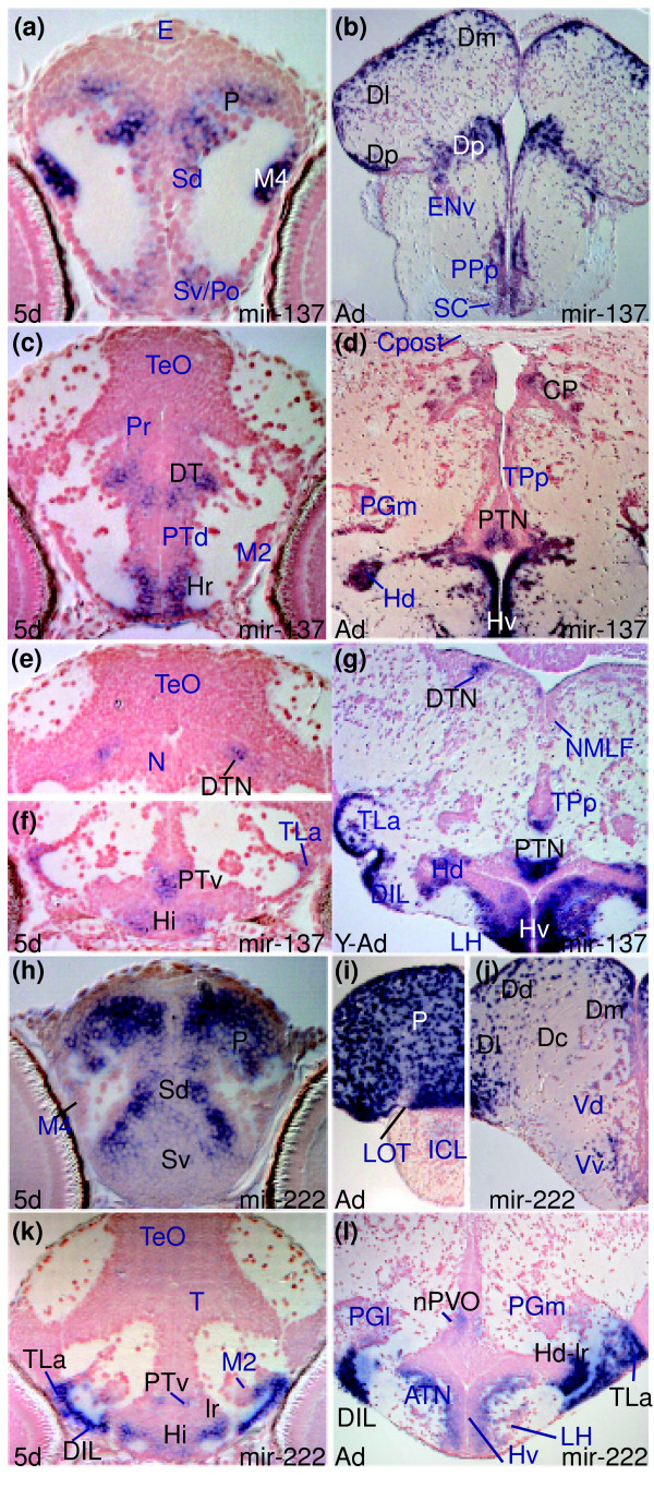
“Like miR-137, miR-222 shows conserved restricted expression in the rostral brain throughout life with domains in the telencephalon (for instance, compare P in Figure 3h with P, Dm, Dd, Dl, and Dc in Figure 3i,j), hypothalamus (DIL, TLa, lr/Hd-lr in Figure 3k,l) and posterior tubercular area (PTv in Figure 3k and nPVO in Figure 3l; see also Additional data file 20, and Tables D and I in Additional”
§ MicroRNAs show a wide diversity of expression profiles in the developing and mature central nervous system > Results > miRNA expression can be largely conserved between larval and adult stages
plain- Asserted: passes — The authors put it forward as true.
- Checkable: passes — It can be checked against evidence.
- Load-bearing: unclear — It is unclear whether the paper’s conclusions depend on it.
- Based: passes — The paper gives a reason why it holds.
A result the paper reports from its own data — it cites no other source for it, so there is none to check. The quote shows it is in the paper.
In the paper
“whereas miR-138 (Additional data file 11, and Table B in Additional data file 27) shows a widespread but more restricted pattern of expression.”
§ MicroRNAs show a wide diversity of expression profiles in the developing and mature central nervous system > Results > miRNAs can be widely expressed in differentiating CNS cells
plaincorrelational- Asserted: passes — The authors put it forward as true.
- Checkable: passes — It can be checked against evidence.
- Load-bearing: unclear — It is unclear whether the paper’s conclusions depend on it.
- Based: passes — The paper gives a reason why it holds.
A result the paper reports from its own data — it cites no other source for it, so there is none to check. The quote shows it is in the paper.
In the paper
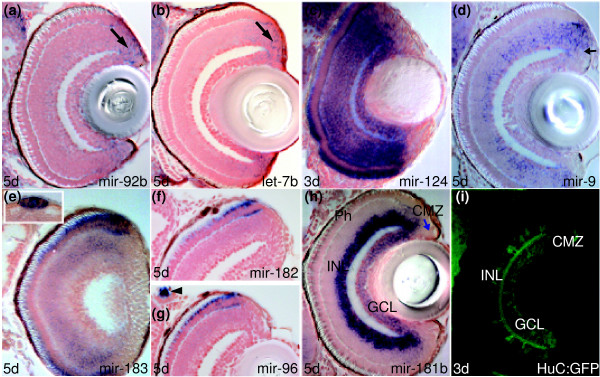
“We observe that both are expressed in cells associated with the visual system, including retinal amacrine cells (INL) and ganglion cells (GCL in Figure 2h; Additional data files 13 and 14).”
§ MicroRNAs show a wide diversity of expression profiles in the developing and mature central nervous system > Results > miRNAs can show spatially localized expression in the larval brain
plaincorrelational- Asserted: passes — The authors put it forward as true.
- Checkable: passes — It can be checked against evidence.
- Load-bearing: unclear — It is unclear whether the paper’s conclusions depend on it.
- Based: passes — The paper gives a reason why it holds.
A result the paper reports from its own data — it cites no other source for it, so there is none to check. The quote shows it is in the paper.
In the paper
“let-7a and let-7c, which differ in their sequence from let-7b by two and one nucleotide, respectively, located outside the seed region, appear to lack this retinal expression, although we cannot exclude that these LNA probes cross-hybridize to various let-7 family members in different brain areas (Additional data files 22-24 and 27-29).”
§ MicroRNAs show a wide diversity of expression profiles in the developing and mature central nervous system > Results > miRNA expression can be restricted to proliferating cells in the larval zebrafish brain
hedged- Asserted: passes — The authors put it forward as true.
- Checkable: passes — It can be checked against evidence.
- Load-bearing: unclear — It is unclear whether the paper’s conclusions depend on it.
- Based: passes — The paper gives a reason why it holds.
A result the paper reports from its own data — it cites no other source for it, so there is none to check. The quote shows it is in the paper.
In the paper
“miR-222 and miR-34 are expressed in neural cells in restricted subdivisions along the rostro-caudal axis of the larval brain.”
§ MicroRNAs show a wide diversity of expression profiles in the developing and mature central nervous system > Results > miRNAs can show spatially localized expression in the larval brain
plain- Asserted: passes — The authors put it forward as true.
- Checkable: passes — It can be checked against evidence.
- Load-bearing: passes — The paper’s conclusions depend on it.
- Based: passes — The paper gives a reason why it holds.
A result the paper reports from its own data — it cites no other source for it, so there is none to check. The quote shows it is in the paper.
In the paper
“Several other miRNAs show robust expression in restricted populations of adult ventricular or periventricular cells, including miR-34b (Figures I and L in Additional data file 25).”
§ MicroRNAs show a wide diversity of expression profiles in the developing and mature central nervous system > Results > miRNA expression can be largely conserved between larval and adult stages
plain- Asserted: passes — The authors put it forward as true.
- Checkable: passes — It can be checked against evidence.
- Load-bearing: unclear — It is unclear whether the paper’s conclusions depend on it.
- Based: passes — The paper gives a reason why it holds.
A result the paper reports from its own data — it cites no other source for it, so there is none to check. The quote shows it is in the paper.
In the paper
“For instance, in contrast to strong and widespread expression in some domains of the larval brain, adult expression of miR-219 is restricted to relatively few cells.”
§ MicroRNAs show a wide diversity of expression profiles in the developing and mature central nervous system > Results > miRNA expression can change between larval and adult stages
plain- Asserted: passes — The authors put it forward as true.
- Checkable: passes — It can be checked against evidence.
- Load-bearing: unclear — It is unclear whether the paper’s conclusions depend on it.
- Based: passes — The paper gives a reason why it holds.
- Probably not a substantial claim. 26
- It may state two separate claims as one. 42
Every source the paper cites for this claim was checked and supports it.
In the paper
“There is a precedent for post-transcriptional regulation of miRNA expression [48,49], although this has not been demonstrated for different miRNAs from the same transcript.”
§ MicroRNAs show a wide diversity of expression profiles in the developing and mature central nervous system > Results > miRNAs of the same family or cluster can show subtle differences in expression in the adult brain
What the sources say
- Supportedcites [48] Obernosterer G, Leuschner PJ, Alenius M, Martinez J: Post-transcriptional regulation of microRNA expression. Rna. 2006, 12: 1161-1167. 10.1261/rna.2322506. source
The Introduction says the authors provide evidence that mammalian miRNA expression can be regulated during processing. The discussion reports that differential processing of precursor miRNAs produces tissue- and developmental-specific expression. The citations attach to the precedent in the first clause, not to the citing authors’ qualification about different miRNAs from the same transcript. (LLM, conf=0.93)
- Supportedcites [49] Thomson JM, Newman M, Parker JS, Morin-Kensicki EM, Wright T, Hammond SM: Extensive post-transcriptional regulation of microRNAs and its implications for cancer. Genes Dev. 2006, 20: 2202-2207. 10.1101/gad.1444406. source
The Results and Discussion report that mature miRNA levels can differ from primary-transcript levels and identify regulated Drosha processing as a post-transcriptional mechanism. This supports the precedent for post-transcriptional regulation to which the citations are attached; the sentence’s same-transcript caveat is not presented as a finding from this source. (LLM, conf=0.92)
plain- Asserted: passes — The authors put it forward as true.
- Checkable: passes — It can be checked against evidence.
- Load-bearing: fails — The paper’s conclusions may not depend on it.
- Based: passes — The paper gives a reason why it holds.
Probably not a substantial claim. The paper’s conclusions may not depend on it. It may state two separate claims as one.
- Supported
The sources with a verdict support this claim; the rest weren’t checked yet, have no full text we could read, or their verdicts aren’t rated yet.
In the paper
“These observations are consistent with the fact that proliferation and production of neurons continues into adulthood in the CNS of zebrafish [46,47].”
§ MicroRNAs show a wide diversity of expression profiles in the developing and mature central nervous system > Results > miRNA expression can be largely conserved between larval and adult stages
What the sources say
- Supportedcites [46] Adolf B, Chapouton P, Lam CS, Topp S, Tannhauser B, Strahle U, Gotz M, Bally-Cuif L: Conserved and acquired features of adult neurogenesis in the zebrafish telencephalon. Dev Biol. 2006, 295: 278-293. 10.1016/j.ydbio.2006.03.023. source
The Introduction states that proliferation and neurogenesis occur throughout life in teleost fish and identifies several sites where newborn neurons form in the adult zebrafish brain. This supports the claim about continued proliferation and neuron production in the zebrafish CNS; it does not imply that every CNS region produces neurons. (LLM, conf=0.93)
- Not checked — no full text of the sourcecites [47] Grandel H, Kaslin J, Ganz J, Wenzel I, Brand M: Neural stem cells and neurogenesis in the adult zebrafish brain: origin, proliferation dynamics, migration and cell fate. Dev Biol. 2006, 295: 263-277. 10.1016/j.ydbio.2006.03.040. source
plain- Asserted: passes — The authors put it forward as true.
- Checkable: passes — It can be checked against evidence.
- Load-bearing: fails — The paper’s conclusions may not depend on it.
- Based: passes — The paper gives a reason why it holds.
Probably not a substantial claim. The paper’s conclusions may not depend on it.
- Supported
The cited sources support only part of this claim — read each source’s verdict below.
In the paper
“Many of the genes in these pathways are implicated in human diseases [43].”
§ MicroRNAs show a wide diversity of expression profiles in the developing and mature central nervous system > Results > miRNA expression can be cell type specific
What the sources say
- Partly supportedcites [43] Banizs B, Pike MM, Millican CL, Ferguson WB, Komlosi P, Sheetz J, Bell PD, Schwiebert EM, Yoder BK: Dysfunctional cilia lead to altered ependyma and choroid plexus function, and result in the formation of hydrocephalus. Development. 2005, 132: 5329-5339. 10.1242/dev.02153. sourceExperimental — accuracy not yet measured
The Discussion states that mutations in IFT genes such as Tg737 cause developmental defects and disease, and the Introduction describes disease in Tg737 mutant mice. The copy does not establish that many IFT-pathway genes are implicated specifically in human diseases; its mention of human hydrocephalus does not identify the responsible genes as IFT genes. Page numbers were not available in the indexed passages. (LLM, conf=0.87)
plaincorrelational- Asserted: passes — The authors put it forward as true.
- Checkable: passes — It can be checked against evidence.
- Load-bearing: fails — The paper’s conclusions may not depend on it.
- Based: passes — The paper gives a reason why it holds.
Probably not a substantial claim. The paper’s conclusions may not depend on it.
- Partly supported
The cited sources that could be checked couldn’t confirm or dispute this claim.
In the paper
“A cluster on chromosome 8 contains both miR-181a and miR-181b but there is an additional copy of miR-181a on chromosome 22 and of miR-181b on chromosome 20 [19,44].”
§ MicroRNAs show a wide diversity of expression profiles in the developing and mature central nervous system > Results > miRNAs of the same family or cluster can show subtle differences in expression in the adult brain
What the sources say
- Could not verifycites [19] Ason B, Darnell DK, Wittbrodt B, Berezikov E, Kloosterman WP, Wittbrodt J, Antin PB, Plasterk RH: Differences in vertebrate microRNA expression. Proc Natl Acad Sci USA. 2006, 103: 14385-14389. 10.1073/pnas.0603529103. sourceExperimental — accuracy not yet measured
The Results and Discussion discusses miRNA copy number and genomic locations, but the passages in our copy concern other miRNAs, not miR-181a or miR-181b. Searches for miR-181 and chromosomes 8, 20, and 22 found no passage establishing the claimed cluster or additional copies. (LLM, conf=0.73)
- Not matchedcites [44] Griffiths-Jones S: miRBase: the microRNA sequence database. Methods Mol Biol. 2006, 342: 129-138.
Couldn’t be matched to a paper in Crossref, so it can’t be checked.
plain- Asserted: passes — The authors put it forward as true.
- Checkable: passes — It can be checked against evidence.
- Load-bearing: unclear — It is unclear whether the paper’s conclusions depend on it.
- Based: passes — The paper gives a reason why it holds.
It may state two separate claims as one.
- Could not verify
The cited sources support only part of this claim — read each source’s verdict below.
In the paper
“Localized translation from mRNAs is mediated in part by mRNA binding proteins that promote translation [59,60] and a counterpart to this could be that miRNAs clear mRNAs at sites where protein activity should be low.”
§ MicroRNAs show a wide diversity of expression profiles in the developing and mature central nervous system > Discussion > Some miRNAs are likely to be co-expressed with their targets and may spatially or temporally modulate protein levels within neural cells
What the sources say
- Could not verifycites [59] Adler CE, Fetter RD, Bargmann CI: UNC-6/Netrin induces neuronal asymmetry and defines the site of axon formation. Nat Neurosci. 2006, 9: 511-518. 10.1038/nn1666. sourceExperimental — accuracy not yet measured
The Results and Discussion describe netrin-dependent localization of signaling proteins during axon formation, not mRNA-binding proteins that promote local translation. Searches for localized translation and RNA-binding proteins found no passage in our copy addressing that mechanism; the References list a separate study on local protein synthesis, but that does not establish the claimed mechanism. (LLM, conf=0.75)
- Partly supportedcites [60] Leung KM, van Horck FP, Lin AC, Allison R, Standart N, Holt CE: Asymmetrical beta-actin mRNA translation in growth cones mediates attractive turning to netrin-1. Nat Neurosci. 2006, 9: 1247-1256. 10.1038/nn1775. sourceExperimental — accuracy not yet measured
The Introduction says RNA-binding proteins localize mRNAs and can help regulate their translation. The Discussion proposes that Vg1RBP may activate β-actin translation, but presents that role as a possibility rather than an established mechanism, so the claim overstates this paper's evidence. (LLM, conf=0.82)
hedgeddirect- Asserted: passes — The authors put it forward as true.
- Checkable: passes — It can be checked against evidence.
- Load-bearing: fails — The paper’s conclusions may not depend on it.
- Based: passes — The paper gives a reason why it holds.
Probably not a substantial claim. The paper’s conclusions may not depend on it.
- Could not verify
The sources with a verdict support this claim; the rest weren’t checked yet, have no full text we could read, or their verdicts aren’t rated yet.
In the paper
“Given that some mRNAs are localized and translated locally within neurons (for example, [57,58]), an attractive possibility is that cell-type specific brain miRNAs regulate spatially localized translation of proteins.”
§ MicroRNAs show a wide diversity of expression profiles in the developing and mature central nervous system > Discussion > Some miRNAs are likely to be co-expressed with their targets and may spatially or temporally modulate protein levels within neural cells
What the sources say
- Not checked — no full text of the sourcecites [57] Raab-Graham KF, Haddick PC, Jan YN, Jan LY: Activity- and mTOR-dependent suppression of Kv1.1 channel mRNA translation in dendrites. Science. 2006, 314: 144-148. 10.1126/science.1131693. source
- Supportedcites [58] Piper M, Holt C: RNA translation in axons. Annu Rev Cell Dev Biol. 2004, 20: 505-523. 10.1146/annurev.cellbio.20.010403.111746. source
The Introduction describes mRNAs localized within dendrites and experimental support for their local translation. The Conclusion also states that mRNAs can be tethered and translated within axons, supporting the claim about local translation within neurons. (LLM, conf=0.94)
plain- Asserted: passes — The authors put it forward as true.
- Checkable: passes — It can be checked against evidence.
- Load-bearing: fails — The paper’s conclusions may not depend on it.
- Based: passes — The paper gives a reason why it holds.
Probably not a substantial claim. The paper’s conclusions may not depend on it.
The cited sources support only part of this claim — read each source’s verdict below.
In the paper
“Certainly, recent studies suggest that there is a huge variability in the level of transcription at equivalent active loci in different cells [55] and perhaps there is comparable variability at repressed loci.”
§ MicroRNAs show a wide diversity of expression profiles in the developing and mature central nervous system > Discussion > Some CNS miRNAs may constitutively survey fluctuating levels of transcriptionally 'silenced' target mRNAs
What the sources say
- Partly supportedcites [55] Raj A, Peskin CS, Tranchina D, Vargas DY, Tyagi S: Stochastic mRNA synthesis in mammalian cells. PLoS Biol. 2006, 4: e309-10.1371/journal.pbio.0040309. sourceExperimental — accuracy not yet measured
The Discussion reports large cell-to-cell variation in mRNA levels, and the Results attribute it to random bursts of gene activation. Those passages support variability in transcription of the same genes across cells, but they do not establish huge variation in transcription levels specifically among loci that are all active. The sentence’s speculation about repressed loci is not attributed to this source. (LLM, conf=0.84)
hedged- Asserted: passes — The authors put it forward as true.
- Checkable: passes — It can be checked against evidence.
- Load-bearing: fails — The paper’s conclusions may not depend on it.
- Based: passes — The paper gives a reason why it holds.
Probably not a substantial claim. The paper’s conclusions may not depend on it.
- Partly supported
The cited sources support only part of this claim — read each source’s verdict below.
In the paper
“It is intriguing that the translation of beta-actin in neuronal processes is regulated by Vg1RBP, a protein that binds the 3' UTR of mRNAs [60] and may, therefore, shield the mRNA from miRNAs.”
§ MicroRNAs show a wide diversity of expression profiles in the developing and mature central nervous system > Discussion > Some miRNAs are likely to be co-expressed with their targets and may spatially or temporally modulate protein levels within neural cells
What the sources say
- Partly supportedcites [60] Leung KM, van Horck FP, Lin AC, Allison R, Standart N, Holt CE: Asymmetrical beta-actin mRNA translation in growth cones mediates attractive turning to netrin-1. Nat Neurosci. 2006, 9: 1247-1256. 10.1038/nn1775. sourceExperimental — accuracy not yet measured
The Results section on Vg1RBP in retinal growth cones reports that Vg1RBP associates with β-actin mRNA, and the reporter-synthesis Results show that the β-actin 3′ UTR is needed for netrin-1-induced local protein synthesis. But the Discussion presents Vg1RBP's activation of β-actin translation as a possibility, not an established mechanism. The citation therefore supports the association and UTR-related context, but overstates direct regulation by Vg1RBP. (LLM, conf=0.84)
plaindirect- Asserted: passes — The authors put it forward as true.
- Checkable: passes — It can be checked against evidence.
- Load-bearing: fails — The paper’s conclusions may not depend on it.
- Based: passes — The paper gives a reason why it holds.
Probably not a substantial claim. The paper’s conclusions may not depend on it.
- Partly supported
Every source the paper cites for this claim was checked and supports it.
In the paper
“mRNAs can be protected from miRNA attack as seen, for instance, in germ cells [7], although we are unaware of examples of such regulation occurring within different cellular compartments.”
§ MicroRNAs show a wide diversity of expression profiles in the developing and mature central nervous system > Discussion > Some miRNAs are likely to be co-expressed with their targets and may spatially or temporally modulate protein levels within neural cells
What the sources say
- Supportedcites [7] Mishima Y, Giraldez AJ, Takeda Y, Fujiwara T, Sakamoto H, Schier AF, Inoue K: Differential regulation of germline mRNAs in soma and germ cells by zebrafish miR-430. Curr Biol. 2006, 16: 2135-2142. 10.1016/j.cub.2006.08.086. source
The Discussion reports that elements in the nanos1 3′ UTR allow expression in zebrafish primordial germ cells despite miRNA-mediated repression. This supports protection from the miRNA’s repressive effects, though the Results clarify that the mRNA remains accessible to miRNAs rather than being physically shielded from them. Page numbers were not available in our copy. (LLM, conf=0.88)
plaindirect- Asserted: passes — The authors put it forward as true.
- Checkable: passes — It can be checked against evidence.
- Load-bearing: fails — The paper’s conclusions may not depend on it.
- Based: passes — The paper gives a reason why it holds.
Probably not a substantial claim. The paper’s conclusions may not depend on it.
- Supported
The sources with a verdict support this claim; the rest weren’t checked yet, have no full text we could read, or their verdicts aren’t rated yet.
In the paper
“For instance, in zebrafish, miR-430 targets a large number of maternally deposited transcripts at the onset of zygotic transcription [6] and many examples of spatially exclusive expression domains of miRNAs and their targets have been documented (for example, [12,53]).”
§ MicroRNAs show a wide diversity of expression profiles in the developing and mature central nervous system > Discussion
What the sources say
- Not checked — no full text of the sourcecites [12] Stark A, Brennecke J, Bushati N, Russell RB, Cohen SM: Animal microRNAs confer robustness to gene expression and have a significant impact on 3'UTR evolution. Cell. 2005, 123: 1133-1146. 10.1016/j.cell.2005.11.023. source
- Supportedcites [53] Li Y, Wang F, Lee JA, Gao FB: MicroRNA-9a ensures the precise specification of sensory organ precursors in Drosophila. Genes Dev. 2006, 20: 2793-2805. 10.1101/gad.1466306. source
In “MiR-9a inhibits Sens expression in non-SOP cells” (page not available), the paper says miRNAs and their targets are often expressed in adjacent cells and presents miR-9a and Sens as an experimental example. This supports the cited statement about documented spatially distinct expression patterns. (LLM, conf=0.86)
plaincorrelational- Asserted: passes — The authors put it forward as true.
- Checkable: passes — It can be checked against evidence.
- Load-bearing: fails — The paper’s conclusions may not depend on it.
- Based: passes — The paper gives a reason why it holds.
Probably not a substantial claim. The paper’s conclusions may not depend on it.
A result the paper reports from its own data — it cites no other source for it, so there is none to check. The quote shows it is in the paper.
In the paper
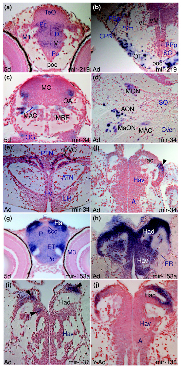
“In contrast, miR-34 expression is absent from forebrain and midbrain and present only in the caudal ventral and lateral isthmus and hindbrain, including ventral and lateral medulla oblongata cells (MO in Figure 5c), presumptive octaval area (OA in Figure 5c), reticular formation cells (intermediate reticular formation-(IMRF) and Mauthner cell (MAC) in Figure 5c; Additional data file 5, and Table D”
§ MicroRNAs show a wide diversity of expression profiles in the developing and mature central nervous system > Results > miRNAs can show spatially localized expression in the larval brain
plain- Asserted: passes — The authors put it forward as true.
- Checkable: passes — It can be checked against evidence.
- Load-bearing: unclear — It is unclear whether the paper’s conclusions depend on it.
- Based: passes — The paper gives a reason why it holds.
It may state two separate claims as one.
A result the paper reports from its own data — it cites no other source for it, so there is none to check. The quote shows it is in the paper.
In the paper
“miR-182 and miR-96 show almost identical expression patterns to miR-183 (Figure 2f,g; and Table E in Additional data file 27), although expression is not as robust.”
§ MicroRNAs show a wide diversity of expression profiles in the developing and mature central nervous system > Results > miRNA expression can be cell type specific
plaincorrelational- Asserted: passes — The authors put it forward as true.
- Checkable: passes — It can be checked against evidence.
- Load-bearing: passes — The paper’s conclusions depend on it.
- Based: passes — The paper gives a reason why it holds.
It may state two separate claims as one.
A result the paper reports from its own data — it cites no other source for it, so there is none to check. The quote shows it is in the paper.
In the paper
“This pattern is highly reminiscent of expression of the huC gene (Figure 2i), which encodes an RNA binding protein expressed in nearly all CNS neurons but the same subsets of retinal cells as miR-181a and miR-181b.”
§ MicroRNAs show a wide diversity of expression profiles in the developing and mature central nervous system > Results > miRNAs can show spatially localized expression in the larval brain
plaincorrelational- Asserted: passes — The authors put it forward as true.
- Checkable: passes — It can be checked against evidence.
- Load-bearing: unclear — It is unclear whether the paper’s conclusions depend on it.
- Based: passes — The paper gives a reason why it holds.
It may state two separate claims as one.
A result the paper reports from its own data — it cites no other source for it, so there is none to check. The quote shows it is in the paper.
In the paper
“Like miR-92b, let-7b expression is restricted to the CMZ of the retina, with expression absent from all mature retinal neurons (arrow in Figure 2b); let-7b has broader expression elsewhere in the larval brain (Additional data file 23).”
§ MicroRNAs show a wide diversity of expression profiles in the developing and mature central nervous system > Results > miRNA expression can be restricted to proliferating cells in the larval zebrafish brain
plain- Asserted: passes — The authors put it forward as true.
- Checkable: passes — It can be checked against evidence.
- Load-bearing: passes — The paper’s conclusions depend on it.
- Based: passes — The paper gives a reason why it holds.
It may state two separate claims as one.
A result the paper reports from its own data — it cites no other source for it, so there is none to check. The quote shows it is in the paper.
In the paper
“The expression of these miRNAs in peripheral sensory neural cells overlaps with miR-200a (Additional data file 16), although miR-200a lacks the CNS and cranial ganglia expression sites common to miR-183, miR-182 and miR-96 (Table E in Additional data file 27).”
§ MicroRNAs show a wide diversity of expression profiles in the developing and mature central nervous system > Results > miRNA expression can be cell type specific
plaincorrelational- Asserted: passes — The authors put it forward as true.
- Checkable: passes — It can be checked against evidence.
- Load-bearing: unclear — It is unclear whether the paper’s conclusions depend on it.
- Based: passes — The paper gives a reason why it holds.
It may state two separate claims as one.
A result the paper reports from its own data — it cites no other source for it, so there is none to check. The quote shows it is in the paper.
In the paper
“Expression is not ubiquitous in neural cells as some areas such as the epithalamus and hypothalamic lateral torus are devoid of expression (Additional data files 2 and 3, and Table A in Additional data file 27).”
§ MicroRNAs show a wide diversity of expression profiles in the developing and mature central nervous system > Results > miRNAs can be widely expressed in both proliferating and differentiating CNS cells
plain- Asserted: passes — The authors put it forward as true.
- Checkable: passes — It can be checked against evidence.
- Load-bearing: passes — The paper’s conclusions depend on it.
- Based: passes — The paper gives a reason why it holds.
It may state two separate claims as one.
A result the paper reports from its own data — it cites no other source for it, so there is none to check. The quote shows it is in the paper.
In the paper
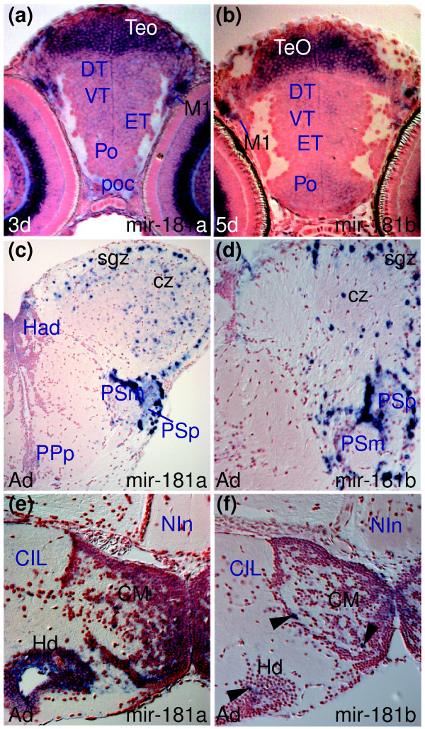
“Both miRNAs are also expressed in migrated pretectal (M1 in Figure 4a,b) and tectal cells (TeO in Figure 4a,b) and more weakly in many differentiating cells throughout the brain, with some stronger sites of expression in central pallium and medulla oblongata (Additional data files 13 and 14, and Table C in Additional data file 27).”
§ MicroRNAs show a wide diversity of expression profiles in the developing and mature central nervous system > Results > miRNAs can show spatially localized expression in the larval brain
plain- Asserted: passes — The authors put it forward as true.
- Checkable: passes — It can be checked against evidence.
- Load-bearing: unclear — It is unclear whether the paper’s conclusions depend on it.
- Based: passes — The paper gives a reason why it holds.
It may state two separate claims as one.
A result the paper reports from its own data — it cites no other source for it, so there is none to check. The quote shows it is in the paper.
In the paper
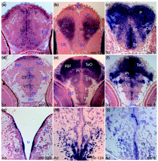
“Similarly showing conserved expression over time, miR-124 expression is excluded from periventricular cells and is detected in most differentiated cells throughout the adult brain as in the larval brain (compare Vd in Figure 1h and Sd in Figure 1b; Additional data file 7, and Tables B and G in Additional data file 27).”
§ MicroRNAs show a wide diversity of expression profiles in the developing and mature central nervous system > Results > miRNA expression can be largely conserved between larval and adult stages
plain- Asserted: passes — The authors put it forward as true.
- Checkable: passes — It can be checked against evidence.
- Load-bearing: passes — The paper’s conclusions depend on it.
- Based: passes — The paper gives a reason why it holds.
It may state two separate claims as one.
A result the paper reports from its own data — it cites no other source for it, so there is none to check. The quote shows it is in the paper.
In the paper
“For instance, miR-222 and miR-221 share largely similar expression in the adult hypothalamus (ATN, LH, Hd in Figure G of Additional data file 20 and Figure O of Additional data file 19) but only miR-222 is expressed in the ventral intermediate hypothalamus at the larval stage (Figure D of Additional data file 20 and Figure B of Additional data file 19; see also Table K in Additional data file 28”
§ MicroRNAs show a wide diversity of expression profiles in the developing and mature central nervous system > Results > miRNAs of the same family or cluster can show subtle differences in expression in the adult brain
plaincorrelational- Asserted: passes — The authors put it forward as true.
- Checkable: passes — It can be checked against evidence.
- Load-bearing: unclear — It is unclear whether the paper’s conclusions depend on it.
- Based: passes — The paper gives a reason why it holds.
It may state two separate claims as one.
A result the paper reports from its own data — it cites no other source for it, so there is none to check. The quote shows it is in the paper.
In the paper
“Additionally, within the retina miR-9 is expressed in maturing cells of the CMZ (which are likely to still be proliferative) but expression is maintained only in amacrine cells of the inner nuclear layer (INL in Figure 2d).”
§ MicroRNAs show a wide diversity of expression profiles in the developing and mature central nervous system > Results > miRNAs can be widely expressed in both proliferating and differentiating CNS cells
hedged- Asserted: passes — The authors put it forward as true.
- Checkable: passes — It can be checked against evidence.
- Load-bearing: unclear — It is unclear whether the paper’s conclusions depend on it.
- Based: passes — The paper gives a reason why it holds.
It may state two separate claims as one.
A result the paper reports from its own data — it cites no other source for it, so there is none to check. The quote shows it is in the paper.
In the paper
“miR-181a and miR-181b show similar expression in the larval brain, and this is largely conserved to adult stages (although there is down-regulation in some areas such as thalamus and tegmentum; Additional data files 13 and 14, and Tables C and H in Additional data file 27).”
§ MicroRNAs show a wide diversity of expression profiles in the developing and mature central nervous system > Results > miRNAs of the same family or cluster can show subtle differences in expression in the adult brain
plaincorrelational- Asserted: passes — The authors put it forward as true.
- Checkable: passes — It can be checked against evidence.
- Load-bearing: unclear — It is unclear whether the paper’s conclusions depend on it.
- Based: passes — The paper gives a reason why it holds.
It may state two separate claims as one.
A result the paper reports from its own data — it cites no other source for it, so there is none to check. The quote shows it is in the paper.
In the paper
“miR-183 is expressed in retinal photoreceptors and weakly in some inner nuclear layer cells, pineal cells that are again likely to be photoreceptors (Figure 2e) and perhaps also in parapineal photoreceptors (Figure C in Additional data file 15).”
§ MicroRNAs show a wide diversity of expression profiles in the developing and mature central nervous system > Results > miRNA expression can be cell type specific
hedged- Asserted: passes — The authors put it forward as true.
- Checkable: passes — It can be checked against evidence.
- Load-bearing: unclear — It is unclear whether the paper’s conclusions depend on it.
- Based: passes — The paper gives a reason why it holds.
It may state two separate claims as one.
The paper states this without a source or data behind it here, so there is nothing to check it against. The quote shows it is in the paper.
In the paper
“Some of the miRNA expression patterns helped us annotate larval fish brain nuclei.”
§ MicroRNAs show a wide diversity of expression profiles in the developing and mature central nervous system > Materials and methods > Neuroanatomical annotation
plaindirect- Asserted: passes — The authors put it forward as true.
- Checkable: passes — It can be checked against evidence.
- Load-bearing: fails — The paper’s conclusions may not depend on it.
- Based: passes — The paper gives a reason why it holds.
Probably not a substantial claim. The paper’s conclusions may not depend on it.
A result the paper reports from its own data — it cites no other source for it, so there is none to check. The quote shows it is in the paper.
In the paper
“For example, larval ventral thalamus (VT in Figure 5a) expresses miR-219 but the corresponding adult thalamic nuclei do not (ventrolateral (VL) and ventromedial (VM) in Figure 5b).”
§ MicroRNAs show a wide diversity of expression profiles in the developing and mature central nervous system > Results > miRNA expression can change between larval and adult stages
plain- Asserted: passes — The authors put it forward as true.
- Checkable: passes — It can be checked against evidence.
- Load-bearing: unclear — It is unclear whether the paper’s conclusions depend on it.
- Based: passes — The paper gives a reason why it holds.
It may state two separate claims as one.
A result the paper reports from its own data — it cites no other source for it, so there is none to check. The quote shows it is in the paper.
In the paper
“Conversely, in adults, miR-219 is expressed in cells, possibly glia, associated with major tracts such as the lateral olfactory tract, post-optic commissure/optic chiasma and tract (poc and OT, respectively, in Figure 5b) whereas the equivalent pathways in larvae are devoid of staining (Figure 5a).”
§ MicroRNAs show a wide diversity of expression profiles in the developing and mature central nervous system > Results > miRNA expression can change between larval and adult stages
hedgedcorrelational- Asserted: passes — The authors put it forward as true.
- Checkable: passes — It can be checked against evidence.
- Load-bearing: unclear — It is unclear whether the paper’s conclusions depend on it.
- Based: passes — The paper gives a reason why it holds.
It may state two separate claims as one.
A result the paper reports from its own data — it cites no other source for it, so there is none to check. The quote shows it is in the paper.
In the paper
“For instance, miR-92b is downregulated in most mature neurons whereas, conversely, miR-124 is absent from proliferative cells and widely expressed in differentiated neurons.”
§ MicroRNAs show a wide diversity of expression profiles in the developing and mature central nervous system > Discussion > Many expression profiles are consistent with the mutual exclusion model of miRNA function
plaincorrelational- Asserted: passes — The authors put it forward as true.
- Checkable: passes — It can be checked against evidence.
- Load-bearing: unclear — It is unclear whether the paper’s conclusions depend on it.
- Based: passes — The paper gives a reason why it holds.
It may state two separate claims as one.
A result the paper reports from its own data — it cites no other source for it, so there is none to check. The quote shows it is in the paper.
In the paper
“For instance, miR-34 shows conserved expression in the larval and adult hindbrain (Mauthner neuron, (MAC) and presumptive octavolateral area (OA) in Figure 5c; MAC and octavolateral nuclei (medial octavolateral (MON), anterior octaval (AON), magnocellular octaval (MaON) in Figure 5d) but also expands to include forebrain and midbrain cells of the habenulae (Had in Figure 5f), posterior tuberculum”
§ MicroRNAs show a wide diversity of expression profiles in the developing and mature central nervous system > Results > miRNA expression can change between larval and adult stages
plaincorrelational- Asserted: passes — The authors put it forward as true.
- Checkable: passes — It can be checked against evidence.
- Load-bearing: passes — The paper’s conclusions depend on it.
- Based: passes — The paper gives a reason why it holds.
It may state two separate claims as one.
A result the paper reports from its own data — it cites no other source for it, so there is none to check. The quote shows it is in the paper.
In the paper
“However, there are sharp transitions between domains of expression and non-expression, with some structures, such as the habenular nuclei, devoid of expression.”
§ MicroRNAs show a wide diversity of expression profiles in the developing and mature central nervous system > Discussion > Many expression profiles are consistent with the mutual exclusion model of miRNA function
plain- Asserted: passes — The authors put it forward as true.
- Checkable: passes — It can be checked against evidence.
- Load-bearing: passes — The paper’s conclusions depend on it.
- Based: passes — The paper gives a reason why it holds.
It may state two separate claims as one.
A result the paper reports from its own data — it cites no other source for it, so there is none to check. The quote shows it is in the paper.
In the paper
“miR-100 also shows robust habenular expression (Figure K in Additional data file 25) while miR-92b and miR-34b are expressed in ventricular cells adjacent to the mature habenular nuclei (Figure J in Additional data file 6, and Figure L in Additional data file 25).”
§ MicroRNAs show a wide diversity of expression profiles in the developing and mature central nervous system > Results > miRNA expression can change between larval and adult stages
plain- Asserted: passes — The authors put it forward as true.
- Checkable: passes — It can be checked against evidence.
- Load-bearing: unclear — It is unclear whether the paper’s conclusions depend on it.
- Based: passes — The paper gives a reason why it holds.
It may state two separate claims as one.
A result the paper reports from its own data — it cites no other source for it, so there is none to check. The quote shows it is in the paper.
In the paper
“It reveals a wide diversity in miRNA expression, ranging from single cell types to the majority of CNS cells and from transient to constitutive expression.”
§ MicroRNAs show a wide diversity of expression profiles in the developing and mature central nervous system > Conclusion
plain- Asserted: passes — The authors put it forward as true.
- Checkable: passes — It can be checked against evidence.
- Load-bearing: passes — The paper’s conclusions depend on it.
- Based: passes — The paper gives a reason why it holds.
It may state two separate claims as one.
A result the paper reports from its own data — it cites no other source for it, so there is none to check. The quote shows it is in the paper.
In the paper
“A few of the miRNAs we examined show expression restricted to very few cells and usually expressing cells share some characteristics of function and/or form.”
§ MicroRNAs show a wide diversity of expression profiles in the developing and mature central nervous system > Discussion > Some miRNAs are likely to be co-expressed with their targets and may spatially or temporally modulate protein levels within neural cells
plaincorrelational- Asserted: passes — The authors put it forward as true.
- Checkable: passes — It can be checked against evidence.
- Load-bearing: passes — The paper’s conclusions depend on it.
- Based: passes — The paper gives a reason why it holds.
It may state two separate claims as one.
A result the paper reports from its own data — it cites no other source for it, so there is none to check. The quote shows it is in the paper.
In the paper
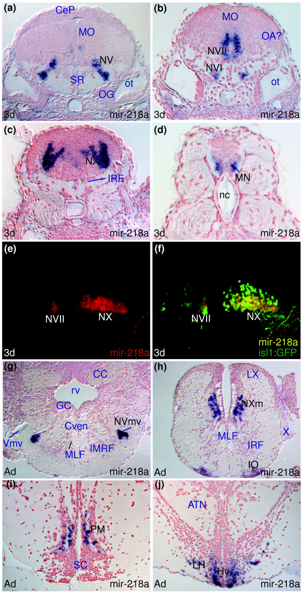
“As described above, miR-222 expression is also largely conserved between larvae and adults (Figure 3h-l) but de novo expression in the adult facial and vagal lobes is also observed (LVII, LX, Figure I in Additional data file 20).”
§ MicroRNAs show a wide diversity of expression profiles in the developing and mature central nervous system > Results > miRNA expression can change between larval and adult stages
plain- Asserted: passes — The authors put it forward as true.
- Checkable: passes — It can be checked against evidence.
- Load-bearing: unclear — It is unclear whether the paper’s conclusions depend on it.
- Based: passes — The paper gives a reason why it holds.
It may state two separate claims as one.
What the paper did — it cites no source for this, so there is none to check. The quote shows it is in the paper.
In the paper
“We observe that in the case of let-7a, miR-92b and miR-153a, one mismatch strongly reduces the hybridization signal. Since there is still some staining left, we cannot exclude some cross-hybridization with other members of the respective miRNA families.”
§ MicroRNAs show a wide diversity of expression profiles in the developing and mature central nervous system > Results
hedgeddirect- Asserted: passes — The authors put it forward as true.
- Checkable: passes — It can be checked against evidence.
- Load-bearing: passes — The paper’s conclusions depend on it.
- Based: passes — The paper gives a reason why it holds.
It may state two separate claims as one.
In the paper
“Although expression of mouse miR-375 in the pituitary has not been ascertained, it is expressed in pancreatic beta cells [45] and appears to function in the regulation of insulin secretion.”
§ MicroRNAs show a wide diversity of expression profiles in the developing and mature central nervous system > Results > miRNA expression can be cell type specific
What the sources say
- Not checked — no full text of the sourcecites [45] Poy MN, Eliasson L, Krutzfeldt J, Kuwajima S, Ma X, Macdonald PE, Pfeffer S, Tuschl T, Rajewsky N, Rorsman P, et al: A pancreatic islet-specific microRNA regulates insulin secretion. Nature. 2004, 432: 226-230. 10.1038/nature03076. source
hedgedconditional- Asserted: passes — The authors put it forward as true.
- Checkable: passes — It can be checked against evidence.
- Load-bearing: fails — The paper’s conclusions may not depend on it.
- Based: passes — The paper gives a reason why it holds.
Probably not a substantial claim. The paper’s conclusions may not depend on it. It may state two separate claims as one.
A conclusion the paper draws from its own results — it cites no other source for it, so there is none to check. The quote shows it is in the paper.
In the paper
“In a similar manner, other miRNAs belonging to a particular cluster seem to largely share expression patterns but also have subtle differences in transcript localization.”
§ MicroRNAs show a wide diversity of expression profiles in the developing and mature central nervous system > Results > miRNAs of the same family or cluster can show subtle differences in expression in the adult brain
hedgedcorrelational- Asserted: passes — The authors put it forward as true.
- Checkable: passes — It can be checked against evidence.
- Load-bearing: unclear — It is unclear whether the paper’s conclusions depend on it.
- Based: passes — The paper gives a reason why it holds.
It may state two separate claims as one.
What the paper did — it cites no source for this, so there is none to check. The quote shows it is in the paper.
In the paper
“miR-181a and miR-181b belong to the same family but differ in three nucleotides outside the seed region, suggesting that LNA probes can discriminate between their transcript expression profiles (Tables K and L in Additional data file 28, and Additional data file 29).”
§ MicroRNAs show a wide diversity of expression profiles in the developing and mature central nervous system > Results > miRNAs can show spatially localized expression in the larval brain
hedgedconditional- Asserted: passes — The authors put it forward as true.
- Checkable: passes — It can be checked against evidence.
- Load-bearing: unclear — It is unclear whether the paper’s conclusions depend on it.
- Based: passes — The paper gives a reason why it holds.
It may state two separate claims as one.
A conclusion the paper draws from its own results — it cites no other source for it, so there is none to check. The quote shows it is in the paper.
In the paper
“This difference in expression may again be due to genomic duplication of the miRNAs.”
§ MicroRNAs show a wide diversity of expression profiles in the developing and mature central nervous system > Results > miRNAs of the same family or cluster can show subtle differences in expression in the adult brain
hedgedconditional- Asserted: passes — The authors put it forward as true.
- Checkable: passes — It can be checked against evidence.
- Load-bearing: fails — The paper’s conclusions may not depend on it.
- Based: passes — The paper gives a reason why it holds.
Probably not a substantial claim. The paper’s conclusions may not depend on it.
A conclusion the paper draws from its own results — it cites no other source for it, so there is none to check. The quote shows it is in the paper.
In the paper
“It is not obvious why there should be differences in miR-222 and miR-221 expression as they are present in the same cluster and one would predict that they are co-transcribed.”
§ MicroRNAs show a wide diversity of expression profiles in the developing and mature central nervous system > Results > miRNAs of the same family or cluster can show subtle differences in expression in the adult brain
hedged- Asserted: passes — The authors put it forward as true.
- Checkable: fails — It may be too vague to check against evidence.
- Load-bearing: fails — The paper’s conclusions may not depend on it.
- Based: passes — The paper gives a reason why it holds.
Probably not a substantial claim. It may be too vague to check against evidence. The paper’s conclusions may not depend on it. It may state two separate claims as one.
A conclusion the paper draws from its own results — it cites no other source for it, so there is none to check. The quote shows it is in the paper.
In the paper
“miR-137 is expressed in the adult midbrain DTN (Figure 3g) and given the good correspondence of localized tegmental expression between 5 dpf and adult brains, we suggest that the cells in the larval tegmentum correspond to the presumptive midbrain DTN (Figure 3e,g; for other nuclei, see Additional data file 10, and Tables C and H in Additional data file 27).”
§ MicroRNAs show a wide diversity of expression profiles in the developing and mature central nervous system > Results > miRNA expression can be largely conserved between larval and adult stages
hedgedcorrelational- Asserted: passes — The authors put it forward as true.
- Checkable: passes — It can be checked against evidence.
- Load-bearing: passes — The paper’s conclusions depend on it.
- Based: passes — The paper gives a reason why it holds.
It may state two separate claims as one.
A conclusion the paper draws from its own results — it cites no other source for it, so there is none to check. The quote shows it is in the paper.
In the paper
“Together these results show that miRNA expression is frequently region or nucleus specific and can vary in the levels of expression from one group of cells to another.”
§ MicroRNAs show a wide diversity of expression profiles in the developing and mature central nervous system > Results > miRNAs can show spatially localized expression in the larval brain
plaincorrelational- Asserted: passes — The authors put it forward as true.
- Checkable: passes — It can be checked against evidence.
- Load-bearing: passes — The paper’s conclusions depend on it.
- Based: passes — The paper gives a reason why it holds.
It may state two separate claims as one.
What the paper did — it cites no source for this, so there is none to check. The quote shows it is in the paper.
In the paper
“The acetylation step in the in situ hybridization was often omitted since it did not change or improve signal.”
§ MicroRNAs show a wide diversity of expression profiles in the developing and mature central nervous system > Materials and methods > In situ hybridization and antibody labeling
plain- Asserted: passes — The authors put it forward as true.
- Checkable: passes — It can be checked against evidence.
- Load-bearing: fails — The paper’s conclusions may not depend on it.
- Based: passes — The paper gives a reason why it holds.
Probably not a substantial claim. The paper’s conclusions may not depend on it.
A claim is checked only against the cited paper’s full text, never its abstract, and none is available for its sources.
In the paper
“As the silencing of loci is generally efficient, one might conclude that for most of the time in most mature neurons, some miRNAs are doing very little indeed, consistent with the idea that miRNAs confer robustness to programs of gene expression [11,12].”
§ MicroRNAs show a wide diversity of expression profiles in the developing and mature central nervous system > Discussion > Some CNS miRNAs may constitutively survey fluctuating levels of transcriptionally 'silenced' target mRNAs
What the sources say
- Not checked — no full text of the sourcecites [11] Farh KK, Grimson A, Jan C, Lewis BP, Johnston WK, Lim LP, Burge CB, Bartel DP: The widespread impact of mammalian microRNAs on mRNA repression and evolution. Science. 2005, 310: 1817-1821. 10.1126/science.1121158. source
- Not checked — no full text of the sourcecites [12] Stark A, Brennecke J, Bushati N, Russell RB, Cohen SM: Animal microRNAs confer robustness to gene expression and have a significant impact on 3'UTR evolution. Cell. 2005, 123: 1133-1146. 10.1016/j.cell.2005.11.023. source
hedgedconditional- Asserted: passes — The authors put it forward as true.
- Checkable: passes — It can be checked against evidence.
- Load-bearing: fails — The paper’s conclusions may not depend on it.
- Based: passes — The paper gives a reason why it holds.
Probably not a substantial claim. The paper’s conclusions may not depend on it. It may state two separate claims as one.
- Rests on an assumption: The silencing of loci is generally efficient.
In the paper
“There are well-described mechanisms for repressing and silencing loci (for example, [54]), but the absolute efficacy of such mechanisms is unknown.”
§ MicroRNAs show a wide diversity of expression profiles in the developing and mature central nervous system > Discussion > Some CNS miRNAs may constitutively survey fluctuating levels of transcriptionally 'silenced' target mRNAs
What the sources say
- Not checked — no full text of the sourcecites [54] Talbert PB, Henikoff S: Spreading of silent chromatin: inaction at a distance. Nat Rev Genet. 2006, 7: 793-803. 10.1038/nrg1920. source
boosted- Asserted: passes — The authors put it forward as true.
- Checkable: passes — It can be checked against evidence.
- Load-bearing: fails — The paper’s conclusions may not depend on it.
- Based: passes — The paper gives a reason why it holds.
Probably not a substantial claim. The paper’s conclusions may not depend on it.
What the paper did — it cites no source for this, so there is none to check. The quote shows it is in the paper.
In the paper
“Additional data file 28 includes Tables J-L, listing the miRNAs analyzed, the family/cluster they belong to and the mismatch LNA probe sequences used to test specificity of corresponding fully matching LNA probes.”
§ MicroRNAs show a wide diversity of expression profiles in the developing and mature central nervous system > Additional data files
plain- Asserted: passes — The authors put it forward as true.
- Checkable: passes — It can be checked against evidence.
- Load-bearing: fails — The paper’s conclusions may not depend on it.
- Based: passes — The paper gives a reason why it holds.
Probably not a substantial claim. The paper’s conclusions may not depend on it.
A conclusion the paper draws from its own results — it cites no other source for it, so there is none to check. The quote shows it is in the paper.
In the paper
“miRNAs that are induced when cells transition from one state to another are likely to target mRNAs that are expressed during the initial state and not required during the second state.”
§ MicroRNAs show a wide diversity of expression profiles in the developing and mature central nervous system > Discussion > Some CNS miRNAs may constitutively survey fluctuating levels of transcriptionally 'silenced' target mRNAs
hedgedcorrelational- Asserted: passes — The authors put it forward as true.
- Checkable: passes — It can be checked against evidence.
- Load-bearing: fails — The paper’s conclusions may not depend on it.
- Based: fails — The paper may give no reason why it holds.
Probably not a substantial claim. The paper’s conclusions may not depend on it. The paper may give no reason why it holds.
A conclusion the paper draws from its own results — it cites no other source for it, so there is none to check. The quote shows it is in the paper.
In the paper
“These differences between larva and adult brain suggest downregulation of expression in most cells in the adult brain and either conserved or de novo expression in a few discrete cell populations (Additional data file 18, Tables B and G in Additional data file 27).”
§ MicroRNAs show a wide diversity of expression profiles in the developing and mature central nervous system > Results > miRNA expression can change between larval and adult stages
hedgeddirect- Asserted: passes — The authors put it forward as true.
- Checkable: passes — It can be checked against evidence.
- Load-bearing: passes — The paper’s conclusions depend on it.
- Based: passes — The paper gives a reason why it holds.
It may state two separate claims as one.
What the paper did — it cites no source for this, so there is none to check. The quote shows it is in the paper.
In the paper
“Penetration of all reagents even in adult brains was generally very good, although in some preparations deep tissue in the brain did not label well and readers should be cautious about interpretation regarding absence of expression in such cases.”
§ MicroRNAs show a wide diversity of expression profiles in the developing and mature central nervous system > Materials and methods > Sectioning
hedged- Asserted: passes — The authors put it forward as true.
- Checkable: passes — It can be checked against evidence.
- Load-bearing: fails — The paper’s conclusions may not depend on it.
- Based: passes — The paper gives a reason why it holds.
Probably not a substantial claim. The paper’s conclusions may not depend on it. It may state two separate claims as one.
In the paper
“For instance, miR-134 transcripts are localized to the dendritic compartments in which they function and miRNA activity is spatially/temporally regulated by extracellular signals [29].”
§ MicroRNAs show a wide diversity of expression profiles in the developing and mature central nervous system > Discussion > Some miRNAs are likely to be co-expressed with their targets and may spatially or temporally modulate protein levels within neural cells
What the sources say
- Not checked — no full text of the sourcecites [29] Schratt GM, Tuebing F, Nigh EA, Kane CG, Sabatini ME, Kiebler M, Greenberg ME: A brain-specific microRNA regulates dendritic spine development. Nature. 2006, 439: 283-289. 10.1038/nature04367. source
plaindirect- Asserted: passes — The authors put it forward as true.
- Checkable: passes — It can be checked against evidence.
- Load-bearing: fails — The paper’s conclusions may not depend on it.
- Based: passes — The paper gives a reason why it holds.
Probably not a substantial claim. The paper’s conclusions may not depend on it. It may state two separate claims as one.
A conclusion the paper draws from its own results — it cites no other source for it, so there is none to check. The quote shows it is in the paper.
In the paper
“The mutual exclusion of miRNAs and their targets may be in either space or in time.”
§ MicroRNAs show a wide diversity of expression profiles in the developing and mature central nervous system > Discussion
hedgedcorrelational- Asserted: passes — The authors put it forward as true.
- Checkable: passes — It can be checked against evidence.
- Load-bearing: fails — The paper’s conclusions may not depend on it.
- Based: passes — The paper gives a reason why it holds.
Probably not a substantial claim. The paper’s conclusions may not depend on it.
A claim is checked only against the cited paper’s full text, never its abstract, and none is available for its sources.
In the paper
“A common feature of miRNA function is one of mutually exclusive expression of miRNAs and their target mRNAs [11,12].”
§ MicroRNAs show a wide diversity of expression profiles in the developing and mature central nervous system > Discussion
What the sources say
- Not checked — no full text of the sourcecites [11] Farh KK, Grimson A, Jan C, Lewis BP, Johnston WK, Lim LP, Burge CB, Bartel DP: The widespread impact of mammalian microRNAs on mRNA repression and evolution. Science. 2005, 310: 1817-1821. 10.1126/science.1121158. source
- Not checked — no full text of the sourcecites [12] Stark A, Brennecke J, Bushati N, Russell RB, Cohen SM: Animal microRNAs confer robustness to gene expression and have a significant impact on 3'UTR evolution. Cell. 2005, 123: 1133-1146. 10.1016/j.cell.2005.11.023. source
plaincorrelational- Asserted: passes — The authors put it forward as true.
- Checkable: passes — It can be checked against evidence.
- Load-bearing: fails — The paper’s conclusions may not depend on it.
- Based: passes — The paper gives a reason why it holds.
Probably not a substantial claim. The paper’s conclusions may not depend on it.
In the paper
“For instance, in zebrafish, miR-430 targets a large number of maternally deposited transcripts at the onset of zygotic transcription [6] and many examples of spatially exclusive expression domains of miRNAs and their targets have been documented (for example, [12,53]).”
§ MicroRNAs show a wide diversity of expression profiles in the developing and mature central nervous system > Discussion
What the sources say
- Not checked — no full text of the sourcecites [6] Giraldez AJ, Mishima Y, Rihel J, Grocock RJ, Van Dongen S, Inoue K, Enright AJ, Schier AF: Zebrafish MiR-430 promotes deadenylation and clearance of maternal mRNAs. Science. 2006, 312: 75-79. 10.1126/science.1122689. source
plaindirect- Asserted: passes — The authors put it forward as true.
- Checkable: passes — It can be checked against evidence.
- Load-bearing: fails — The paper’s conclusions may not depend on it.
- Based: passes — The paper gives a reason why it holds.
Probably not a substantial claim. The paper’s conclusions may not depend on it.
In the paper
“mRNAs from genes required for general cellular machinery are protected from miRNA action through possession of short 3' UTRs that are poor miRNA targets and genes required for specific cellular functions of the specific cell type are often miRNA anti-targets - that is, their 3' UTRs lack sequences that would enable binding of co-expressed miRNAs [12].”
§ MicroRNAs show a wide diversity of expression profiles in the developing and mature central nervous system > Discussion > Some CNS miRNAs may constitutively survey fluctuating levels of transcriptionally 'silenced' target mRNAs
What the sources say
- Not checked — no full text of the sourcecites [12] Stark A, Brennecke J, Bushati N, Russell RB, Cohen SM: Animal microRNAs confer robustness to gene expression and have a significant impact on 3'UTR evolution. Cell. 2005, 123: 1133-1146. 10.1016/j.cell.2005.11.023. source
plaindirect- Asserted: passes — The authors put it forward as true.
- Checkable: passes — It can be checked against evidence.
- Load-bearing: unclear — It is unclear whether the paper’s conclusions depend on it.
- Based: passes — The paper gives a reason why it holds.
It may state two separate claims as one.
A conclusion the paper draws from its own results — it cites no other source for it, so there is none to check. The quote shows it is in the paper.
In the paper
“The first is to function in neural stem cells/progenitors and the second is to facilitate the clearance of target mRNAs at spatial or temporal transitions.”
§ MicroRNAs show a wide diversity of expression profiles in the developing and mature central nervous system > Conclusion
plaindirect- Asserted: passes — The authors put it forward as true.
- Checkable: passes — It can be checked against evidence.
- Load-bearing: passes — The paper’s conclusions depend on it.
- Based: passes — The paper gives a reason why it holds.
It may state two separate claims as one.
In the paper
“However, miR-134 is localized to dendrites and appears to modulate the levels of activity of a kinase that influences dendrite morphogenesis [29].”
§ MicroRNAs show a wide diversity of expression profiles in the developing and mature central nervous system > Discussion > Some miRNAs are likely to be co-expressed with their targets and may spatially or temporally modulate protein levels within neural cells
What the sources say
- Not checked — no full text of the sourcecites [29] Schratt GM, Tuebing F, Nigh EA, Kane CG, Sabatini ME, Kiebler M, Greenberg ME: A brain-specific microRNA regulates dendritic spine development. Nature. 2006, 439: 283-289. 10.1038/nature04367. source
hedgedconditional- Asserted: passes — The authors put it forward as true.
- Checkable: passes — It can be checked against evidence.
- Load-bearing: fails — The paper’s conclusions may not depend on it.
- Based: passes — The paper gives a reason why it holds.
Probably not a substantial claim. The paper’s conclusions may not depend on it. It may state two separate claims as one.
In the paper
“Major changes in the transcriptome that occur when cells transition between states are thought to be brought about by transcriptional mechanisms independent of miRNA function and so it seems likely that many target mRNAs will not be actively transcribed once a transition has occurred (for example, [6]).”
§ MicroRNAs show a wide diversity of expression profiles in the developing and mature central nervous system > Discussion > Some CNS miRNAs may constitutively survey fluctuating levels of transcriptionally 'silenced' target mRNAs
What the sources say
- Not checked — no full text of the sourcecites [6] Giraldez AJ, Mishima Y, Rihel J, Grocock RJ, Van Dongen S, Inoue K, Enright AJ, Schier AF: Zebrafish MiR-430 promotes deadenylation and clearance of maternal mRNAs. Science. 2006, 312: 75-79. 10.1126/science.1122689. source
hedgedconditional- Asserted: passes — The authors put it forward as true.
- Checkable: passes — It can be checked against evidence.
- Load-bearing: fails — The paper’s conclusions may not depend on it.
- Based: passes — The paper gives a reason why it holds.
Probably not a substantial claim. The paper’s conclusions may not depend on it. It may state two separate claims as one.
A conclusion the paper draws from its own results — it cites no other source for it, so there is none to check. The quote shows it is in the paper.
In the paper
“This analysis of habenular miRNA profiles illustrates that specific brain nuclei can express combinations of different miRNAs, some throughout life and some associated with differentiation.”
§ MicroRNAs show a wide diversity of expression profiles in the developing and mature central nervous system > Results > miRNA expression can change between larval and adult stages
plaincorrelational- Asserted: passes — The authors put it forward as true.
- Checkable: passes — It can be checked against evidence.
- Load-bearing: passes — The paper’s conclusions depend on it.
- Based: passes — The paper gives a reason why it holds.
It may state two separate claims as one.
A claim is checked only against the cited paper’s full text, never its abstract, and none is available for its sources.
In the paper
“This profile is one of the best conserved miRNA patterns, as a similar restriction to mature neurons is seen for mouse and fly miR-124 [12,30].”
§ MicroRNAs show a wide diversity of expression profiles in the developing and mature central nervous system > Discussion > Many expression profiles are consistent with the mutual exclusion model of miRNA function
What the sources say
- Not checked — no full text of the sourcecites [12] Stark A, Brennecke J, Bushati N, Russell RB, Cohen SM: Animal microRNAs confer robustness to gene expression and have a significant impact on 3'UTR evolution. Cell. 2005, 123: 1133-1146. 10.1016/j.cell.2005.11.023. source
- Not checked — no full text of the sourcecites [30] Smirnova L, Grafe A, Seiler A, Schumacher S, Nitsch R, Wulczyn FG: Regulation of miRNA expression during neural cell specification. Eur J Neurosci. 2005, 21: 1469-1477. source
plaincorrelational- Asserted: passes — The authors put it forward as true.
- Checkable: passes — It can be checked against evidence.
- Load-bearing: unclear — It is unclear whether the paper’s conclusions depend on it.
- Based: passes — The paper gives a reason why it holds.
It may state two separate claims as one.
A conclusion the paper draws from its own results — it cites no other source for it, so there is none to check. The quote shows it is in the paper.
In the paper
“In such situations, the miRNA is expressed at high levels in comparison to the target mRNA and the expectation is that miRNA function is to maintain low levels of target gene activity.”
§ MicroRNAs show a wide diversity of expression profiles in the developing and mature central nervous system > Discussion
plainconditional- Asserted: passes — The authors put it forward as true.
- Checkable: passes — It can be checked against evidence.
- Load-bearing: unclear — It is unclear whether the paper’s conclusions depend on it.
- Based: passes — The paper gives a reason why it holds.
It may state two separate claims as one.
What the paper did — it cites no source for this, so there is none to check. The quote shows it is in the paper.
In the paper
“Neuroanatomical documentation of the zebrafish brain, particularly at larval stages, is far from complete and so our annotation is not always definitive.”
§ MicroRNAs show a wide diversity of expression profiles in the developing and mature central nervous system > Materials and methods > Neuroanatomical annotation
plaindirect- Asserted: passes — The authors put it forward as true.
- Checkable: passes — It can be checked against evidence.
- Load-bearing: fails — The paper’s conclusions may not depend on it.
- Based: passes — The paper gives a reason why it holds.
Probably not a substantial claim. The paper’s conclusions may not depend on it. It may state two separate claims as one.
The paper relies on this without support.
Expression profiles of miRNAs allow generalized predictions regarding miRNA roles.
“Although one cannot make specific conclusions regarding function based upon expression pattern alone, expression profiles do allow one to make generalized predictions regarding miRNA roles.”
The paper relies on this without support.
The 38 selected miRNAs are representative of conserved vertebrate miRNAs in the CNS.
“Based on our previous miRNA expression analyses [18,24], we selected 38 conserved vertebrate miRNAs from different families and with distinct expression profiles in the CNS and studied their expression in zebrafish neural tissue from development into adulthood.”
The paper relies on this without support.
In situ hybridization signal with LNA probes reliably reports the expression of the targeted miRNA.
“In order to survey the expression patterns of miRNAs in the brain, we performed in situ hybridizations with locked nucleic acid (LNA) probes to 38 different miRNAs (Table J in Additional data file 28,) at 3 and/or 5 days and/or 6 weeks (young adult ('Y-Ad' in Additional data files 1-29)) and/or adult zebrafish (adult ('A' in Additional data files 1-29)).”
The paper relies on this without support.
The silencing of loci is generally efficient.
“As the silencing of loci is generally efficient”
The paper relies on this without support.
Other protein coding genes not required by the cell are transcriptionally silenced.
“Other protein coding genes not required by the cell are transcriptionally silenced”
The paper relies on this without support.
In some instances, miRNAs are expressed in the same cells at the same time as their target genes.
“in some instances, miRNAs are expressed in the same cells at the same time as their target genes”
The paper relies on this without support.
There is a great deal of regulation of protein activity levels within cells but there are not many examples of miRNAs involved in such regulation.
“There is a great deal of regulation of protein activity levels within cells but there are not many examples of miRNAs involved in such regulation.”
The paper relies on this without support.
Each neural cell requires the expression and function of a certain proportion of all possible protein coding genes.
“each neural cell requires the expression and function of a certain proportion of all possible protein coding genes”
The paper relies on this without support.
The mutual exclusion model of miRNA function may underlie most functions of animal miRNAs.
“The mutual exclusion model of miRNA function may underlie most functions of animal miRNAs”
Claims other papers make about this one
Each is checked against this paper’s own text: does it say what the citing paper says it does?
The source backs part of the claim, not all of it — or backs it for a different population, condition or comparison.
In the citing paper
“Notably, miR-219 shows strong expression in OLs, consistent with its tight association with glial cells in the zebrafish brain (Kapsimali et al., 2007).”
From Identification of Dynamically Regulated MicroRNA and mRNA Networks in Developing Oligodendrocytes (2008) · cites this paper as Kapsimali et al., 2007
Experimental — accuracy not yet measuredThe cited source says what the claim attributes to it.
In the citing paper
“recent work indicates that miR-218 is en-riched in MNs of other organisms (Kapsimali et al., 2007;Punnamoottil et al., 2015)”
From MicroRNA Profiling Reveals Marker of Motor Neuron Disease in ALS Models (2017) · cites this paper as Kapsimali et al., 2007
The cited source says what the claim attributes to it.
In the citing paper
“miR-124 is known to be expressed in the CNS by neuronal cells23,39–41.”
From MicroRNA-124 promotes microglia quiescence and suppresses EAE by deactivating macrophages via the C/EBP-α–PU.1 pathway (2011) · cites this paper as [41]
Citations
Cites
Lewis, B. P., Burge, C. B., & Bartel, D. P. (2005) · Cell · doi:10.1016/j.cell.2004.12.035 · Cited by 11,855
The nuclear RNase III Drosha initiates microRNA processing
Lee, Y., Ahn, C., Han, J., Choi, H., Kim, J., Yim, J., Lee, J., Provost, P., Rådmark, O., Kim, S., et al. (2003) · Nature · doi:10.1038/nature01957 · Cited by 5,299
Role for a bidentate ribonuclease in the initiation step of RNA interference
Bernstein, E., Caudy, A. A., Hammond, S. M., & Hannon, G. J. (2001) · Nature · doi:10.1038/35053110 · Cited by 5,018
Microarray analysis shows that some microRNAs downregulate large numbers of target mRNAs
Lim, L. P., Lau, N. C., Garrett-Engele, P., Grimson, A., Schelter, J. M., Castle, J., Bartel, D. P., Linsley, P. S., & Johnson, J. M. (2005) · Nature · doi:10.1038/nature03315 · Cited by 4,755
Identification of tissue-specific microRNAs from mouse
Lagos-Quintana, M., Rauhut, R., Yalcin, A., Meyer, J., Lendeckel, W., & Tuschl, T. (2002) · Current Biology · doi:10.1016/s0960-9822(02)00809-6 · Cited by 3,355
MicroRNA-21 Is an Antiapoptotic Factor in Human Glioblastoma Cells
Chan, J. A., Krichevsky, A. M., & Kosik, K. S. (2005) · Cancer Research · doi:10.1158/0008-5472.can-05-0137 · Cited by 2,557
A pancreatic islet-specific microRNA regulates insulin secretion
Poy, M. N., Eliasson, L., Krutzfeldt, J., Kuwajima, S., Ma, X., MacDonald, P. E., Pfeffer, S., Tuschl, T., Rajewsky, N., Rorsman, P., et al. (2004) · Nature · doi:10.1038/nature03076 · Cited by 2,102
The Diverse Functions of MicroRNAs in Animal Development and Disease
Kloosterman, W. P., & Plasterk, R. H. A. (2006) · Developmental Cell · doi:10.1016/j.devcel.2006.09.009 · Cited by 2,082
Self-citationMiranda, K. C., Huynh, T., Tay, Y., Ang, Y.-S., Tam, W.-L., Thomson, A. M., Lim, B., & Rigoutsos, I. (2006) · Cell · doi:10.1016/j.cell.2006.07.031 · Cited by 2,063
Identification of hundreds of conserved and nonconserved human microRNAs
Bentwich, I., Avniel, A., Karov, Y., Aharonov, R., Gilad, S., Barad, O., Barzilai, A., Einat, P., Einav, U., Meiri, E., et al. (2005) · Nature Genetics · doi:10.1038/ng1590 · Cited by 1,841
Cited by
Sorted by citations
Items 101–200 of 376
Neural Stem Cell-Derived Exosomes Regulate Neural Stem Cell Differentiation Through miR-9-Hes1 Axis
Yuan, P., Ding, L., Chen, H., Wang, Y., Li, C., Zhao, S., Yang, X., Ma, Y., Zhu, J., Qi, X., et al. (2021) · Frontiers in Cell and Developmental Biology · doi:10.3389/fcell.2021.601600 · Cited by 93
MicroRNA-92b regulates the development of intermediate cortical progenitors in embryonic mouse brain
Nowakowski, T. J., Fotaki, V., Pollock, A., Sun, T., Pratt, T., & Price, D. J. (2013) · Proceedings of the National Academy of Sciences · doi:10.1073/pnas.1219385110 · Cited by 93
Yuan, Z., Sun, X. S., Liu, H., & Xie, J. (2011) · PLoS ONE · doi:10.1371/journal.pone.0017666 · Cited by 91
Evolutionary conservation and conversion of Foxg1 function in brain development
Kumamoto, T., & Hanashima, C. (2017) · Development Growth & Differentiation · doi:10.1111/dgd.12367 · Cited by 92
Irmady, K., Jackman, K. A., Padow, V. A., Shahani, N., Martin, L. A., Cerchietti, L. C. A., Unsicker, K., Iadecola, C., & Hempstead, B. L. (2014) · Journal of Neuroscience · doi:10.1523/jneurosci.1982-13.2014 · Cited by 90
Epigenetics, microRNA, and addiction
Kenny, P. J. (2014) · Dialogues in Clinical Neuroscience · doi:10.31887/dcns.2014.16.3/pkenny · Cited by 93
miR-34 is maternally inherited in Drosophila melanogaster and Danio rerio
Soni, K., Choudhary, A., Patowary, A., Singh, A. R., Bhatia, S., Sivasubbu, S., Chandrasekaran, S., & Pillai, B. (2013) · Nucleic Acids Research · doi:10.1093/nar/gkt139 · Cited by 90
Wang, Y., Li, W., Zang, X., Chen, N., Liu, T., Tsonis, P. A., & Huang, Y. (2012) · Investigative Ophthalmology & Visual Science · doi:10.1167/iovs.12-10904 · Cited by 88
Alsaweed, M., Lai, C. T., Hartmann, P. E., Geddes, D. T., & Kakulas, F. (2016) · PLoS ONE · doi:10.1371/journal.pone.0152610 · Cited by 93
The regulatory roles of non-coding RNAs in nerve injury and regeneration
Yu, B., Zhou, S., Yi, S., & Gu, X. (2015) · Progress in Neurobiology · doi:10.1016/j.pneurobio.2015.09.006 · Cited by 87
MicroRNAs in Adult and Embryonic Neurogenesis
Liu, C., & Zhao, X. (2009) · NeuroMolecular Medicine · doi:10.1007/s12017-009-8077-y · Cited by 87
Differential expression patterns of conserved miRNAs and isomiRs during Atlantic halibut development
Bizuayehu, T. T., Lanes, C. F. C., Furmanek, T., Karlsen, B. O., Fernandes, J. M. O., Johansen, S., & Babiak, I. (2012) · BMC Genomics · doi:10.1186/1471-2164-13-11 · Cited by 85
Modes of Aβ toxicity in Alzheimer’s disease
Götz, J., Eckert, A., Matamales, M., Ittner, L. M., & Liu, X. (2011) · Cellular and Molecular Life Sciences · doi:10.1007/s00018-011-0750-2 · Cited by 87
MicroRNAs control neurobehavioral development and function in zebrafish
Tal, T. L., Franzosa, J. A., Tilton, S. C., Philbrick, K. A., Iwaniec, U. T., Turner, R. T., Waters, K. M., & Tanguay, R. L. (2012) · The FASEB Journal · doi:10.1096/fj.11-194464 · Cited by 84
MicroRNA Profiling Reveals Marker of Motor Neuron Disease in ALS Models
Hoye, M. L., Koval, E. D., Wegener, A. J., Hyman, T. S., Yang, C., O'Brien, D. R., Miller, R. L., Cole, T., Schoch, K. M., Shen, T., et al. (2017) · The Journal of Neuroscience · doi:10.1523/jneurosci.3582-16.2017 · Cited by 82
Roy, B., Lee, E. J., Li, T., & Rampersaud, M. (2022) · Genes · doi:10.3390/genes13030425 · Cited by 83
Deciphering Genetic Disease in the Genomic Era: The Model of GnRH Deficiency
Sykiotis, G. P., Pitteloud, N., Seminara, S. B., Kaiser, U. B., & Crowley, W. F. (2010) · Science Translational Medicine · doi:10.1126/scitranslmed.3000288 · Cited by 80
The role of microRNAs in synaptic plasticity, major affective disorders and suicidal behavior
Serafini, G., Pompili, M., Innamorati, M., Giordano, G., Montebovi, F., Sher, L., Dwivedi, Y., & Girardi, P. (2012) · Neuroscience Research · doi:10.1016/j.neures.2012.04.001 · Cited by 82
Juhila, J., Sipilä, T., Icay, K., Nicorici, D., Ellonen, P., Kallio, A., Korpelainen, E., Greco, D., & Hovatta, I. (2011) · PLoS ONE · doi:10.1371/journal.pone.0021495 · Cited by 81
Emergence of Extracellular Vesicles as “Liquid Biopsy” for Neurological Disorders: Boom or Bust
Kumar, A., Nader, M. A., & Deep, G. (2024) · Pharmacological Reviews · doi:10.1124/pharmrev.122.000788 · Cited by 79
MicroRNA Function in the Nervous System
Fiore, R., Khudayberdiev, S., Saba, R., & Schratt, G. M. (2011) · Progress in molecular biology and translational science · doi:10.1016/b978-0-12-415795-8.00004-0 · Cited by 80
Insertion of MicroRNA Targets into the Flavivirus Genome Alters Its Highly Neurovirulent Phenotype
Heiss, B. L., Maximova, O. A., & Pletnev, A. G. (2010) · Journal of Virology · doi:10.1128/jvi.02091-10 · Cited by 78
Dumortier, O., Hinault, C., Gautier, N., Patouraux, S., Casamento, V., & OBBERGHEN, E. V. (2014) · Diabetes · doi:10.2337/db13-1431 · Cited by 79
“Seq-ing” Insights into the Epigenetics of Neuronal Gene Regulation
Telese, F., Gamliel, A., Skowronska‐Krawczyk, D., García-Bassets, I., & Rosenfeld, M. G. (2013) · Neuron · doi:10.1016/j.neuron.2013.01.034 · Cited by 81
Jiang, Y. Z., Xu, B., Chen, J., Sui, Y., Ren, L., Li, J., Zhang, H., Guo, L., & Sun, X. (2018) · Medical Science Monitor · doi:10.12659/msm.908765 · Cited by 76
Kapsimali, M., & Barlow, L. A. (2012) · Seminars in Cell and Developmental Biology · doi:10.1016/j.semcdb.2012.11.002 · Cited by 76
Self-citationRezaee, D., Saadatpour, F., Akbari, N., Zoghi, A., Najafi, S., Beyranvand, P., Rarani, F. Z., Rashidi, M. A., Bagheri‐Mohammadi, S., & Bakhtiari, M. (2023) · Ageing Research Reviews · doi:10.1016/j.arr.2023.102090 · Cited by 76
Identification and target prediction of miRNAs specifically expressed in rat neural tissue
Hua, Y., Tang, Z.-Y., Tu, K., Zhu, L., Li, Y., Xie, L., & Xiao, H. (2009) · BMC Genomics · doi:10.1186/1471-2164-10-214 · Cited by 73
Alterations of miR-132 are novel diagnostic biomarkers in peripheral blood of schizophrenia patients
Yu, H.-C., Wu, J., Zhang, H.-X., Zhang, G.-L., Sui, J., Tong, W.-W., Zhang, X.-Y., Nie, L.-L., Duan, J.-H., Zhang, L.-R., et al. (2015) · Progress in Neuro-Psychopharmacology and Biological Psychiatry · doi:10.1016/j.pnpbp.2015.05.007 · Cited by 71
Evolution of MicroRNAs and the Diversification of Species
Loh, Y. E., Yi, S. V., & Streelman, J. T. (2010) · Genome Biology and Evolution · doi:10.1093/gbe/evq085 · Cited by 72
Non-coding RNAs as Emerging Regulators of Neural Injury Responses and Regeneration
Zhou, S., Ding, F., & Gu, X. (2016) · Neuroscience Bulletin · doi:10.1007/s12264-016-0028-7 · Cited by 71
RNA on the brain: emerging layers of post‐transcriptional regulation in cerebral cortex development
Lennox, A. L., Mao, H., & Silver, D. L. (2017) · Wiley Interdisciplinary Reviews Developmental Biology · doi:10.1002/wdev.290 · Cited by 73
MicroRNA 218 Mediates the Effects of Tbx5a Over-Expression on Zebrafish Heart Development
Chiavacci, E., Dolfi, L., Verduci, L., Meghini, F., Gestri, G., Mercatanti, A., Wilson, S. W., Cremisi, F., & Pitto, L. (2012) · PLoS ONE · doi:10.1371/journal.pone.0050536 · Cited by 69
Self-citationMicroRNAs: Not “Fine-Tuners” but Key Regulators of Neuronal Development and Function
Davis, G. M., Haas, M., & Pocock, R. (2015) · Frontiers in Neurology · doi:10.3389/fneur.2015.00245 · Cited by 69
Drosophila miR-124 regulates neuroblast proliferation through its target anachronism
Weng, R., & Cohen, S. M. (2012) · Development · doi:10.1242/dev.075143 · Cited by 71
Wang, Y., Zhang, Y., Huang, J. C., Chen, X., Gu, X., Wang, Y., Zeng, L., & Yang, G. (2014) · BMC Neurology · doi:10.1186/1471-2377-14-77 · Cited by 68
Gubern, C., Camós, S., Ballesteros, I., Rodríguez-Romanos, R., Romera, V. G., Cañadas, R., Lizasoaín, I., Moro, M. Á., Serena, J. L., Mallolas, J., et al. (2013) · FEBS Journal · doi:10.1111/febs.12546 · Cited by 69
MicroRNA: Basic concepts and implications for regeneration and repair of neurodegenerative diseases
Saraiva, C., Esteves, M., & Bernardino, L. I. (2017) · Biochemical Pharmacology · doi:10.1016/j.bcp.2017.07.008 · Cited by 68
MicroRNA‐124: An emerging therapeutic target in cancer
Jia, X., Wang, X., Guo, X., Ji, J., Lou, G., Zhao, J., Zhou, W., Guo, M., Zhang, M., Li, C., et al. (2019) · Cancer Medicine · doi:10.1002/cam4.2489 · Cited by 68
Wang, Y., Dong, X., Li, Z., Wang, W., Tian, J., & Chen, J. (2014) · Journal of the Neurological Sciences · doi:10.1016/j.jns.2014.02.002 · Cited by 67
Leal‐Galicia, P., Chávez–Hernández, M. E., Mata, F., Mata-Luévanos, J., Rodríguez–Serrano, L. M., Tapia-de-Jesús, A., & Buenrostro-Jáuregui, M. H. (2021) · International Journal of Molecular Sciences · doi:10.3390/ijms222111489 · Cited by 70
MicroRNAs Regulation Modulated Self-Renewal and Lineage Differentiation of Stem Cells
Liu, S., Fu, R., Yu, H.-H., Li, K.-W., Tsai, C., Shyu, W., & Lin, S. (2009) · Cell Transplantation · doi:10.3727/096368909x471224 · Cited by 64
The miR-92b functions as a potential oncogene by targeting on Smad3 in glioblastomas
Wu, Z. B., Cai, L., Lin, S., Lu, J. L., Yao, Y., & Zhou, L. F. (2013) · Brain Research · doi:10.1016/j.brainres.2013.07.031 · Cited by 63
miR-153 Regulates SNAP-25, Synaptic Transmission, and Neuronal Development
Wei, C., Thatcher, E. J., Olena, A. F., Cha, D. J., Perdigoto, A. L., Marshall, A. F., Carter, B. D., Broadie, K. S., & Patton, J. G. (2013) · PLoS ONE · doi:10.1371/journal.pone.0057080 · Cited by 67
Epigenetic programming of hypoxic–ischemic encephalopathy in response to fetal hypoxia
Ma, Q., & Zhang, L. (2014) · Progress in Neurobiology · doi:10.1016/j.pneurobio.2014.11.001 · Cited by 63
MicroRNAs in brain development and cerebrovascular pathophysiology
Ma, Q., Zhang, L., & Pearce, W. J. (2019) · American Journal of Physiology-Cell Physiology · doi:10.1152/ajpcell.00022.2019 · Cited by 64
Liu, T., Im, W., Mook‐Jung, I., & Kim, M. (2015) · Neural Regeneration Research · doi:10.4103/1673-5374.156978 · Cited by 61
The role of genetic and epigenetic alterations in neuroblastoma disease pathogenesis
Domingo-Fernandéz, R., Watters, K. M., Piskareva, O., Stallings, R. L., & Bray, I. M. (2012) · Pediatric Surgery International · doi:10.1007/s00383-012-3239-7 · Cited by 61
Hudish, L. I., Blasky, A. J., & Appel, B. (2013) · Developmental Cell · doi:10.1016/j.devcel.2013.10.015 · Cited by 60
MicroRNA Expression Profiling of the Porcine Developing Brain
Podolska, A., Kaczkowski, B., Busk, P. K., Søkilde, R., Litman, T., Fredholm, M., & Cirera, S. (2011) · PLoS ONE · doi:10.1371/journal.pone.0014494 · Cited by 59
Zhou, J., Benito‐Martín, A., Mighty, J., Chang, L., Ghoroghi, S., Wu, H., Wong, M., Guariglia, S. R., Baranov, P. Y., Young, M. J., et al. (2018) · Scientific Reports · doi:10.1038/s41598-018-20421-1 · Cited by 59
Ng, T. K., Yung, J. S. Y., Choy, K. W., Cao, D., Leung, C. K.-S., Cheung, H. S., & Pang, C. P. (2015) · Scientific Reports · doi:10.1038/srep16429 · Cited by 60
Li, W., Jiang, Y., Wang, Y., Yang, S., Bi, X., Pan, X., Ma, A., & Li, W. (2018) · Neuroscience Letters · doi:10.1016/j.neulet.2018.03.041 · Cited by 58
miR-34b/c Regulates Wnt1 and Enhances Mesencephalic Dopaminergic Neuron Differentiation
Gregorio, R. D., Pulcrano, S., Sanctis, C. D., Volpicelli, F., Guatteo, E., von Oerthel, L., Latagliata, E. C., Esposito, R., Piscitelli, R. M., Perrone‐Capano, C., et al. (2018) · Stem Cell Reports · doi:10.1016/j.stemcr.2018.02.006 · Cited by 56
New meaning in the message: Noncoding RNAs and their role in retinal development
Rapicavoli, N. A., & Blackshaw, S. (2009) · Developmental Dynamics · doi:10.1002/dvdy.21844 · Cited by 55
Non-coding RNAs—Novel targets in neurotoxicity
Tal, T. L., & Tanguay, R. L. (2012) · NeuroToxicology · doi:10.1016/j.neuro.2012.02.013 · Cited by 55
MicroRNAs in pancreas development
Dumortier, O., & OBBERGHEN, E. V. (2012) · Diabetes Obesity and Metabolism · doi:10.1111/j.1463-1326.2012.01656.x · Cited by 55
Barbano, R., Pasculli, B., Rendina, M., Fontana, A., Fusilli, C., Copetti, M., Castellana, S., Valori, V. M., Morritti, M. G., Graziano, P., et al. (2017) · Scientific Reports · doi:10.1038/srep45283 · Cited by 53
Computational Identification of Ciona intestinalis MicroRNAs
Keshavan, R., Virata, M. J., Keshavan, A., & Zeller, R. W. (2010) · ZOOLOGICAL SCIENCE · doi:10.2108/zsj.27.162 · Cited by 52
MicroRNAs in Vascular Eye Diseases
Liu, C., Huang, S., Britton, W. R., & Chen, J. (2020) · International Journal of Molecular Sciences · doi:10.3390/ijms21020649 · Cited by 55
Identification and Characterization of 63 MicroRNAs in the Asian Seabass Lates calcarifer
Xia, J., He, X. P., Bai, Z. Y., & Yue, G. H. (2011) · PLoS ONE · doi:10.1371/journal.pone.0017537 · Cited by 52
Diverse Functions of MicroRNAs in Nervous System Development
Cochella, L., & Hobert, O. (2012) · Current topics in developmental biology/Current Topics in Developmental Biology · doi:10.1016/b978-0-12-387038-4.00005-7 · Cited by 54
MicroRNA expression changes during zebrafish development induced by perfluorooctane sulfonate
Zhang, L., Li, Y., Zeng, H., Wei, J., Wan, Y., Chen, J., & Xu, S. (2010) · Journal of Applied Toxicology · doi:10.1002/jat.1583 · Cited by 53
The Path to microRNA Therapeutics in Psychiatric and Neurodegenerative Disorders
Chan, A. W. S., & Kocerha, J. (2012) · Frontiers in Genetics · doi:10.3389/fgene.2012.00082 · Cited by 54
A Dicer-miR-107 Interaction Regulates Biogenesis of Specific miRNAs Crucial for Neurogenesis
Ristori, E., Lopez‐Ramirez, M. A., Narayanan, A., Hill-Terán, G., Moro, A., Calvo, C., Thomas, J., & Nicoli, S. (2015) · Developmental Cell · doi:10.1016/j.devcel.2014.12.013 · Cited by 54
Latasa, M. J., Cisneros, E., & Frade, J. M. (2009) · The International Journal of Developmental Biology · doi:10.1387/ijdb.082721ml · Cited by 61
MicroRNA expression profiling during the life cycle of the silkworm (Bombyx mori)
Liu, S., Zhang, L., Li, Q., Zhao, P., Duan, J., Cheng, D., Xiang, Z., & Xia, Q. (2009) · BMC Genomics · doi:10.1186/1471-2164-10-455 · Cited by 51
RetractedZhao, K., Liang, G., Sun, X., & Guan, L. L. (2016) · BMC Genomics · doi:10.1186/s12864-016-2962-1 · Cited by 51
The Therapeutic Potential of microRNAs in Nervous System Damage, Degeneration, and Repair
Hutchison, E. R., Okun, E., & Mattson, M. P. (2009) · NeuroMolecular Medicine · doi:10.1007/s12017-009-8086-x · Cited by 52
Lung microRNA: from development to disease
Nana-Sinkam, S. P., Karsies, T. J., Riscili, B. P., Ezzie, M. E., & Piper, M. G. (2009) · Expert Review of Respiratory Medicine · doi:10.1586/ers.09.30 · Cited by 49
Geoffroy, A., Kerek, R., Pourié, G., Helle, D., Guéant, J., Daval, J., & Bossenmeyer‐Pourié, C. (2016) · Molecular Neurobiology · doi:10.1007/s12035-016-0035-8 · Cited by 49
Kou, X., Chen, D., & Chen, N. (2019) · International Journal of Molecular Sciences · doi:10.3390/ijms20071591 · Cited by 52
Non-coding RNA and pseudogenes in neurodegenerative diseases: “The (un)Usual Suspects”
Costa, V., Esposito, R., Aprile, M., & Ciccodicola, A. (2012) · Frontiers in Genetics · doi:10.3389/fgene.2012.00231 · Cited by 49
The emerging role of tumor-suppressive microRNA-218 in targeting glioblastoma stemness
Gao, X., & Jin, W. (2014) · Cancer Letters · doi:10.1016/j.canlet.2014.07.011 · Cited by 49
Role of miRNAs and epigenetics in neural stem cell fate determination
Lopez‐Ramirez, M. A., & Nicoli, S. (2013) · Epigenetics · doi:10.4161/epi.27536 · Cited by 48
Fgf signaling controls pharyngeal taste bud formation through miR-200 and Delta-Notch activity
Kapsimali, M., Kaushik, A.-L., Gibon, G., Dirian, L., Ernest, S., & Rosa, F. (2011) · Development · doi:10.1242/dev.058669 · Cited by 48
Self-citationCopper-Induced Deregulation of microRNA Expression in the Zebrafish Olfactory System
Wang, L., Bammler, T. K., Beyer, R. P., & Gallagher, E. P. (2013) · Environmental Science & Technology · doi:10.1021/es400615q · Cited by 47
Herzog, R., Zendedel, A., Lammerding, L., Beyer, C., & Slowik, A. (2016) · The Journal of Steroid Biochemistry and Molecular Biology · doi:10.1016/j.jsbmb.2016.11.018 · Cited by 47
The role of microRNAs in newborn brain development and hypoxic ischaemic encephalopathy
Ponnusamy, V., & Yip, P. K. (2019) · Neuropharmacology · doi:10.1016/j.neuropharm.2018.11.041 · Cited by 47
McNeill, E. M., Warinner, C., Alkins, S., Taylor, A., Heggeness, H., DeLuca, T. F., Fulga, T. A., Wall, D. P., Griffith, L. C., & Van Vactor, D. (2020) · Nature Communications · doi:10.1038/s41467-020-14761-8 · Cited by 51
MicroRNAs in Neurocognitive Dysfunctions: New Molecular Targets for Pharmacological Treatments?
Nadim, W. D., Simion, V., Bénédetti, H., Pichon, C., Baril, P., & Morisset‐Lopez, S. (2016) · Current Neuropharmacology · doi:10.2174/1570159x14666160709001441 · Cited by 46
Hu, V. W. (2013) · Child Development · doi:10.1111/j.1467-8624.2012.01759.x · Cited by 45
Wang, Y., Yuan, Y., Gao, Y., Li, X., Tian, F., Liu, F., Du, R., Li, P., Wang, F., Xu, S., et al. (2019) · Brain and Development · doi:10.1016/j.braindev.2019.04.010 · Cited by 46
Deciphering the Function and Regulation of microRNAs in Alzheimer’s Disease and Parkinson’s Disease
Qiu, L., Zhang, W., Tan, E., & Zeng, L. (2014) · ACS Chemical Neuroscience · doi:10.1021/cn500149w · Cited by 45
Shi, L., Ko, M. L., & Ko, G. Y. ‐. (2009) · Journal of Biological Chemistry · doi:10.1074/jbc.m109.033993 · Cited by 45
Vaccaro, T. D. S., Sorrentino, J. M., Salvador, S., Veit, T. D., Souza, D. O., & Almeida, R. F. (2018) · Behavioral Sciences · doi:10.3390/bs8080075 · Cited by 44
The Role of microRNAs in Drug Addiction
Pietrzykowski, A. Z. (2010) · International review of neurobiology · doi:10.1016/s0074-7742(10)91001-5 · Cited by 44
Epigenetic control on cell fate choice in neural stem cells
Hu, X.-L., Wang, Y., & Shen, Q. (2012) · Protein & Cell · doi:10.1007/s13238-012-2916-6 · Cited by 45
The crucial role and regulations of miRNAs in zebrafish development
Bhattacharya, M., Sharma, A. R., Sharma, G., Patra, B. C., Nam, J., Chakraborty, C., & Lee, S. (2016) · PROTOPLASMA · doi:10.1007/s00709-015-0931-1 · Cited by 44
Agrawal, R. G., Owen, J. A., Levin, P. S., Hewetson, A., Berman, A., Franklin, S. R., Hogue, R. J., Chen, Y., Walz, C., Colvard, B. D., et al. (2013) · Alcoholism Clinical and Experimental Research · doi:10.1111/acer.12288 · Cited by 42
Single-nucleotide polymorphisms among microRNA: big effects on cancer
Song, F., & Chen, K. (2011) · 癌症:英文版 · doi:10.5732/cjc.011.10142 · Cited by 42
Ahmed, S. H., Espinoza‐Sánchez, N. A., El‐Damen, A., Fahim, S. A., Badawy, M. A., Greve, B., El‐Shinawi, M., Götte, M., & Ibrahim, S. (2021) · PLoS ONE · doi:10.1371/journal.pone.0250642 · Cited by 42
Directed self-assembly of a xenogeneic vascularized endocrine pancreas for type 1 diabetes
Citro, A., Neroni, A., Pignatelli, C., Campo, F., Policardi, M., Monieri, M., Pellegrini, S., Dugnani, E., Manenti, F., Maffia, M. C., et al. (2023) · Nature Communications · doi:10.1038/s41467-023-36582-1 · Cited by 41
Choline the brain and neurodegeneration insights from epigenetics
Bekdash, R. A. (2017) · Frontiers in bioscience · doi:10.2741/4636 · Cited by 43
(2015) · PubMed Central · Cited by 40
MicroRNAs in the pathogenesis of neuroblastoma
Schulte, J. H., Horn, S., Schlierf, S., Schramm, A. R., Heukamp, L. C., Christiansen, H., Buettner, R., Berwanger, B., & Eggert, A. (2008) · Cancer Letters · doi:10.1016/j.canlet.2008.06.010 · Cited by 40
Roles of microRNAs beyond development — Metabolism and neural plasticity
Aumiller, V., & Förstemann, K. (2008) · Biochimica et Biophysica Acta (BBA) - Gene Regulatory Mechanisms · doi:10.1016/j.bbagrm.2008.04.008 · Cited by 39
Tatro, E. T., Scott, E. R., Nguyen, T. B., Salaria, S., Banerjee, S., Moore, D. J., Masliah, E., Achim, C. L., & Everall, I. (2010) · PLoS ONE · doi:10.1371/journal.pone.0010337 · Cited by 40
MiR-375 attenuates injury of cerebral ischemia/reperfusion via targetting Ctgf
Ou, J., Kou, L., Liang, L., & Tang, C. (2017) · Bioscience Reports · doi:10.1042/bsr20171242 · Cited by 39
Reproductive role of miRNA in the hypothalamic-pituitary axis
Cao, C., Ding, Y., Kong, X., Feng, G., Xiang, W., Chen, L., Yang, F., Zhang, K., Chu, M., Wang, P., et al. (2018) · Molecular and Cellular Neuroscience · doi:10.1016/j.mcn.2018.01.008 · Cited by 40
Metadata
- Authors
- Kapsimali, M., Kloosterman, W. P., de Bruijn, E., Rosa, F., Plasterk, R. H., & Wilson, S. W.
- Affiliations
- University College London (GB)
- Corresponding author
- Plasterk, Ronald HA
- Wilson, Stephen W
- Venue
- Genome Biology
- Published
- 2007-08-21
- Volume
- 8
- Issue
- 8
- Pages
- R173-R173
- ISSN
- 1465-6906 (ISSN-L)
- 1474-760X
- PMC
- PMC2375003
- OpenAlex
- W2096304780
- Semantic Scholar
- c50acd2cdddea4ee8afdc71bad1ab41d1a25196d
- Cited by
- 376
- References
- 69
- Citations by year
- 2026: 3 · 2025: 4 · 2024: 7 · 2023: 11 · 2022: 10 · 2021: 16 · 2020: 17 · 2019: 10 · 2018: 22 · 2017: 16 · 2016: 21 · 2015: 22 · 2014: 31 · 2013: 36 · 2012: 37
- FWCI
- 9.50
- Type
- journal article
- Language
- en
- Open access
- gold
- doi.org
- Locations
- Genome biology (publishedVersion, cc-by)
- PubMed Central (publishedVersion, cc-by)
- PubMed (publishedVersion)
- HAL (submittedVersion)
- UCL Discovery (University College London) (submittedVersion)
- Funders
- Wellcome Trust
- Institut National de la Santé et de la Recherche Médicale
- ZonMw
- Centre National de la Recherche Scientifique
- Biotechnology and Biological Sciences Research Council
- Topics
- Life Sciences › Biochemistry, Genetics and Molecular Biology › Cancer Research › MicroRNA in disease regulation
- Life Sciences › Biochemistry, Genetics and Molecular Biology › Molecular Biology › Developmental Biology and Gene Regulation
- Life Sciences › Neuroscience › Developmental Neuroscience › Neurogenesis and neuroplasticity mechanisms
- Keywords
- microRNA, central nervous system development, zebrafish brain, microRNA expression profiles, cell type-specific gene expression, neuronal precursors, neural stem cells, neuronal differentiation, motor neurons, telencephalon, miR-124, miR-9, miR-222, miR-92b, spatiotemporal gene expression
- MeSH terms
- Animals, Brain, Cell Differentiation, Larva, Zebrafish, Gene Expression Regulation, Developmental, Oligonucleotide Array Sequence Analysis, Gene Expression Profiling, MicroRNAs, Organogenesis
About these checks
Registry, retraction, Cites and statistics results are computed, not judged, with one exception: a printed reference that no index lists is read once by a model before it counts as a citation error. Claims are extracted by a model and kept only when their quote appears verbatim in the text; each cited source is then checked, with a model’s help, for whether it says what the claim relies on. Those verdicts can be wrong — read the claim’s quote and the cited source before relying on one.
A language model drew the claims from the paper on 2026-11-06, under version 3 of the claim definition (model deepseek-v4.1-flash, prompt extract-v3).
A second model labelled each claim on 2026-11-06 (model jev-1.13-20260917, definition revision gold-guideline-v0.2).
Cited sources are checked with a language model’s help (gpt-6-sol).
Statistics and arithmetic are recomputed, not judged.
Confidence bands are shown only for fields calibrated on our annotated test set. None is calibrated yet, so no band is shown.
What counts as a claim ›How we measure confidence ›How we check this ›
Are you an author, or do you see a problem with this page? Request a review
