

The DOI matches its Crossref record; 58 of the 58 references it cites were found in a registry or an index.
Impact of Tau on Neurovascular Pathology in Alzheimer's Disease
Abstract
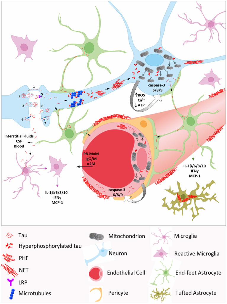
Alzheimer's disease (AD) is a chronic neurodegenerative disorder and the most prevalent cause of dementia. The main cerebral histological hallmarks are represented by parenchymal insoluble deposits of amyloid beta (Aβ plaques) and neurofibrillary tangles (NFT), intracellular filamentous inclusions of tau, a microtubule-associated protein. It is well-established that cerebrovascular dysfunction is an early feature of AD pathology, but the detrimental mechanisms leading to blood vessel impairment and the associated neurovascular deregulation are not fully understood. In 90% of AD cases, Aβ deposition around the brain vasculature, known as cerebral amyloid angiopathy (CAA), alters blood brain barrier (BBB) essential functions. While the effects of vascular Aβ accumulation are better documented, the scientific community has only recently started to consider the impact of tau on neurovascular pathology in AD. Emerging compelling evidence points to transmission of neuronal tau to different brain cells, including astrocytes, as well as to the release of tau into brain interstitial fluids, which may lead to perivascular neurofibrillar tau accumulation and toxicity, affecting vessel architecture, cerebral blood flow (CBF), and vascular permeability. BBB integrity and functionality may therefore be impacted by pathological tau, consequentially accelerating the progression of the disease. Tau aggregates have also been shown to induce mitochondrial damage: it is known that tau impairs mitochondrial localization, distribution and dynamics, alters ATP and reactive oxygen species production, and compromises oxidative phosphorylation systems. In light of this previous knowledge, we postulate that tau can initiate neurovascular pathology in AD through mitochondrial dysregulation. In this review, we will explore the literature investigating tau pathology contribution to the malfunction of the brain vasculature and neurovascular unit, and its association with mitochondrial alterations and caspase activation, in cellular, animal, and human studies of AD and tauopathies.
Overview
The claims and statistics of this paper haven’t been checked yet.
Run the check — free, no account needed. It reads the open-access text and checks the sources it cites.
Integrity signals
Registry
Cites
Statistics
Figures
Verification
Claims the paper makes
The claims and statistics of this paper haven’t been checked yet.
Run the check — free, no account needed. It reads the open-access text and checks the sources it cites.
Claims other papers make about this one
Each is checked against this paper’s own text: does it say what the citing paper says it does?
The source backs part of the claim, not all of it — or backs it for a different population, condition or comparison.
In the citing paper
“which have been demonstrated to play an important role in neurodevelopment and neurodegenerative diseases like AD108.”
From GRAMD1B is a regulator of lipid homeostasis, autophagic flux and phosphorylated tau (2025) · cites this paper as [108]
Experimental — accuracy not yet measured
Self-check
Percentages, sums and reported differences re-computed from the paper’s own numbers.
The self-check needs the paper’s full text.
Citations
Cites
Neuropathological stageing of Alzheimer-related changes.
Braak, H., & Braak, E. (1991) · Acta Neuropathologica · doi:10.1007/bf00308809 · Cited by 16,123
Zhang, Y., Chen, K., Sloan, S. A., Bennett, M. L., Scholze, A. R., O'Keeffe, S., Phatnani, H. P., Guarnieri, P., Caneda, C., Ruderisch, N., et al. (2014) · The Journal of Neuroscience · doi:10.1523/jneurosci.1860-14.2014 · Cited by 4,875
Neurovascular pathways to neurodegeneration in Alzheimer’s disease and other disorders
Zlokovic, B. V. (2011) · Nature Reviews Neuroscience · doi:10.1038/nrn3114 · Cited by 2,969
The blood-brain barrier/neurovascular unit in health and disease
Hawkins, B. T., & Davis, T. P. (2005) · Pharmacological Reviews · doi:10.1124/pr.57.2.4 · Cited by 2,661
Neurovascular regulation in the normal brain and in Alzheimer's disease
Iadecola, C. (2004) · Nature Reviews Neuroscience · doi:10.1038/nrn1387 · Cited by 2,213
Self-propagation of pathogenic protein aggregates in neurodegenerative diseases
Jucker, M., & Walker, L. C. (2013) · Nature · doi:10.1038/nature12481 · Cited by 1,663
Palmqvist, S., Janelidze, S., Quiroz, Y. T., Zetterberg, H., Lopera, F., Stomrud, E., Su, Y., Chen, Y., Serrano, G. E., Leuzy, A., et al. (2020) · JAMA · doi:10.1001/jama.2020.12134 · Cited by 1,603
Propagation of Tau Pathology in a Model of Early Alzheimer's Disease
de Calignon, A., Polydoro, M., Suárez-Calvet, M., William, C., Adamowicz, D. H., Kopeikina, K. J., Pitstick, R., Sahara, N., Ashe, K. H., Carlson, G. A., et al. (2012) · Neuron · doi:10.1016/j.neuron.2011.11.033 · Cited by 1,427
Patterns of Brain Activation in People at Risk for Alzheimer's Disease
Bookheimer, S. Y., Strojwas, M. H., Cohen, M. S., Saunders, A. M., Pericak-Vance, M. A., Mazziotta, J. C., & Small, G. W. (2000) · New England Journal of Medicine · doi:10.1056/nejm200008173430701 · Cited by 1,328
Propagation of Tau Misfolding from the Outside to the Inside of a Cell
Frost, B., Jacks, R. L., & Diamond, M. I. (2009) · Journal of Biological Chemistry · doi:10.1074/jbc.m808759200 · Cited by 1,252
Research works printed but not linked: 213
- Cummings, 2004, Alzheimer's disease, N Engl J Med., 351, 56, 10.1056/NEJMra040223 · DOI · Paper page
- Caselli, 2006, Alzheimer's disease a century later, J Clin Psychiatry., 67, 1784, 10.4088/JCP.v67n1118 · DOI
- Querfurth, 2010, Alzheimer's disease, N Engl J Med., 362, 329, 10.1056/NEJMra0909142 · DOI · Paper page
- Kalaria, 2000, The role of cerebral ischemia in Alzheimer's disease, Neurobiol Aging., 21, 321, 10.1016/S0197-4580(00)00125-1 · DOI
- de la Torre, 2004, Is Alzheimer's disease a neurodegenerative or a vascular disorder? Data, dogma, and dialectics, Lancet Neurol., 3, 184, 10.1016/S1474-4422(04)00683-0 · DOI · Paper page
- Zlokovic, 2005, Neurovascular mechanisms of Alzheimer's neurodegeneration, Trends Neurosci., 28, 202, 10.1016/j.tins.2005.02.001 · DOI · Paper page
- Finder, 2007, Amyloid-beta aggregation, Neurodegener Dis., 4, 13, 10.1159/000100355 · DOI
- Fossati, 2010, Differential activation of mitochondrial apoptotic pathways by vasculotropic amyloid-beta variants in cells composing the cerebral vessel walls, FASEB J., 24, 229, 10.1096/fj.09-139584 · DOI
- Muller-Hill, 1989, Molecular biology of Alzheimer's disease, Annu Rev Biochem., 58, 287, 10.1146/annurev.bi.58.070189.001443 · DOI
- Fossati, 2012, TRAIL death receptors DR4 and DR5 mediate cerebral microvascular endothelial cell apoptosis induced by oligomeric Alzheimer's abeta, Cell Death Dis., 3, e321, 10.1038/cddis.2012.55 · DOI
- Fossati, 2012, Insights into caspase-mediated apoptotic pathways induced by amyloid-beta in cerebral microvascular endothelial cells, Neurodegener Dis., 10, 324, 10.1159/000332821 · DOI
- Ghiso, 2014, Amyloidosis associated with cerebral amyloid angiopathy: cell signaling pathways elicited in cerebral endothelial cells, J Alzheimers Dis., 42, S167, 10.3233/JAD-140027 · DOI
- Fossati, 2013, Differential contribution of isoaspartate post-translational modifications to the fibrillization and toxic properties of amyloid beta and the Asn23 Iowa mutation, Biochem J., 456, 347, 10.1042/BJ20130652 · DOI
- Drubin, 1986, Tau protein function in living cells, J Cell Biol., 103, 2739, 10.1083/jcb.103.6.2739 · DOI
- Drechsel, 1992, Modulation of the dynamic instability of tubulin assembly by the microtubule-associated protein tau, Mol Biol Cell., 3, 1141, 10.1091/mbc.3.10.1141 · DOI
- Khatoon, 1992, Brain levels of microtubule-associated protein tau are elevated in Alzheimer's disease: a radioimmuno-slot-blot assay for nanograms of the protein, J Neurochem., 59, 750, 10.1111/j.1471-4159.1992.tb09432.x · DOI
- Khatoon, 1994, Levels of normal and abnormally phosphorylated tau in different cellular and regional compartments of Alzheimer disease and control brains, FEBS Lett., 351, 80, 10.1016/0014-5793(94)00829-9 · DOI
- Vidal, 2000, Senile dementia associated with amyloid beta protein angiopathy and tau perivascular pathology but not neuritic plaques in patients homozygous for the APOE-epsilon4 allele, Acta Neuropathol., 100, 1, 10.1007/s004010051186 · DOI
- Kovac, 2009, Human truncated tau is using a different mechanism from amyloid-beta to damage the blood-brain barrier, J Alzheimers Dis., 18, 897, 10.3233/JAD-2009-1197 · DOI
- Merlini, 2016, Tau pathology-dependent remodelling of cerebral arteries precedes Alzheimer's disease-related microvascular cerebral amyloid angiopathy, Acta Neuropathol., 131, 737, 10.1007/s00401-016-1560-2 · DOI
- Blair, 2015, Tau depletion prevents progressive blood-brain barrier damage in a mouse model of tauopathy, Acta Neuropathol Commun., 3, 8, 10.1186/s40478-015-0186-2 · DOI · Paper page
- Farquhar, 1963, Junctional complexes in various epithelia, J Cell Biol., 17, 375, 10.1083/jcb.17.2.375 · DOI
- Abbott, 2010, Structure and function of the blood-brain barrier, Neurobiol Dis., 37, 13, 10.1016/j.nbd.2009.07.030 · DOI · Paper page
- Oksanen, 2019, Astrocyte alterations in neurodegenerative pathologies and their modeling in human induced pluripotent stem cell platforms, Cell Mol Life Sci., 76, 2739, 10.1007/s00018-019-03111-7 · DOI
- Farina, 2007, Astrocytes are active players in cerebral innate immunity, Trends Immunol., 28, 138, 10.1016/j.it.2007.01.005 · DOI · Paper page
- Streit, 2005, Role of microglia in the central nervous system's immune response, Neurol Res., 27, 685, 10.1179/016164105X49463 · DOI
- Ajami, 2007, Local self-renewal can sustain CNS microglia maintenance and function throughout adult life, Nat Neurosci., 10, 1538, 10.1038/nn2014 · DOI · Paper page
- Persidsky, 1999, Microglial and astrocyte chemokines regulate monocyte migration through the blood-brain barrier in human immunodeficiency virus-1 encephalitis, Am J Pathol., 155, 1599, 10.1016/S0002-9440(10)65476-4 · DOI
- Hudson, 2005, Astrocytes and microglia differentially regulate trafficking of lymphocyte subsets across brain endothelial cells, Brain Res., 1058, 148, 10.1016/j.brainres.2005.07.071 · DOI
- Rogers, 1988, Expression of immune system-associated antigens by cells of the human central nervous system: relationship to the pathology of Alzheimer's disease, Neurobiol Aging., 9, 339, 10.1016/S0197-4580(88)80079-4 · DOI · Paper page
- Sheng, 2001, Interleukin-1 promotion of MAPK-p38 overexpression in experimental animals and in Alzheimer's disease: potential significance for tau protein phosphorylation, Neurochem Int., 39, 341, 10.1016/S0197-0186(01)00041-9 · DOI · Paper page
- Hayes, 2002, Pathological relationships between microglial cell activity and tau and amyloid beta protein in patients with Alzheimer's disease, Neurosci Lett., 331, 171, 10.1016/S0304-3940(02)00888-1 · DOI
- Ostergaard, 2013, The capillary dysfunction hypothesis of Alzheimer's disease, Neurobiol Aging., 34, 1018, 10.1016/j.neurobiolaging.2012.09.011 · DOI
- Buee, 1997, Brain microvascular changes in Alzheimer's disease and other dementias, Ann N Y Acad Sci., 826, 7, 10.1111/j.1749-6632.1997.tb48457.x · DOI
- Bell, 2009, Neurovascular mechanisms and blood-brain barrier disorder in Alzheimer's disease, Acta Neuropathol., 118, 103, 10.1007/s00401-009-0522-3 · DOI
- Dehmelt, 2005, The MAP2/Tau family of microtubule-associated proteins, Genome Biol., 6, 204, 10.1186/gb-2004-6-1-204 · DOI · Paper page
- Halpain, 2006, The MAP1 family of microtubule-associated proteins, Genome Biol., 7, 224, 10.1186/gb-2006-7-6-224 · DOI · Paper page
- Ding, 2006, Site-specific phosphorylation and caspase cleavage differentially impact tau-microtubule interactions and tau aggregation, J Biol Chem., 281, 19107, 10.1074/jbc.M511697200 · DOI
- McMillan, 2011, Truncation of tau at E391 promotes early pathologic changes in transgenic mice, J Neuropathol Exp Neurol., 70, 1006, 10.1097/NEN.0b013e31823557fb · DOI
- Kolarova, 2012, Structure and pathology of tau protein in Alzheimer disease, Int J Alzheimers Dis., 2012, 731526, 10.1155/2012/731526 · DOI
- Dolan, 2010, The role of tau kinases in Alzheimer's disease, Curr Opin Drug Discov Devel., 13, 595
- Rodriguez-Martin, 2013, Tau phosphorylation affects its axonal transport and degradation, Neurobiol Aging., 34, 2146, 10.1016/j.neurobiolaging.2013.03.015 · DOI
- Binder, 1985, The distribution of tau in the mammalian central nervous system, J Cell Biol., 101, 1371, 10.1083/jcb.101.4.1371 · DOI · Paper page
- Arrasate, 2000, Tau dephosphorylation at tau-1 site correlates with its association to cell membrane, Neurochem Res., 25, 43, 10.1023/A:1007583214722 · DOI
- Maccioni, 2001, The protein kinase Cdk5. Structural aspects, roles in neurogenesis and involvement in Alzheimer's pathology, Eur J Biochem., 268, 1518, 10.1046/j.1432-1327.2001.02024.x · DOI
- Hernandez, 2013, GSK3 and tau: two convergence points in Alzheimer's disease, J Alzheimers Dis., 33, S141, 10.3233/JAD-2012-129025 · DOI · Paper page
- Wolfe, 2012, The role of tau in neurodegenerative diseases and its potential as a therapeutic target, Scientifica., 2012, 796024, 10.6064/2012/796024 · DOI
- Liu, 2016, The role of Cdk5 in Alzheimer's disease, Mol Neurobiol., 53, 4328, 10.1007/s12035-015-9369-x · DOI
- Crespo-Biel, 2012, Protein tau: prime cause of synaptic and neuronal degeneration in Alzheimer's disease, Int J Alzheimers Dis., 2012, 251426, 10.1155/2012/251426 · DOI
- de Calignon, 2010, Caspase activation precedes and leads to neurodegeneration in a murine model of Alzheimer's disease, Med Sci., 26, 787, 10.1051/medsci/20102610787 · DOI
- Alonso, 2001, Hyperphosphorylation induces self-assembly of tau into tangles of paired helical filaments/straight filaments, Proc Natl Acad Sci USA., 98, 6923, 10.1073/pnas.121119298 · DOI · Paper page
- Liu, 2007, Site-specific effects of tau phosphorylation on its microtubule assembly activity and self-aggregation, Eur J Neurosci., 26, 3429, 10.1111/j.1460-9568.2007.05955.x · DOI · Paper page
- Liu, 2008, Tau exon 10 alternative splicing and tauopathies, Mol Neurodegener., 3, 8, 10.1186/1750-1326-3-8 · DOI · Paper page
- Beharry, 2014, Tau-induced neurodegeneration: mechanisms and targets, Neurosci Bull., 30, 346, 10.1007/s12264-013-1414-z · DOI
- Park, 2005, The generation of a 17 kDa neurotoxic fragment: an alternative mechanism by which tau mediates beta-amyloid-induced neurodegeneration, J Neurosci., 25, 5365, 10.1523/JNEUROSCI.1125-05.2005 · DOI · Paper page
- Hanger, 2010, Tau cleavage and tau aggregation in neurodegenerative disease, Biochem Soc Trans., 38, 1016, 10.1042/BST0381016 · DOI
- D'Amelio, 2011, Caspase-3 triggers early synaptic dysfunction in a mouse model of Alzheimer's disease, Nat Neurosci., 14, 69, 10.1038/nn.2709 · DOI · Paper page
- Rohn, 2002, Caspase-9 activation and caspase cleavage of tau in the Alzheimer's disease brain, Neurobiol Dis., 11, 341, 10.1006/nbdi.2002.0549 · DOI
- Goedert, 2010, The propagation of prion-like protein inclusions in neurodegenerative diseases, Trends Neurosci., 33, 317, 10.1016/j.tins.2010.04.003 · DOI
- Yamada, 2011, In vivo microdialysis reveals age-dependent decrease of brain interstitial fluid tau levels in P301S human tau transgenic mice, J Neurosci., 31, 13110, 10.1523/JNEUROSCI.2569-11.2011 · DOI · Paper page
- Johnson, 1997, The tau protein in human cerebrospinal fluid in Alzheimer's disease consists of proteolytically derived fragments, J Neurochem., 68, 430, 10.1046/j.1471-4159.1997.68010430.x · DOI · Paper page
- Dage, 2016, Levels of tau protein in plasma are associated with neurodegeneration and cognitive function in a population-based elderly cohort, Alzheimers Dement., 12, 1226, 10.1016/j.jalz.2016.06.001 · DOI · Paper page
- Deters, 2017, Plasma tau association with brain atrophy in mild cognitive impairment and Alzheimer's disease, J Alzheimers Dis., 58, 1245, 10.3233/JAD-161114 · DOI
- Fossati, 2019, Plasma tau complements CSF tau and P-tau in the diagnosis of Alzheimer's disease, Alzheimers Dement., 11, 483, 10.1016/j.dadm.2019.05.001 · DOI
- Zetterberg, 2013, Plasma tau levels in Alzheimer's disease, Alzheimers Res Ther., 5, 9, 10.1186/alzrt163 · DOI
- Blennow, 2018, The past and the future of Alzheimer's disease fluid biomarkers, J Alzheimers Dis., 62, 1125, 10.3233/JAD-170773 · DOI
- Mielke, 2017, Association of plasma total tau level with cognitive decline and risk of mild cognitive impairment or dementia in the mayo clinic study on aging, JAMA Neurol., 74, 1073, 10.1001/jamaneurol.2017.1359 · DOI
- Karikari, 2020, Blood phosphorylated tau 181 as a biomarker for Alzheimer's disease: a diagnostic performance and prediction modelling study using data from four prospective cohorts, Lancet Neurol., 19, 422, 10.1016/S1474-4422(20)30071-5 · DOI · Paper page
- Janelidze, 2020, Plasma P-tau181 in Alzheimer's disease: relationship to other biomarkers, differential diagnosis, neuropathology and longitudinal progression to Alzheimer's dementia, Nat Med., 26, 379, 10.1038/s41591-020-0755-1 · DOI · Paper page
- Tarasoff-Conway, 2015, Clearance systems in the brain-implications for Alzheimer disease, Nat Rev Neurol., 11, 457, 10.1038/nrneurol.2015.119 · DOI · Paper page
- Carare, 2020, Clearance of interstitial fluid (ISF) and CSF (CLIC) group-part of vascular professional interest area (PIA): cerebrovascular disease and the failure of elimination of amyloid-beta from the brain and retina with age and Alzheimer's disease-opportunities for therapy, Alzheimers Dement., 12, e12053, 10.1002/dad2.12053 · DOI
- Furman, 2017, Widespread tau seeding activity at early braak stages, Acta Neuropathol., 133, 91, 10.1007/s00401-016-1644-z · DOI
- Kaufman, 2017, Characterization of tau prion seeding activity and strains from formaldehyde-fixed tissue, Acta Neuropathol Commun., 5, 41, 10.1186/s40478-017-0442-8 · DOI
- Pooler, 2013, Physiological release of endogenous tau is stimulated by neuronal activity, EMBO Rep., 14, 389, 10.1038/embor.2013.15 · DOI · Paper page
- Katsinelos, 2018, Unconventional secretion mediates the trans-cellular spreading of tau, Cell Rep., 23, 2039, 10.1016/j.celrep.2018.04.056 · DOI
- Merezhko, 2018, Secretion of tau via an unconventional non-vesicular mechanism, Cell Rep., 25, 2027, 10.1016/j.celrep.2018.10.078 · DOI · Paper page
- Polanco, 2016, Extracellular vesicles isolated from the brains of rTg4510 mice seed tau protein aggregation in a threshold-dependent manner, J Biol Chem., 291, 12445, 10.1074/jbc.M115.709485 · DOI
- Dujardin, 2014, Ectosomes: a new mechanism for non-exosomal secretion of tau protein, PLoS ONE., 9, e100760, 10.1371/journal.pone.0100760 · DOI
- Abounit, 2016, Tunneling nanotubes: a possible highway in the spreading of tau and other prion-like proteins in neurodegenerative diseases, Prion., 10, 344, 10.1080/19336896.2016.1223003 · DOI · Paper page
- Tardivel, 2016, Tunneling nanotube (TNT)-mediated neuron-to neuron transfer of pathological tau protein assemblies, Acta Neuropathol Commun., 4, 117, 10.1186/s40478-016-0386-4 · DOI · Paper page
- Santa-Maria, 2012, Paired helical filaments from Alzheimer disease brain induce intracellular accumulation of tau protein in aggresomes, J Biol Chem., 287, 20522, 10.1074/jbc.M111.323279 · DOI
- Wu, 2013, Small misfolded Tau species are internalized via bulk endocytosis and anterogradely and retrogradely transported in neurons, J Biol Chem., 288, 1856, 10.1074/jbc.M112.394528 · DOI · Paper page
- Calafate, 2016, Loss of bin1 promotes the propagation of tau pathology, Cell Rep., 17, 931, 10.1016/j.celrep.2016.09.063 · DOI · Paper page
- Evans, 2018, Extracellular monomeric and aggregated tau efficiently enter human neurons through overlapping but distinct pathways, Cell Rep., 22, 3612, 10.1016/j.celrep.2018.03.021 · DOI
- Falcon, 2015, Conformation determines the seeding potencies of native and recombinant Tau aggregates, J Biol Chem., 290, 1049, 10.1074/jbc.M114.589309 · DOI
- Falcon, 2018, Galectin-8-mediated selective autophagy protects against seeded tau aggregation, J Biol Chem., 293, 2438, 10.1074/jbc.M117.809293 · DOI
- Wu, 2016, Neuronal activity enhances tau propagation and tau pathology in vivo, Nat Neurosci, 19, 1085, 10.1038/nn.4328 · DOI · Paper page
- Togo, 2002, Tau accumulation in astrocytes in progressive supranuclear palsy is a degenerative rather than a reactive process, Acta Neuropathol., 104, 398, 10.1007/s00401-002-0569-x · DOI
- Buee, 1999, Comparative biochemistry of tau in progressive supranuclear palsy, corticobasal degeneration, FTDP-17 and pick's disease, Brain Pathol., 9, 681, 10.1111/j.1750-3639.1999.tb00550.x · DOI
- Ferrer, 2018, Aging-related tau astrogliopathy (ARTAG): not only tau phosphorylation in astrocytes, Brain Pathol., 28, 965, 10.1111/bpa.12593 · DOI
- Odawara, 1995, Investigation of tau-2 positive microglia-like cells in the subcortical nuclei of human neurodegenerative disorders, Neurosci Lett., 192, 145, 10.1016/0304-3940(95)11595-N · DOI · Paper page
- Leyns, 2017, Glial contributions to neurodegeneration in tauopathies, Mol Neurodegener., 12, 50, 10.1186/s13024-017-0192-x · DOI · Paper page
- Hallmann, 2017, Astrocyte pathology in a human neural stem cell model of frontotemporal dementia caused by mutant TAU protein, Sci Rep., 7, 42991, 10.1038/srep42991 · DOI
- Walker, 2020, Glial tauopathy: neurons optional?, J Exp Med., 217, e20191915, 10.1084/jem.20191915 · DOI
- Martini-Stoica, 2018, TFEB enhances astroglial uptake of extracellular tau species and reduces tau spreading, J Exp Med., 215, 2355, 10.1084/jem.20172158 · DOI · Paper page
- Asai, 2015, Depletion of microglia and inhibition of exosome synthesis halt tau propagation, Nat Neurosci., 18, 1584, 10.1038/nn.4132 · DOI · Paper page
- De Marco, Cell-based assay to study antibody-mediated tau clearance by microglia, J Vis Exp, 10.3791/58576 · DOI
- Nilson, 2017, Tau oligomers associate with inflammation in the brain and retina of tauopathy mice and in neurodegenerative diseases, J Alzheimers Dis., 55, 1083, 10.3233/JAD-160912 · DOI
- Jaworski, 2011, Dendritic degeneration, neurovascular defects, and inflammation precede neuronal loss in a mouse model for tau-mediated neurodegeneration, Am J Pathol., 179, 2001, 10.1016/j.ajpath.2011.06.025 · DOI
- Bennett, 2018, Tau induces blood vessel abnormalities and angiogenesis-related gene expression in P301L transgenic mice and human Alzheimer's disease, Proc Natl Acad Sci USA., 115, E1289, 10.1073/pnas.1710329115 · DOI · Paper page
- Kim, 2018, Assessment of extent and role of tau in subcortical vascular cognitive impairment using 18F-AV1451 positron emission tomography imaging, JAMA Neurol., 75, 999, 10.1001/jamaneurol.2018.0975 · DOI
- Majerova, 2018, Cerebrovascular inflammation is associated with tau pathology in guam parkinsonism dementia, J Neural Transm., 125, 1013, 10.1007/s00702-018-1883-3 · DOI
- Castillo-Carranza, 2015, Tau immunotherapy modulates both pathological tau and upstream amyloid pathology in an Alzheimer's disease mouse model, J Neurosci., 35, 4857, 10.1523/JNEUROSCI.4989-14.2015 · DOI · Paper page
- Laurent, 2017, Hippocampal T cell infiltration promotes neuroinflammation and cognitive decline in a mouse model of tauopathy, Brain., 140, 184, 10.1093/brain/aww270 · DOI · Paper page
- Majerova, 2019, Trafficking of immune cells across the blood-brain barrier is modulated by neurofibrillary pathology in tauopathies, PLoS ONE., 14, e0217216, 10.1371/journal.pone.0217216 · DOI
- Davalos, 2012, Fibrinogen-induced perivascular microglial clustering is required for the development of axonal damage in neuroinflammation, Nat Commun., 3, 1227, 10.1038/ncomms2230 · DOI · Paper page
- Lecrux, 2011, The neurovascular unit in brain function and disease, Acta Physiol., 203, 47, 10.1111/j.1748-1716.2011.02256.x · DOI · Paper page
- Wang, 2013, Expression of Tau40 induces activation of cultured rat microglial cells, PLoS ONE., 8, e76057, 10.1371/journal.pone.0076057 · DOI · Paper page
- Forman, 2005, Transgenic mouse model of tau pathology in astrocytes leading to nervous system degeneration, J Neurosci., 25, 3539, 10.1523/JNEUROSCI.0081-05.2005 · DOI · Paper page
- Castillo-Carranza, 2017, Cerebral microvascular accumulation of tau oligomers in Alzheimer's disease and related tauopathies, Aging Dis., 8, 257, 10.14336/AD.2017.0112 · DOI · Paper page
- Rohn, 2013, Immunolocalization of an amino-terminal fragment of apolipoprotein E in the pick's disease brain, PLoS ONE., 8, e80180, 10.1371/journal.pone.0080180 · DOI
- Buee, 1994, Pathological alterations of the cerebral microvasculature in Alzheimer's disease and related dementing disorders, Acta Neuropathol., 87, 469, 10.1007/BF00294173 · DOI
- Arima, 2006, Ultrastructural characteristics of tau filaments in tauopathies: immuno-electron microscopic demonstration of tau filaments in tauopathies, Neuropathology., 26, 475, 10.1111/j.1440-1789.2006.00669.x · DOI
- Arima, 1997, Ultrastructural characterization of the tau-immunoreactive tubules in the oligodendroglial perikarya and their inner loop processes in progressive supranuclear palsy, Acta Neuropathol., 93, 558, 10.1007/s004010050652 · DOI
- Kouri, 2011, Corticobasal degeneration: a pathologically distinct 4R tauopathy, Nat Rev Neurol., 7, 263, 10.1038/nrneurol.2011.43 · DOI · Paper page
- Takeda, 2012, Pick's disease, Adv Exp Med Biol., 724, 300, 10.1007/978-1-4614-0653-2_23 · DOI
- Trickler, 2005, Brain microvessel endothelial cell responses to tumor necrosis factor-alpha involve a nuclear factor kappa B (NF-kappaB) signal transduction pathway, Brain Res., 1048, 24, 10.1016/j.brainres.2005.04.028 · DOI
- Wu, 2010, Ac-YVAD-CMK decreases blood-brain barrier degradation by inhibiting caspase-1 activation of interleukin-1beta in intracerebral hemorrhage mouse model, Transl Stroke Res., 1, 57, 10.1007/s12975-009-0002-z · DOI
- Ohara, 2000, Adrenergic mediation of TNF alpha-stimulated ICAM-1 expression on human brain microvascular endothelial cells, Acta Neurochir Suppl., 76, 117, 10.1007/978-3-7091-6346-7_24 · DOI
- Ruprecht, 2001, Effects of oncostatin M on human cerebral endothelial cells and expression in inflammatory brain lesions, J Neuropathol Exp Neurol., 60, 1087, 10.1093/jnen/60.11.1087 · DOI
- Sonar, 2017, Differentiation and transmigration of CD4 T cells in neuroinflammation and autoimmunity, Front Immunol., 8, 1695, 10.3389/fimmu.2017.01695 · DOI
- Koren, 2009, Chaperone signalling complexes in Alzheimer's disease, J Cell Mol Med., 13, 619, 10.1111/j.1582-4934.2008.00557.x · DOI
- Leszczynski, 2002, Non-thermal activation of the hsp27/p38MAPK stress pathway by mobile phone radiation in human endothelial cells: molecular mechanism for cancer- and blood-brain barrier-related effects, Differentiation., 70, 120, 10.1046/j.1432-0436.2002.700207.x · DOI
- Isogai, 2001, Plasminogen activator inhibitor-1 promotes angiogenesis by stimulating endothelial cell migration toward fibronectin, Cancer Res., 61, 5587
- Czekay, 2011, PAI-1: an integrator of cell signaling and migration, Int J Cell Biol., 2011, 562481, 10.1155/2011/562481 · DOI
- Jeon, 2012, Plasminogen activator inhibitor type 1 regulates microglial motility and phagocytic activity, J Neuroinflammation., 9, 149, 10.1186/1742-2094-9-149 · DOI
- Stewart, 1992, A morphometric study of the blood-brain barrier in Alzheimer's disease, Lab Invest., 67, 734
- Claudio, 1996, Ultrastructural features of the blood-brain barrier in biopsy tissue from Alzheimer's disease patients, Acta Neuropathol., 91, 6, 10.1007/s004010050386 · DOI
- Miyakawa, 1988, Morphological changes of microvessels in the brain with Alzheimer's disease, Jpn J Psychiatry Neurol., 42, 819, 10.1111/j.1440-1819.1988.tb01171.x · DOI
- Hashimura, 1991, Morphological changes of blood vessels in the brain with Alzheimer's disease, Jpn J Psychiatry Neurol., 45, 661, 10.1111/j.1440-1819.1991.tb01187.x · DOI
- Kimura, 1991, Observations of microvessels in the brain with Alzheimer's disease by the scanning electron microscopy, Jpn J Psychiatry Neurol., 45, 671, 10.1111/j.1440-1819.1991.tb01189.x · DOI
- Higuchi, 1987, Ultrastructural changes of blood vessels in the cerebral cortex in Alzheimer's disease, Jpn J Psychiatry Neurol., 41, 283, 10.1111/j.1440-1819.1987.tb00414.x · DOI
- Yamashita, 1991, Vascular changes in the brains with Alzheimer's disease, Jpn J Psychiatry Neurol., 45, 79, 10.1111/j.1440-1819.1991.tb00509.x · DOI
- Greenberg, 2004, Amyloid angiopathy-related vascular cognitive impairment, Stroke., 35, 2616, 10.1161/01.STR.0000143224.36527.44 · DOI · Paper page
- Vinters, 2003, Amyloidosis of cerebral arteries, Adv Neurol., 92, 105
- Fann, 2018, Long-term risk of dementia among people with traumatic brain injury in Denmark: a population-based observational cohort study, Lancet Psychiatry., 5, 424, 10.1016/S2215-0366(18)30065-8 · DOI · Paper page
- Katsumoto, 2019, Tau pathology in chronic traumatic encephalopathy and Alzheimer's disease: similarities and differences, Front Neurol., 10, 980, 10.3389/fneur.2019.00980 · DOI
- McKee, 2020, The neuropathology of chronic traumatic encephalopathy: the status of the literature, Semin Neurol., 40, 359, 10.1055/s-0040-1713632 · DOI
- Stein, 2014, Chronic traumatic encephalopathy: a spectrum of neuropathological changes following repetitive brain trauma in athletes and military personnel, Alzheimers Res Ther., 6, 4, 10.1186/alzrt234 · DOI
- Schultz, 2004, High prevalence of thorn-shaped astrocytes in the aged human medial temporal lobe, Neurobiol Aging., 25, 397, 10.1016/S0197-4580(03)00113-1 · DOI
- McAleese, 2015, Cortical tau load is associated with white matter hyperintensities, Acta Neuropathol Commun., 3, 60, 10.1186/s40478-015-0240-0 · DOI · Paper page
- Nunnari, 2012, Mitochondria: in sickness and in health, Cell., 148, 1145, 10.1016/j.cell.2012.02.035 · DOI · Paper page
- Chen, 2009, Mitochondrial dynamics–fusion, fission, movement, and mitophagy–in neurodegenerative diseases, Hum Mol Genet., 18, R169, 10.1093/hmg/ddp326 · DOI
- Santos, 2010, A synergistic dysfunction of mitochondrial fission/fusion dynamics and mitophagy in Alzheimer's disease, J Alzheimers Dis., 20, S401, 10.3233/JAD-2010-100666 · DOI
- Itoh, 2013, Mitochondrial dynamics in neurodegeneration, Trends Cell Biol., 23, 64, 10.1016/j.tcb.2012.10.006 · DOI
- Kageyama, 2011, Mitochondrial division: molecular machinery and physiological functions, Curr Opin Cell Biol., 23, 427, 10.1016/j.ceb.2011.04.009 · DOI
- Picard, 2013, Mitochondrial morphology transitions and functions: implications for retrograde signaling?, Am J Physiol Regul Integr Comp Physiol., 304, R393, 10.1152/ajpregu.00584.2012 · DOI
- Wang, 2014, Oxidative stress and mitochondrial dysfunction in Alzheimer's disease, Biochim Biophys Acta., 1842, 1240, 10.1016/j.bbadis.2013.10.015 · DOI · Paper page
- Li, 2016, Human wild-type full-length tau accumulation disrupts mitochondrial dynamics and the functions via increasing mitofusins, Sci Rep., 6, 24756, 10.1038/srep24756 · DOI
- Placido, 2017, Phosphatase 2A inhibition affects endoplasmic reticulum and mitochondria homeostasis via cytoskeletal alterations in brain endothelial cells, Mol Neurobiol., 54, 154, 10.1007/s12035-015-9640-1 · DOI
- Kandimalla, 2016, Reduced dynamin-related protein 1 protects against phosphorylated Tau-induced mitochondrial dysfunction and synaptic damage in Alzheimer's disease, Hum Mol Genet., 25, 4881, 10.1093/hmg/ddw312 · DOI
- Manczak, 2012, Abnormal interaction between the mitochondrial fission protein Drp1 and hyperphosphorylated tau in Alzheimer's disease neurons: implications for mitochondrial dysfunction and neuronal damage, Hum Mol Genet., 21, 2538, 10.1093/hmg/dds072 · DOI
- Manczak, 2011, Impaired mitochondrial dynamics and abnormal interaction of amyloid beta with mitochondrial protein Drp1 in neurons from patients with Alzheimer's disease: implications for neuronal damage, Hum Mol Genet., 20, 2495, 10.1093/hmg/ddr139 · DOI
- Bonda, 2010, Mitochondrial dynamics in Alzheimer's disease: opportunities for future treatment strategies, Drugs Aging., 27, 181, 10.2165/11532140-000000000-00000 · DOI
- Kopeikina, 2011, Tau accumulation causes mitochondrial distribution deficits in neurons in a mouse model of tauopathy and in human Alzheimer's disease brain, Am J Pathol., 179, 2071, 10.1016/j.ajpath.2011.07.004 · DOI
- Jacobson, 2004, Interplay between mitochondria and cellular calcium signalling, Mol Cell Biochem., 209, 10.1023/B:MCBI.0000009869.29827.df · DOI
- Mattson, 2008, Mitochondria in neuroplasticity and neurological disorders, Neuron., 60, 748, 10.1016/j.neuron.2008.10.010 · DOI · Paper page
- Parodi-Rullan, 2019, Endothelial mitochondrial dysfunction in cerebral amyloid angiopathy and Alzheimer's disease, J Alzheimers Dis., 72, 1019, 10.3233/JAD-190357 · DOI
- Fossati, 2016, The carbonic anhydrase inhibitor methazolamide prevents amyloid beta-induced mitochondrial dysfunction and caspase activation protecting neuronal and glial cells in vitro and in the mouse brain, Neurobiol Dis., 86, 29, 10.1016/j.nbd.2015.11.006 · DOI
- Solesio, 2018, Carbonic anhydrase inhibition selectively prevents amyloid beta neurovascular mitochondrial toxicity, Aging Cell, 17, e12787, 10.1111/acel.12787 · DOI · Paper page
- Calkins, 2011, Impaired mitochondrial biogenesis, defective axonal transport of mitochondria, abnormal mitochondrial dynamics and synaptic degeneration in a mouse model of Alzheimer's disease, Hum Mol Genet., 20, 4515, 10.1093/hmg/ddr381 · DOI · Paper page
- Balietti, 2013, Early selective vulnerability of synapses and synaptic mitochondria in the hippocampal CA1 region of the Tg2576 mouse model of Alzheimer's disease, J Alzheimers Dis., 34, 887, 10.3233/JAD-121711 · DOI
- Du, 2010, Early deficits in synaptic mitochondria in an Alzheimer's disease mouse model, Proc Natl Acad Sci USA., 107, 18670, 10.1073/pnas.1006586107 · DOI · Paper page
- Gibson, 2010, A mitocentric view of Alzheimer's disease suggests multi-faceted treatments, J Alzheimers Dis., 20, S591, 10.3233/JAD-2010-100336 · DOI · Paper page
- Varghese, 2011, Mitochondrial bioenergetics is defective in presymptomatic Tg2576 AD mice, Transl Neurosci., 2, 1, 10.2478/s13380-011-0011-8 · DOI
- Amadoro, 2010, A NH2 tau fragment targets neuronal mitochondria at AD synapses: possible implications for neurodegeneration, J Alzheimers Dis., 21, 445, 10.3233/JAD-2010-100120 · DOI
- David, 2005, Proteomic and functional analyses reveal a mitochondrial dysfunction in P301L tau transgenic mice, J Biol Chem., 280, 23802, 10.1074/jbc.M500356200 · DOI · Paper page
- Schulz, 2012, A new link to mitochondrial impairment in tauopathies, Mol Neurobiol., 46, 205, 10.1007/s12035-012-8308-3 · DOI
- Britti, 2020, Tau inhibits mitochondrial calcium efflux and makes neurons vulnerable to calcium-induced cell death, Cell Calcium., 86, 102150, 10.1016/j.ceca.2019.102150 · DOI
- Mutisya, 1994, Cortical cytochrome oxidase activity is reduced in Alzheimer's disease, J Neurochem., 63, 2179, 10.1046/j.1471-4159.1994.63062179.x · DOI
- Schagger, 1995, Human diseases with defects in oxidative phosphorylation. 2. F1F0 ATP-synthase defects in Alzheimer disease revealed by blue native polyacrylamide gel electrophoresis, Eur J Biochem, 227, 916, 10.1111/j.1432-1033.1995.tb20219.x · DOI
- Kim, 2001, The reduction of NADH ubiquinone oxidoreductase 24- and 75-kDa subunits in brains of patients with Down syndrome and Alzheimer's disease, Life Sci., 68, 2741, 10.1016/S0024-3205(01)01074-8 · DOI
- Maurer, 2000, A selective defect of cytochrome c oxidase is present in brain of Alzheimer disease patients, Neurobiol Aging., 21, 455, 10.1016/S0197-4580(00)00112-3 · DOI · Paper page
- Wang, 2005, Increased oxidative damage in nuclear and mitochondrial DNA in Alzheimer's disease, J Neurochem., 93, 953, 10.1111/j.1471-4159.2005.03053.x · DOI
- Aliyev, 2005, Mitochondria DNA deletions in atherosclerotic hypoperfused brain microvessels as a primary target for the development of Alzheimer's disease, J Neurol Sci., 285, 10.1016/j.jns.2004.11.040 · DOI
- Haworth, 1979, The Ca2+-induced membrane transition in mitochondria. II. Nature of the Ca2+ trigger site, Arch Biochem Biophys., 195, 460, 10.1016/0003-9861(79)90372-2 · DOI · Paper page
- Hunter, 1979, The Ca2+-induced membrane transition in mitochondria. I. The protective mechanisms, Arch Biochem Biophys., 195, 453, 10.1016/0003-9861(79)90371-0 · DOI
- Hunter, 1979, The Ca2+-induced membrane transition in mitochondria. III. Transitional Ca2+ release, Arch Biochem Biophys., 195, 468, 10.1016/0003-9861(79)90373-4 · DOI · Paper page
- Kroemer, 2007, Mitochondrial cell death control in familial parkinson disease, PLoS Biol., 5, e206, 10.1371/journal.pbio.0050206 · DOI
- Bonora, 2013, Role of the c subunit of the FO ATP synthase in mitochondrial permeability transition, Cell Cycle., 12, 674, 10.4161/cc.23599 · DOI
- Bernardi, 2015, The mitochondrial permeability transition pore: molecular nature and role as a target in cardioprotection, J Mol Cell Cardiol., 78, 100, 10.1016/j.yjmcc.2014.09.023 · DOI
- Bernardi, 2015, From ATP to PTP and back: a dual function for the mitochondrial ATP synthase, Circ Res., 116, 1850, 10.1161/CIRCRESAHA.115.306557 · DOI
- Jonas, 2015, Cell death disguised: the mitochondrial permeability transition pore as the c-subunit of the F(1)F(O) ATP synthase, Pharmacol Res., 99, 382, 10.1016/j.phrs.2015.04.013 · DOI
- Rao, 2014, Mitochondrial permeability transition pore is a potential drug target for neurodegeneration, Biochim Biophys Acta., 1842, 1267, 10.1016/j.bbadis.2013.09.003 · DOI
- Manczak, 2012, Abnormal interaction of VDAC1 with amyloid beta and phosphorylated tau causes mitochondrial dysfunction in Alzheimer's disease, Hum Mol Genet., 21, 5131, 10.1093/hmg/dds360 · DOI · Paper page
- Cerretti, 1992, Molecular cloning of the interleukin-1 beta converting enzyme, Science., 256, 97, 10.1126/science.1373520 · DOI
- Nicholson, 1995, Identification and inhibition of the ICE/CED-3 protease necessary for mammalian apoptosis, Nature., 376, 37, 10.1038/376037a0 · DOI
- Alnemri, 1996, Human ICE/CED-3 protease nomenclature, Cell., 87, 171, 10.1016/S0092-8674(00)81334-3 · DOI
- Liu, 1996, Induction of apoptotic program in cell-free extracts: requirement for dATP and cytochrome c, Cell., 86, 147, 10.1016/S0092-8674(00)80085-9 · DOI · Paper page
- Thornberry, 1998, Caspases: enemies within, Science., 281, 1312, 10.1126/science.281.5381.1312 · DOI
- Hengartner, 2000, The biochemistry of apoptosis, Nature., 407, 770, 10.1038/35037710 · DOI · Paper page
- Shi, 2002, Mechanisms of caspase activation and inhibition during apoptosis, Mol Cell., 9, 459, 10.1016/S1097-2765(02)00482-3 · DOI
- Riedl, 2004, Molecular mechanisms of caspase regulation during apoptosis, Nat Rev Mol Cell Biol., 5, 897, 10.1038/nrm1496 · DOI
- Hyman, 2012, Apoptotic and non-apoptotic roles of caspases in neuronal physiology and pathophysiology, Nat Rev Neurosci., 13, 395, 10.1038/nrn3228 · DOI
- Bamberger, 2002, Inflammation, apoptosis, and Alzheimer's disease, Neuroscientist., 8, 276, 10.1177/1073858402008003013 · DOI
- Supnet, 2010, The dysregulation of intracellular calcium in Alzheimer disease, Cell Calcium., 47, 183, 10.1016/j.ceca.2009.12.014 · DOI
- Chen, 2012, Oxidative stress in neurodegenerative diseases, Neural Regen Res., 7, 376, 10.3969/j.issn.1673-5374.2012.05.009 · DOI
- Glushakova, 2018, Role of caspase-3-mediated apoptosis in chronic caspase-3-cleaved tau accumulation and blood-brain barrier damage in the corpus callosum after traumatic brain injury in rats, J Neurotrauma., 35, 157, 10.1089/neu.2017.4999 · DOI
- Selznick, 1999, In situ immunodetection of neuronal caspase-3 activation in Alzheimer disease, J Neuropathol Exp Neurol., 58, 1020, 10.1097/00005072-199909000-00012 · DOI
- Su, 2001, Activated caspase-3 expression in Alzheimer's and aged control brain: correlation with Alzheimer pathology, Brain Res., 898, 350, 10.1016/S0006-8993(01)02018-2 · DOI
200 of 213 shown
Cited by
Sorted by citations
Association of small vessel disease with tau pathology
Kapasi, A., Yu, L., Petyuk, V., Arfanakis, K., Bennett, D. A., & Schneider, J. A. (2022) · Acta Neuropathologica · doi:10.1007/s00401-021-02397-x · Cited by 55
The association between white matter hyperintensities and amyloid and tau deposition
Alban, S. L., Lynch, K. M., Ringman, J. M., Toga, A. W., Chui, H. C., Sepehrband, F., & Choupan, J. (2023) · NeuroImage: Clinical · doi:10.1016/j.nicl.2023.103383 · Cited by 35
GRAMD1B is a regulator of lipid homeostasis, autophagic flux and phosphorylated tau
Acosta Ingram, D., Turkes, E., Kim, T. Y., Vo, S., Sweeney, N., Bonte, M.-A., Rutherford, R., Julian, D. L., Pan, M., Marsh, J., et al. (2025) · Nature Communications · doi:10.1038/s41467-025-58585-w · Cited by 12
Metadata
- Authors
- Canepa, E., & Fossati, S.
- Venue
- Frontiers in Neurology
- Publisher
- Frontiers Media SA
- Published
- 2021-01-07
- Volume
- 11
- ISSN
- 1664-2295
- PMC
- PMC7817626
- Cited by
- 67
- References
- 58
- Funders
- National Institutes of Health
- Edward N. and Della L. Thome Memorial Foundation
- Alzheimer's Association
About these checks
Registry, retraction, Cites and statistics results are computed, not judged, with one exception: a printed reference that no index lists is read once by a model before it counts as a citation error. Claims are extracted by a model and kept only when their quote appears verbatim in the text; each cited source is then checked, with a model’s help, for whether it says what the claim relies on. Those verdicts can be wrong — read the claim’s quote and the cited source before relying on one.
Statistics and arithmetic are recomputed, not judged.
Confidence bands are shown only for fields calibrated on our annotated test set. None is calibrated yet, so no band is shown.
What counts as a claim ›How we measure confidence ›How we check this ›
Are you an author, or do you see a problem with this page? Request a review
