

The DOI matches its Crossref record; 178 of the 179 references it cites were found in a registry or an index; 1 of the 191 sources its claims cite were checked for support.
AMPK and Exercise: Glucose Uptake and Insulin Sensitivity
Open accessAbstract
AMPK is an evolutionary conserved sensor of cellular energy status that is activated during exercise. Pharmacological activation of AMPK promotes glucose uptake, fatty acid oxidation, mitochondrial biogenesis, and insulin sensitivity; processes that are reduced in obesity and contribute to the development of insulin resistance. AMPK deficient mouse models have been used to provide direct genetic evidence either supporting or refuting a role for AMPK in regulating these processes. Exercise promotes glucose uptake by an insulin dependent mechanism involving AMPK. Exercise is important for improving insulin sensitivity; however, it is not known if AMPK is required for these improvements. Understanding how these metabolic processes are regulated is important for the development of new strategies that target obesity-induced insulin resistance. This review will discuss the involvement of AMPK in regulating skeletal muscle metabolism (glucose uptake, glycogen synthesis, and insulin sensitivity).
Overview
AI snapshot
Written from the paper’s full text.
TL;DR
This review argues that AMPK is activated by exercise and is important for regulating skeletal muscle glucose uptake during exercise, while its requirement for exercise-induced improvements in insulin sensitivity remains unresolved. It synthesizes genetic mouse-model and human/rodent exercise evidence on AMPK, TBC1D1/TBC1D4, glycogen synthesis, and alternative insulin-sensitizing mechanisms.
Key findings
- AMPK is activated during exercise in an intensity-dependent manner: α2 activity increases from 40% VO2max, whereas α1 activity increases during high-intensity contractions equivalent to >100% VO2max.
- Muscle-specific AMPK β1β2 KO mice have undetectable AMPK activity and show reduced voluntary wheel activity (75%), maximal exercise capacity (95%), mitochondrial content (30%), and exercise-mediated skeletal muscle glucose uptake (55% to 70%); young mice have normal insulin sensitivity.
- TBC1D1 and TBC1D4 are implicated in contraction/exercise glucose uptake: TBC1D1 4P mutants reduce contraction-mediated glucose uptake by 25% to 30%, TBC1D4 4P mutant inhibits insulin-stimulated glucose uptake in vivo by ~50%, and TBC1D1 expression is reduced by ~50% in skeletal muscle of AMPK α2 KO, α2 KD, α2iTg, and β1β2 M-KO mice.
- Exercise improves insulin sensitivity, but whether AMPK is required is not known; proposed mechanisms include TBC1D4, TBC1D1, IL-6 (which can increase ~100-fold with exercise), adiponectin, glycogen synthesis/levels, and mTOR/p70 S6K signaling.
Methods
- Narrative review; the abstract states it will discuss AMPK involvement in skeletal muscle metabolism (glucose uptake, glycogen synthesis, insulin sensitivity).
- Synthesizes studies using whole-body and muscle-specific AMPK mouse models (α1, α2, β1, β2, γ3 KO; β1β2 M-KO; α2 KD/α2iTg; LKB1-MKO) plus human and rodent exercise/contraction studies.
- Main analysis is qualitative synthesis of cited literature; no systematic search, meta-analysis, or primary data collection is described.
Limitations
- The paper states that it is not known if AMPK is required for exercise-induced improvements in insulin sensitivity and that further studies are required to fully define the role of AMPK in mediating these effects.
- The paper notes inconsistent observations and unresolved mechanisms, including TBC1D4/TBC1D1 phosphorylation, IL-6 effects, and glycogen/AMPK relationships.
- Evident from the text: this is a narrative review without systematic methods or new data, so conclusions depend on the cited models and may be affected by isoform compensation, mouse strain/background differences, and reliance on pharmacological or supraphysiological stimuli.
Model-written summary. Check it against the paper before relying on it.
Integrity signals
Registry
Cites
Statistics
Figures
Verification
Claims the paper makes
111 Not checked yet · 22 Own interpretation · 2 No basis given · 1 Partly supported · accuracy not yet measured
The cited sources support only part of this claim — read each source’s verdict below.
In the paper
“Unlike TBC1D4, TBC1D1 requires 14-3-3 binding in response to contraction and AICAR but not insulin in both human and rodent skeletal muscle [74,117].”
§ AMPK and Exercise: Glucose Uptake and Insulin Sensitivity > EXERCISE AND INSULIN SENSITIVITY
What the sources say
- Partly supportedcites [74] Pehmoller, 2009, Am J Physiol Endocrinol Metab sourceExperimental — accuracy not yet measured
The Discussion reports that contraction and AICAR increase 14-3-3 binding to TBC1D1, whereas insulin does not, in mouse EDL muscle. This paper does not establish the same pattern in human muscle or show that 14-3-3 binding is required for a TBC1D1 response; the Discussion says its physiological significance is unclear. (LLM, conf=0.88)
- Not checked yetcites [117] Frosig, 2010, J Physiol source
plaindirect- Asserted: passes — The authors put it forward as true.
- Checkable: passes — It can be checked against evidence.
- Load-bearing: passes — The paper’s conclusions depend on it.
- Based: passes — The paper gives a reason why it holds.
- Partly supported
In the paper
“As anticipated, AMPK β1β2M-KO mice have undetectable levels of AMPK activity at rest or during exercise [30].”
§ AMPK and Exercise: Glucose Uptake and Insulin Sensitivity > AMPK MOUSE MODELS USED TO STUDY SKELETAL MUSCLE METABOLISM > AMPK β
What the sources say
- Not checked yetcites [30] O'Neill, 2011, Proc Natl Acad Sci U S A source
plain- Asserted: passes — The authors put it forward as true.
- Checkable: passes — It can be checked against evidence.
- Load-bearing: passes — The paper’s conclusions depend on it.
- Based: passes — The paper gives a reason why it holds.
In the paper
“Interestingly, young (9 to 12 weeks) AMPK β1β2M-KO mice have similar whole-body weight and adiposity, and consistent with this, whole-body and skeletal muscle specific insulin sensitivity is similar when compared to wild-type littermates [30].”
§ AMPK and Exercise: Glucose Uptake and Insulin Sensitivity > AMPK MOUSE MODELS USED TO STUDY SKELETAL MUSCLE METABOLISM > AMPK β
What the sources say
- Not checked yetcites [30] O'Neill, 2011, Proc Natl Acad Sci U S A source
plain- Asserted: passes — The authors put it forward as true.
- Checkable: passes — It can be checked against evidence.
- Load-bearing: passes — The paper’s conclusions depend on it.
- Based: passes — The paper gives a reason why it holds.
In the paper
“Importantly, GLUT4 translocation during exercise is normal in skeletal muscle of type 2 diabetics [64], and exercise also improves insulin sensitivity.”
§ AMPK and Exercise: Glucose Uptake and Insulin Sensitivity > AMPK AND EXERCISE
What the sources say
- Not checked yetcites [64] Kennedy, 1999, Diabetes source
plain- Asserted: passes — The authors put it forward as true.
- Checkable: passes — It can be checked against evidence.
- Load-bearing: passes — The paper’s conclusions depend on it.
- Based: passes — The paper gives a reason why it holds.
None of the sources cited for this claim have been checked yet.
In the paper
“Consistent with this, AMPK is activated during exercise in both rodents and humans [51-60], in an intensity-dependent manner [57,59-61].”
§ AMPK and Exercise: Glucose Uptake and Insulin Sensitivity > AMPK AND EXERCISE
What the sources say
- Not checked yetcites [51] Hayashi, 1998, Diabetes source
- Not checked yetcites [52] Hutber, 1997, Am J Physiol
- Not checked yetcites [53] Ihlemann, 1999, Am J Physiol
- Not checked yetcites [54] Rasmussen, 1998, J Appl Physiol source
- Not checked yetcites [55] Rasmussen, 1997, J Appl Physiol source
- Not checked yetcites [56] Vavvas, 1997, J Biol Chem source
- Not checked yetcites [57] Wojtaszewski, 2000, J Physiol source
- Not checked yetcites [58] Chen, 2003, Diabetes source
- Not checked yetcites [59] Fujii, 2000, Biochem Biophys Res Commun source
- Not checked yetcites [60] Winder, 1996, Am J Physiol
plaindirect- Asserted: passes — The authors put it forward as true.
- Checkable: passes — It can be checked against evidence.
- Load-bearing: passes — The paper’s conclusions depend on it.
- Based: passes — The paper gives a reason why it holds.
None of the sources cited for this claim have been checked yet.
In the paper
“Skeletal muscle is responsible for around 80% of insulin-stimulated glucose disposal [23-25]; therefore, responsiveness to insulin in this tissue is important for regulating whole-body insulin sensitivity.”
§ AMPK and Exercise: Glucose Uptake and Insulin Sensitivity > INTRODUCTION > Insulin resistance
What the sources say
plain- Asserted: passes — The authors put it forward as true.
- Checkable: passes — It can be checked against evidence.
- Load-bearing: passes — The paper’s conclusions depend on it.
- Based: passes — The paper gives a reason why it holds.
A conclusion the paper draws from its own results — it cites no other source for it, so there is none to check. The quote shows it is in the paper.
In the paper
“Skeletal muscle is responsible for around 80% of insulin-stimulated glucose disposal [23-25]; therefore, responsiveness to insulin in this tissue is important for regulating whole-body insulin sensitivity.”
§ AMPK and Exercise: Glucose Uptake and Insulin Sensitivity > INTRODUCTION > Insulin resistance
plaindirect- Asserted: passes — The authors put it forward as true.
- Checkable: passes — It can be checked against evidence.
- Load-bearing: passes — The paper’s conclusions depend on it.
- Based: passes — The paper gives a reason why it holds.
None of the sources cited for this claim have been checked yet.
In the paper
“AMPK α2 KO and α2KD mice have reduced AMPK T172 phosphorylation and α2 activity in skeletal muscle, but enhanced or modestly reduced α1 activity, respectively [31,33,34].”
§ AMPK and Exercise: Glucose Uptake and Insulin Sensitivity > AMPK MOUSE MODELS USED TO STUDY SKELETAL MUSCLE METABOLISM > AMPK α
What the sources say
plaindirect- Asserted: passes — The authors put it forward as true.
- Checkable: passes — It can be checked against evidence.
- Load-bearing: passes — The paper’s conclusions depend on it.
- Based: passes — The paper gives a reason why it holds.
None of the sources cited for this claim have been checked yet.
In the paper
“Both AMPK α1 and α2 are activated during exercise; however, α1 activity appears to be increased during high intensity muscle contractions, which are equivalent to >100% maximal oxygen consumption (VO2 Max; measure of maximal exercise capacity) [59,62,63].”
§ AMPK and Exercise: Glucose Uptake and Insulin Sensitivity > AMPK AND EXERCISE
What the sources say
hedgeddirect- Asserted: passes — The authors put it forward as true.
- Checkable: passes — It can be checked against evidence.
- Load-bearing: unclear — It is unclear whether the paper’s conclusions depend on it.
- Based: passes — The paper gives a reason why it holds.
In the paper
“Exercise can also regulate glucose uptake by an insulin-independent mechanism involving AMPK [30].”
§ AMPK and Exercise: Glucose Uptake and Insulin Sensitivity > AMPK AND EXERCISE
What the sources say
- Not checked yetcites [30] O'Neill, 2011, Proc Natl Acad Sci U S A source
plaindirect- Asserted: passes — The authors put it forward as true.
- Checkable: passes — It can be checked against evidence.
- Load-bearing: passes — The paper’s conclusions depend on it.
- Based: passes — The paper gives a reason why it holds.
- It may state two separate claims as one. 42
- Probably not a substantial claim. 4
In the paper
“Their phenotype is more dramatic compared to whole-body AMPK β2 KO mice [28] and is characterized by dramatically reduced voluntary wheel activity (75%), maximal exercise capacity (95%), mitochondrial content (30%) and exercise-mediated glucose uptake in skeletal muscle (55% to 70%) [30].”
§ AMPK and Exercise: Glucose Uptake and Insulin Sensitivity > AMPK MOUSE MODELS USED TO STUDY SKELETAL MUSCLE METABOLISM > AMPK β
What the sources say
- Not checked yetcites [30] O'Neill, 2011, Proc Natl Acad Sci U S A source
plaindirect- Asserted: passes — The authors put it forward as true.
- Checkable: passes — It can be checked against evidence.
- Load-bearing: passes — The paper’s conclusions depend on it.
- Based: passes — The paper gives a reason why it holds.
It may state two separate claims as one.
In the paper
“Interestingly, young (9 to 12 weeks) AMPK β1β2M-KO mice have similar whole-body weight and adiposity, and consistent with this, whole-body and skeletal muscle specific insulin sensitivity is similar when compared to wild-type littermates [30].”
§ AMPK and Exercise: Glucose Uptake and Insulin Sensitivity > AMPK MOUSE MODELS USED TO STUDY SKELETAL MUSCLE METABOLISM > AMPK β
What the sources say
- Not checked yetcites [30] O'Neill, 2011, Proc Natl Acad Sci U S A source
plain- Asserted: passes — The authors put it forward as true.
- Checkable: passes — It can be checked against evidence.
- Load-bearing: passes — The paper’s conclusions depend on it.
- Based: passes — The paper gives a reason why it holds.
It may state two separate claims as one.
The paper states this without a source or data behind it here, so there is nothing to check it against. The quote shows it is in the paper.
In the paper
“In contrast, α2 activity is increased at low intensity exercise starting at 40% VO2 Max and increases progressively with exercise intensity.”
§ AMPK and Exercise: Glucose Uptake and Insulin Sensitivity > AMPK AND EXERCISE
plaindirect- Asserted: passes — The authors put it forward as true.
- Checkable: passes — It can be checked against evidence.
- Load-bearing: unclear — It is unclear whether the paper’s conclusions depend on it.
- Based: passes — The paper gives a reason why it holds.
It may state two separate claims as one.
In the paper
“Interestingly, transgenic mice harboring the R225Q mutation in AMPK γ3 subunit have higher AMPK activity and glycogen levels and are protected from diet-induced insulin resistance [4], which is a likely effect of increased fatty acid oxidation and subsequent reduction in skeletal muscle lipids.”
§ AMPK and Exercise: Glucose Uptake and Insulin Sensitivity > AMPK MOUSE MODELS USED TO STUDY SKELETAL MUSCLE METABOLISM > AMPK γ
What the sources say
- Not checked yetcites [4] Barnes, 2004, J Biol Chem source
plaindirect- Asserted: passes — The authors put it forward as true.
- Checkable: passes — It can be checked against evidence.
- Load-bearing: unclear — It is unclear whether the paper’s conclusions depend on it.
- Based: passes — The paper gives a reason why it holds.
It may state two separate claims as one.
In the paper
“Whole-body AMPK α2 KO mice have reduced insulin secretion and whole-body insulin-stimulated glucose utilization due to elevated catecholamine levels and a hyperactive sympathetic nervous system [33].”
§ AMPK and Exercise: Glucose Uptake and Insulin Sensitivity > AMPK MOUSE MODELS USED TO STUDY SKELETAL MUSCLE METABOLISM > AMPK α
What the sources say
- Not checked yetcites [33] Viollet, 2003, J Clin Invest source
plaindirect- Asserted: passes — The authors put it forward as true.
- Checkable: passes — It can be checked against evidence.
- Load-bearing: passes — The paper’s conclusions depend on it.
- Based: passes — The paper gives a reason why it holds.
It may state two separate claims as one.
In the paper
“In contrast, AMPK β2 KO mice have reduced AMPK activity in skeletal muscle but not liver or adipose, and have a compensatory increase in AMPK β1 protein expression and maintained α1 expression in skeletal muscle [28].”
§ AMPK and Exercise: Glucose Uptake and Insulin Sensitivity > AMPK MOUSE MODELS USED TO STUDY SKELETAL MUSCLE METABOLISM > AMPK β
What the sources say
- Not checked yetcites [28] Steinberg, 2010, J Biol Chem source
plaindirect- Asserted: passes — The authors put it forward as true.
- Checkable: passes — It can be checked against evidence.
- Load-bearing: unclear — It is unclear whether the paper’s conclusions depend on it.
- Based: passes — The paper gives a reason why it holds.
It may state two separate claims as one.
In the paper
“However, an alternative muscle-specific mouse model on a model FVB background, rather than a C57Bl6 background, overexpressing a muscle-specific KD AMPKα2 B157A mutation (α2iTg), are glucose intolerant and develop exacerbated skeletal muscle insulin resistance when fed a high-fat diet for 30 weeks, which occurs independently of an increase in muscle lipids [37].”
§ AMPK and Exercise: Glucose Uptake and Insulin Sensitivity > AMPK MOUSE MODELS USED TO STUDY SKELETAL MUSCLE METABOLISM > AMPK α
What the sources say
- Not checked yetcites [37] Fujii, 2008, Diabetes source
plaindirect- Asserted: passes — The authors put it forward as true.
- Checkable: passes — It can be checked against evidence.
- Load-bearing: passes — The paper’s conclusions depend on it.
- Based: passes — The paper gives a reason why it holds.
It may state two separate claims as one.
In the paper
“AMPK β2 KO mice are characterized by reduced exercise capacity and increased susceptibility to develop insulin resistance on a high-fat diet possibly due to greater ceramide accumulation in skeletal muscle [28].”
§ AMPK and Exercise: Glucose Uptake and Insulin Sensitivity > AMPK MOUSE MODELS USED TO STUDY SKELETAL MUSCLE METABOLISM > AMPK β
What the sources say
- Not checked yetcites [28] Steinberg, 2010, J Biol Chem source
hedgeddirect- Asserted: passes — The authors put it forward as true.
- Checkable: passes — It can be checked against evidence.
- Load-bearing: passes — The paper’s conclusions depend on it.
- Based: passes — The paper gives a reason why it holds.
It may state two separate claims as one.
None of the sources cited for this claim have been checked yet.
In the paper
“Whole-body γ3 KO mice have reduced AMPK activity at rest, during exercise and in response to fasting, but normal insulin sensitivity under basal conditions and glucose uptake during exercise [4,41].”
§ AMPK and Exercise: Glucose Uptake and Insulin Sensitivity > AMPK MOUSE MODELS USED TO STUDY SKELETAL MUSCLE METABOLISM > AMPK γ
What the sources say
plain- Asserted: passes — The authors put it forward as true.
- Checkable: passes — It can be checked against evidence.
- Load-bearing: passes — The paper’s conclusions depend on it.
- Based: passes — The paper gives a reason why it holds.
It may state two separate claims as one.
A conclusion the paper draws from its own results — it cites no other source for it, so there is none to check. The quote shows it is in the paper.
In the paper
“These studies highlight an important role for AMPK in regulating exercise capacity, mitochondrial content and glucose uptake during exercise, but not insulin sensitivity.”
§ AMPK and Exercise: Glucose Uptake and Insulin Sensitivity > AMPK MOUSE MODELS USED TO STUDY SKELETAL MUSCLE METABOLISM > AMPK β
plaindirect- Asserted: passes — The authors put it forward as true.
- Checkable: passes — It can be checked against evidence.
- Load-bearing: passes — The paper’s conclusions depend on it.
- Based: passes — The paper gives a reason why it holds.
It may state two separate claims as one.
In the paper
“AMPK β1 KO mice have reduced AMPK activity in liver but not skeletal muscle, are hyperphagic and as a result have lower body weight, adiposity and hepatic lipid accumulation, and are protected from high-fat diet induced insulin resistance [27].”
§ AMPK and Exercise: Glucose Uptake and Insulin Sensitivity > AMPK MOUSE MODELS USED TO STUDY SKELETAL MUSCLE METABOLISM > AMPK β
What the sources say
- Not checked yetcites [27] Dzamko, 2010, J Biol Chem source
plaindirect- Asserted: passes — The authors put it forward as true.
- Checkable: passes — It can be checked against evidence.
- Load-bearing: unclear — It is unclear whether the paper’s conclusions depend on it.
- Based: passes — The paper gives a reason why it holds.
It may state two separate claims as one.
None of the sources cited for this claim have been checked yet.
In the paper
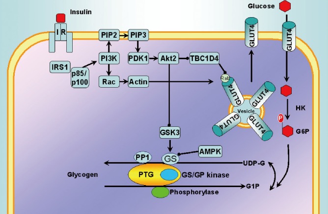
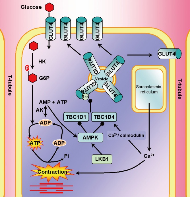
“1) PI3 kinase inhibitor wortmannin inhibits insulin but not contraction-stimulated glucose uptake. Contraction does not promote IRS-1 autophosphorylation or increase PI3 kinase activity [103-106].”
§ AMPK and Exercise: Glucose Uptake and Insulin Sensitivity > REGULATION OF GLUCOSE UPTAKE > Exercise and glucose uptake
What the sources say
plain- Asserted: passes — The authors put it forward as true.
- Checkable: passes — It can be checked against evidence.
- Load-bearing: passes — The paper’s conclusions depend on it.
- Based: passes — The paper gives a reason why it holds.
It may state two separate claims as one.
None of the sources cited for this claim have been checked yet.
In the paper
“1) PI3 kinase inhibitor wortmannin inhibits insulin but not contraction-stimulated glucose uptake. Contraction does not promote IRS-1 autophosphorylation or increase PI3 kinase activity [103-106].”
§ AMPK and Exercise: Glucose Uptake and Insulin Sensitivity > REGULATION OF GLUCOSE UPTAKE > Exercise and glucose uptake
What the sources say
plaindirect- Asserted: passes — The authors put it forward as true.
- Checkable: passes — It can be checked against evidence.
- Load-bearing: passes — The paper’s conclusions depend on it.
- Based: passes — The paper gives a reason why it holds.
It may state two separate claims as one.
In the paper
“In mice, whole-body mutation of S642 (S649 in mouse) to alanine (TBC1D4 T649A KI) reduces whole-body glucose and insulin intolerance and impairs insulin-stimulated glucose uptake and GLUT4 translocation in muscle [75].”
§ AMPK and Exercise: Glucose Uptake and Insulin Sensitivity > REGULATION OF GLUCOSE UPTAKE > TBC1D4
What the sources say
- Not checked yetcites [75] Chen, 2011, Cell Metab source
plaindirect- Asserted: passes — The authors put it forward as true.
- Checkable: passes — It can be checked against evidence.
- Load-bearing: passes — The paper’s conclusions depend on it.
- Based: passes — The paper gives a reason why it holds.
It may state two separate claims as one.
A conclusion the paper draws from its own results — it cites no other source for it, so there is none to check. The quote shows it is in the paper.
In the paper
“These studies suggest that targeting AMPK may enhance exercise performance and be protective against insulin resistance disease development in obesity.”
§ AMPK and Exercise: Glucose Uptake and Insulin Sensitivity > AMPK MOUSE MODELS USED TO STUDY SKELETAL MUSCLE METABOLISM > AMPK γ
hedgedconditional- Asserted: passes — The authors put it forward as true.
- Checkable: passes — It can be checked against evidence.
- Load-bearing: passes — The paper’s conclusions depend on it.
- Based: passes — The paper gives a reason why it holds.
It may state two separate claims as one.
In the paper
“Phosphorylation of TBC1D1 at S237 appears to be the predominant site phosphorylated by AMPK; however, TBC1D1 PAS phosphorylation does not appear to correlate well with S237 phosphorylation [74].”
§ AMPK and Exercise: Glucose Uptake and Insulin Sensitivity > EXERCISE AND INSULIN SENSITIVITY
What the sources say
- Not checked yetcites [74] Pehmoller, 2009, Am J Physiol Endocrinol Metab source
hedgedcorrelational- Asserted: passes — The authors put it forward as true.
- Checkable: passes — It can be checked against evidence.
- Load-bearing: unclear — It is unclear whether the paper’s conclusions depend on it.
- Based: passes — The paper gives a reason why it holds.
It may state two separate claims as one.
In the paper
“Similarly, Howlett et al. [124] found that although 3 hours of cycling increased 14-3-3 binding to TBC1D4 immediately following exercise, this had returned to baseline despite maintained TBC1D4 phosphorylation 3 hours postexercise.”
§ AMPK and Exercise: Glucose Uptake and Insulin Sensitivity > EXERCISE AND INSULIN SENSITIVITY
What the sources say
- Not checked yetcites [124] Howlett, 2008, Am J Physiol Endocrinol Metab source
plaindirect- Asserted: passes — The authors put it forward as true.
- Checkable: passes — It can be checked against evidence.
- Load-bearing: passes — The paper’s conclusions depend on it.
- Based: passes — The paper gives a reason why it holds.
It may state two separate claims as one.
None of the sources cited for this claim have been checked yet.
In the paper
“TBC1D1 phosphorylation is enhanced immediately following exercise but returns to basal levels 3-4 and 27 hours post-exercise in rats, suggesting that the involvement of TBC1D1 in the insulin sensitizing effects of exercise are different from that of TBC1D4.”
§ AMPK and Exercise: Glucose Uptake and Insulin Sensitivity > EXERCISE AND INSULIN SENSITIVITY
What the sources say
plaindirect- Asserted: passes — The authors put it forward as true.
- Checkable: passes — It can be checked against evidence.
- Load-bearing: passes — The paper’s conclusions depend on it.
- Based: passes — The paper gives a reason why it holds.
It may state two separate claims as one.
None of the sources cited for this claim have been checked yet.
In the paper
“TBC1D1 PAS phosphorylation is slightly increased in α2iTg and α2KO mice but unresponsive in AMPK KD and β1β2 M-KO mice following contractions [30,74], suggesting that AMPK is important for PAS phosphorylation in response to contraction.”
§ AMPK and Exercise: Glucose Uptake and Insulin Sensitivity > REGULATION OF GLUCOSE UPTAKE > TBC1D1
What the sources say
plain- Asserted: passes — The authors put it forward as true.
- Checkable: passes — It can be checked against evidence.
- Load-bearing: passes — The paper’s conclusions depend on it.
- Based: passes — The paper gives a reason why it holds.
It may state two separate claims as one.
None of the sources cited for this claim have been checked yet.
In the paper
“TBC1D1 T590 is also predominately phosphorylated in response to muscle contractions [74]; however, this has not been observed in all studies [102].”
§ AMPK and Exercise: Glucose Uptake and Insulin Sensitivity > REGULATION OF GLUCOSE UPTAKE > TBC1D1
What the sources say
plaindirect- Asserted: passes — The authors put it forward as true.
- Checkable: passes — It can be checked against evidence.
- Load-bearing: unclear — It is unclear whether the paper’s conclusions depend on it.
- Based: passes — The paper gives a reason why it holds.
It may state two separate claims as one.
In the paper
“These studies have found that whilst TBC1D4 T642 and PAS phosphorylation is increased immediately after exercise and 3 hours postexercise consistent with enhanced insulin-stimulated glucose uptake; following in vitro contractions TBC1D4 phosphorylation had returned to resting levels 3 hours postexercise despite having an additive effect on glucose uptake in response to insulin [123].”
§ AMPK and Exercise: Glucose Uptake and Insulin Sensitivity > EXERCISE AND INSULIN SENSITIVITY
What the sources say
- Not checked yetcites [123] Funai, 2010, Am J Physiol Endocrinol Metab source
plaindirect- Asserted: passes — The authors put it forward as true.
- Checkable: passes — It can be checked against evidence.
- Load-bearing: passes — The paper’s conclusions depend on it.
- Based: passes — The paper gives a reason why it holds.
It may state two separate claims as one.
None of the sources cited for this claim have been checked yet.
In the paper
“It appears that only when AMPK activity is completely abolished, as in β1β2 M-KO mice, that more dramatic reductions in glucose uptake are observed [30], which are similar to that observed in LKB1-MKO mice [18].”
§ AMPK and Exercise: Glucose Uptake and Insulin Sensitivity > REGULATION OF GLUCOSE UPTAKE > TBC1D1
What the sources say
hedgeddirect- Asserted: passes — The authors put it forward as true.
- Checkable: passes — It can be checked against evidence.
- Load-bearing: passes — The paper’s conclusions depend on it.
- Based: passes — The paper gives a reason why it holds.
It may state two separate claims as one.
In the paper
“Koshinaka et al. [143] found that whilst AMPK activation increased with exercise intensity, there were not greater improvements in insulin sensitivity when HIT was compared to LIT exercise groups.”
§ AMPK and Exercise: Glucose Uptake and Insulin Sensitivity > AMPK AND GLYCOGEN SYNTHESIS > Glycogen synthesis following exercise
What the sources say
- Not checked yetcites [143] Koshinaka, 2008, Metabolism source
plaindirect- Asserted: passes — The authors put it forward as true.
- Checkable: passes — It can be checked against evidence.
- Load-bearing: passes — The paper’s conclusions depend on it.
- Based: passes — The paper gives a reason why it holds.
It may state two separate claims as one.
In the paper
“In regards to AMPK, the AMPK β subunits contain a central domain, referred to as the carbohydrate-binding motif (CBM) or glycogen-binding domain that allows AMPK to bind to glycogen [129].”
§ AMPK and Exercise: Glucose Uptake and Insulin Sensitivity > AMPK AND GLYCOGEN SYNTHESIS
What the sources say
- Not checked yetcites [129] Polekhina, 2003, Curr Biol source
plaindirect- Asserted: passes — The authors put it forward as true.
- Checkable: passes — It can be checked against evidence.
- Load-bearing: fails — The paper’s conclusions may not depend on it.
- Based: passes — The paper gives a reason why it holds.
Probably not a substantial claim. The paper’s conclusions may not depend on it.
In the paper
“S6K phosphorylation of IRS1 at S1101 is increased in genetic and diet-induced obese mice and has shown to induce insulin resistance [154].”
§ AMPK and Exercise: Glucose Uptake and Insulin Sensitivity > ALTERNATIVE MECHANISMS LINKING AMPK TO INSULIN SENSITIVITY > mTOR
What the sources say
- Not checked yetcites [154] Tremblay, 2007, Proc Natl Acad Sci U S A source
plaindirect- Asserted: passes — The authors put it forward as true.
- Checkable: passes — It can be checked against evidence.
- Load-bearing: passes — The paper’s conclusions depend on it.
- Based: passes — The paper gives a reason why it holds.
It may state two separate claims as one.
In the paper
“GS is phosphorylated on at least nine sites and insulin and contraction dephosphorylates both C- and N terminal sites [126].”
§ AMPK and Exercise: Glucose Uptake and Insulin Sensitivity > AMPK AND GLYCOGEN SYNTHESIS
What the sources say
- Not checked yetcites [126] Jensen, 2009, Arch Physiol Biochem source
plaindirect- Asserted: passes — The authors put it forward as true.
- Checkable: passes — It can be checked against evidence.
- Load-bearing: unclear — It is unclear whether the paper’s conclusions depend on it.
- Based: passes — The paper gives a reason why it holds.
It may state two separate claims as one.
In the paper
“The CBM is closely related to isoamylase domains found in glycogen and starch branching enzymes [129].”
§ AMPK and Exercise: Glucose Uptake and Insulin Sensitivity > AMPK AND GLYCOGEN SYNTHESIS
What the sources say
- Not checked yetcites [129] Polekhina, 2003, Curr Biol source
plaincorrelational- Asserted: passes — The authors put it forward as true.
- Checkable: passes — It can be checked against evidence.
- Load-bearing: fails — The paper’s conclusions may not depend on it.
- Based: passes — The paper gives a reason why it holds.
Probably not a substantial claim. The paper’s conclusions may not depend on it.
In the paper
“Glycogen can also inhibit AMPK, suggesting that AMPK may regulate cellular energy status through glycogen sensing [130].”
§ AMPK and Exercise: Glucose Uptake and Insulin Sensitivity > AMPK AND GLYCOGEN SYNTHESIS
What the sources say
- Not checked yetcites [130] McBride, 2009, Cell Metab source
hedgedconditional- Asserted: passes — The authors put it forward as true.
- Checkable: passes — It can be checked against evidence.
- Load-bearing: unclear — It is unclear whether the paper’s conclusions depend on it.
- Based: passes — The paper gives a reason why it holds.
It may state two separate claims as one.
A conclusion the paper draws from its own results — it cites no other source for it, so there is none to check. The quote shows it is in the paper.
In the paper
“These studies suggest that whilst AMPK may be involved in insulin sensitizing effects of exercise, alternative pathways and measures other than glycogen levels and AMPK activity should be considered when determining the level of exercise-mediated improvements in insulin sensitivity.”
§ AMPK and Exercise: Glucose Uptake and Insulin Sensitivity > AMPK AND GLYCOGEN SYNTHESIS > Glycogen synthesis following exercise
hedged- Asserted: passes — The authors put it forward as true.
- Checkable: fails — It may be too vague to check against evidence.
- Load-bearing: passes — The paper’s conclusions depend on it.
- Based: passes — The paper gives a reason why it holds.
Probably not a substantial claim. It may be too vague to check against evidence.
In the paper
“Recently, acute treatment with AICAR was shown to promote glycogen synthesis in skeletal muscle through a G6P-GS dependent pathway; an effect that is blunted in GS R582A KI mice [137].”
§ AMPK and Exercise: Glucose Uptake and Insulin Sensitivity > AMPK AND GLYCOGEN SYNTHESIS
What the sources say
- Not checked yetcites [137] Hunter, 2011, Diabetes source
plaindirect- Asserted: passes — The authors put it forward as true.
- Checkable: passes — It can be checked against evidence.
- Load-bearing: passes — The paper’s conclusions depend on it.
- Based: passes — The paper gives a reason why it holds.
It may state two separate claims as one.
In the paper
“Instead, activation of AMPK by glucose deprivation or AICAR promotes tyrosine phosphorylation of IR and IRS-1 as well as Akt T308 and S473 phosphorylation [146].”
§ AMPK and Exercise: Glucose Uptake and Insulin Sensitivity > ALTERNATIVE MECHANISMS LINKING AMPK TO INSULIN SENSITIVITY > IR and IRS
What the sources say
- Not checked yetcites [146] Chopra, 2012, Diabetologia source
plaindirect- Asserted: passes — The authors put it forward as true.
- Checkable: passes — It can be checked against evidence.
- Load-bearing: passes — The paper’s conclusions depend on it.
- Based: passes — The paper gives a reason why it holds.
It may state two separate claims as one.
In the paper
“Endurance training has been shown to inhibit mTOR/p70 S6K signaling and reduce IRS1 serine phosphorylation in rat skeletal muscle [157], and recent studies have demonstrated that AMPK is involved in the suppression of mTOR signaling [39].”
§ AMPK and Exercise: Glucose Uptake and Insulin Sensitivity > ALTERNATIVE MECHANISMS LINKING AMPK TO INSULIN SENSITIVITY > mTOR
What the sources say
- Not checked yetcites [157] Glynn, 2008, Appl Physiol Nutr Metab source
boosteddirect- Asserted: passes — The authors put it forward as true.
- Checkable: passes — It can be checked against evidence.
- Load-bearing: passes — The paper’s conclusions depend on it.
- Based: passes — The paper gives a reason why it holds.
It may state two separate claims as one.
In the paper
“It has been reported that AMPK rapidly phosphorylates IRS1 on S789 in vitro and treatment with AICAR increases IRS1 S789 phosphorylation in mouse C2C12 myotubes, suggesting a possible link between the potentiating effects of exercise on insulin signaling [145].”
§ AMPK and Exercise: Glucose Uptake and Insulin Sensitivity > ALTERNATIVE MECHANISMS LINKING AMPK TO INSULIN SENSITIVITY > IR and IRS
What the sources say
- Not checked yetcites [145] Jakobsen, 2001, J Biol Chem source
hedgeddirect- Asserted: passes — The authors put it forward as true.
- Checkable: passes — It can be checked against evidence.
- Load-bearing: unclear — It is unclear whether the paper’s conclusions depend on it.
- Based: passes — The paper gives a reason why it holds.
It may state two separate claims as one.
In the paper
“Similarly, Kim et al. [144] showed that whilst the different contraction protocols altered the level of AMPK activation, there were no differences in the level of improvement in insulin sensitivity.”
§ AMPK and Exercise: Glucose Uptake and Insulin Sensitivity > AMPK AND GLYCOGEN SYNTHESIS > Glycogen synthesis following exercise
What the sources say
- Not checked yetcites [144] Kim, 2004, J Appl Physiol source
plain- Asserted: passes — The authors put it forward as true.
- Checkable: passes — It can be checked against evidence.
- Load-bearing: passes — The paper’s conclusions depend on it.
- Based: passes — The paper gives a reason why it holds.
It may state two separate claims as one.
None of the sources cited for this claim have been checked yet.
In the paper
“Recently, mutations in the CBM have been shown to prevent glycogen binding and reduce total AMPK activity in cell-free assays [130] and in vivo [131].”
§ AMPK and Exercise: Glucose Uptake and Insulin Sensitivity > AMPK AND GLYCOGEN SYNTHESIS
What the sources say
plaindirect- Asserted: passes — The authors put it forward as true.
- Checkable: passes — It can be checked against evidence.
- Load-bearing: unclear — It is unclear whether the paper’s conclusions depend on it.
- Based: passes — The paper gives a reason why it holds.
It may state two separate claims as one.
In the paper
“Interestingly, AMPK activation by AICAR promotes glucose uptake and increases G6P, which can be used for glycogen synthesis or metabolized through glycolysis to produce ATP [127].”
§ AMPK and Exercise: Glucose Uptake and Insulin Sensitivity > AMPK AND GLYCOGEN SYNTHESIS
What the sources say
- Not checked yetcites [127] Bouskila, 2010, Cell Metab source
plaindirect- Asserted: passes — The authors put it forward as true.
- Checkable: passes — It can be checked against evidence.
- Load-bearing: passes — The paper’s conclusions depend on it.
- Based: passes — The paper gives a reason why it holds.
It may state two separate claims as one.
In the paper
“This domain is highly conserved between isoforms and across species, ranging from mammals, fruitfly (Drosophila melanogaster), worm (Caenorhabditis elegans), yeast (Saccharomyces cerevisiae), and plants [129].”
§ AMPK and Exercise: Glucose Uptake and Insulin Sensitivity > AMPK AND GLYCOGEN SYNTHESIS
What the sources say
- Not checked yetcites [129] Polekhina, 2003, Curr Biol source
plain- Asserted: passes — The authors put it forward as true.
- Checkable: passes — It can be checked against evidence.
- Load-bearing: fails — The paper’s conclusions may not depend on it.
- Based: passes — The paper gives a reason why it holds.
Probably not a substantial claim. The paper’s conclusions may not depend on it.
In the paper
“The following year, Nieto-Vazquez et al. [166] found that the differential effects of IL-6 may be due to the amount of exposure to the cytokine, whereby, chronic treatment induces insulin resistance via mechanisms involving protein-tyrosine phosphatase 1B, JNK activation and SOCS3-mediated inhibition of insulin signaling, while acute exposure enhances insulin-stimulated glucose uptake via the LKB1”
§ AMPK and Exercise: Glucose Uptake and Insulin Sensitivity > ALTERNATIVE MECHANISMS LINKING AMPK TO INSULIN SENSITIVITY > IL-6
What the sources say
- Not checked yetcites [166] Nieto-Vazquez, 2008, Diabetes source
hedgeddirect- Asserted: passes — The authors put it forward as true.
- Checkable: passes — It can be checked against evidence.
- Load-bearing: passes — The paper’s conclusions depend on it.
- Based: passes — The paper gives a reason why it holds.
It may state two separate claims as one.
In the paper
“Recent studies have shown that the activation of AMPK by adiponectin enhances the ability of insulin to stimulate IRS1 and Akt phosphorylation concurrently, with reduced S6K1 phosphorylation at T389 as well as IRS1 phosphorylation at S302 and S636/639 [160].”
§ AMPK and Exercise: Glucose Uptake and Insulin Sensitivity > ALTERNATIVE MECHANISMS LINKING AMPK TO INSULIN SENSITIVITY > Adiponectin
What the sources say
- Not checked yetcites [160] Wang, 2007, J Biol Chem source
plaindirect- Asserted: passes — The authors put it forward as true.
- Checkable: passes — It can be checked against evidence.
- Load-bearing: passes — The paper’s conclusions depend on it.
- Based: passes — The paper gives a reason why it holds.
It may state two separate claims as one.
None of the sources cited for this claim have been checked yet.
In the paper
“Their study and Geiger et al. [164] also suggested that IL-6 is additive to insulin in stimulating glucose uptake; however, this was not observed by all studies [165] who found that IL-6 regulated glucose uptake and glycogen synthesis via a PI3-dependent pathway and not AMPK; given human myotubes targeted for AMPK depletion by siRNA did not have impaired insulin sensitivity.”
§ AMPK and Exercise: Glucose Uptake and Insulin Sensitivity > ALTERNATIVE MECHANISMS LINKING AMPK TO INSULIN SENSITIVITY > IL-6
What the sources say
hedgeddirect- Asserted: passes — The authors put it forward as true.
- Checkable: passes — It can be checked against evidence.
- Load-bearing: unclear — It is unclear whether the paper’s conclusions depend on it.
- Based: passes — The paper gives a reason why it holds.
It may state two separate claims as one.
None of the sources cited for this claim have been checked yet.
In the paper
“AMPK γ3 expression is reduced following strength training in obese type 2 diabetic humans and endurance training in healthy men [10,49].”
§ AMPK and Exercise: Glucose Uptake and Insulin Sensitivity > CHRONIC EXERCISE TRAINING
What the sources say
plaindirect- Asserted: passes — The authors put it forward as true.
- Checkable: passes — It can be checked against evidence.
- Load-bearing: passes — The paper’s conclusions depend on it.
- Based: passes — The paper gives a reason why it holds.
It may state two separate claims as one.
In the paper
“Carey et al. [163] was one of the first studies to show that IL-6 increases glucose disposal in humans and glucose uptake in L6 myotubes; an effect dependent on AMPK signaling.”
§ AMPK and Exercise: Glucose Uptake and Insulin Sensitivity > ALTERNATIVE MECHANISMS LINKING AMPK TO INSULIN SENSITIVITY > IL-6
What the sources say
- Not checked yetcites [163] Carey, 2006, Diabetes source
plaindirect- Asserted: passes — The authors put it forward as true.
- Checkable: passes — It can be checked against evidence.
- Load-bearing: passes — The paper’s conclusions depend on it.
- Based: passes — The paper gives a reason why it holds.
It may state two separate claims as one.
In the paper
“Recently, AMPK was found to phosphorylate the IR in response to glucose deprivation; conditions that also suppresses mTOR activity [146].”
§ AMPK and Exercise: Glucose Uptake and Insulin Sensitivity > ALTERNATIVE MECHANISMS LINKING AMPK TO INSULIN SENSITIVITY > mTOR
What the sources say
- Not checked yetcites [146] Chopra, 2012, Diabetologia source
plaindirect- Asserted: passes — The authors put it forward as true.
- Checkable: passes — It can be checked against evidence.
- Load-bearing: unclear — It is unclear whether the paper’s conclusions depend on it.
- Based: passes — The paper gives a reason why it holds.
It may state two separate claims as one.
In the paper
“Interestingly, a two week dietary intervention to induce weight loss improved insulin sensitivity in obese humans but did not alter TBC1D1 phosphorylation. Instead, the dietary intervention did increase TBC1D1 S231 and 660 but not S700 or PAS phosphorylation in response to exercise [175].”
§ AMPK and Exercise: Glucose Uptake and Insulin Sensitivity > CHRONIC EXERCISE TRAINING
What the sources say
- Not checked yetcites [175] Jessen, 2011, Am J Physiol Endocrinol Metab source
plaindirect- Asserted: passes — The authors put it forward as true.
- Checkable: passes — It can be checked against evidence.
- Load-bearing: passes — The paper’s conclusions depend on it.
- Based: passes — The paper gives a reason why it holds.
It may state two separate claims as one.
In the paper
“TBC1D4 phosphorylation (S318, 341, 588, 642, and 751) is reduced in skeletal muscle from obese type 2 diabetics under basal conditions, but can be restored to comparable levels of nondiabetics by 10 weeks of endurance training [81].”
§ AMPK and Exercise: Glucose Uptake and Insulin Sensitivity > CHRONIC EXERCISE TRAINING
What the sources say
- Not checked yetcites [81] Vind, 2011, Diabetologia source
plaindirect- Asserted: passes — The authors put it forward as true.
- Checkable: passes — It can be checked against evidence.
- Load-bearing: passes — The paper’s conclusions depend on it.
- Based: passes — The paper gives a reason why it holds.
It may state two separate claims as one.
In the paper
“Aerobic training increases circulating adiponectin and adiponectin receptor mRNA in skeletal muscle [161], further supporting a role for AMPK signaling in improving insulin sensitivity during exercise.”
§ AMPK and Exercise: Glucose Uptake and Insulin Sensitivity > ALTERNATIVE MECHANISMS LINKING AMPK TO INSULIN SENSITIVITY > Adiponectin
What the sources say
- Not checked yetcites [161] Bluher, 2006, J Clin Endocrinol Metab source
plaindirect- Asserted: passes — The authors put it forward as true.
- Checkable: passes — It can be checked against evidence.
- Load-bearing: passes — The paper’s conclusions depend on it.
- Based: passes — The paper gives a reason why it holds.
It may state two separate claims as one.
The paper relies on this without support.
Exercise improves insulin sensitivity.
“Importantly, GLUT4 translocation during exercise is normal in skeletal muscle of type 2 diabetics [64], and exercise also improves insulin sensitivity.”
“Exercise improves insulin sensitivity and it was originally proposed that this may involve the direct interaction of AMPK with IRS1 [145].”
The paper relies on this without support.
Obesity and type 2 diabetes are causally linked through their association with skeletal muscle insulin resistance.
“Obesity and type 2 diabetes are casually linked through their association with skeletal muscle insulin resistance.”
The paper relies on this without support.
AICAR is a specific activator of AMPK.
“Skeletal muscle-specific deletion of LKB1 (LKB1-MKO) from mice results in greatly reduced α2 AMPK T172 phosphorylation following activation by AICAR (a cell-permeable adenosine analog that can be phosphorylated to form 5-aminoimidazoel4-carboxamide-1-d-riborfuronosil-5'monophosphate) or muscle contractions/exercise [17-19]”
The paper relies on this without support.
Exercise enhances insulin-stimulated glucose uptake, an effect that can persist for hours following an acute bout of exercise.
“Exercise enhances insulin-stimulated glucose uptake, an effect that can persist for hours following an acute bout of exercise.”
The paper relies on this without support.
Glycogen synthesis is increased following exercise and during insulin stimulation, and insulin-stimulated glycogen synthesis is enhanced following exercise.
“Glycogen synthesis is increased following exercise and during insulin stimulation, and insulin-stimulated glycogen synthesis is enhanced following exercise.”
The paper relies on this without support.
Contraction and insulin promote glycogen synthesis by distinct mechanisms but both involve multisite dephosphorylation and activation of glycogen synthase (GS).
“Contraction and insulin promote glycogen synthesis by distinct mechanisms but both involve multisite dephosphorylation and activation of glycogen synthase (GS).”
The paper relies on this without support.
In the case of insulin-stimulated glycogen synthesis, insulin signaling thru Akt inactivates GSK3 by phosphorylation at S9 and 21, and this in turn activates GS.
“In the case of insulin-stimulated glycogen synthesis, insulin signaling thru Akt inactivates GSK3 by phosphorylation at S9 and 21, and this in turn activates GS.”
The paper relies on this without support.
Insulin-stimulated glucose transport enhances cellular G6P, which in turn promotes glycogen synthesis.
“Insulin-stimulated glucose transport enhances cellular G6P, which in turn promotes glycogen synthesis.”
The paper relies on this without support.
Exercise promotes glycogen resynthesis.
“Exercise promotes glycogen resynthesis and this may be important for improving insulin sensitivity postexercise.”
The paper relies on this without support.
JNK and IKK can phosphorylate serine residues on IRS1 and impair insulin signaling.
“JNK and IKK can phosphorylate serine residues on IRS1 and impair insulin signaling; therefore, these studies may suggest that activation of AMPK during exercise may help restore insulin signaling that is impaired by these serine kinases in obesity.”
The paper relies on this without support.
Circulating IL-6 levels are elevated in obesity and during exercise.
“Circulating IL-6 levels are elevated in obesity and during exercise; however, in obesity high levels of IL-6 have been linked to insulin resistance whereas during exercise IL-6 may enhance glucose uptake via AMPK and improve insulin sensitivity given IL-6 is additive to insulin in promoting glucose uptake.”
The paper relies on this without support.
Both acute exercise and AICAR activate AMPK and increase insulin sensitivity.
“Both acute exercise and AICAR activate AMPK and increase insulin sensitivity.”
Claims other papers make about this one
Each is checked against this paper’s own text: does it say what the citing paper says it does?
In the citing paper
“Exercise activates AMPK, which in turn enhances glucose uptake into muscle and improves insulin sensitivity thus helping to offset the negative effects of obesity, diabetes and cardiovascular disease and thereby reducing morbidity and improving healthspan (197).”
From A Critical Review of the Evidence That Metformin Is a Putative Anti-Aging Drug That Enhances Healthspan and Extends Lifespan (2021) · cites this paper as [197]
Self-check
Percentages, sums and reported differences re-computed from the paper’s own numbers.
Each contradiction between two passages listed here was confirmed by an independent check.
Rated minor: it would not touch a result — a value no result rests on, a p-value that stays on the same side of the significance level, or a conflict that is clerical, resolved elsewhere in the paper, or outside its own findings.
In the paper
§ AMPK and Exercise: Glucose Uptake and Insulin Sensitivity > Abstract
“Exercise promotes glucose uptake by an insulin dependent mechanism involving AMPK.”
§ AMPK and Exercise: Glucose Uptake and Insulin Sensitivity > AMPK AND EXERCISE
“Exercise can also regulate glucose uptake by an insulin-independent mechanism involving AMPK [30].”
Rated minor: it would not touch a result — a value no result rests on, a p-value that stays on the same side of the significance level, or a conflict that is clerical, resolved elsewhere in the paper, or outside its own findings.
In the paper
§ AMPK and Exercise: Glucose Uptake and Insulin Sensitivity > REGULATION OF GLUCOSE UPTAKE > TBC1D1
“An et al. [95] has shown that overexpression of TBC1D1 by mutation of four potential AMPK/Akt phosphorylation sites (4P) (S231, T499, T590, S621)”
§ AMPK and Exercise: Glucose Uptake and Insulin Sensitivity > REGULATION OF GLUCOSE UPTAKE > TBC1D1
“An et al. [95] mutated conserved Akt site (T596) and 3 predicted AMPK sites (S231, T499, S621)”
Rated minor: it would not touch a result — a value no result rests on, a p-value that stays on the same side of the significance level, or a conflict that is clerical, resolved elsewhere in the paper, or outside its own findings.
In the paper
§ AMPK and Exercise: Glucose Uptake and Insulin Sensitivity > AMPK MOUSE MODELS USED TO STUDY SKELETAL MUSCLE METABOLISM > AMPK β
“AMPK β1 KO mice have reduced AMPK activity in liver but not skeletal muscle, are hyperphagic”
§ AMPK and Exercise: Glucose Uptake and Insulin Sensitivity > References
“AMPK beta1 deletion reduces appetite, preventing obesity and hepatic insulin resistance”
Rated minor: it would not touch a result — a value no result rests on, a p-value that stays on the same side of the significance level, or a conflict that is clerical, resolved elsewhere in the paper, or outside its own findings.
In the paper
§ AMPK and Exercise: Glucose Uptake and Insulin Sensitivity > REGULATION OF GLUCOSE UPTAKE > TBC1D4
“(TBC1D4 T649A KI) reduces whole-body glucose and insulin intolerance and impairs insulin-stimulated glucose uptake and GLUT4 translocation in muscle [75].”
§ AMPK and Exercise: Glucose Uptake and Insulin Sensitivity > References
“Mice with AS160/TBC1D4-Thr649Ala knockin mutation are glucose intolerant with reduced insulin sensitivity and altered GLUT4 trafficking”
Rated minor: it would not touch a result — a value no result rests on, a p-value that stays on the same side of the significance level, or a conflict that is clerical, resolved elsewhere in the paper, or outside its own findings.
In the paper
§ AMPK and Exercise: Glucose Uptake and Insulin Sensitivity > REGULATION OF GLUCOSE UPTAKE > TBC1D4
“In mice, whole-body mutation of S642 (S649 in mouse) to alanine (TBC1D4 T649A KI)”
§ AMPK and Exercise: Glucose Uptake and Insulin Sensitivity > REGULATION OF GLUCOSE UPTAKE > TBC1D4
“Correlation studies suggest that T642 (human and T649 in mouse) is the predominant Akt site on TBC1D4 recognized by the PAS antibody [74].”
Citations
Cites
1 retracted
Dual Role of Interleukin-6 in Regulating Insulin Sensitivity in Murine Skeletal Muscle
Nieto-Vazquez, I., Fernández-Veledo, S., de Alvaro, C., & Lorenzo, M. (2008) · Diabetes · doi:10.2337/db07-1062 · Cited by 209
RetractedPhosphorylation and regulation of Akt/PKB by the rictor-mTOR complex
Sarbassov, D. D., Guertin, D. A., Ali, S. M., & Sabatini, D. M. (2005) · Science · doi:10.1126/science.1106148 · Cited by 6,216
Insulin signalling and the regulation of glucose and lipid metabolism
Saltiel, A. R., & Kahn, C. R. (2001) · Nature · doi:10.1038/414799a · Cited by 5,326
Mechanism of activation of protein kinase B by insulin and IGF‐1.
Alessi, D. R., Andjelković, M., Caudwell, B., Cron, P. D., MORRICE, N. A., Cohen, P., & Hemmings, B. A. (1996) · The EMBO Journal · doi:10.1002/j.1460-2075.1996.tb01045.x · Cited by 3,014
Alessi, D. R., James, S. R., Downes, C. P., Holmes, A. B., Gaffney, P. R. J., Reese, C. B., & Cohen, P. (1997) · Current Biology · doi:10.1016/s0960-9822(06)00122-9 · Cited by 2,874
Muscle as an Endocrine Organ: Focus on Muscle-Derived Interleukin-6
Pedersen, B. K., & Febbraio, M. A. (2008) · Physiological Reviews · doi:10.1152/physrev.90100.2007 · Cited by 2,388
Skeletal Muscle Insulin Resistance Is the Primary Defect in Type 2 Diabetes
DeFronzo, R. A., & Tripathy, D. (2009) · Diabetes Care · doi:10.2337/dc09-s302 · Cited by 2,102 · cited 1×
“Skeletal muscle is responsible for around 80% of insulin-stimulated glucose disposal [23-25]; therefore, responsiveness to insulin in this tissue is important for regulating whole-body insulin sensitivity.”
BackgroundSerum retinol binding protein 4 contributes to insulin resistance in obesity and type 2 diabetes
Yang, Q., Graham, T. E., Mody, N., Preitner, F., Peroni, O. D., Zabolotny, J. M., Kotani, K., Quadro, L., & Kahn, B. B. (2005) · Nature · doi:10.1038/nature03711 · Cited by 2,086
Hawley, S. A., Boudeau, J., Reid, J. L., Mustard, K. J., Udd, L., Mäkelä, T. P., Alessi, D. R., & Hardie, D. G. (2003) · Journal of Biology · doi:10.1186/1475-4924-2-28 · Cited by 1,694
Absence of S6K1 protects against age- and diet-induced obesity while enhancing insulin sensitivity
Um, S. H., Frigerio, F., Watanabe, M., Picard, F., Joaquin, M., Sticker, M., Fumagalli, S., Allegrini, P. R., Kozma, S. C., Auwerx, J., et al. (2004) · Nature · doi:10.1038/nature02866 · Cited by 1,677
Research works printed but not linked: 10
- Arad, 2002, J Clin Invest, 109, 357, 10.1172/jci0214571 · DOI
- Hutber, 1997, Am J Physiol, 272, E262
- Ihlemann, 1999, Am J Physiol, 277, E208
- Winder, 1996, Am J Physiol, 270, E299
- Martin, 1995, Am J Physiol, 269, E583
- Han, 1995, Am J Physiol, 269, R544
- King, 1993, Am J Physiol, 265, R447
- Steinberg, 2010, J Biol Chem, 285, 37198
- Fischer, 2006, Exerc Immunol Rev, 12, 6
- Friedrichsen, Mol Cell Endocrinol
Cited by
Sorted by citations
Regulation and function of AMPK in physiology and diseases
Jeon, S. (2016) · Experimental & Molecular Medicine · doi:10.1038/emm.2016.81 · Cited by 1,149
AMPK, insulin resistance, and the metabolic syndrome
Ruderman, N. B., Carling, D., Prentki, M., & Cacicedo, J. M. (2013) · Journal of Clinical Investigation · doi:10.1172/jci67227 · Cited by 886
AMPK in skeletal muscle function and metabolism
Kjøbsted, R., Hingst, J. R., Fentz, J., Foretz, M., Sanz, M.-N., Pehmøller, C. K., Shum, M., Marette, A., Mounier, R., Treebak, J. T., et al. (2017) · The FASEB Journal · doi:10.1096/fj.201700442r · Cited by 517
AMPK activation: a therapeutic target for type 2 diabetes?
Saha, A. K., Coughlan, K. A., Valentine, R. J., & Ruderman, N. B. (2014) · Diabetes Metabolic Syndrome and Obesity · doi:10.2147/dmso.s43731 · Cited by 458
Systemic pan-AMPK activator MK-8722 improves glucose homeostasis but induces cardiac hypertrophy
Myers, R. W., Guan, H.-P., Ehrhart, J., Petrov, A., Prahalada, S., Tozzo, E., Yang, X., Kurtz, M. M., Trujillo, M., Gonzalez Trotter, D., et al. (2017) · Science · doi:10.1126/science.aah5582 · Cited by 346
Mohammed, I., Hollenberg, M. D., Ding, H., & Triggle, C. R. (2021) · Frontiers in Endocrinology · doi:10.3389/fendo.2021.718942 · Cited by 278
AMPK controls exercise endurance, mitochondrial oxidative capacity, and skeletal muscle integrity.
Lantier, L., Fentz, J., Mounier, R., Leclerc, J., Treebak, J. T., Pehmøller, C., Sanz, N., Sakakibara, I., Saint‐Amand, E., Rimbaud, S., et al. (2014) · The FASEB Journal · doi:10.1096/fj.14-250449 · Cited by 228
The Regulation of Fat Metabolism during Aerobic Exercise
Muscella, A., Stefàno, E., Lunetti, P., Capobianco, L., & Marsigliante, S. (2020) · Biomolecules · doi:10.3390/biom10121699 · Cited by 237
Effects of metformin on weight loss: potential mechanisms
Malin, S. K., & Kashyap, S. R. (2014) · Current Opinion in Endocrinology, Diabetes & Obesity · doi:10.1097/med.0000000000000095 · Cited by 223
TXNIP/VDUP1 attenuates steatohepatitis via autophagy and fatty acid oxidation
Park, H.-S., Song, J.-W., Park, J.-H., Lim, B.-K., Moon, O.-S., Son, H.-Y., Lee, J.-H., Gao, B., Won, Y.-S., & Kwon, H.-J. (2021) · Autophagy · doi:10.1080/15548627.2020.1834711 · Cited by 219
Sharma, A., Anand, S. K., Singh, N., Dwivedi, U. N., & Kakkar, P. (2023) · Experimental Cell Research · doi:10.1016/j.yexcr.2023.113614 · Cited by 207
Manna, P., Achari, A. E., & Jain, S. K. (2017) · Archives of Biochemistry and Biophysics · doi:10.1016/j.abb.2017.01.002 · Cited by 181
Rupasinghe, H. P. V., Sekhon-Loodu, S., Mantso, T., & Panayiotidis, M. I. (2016) · Pharmacology & Therapeutics · doi:10.1016/j.pharmthera.2016.06.005 · Cited by 178
Exercise, Energy Intake, Glucose Homeostasis, and the Brain
van Praag, H., Fleshner, M., Schwartz, M. W., & Mattson, M. P. (2014) · Journal of Neuroscience · doi:10.1523/jneurosci.2814-14.2014 · Cited by 157
Akt activation: A potential strategy to ameliorate insulin resistance
Zhang, Z., Liu, H., & Liu, J. (2017) · Diabetes Research and Clinical Practice · doi:10.1016/j.diabres.2017.10.004 · Cited by 143
Metformin; an old antidiabetic drug with new potentials in bone disorders
Bahrambeigi, S., Yousefi, B., Rahimi, M., & Shafiei‐Irannejad, V. (2018) · Biomedicine & Pharmacotherapy · doi:10.1016/j.biopha.2018.11.032 · Cited by 125
Human muscle fibre type‐specific regulation of AMPK and downstream targets by exercise
Steenberg, D. E., Albers, P. H., Prats, C., Baba, O., Birk, J. B., & Wojtaszewski, J. F. P. (2015) · The Journal of Physiology · doi:10.1113/jphysiol.2014.283267 · Cited by 121
Neurogenic obesity and systemic inflammation following spinal cord injury: A review
Farkas, G. J., & Gater, D. R. (2017) · Journal of Spinal Cord Medicine · doi:10.1080/10790268.2017.1357104 · Cited by 119
Steneberg, P., Lindahl, E., Dahl, U., Lidh, E., Straseviçiene, J., Backlund, F., Kjellkvist, E., Berggren, E., Lundberg, I., Bergqvist, I., et al. (2018) · JCI Insight · doi:10.1172/jci.insight.99114 · Cited by 110
Huang, Q., Chen, L., Teng, H., Song, H., Wu, X., & Xu, M. (2015) · Journal of Functional Foods · doi:10.1016/j.jff.2015.09.020 · Cited by 110
Effects of exercise on cellular and tissue aging
Carapeto, P. V., & Aguayo-Mazzucato, C. (2021) · Aging · doi:10.18632/aging.203051 · Cited by 107
“Recent studies support the notion that IL-6 can activate pathways that have insulin-sensitizing effects [113, 114] by activating AMPK in skeletal muscle, leading to increased glucose uptake and translocation of the glucose transporter GLUT4 from intracellular compartments to the plasma membrane [115].”
BackgroundPark, M.-J., Kim, D., Lim, S.-K., Choi, J., Kim, J.-C., Yoon, K. C., Lee, J.-B., Lee, J.-H., Han, H. J., Choi, I., et al. (2014) · Journal of Hepatology · doi:10.1016/j.jhep.2014.06.032 · Cited by 103
Excess Accumulation of Lipid Impairs Insulin Sensitivity in Skeletal Muscle
Park, S. S., & Seo, Y. (2020) · International Journal of Molecular Sciences · doi:10.3390/ijms21061949 · Cited by 111
4-1BB signaling activates glucose and fatty acid metabolism to enhance CD8+ T cell proliferation
Choi, B. K., Lee, D. Y., Lee, D. G., Kim, Y. H., Kim, S.-H., Oh, H. S., Han, C., & Kwon, B. S. (2016) · Cellular and Molecular Immunology · doi:10.1038/cmi.2016.02 · Cited by 93
Lee, J. O., Lee, H. J., Lee, Y. W., Han, J. A., Kang, M. J., Moon, J., Shin, M.-J., Lee, H. J., Chung, J. H., Lee, J., et al. (2020) · FEBS Journal · doi:10.1111/febs.15301 · Cited by 97
Xu, C., Liu, W., Zhang, D., Cao, X., Shi, H., & Li, X. (2017) · Aquaculture · doi:10.1016/j.aquaculture.2017.10.022 · Cited by 91
Interactive Roles for AMPK and Glycogen from Cellular Energy Sensing to Exercise Metabolism
Janzen, N. R., Whitfield, J., & Hoffman, N. J. (2018) · International Journal of Molecular Sciences · doi:10.3390/ijms19113344 · Cited by 88
Teng, H., Chen, L., Fang, T., Yuan, B., & Lin, Q. (2016) · Journal of Functional Foods · doi:10.1016/j.jff.2016.10.033 · Cited by 86
Chen, B.-L., Wang, L.-T., Huang, K.-H., Wang, C., Chiang, C.-K., & Liu, S.-H. (2014) · Journal of Nutritional Biochemistry · doi:10.1016/j.jnutbio.2014.05.013 · Cited by 89
Glucose uptake in human brown adipose tissue is impaired upon fasting-induced insulin resistance
Hanssen, M. J. W., Wierts, R., Hoeks, J., Gemmink, A., Brans, B. T., Mottaghy, F. M., Schrauwen, P., & Lichtenbelt, W. D. V. M. (2014) · Diabetologia · doi:10.1007/s00125-014-3465-8 · Cited by 88
Hypothalamic AMPK as a Mediator of Hormonal Regulation of Energy Balance
Wang, B., & Cheng, K. K. Y. (2018) · International Journal of Molecular Sciences · doi:10.3390/ijms19113552 · Cited by 87
Sakurai, T., Ogasawara, J., Kizaki, T., Sato, S., Ishibashi, Y., Takahashi, M., Kobayashi, O., Oh‐ishi, S., Nagasawa, J., Takahashi, K., et al. (2013) · International Journal of Endocrinology · doi:10.1155/2013/801743 · Cited by 87
Krishan, S., Richardson, D. R., & Sahni, S. (2014) · Molecular Pharmacology · doi:10.1124/mol.114.095810 · Cited by 80
Lee, K., Tripathy, D., Demark‐Wahnefried, W., Courneya, K. S., Sami, N., Bernstein, L., SPICER, D. V., Buchanan, T. A., Mortimer, J. E., & Dieli-Conwright, C. M. (2019) · JAMA Oncology · doi:10.1001/jamaoncol.2019.0038 · Cited by 76
Johnson, R., de Beer, D., Dludla, P. V., Ferreira, D., Muller, C. J. F., & Joubert, E. (2018) · Planta Medica · doi:10.1055/s-0044-100622 · Cited by 79
Varshney, R., Mishra, R., Das, N., Sircar, D., & Roy, P. (2019) · Journal of Functional Foods · doi:10.1016/j.jff.2019.05.004 · Cited by 70
The IL-6 Paradox: Context Dependent Interplay of SOCS3 and AMPK
Sarvas, J. L. (2013) · Journal of Diabetes & Metabolism · doi:10.4172/2155-6156.s13-003 · Cited by 69
Cocoa Flavanol Supplementation and Exercise: A Systematic Review
Decroix, L., Soares, D. D., MEEUSEN, R. R., Heyman, E., & Tonoli, C. (2018) · Sports Medicine · doi:10.1007/s40279-017-0849-1 · Cited by 64
Aerobic Exercise Training Decreases Hepatic Asprosin in Diabetic Rats
Ko, J. R., Seo, D. Y., Kim, T. N., Park, S. H., Kwak, H., Ko, K. S., Rhee, B. D., & Han, J. (2019) · Journal of Clinical Medicine · doi:10.3390/jcm8050666 · Cited by 66
Zhang, J., Tam, W., Hounsri, K., Kusuyama, J., & Wu, X. V. (2023) · Archives of Physical Medicine and Rehabilitation · doi:10.1016/j.apmr.2023.10.005 · Cited by 77
Discovery of MK-8722: A systemic, direct pan-activator of amp-activated protein kinase
Feng, D., Biftu, T., Romero, F. A., Kekec, A., Dropinski, J., Kassick, A., Xu, S., Kurtz, M. M., Gollapudi, A., Shao, Q., et al. (2018) · ACS Medicinal Chemistry Letters · doi:10.1021/acsmedchemlett.7b00417 · Cited by 64
Kim, N., Nam, M., Kang, M. S., Lee, J. O., Lee, Y. W., Hwang, G., & Kim, H. S. (2017) · Scientific Reports · doi:10.1038/srep41066 · Cited by 65
Acute resistance exercise-induced IGF1 expression and subsequent GLUT4 translocation
Kido, K., Ato, S., Yokokawa, T., Makanae, Y., Sato, K., & Fujita, S. (2016) · Physiological Reports · doi:10.14814/phy2.12907 · Cited by 62
Ferrari, F., Bock, P. M., Motta, M. T., & Helal, L. C. A. S. (2019) · Arquivos Brasileiros de Cardiologia · doi:10.5935/abc.20190224 · Cited by 90
Choi, J., Lee, J.-H., Koh, I., Shim, J., Park, J., Jeon, J. Y., Yun, M., Kim, S. H., Yook, J. I., Kim, E. H., et al. (2016) · Oncotarget · doi:10.18632/oncotarget.11595 · Cited by 61
Subcutaneous adipose tissue accumulation protects systemic glucose tolerance and muscle metabolism
Booth, A. D., Magnuson, A. M., Fouts, J. K., Wei, Y., Wang, D., Pagliassotti, M. J., & Foster, M. T. (2018) · Adipocyte · doi:10.1080/21623945.2018.1525252 · Cited by 61
Ngoei, K. R. W., Langendorf, C. G., Ling, N. X. Y., Hoque, A., Varghese, S., Camerino, M. A., Walker, S. R., Bozikis, Y. E., Dite, T. A., Ovens, A. J., et al. (2018) · Cell chemical biology · doi:10.1016/j.chembiol.2018.03.008 · Cited by 60
Kong, J., Xie, Y., Fan, R., Wang, Q., Luo, Y., & Dong, P. (2025) · European journal of medical research · doi:10.1186/s40001-025-02751-9 · Cited by 74
Mattson, M. P. (2014) · Journal of Parkinson s Disease · doi:10.3233/jpd-130335 · Cited by 60
Zhang, X., Zhang, T.-T., Song, D., Zhou, J. H., Han, R., Qin, Z., & Sheng, R. (2015) · Molecular Brain · doi:10.1186/s13041-015-0112-3 · Cited by 57
Castro, V., Skowrońska, M., Lombardi, J., He, J., Seth, N., Velichkovska, M., & Toborek, M. J. (2017) · Journal of Cerebral Blood Flow & Metabolism · doi:10.1177/0271678x17720816 · Cited by 55
Li, Z., Ni, C.-L., Yao, Z., Chen, L.-M., & Niu, W.-Y. (2014) · Metabolism · doi:10.1016/j.metabol.2014.05.008 · Cited by 56
The Emerging Role of Thioredoxin-Interacting Protein in Myocardial Ischemia/Reperfusion Injury
Wang, B. F., & Yoshioka, J. (2016) · Journal of Cardiovascular Pharmacology and Therapeutics · doi:10.1177/1074248416675731 · Cited by 55
Marignac, V. L. M., Smith, S. E., Toban, N., Bazile, M., & Aloyz, R. S. (2013) · Oncotarget · doi:10.18632/oncotarget.1508 · Cited by 54
Choi, J., Pichiah, P. B. T., Kim, M., & Cha, Y. (2016) · Journal of Clinical Biochemistry and Nutrition · doi:10.3164/jcbn.15-30 · Cited by 53
Han, J., Tuấn, N. Q., Park, M., Quan, K. T., Oh, J., Heo, K., Na, M., & Myung, C. (2018) · Molecular Nutrition & Food Research · doi:10.1002/mnfr.201700769 · Cited by 54
Pharmacological Activities of Mogrosides
Liu, C., Dai, L., Liu, Y., Dou, D., Sun, Y., & Ma, L. (2018) · Future Medicinal Chemistry · doi:10.4155/fmc-2017-0255 · Cited by 52
Bahrambeigi, S., & Shafiei‐Irannejad, V. (2019) · Biochemical Pharmacology · doi:10.1016/j.bcp.2019.113787 · Cited by 51
The AMPK-SIRT signaling network regulates glucose tolerance under calorie restriction conditions
Silvestre, M. P., Viollet, B., Caton, P., Leclerc, J., Sakakibara, I., Foretz, M., Holness, M., & Sugden, M. C. (2014) · Life Sciences · doi:10.1016/j.lfs.2014.01.080 · Cited by 54
Chauhan, P., Tamrakar, A. K., Mahajan, S. K., & Prasad, G. B. K. S. (2018) · Life Sciences · doi:10.1016/j.lfs.2018.10.027 · Cited by 49
Ferdowsi, P. V., Ahuja, K., Beckett, J. M., & Myers, S. (2021) · Cells · doi:10.3390/cells10061560 · Cited by 50
Canbolat, E., & Çakiroğlu, F. P. (2022) · Critical Reviews in Food Science and Nutrition · doi:10.1080/10408398.2022.2087595 · Cited by 48
Hsu, C., Wu, M., Chen, C., Tsai, C., Hsu, H., & Tang, C. (2013) · Cell Communication and Signaling · doi:10.1186/1478-811x-11-68 · Cited by 46
Díaz-Guerra, A., Villena-Gutiérrez, R., Clemente‐Moragón, A., Gómez, M., Oliver, E., Fernández-Tocino, M., Galán‐Arriola, C., Cádiz, L., & Ibáñez, B. (2024) · JACC CardioOncology · doi:10.1016/j.jaccao.2024.02.005 · Cited by 45
Seo, K., Lee, J., Choi, R., Lee, H.-I., Lee, J., Jeong, Y.-K., Kim, M., & Lee, M. (2014) · Food & Function · doi:10.1039/c4fo00135d · Cited by 44
Lee, J. O., Kim, N., Lee, H. J., Moon, J. W., Lee, S. K., Kim, S. J., Kim, J. K., Park, S. H., & Kim, H. S. (2015) · Journal of Cellular Biochemistry · doi:10.1002/jcb.25100 · Cited by 51
Adil, M., Mansoori, M. N., Singh, D. R., Kandhare, A. D., & Sharma, M. (2017) · Biomedicine & Pharmacotherapy · doi:10.1016/j.biopha.2017.08.001 · Cited by 42
Chen, S.-P., Lin, S., Chen, T., Ng, H. S., Yim, H.-S., Leong, M. K., & Weng, C. (2021) · Biomedicine & Pharmacotherapy · doi:10.1016/j.biopha.2021.112333 · Cited by 42
Cellular and Molecular Regulation of Exercise—A Neuronal Perspective
Reddy, I., Yadav, Y., & Dey, C. S. (2022) · Cellular and Molecular Neurobiology · doi:10.1007/s10571-022-01272-x · Cited by 46
The acute vs. chronic effect of exercise on insulin sensitivity: nothing lasts forever
DiMenna, F. J., & Arad, A. D. (2020) · Cardiovascular Endocrinology & Metabolism · doi:10.1097/xce.0000000000000239 · Cited by 46
Joshi, D. M., Patel, J., & Bhatt, H. G. (2021) · Network Modeling Analysis in Health Informatics and Bioinformatics · doi:10.1007/s13721-020-00274-3
Care of the Athlete With Type 1 Diabetes Mellitus: A Clinical Review
Horton, W. B., & Subauste, J. S. (2016) · Intern J Endocrinol Metab · doi:10.5812/ijem.36091 · Cited by 43
Wang, L., Yang, Q., Yu, J., Wang, Q., Wu, X., Cao, Q., Zhang, Z., Feng, Z., & He, H. (2024) · Redox Biology · doi:10.1016/j.redox.2024.103079 · Cited by 39
Wang, P., Jiang, L., Zhou, N., Zhou, H., Liu, H., Zhao, W., Zhang, H., Zhang, X., & Hu, Z. (2018) · Oncotarget · doi:10.18632/oncotarget.23877 · Cited by 41
Role of AMPK and PPARγ1 in exercise-induced lipoprotein lipase in skeletal muscle
Sasaki, T., Nakata, R., Inoue, H., Shimizu, M., Inoue, J., & Sato, R. (2014) · American Journal of Physiology-Endocrinology and Metabolism · doi:10.1152/ajpendo.00691.2013 · Cited by 39
Wang, X., Zhu, Y., Zhu, Y., Sun, Y.-L., Huang, J., Li, Z., Wang, Y., Wu, J., Qin, Z., & Lin, F. (2021) · Acta Pharmacologica Sinica · doi:10.1038/s41401-021-00720-6 · Cited by 40
Xing, W., Li, Y., Zhang, H., Mi, C., Hou, Z., Quon, M. J., & Gao, F. (2013) · American Journal of Physiology-Heart and Circulatory Physiology · doi:10.1152/ajpheart.00290.2013 · Cited by 39
Jang, C. H., Moon, N., Lee, J., Kwon, M. J., Oh, J., & Kim, J. (2022) · Antioxidants · doi:10.3390/antiox11040626 · Cited by 36
Bakar, M. H. A., Cheng, K., Sarmidi, M. R., Yaakob, H., & Huri, H. Z. (2015) · Molecules · doi:10.3390/molecules20058242 · Cited by 37
RetractedIn Defense of Sugar: A Critique of Diet-Centrism
Archer, E. (2018) · Progress in Cardiovascular Diseases · doi:10.1016/j.pcad.2018.04.007 · Cited by 39
Juszczak, F., Vlassembrouck, M., Botton, O., Zwakhals, T., Decarnoncle, M., Tassin, A., Caron, N., & Declèves, A. (2020) · International Journal of Molecular Sciences · doi:10.3390/ijms22010350 · Cited by 35
Asrafuzzaman, M., Rahman, M. M., Mandal, M., Marjuque, M., Bhowmik, A., Rokeya, B., Hassan, Z., & Faruque, M. O. (2018) · Journal of Taibah University Medical Sciences · doi:10.1016/j.jtumed.2018.02.009 · Cited by 41
Park, S., Turner, K., Zheng, D., Brault, J. J., Zou, K., Chaves, A., Nielsen, T. S., Tanner, C. J., Treebak, J. T., & Houmard, J. A. (2018) · The Journal of Physiology · doi:10.1113/jp276990 · Cited by 35
Liu, J., Lu, J., Zhang, L., Liu, Y., Zhang, Y., Gao, Y., Yuan, X., Xiang, M., & Tang, Q. (2022) · Biomedicine & Pharmacotherapy · doi:10.1016/j.biopha.2022.114080 · Cited by 37
O'Neill, H. M., Lally, J., Galić, S., Pulinilkunnil, T., Ford, R. J., Dyck, J. R. B., van Denderen, B. J., Kemp, B. E., & Steinberg, G. R. (2015) · Physiological Reports · doi:10.14814/phy2.12444 · Cited by 33
Self-citationImpact of Isoorientin on Metabolic Activity and Lipid Accumulation in Differentiated Adipocytes
Ziqubu, K., Muller, C. J. F., Dludla, P. V., Mthembu, S. X. H., Obonye, N., Louw, J., Kappo, A. P., Silvestri, S., Orlando, P., Tiano, L., et al. (2020) · Molecules · doi:10.3390/molecules25081773 · Cited by 31
Action Mechanism of Metformin and Its Application in Hematological Malignancy Treatments: A Review
Zhang, Y., Zhou, F., Guan, J., Zhou, L., & Chen, B. (2023) · Biomolecules · doi:10.3390/biom13020250 · Cited by 38
Kim, M. J., Koo, Y. D., Kim, M., Lim, S. K., Park, Y. J., Chung, S. S., Jang, H. C., & Park, K. S. (2016) · Diabetes & Metabolism Journal · doi:10.4093/dmj.2016.40.5.406 · Cited by 31
The Potential Therapeutic Role of Metformin in Diabetic and Non-Diabetic Bone Impairment
Mu, W., Liang, G., Feng, Y., Jiang, Y., & Qu, F. (2022) · Pharmaceuticals · doi:10.3390/ph15101274 · Cited by 30
Jin, M., Shi, G.-R., Tang, S., Nan-Qin, Qiao, W., & Duan, H. (2013) · Fitoterapia · doi:10.1016/j.fitote.2013.07.024 · Cited by 30
Wang, T., Niu, Y., Liu, S., Yuan, H., Liu, X., & Fu, L. (2018) · Biochimica et Biophysica Acta (BBA) - Molecular Basis of Disease · doi:10.1016/j.bbadis.2018.07.023 · Cited by 29
Kim, D.-Y., Lim, S., Suk, K., & Lee, W. (2019) · Biochemistry and Cell Biology · doi:10.1139/bcb-2019-0088 · Cited by 30
Oleuropein alleviates gestational diabetes mellitus by activating AMPK signaling
Zhang, Z., Zhao, H., & Wang, A. (2020) · Endocrine Connections · doi:10.1530/ec-20-0466 · Cited by 29
The Molecular Signature of High-intensity Training in the Human Body
Wahl, P., Bloch, W., & Proschinger, S. (2021) · International Journal of Sports Medicine · doi:10.1055/a-1551-9294 · Cited by 37
Dayarathne, L. A., Ranaweera, S. S., Natraj, P., Rajan, P., Lee, Y. J., & Han, C. (2021) · Journal of Veterinary Science · doi:10.4142/jvs.2021.22.e92 · Cited by 29
Guan, Y., Sun, H., Chen, H., Li, P., Shan, Y., & Li, X. (2021) · Food & Function · doi:10.1039/d0fo02965c · Cited by 28
Ost, M., Werner, F., Dokas, J., Klaus, S., & Voigt, A. (2014) · PLoS ONE · doi:10.1371/journal.pone.0094689 · Cited by 27
Wang, T., Zheng, L. P., Wang, S., Zhao, M., & Liu, X. (2022) · Food Science and Human Wellness · doi:10.1016/j.fshw.2022.06.020 · Cited by 27
Montero, D., Houben, A. J. H. M., Koster, A., Muris, D. M. J., Schram, M. T., Gronenschild, E. H. B. M., Sep, S. J. S., Henry, R. M. A., Kallen, C., Schaper, N. C., et al. (2016) · The Journal of Clinical Endocrinology & Metabolism · doi:10.1210/jc.2016-1526 · Cited by 26
Recent advances in sea cucumber peptide: Production, bioactive properties, and prospects
Man, J., El‐Aty, A. M. A., Wang, Z., & Tan, M. (2022) · Food Frontiers · doi:10.1002/fft2.196 · Cited by 26
Metadata
- Authors
- O'Neill, H. M.
- Affiliations
- Protein Chemistry and Metabolism Unit, St. Vincent's Institute of Medical Research, Fitzroy, Australia.
- Venue
- Diabetes & Metabolism Journal
- Publisher
- Korean Diabetes Association
- Published
- 2013
- Volume
- 37
- Issue
- 1
- Pages
- 1
- ISSN
- 2233-6079 (ISSN-L)
- 2233-6087
- PMC
- PMC3579147
- OpenAlex
- W2134304387
- Semantic Scholar
- 7f2feca848d0903c0145aa7ea795eab629122e01
- Cited by
- 295
- References
- 179
- Citations by year
- 2026: 26 · 2025: 21 · 2024: 23 · 2023: 23 · 2022: 24 · 2021: 24 · 2020: 20 · 2019: 17 · 2018: 31 · 2017: 17 · 2016: 13 · 2015: 25 · 2014: 22 · 2013: 7
- FWCI
- 10.00
- Type
- journal article
- Language
- en
- Open access
- gold
- doi.org
- Licence
- Locations
- Diabetes & Metabolism Journal (publishedVersion, cc-by-nc)
- Europe PMC (PubMed Central) (publishedVersion, cc-by-nc)
- PubMed Central (publishedVersion, cc-by-nc)
- PubMed (publishedVersion)
- DOAJ (DOAJ: Directory of Open Access Journals) (submittedVersion, cc-by-sa)
- Topics
- Life Sciences › Biochemistry, Genetics and Molecular Biology › Molecular Biology › Metabolism, Diabetes, and Cancer
- Health Sciences › Medicine › Physiology (medical) › Adipose Tissue and Metabolism
- Health Sciences › Medicine › Endocrinology, Diabetes and Metabolism › Natural Antidiabetic Agents Studies
- Keywords
- AMPK, physical activity, glucose uptake, insulin sensitivity, insulin resistance, obesity, skeletal muscle metabolism, glycogen synthesis, fatty acid oxidation, mitochondrial biogenesis
About these checks
Registry, retraction, Cites and statistics results are computed, not judged, with one exception: a printed reference that no index lists is read once by a model before it counts as a citation error. Claims are extracted by a model and kept only when their quote appears verbatim in the text; each cited source is then checked, with a model’s help, for whether it says what the claim relies on. Those verdicts can be wrong — read the claim’s quote and the cited source before relying on one.
A language model drew the claims from the paper on 2026-10-10, under version 3 of the claim definition (model deepseek-v4.1-flash, prompt extract-v3).
A second model labelled each claim on 2026-10-10 (model jev-1.13-20260917, definition revision gold-guideline-v0.2).
Cited sources are checked with a language model’s help (gpt-6-sol).
Statistics and arithmetic are recomputed, not judged.
Confidence bands are shown only for fields calibrated on our annotated test set. None is calibrated yet, so no band is shown.
What counts as a claim ›How we measure confidence ›How we check this ›
Are you an author, or do you see a problem with this page? Request a review
