

The DOI matches its Crossref record; 172 of the 172 references it cites were found in a registry or an index.
Targeted therapy using engineered extracellular vesicles: principles and strategies for membrane modification
Open accessAbstract
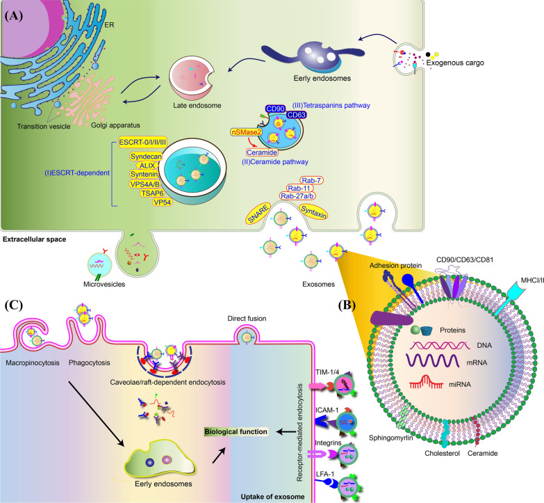
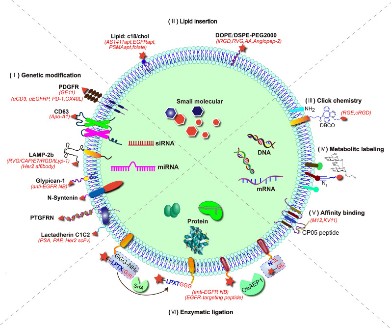
Extracellular vesicles (EVs) are 30–150 nm membrane-bound vesicles naturally secreted by cells and play important roles in intercellular communication by delivering regulatory molecules such as proteins, lipids, nucleic acids and metabolites to recipient cells. As natural nano-carriers, EVs possess desirable properties such as high biocompatibility, biological barrier permeability, low toxicity, and low immunogenicity, making them potential therapeutic delivery vehicles. EVs derived from specific cells have inherent targeting capacity towards specific cell types, which is yet not satisfactory enough for targeted therapy development and needs to be improved. Surface modifications endow EVs with targeting abilities, significantly improving their therapeutic efficiency. Herein, we first briefly introduce the biogenesis, composition, uptake and function of EVs, and review the cargo loading approaches for EVs. Then, we summarize the recent advances in surface engineering strategies of EVs, focusing on the applications of engineered EVs for targeted therapy. Altogether, EVs hold great promise for targeted delivery of various cargos, and targeted modifications show promising effects on multiple diseases. Graphical Abstract
Overview
The claims and statistics of this paper haven’t been checked yet.
Run the check — free, no account needed. It reads the open-access text and checks the sources it cites.
Integrity signals
Registry
Cites
Statistics
Figures
Verification
Claims the paper makes
The claims and statistics of this paper haven’t been checked yet.
Run the check — free, no account needed. It reads the open-access text and checks the sources it cites.
Claims other papers make about this one
Each is checked against this paper’s own text: does it say what the citing paper says it does?
In the citing paper
“In addition, exosomes can be genetically engineered, chemically surface modified or enzymatically engineered to introduce peptides and proteins to enhance their targeting ability [42, 83–86].”
From Exosome for mRNA delivery: Strategies and therapeutic applications (2024) · cites this paper as [85]
Self-check
Percentages, sums and reported differences re-computed from the paper’s own numbers.
The self-check needs the paper’s full text.
Citations
Cites
Théry, C., Witwer, K. W., Aikawa, E., Alcaraz, M. J., Anderson, J. D., Andriantsitohaina, R., Antoniou, A., Arab, T., Archer, F., Atkin‐Smith, G. K., et al. (2018) · Journal of Extracellular Vesicles · doi:10.1080/20013078.2018.1535750 · Cited by 11,099
Shedding light on the cell biology of extracellular vesicles
van Niel, G., D'Angelo, G., & Raposo, G. (2018) · Nature Reviews Molecular Cell Biology · doi:10.1038/nrm.2017.125 · Cited by 8,369
Tumour exosome integrins determine organotropic metastasis
Hoshino, A., Costa-Silva, B., Shen, T.-L., Rodrigues, G., Hashimoto, A., Tesic Mark, M., Molina, H., Kohsaka, S., Di Giannatale, A., Ceder, S., et al. (2015) · Nature · doi:10.1038/nature15756 · Cited by 5,101
Delivery of siRNA to the mouse brain by systemic injection of targeted exosomes
Alvarez-Erviti, L., Seow, Y., Yin, H., Betts, C., Lakhal, S., & Wood, M. J. A. (2011) · Nature Biotechnology · doi:10.1038/nbt.1807 · Cited by 4,930
Ceramide Triggers Budding of Exosome Vesicles into Multivesicular Endosomes
Trajković, K., Hsu, C., Chiantia, S., Rajendran, L., Wenzel, D., Wieland, F., Schwille, P., Brügger, B., & Simons, M. (2008) · Science · doi:10.1126/science.1153124 · Cited by 3,760
Glypican-1 identifies cancer exosomes and detects early pancreatic cancer
Melo, S. A., Luecke, L. B., Kahlert, C., Fernandez, A. F., Gammon, S. T., Kaye, J., LeBleu, V. S., Mittendorf, E. A., Weitz, J., Rahbari, N., et al. (2015) · Nature · doi:10.1038/nature14581 · Cited by 2,880
HaloTag: A Novel Protein Labeling Technology for Cell Imaging and Protein Analysis
Los, G. V., Encell, L. P., McDougall, M. G., Hartzell, D. D., Karassina, N., Zimprich, C., Wood, M. G., Learish, R., Ohana, R. F., Urh, M., et al. (2008) · ACS Chemical Biology · doi:10.1021/cb800025k · Cited by 2,624
mRNA-based therapeutics--developing a new class of drugs
Sahin, U., Karikó, K., & Türeci, Ö. (2014) · Nature Reviews Drug Discovery · doi:10.1038/nrd4278 · Cited by 2,474
Exosomes as drug delivery vehicles for Parkinson’s disease therapy
Haney, M. J., Klyachko, N. L., Zhao, Y., Gupta, R., Plotnikova, E. G., He, Z., Patel, T., Piroyan, A., Sokolsky, M., Kabanov, A. V., et al. (2015) · Journal of Controlled Release · doi:10.1016/j.jconrel.2015.03.033 · Cited by 2,009
The exosome journey: from biogenesis to uptake and intracellular signalling
Gurung, S., Perocheau, D. P. B., Touramanidou, L., & Baruteau, J. (2021) · Cell Communication and Signaling · doi:10.1186/s12964-021-00730-1 · Cited by 1,912
The biology and function of exosomes in cancer
Kalluri, R. (2016) · Journal of Clinical Investigation · doi:10.1172/jci81135 · Cited by 1,880
Concise review: MSC-derived exosomes for cell-free therapy
Phinney, D. G., & Pittenger, M. F. (2017) · Stem Cells · doi:10.1002/stem.2575 · Cited by 1,834
Biogenesis and secretion of exosomes
Kowal, J., Tkach, M., & Théry, C. (2014) · Current Opinion in Cell Biology · doi:10.1016/j.ceb.2014.05.004 · Cited by 1,781
Systemically Injected Exosomes Targeted to EGFR Deliver Antitumor MicroRNA to Breast Cancer Cells
Ohno, S.-I., Takanashi, M., Sudo, K., Ueda, S., Ishikawa, A., Matsuyama, N., Fujita, K., Mizutani, T., Ohgi, T., Ochiya, T., et al. (2013) · Molecular Therapy · doi:10.1038/mt.2012.180 · Cited by 1,666
Engineering exosomes for targeted drug delivery
Liang, Y., Duan, L., Lu, J., & Xia, J. (2021) · Theranostics · doi:10.7150/thno.52570 · Cited by 1,547
Self-citationDevelopment of exosome-encapsulated paclitaxel to overcome MDR in cancer cells
Kim, M. S., Haney, M. J., Zhao, Y., Mahajan, V., Deygen, I., Klyachko, N. L., Inskoe, E., Piroyan, A., Sokolsky, M., Okolie, O., et al. (2016) · Nanomedicine: Nanotechnology, Biology and Medicine · doi:10.1016/j.nano.2015.10.012 · Cited by 1,410
Exosomes: composition, biogenesis, and mechanisms in cancer metastasis and drug resistance
Mashouri, L., Yousefi, H., Aref, A. R., Ahadi, A. M., Molaei, F., & Alahari, S. K. (2019) · Molecular Cancer · doi:10.1186/s12943-019-0991-5 · Cited by 1,363
Zhuang, X., Xiang, X., Grizzle, W., Sun, D., Zhang, S., Axtell, R. C., Ju, S., Mu, J., Zhang, L., Steinman, L., et al. (2011) · Molecular Therapy · doi:10.1038/mt.2011.164 · Cited by 1,356
Exosome-Mediated Metastasis: Communication from a Distance
Wortzel, I., Dror, S., Kenific, C. M., & Lyden, D. C. (2019) · Developmental Cell · doi:10.1016/j.devcel.2019.04.011 · Cited by 1,294
Extracellular vesicles as drug delivery systems: Why and how?
Elsharkasy, O. M., Nordin, J. Z., Hagey, D. W., de Jong, O. G., Schiffelers, R. M., Andaloussi, S. E., & Vader, P. (2020) · Advanced Drug Delivery Reviews · doi:10.1016/j.addr.2020.04.004 · Cited by 1,235
Surface functionalized exosomes as targeted drug delivery vehicles for cerebral ischemia therapy
Tian, T., Zhang, H.-X., He, C.-P., Fan, S., Zhu, Y.-L., Qi, C., Huang, N.-P., Xiao, Z.-D., Lu, Z.-H., Tannous, B. A., et al. (2018) · Biomaterials · doi:10.1016/j.biomaterials.2017.10.012 · Cited by 1,154
Strategies for delivering therapeutics across the blood–brain barrier
Terstappen, G. C., Meyer, A. H., Bell, R. D., & Zhang, W. (2021) · Nature Reviews Drug Discovery · doi:10.1038/s41573-021-00139-y · Cited by 1,138
Exosomes: Therapy delivery tools and biomarkers of diseases
Barile, L., & Vassalli, G. (2017) · Pharmacology & Therapeutics · doi:10.1016/j.pharmthera.2017.02.020 · Cited by 1,053
Yang, T., Martin, P. B., Fogarty, B., Brown, A. H., Schurman, K., Phipps, R. J., Yin, V. P., Lockman, P. R., & Bai, S. (2015) · Pharmaceutical Research · doi:10.1007/s11095-014-1593-y · Cited by 980
Christianson, H. C., Svensson, K. J., van Kuppevelt, T. H., Li, J.-P., & Belting, M. (2013) · Proceedings of the National Academy of Sciences · doi:10.1073/pnas.1304266110 · Cited by 903
Exosome-based liquid biopsies in cancer: opportunities and challenges
Wang, Y., Hurley, J. D., Roberts, D., Chakrabortty, S. K., Enderle, D., Noerholm, M., Breakefield, X. O., & Skog, J. (2021) · Annals of Oncology · doi:10.1016/j.annonc.2021.01.074 · Cited by 882
Pascucci, L., Coccè, V., Bonomi, A., Ami, D., Ceccarelli, P., Ciusani, E., Viganò, L., Locatelli, A., Sisto, F., Doglia, S. M., et al. (2014) · Journal of Controlled Release · doi:10.1016/j.jconrel.2014.07.042 · Cited by 846
Fuhrmann, G., Serio, A., Mazo, M. M., Nair, R., & Stevens, M. M. (2014) · Journal of Controlled Release · doi:10.1016/j.jconrel.2014.11.029 · Cited by 770
Jia, G., Han, Y., An, Y., Ding, Y., He, C., Wang, X., & Tang, Q. (2018) · Biomaterials · doi:10.1016/j.biomaterials.2018.06.029 · Cited by 753
Kojima, R., Bojar, D., Rizzi, G., Hamri, G. C.-E., El-Baba, M. D., Saxena, P., Ausländer, S., Tan, K. R., & Fussenegger, M. (2018) · Nature Communications · doi:10.1038/s41467-018-03733-8 · Cited by 748
Biodistribution and delivery efficiency of unmodified tumor-derived exosomes
Smyth, T., Kullberg, M., Malik, N., Smith-Jones, P. M., Graner, M. W., & Anchordoquy, T. J. (2014) · Journal of Controlled Release · doi:10.1016/j.jconrel.2014.12.013 · Cited by 738
Tao, S., Yuan, T., Zhang, Y.-L., Yin, W., Guo, S., & Zhang, C. (2016) · Theranostics · doi:10.7150/thno.17133 · Cited by 734
Saponins as cytotoxic agents: a review
Podolak, I., Galanty, A., & Sobolewska, D. (2010) · Phytochemistry Reviews · doi:10.1007/s11101-010-9183-z · Cited by 718
Mesenchymal stem cell secretes microparticles enriched in pre-microRNAs
Chen, T. S., Lai, R. C., Lee, M. M., Choo, A. B. H., Lee, C. N., & Lim, S. K. (2009) · Nucleic Acids Research · doi:10.1093/nar/gkp857 · Cited by 672
Kim, M. S., Haney, M. J., Zhao, Y., Yuan, D., Deygen, I., Klyachko, N. L., Kabanov, A. V., & Batrakova, E. V. (2018) · Nanomedicine Nanotechnology Biology and Medicine · doi:10.1016/j.nano.2017.09.011 · Cited by 663
Lou, G., Song, X., Yang, F., Wu, S., Wang, J., Chen, Z., & Liu, Y. (2015) · Journal of Hematology & Oncology · doi:10.1186/s13045-015-0220-7 · Cited by 651
Mesenchymal Stem Cell-Derived Exosomes: Applications in Regenerative Medicine
Hade, M. D., Suire, C. N., & Suo, Z. (2021) · Cells · doi:10.3390/cells10081959 · Cited by 642
Exosomes and Exosome-Inspired Vesicles for Targeted Drug Delivery
Antimisiaris, S. G., Mourtas, S., & Marazioti, A. (2018) · Pharmaceutics · doi:10.3390/pharmaceutics10040218 · Cited by 631
Liang, G., Zhu, Y., Ali, D. J., Tian, T., Xu, H., Si, K., Sun, B., Chen, B., & Xiao, Z. (2020) · Journal of Nanobiotechnology · doi:10.1186/s12951-019-0563-2 · Cited by 619
Mesenchymal stem cells: Cell therapy and regeneration potential
Brown, C., McKee, C., Bakshi, S., Walker, K., Hakman, E., Halassy, S., Svinarich, D. M., Dodds, R. A., Govind, C. K., & Chaudhry, G. R. (2019) · Journal of Tissue Engineering and Regenerative Medicine · doi:10.1002/term.2914 · Cited by 612
Macrophage exosomes as natural nanocarriers for protein delivery to inflamed brain
Yuan, D., Zhao, Y., Banks, W. A., Bullock, K. M., Haney, M., Batrakova, E., & Kabanov, A. V. (2017) · Biomaterials · doi:10.1016/j.biomaterials.2017.07.011 · Cited by 607
Pérez-Hernández, D., Gutiérrez‐Vázquez, C., Jorge, I., López‐Martín, S., Ursa, Á., Sánchez‐Madrid, F., Vázquez, J., & Yáñez‐Mó, M. (2013) · Journal of Biological Chemistry · doi:10.1074/jbc.m112.445304 · Cited by 554
Exosome Mediated Delivery of miR-124 Promotes Neurogenesis after Ischemia
Yang, J., Zhang, X., Chen, X., Wang, L., & Yang, G. (2017) · Molecular Therapy - Nucleic Acids · doi:10.1016/j.omtn.2017.04.010 · Cited by 547
Witwer, K. W., & Théry, C. (2019) · Journal of Extracellular Vesicles · doi:10.1080/20013078.2019.1648167 · Cited by 546
Nassar, W., El‐Ansary, M., Sabry, D., Mostafa, M. A., Fayad, T., Kotb, E., Temraz, M., Saad, A.-N., Essa, W. A., & Adel, H. (2016) · Biomaterials Research · doi:10.1186/s40824-016-0068-0 · Cited by 525
Biodistribution of extracellular vesicles following administration into animals: A systematic review
Kang, M., Jordan, V., Blenkiron, C., & Chamley, L. (2021) · Journal of Extracellular Vesicles · doi:10.1002/jev2.12085 · Cited by 519
Exosomes: New players in cell–cell communication
Bang, C., & Thum, T. (2012) · The International Journal of Biochemistry & Cell Biology · doi:10.1016/j.biocel.2012.08.007 · Cited by 517
Salunkhe, S. A., Dheeraj, Basak, M., Chitkara, D., & Mittal, A. (2020) · Journal of Controlled Release · doi:10.1016/j.jconrel.2020.07.042 · Cited by 513
CAR exosomes derived from effector CAR-T cells have potent antitumour effects and low toxicity
Fu, W., Lei, C., Liu, S., Cui, Y., Wang, C., Qian, K., Li, T., Shen, Y., Fan, X., Lin, F., et al. (2019) · Nature Communications · doi:10.1038/s41467-019-12321-3 · Cited by 510
Kugeratski, F. G., Hodge, K., Lilla, S., McAndrews, K. M., Zhou, X., Hwang, R. F., Zanivan, S. R., & Kalluri, R. (2021) · Nature Cell Biology · doi:10.1038/s41556-021-00693-y · Cited by 502
Pi, F., Binzel, D. W., Lee, T. J., Li, Z., Sun, M., Rychahou, P. G., Li, H., Haque, F., Wang, S., Croce, C. M., et al. (2017) · Nature Nanotechnology · doi:10.1038/s41565-017-0012-z · Cited by 499
Exosome-mediated delivery of hydrophobically modified siRNA for huntingtin mRNA silencing
Didiot, M.-C., Hall, L. M., Coles, A. H., Haraszti, R. A., Godinho, B. M., Chase, K., Sapp, E., Ly, S., Alterman, J. F., Hassler, M. R., et al. (2016) · Molecular Therapy · doi:10.1038/mt.2016.126 · Cited by 483
Kim, S. M., Yang, Y., Oh, S. J., Hong, Y., Seo, M., & Jang, M. (2017) · Journal of Controlled Release · doi:10.1016/j.jconrel.2017.09.013 · Cited by 481 · cited 1×
“Kim et al. reported that CRISPR–Cas9 loaded EVs could efficiently inhibit the targeting gene, PARP-1 , and enhance the chemosensitivity of cisplatin in ovarian cancer [106].”
BackgroundCellular Functions and Molecular Mechanisms of the ESCRT Membrane-Scission Machinery
Christ, L., Raiborg, C., Wenzel, E. M., Campsteijn, C., & Stenmark, H. (2016) · Trends in Biochemical Sciences · doi:10.1016/j.tibs.2016.08.016 · Cited by 474
Blood Exosomes Endowed with Magnetic and Targeting Properties for Cancer Therapy
Qi, H., Liu, C., Long, L., Ren, Y., Zhang, S., Chang, X., Qian, X., Jia, H., Zhao, J., Sun, J., et al. (2016) · ACS Nano · doi:10.1021/acsnano.5b06939 · Cited by 459
Kooijmans, S. A. A., Gomez‐Aleza, C., Roffler, S. R., van Solinge, W. W., Vader, P., & Schiffelers, R. M. (2016) · Journal of Extracellular Vesicles · doi:10.3402/jev.v5.31053 · Cited by 447
Kim, D.-K., Nishida, H., An, S. Y., Shetty, A. K., Bartosh, T. J., & Prockop, D. J. (2015) · Proceedings of the National Academy of Sciences · doi:10.1073/pnas.1522297113 · Cited by 436
Anchor peptide captures, targets, and loads exosomes of diverse origins for diagnostics and therapy
Gao, X., Ran, N., Dong, X., Zuo, B., Yang, R., Zhou, Q., Moulton, H. M., Seow, Y., & Yin, H. (2018) · Science Translational Medicine · doi:10.1126/scitranslmed.aat0195 · Cited by 426
In Vivo Neuroimaging of Exosomes Using Gold Nanoparticles
Betzer, O., Perets, N., Angel, A., Motiei, M., Sadan, T., Yadid, G., Offen, D., & Popovtzer, R. (2017) · ACS Nano · doi:10.1021/acsnano.7b04495 · Cited by 411
Yin, J., Straight, P. D., McLoughlin, S. M., Zhou, Z., Lin, A. J., Golan, D. E., Kelleher, N. L., Kolter, R., & Walsh, C. T. (2005) · Proceedings of the National Academy of Sciences · doi:10.1073/pnas.0507705102 · Cited by 408
MVB vesicle formation: ESCRT-dependent, ESCRT-independent and everything in between
Babst, M. (2011) · Current Opinion in Cell Biology · doi:10.1016/j.ceb.2011.04.008 · Cited by 399
CD169 mediates the capture of exosomes in spleen and lymph node
Saunderson, S. C., Dunn, A. C., Crocker, P. R., & McLellan, A. D. (2013) · Blood · doi:10.1182/blood-2013-03-489732 · Cited by 396
Liang, Y., Xu, X., Li, X., Xiong, J., Li, B., Duan, L., Wang, D., & Xia, J. (2020) · ACS Applied Materials & Interfaces · doi:10.1021/acsami.0c10458 · Cited by 391
Self-citationChallenges in the development and establishment of exosome-based drug delivery systems
Wang, J., Chen, D., & Ho, E. A. (2020) · Journal of Controlled Release · doi:10.1016/j.jconrel.2020.10.020 · Cited by 391
Survivin: A new target for anti-cancer therapy
Ryan, B. M., O’Donovan, N., & Duffy, M. J. (2009) · Cancer Treatment Reviews · doi:10.1016/j.ctrv.2009.05.003 · Cited by 378
Xu, X., Liang, Y., Li, X., Ouyang, K., Wang, M., Cao, T., Li, W., Liu, J., Xiong, J., Li, B., et al. (2021) · Biomaterials · doi:10.1016/j.biomaterials.2020.120539 · Cited by 377
Self-citationNanoparticle Delivery of CRISPR/Cas9 for Genome Editing
Duan, L., Ouyang, K., Xu, X., Xu, L., Wen, C., Zhou, X., Qin, Z., Xu, Z., Sun, W., & Liang, Y. (2021) · Frontiers in Genetics · doi:10.3389/fgene.2021.673286 · Cited by 377
Self-citationYang, L., Han, B., Zhang, Z., Wang, S., Bai, Y., Zhang, Y., Tang, Y., Du, L., Xu, L., Wu, F., et al. (2020) · Circulation · doi:10.1161/circulationaha.120.045765 · Cited by 376
Gee, P., Lung, M. S. Y., Okuzaki, Y., Sasakawa, N., Iguchi, T., Makita, Y., Hozumi, H., Miura, Y., Yang, L. F., Iwasaki, M., et al. (2020) · Nature Communications · doi:10.1038/s41467-020-14957-y · Cited by 372
Cui, G.-H., Guo, H.-D., Li, H., Zhai, Y., Gong, Z., Wu, J., Liu, J.-S., Dong, Y., Hou, S., & Liu, J. (2019) · Immunity & Ageing · doi:10.1186/s12979-019-0150-2 · Cited by 359
Oncogene Knockdown via Active Loading of Small RNAs into Extracellular Vesicles by Sonication
Lamichhane, T. N., Jeyaram, A., Patel, D. B., Parajuli, B., Livingston, N. K., Arumugasaamy, N., Schardt, J. S., & Jay, S. M. (2016) · Cellular and Molecular Bioengineering · doi:10.1007/s12195-016-0457-4 · Cited by 347
Wang, H., Sui, H., Zheng, Y., Jiang, Y., Shi, Y., Liang, J., & Zhao, L. (2019) · Nanoscale · doi:10.1039/c9nr01255a · Cited by 324
Fibronectin on the Surface of Myeloma Cell-derived Exosomes Mediates Exosome-Cell Interactions
Purushothaman, A., Bandari, S., Liu, J., Mobley, J. A., Brown, E. E., & Sanderson, R. D. (2015) · Journal of Biological Chemistry · doi:10.1074/jbc.m115.686295 · Cited by 318
Kim, G., Kim, M., Lee, Y., Byun, J. W., Hwang, D. W., & Lee, M. (2019) · Journal of Controlled Release · doi:10.1016/j.jconrel.2019.11.009 · Cited by 317
Yamashita, T., Takahashi, Y., & Takakura, Y. (2018) · Biological and Pharmaceutical Bulletin · doi:10.1248/bpb.b18-00133 · Cited by 309
COVID-19 mRNA vaccines: Platforms and current developments
Szabó, G. T., Mahiny, A. J., & Vlatkovic, I. (2022) · Molecular Therapy · doi:10.1016/j.ymthe.2022.02.016 · Cited by 308
Cheng, L., Zhang, X., Tang, J., Lv, Q., & Liu, J. (2021) · Biomaterials · doi:10.1016/j.biomaterials.2021.120964 · Cited by 307
Nakase, I., & Futaki, S. (2015) · Scientific Reports · doi:10.1038/srep10112 · Cited by 297
Internalization of Exosomes through Receptor-Mediated Endocytosis
Gonda, A., Kabagwira, J., Senthil, G. N., & Wall, N. R. (2019) · Molecular Cancer Research · doi:10.1158/1541-7786.mcr-18-0891 · Cited by 296
Shao, J., Zaro, J., & Shen, Y. (2020) · International Journal of Nanomedicine · doi:10.2147/ijn.s281890 · Cited by 295
Reprogramming Exosomes as Nanoscale Controllers of Cellular Immunity
Cheng, Q., Shi, X., Han, M., Smbatyan, G., Lenz, H., & Zhang, Y. (2018) · Journal of the American Chemical Society · doi:10.1021/jacs.8b10047 · Cited by 294
Targeted exosome-mediated delivery of opioid receptor Mu siRNA for the treatment of morphine relapse
Liu, Y., Li, D., Liu, Z., Zhou, Y., Chu, D., Li, X., Jiang, X., Hou, D., Chen, X., Chen, Y., et al. (2015) · Scientific Reports · doi:10.1038/srep17543 · Cited by 288
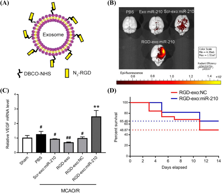
Self-citationExosome-mediated targeted delivery of miR-210 for angiogenic therapy after cerebral ischemia in mice
Zhang, H., Wu, J. S., Wu, J., Fan, Q. W., Zhou, J., Wu, J., Liu, S., Zang, J., Ye, J., Xiao, M., et al. (2019) · Journal of Nanobiotechnology · doi:10.1186/s12951-019-0461-7 · Cited by 284
Liang, G., Kan, S., Zhu, Y., Feng, S., Feng, W., & Gao, S. (2018) · International Journal of Nanomedicine · doi:10.2147/ijn.s154458 · Cited by 276
Aptamer-Conjugated Extracellular Nanovesicles for Targeted Drug Delivery
Wan, Y., Wang, L., Zhu, C., Zheng, Q., Wang, G., Tong, J., Fang, Y., Xia, Y., Cheng, G., He, X., et al. (2017) · Cancer Research · doi:10.1158/0008-5472.can-17-2880 · Cited by 261
Approaches to surface engineering of extracellular vesicles
Richter, M., Vader, P., & Fuhrmann, G. (2021) · Advanced Drug Delivery Reviews · doi:10.1016/j.addr.2021.03.020 · Cited by 258
Maturation of reticulocytes: formation of exosomes as a mechanism for shedding membrane proteins
Johnstone, R. M. (1992) · Biochemistry and Cell Biology · doi:10.1139/o92-028 · Cited by 255
Hao, S., Bai, O., Li, F., Yuan, J., Laferté, S., & Xiang, J. (2006) · Immunology · doi:10.1111/j.1365-2567.2006.02483.x · Cited by 253
A platform for actively loading cargo RNA to elucidate limiting steps in EV‐mediated delivery
Hung, M. E., & Leonard, J. N. (2016) · Journal of Extracellular Vesicles · doi:10.3402/jev.v5.31027 · Cited by 251
Pham, T. C., Jayasinghe, M. K., Pham, T. T., Yang, Y., Wei, L., Usman, W. M., Chen, H., Pirisinu, M., Gong, J., Kim, S., et al. (2021) · Journal of Extracellular Vesicles · doi:10.1002/jev2.12057 · Cited by 250
Tian, T., Cao, L., He, C. H., Ye, Q., Liang, R., You, W., Zhang, H., Wu, J., Ye, J., Tannous, B. A., et al. (2021) · Theranostics · doi:10.7150/thno.56367 · Cited by 243
Kalani, A., Chaturvedi, P., Kamat, P. K., Maldonado, C. J., Bauer, P. M., Joshua, I. G., Tyagi, S. C., & Tyagi, N. (2016) · The International Journal of Biochemistry & Cell Biology · doi:10.1016/j.biocel.2016.09.002 · Cited by 237
Inside(sight) of tiny communicator: exosome biogenesis, secretion, and uptake
Jadli, A. S., Ballasy, N., Edalat, P., & Patel, V. B. (2020) · Molecular and Cellular Biochemistry · doi:10.1007/s11010-020-03703-z · Cited by 225
Hypoxia and extracellular vesicles: A review on methods, vesicular cargo and functions
Korvenlaita, N., Pistono, C., Huremagic, B., Jolkkonen, J., Giugno, R., & Malm, T. (2020) · Journal of Extracellular Vesicles · doi:10.1002/jev2.12002 · Cited by 221
Drug loading techniques for exosome-based drug delivery systems
Xi, X.-M., Cheng-Meng, Xia, S.-J., & Lu, R. (2021) · Die Pharmazie · doi:10.31083/ph.2021.0128 · Cited by 218
Tumor-exosomes and leukocyte activation: an ambivalent crosstalk
Zech, D., Rana, S., Büchler, M. W., & Zöller, M. (2012) · Cell Communication and Signaling · doi:10.1186/1478-811x-10-37 · Cited by 217
Chondrocyte-specific genomic editing enabled by hybrid exosomes for osteoarthritis treatment
Liang, Y., Xu, X., Xu, L., Iqbal, Z., Ouyang, K., Zhang, H., Wen, C., Duan, L., & Xia, J. (2022) · Theranostics · doi:10.7150/thno.69368 · Cited by 217
Self-citationZou, J., Shi, M., Liu, X., Jin, C. Y., Xing, X., Qiu, L., & Tan, W. (2019) · Analytical Chemistry · doi:10.1021/acs.analchem.8b05204 · Cited by 215
Lv, Q., Cheng, L., Lu, Y., Zhang, X., Wang, Y., Deng, J., Zhou, J., Liu, B., & Liu, J. (2020) · Advanced Science · doi:10.1002/advs.202000515 · Cited by 211
Molecular Recognition-Based DNA Nanoassemblies on the Surfaces of Nanosized Exosomes
Wan, S., Zhang, L., Wang, S., Liu, Y., Wu, C. S., Cui, C., Sun, H. R., Shi, M., Jiang, Y. Y., Li, L., et al. (2017) · Journal of the American Chemical Society · doi:10.1021/jacs.7b00319 · Cited by 210
Li, L., He, D., Guo, Q., Zhang, Z., Ru, D., Wang, L., Gong, K., Liu, F., Duan, Y., & Li, H. (2022) · Journal of Nanobiotechnology · doi:10.1186/s12951-022-01264-5 · Cited by 206
LAMP2A regulates the loading of proteins into exosomes
Ferreira, J. V., Soares, A. D. R., Ramalho, J. S., Carvalho, C. M., Cardoso, M. H., Pintado, P. A., Carvalho, A. S., Beck, H. C., Matthiesen, R., Zuzarte, M., et al. (2022) · Science Advances · doi:10.1126/sciadv.abm1140 · Cited by 201
Drug loading techniques for exosome-based drug delivery systems.
Xi, X.-M., Xia, S.-J., & Lu, R. (2021) · PubMed · doi:10.1691/ph.2021.0128 · Cited by 200
Wang, Y., Chen, X., Tian, B., Liu, J., Yang, L., Zeng, L., Chen, T., Hong, A., & Wang, X. (2017) · Theranostics · doi:10.7150/thno.16532 · Cited by 198
Exosome: a significant nano-scale drug delivery carrier
Peng, H., Ji, W., Zhao, R., Yang, J., Lü, Z., Li, Y., & Zhang, X. (2020) · Journal of Materials Chemistry B · doi:10.1039/d0tb01499k · Cited by 192
Wilhelm, J., Kühn, S., Tarnawski, M., Gotthard, G., Tünnermann, J., Tänzer, T., Karpenko, J., Mertes, N., Xue, L., Uhrig, U., et al. (2021) · Biochemistry · doi:10.1021/acs.biochem.1c00258 · Cited by 184
Hatlem, D., Trunk, T., Linke, D., & Leo, J. C. (2019) · International Journal of Molecular Sciences · doi:10.3390/ijms20092129 · Cited by 183
Wang, J.-H., Forterre, A. V., Zhao, J., Frimannsson, D. O., Delcayre, A., Antes, T. J., Efron, B., Jeffrey, S. S., Pegram, M. D., & Matin, A. C. (2018) · Molecular Cancer Therapeutics · doi:10.1158/1535-7163.mct-17-0827 · Cited by 181
Isolation of anti-CD22 Fv with high affinity by Fv display on human cells
Ho, M., Nagata, S., & Pastan, I. H. (2006) · Proceedings of the National Academy of Sciences · doi:10.1073/pnas.0603653103 · Cited by 180
ESCRT-dependent cargo sorting at multivesicular endosomes
Frankel, E. B., & Audhya, A. (2017) · Seminars in Cell and Developmental Biology · doi:10.1016/j.semcdb.2017.08.020 · Cited by 178
Journey of siRNA: Clinical Developments and Targeted Delivery
Nikam, R. R., & Gore, K. R. (2018) · Nucleic Acid Therapeutics · doi:10.1089/nat.2017.0715 · Cited by 175
Wang, M., Altınoglu, S., Takeda, Y. S., & Xu, Q. (2015) · PLoS ONE · doi:10.1371/journal.pone.0141860 · Cited by 171
Kooijmans, S. A. A., Gitz-Francois, J. J., Schiffelers, R. M., & Vader, P. (2018) · Nanoscale · doi:10.1039/c7nr06966a · Cited by 169
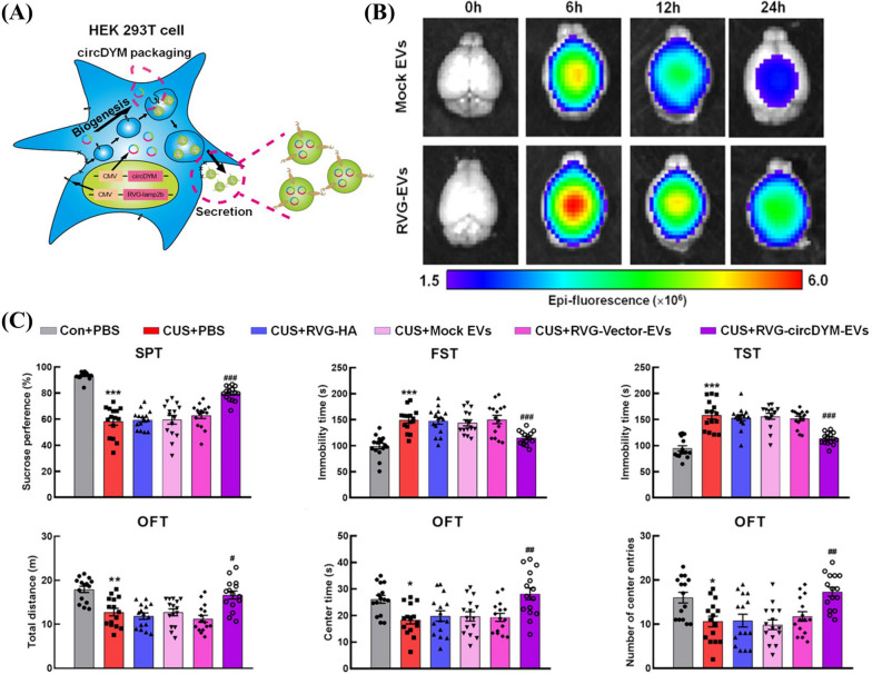
Extracellular vesicle‐mediated delivery of circDYM alleviates CUS‐induced depressive‐like behaviours
Yu, X., Bai, Y., Han, B., Ju, M., Tang, T., Shen, L., Li, M., Yang, L., Zhang, Z., Hu, G., et al. (2022) · Journal of Extracellular Vesicles · doi:10.1002/jev2.12185 · Cited by 167
Wang, J., Li, W., Lu, Z., Zhang, L., Hu, Y., Li, Q., Du, W., Feng, X., Jia, H., & Liu, B. (2017) · Nanoscale · doi:10.1039/c7nr04425a · Cited by 164
Rountree, R. B., Mandl, S. J., Nachtwey, J. M., Dalpozzo, K., Do, L., Lombardo, J. R., Schoonmaker, P. L., Brinkmann, K., Dirmeier, U., Laus, R., et al. (2011) · Cancer Research · doi:10.1158/0008-5472.can-10-4076 · Cited by 163
Delivery approaches for CRISPR/Cas9 therapeutics in vivo: advances and challenges
Luther, D. C., Lee, Y. W., Nagaraj, H., Scaletti, F., & Rotello, V. M. (2018) · Expert Opinion on Drug Delivery · doi:10.1080/17425247.2018.1517746 · Cited by 162
Liang, Y., Iqbal, Z., Lu, J., Wang, J., Zhang, H., Chen, X., Duan, L., & Xia, J. (2023) · Molecular Therapy · doi:10.1016/j.ymthe.2022.10.008 · Cited by 162
Self-citationSensing of latent EBV infection through exosomal transfer of 5′pppRNA
Baglio, S. R., van Eijndhoven, M. A. J., Koppers‐Lalic, D., Berenguer, J., Lougheed, S. M., Gibbs, S., Léveillé, N., Rinkel, R. N. P. M., Hopmans, E. S., Swaminathan, S., et al. (2016) · Proceedings of the National Academy of Sciences · doi:10.1073/pnas.1518130113 · Cited by 161
Yang, J., Wu, S., Hou, L., Zhu, D., Yin, S., Yang, G., & Wang, Y. (2020) · Molecular Therapy Nucleic Acids · doi:10.1016/j.omtn.2020.06.013 · Cited by 154
Bai, J., Duan, J., Liu, R., Du, Y.-F., Luo, Q., Cui, Y., Su, Z.-B., Xu, J., Xie, Y., & Lü, W.-L. (2019) · Asian Journal of Pharmaceutical Sciences · doi:10.1016/j.ajps.2019.04.002 · Cited by 144
Ren, X., Zhao, Y., Xue, F., Zheng, Y., Huang, H., Wang, W., Chang, Y., Yang, H., & Zhang, J. (2019) · Molecular Therapy — Nucleic Acids · doi:10.1016/j.omtn.2019.07.008 · Cited by 143
Shimoda, A., Tahara, Y., Sawada, S., Sasaki, Y., & Akiyoshi, K. (2017) · Biochemical and Biophysical Research Communications · doi:10.1016/j.bbrc.2017.07.126 · Cited by 142
Strategies for Targeted Delivery of Exosomes to the Brain: Advantages and Challenges
Choi, H., Choi, K., Kim, D., Oh, B.-K., Yim, H. Y., Jo, S., & Choi, C. (2022) · Pharmaceutics · doi:10.3390/pharmaceutics14030672 · Cited by 142
Jash, A., Ubeyitogullari, A., & Rizvi, S. S. H. (2021) · Journal of Materials Chemistry B · doi:10.1039/d1tb00126d · Cited by 139
Koch, R., Aung, T., Vogel, D., Chapuy, B., Wenzel, D., Becker, S., Sinzig, U., Venkataramani, V., von Mach, T., Jacob, R., et al. (2015) · Clinical Cancer Research · doi:10.1158/1078-0432.ccr-15-0577 · Cited by 138
Meliani, A., Boisgerault, F., Fitzpatrick, Z., Marmier, S., Leborgne, C., Collaud, F., Simon Sola, M., Charles, S., Ronzitti, G., Vignaud, A., et al. (2017) · Blood Advances · doi:10.1182/bloodadvances.2017010181 · Cited by 127
Aspe, J. R., Diaz Osterman, C. J., Jutzy, J. M. S., Deshields, S., Whang, S., & Wall, N. R. (2014) · Journal of Extracellular Vesicles · doi:10.3402/jev.v3.23244 · Cited by 126
Rong, Y., Zhang, J., Jiang, D., Ji, C., Liu, W., Wang, J., Ge, X., Tang, P., Yu, S., Cui, W., et al. (2020) · Acta Biomaterialia · doi:10.1016/j.actbio.2020.12.034 · Cited by 124
Exosome-mediated delivery of an anti-angiogenic peptide inhibits pathological retinal angiogenesis
Dong, X., Yi, L., Yu, Z., Wang, T., Liu, Y., Han, G., Zhang, X., Li, Y., Song, Y., Xu, H., et al. (2021) · Theranostics · doi:10.7150/thno.54755 · Cited by 124
Goh, W. J., Lee, C. K., Zou, S., Woon, E. C. Y., Czarny, B., & Pastorin, G. (2017) · International Journal of Nanomedicine · doi:10.2147/ijn.s131786 · Cited by 122
Roberts-Dalton, H., Cocks, A., Falcón‐Pérez, J. M., Sayers, E. J., Webber, J. P., Watson, P., Clayton, A., & Jones, A. T. (2017) · Nanoscale · doi:10.1039/c7nr04128d · Cited by 121
Gupta, D., Wiklander, O. P. B., Görgens, A., Conceição, M., Corso, G., Liang, X., Seow, Y., Balusu, S., Feldin, U., Bostancıoğlu, R. B., et al. (2021) · Nature Biomedical Engineering · doi:10.1038/s41551-021-00792-z · Cited by 119
Engineered Cas9 extracellular vesicles as a novel gene editing tool
Osteikoetxea, X., da Silva, A. M., Lázaro‐Ibáñez, E., Salmond, N., Shatnyeva, O., Stein, J., Schick, J., Wren, S. A. C., Lindgren, J., Firth, M. A., et al. (2022) · Journal of Extracellular Vesicles · doi:10.1002/jev2.12225 · Cited by 118
Facile metabolic glycan labeling strategy for exosome tracking
Lee, T. S., Kim, Y., Zhang, W., Song, I. H., & Tung, C. (2018) · Biochimica et Biophysica Acta (BBA) - General Subjects · doi:10.1016/j.bbagen.2018.02.001 · Cited by 110
Site-Specific Sequential Protein Labeling Catalyzed by a Single Recombinant Ligase
Rehm, F. B. H., Harmand, T. J., Yap, K., Durek, T., Craik, D. J., & Ploegh, H. L. (2019) · Journal of the American Chemical Society · doi:10.1021/jacs.9b09166 · Cited by 108
Lewis, N. D., Sia, C. L., Kirwin, K., Haupt, S., Mahimkar, G., Zi, T., Xu, K., Dooley, K., Jang, S. C., Choi, B. D., et al. (2020) · Molecular Cancer Therapeutics · doi:10.1158/1535-7163.mct-20-0484 · Cited by 107
Ma, X., Wang, J., Li, J., Ma, C., Chen, S., Lei, W., Yang, Y., Liu, S., Bihl, J. C., & Chen, C. (2018) · Cellular Physiology and Biochemistry · doi:10.1159/000488635 · Cited by 103
Yang, Z., Yang, Y., Xu, Y., Jiang, W., Shao, Y., Xing, J., Chen, Y., & Han, Y. (2021) · Stem Cell Research & Therapy · doi:10.1186/s13287-021-02528-x · Cited by 95
Exosome: A New Player in Translational Nanomedicine
Aheget, H., Tristán‐Manzano, M., Mazini, L., Cortijo-Gutierréz, M., Galindo‐Moreno, P., Herrera, C., Martı́n, F., Marchal, J. A., & Benabdellah, K. (2020) · Journal of Clinical Medicine · doi:10.3390/jcm9082380 · Cited by 93
Kim, M., Kim, G., Hwang, D. W., & Lee, M. (2019) · Journal of Biomedical Nanotechnology · doi:10.1166/jbn.2019.2866 · Cited by 92
Khalaj, K., Figueira, R. L., Antounians, L., Lauriti, G., & Zani, A. (2020) · Journal of Extracellular Vesicles · doi:10.1080/20013078.2020.1795365 · Cited by 90
Eliciting anti-cancer immunity by genetically engineered multifunctional exosomes
Cheng, Q., Dai, Z., Smbatyan, G., Epstein, A. L., Lenz, H., & Zhang, Y. (2022) · Molecular Therapy · doi:10.1016/j.ymthe.2022.06.013 · Cited by 90
An exosome pathway without an ESCRT
Kenific, C. M., Zhang, H., & Lyden, D. C. (2020) · Cell Research · doi:10.1038/s41422-020-00418-0 · Cited by 89
Genetic Code Expansion: Inception, Development, Commercialization
Manandhar, M., Chun, E., & Romesberg, F. E. (2021) · Journal of the American Chemical Society · doi:10.1021/jacs.0c11938 · Cited by 86
Exosomes as Targeted Delivery Platform of CRISPR/Cas9 for Therapeutic Genome Editing
Duan, L., Ouyang, K., Wang, J., Xu, L., Xu, X., Wen, C., Xie, Y., Liang, Y., & Xia, J. (2021) · ChemBioChem · doi:10.1002/cbic.202100359 · Cited by 82
Self-citationSite‐Specific Protein Labeling with SNAP‐Tags
Cole, N. B. (2013) · Current Protocols in Protein Science · doi:10.1002/0471140864.ps3001s73 · Cited by 80
Shah, N., Ishii, M., Brandon, C. S., Ablonczy, Z., Cai, J., Liu, Y., Chou, C. J., & Rohrer, B. (2018) · Biochimica et Biophysica Acta (BBA) - Molecular Basis of Disease · doi:10.1016/j.bbadis.2018.04.016 · Cited by 77
Extracellular Vesicles: The Next Generation Theranostic Nanomedicine for Inflammatory Bowel Disease
Li, D., Yang, M., Xu, J., Xu, H., Zhu, M., Liang, Y., Zhang, Y., Tian, C., Nie, Y., Shi, R., et al. (2022) · International Journal of Nanomedicine · doi:10.2147/ijn.s370784 · Cited by 76
Self-citationDelivery of siRNA based on engineered exosomes for glioblastoma therapy by targeting STAT3
Liang, S., Zuo, F.-F., Yin, B., & Ye, B. (2022) · Biomaterials Science · doi:10.1039/d1bm01723c · Cited by 75
The role of microvesicles and its active molecules in regulating cellular biology
Lv, Y., Tan, J., Miao, Y., & Zhang, Q. (2019) · Journal of Cellular and Molecular Medicine · doi:10.1111/jcmm.14667 · Cited by 74
Nanomaterials for mRNA ‐based therapeutics: Challenges and opportunities
Li, D., Liu, Q., Yang, M., Xu, H., Zhu, M., Zhang, Y., Xu, J., Tian, C., Yao, J., Wang, L., et al. (2023) · Bioengineering & Translational Medicine · doi:10.1002/btm2.10492 · Cited by 74
Self-citationWu, J., Li, Y., Wang, J., Hu, X.-B., Huang, S. Q., Luo, S., & Xiang, D. (2021) · Journal of Nanobiotechnology · doi:10.1186/s12951-021-01153-3 · Cited by 73
Liang, Y., Iqbal, Z., Wang, J., Xu, L., Xu, X., Ouyang, K., Zhang, H., Lu, J., Duan, L., & Xia, J. (2022) · Biomaterials Science · doi:10.1039/d2bm00480a · Cited by 72
Self-citationBiomaterial-assisted targeted and controlled delivery of CRISPR/Cas9 for precise gene editing
Iqbal, Z., Rehman, K., Xia, J., Shabbir, M., Zaman, M., Liang, Y., & Duan, L. (2023) · Biomaterials Science · doi:10.1039/d2bm01636b · Cited by 71
Self-citationFernández-Francos, S., Eiró, N., González-Galiano, N., & Vizoso, F. J. (2021) · International Journal of Molecular Sciences · doi:10.3390/ijms22157850 · Cited by 70
Liu, M., Hu, Y., & Chen, G. (2020) · Frontiers in Oncology · doi:10.3389/fonc.2020.01453 · Cited by 67
Targeted delivery of exosomal miR-484 reprograms tumor vasculature for chemotherapy sensitization
Zhao, Z. X., Shuang, T., Gao, Y., Fan, L., Zhang, J., He, W., Qu, L., Chen, B., & Hao, Q. (2022) · Cancer Letters · doi:10.1016/j.canlet.2022.01.011 · Cited by 66
Osteoclast-targeted delivery of anti-miRNA oligonucleotides by red blood cell extracellular vesicles
Xu, L., Xu, X., Liang, Y., Wen, C., Ouyang, K., Huang, J., Xiao, Y., Deng, X., Xia, J., & Duan, L. (2023) · Journal of Controlled Release · doi:10.1016/j.jconrel.2023.04.043 · Cited by 65
Self-citationTumor-specific delivery of KRAS siRNA with iRGD-exosomes efficiently inhibits tumor growth
Zhou, Y., Yuan, Y., Liu, M., Hu, X., Quan, Y., & Chen, X. (2019) · ExRNA · doi:10.1186/s41544-019-0034-9 · Cited by 63
Li, X., Yu, Q., Zhao, R., Guo, X., Liu, C., Zhang, K., Zhang, W., Liu, J., Yu, J., Wang, S., et al. (2022) · Frontiers in Oncology · doi:10.3389/fonc.2022.898156 · Cited by 61
Guo, Y., Wan, Z., Zhao, P., Wei, M., Liu, Y., Bu, T., Sun, W., Li, Z., & Yuan, L. (2021) · Journal of Nanobiotechnology · doi:10.1186/s12951-021-01145-3 · Cited by 60
The role of microvesicles containing microRNAs in vascular endothelial dysfunction
Shu, Z., Tan, J., Miao, Y., & Zhang, Q. (2019) · Journal of Cellular and Molecular Medicine · doi:10.1111/jcmm.14716 · Cited by 59
Adeno-Associated Virus (AAV) Gene Delivery: Dissecting Molecular Interactions upon Cell Entry
Large, E. E., Silveria, M., Zane, G. M., Weerakoon, O., & Chapman, M. S. (2021) · Viruses · doi:10.3390/v13071336 · Cited by 57
Zöller, M. (2015) · Methods in molecular biology · doi:10.1007/978-1-4939-3204-7_7 · Cited by 48
Role of lactadherin in the clearance of phosphatidylserine‐expressing red blood cells
Dasgupta, S. K., Abdel‐Monem, H., Guchhait, P., Nagata, S., & Thiagarajan, P. (2008) · Transfusion · doi:10.1111/j.1537-2995.2008.01841.x · Cited by 38
Nanoliposome is a Promising Carrier of Protein and Peptide Biomolecule for the Treatment of Cancer
Giri, T. K., Giri, A., Barman, T. K., & Maity, S. (2016) · Anti-Cancer Agents in Medicinal Chemistry · doi:10.2174/1871520616666151116121821 · Cited by 34
Mesenchymal Stem Cell-derived Exosomes: Novel Therapeutic Approach for Inflammatory Bowel Diseases
Tian, C., Yang, M., Xu, H., Zhu, M., Zhang, Y., Yao, J., Wang, L., Liang, Y., & Li, D. (2023) · Stem Cells International · doi:10.1155/2023/4245704 · Cited by 34
Self-citationAAV6 Vexosomes Mediate Robust Suicide Gene Delivery in a Murine Model of Hepatocellular Carcinoma
Khan, N., Maurya, S., Bammidi, S., & Jayandharan, G. R. (2020) · Molecular Therapy — Methods & Clinical Development · doi:10.1016/j.omtm.2020.03.006 · Cited by 32
Endothelial microvesicles in hypoxic hypoxia diseases
Deng, F., Wang, S., Xu, R., Yu, W., Wang, X., & Zhang, L. (2018) · Journal of Cellular and Molecular Medicine · doi:10.1111/jcmm.13671 · Cited by 26
Characteristics of erythrocyte-derived microvesicles and its relation with atherosclerosis
Li, K., Zheng, L., Wang, Q., & Hu, Y. (2016) · Atherosclerosis · doi:10.1016/j.atherosclerosis.2016.10.043 · Cited by 22
Wang, X.-W., & Zhang, W. (2019) · Methods in molecular biology · doi:10.1007/978-1-4939-9654-4_19 · Cited by 10
Cited by
Sorted by citations
Xu, G., Jin, J., Fu, Z., Wang, G., Lei, X., Xu, J., & Wang, J. (2025) · Signal Transduction and Targeted Therapy · doi:10.1038/s41392-025-02312-w · Cited by 320
Brucea javanica derived exosome-like nanovesicles deliver miRNAs for cancer therapy
Yan, G., Xiao, Q., Zhao, J., Chen, H., Xu, Y., Tan, M., & Peng, L. (2024) · Journal of Controlled Release · doi:10.1016/j.jconrel.2024.01.060 · Cited by 174
Exosome for mRNA delivery: Strategies and therapeutic applications
Iqbal, Z., Rehman, K., Mahmood, A., Shabbir, M., Liang, Y., Duan, L., & Zeng, H. (2024) · Journal of Nanobiotechnology · doi:10.1186/s12951-024-02634-x · Cited by 144
Self-citationSerrano, D. R., Juste, F., Anaya, B. J., Ramirez, B. I., Sánchez-Guirales, S. A., Moyota, J. M. Q., Hernandez, E. M., Simon, J. A., Trallero, J. M., Serrano, C., et al. (2025) · Pharmaceutics · doi:10.3390/pharmaceutics17101336 · Cited by 133
Extracellular Vesicles for Clinical Diagnostics: From Bulk Measurements to Single-Vesicle Analysis
Tran, H. L., Zheng, W., Issadore, D., Im, H., Cho, Y., Zhang, Y., Liu, D., Liu, Y., Li, B., Liu, F., et al. (2025) · ACS Nano · doi:10.1021/acsnano.5c00706 · Cited by 117
Metadata
- Authors
- Liu, Q., Li, D., Pan, X., & Liang, Y.
- Affiliations
- Shenzhen University (CN)
- Jinan University (CN)
- Corresponding author
- Pan, Xiaohua
- Liang, Yujie
- Venue
- Journal of Nanobiotechnology
- Published
- 2023-09-16
- Volume
- 21
- Issue
- 1
- Pages
- 334-334
- ISSN
- 1477-3155 (ISSN-L)
- PMC
- PMC10505332
- OpenAlex
- W4386799292
- Cited by
- 201
- References
- 172
- Citations by year
- 2026: 96 · 2025: 65 · 2024: 36 · 2023: 4
- FWCI
- 5.30
- Type
- journal article
- Language
- en
- Open access
- gold
- doi.org
- Locations
- Journal of Nanobiotechnology (publishedVersion, cc-by)
- PubMed Central (publishedVersion, cc-by)
- PubMed (publishedVersion)
- DOAJ (DOAJ: Directory of Open Access Journals) (submittedVersion, cc-by-sa)
- Funders
- Sanming Project of Medicine in Shenzhen (SZGSP013, JSGG20191129094218565, SZSM202106019, SZXK042, GJHZ20190820115203714)
- Basic and Applied Basic Research Foundation of Guangdong Province (2023A1515011936)
- Topics
- Life Sciences › Biochemistry, Genetics and Molecular Biology › Molecular Biology › Extracellular vesicles in disease
- Physical Sciences › Materials Science › Biomaterials › Nanoparticle-Based Drug Delivery
- Life Sciences › Biochemistry, Genetics and Molecular Biology › Molecular Biology › RNA Interference and Gene Delivery
- Keywords
- extracellular vesicles, engineered extracellular vesicles, membrane modification, surface engineering, targeted delivery, cargo loading, EV biogenesis, EV uptake, intercellular communication, therapeutic delivery vehicles, nanocarriers
- MeSH terms
- Extracellular Vesicles, Biological Transport, Cell Communication, Nucleic Acids, Permeability
About these checks
Registry, retraction, Cites and statistics results are computed, not judged, with one exception: a printed reference that no index lists is read once by a model before it counts as a citation error. Claims are extracted by a model and kept only when their quote appears verbatim in the text; each cited source is then checked, with a model’s help, for whether it says what the claim relies on. Those verdicts can be wrong — read the claim’s quote and the cited source before relying on one.
Statistics and arithmetic are recomputed, not judged.
Confidence bands are shown only for fields calibrated on our annotated test set. None is calibrated yet, so no band is shown.
What counts as a claim ›How we measure confidence ›How we check this ›
Are you an author, or do you see a problem with this page? Request a review
