

The DOI matches its Crossref record; 134 of the 134 references it cites were found in a registry or an index; 12 of the 112 sources its claims cite were checked for support.
Fecal microbiota transfer between young and aged mice reverses hallmarks of the aging gut, eye, and brain
Open accessAbstract
Background: Altered intestinal microbiota composition in later life is associated with inflammaging, declining tissue function, and increased susceptibility to age-associated chronic diseases, including neurodegenerative dementias. Here, we tested the hypothesis that manipulating the intestinal microbiota influences the development of major comorbidities associated with aging and, in particular, inflammation affecting the brain and retina. Methods: Using fecal microbiota transplantation, we exchanged the intestinal microbiota of young (3 months), old (18 months), and aged (24 months) mice. Whole metagenomic shotgun sequencing and metabolomics were used to develop a custom analysis workflow, to analyze the changes in gut microbiota composition and metabolic potential. Effects of age and microbiota transfer on the gut barrier, retina, and brain were assessed using protein assays, immunohistology, and behavioral testing. Results: We show that microbiota composition profiles and key species enriched in young or aged mice are successfully transferred by FMT between young and aged mice and that FMT modulates resulting metabolic pathway profiles. The transfer of aged donor microbiota into young mice accelerates age-associated central nervous system (CNS) inflammation, retinal inflammation, and cytokine signaling and promotes loss of key functional protein in the eye, effects which are coincident with increased intestinal barrier permeability. Conversely, these detrimental effects can be reversed by the transfer of young donor microbiota. Conclusions: These findings demonstrate that the aging gut microbiota drives detrimental changes in the gut–brain and gut–retina axes suggesting that microbial modulation may be of therapeutic benefit in preventing inflammation-related tissue decline in later life. Graphical abstract:
Overview
AI snapshot
Written from the paper’s full text.
TL;DR
Heterochronic fecal microbiota transfer (FMT) between young (3-month), old (18-month), and aged (24-month) C57BL/6 mice showed that aged donor microbiota transferred into young mice increased CNS microglial activation, retinal inflammation, and intestinal barrier permeability, while young donor microbiota transferred into aged mice reversed these changes. The findings support a causal role for the aging gut microbiota in gut–brain and gut–retina axes.
Key findings
- FMT shifted recipient microbiota toward donor-age profiles: aged donor microbiota into young mice enriched *Prevotella* sp. MGM2, *Lactobacillus intestinalis*, and *L. johnsonii*; young donor microbiota into aged mice enriched *Bifidobacterium animalis*, *Akkermansia muciniphila*, *Eubacterium* sp14–2, and *T. muris*. Time point accounted for 50% of data variance (R²adj = 50%; permutational ANOVA on dbRDA F = 55.8, df = 3, P < .01).
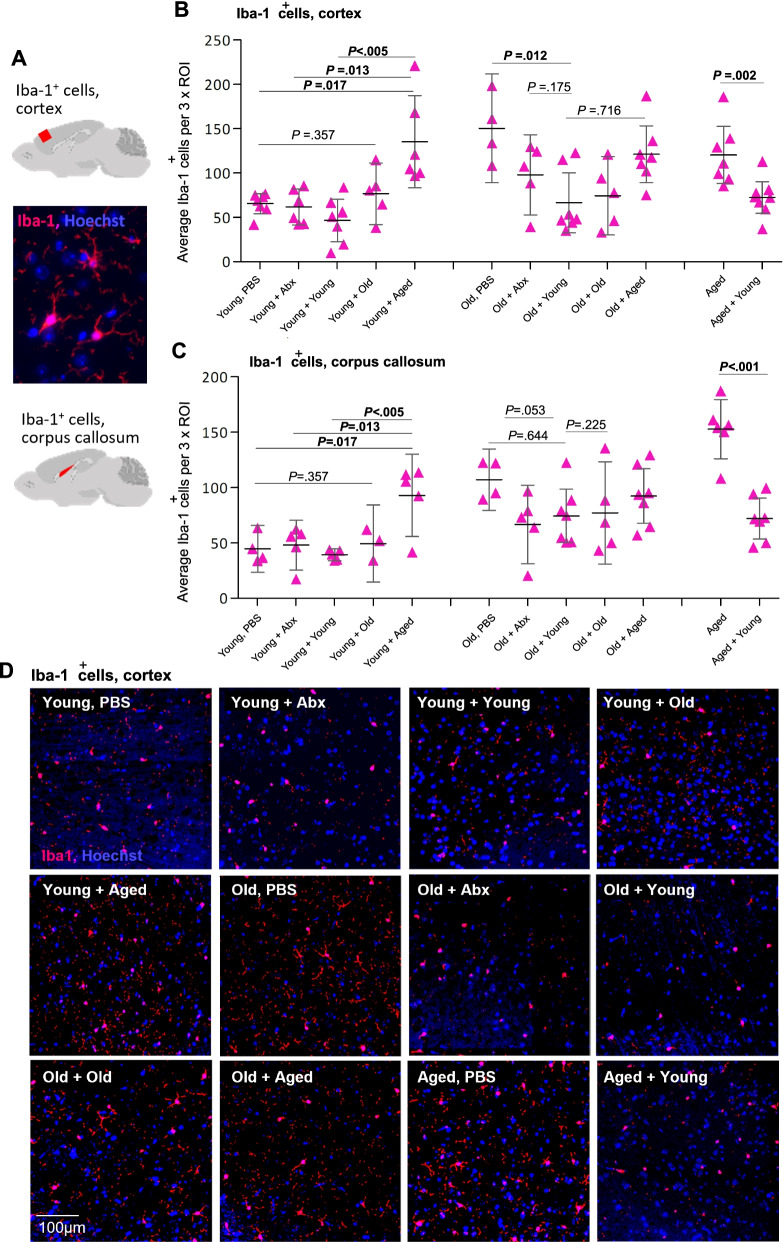
- Aged donor FMT into young mice increased Iba-1+ microglia in cortex and corpus callosum to aged-like levels; young donor FMT into aged mice decreased them. Baseline PBS young vs. aged: cortex P = .005, corpus callosum P < .001.
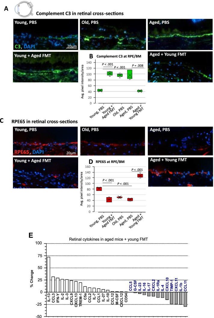
- In retina, aged donor FMT into young mice increased complement C3 at the RPE/BM interface, while young donor FMT into aged mice reduced C3 and restored RPE65 to young-like levels; young donor FMT into aged mice reduced retinal CCL11, CXCL11, and IL-1β and most increased IL-13.
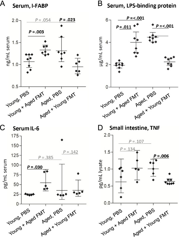
- Barrier/systemic markers: serum I-FABP increased ∼30% in young mice receiving aged donor microbiota and decreased ∼30% in aged mice receiving young donor microbiota; LBP was approximately two times higher in aged than young mice and moved toward donor-age levels after FMT; serum IL-6 was approximately twice as high in young mice receiving aged donor microbiota; intestinal TNF was reduced by around 40% in aged mice receiving young donor microbiota. Y-maze and novel object recognition showed no significant improvement in aged mice receiving young donor microbiota.
Methods
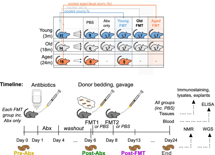
- Male C57BL/6J mice aged 3, 18, or 24 months from the same colony were antibiotic-depleted, then received heterochronic or coeval FMT via oral gavage and soiled bedding; controls received PBS or antibiotics only. Fecal pellets were collected at Pre-Abx, Post-Abx, Post-FMT (7 days), and end (18 days).
- Whole metagenomic shotgun sequencing (MetaPhlAn3, HUMAnN3), 1H-NMR fecal metabolomics, ELISAs/Luminex, immunohistology for Iba-1, complement C3, and RPE65, retinal cytokine array, and Y-maze/novel object recognition were used. Analyses included linear mixed models, PERMANOVA/dbRDA, PLS-DA, and Kruskal–Wallis tests.
- Sample sizes included groups of seven mice sub-housed as three or four; Iba-1 counts n = 5–7 mice/group; retinal staining n = 5 mice/group; retinal cytokine array n = 8 mice/group; behavior n = 10 aged FMT, n = 10 aged control, n = 11 young baseline.
Limitations
- Stated: the old (18-month) group had inter-individual variation and did not form a discrete cluster, so further metagenomic analysis focused on young and aged mice; limited availability of 24-month mice meant only heterochronic FMT and no-transfer controls were possible for that age group.
- Stated: by 18 days post-FMT, transplanted microbiota more closely resembled baseline pre-antibiotic profiles (Procrustes correlation = 25%, P = .05), raising questions about long-term persistence; behavioral tests showed no significant improvement; procedures were optimized for the bacterial fraction, so fungal, viral, and archaeal contributions cannot be excluded; antibiotics may penetrate the blood–brain barrier and theoretically affect microglial reactivity; and it is difficult to distinguish changes in microglial cell number from changes in Iba-1 expression.
- Evident from design: the endpoint was short-term (18 days), no germ-free or single-species colonization experiments were performed, and extrapolation to elderly humans would require comparable FMT studies.
Model-written summary. Check it against the paper before relying on it.
Integrity signals
Registry
Cites
Statistics
Figures
Verification
Claims the paper makes
84 Own result · 44 Not checked yet · 33 Own interpretation · 12 Own method · 2 Partly supported · 1 Supported · accuracy not yet measured
A result the paper reports from its own data — it cites no other source for it, so there is none to check. The quote shows it is in the paper.
In the paper
“and promotes loss of key functional protein in the eye”
§ Fecal microbiota transfer between young and aged mice reverses hallmarks of the aging gut, eye, and brain > Abstract > Results
plaindirect- Asserted: passes — The authors put it forward as true.
- Checkable: passes — It can be checked against evidence.
- Load-bearing: passes — The paper’s conclusions depend on it.
- Based: passes — The paper gives a reason why it holds.
A result the paper reports from its own data — it cites no other source for it, so there is none to check. The quote shows it is in the paper.
In the paper
“Conversely, these detrimental effects can be reversed by the transfer of young donor microbiota.”
- Also stated in: § Background · plain
“age-associated inflammatory changes were reversed in aged mice by transfer of young donor microbiota”
§ Fecal microbiota transfer between young and aged mice reverses hallmarks of the aging gut, eye, and brain > Abstract > Results
plaindirect- Asserted: passes — The authors put it forward as true.
- Checkable: passes — It can be checked against evidence.
- Load-bearing: passes — The paper’s conclusions depend on it.
- Based: passes — The paper gives a reason why it holds.
- Also stated in: § Background · plain
A result the paper reports from its own data — it cites no other source for it, so there is none to check. The quote shows it is in the paper.
In the paper
“effects which are coincident with increased intestinal barrier permeability”
§ Fecal microbiota transfer between young and aged mice reverses hallmarks of the aging gut, eye, and brain > Abstract > Results
plaincorrelational- Asserted: passes — The authors put it forward as true.
- Checkable: passes — It can be checked against evidence.
- Load-bearing: passes — The paper’s conclusions depend on it.
- Based: passes — The paper gives a reason why it holds.
A result the paper reports from its own data — it cites no other source for it, so there is none to check. The quote shows it is in the paper.
In the paper
“We show that microbiota composition profiles and key species enriched in young or aged mice are successfully transferred by FMT between young and aged mice”
- Also stated in: § Background · plain
“We show that FMT from between young and aged mice replaced the recipient microbiota composition with a composition resembling the donor, enriched for particular bacterial species”
§ Fecal microbiota transfer between young and aged mice reverses hallmarks of the aging gut, eye, and brain > Abstract > Results
plaindirect- Asserted: passes — The authors put it forward as true.
- Checkable: passes — It can be checked against evidence.
- Load-bearing: passes — The paper’s conclusions depend on it.
- Based: passes — The paper gives a reason why it holds.
- Also stated in: § Background · plain
A result the paper reports from its own data — it cites no other source for it, so there is none to check. The quote shows it is in the paper.
In the paper
“The transfer of aged donor microbiota into young mice accelerates age-associated central nervous system (CNS) inflammation”
§ Fecal microbiota transfer between young and aged mice reverses hallmarks of the aging gut, eye, and brain > Abstract > Results
plaindirect- Asserted: passes — The authors put it forward as true.
- Checkable: passes — It can be checked against evidence.
- Load-bearing: passes — The paper’s conclusions depend on it.
- Based: passes — The paper gives a reason why it holds.
A result the paper reports from its own data — it cites no other source for it, so there is none to check. The quote shows it is in the paper.
In the paper
“retinal inflammation, and cytokine signaling”
§ Fecal microbiota transfer between young and aged mice reverses hallmarks of the aging gut, eye, and brain > Abstract > Results
plaindirect- Asserted: passes — The authors put it forward as true.
- Checkable: passes — It can be checked against evidence.
- Load-bearing: passes — The paper’s conclusions depend on it.
- Based: passes — The paper gives a reason why it holds.
A result the paper reports from its own data — it cites no other source for it, so there is none to check. The quote shows it is in the paper.
In the paper
“aged mice receiving young donor microbiota had reduced circulating concentrations of LBP, to levels comparable to young mice”
§ Fecal microbiota transfer between young and aged mice reverses hallmarks of the aging gut, eye, and brain > Results > Heterochronic FMT reverses age-associated breakdown of epithelial barrier integrity and systemic inflammation
plaindirect- Asserted: passes — The authors put it forward as true.
- Checkable: passes — It can be checked against evidence.
- Load-bearing: passes — The paper’s conclusions depend on it.
- Based: passes — The paper gives a reason why it holds.
A result the paper reports from its own data — it cites no other source for it, so there is none to check. The quote shows it is in the paper.
In the paper
“while pre- and post-transplant levels were not significantly different in young mice receiving an aged donor microbiota”
§ Fecal microbiota transfer between young and aged mice reverses hallmarks of the aging gut, eye, and brain > Results > Heterochronic FMT reverses age-associated breakdown of epithelial barrier integrity and systemic inflammation
plain- Asserted: passes — The authors put it forward as true.
- Checkable: passes — It can be checked against evidence.
- Load-bearing: unclear — It is unclear whether the paper’s conclusions depend on it.
- Based: passes — The paper gives a reason why it holds.
A result the paper reports from its own data — it cites no other source for it, so there is none to check. The quote shows it is in the paper.
In the paper
“No significant differences were seen between the old (18 months) and aged (24 months) control, PBS-treated groups.”
§ Fecal microbiota transfer between young and aged mice reverses hallmarks of the aging gut, eye, and brain > Results > The intestinal microbiota regulates microglial activation in the CNS
plain- Asserted: passes — The authors put it forward as true.
- Checkable: passes — It can be checked against evidence.
- Load-bearing: unclear — It is unclear whether the paper’s conclusions depend on it.
- Based: passes — The paper gives a reason why it holds.
A result the paper reports from its own data — it cites no other source for it, so there is none to check. The quote shows it is in the paper.
In the paper
“In young mice, there were no significant differences in Iba-1+ cell counts between mice receiving mock transfer, coeval (young) transfer, or old (18 months) donor microbiota.”
§ Fecal microbiota transfer between young and aged mice reverses hallmarks of the aging gut, eye, and brain > Results > The intestinal microbiota regulates microglial activation in the CNS
plain- Asserted: passes — The authors put it forward as true.
- Checkable: passes — It can be checked against evidence.
- Load-bearing: passes — The paper’s conclusions depend on it.
- Based: passes — The paper gives a reason why it holds.
- It may state two separate claims as one. 43
- Probably not a substantial claim. 38
The cited sources support only part of this claim — read each source’s verdict below.
In the paper
“These changes can adversely affect the host metabolism and immunity [25–27, 40, 64, 86, 110, 112, 118] and are associated with the development of cardiovascular, autoimmune, metabolic, and neurodegenerative disorders [17, 90, 122, 132].”
§ Fecal microbiota transfer between young and aged mice reverses hallmarks of the aging gut, eye, and brain > Background
What the sources say
- Partly supportedcites [25] Claesson MJ, Cusack S, ’Sullivan O, O, Greene-Diniz R, De Weerd H, Flannery E, et al. Composition, variability, and temporal stability of the intestinal microbiota of the elderly. Proc Natl Acad Sci U S A. 2011;108(SUPPL. 1):4586–91. https://doi.org/10.1073/pnas.1000097107. sourceExperimental — accuracy not yet measured
The Abstract links intestinal microbiota alterations to obesity and inflammatory bowel disease, but says the health ramifications of microbiota changes in older people are still being explored. It does not link those aging-related changes to the development of cardiovascular or neurodegenerative disorders, or establish the claimed adverse effects on host metabolism and immunity. (LLM, conf=0.86)
- Partly supportedcites [26] Claesson MJ, Jeffery IB, Conde S, Power SE, O’connor EM, Cusack S, et al. Gut microbiota composition correlates with diet and health in the elderly. Nature. 2012;488(7410):178–84. https://doi.org/10.1038/nature11319. sourceExperimental — accuracy not yet measured
The introduction (p. 5) links microbiota changes to inflammatory and metabolic disorders, and the study reports associations between elderly participants’ microbiota and markers of inflammation (p. 3). This supports the immune and metabolic aspects, but the paper does not establish associations with the development of autoimmune or neurodegenerative disorders. It notes a weaker association with blood pressure (p. 9), not cardiovascular disease development. (LLM, conf=0.85)
- Partly supportedcites [27] Clark RI, Salazar A, Yamada R, Fitz-Gibbon S, Morselli M, Alcaraz J, et al. Distinct shifts in microbiota composition during drosophila aging impair intestinal function and drive mortality. Cell Rep. 2015;12(10):1656–67. https://doi.org/10.1016/j.celrep.2015.08.004. sourceExperimental — accuracy not yet measured
The Abstract reports that age-related microbiota changes in Drosophila contribute to immune gene activation, but it does not establish an adverse effect on host metabolism. The opening passage and Discussion link dysbiosis or intestinal barrier dysfunction to cardiovascular, neurological, inflammatory, and metabolic disorders, but do not specifically mention autoimmune disorders. Those human disease links are background associations, not outcomes demonstrated by this fly study. (LLM, conf=0.86)
- Partly supportedcites [40] Fransen F, van Beek AA, Borghuis T, El Aidy S, Hugenholtz F, van der Gaast-de Jongh C, et al. Aged gut microbiota contributes to systemical inflammaging after transfer to germ-free mice. Front Immunol. 2017;8(NOV):1385. https://doi.org/10.3389/fimmu.2017.01385. sourceExperimental — accuracy not yet measured
The Introduction says gut microbiota affects host immunity and metabolism, and the Discussion reports that aged-microbiota transfer induced characteristics of immunosenescence in young germ-free mice. The paper also links inflammaging to dementia and cardiovascular disease and discusses metabolic syndrome, but it does not establish that aging-related microbiota changes are associated with development of every listed disorder, particularly autoimmune disorders. (LLM, conf=0.85)
- Partly supportedcites [64] Langille MGII, Meehan CJ, Koenig JE, Dhanani AS, Rose RA, Howlett SE, et al. Microbial shifts in the aging mouse gut. Microbiome. 2014;2(1). https://doi.org/10.1186/s40168-014-0050-9. sourceExperimental — accuracy not yet measured
The Background links age-related microbiome shifts to likely increased inflammation and describes microbial effects on immune function; the Conclusions suggest possible effects on host vitamin availability and carbohydrate metabolism. The Background also mentions an association between microbiome perturbations and autoimmune disease. Our copy does not support the full list of cardiovascular, metabolic, and neurodegenerative disorders, nor does it establish that the observed aging-related shifts adversely affect host metabolism. (LLM, conf=0.84)
- Not checked yetcites [86] O’Toole PW, Jeffery IB. Gut microbiota and aging: Science. American Association for the Advancement of Science; 2015. https://doi.org/10.1126/science.aac8469. source
- Supportedcites [110] Smith P, Willemsen D, Popkes M, Metge F, Gandiwa E, Reichard M, et al. Regulation of life span by the gut microbiota in the short-lived African turquoise killifish. ELife. 2017;6(August). https://doi.org/10.7554/eLife.27014. source
The citation is attached to the first clause, about effects on host metabolism and immunity; the disorder list has a separate citation group. In the Results section on microbial transfer (page unavailable), young- and old-microbiota recipients differed in intestinal defense responses, hyaluronic acid metabolism, and expression of a TOR-pathway gene. The paper links the altered metabolism to inflammation and deregulated immune responses, supporting the hedged claim in its killifish model. (LLM, conf=0.85)
- Partly supportedcites [112] Stebegg M, Silva-Cayetano A, Innocentin S, Jenkins TP, Cantacessi C, Gilbert C, et al. Heterochronic faecal transplantation boosts gut germinal centres in aged mice. Nat Commun. 2019;10(1). https://doi.org/10.1038/s41467-019-10430-7. sourceExperimental — accuracy not yet measured
The Introduction says gut microorganisms affect host metabolism and immune responses, and links age-related microbiota changes to immune inflammaging. The Discussion also reports that transfer of an aged microbiome causes systemic inflammation. Our copy does not establish an adverse effect of age-related microbiota changes on host metabolism; the disorder list has a separate citation group in the citing sentence. (LLM, conf=0.84)
- Partly supportedcites [118] Thevaranjan N, Puchta A, Schulz C, Avee Naidoo JC, Szamosi CP, Verschoor DL, et al. Age-associated microbial dysbiosis promotes intestinal permeability, systemic inflammation, and macrophage dysfunction. Cell Host Microbe. 2017;21(4):455–466.e4. https://doi.org/10.1016/j.chom.2017.03.002. sourceExperimental — accuracy not yet measured
The Introduction and Discussion report that age-associated gut dysbiosis promotes systemic inflammation and impairs macrophage antibacterial function, supporting the immunity portion. The cited paper does not establish an adverse effect on host metabolism. The disorder associations appear after a separate citation group in the citing sentence, so they are not attributed to this source. (LLM, conf=0.83)
- Partly supportedcites [17] Boulangé CL, Neves AL, Chilloux J, Nicholson JK, Dumas ME. Impact of the gut microbiota on inflammation, obesity, and metabolic disease. Genome Med. 2016. https://doi.org/10.1186/s13073-016-0303-2. sourceExperimental — accuracy not yet measured
In “The essential role of the gut microbiota in human health” (page not available), the review says changes in the intestinal ecosystem can disrupt host–microbe symbiosis and affect metabolism and immune function. Its Fig. 4 caption links dysbiosis with metabolic disease and increased likelihood of cardiovascular disease. The cited passages do not establish that these are specifically aging-related changes or link them to autoimmune or neurodegenerative disorders. (LLM, conf=0.86)
plaincorrelational- Asserted: passes — The authors put it forward as true.
- Checkable: passes — It can be checked against evidence.
- Load-bearing: passes — The paper’s conclusions depend on it.
- Based: passes — The paper gives a reason why it holds.
It may state two separate claims as one.
- Partly supported
Every source the paper cites for this claim was checked and supports it.
In the paper
“There is some evidence for improved cognition with probiotic use in those with pre-existing cognitive impairment [31]”
§ Fecal microbiota transfer between young and aged mice reverses hallmarks of the aging gut, eye, and brain > Discussion > The intestinal microbiota regulates CNS microglia activation
What the sources say
- Supportedcites [31] Coutts L, Ibrahim K, Tan QY, Lim SER, Cox NJ, Roberts HC. Can probiotics, prebiotics and synbiotics improve functional outcomes for older people: a systematic review. Eur Geriatr Med. 2020. https://doi.org/10.1007/s41999-020-00396-x Springer Science and Business Media Deutschland GmbH. source
The Results (Cognition and mood) state that four probiotic studies reported a positive effect on cognition in participants with pre-existing cognitive impairment. The Discussion describes the evidence as limited and notes study-quality concerns, consistent with the claim’s cautious wording. (LLM, conf=0.94)
hedgedconditional- Asserted: passes — The authors put it forward as true.
- Checkable: passes — It can be checked against evidence.
- Load-bearing: fails — The paper’s conclusions may not depend on it.
- Based: passes — The paper gives a reason why it holds.
Probably not a substantial claim. The paper’s conclusions may not depend on it.
- Supported
The cited sources support only part of this claim — read each source’s verdict below.
In the paper
“Deficiencies in B vitamins are implicated in retinitis pigmentosa and glaucoma, for which improved outcomes can be achieved by dietary supplementation [24, 114].”
§ Fecal microbiota transfer between young and aged mice reverses hallmarks of the aging gut, eye, and brain > Discussion > The intestinal microbiota regulates retinal inflammation
What the sources say
- Partly supportedcites [24] Chou T-H, Romano GL, Amato R, Porciatti V. Nicotinamide-rich diet in DBA/2J mice preserves retinal ganglion cell metabolic function as assessed by PERG adaptation to flicker. Nutrients. 2020;12(7):1910. https://doi.org/10.3390/nu12071910. sourceExperimental — accuracy not yet measured
The Abstract and Discussion report that a nicotinamide-rich diet preserved retinal ganglion cell function and reduced cell loss in a mouse model of glaucoma. This supports improved outcomes with dietary supplementation in that model, but not the broader implication of improved outcomes in people with glaucoma. The paper does not address retinitis pigmentosa. (LLM, conf=0.86)
- Partly supportedcites [114] Sugano E, Tabata K, Takezawa T, Shiraiwa R, Muraoka H, Metoki T, et al. N-methyl-N-nitrosourea-induced photoreceptor degeneration is inhibited by nicotinamide via the blockade of upstream events before the phosphorylation of signalling proteins. Biomed Res Int. 2019;2019. https://doi.org/10.1155/2019/3238719. sourceExperimental — accuracy not yet measured
The Introduction identifies nicotinamide as vitamin B3, and the Results (§3.1) report preserved retinal morphology in nicotinamide-treated rats with chemically induced photoreceptor degeneration. But the Methods (§2.3) specify an injection, not dietary supplementation; the study does not show improved outcomes in people with retinitis pigmentosa or glaucoma. The Introduction describes retinitis pigmentosa as genetic rather than linking it to B-vitamin deficiency. (LLM, conf=0.86)
plaindirect- Asserted: passes — The authors put it forward as true.
- Checkable: passes — It can be checked against evidence.
- Load-bearing: fails — The paper’s conclusions may not depend on it.
- Based: passes — The paper gives a reason why it holds.
Probably not a substantial claim. The paper’s conclusions may not depend on it.
- Partly supported
A result the paper reports from its own data — it cites no other source for it, so there is none to check. The quote shows it is in the paper.
In the paper
“and that FMT modulates resulting metabolic pathway profiles”
- Also stated in: § Background · plain
“and altered the metabolic potential of the resulting gut microbial composition”
§ Fecal microbiota transfer between young and aged mice reverses hallmarks of the aging gut, eye, and brain > Abstract > Results
plaindirect- Asserted: passes — The authors put it forward as true.
- Checkable: passes — It can be checked against evidence.
- Load-bearing: passes — The paper’s conclusions depend on it.
- Based: passes — The paper gives a reason why it holds.
It may state two separate claims as one.
- Also stated in: § Background · plain
A result the paper reports from its own data — it cites no other source for it, so there is none to check. The quote shows it is in the paper.
In the paper
“Our cytokine analysis also included IL-12, IL-17, IL-1β, and IL-27, but none of these was above the assay threshold of detection in any group”
§ Fecal microbiota transfer between young and aged mice reverses hallmarks of the aging gut, eye, and brain > Results > Heterochronic FMT reverses age-associated breakdown of epithelial barrier integrity and systemic inflammation
plain- Asserted: passes — The authors put it forward as true.
- Checkable: passes — It can be checked against evidence.
- Load-bearing: fails — The paper’s conclusions may not depend on it.
- Based: passes — The paper gives a reason why it holds.
Probably not a substantial claim. The paper’s conclusions may not depend on it.
A result the paper reports from its own data — it cites no other source for it, so there is none to check. The quote shows it is in the paper.
In the paper
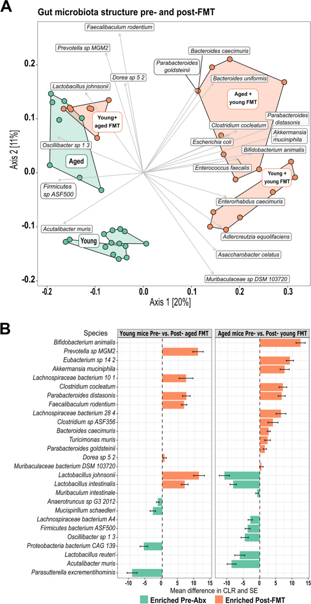
“The FMT induced significant alterations in the fecal microbiota of recipients (Fig. 5C, D, Figs. S7 and S8, permutational ANOVA on dbRDA F = 55.8, df = 3, P < .01) with experimental time point accounting for 50% of the data variance (R2adj = 50%).”
§ Fecal microbiota transfer between young and aged mice reverses hallmarks of the aging gut, eye, and brain > Results > Donor-derived species define the intestinal microbiota composition of FMT recipients
plaindirect- Asserted: passes — The authors put it forward as true.
- Checkable: passes — It can be checked against evidence.
- Load-bearing: passes — The paper’s conclusions depend on it.
- Based: passes — The paper gives a reason why it holds.
It may state two separate claims as one.
A result the paper reports from its own data — it cites no other source for it, so there is none to check. The quote shows it is in the paper.
In the paper
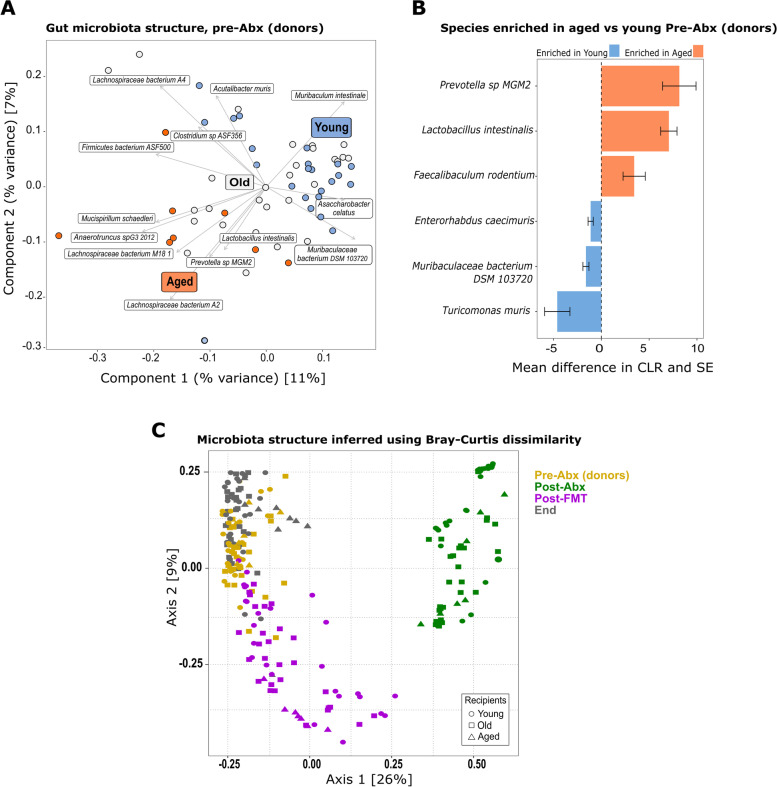
“Transfer of aged donor microbiota into young mice dramatically altered the recipients’ fecal microbiota composition (pairwise PERMANOVA P = .01, after BH correction) and resulted in a microbiota profile resembling that of aged mice at baseline”
§ Fecal microbiota transfer between young and aged mice reverses hallmarks of the aging gut, eye, and brain > Results > Donor-derived species define the intestinal microbiota composition of FMT recipients
boosteddirect- Asserted: passes — The authors put it forward as true.
- Checkable: passes — It can be checked against evidence.
- Load-bearing: passes — The paper’s conclusions depend on it.
- Based: passes — The paper gives a reason why it holds.
It may state two separate claims as one.
A result the paper reports from its own data — it cites no other source for it, so there is none to check. The quote shows it is in the paper.
In the paper
“In this case, the altered profile was distinct from that of young baseline mice, with the observed shifts following those of young mice transplanted with a young donor microbiota”
§ Fecal microbiota transfer between young and aged mice reverses hallmarks of the aging gut, eye, and brain > Results > Donor-derived species define the intestinal microbiota composition of FMT recipients
plaincorrelational- Asserted: passes — The authors put it forward as true.
- Checkable: passes — It can be checked against evidence.
- Load-bearing: unclear — It is unclear whether the paper’s conclusions depend on it.
- Based: passes — The paper gives a reason why it holds.
It may state two separate claims as one.
A result the paper reports from its own data — it cites no other source for it, so there is none to check. The quote shows it is in the paper.
In the paper
“The young (3 months) and aged (24 months) groups formed separate clusters in the ordination; however, the old (18 months) group did not form a separate cluster and shared features with both young and aged mice”
§ Fecal microbiota transfer between young and aged mice reverses hallmarks of the aging gut, eye, and brain > Results > Donor-derived species define the intestinal microbiota composition of FMT recipients
plaincorrelational- Asserted: passes — The authors put it forward as true.
- Checkable: passes — It can be checked against evidence.
- Load-bearing: unclear — It is unclear whether the paper’s conclusions depend on it.
- Based: passes — The paper gives a reason why it holds.
It may state two separate claims as one.
A result the paper reports from its own data — it cites no other source for it, so there is none to check. The quote shows it is in the paper.
In the paper
“The old (18 months) group of animals had significant inter-individual variation in the microbiota composition and did not form a discrete cluster in the ordination”
§ Fecal microbiota transfer between young and aged mice reverses hallmarks of the aging gut, eye, and brain > Results > Donor-derived species define the intestinal microbiota composition of FMT recipients
plain- Asserted: passes — The authors put it forward as true.
- Checkable: passes — It can be checked against evidence.
- Load-bearing: passes — The paper’s conclusions depend on it.
- Based: passes — The paper gives a reason why it holds.
It may state two separate claims as one.
A result the paper reports from its own data — it cites no other source for it, so there is none to check. The quote shows it is in the paper.
In the paper
“The most significantly enriched species in aged mice receiving young donor microbiota (Bifidobacterium animalis, Eubacterium sp. 14–2, A. muciniphila, and Clostridium cocleatum) were also enriched in young mice recolonized with young donor microbiota, although others (Parabacteroides distasonis, Lachnospiraceae 28–4, Clostridium ASF356) were only enriched in the aged recipients”
§ Fecal microbiota transfer between young and aged mice reverses hallmarks of the aging gut, eye, and brain > Results > Donor-derived species define the intestinal microbiota composition of FMT recipients
plaincorrelational- Asserted: passes — The authors put it forward as true.
- Checkable: passes — It can be checked against evidence.
- Load-bearing: unclear — It is unclear whether the paper’s conclusions depend on it.
- Based: passes — The paper gives a reason why it holds.
It may state two separate claims as one.
A result the paper reports from its own data — it cites no other source for it, so there is none to check. The quote shows it is in the paper.
In the paper
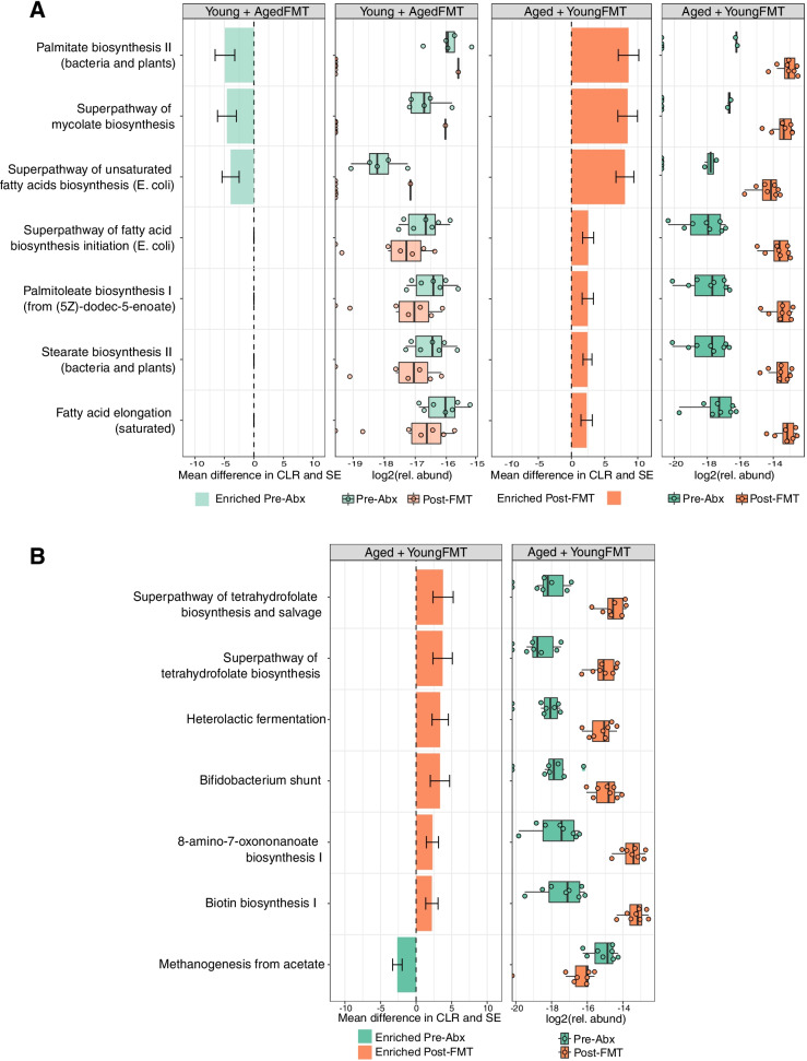
“Distance-based redundancy analysis (dbRDA) showed high inter-individual variation with only 24% of variance explained by treatment groupings (4% of variance was explained by cage groupings).”
§ Fecal microbiota transfer between young and aged mice reverses hallmarks of the aging gut, eye, and brain > Results > Heterochronic FMT alters lipid and vitamin metabolism
plaincorrelational- Asserted: passes — The authors put it forward as true.
- Checkable: passes — It can be checked against evidence.
- Load-bearing: unclear — It is unclear whether the paper’s conclusions depend on it.
- Based: passes — The paper gives a reason why it holds.
It may state two separate claims as one.
In the paper
“Young flies fed homogenates from aged flies had significantly decreased lifespan and increased incidence of intestinal barrier dysfunction, compared with flies fed homogenates from young animals [27].”
§ Fecal microbiota transfer between young and aged mice reverses hallmarks of the aging gut, eye, and brain > Background
What the sources say
- Not checked yetcites [27] Clark RI, Salazar A, Yamada R, Fitz-Gibbon S, Morselli M, Alcaraz J, et al. Distinct shifts in microbiota composition during drosophila aging impair intestinal function and drive mortality. Cell Rep. 2015;12(10):1656–67. https://doi.org/10.1016/j.celrep.2015.08.004. source
plaindirect- Asserted: passes — The authors put it forward as true.
- Checkable: passes — It can be checked against evidence.
- Load-bearing: unclear — It is unclear whether the paper’s conclusions depend on it.
- Based: passes — The paper gives a reason why it holds.
It may state two separate claims as one.
None of the sources cited for this claim have been checked yet.
In the paper
“The altered microbial composition has also been associated with neovascularization and immune cell activation in retinal disease models [4, 50, 99].”
§ Fecal microbiota transfer between young and aged mice reverses hallmarks of the aging gut, eye, and brain > Background
What the sources say
- Not checked yetcites [4] Andriessen EMMAMA, Wilson AM, Mawambo G, Dejda A, Miloudi K, Sennlaub F, et al. Gut microbiota influences pathological angiogenesis in obesity-driven choroidal neovascularization. EMBO Mol Med. 2016;8(12):1366–79. https://doi.org/10.15252/emmm.201606531. source
- Not checked yetcites [50] Horai R, Zárate-Bladés CR, Dillenburg-Pilla P, Chen J, Kielczewski JL, Silver PB, et al. Microbiota-dependent activation of an autoreactive T cell receptor provokes autoimmunity in an immunologically privileged site. Immunity. 2015;43(2):343–53. https://doi.org/10.1016/j.immuni.2015.07.014. source
- Not checked yetcites [99] Rowan S, Jiang S, Korem T, Szymanski J, Chang M-LL, Szelog J, et al. Involvement of a gut–retina axis in protection against dietary glycemia-induced age-related macular degeneration. Proc Natl Acad Sci U S A. 2017;114(22):E4472–81. https://doi.org/10.1073/pnas.1702302114. source
plaincorrelational- Asserted: passes — The authors put it forward as true.
- Checkable: passes — It can be checked against evidence.
- Load-bearing: fails — The paper’s conclusions may not depend on it.
- Based: passes — The paper gives a reason why it holds.
Probably not a substantial claim. The paper’s conclusions may not depend on it.
None of the sources cited for this claim have been checked yet.
In the paper
“This, together with declining immune cell function and immunosenescence, promotes a state of elevated systemic inflammation termed “inflammaging” [39, 40, 118], contributing to the functional decline in the intestine and CNS in old age [10, 13, 48].”
§ Fecal microbiota transfer between young and aged mice reverses hallmarks of the aging gut, eye, and brain > Background
What the sources say
- Not checked yetcites [39] Franceschi C, Garagnani P, Parini P, Giuliani C, Santoro A. Inflammaging: a new immune–metabolic viewpoint for age-related diseases. Nat Rev Endocrinol. 2018. https://doi.org/10.1038/s41574-018-0059-4 Nature Publishing Group. source
- Not checked yetcites [40] Fransen F, van Beek AA, Borghuis T, El Aidy S, Hugenholtz F, van der Gaast-de Jongh C, et al. Aged gut microbiota contributes to systemical inflammaging after transfer to germ-free mice. Front Immunol. 2017;8(NOV):1385. https://doi.org/10.3389/fimmu.2017.01385. source
- Not checked yetcites [118] Thevaranjan N, Puchta A, Schulz C, Avee Naidoo JC, Szamosi CP, Verschoor DL, et al. Age-associated microbial dysbiosis promotes intestinal permeability, systemic inflammation, and macrophage dysfunction. Cell Host Microbe. 2017;21(4):455–466.e4. https://doi.org/10.1016/j.chom.2017.03.002. source
- Not checked yetcites [10] Becker L, Nguyen L, Gill J, Kulkarni S, Pasricha PJ, Habtezion A. Age-dependent shift in macrophage polarisation causes inflammation-mediated degeneration of enteric nervous system. Gut. 2018;67(5):827–36. https://doi.org/10.1136/gutjnl-2016-312940. source
- Not checked yetcites [13] Bevan-Jones WR, Cope TE, Simon Jones P, Kaalund SS, Passamonti L, Allinson K, et al. Neuroinflammation and protein aggregation co-localize across the frontotemporal dementia spectrum. Brain J Neurol. 2020;143(3):1010–26. https://doi.org/10.1093/brain/awaa033. source
- Not checked yetcites [48] Heneka MT. Microglia take Centre stage in neurodegenerative disease. Nat Rev Immunol. 2019;19(2):79–80. https://doi.org/10.1038/s41577-018-0112-5. source
plaindirect- Asserted: passes — The authors put it forward as true.
- Checkable: passes — It can be checked against evidence.
- Load-bearing: passes — The paper’s conclusions depend on it.
- Based: passes — The paper gives a reason why it holds.
It may state two separate claims as one.
None of the sources cited for this claim have been checked yet.
In the paper
“Similar beneficial effects have been reported in aged mice receiving microbiota from young mice to extend life span [8, 61] or halt the functional decline in the mucosal immune system [112].”
§ Fecal microbiota transfer between young and aged mice reverses hallmarks of the aging gut, eye, and brain > Background
What the sources say
- Not checked yetcites [8] Bárcena C, Valdés-Mas R, Mayoral P, Garabaya C, Durand S, Rodríguez F, et al. Healthspan and lifespan extension by fecal microbiota transplantation into progeroid mice. Nat Med. 2019;25(8):1234–42. https://doi.org/10.1038/s41591-019-0504-5. source
- Not checked yetcites [61] Kundu P, Lee HU, Garcia-Perez I, Tay EXY, Kim H, Faylon LE, et al. Neurogenesis and prolongevity signaling in young germ-free mice transplanted with the gut microbiota of old mice. Sci Transl Med. 2019;11(518). https://doi.org/10.1126/scitranslmed.aau4760. source
- Not checked yetcites [112] Stebegg M, Silva-Cayetano A, Innocentin S, Jenkins TP, Cantacessi C, Gilbert C, et al. Heterochronic faecal transplantation boosts gut germinal centres in aged mice. Nat Commun. 2019;10(1). https://doi.org/10.1038/s41467-019-10430-7. source
plaindirect- Asserted: passes — The authors put it forward as true.
- Checkable: passes — It can be checked against evidence.
- Load-bearing: unclear — It is unclear whether the paper’s conclusions depend on it.
- Based: passes — The paper gives a reason why it holds.
It may state two separate claims as one.
In the paper
“In the short-lived African killifish, the transfer of young donor microbiota to aged individuals ameliorated behavioral decline and extended lifespan [110].”
§ Fecal microbiota transfer between young and aged mice reverses hallmarks of the aging gut, eye, and brain > Background
What the sources say
- Not checked yetcites [110] Smith P, Willemsen D, Popkes M, Metge F, Gandiwa E, Reichard M, et al. Regulation of life span by the gut microbiota in the short-lived African turquoise killifish. ELife. 2017;6(August). https://doi.org/10.7554/eLife.27014. source
plaindirect- Asserted: passes — The authors put it forward as true.
- Checkable: passes — It can be checked against evidence.
- Load-bearing: fails — The paper’s conclusions may not depend on it.
- Based: passes — The paper gives a reason why it holds.
Probably not a substantial claim. The paper’s conclusions may not depend on it. It may state two separate claims as one.
None of the sources cited for this claim have been checked yet.
In the paper
“Age-related macular degeneration (AMD) is associated with chronic inflammation [29, 65] and altered fecal microbial metabolite profiles [100].”
§ Fecal microbiota transfer between young and aged mice reverses hallmarks of the aging gut, eye, and brain > Background
What the sources say
- Not checked yetcites [29] Combadière C, Feumi C, Raoul W, Keller N, Rodéro M, Pézard A, et al. CX3CR1-dependent subretinal microglia cell accumulation is associated with cardinal features of age-related macular degeneration. J Clin Investig. 2007;117(10):2920–8. https://doi.org/10.1172/JCI31692. source
- Not checked yetcites [65] Langmann T. Microglia activation in retinal degeneration. J Leukoc Biol. 2007;81(6):1345–51. https://doi.org/10.1189/jlb.0207114. source
- Not checked yetcites [100] Rowan S, Taylor A. The role of microbiota in retinal disease. Adv Exp Med Biol. 2018;1074:429–35. https://doi.org/10.1007/978-3-319-75402-4_53. source
plaincorrelational- Asserted: passes — The authors put it forward as true.
- Checkable: passes — It can be checked against evidence.
- Load-bearing: passes — The paper’s conclusions depend on it.
- Based: passes — The paper gives a reason why it holds.
It may state two separate claims as one.
What the paper did — it cites no source for this, so there is none to check. The quote shows it is in the paper.
In the paper
“We selected all lmm-transformation combinations that had roughly random and normally distributed residuals.”
§ Fecal microbiota transfer between young and aged mice reverses hallmarks of the aging gut, eye, and brain > Methods > Experimental procedures
plain- Asserted: passes — The authors put it forward as true.
- Checkable: passes — It can be checked against evidence.
- Load-bearing: fails — The paper’s conclusions may not depend on it.
- Based: passes — The paper gives a reason why it holds.
Probably not a substantial claim. The paper’s conclusions may not depend on it.
A conclusion the paper draws from its own results — it cites no other source for it, so there is none to check. The quote shows it is in the paper.
In the paper
“Together, our data support a causal role for the gut microbiota–retina axis in regulating retinal inflammation and age-related cytokine signaling, along with the expression of a key functional visual protein (RPE65).”
§ Fecal microbiota transfer between young and aged mice reverses hallmarks of the aging gut, eye, and brain > Results > Heterochronic FMT reverses age-associated retinal inflammation
plaindirect- Asserted: passes — The authors put it forward as true.
- Checkable: passes — It can be checked against evidence.
- Load-bearing: passes — The paper’s conclusions depend on it.
- Based: passes — The paper gives a reason why it holds.
It may state two separate claims as one.
What the paper did — it cites no source for this, so there is none to check. The quote shows it is in the paper.
In the paper
“One sample from the aged Pre-Abx group was removed as it clearly represented an outlier in the ordination.”
§ Fecal microbiota transfer between young and aged mice reverses hallmarks of the aging gut, eye, and brain > Methods > Experimental procedures
boostedSingle case- Asserted: passes — The authors put it forward as true.
- Checkable: passes — It can be checked against evidence.
- Load-bearing: fails — The paper’s conclusions may not depend on it.
- Based: passes — The paper gives a reason why it holds.
Probably not a substantial claim. The paper’s conclusions may not depend on it.
What the paper did — it cites no source for this, so there is none to check. The quote shows it is in the paper.
In the paper
“Animals not achieving 10 s were excluded from the analysis. Similarly, animals not achieving a cumulative 10 s with the familiar object were excluded.”
§ Fecal microbiota transfer between young and aged mice reverses hallmarks of the aging gut, eye, and brain > Methods > Behavioral tests
plain- Asserted: passes — The authors put it forward as true.
- Checkable: passes — It can be checked against evidence.
- Load-bearing: fails — The paper’s conclusions may not depend on it.
- Based: passes — The paper gives a reason why it holds.
Probably not a substantial claim. The paper’s conclusions may not depend on it. It may state two separate claims as one.
A conclusion the paper draws from its own results — it cites no other source for it, so there is none to check. The quote shows it is in the paper.
In the paper
“Together, these data demonstrate that the aged microbiota promotes the breakdown of epithelial barrier integrity, the translocation of bacterial products, and the elevated serum levels of pro-inflammatory cytokines, all of which were reversed by transplantation with young donor microbiota.”
§ Fecal microbiota transfer between young and aged mice reverses hallmarks of the aging gut, eye, and brain > Results > Heterochronic FMT reverses age-associated breakdown of epithelial barrier integrity and systemic inflammation
boosteddirect- Asserted: passes — The authors put it forward as true.
- Checkable: passes — It can be checked against evidence.
- Load-bearing: passes — The paper’s conclusions depend on it.
- Based: passes — The paper gives a reason why it holds.
It may state two separate claims as one.
What the paper did — it cites no source for this, so there is none to check. The quote shows it is in the paper.
In the paper
“Groups of young (3 months), old (18 months), or aged (24 months) male mice were randomly re-assigned to experimental cages 2 weeks prior to the start of the experiment.”
§ Fecal microbiota transfer between young and aged mice reverses hallmarks of the aging gut, eye, and brain > Methods > Experimental procedures
plain- Asserted: passes — The authors put it forward as true.
- Checkable: passes — It can be checked against evidence.
- Load-bearing: fails — The paper’s conclusions may not depend on it.
- Based: passes — The paper gives a reason why it holds.
Probably not a substantial claim. The paper’s conclusions may not depend on it.
What the paper did — it cites no source for this, so there is none to check. The quote shows it is in the paper.
In the paper
“All experiments were approved by the UEA Animal Welfare and Ethical Review Body”
§ Fecal microbiota transfer between young and aged mice reverses hallmarks of the aging gut, eye, and brain > Methods > Experimental model details
plain- Asserted: passes — The authors put it forward as true.
- Checkable: passes — It can be checked against evidence.
- Load-bearing: fails — The paper’s conclusions may not depend on it.
- Based: passes — The paper gives a reason why it holds.
Probably not a substantial claim. The paper’s conclusions may not depend on it.
In the paper
“Chronic inflammation and microglial overactivation in the brain contribute to neurodegenerative pathology, which in turn can lead to cognitive decline”
§ Fecal microbiota transfer between young and aged mice reverses hallmarks of the aging gut, eye, and brain > Results > The intestinal microbiota regulates microglial activation in the CNS
What the sources say
- Not checked yetcites [48] Heneka MT. Microglia take Centre stage in neurodegenerative disease. Nat Rev Immunol. 2019;19(2):79–80. https://doi.org/10.1038/s41577-018-0112-5. source
plaindirect- Asserted: passes — The authors put it forward as true.
- Checkable: passes — It can be checked against evidence.
- Load-bearing: passes — The paper’s conclusions depend on it.
- Based: passes — The paper gives a reason why it holds.
Probably not a substantial claim. It may state two separate claims as one.
What the paper did — it cites no source for this, so there is none to check. The quote shows it is in the paper.
In the paper
“To avoid confounding impacts of vendor influences on the microbiota of different age groups, our study used mice of three different ages from the same colony, which were all bred and maintained in the same environment for their entire lifespan.”
- Also stated in: § Experimental model details · plain
“Male C57BL/6J mice aged 3, 18, or 24 months were used in this study and were all bred and housed in the same room.”
§ Fecal microbiota transfer between young and aged mice reverses hallmarks of the aging gut, eye, and brain > Background
plainn = 3- Asserted: passes — The authors put it forward as true.
- Checkable: passes — It can be checked against evidence.
- Load-bearing: passes — The paper’s conclusions depend on it.
- Based: passes — The paper gives a reason why it holds.
It may state two separate claims as one.
- Also stated in: § Experimental model details · plain
What the paper did — it cites no source for this, so there is none to check. The quote shows it is in the paper.
In the paper
“Fecal pellets, blood, and tissue collections and FMT interventions were all performed at the same time of day across all cages to account for circadian rhythm variability in feeding and microbiota/metabolite composition.”
§ Fecal microbiota transfer between young and aged mice reverses hallmarks of the aging gut, eye, and brain > Methods > Experimental procedures
plain- Asserted: passes — The authors put it forward as true.
- Checkable: passes — It can be checked against evidence.
- Load-bearing: fails — The paper’s conclusions may not depend on it.
- Based: passes — The paper gives a reason why it holds.
Probably not a substantial claim. The paper’s conclusions may not depend on it.
What the paper did — it cites no source for this, so there is none to check. The quote shows it is in the paper.
In the paper
“we accounted for potential inter-cage variability, both before FMT by randomizing housing in the weeks prior to the start of the experiment and post-FMT by factoring inter-cage variation into our statistical analyses of the metagenomic data.”
§ Fecal microbiota transfer between young and aged mice reverses hallmarks of the aging gut, eye, and brain > Results > Donor-derived species define the intestinal microbiota composition of FMT recipients
plain- Asserted: passes — The authors put it forward as true.
- Checkable: passes — It can be checked against evidence.
- Load-bearing: fails — The paper’s conclusions may not depend on it.
- Based: passes — The paper gives a reason why it holds.
Probably not a substantial claim. The paper’s conclusions may not depend on it. It may state two separate claims as one.
A conclusion the paper draws from its own results — it cites no other source for it, so there is none to check. The quote shows it is in the paper.
In the paper
“Collectively, these data demonstrate that the intestinal microbiota strongly influences microglia activation and that heterochronic FMT modulates levels of Iba-1+ microglia, increasing the levels in young adult mice transplanted with an aged donor microbiota and reducing them in the CNS of aged animals receiving a young donor microbiota.”
§ Fecal microbiota transfer between young and aged mice reverses hallmarks of the aging gut, eye, and brain > Results > The intestinal microbiota regulates microglial activation in the CNS
boosteddirect- Asserted: passes — The authors put it forward as true.
- Checkable: passes — It can be checked against evidence.
- Load-bearing: passes — The paper’s conclusions depend on it.
- Based: passes — The paper gives a reason why it holds.
It may state two separate claims as one.
What the paper did — it cites no source for this, so there is none to check. The quote shows it is in the paper.
In the paper
“Importantly, we also accounted for any potential cage grouping or housing effects in our analyses.”
§ Fecal microbiota transfer between young and aged mice reverses hallmarks of the aging gut, eye, and brain > Background
plain- Asserted: passes — The authors put it forward as true.
- Checkable: passes — It can be checked against evidence.
- Load-bearing: fails — The paper’s conclusions may not depend on it.
- Based: passes — The paper gives a reason why it holds.
Probably not a substantial claim. The paper’s conclusions may not depend on it.
A conclusion the paper draws from its own results — it cites no other source for it, so there is none to check. The quote shows it is in the paper.
In the paper
“the latter in keeping with the increase in proportional abundance of Bifidobacterium spp. in this group”
§ Fecal microbiota transfer between young and aged mice reverses hallmarks of the aging gut, eye, and brain > Results > Heterochronic FMT alters lipid and vitamin metabolism
plaincorrelational- Asserted: passes — The authors put it forward as true.
- Checkable: passes — It can be checked against evidence.
- Load-bearing: fails — The paper’s conclusions may not depend on it.
- Based: passes — The paper gives a reason why it holds.
Probably not a substantial claim. The paper’s conclusions may not depend on it.
A conclusion the paper draws from its own results — it cites no other source for it, so there is none to check. The quote shows it is in the paper.
In the paper
“This adaptation over time however did not limit the impact of the transplanted microbiota on tissues, in which changes in key functional cell types and biomarkers of health were apparent at 18 days post-FMT, the point of tissue collection.”
§ Fecal microbiota transfer between young and aged mice reverses hallmarks of the aging gut, eye, and brain > Discussion > Signature microbes of the aging GI tract pre- and post-FMT
plaindirect- Asserted: passes — The authors put it forward as true.
- Checkable: passes — It can be checked against evidence.
- Load-bearing: passes — The paper’s conclusions depend on it.
- Based: passes — The paper gives a reason why it holds.
It may state two separate claims as one.
In the paper
“In mammalian models, altered microglial morphology and function, and elevated expression of microglial activation markers are seen with advancing age [82].”
§ Fecal microbiota transfer between young and aged mice reverses hallmarks of the aging gut, eye, and brain > Discussion > The intestinal microbiota regulates CNS microglia activation
What the sources say
- Not checked yetcites [82] Mosher KI, Wyss-Coray T. Microglial dysfunction in brain aging and Alzheimer’s disease. Biochem Pharmacol. 2014. https://doi.org/10.1016/j.bcp.2014.01.008. source
plaincorrelational- Asserted: passes — The authors put it forward as true.
- Checkable: passes — It can be checked against evidence.
- Load-bearing: passes — The paper’s conclusions depend on it.
- Based: passes — The paper gives a reason why it holds.
Probably not a substantial claim. It may state two separate claims as one.
A conclusion the paper draws from its own results — it cites no other source for it, so there is none to check. The quote shows it is in the paper.
In the paper
“Our data support the suggestion that altered gut microbiota in old age contributes to intestinal and systemic inflammation, and so may contribute to driving inflammatory pathologies of aged organs.”
§ Fecal microbiota transfer between young and aged mice reverses hallmarks of the aging gut, eye, and brain > Discussion
hedgedconditional- Asserted: passes — The authors put it forward as true.
- Checkable: passes — It can be checked against evidence.
- Load-bearing: passes — The paper’s conclusions depend on it.
- Based: passes — The paper gives a reason why it holds.
It may state two separate claims as one.
None of the sources cited for this claim have been checked yet.
In the paper
“Prevotella species associated with detrimental impacts on intestinal barrier integrity are enriched in IBS patients and in models of intestinal inflammation [52, 113] and may promote inflammatory arthritis [3, 104, 126].”
§ Fecal microbiota transfer between young and aged mice reverses hallmarks of the aging gut, eye, and brain > Discussion > Signature microbes of the aging GI tract pre- and post-FMT
What the sources say
- Not checked yetcites [52] Iljazovic A, Roy U, Gálvez EJCC, Lesker TR, Zhao B, Gronow A, et al. Perturbation of the gut microbiome by Prevotella spp. enhances host susceptibility to mucosal inflammation. Mucosal Immunol. 2020;May:1–12. https://doi.org/10.1038/s41385-020-0296-4. source
- Not checked yetcites [113] Su T, Liu R, Lee A, Long Y, Lijun D, Lai S, et al. Altered intestinal microbiota with increased abundance of Prevotella is associated with high risk of diarrhea-predominant irritable bowel syndrome. Gastroenterol Res Pract. 2018;2018. https://doi.org/10.1155/2018/6961783. source
- Not checked yetcites [3] Alpizar-Rodriguez D, Lesker TR, Gronow A, Gilbert B, Raemy E, Lamacchia C, et al. Prevotella copri in individuals at risk for rheumatoid arthritis. Ann Rheum Dis. 2019;78(5):590–3. https://doi.org/10.1136/annrheumdis-2018-214514. source
- Not checked yetcites [104] Scher JU, Sczesnak A, Longman RS, Segata N, Ubeda C, Bielski C, et al. Expansion of intestinal Prevotella copri correlates with enhanced susceptibility to arthritis. ELife. 2013;2013(2):1202. https://doi.org/10.7554/eLife.01202.001. source
- Not checked yetcites [126] Wells PM, Adebayo AS, Bowyer RCE, Freidin MB, Finckh A, Strowig T, et al. Associations between gut microbiota and genetic risk for rheumatoid arthritis in the absence of disease: a cross-sectional study. Lancet Rheumatol. 2020;2(7):e418–27. https://doi.org/10.1016/S2665-9913(20)30064-3. source
hedgedconditional- Asserted: passes — The authors put it forward as true.
- Checkable: passes — It can be checked against evidence.
- Load-bearing: fails — The paper’s conclusions may not depend on it.
- Based: passes — The paper gives a reason why it holds.
Probably not a substantial claim. The paper’s conclusions may not depend on it. It may state two separate claims as one.
A conclusion the paper draws from its own results — it cites no other source for it, so there is none to check. The quote shows it is in the paper.
In the paper
“In summary, heterochronic FMT resulted in significant shifts in lipid and vitamin metabolism pathways, increasing their proportional abundance in aged mice transplanted with the microbiota of young donors and decreasing their abundance in young mice receiving aged donor microbiota.”
§ Fecal microbiota transfer between young and aged mice reverses hallmarks of the aging gut, eye, and brain > Results > Heterochronic FMT alters lipid and vitamin metabolism
plaindirect- Asserted: passes — The authors put it forward as true.
- Checkable: passes — It can be checked against evidence.
- Load-bearing: passes — The paper’s conclusions depend on it.
- Based: passes — The paper gives a reason why it holds.
It may state two separate claims as one.
A conclusion the paper draws from its own results — it cites no other source for it, so there is none to check. The quote shows it is in the paper.
In the paper
“There was also a reduction in methanogenesis suggestive of some impact on gut archaea”
§ Fecal microbiota transfer between young and aged mice reverses hallmarks of the aging gut, eye, and brain > Results > Heterochronic FMT alters lipid and vitamin metabolism
hedgedconditional- Asserted: passes — The authors put it forward as true.
- Checkable: passes — It can be checked against evidence.
- Load-bearing: fails — The paper’s conclusions may not depend on it.
- Based: passes — The paper gives a reason why it holds.
Probably not a substantial claim. The paper’s conclusions may not depend on it.
A conclusion the paper draws from its own results — it cites no other source for it, so there is none to check. The quote shows it is in the paper.
In the paper
“We show that aged donor microbiota transfer to young mice drives inflammation and loss of integrity in the intestinal epithelial barrier elevated systemic and tissue markers of inflammaging, and upregulated inflammation in the retina and brain.”
§ Fecal microbiota transfer between young and aged mice reverses hallmarks of the aging gut, eye, and brain > Discussion
plaindirect- Asserted: passes — The authors put it forward as true.
- Checkable: passes — It can be checked against evidence.
- Load-bearing: passes — The paper’s conclusions depend on it.
- Based: passes — The paper gives a reason why it holds.
It may state two separate claims as one.
None of the sources cited for this claim have been checked yet.
In the paper
“and are associated with promoting intestinal and extra-intestinal health and healthy aging.”
§ Fecal microbiota transfer between young and aged mice reverses hallmarks of the aging gut, eye, and brain > Discussion > Signature microbes of the aging GI tract pre- and post-FMT
What the sources say
- Not checked yetcites [38] Everard A, Belzer C, Geurts L, Ouwerkerk JP, Druart C, Bindels LB, et al. Cross-talk between Akkermansia muciniphila and intestinal epithelium controls diet-induced obesity. Proc Natl Acad Sci U S A. 2013;110(22):9066–71. https://doi.org/10.1073/pnas.1219451110. source
- Not checked yetcites [73] van der Lugt B, van Beek AA, Aalvink S, Meijer B, Sovran B, Vermeij WP, et al. Akkermansia muciniphila ameliorates the age-related decline in colonic mucus thickness and attenuates immune activation in accelerated aging Ercc1-/Δ7 mice. Immun Ageing. 2019;16:6. https://doi.org/10.1186/s12979-019-0145-z. source
- Not checked yetcites [8] Bárcena C, Valdés-Mas R, Mayoral P, Garabaya C, Durand S, Rodríguez F, et al. Healthspan and lifespan extension by fecal microbiota transplantation into progeroid mice. Nat Med. 2019;25(8):1234–42. https://doi.org/10.1038/s41591-019-0504-5. source
plaincorrelational- Asserted: passes — The authors put it forward as true.
- Checkable: passes — It can be checked against evidence.
- Load-bearing: unclear — It is unclear whether the paper’s conclusions depend on it.
- Based: passes — The paper gives a reason why it holds.
It may state two separate claims as one.
None of the sources cited for this claim have been checked yet.
In the paper
“metabolites previously associated with beneficial effects in aging [74, 94]”
§ Fecal microbiota transfer between young and aged mice reverses hallmarks of the aging gut, eye, and brain > Results > Heterochronic FMT alters lipid and vitamin metabolism
What the sources say
- Not checked yetcites [74] Ma J, Hong Y, Zheng N, Xie G, Yuanzhi Lyu YG, Xi C, et al. Gut microbiota remodeling reverses aging-associated inflammation and dysregulation of systemic bile acid homeostasis in mice sex-specifically. Gut Microbes. 2020;11(5):1450–74. https://doi.org/10.1080/19490976.2020.1763770. source
- Not checked yetcites [94] Ramos-Molina B, Queipo-Ortuño MI, Lambertos A, Tinahones FJ, Peñafiel R. Dietary and gut microbiota polyamines in obesity- and age-related diseases. Front Nutr. 2019. https://doi.org/10.3389/fnut.2019.00024. source
plaincorrelational- Asserted: passes — The authors put it forward as true.
- Checkable: passes — It can be checked against evidence.
- Load-bearing: fails — The paper’s conclusions may not depend on it.
- Based: passes — The paper gives a reason why it holds.
Probably not a substantial claim. The paper’s conclusions may not depend on it.
A conclusion the paper draws from its own results — it cites no other source for it, so there is none to check. The quote shows it is in the paper.
In the paper
“The transferred aged microbiota and resulting recipient microbiota composition were distinguished by enrichment for Prevotella, Lacnospiraceae, and Facecalibaculum species and depletion of long-chain fatty acid synthesis.”
§ Fecal microbiota transfer between young and aged mice reverses hallmarks of the aging gut, eye, and brain > Discussion
plaincorrelational- Asserted: passes — The authors put it forward as true.
- Checkable: passes — It can be checked against evidence.
- Load-bearing: passes — The paper’s conclusions depend on it.
- Based: passes — The paper gives a reason why it holds.
It may state two separate claims as one.
In the paper
“Furthermore, A. muciniphila is depleted in models of inflammation and metabolic disorders and displays probiotic potential, by regulating mucus production and epithelial barrier integrity and by reducing serum LPS [38].”
§ Fecal microbiota transfer between young and aged mice reverses hallmarks of the aging gut, eye, and brain > Discussion > Signature microbes of the aging GI tract pre- and post-FMT
What the sources say
- Not checked yetcites [38] Everard A, Belzer C, Geurts L, Ouwerkerk JP, Druart C, Bindels LB, et al. Cross-talk between Akkermansia muciniphila and intestinal epithelium controls diet-induced obesity. Proc Natl Acad Sci U S A. 2013;110(22):9066–71. https://doi.org/10.1073/pnas.1219451110. source
plaindirect- Asserted: passes — The authors put it forward as true.
- Checkable: passes — It can be checked against evidence.
- Load-bearing: fails — The paper’s conclusions may not depend on it.
- Based: passes — The paper gives a reason why it holds.
Probably not a substantial claim. The paper’s conclusions may not depend on it. It may state two separate claims as one.
A conclusion the paper draws from its own results — it cites no other source for it, so there is none to check. The quote shows it is in the paper.
In the paper
“Our data show that transplanting an aged donor microbiota into young recipients results in elevated intestinal concentrations of TNF, and elevated surrogate biomarkers of altered epithelial barrier permeability (I-FABP, LBP), supporting a driving role for the age-associated microbiota in the breakdown of barrier integrity and promotion of systemic inflammatory responses.”
§ Fecal microbiota transfer between young and aged mice reverses hallmarks of the aging gut, eye, and brain > Discussion > The aged intestinal microbiota drives barrier permeability and inflammation
boosteddirect- Asserted: passes — The authors put it forward as true.
- Checkable: passes — It can be checked against evidence.
- Load-bearing: passes — The paper’s conclusions depend on it.
- Based: passes — The paper gives a reason why it holds.
It may state two separate claims as one.
A conclusion the paper draws from its own results — it cites no other source for it, so there is none to check. The quote shows it is in the paper.
In the paper
“Collectively, our results showed that heterochronic FMT resulted in significant alteration of the recipient microbiota structure, enriching for bacterial species distinctive of the donor age group, and that both donor and recipient were influential in the resulting microbiota composition.”
§ Fecal microbiota transfer between young and aged mice reverses hallmarks of the aging gut, eye, and brain > Results > Donor-derived species define the intestinal microbiota composition of FMT recipients
plaindirect- Asserted: passes — The authors put it forward as true.
- Checkable: passes — It can be checked against evidence.
- Load-bearing: passes — The paper’s conclusions depend on it.
- Based: passes — The paper gives a reason why it holds.
It may state two separate claims as one.
In the paper
“However, some species are currently thought to modulate cholesterol and bile acid metabolism and are considered potential therapeutics [43].”
§ Fecal microbiota transfer between young and aged mice reverses hallmarks of the aging gut, eye, and brain > Discussion > Signature microbes of the aging GI tract pre- and post-FMT
What the sources say
- Not checked yetcites [43] Ghosh TS, Das M, Jeffery IB, O’Toole PW, O’Toole PW. Adjusting for age improves identification of gut microbiome alterations in multiple diseases. ELife. 2020;9(March). https://doi.org/10.7554/eLife.50240. source
hedgedconditional- Asserted: passes — The authors put it forward as true.
- Checkable: passes — It can be checked against evidence.
- Load-bearing: fails — The paper’s conclusions may not depend on it.
- Based: passes — The paper gives a reason why it holds.
Probably not a substantial claim. The paper’s conclusions may not depend on it. It may state two separate claims as one.
None of the sources cited for this claim have been checked yet.
In the paper
“B. animalis, a probiotic species positively associated with longevity in mice [78] and negatively correlated with inflammation and human obesity [81]”
§ Fecal microbiota transfer between young and aged mice reverses hallmarks of the aging gut, eye, and brain > Discussion > Signature microbes of the aging GI tract pre- and post-FMT
What the sources say
- Not checked yetcites [78] Matsumoto M, Kurihara S, Kibe R, Ashida H, Benno Y. Longevity in mice is promoted by probiotic-induced suppression of colonic senescence dependent on upregulation of gut bacterial polyamine production. PLoS One. 2011;6(8). https://doi.org/10.1371/journal.pone.0023652. source
- Not checked yetcites [81] Million M, Angelakis E, Maraninchi M, Henry M, Giorgi R, Valero R, et al. Correlation between body mass index and gut concentrations of lactobacillus reuteri, Bifidobacterium animalis, Methanobrevibacter smithii and Escherichia coli. Int J Obes. 2013;37(11):1460–6. https://doi.org/10.1038/ijo.2013.20. source
plaincorrelational- Asserted: passes — The authors put it forward as true.
- Checkable: passes — It can be checked against evidence.
- Load-bearing: fails — The paper’s conclusions may not depend on it.
- Based: passes — The paper gives a reason why it holds.
Probably not a substantial claim. The paper’s conclusions may not depend on it. It may state two separate claims as one.
None of the sources cited for this claim have been checked yet.
In the paper
“In mouse models of accelerated aging, supplementation with A. muciniphila can also attenuate age-related decline in colon thickness and immune activation [73], and exert beneficial effects on lifespan and healthspan [8].”
§ Fecal microbiota transfer between young and aged mice reverses hallmarks of the aging gut, eye, and brain > Discussion > Signature microbes of the aging GI tract pre- and post-FMT
What the sources say
- Not checked yetcites [73] van der Lugt B, van Beek AA, Aalvink S, Meijer B, Sovran B, Vermeij WP, et al. Akkermansia muciniphila ameliorates the age-related decline in colonic mucus thickness and attenuates immune activation in accelerated aging Ercc1-/Δ7 mice. Immun Ageing. 2019;16:6. https://doi.org/10.1186/s12979-019-0145-z. source
- Not checked yetcites [8] Bárcena C, Valdés-Mas R, Mayoral P, Garabaya C, Durand S, Rodríguez F, et al. Healthspan and lifespan extension by fecal microbiota transplantation into progeroid mice. Nat Med. 2019;25(8):1234–42. https://doi.org/10.1038/s41591-019-0504-5. source
plaindirect- Asserted: passes — The authors put it forward as true.
- Checkable: passes — It can be checked against evidence.
- Load-bearing: unclear — It is unclear whether the paper’s conclusions depend on it.
- Based: passes — The paper gives a reason why it holds.
It may state two separate claims as one.
What the paper did — it cites no source for this, so there is none to check. The quote shows it is in the paper.
In the paper
“Our procedures were optimized to assess the bacterial fraction of the intestinal microbiota, and as such, we cannot exclude potential contributions from other microbial constituents in the effects we describe.”
§ Fecal microbiota transfer between young and aged mice reverses hallmarks of the aging gut, eye, and brain > Discussion > Signature microbes of the aging GI tract pre- and post-FMT
plain- Asserted: passes — The authors put it forward as true.
- Checkable: passes — It can be checked against evidence.
- Load-bearing: fails — The paper’s conclusions may not depend on it.
- Based: passes — The paper gives a reason why it holds.
Probably not a substantial claim. The paper’s conclusions may not depend on it.
A conclusion the paper draws from its own results — it cites no other source for it, so there is none to check. The quote shows it is in the paper.
In the paper
“This suggests that the aged donor microbiota engraftment was strongest in the young recipients, while the transfer of young donor microbiota was more susceptible to modification by the influence of the FMT procedure, or the recipient intestinal environment, or both.”
§ Fecal microbiota transfer between young and aged mice reverses hallmarks of the aging gut, eye, and brain > Results > Donor-derived species define the intestinal microbiota composition of FMT recipients
hedgeddirect- Asserted: passes — The authors put it forward as true.
- Checkable: passes — It can be checked against evidence.
- Load-bearing: unclear — It is unclear whether the paper’s conclusions depend on it.
- Based: passes — The paper gives a reason why it holds.
It may state two separate claims as one.
None of the sources cited for this claim have been checked yet.
In the paper
“In agreement with our findings and interpretation is the observation that the microbiota from aged donors exacerbates paracellular permeability and inflammation in germ-free mice [40, 118].”
§ Fecal microbiota transfer between young and aged mice reverses hallmarks of the aging gut, eye, and brain > Discussion > The aged intestinal microbiota drives barrier permeability and inflammation
What the sources say
- Not checked yetcites [40] Fransen F, van Beek AA, Borghuis T, El Aidy S, Hugenholtz F, van der Gaast-de Jongh C, et al. Aged gut microbiota contributes to systemical inflammaging after transfer to germ-free mice. Front Immunol. 2017;8(NOV):1385. https://doi.org/10.3389/fimmu.2017.01385. source
- Not checked yetcites [118] Thevaranjan N, Puchta A, Schulz C, Avee Naidoo JC, Szamosi CP, Verschoor DL, et al. Age-associated microbial dysbiosis promotes intestinal permeability, systemic inflammation, and macrophage dysfunction. Cell Host Microbe. 2017;21(4):455–466.e4. https://doi.org/10.1016/j.chom.2017.03.002. source
plaindirect- Asserted: passes — The authors put it forward as true.
- Checkable: passes — It can be checked against evidence.
- Load-bearing: fails — The paper’s conclusions may not depend on it.
- Based: passes — The paper gives a reason why it holds.
Probably not a substantial claim. The paper’s conclusions may not depend on it.
A conclusion the paper draws from its own results — it cites no other source for it, so there is none to check. The quote shows it is in the paper.
In the paper
“That these effects are reversed in aged recipients of young donor microbiota supports this hypothesis, suggesting a positive impact of the microbiota of young mice on barrier integrity, or that removal of age-associated microbiota allows for barrier recovery.”
§ Fecal microbiota transfer between young and aged mice reverses hallmarks of the aging gut, eye, and brain > Discussion > The aged intestinal microbiota drives barrier permeability and inflammation
hedgedconditional- Asserted: passes — The authors put it forward as true.
- Checkable: passes — It can be checked against evidence.
- Load-bearing: passes — The paper’s conclusions depend on it.
- Based: passes — The paper gives a reason why it holds.
It may state two separate claims as one.
A conclusion the paper draws from its own results — it cites no other source for it, so there is none to check. The quote shows it is in the paper.
In the paper
“Conversely, transfer of aged mice with young donor microbiota, which was distinguished by enrichment Bifidobacteria, Eubacteria, and Akkermansia species, and enrichment for B vitamin biosynthesis and lipid synthesis pathways reversed inflammatory changes in the aged gut, brain, and retina.”
§ Fecal microbiota transfer between young and aged mice reverses hallmarks of the aging gut, eye, and brain > Discussion
plaindirect- Asserted: passes — The authors put it forward as true.
- Checkable: passes — It can be checked against evidence.
- Load-bearing: passes — The paper’s conclusions depend on it.
- Based: passes — The paper gives a reason why it holds.
It may state two separate claims as one.
None of the sources cited for this claim have been checked yet.
In the paper
“Members of the Bifidobacteriaceae, Akkermansiaceae, and Eubacteriaceae families that are associated with beneficial effects on intestinal and extra-intestinal health in both mice and humans”
§ Fecal microbiota transfer between young and aged mice reverses hallmarks of the aging gut, eye, and brain > Results > Donor-derived species define the intestinal microbiota composition of FMT recipients
What the sources say
- Not checked yetcites [8] Bárcena C, Valdés-Mas R, Mayoral P, Garabaya C, Durand S, Rodríguez F, et al. Healthspan and lifespan extension by fecal microbiota transplantation into progeroid mice. Nat Med. 2019;25(8):1234–42. https://doi.org/10.1038/s41591-019-0504-5. source
- Not checked yetcites [38] Everard A, Belzer C, Geurts L, Ouwerkerk JP, Druart C, Bindels LB, et al. Cross-talk between Akkermansia muciniphila and intestinal epithelium controls diet-induced obesity. Proc Natl Acad Sci U S A. 2013;110(22):9066–71. https://doi.org/10.1073/pnas.1219451110. source
- Not checked yetcites [43] Ghosh TS, Das M, Jeffery IB, O’Toole PW, O’Toole PW. Adjusting for age improves identification of gut microbiome alterations in multiple diseases. ELife. 2020;9(March). https://doi.org/10.7554/eLife.50240. source
- Not checked yetcites [73] van der Lugt B, van Beek AA, Aalvink S, Meijer B, Sovran B, Vermeij WP, et al. Akkermansia muciniphila ameliorates the age-related decline in colonic mucus thickness and attenuates immune activation in accelerated aging Ercc1-/Δ7 mice. Immun Ageing. 2019;16:6. https://doi.org/10.1186/s12979-019-0145-z. source
- Not checked yetcites [78] Matsumoto M, Kurihara S, Kibe R, Ashida H, Benno Y. Longevity in mice is promoted by probiotic-induced suppression of colonic senescence dependent on upregulation of gut bacterial polyamine production. PLoS One. 2011;6(8). https://doi.org/10.1371/journal.pone.0023652. source
- Not checked yetcites [81] Million M, Angelakis E, Maraninchi M, Henry M, Giorgi R, Valero R, et al. Correlation between body mass index and gut concentrations of lactobacillus reuteri, Bifidobacterium animalis, Methanobrevibacter smithii and Escherichia coli. Int J Obes. 2013;37(11):1460–6. https://doi.org/10.1038/ijo.2013.20. source
plaincorrelational- Asserted: passes — The authors put it forward as true.
- Checkable: passes — It can be checked against evidence.
- Load-bearing: fails — The paper’s conclusions may not depend on it.
- Based: passes — The paper gives a reason why it holds.
Probably not a substantial claim. The paper’s conclusions may not depend on it.
In the paper
“CNS microglial populations constantly and rapidly remodel [6], making it difficult to discriminate between changes in cell number versus changes in Iba-1 expression in the setting of FMT.”
§ Fecal microbiota transfer between young and aged mice reverses hallmarks of the aging gut, eye, and brain > Discussion > The intestinal microbiota regulates CNS microglia activation
What the sources say
- Not checked yetcites [6] Askew K, Li K, Olmos-Alonso A, Garcia-Moreno F, Liang Y, Richardson P, et al. Coupled proliferation and apoptosis maintain the rapid turnover of microglia in the adult brain. Cell Rep. 2017;18(2):391–405. https://doi.org/10.1016/j.celrep.2016.12.041. source
plaindirect- Asserted: passes — The authors put it forward as true.
- Checkable: passes — It can be checked against evidence.
- Load-bearing: unclear — It is unclear whether the paper’s conclusions depend on it.
- Based: passes — The paper gives a reason why it holds.
It may state two separate claims as one.
In the paper
“Transfer of young donor microbiota results in a decrease in aged mice of cytokines involved in inflammatory immune cell recruitment and pro-inflammatory signaling, including CCL11 (eotaxin), CXCL11, and IL-1β [23, 62, 84], all of which are elevated in aged mouse retinae [108].”
§ Fecal microbiota transfer between young and aged mice reverses hallmarks of the aging gut, eye, and brain > Discussion > The intestinal microbiota regulates retinal inflammation
What the sources say
- Not checked yetcites [108] Shinhmar H, Hogg C, Jeffery G. Long wavelength light that improves aged mitochondrial function selectively increases cytokine expression in serum and the retina. BioRxiv. 2021:10.468030. https://doi.org/10.1101/2021.11.10.468030. source
plaincorrelational- Asserted: passes — The authors put it forward as true.
- Checkable: passes — It can be checked against evidence.
- Load-bearing: passes — The paper’s conclusions depend on it.
- Based: passes — The paper gives a reason why it holds.
It may state two separate claims as one.
None of the sources cited for this claim have been checked yet.
In the paper
“Deficiencies in B vitamins are implicated in retinitis pigmentosa and glaucoma, for which improved outcomes can be achieved by dietary supplementation [24, 114].”
§ Fecal microbiota transfer between young and aged mice reverses hallmarks of the aging gut, eye, and brain > Discussion > The intestinal microbiota regulates retinal inflammation
What the sources say
- Not checked yetcites [24] Chou T-H, Romano GL, Amato R, Porciatti V. Nicotinamide-rich diet in DBA/2J mice preserves retinal ganglion cell metabolic function as assessed by PERG adaptation to flicker. Nutrients. 2020;12(7):1910. https://doi.org/10.3390/nu12071910. source
- Not checked yetcites [114] Sugano E, Tabata K, Takezawa T, Shiraiwa R, Muraoka H, Metoki T, et al. N-methyl-N-nitrosourea-induced photoreceptor degeneration is inhibited by nicotinamide via the blockade of upstream events before the phosphorylation of signalling proteins. Biomed Res Int. 2019;2019. https://doi.org/10.1155/2019/3238719. source
plaincorrelational- Asserted: passes — The authors put it forward as true.
- Checkable: passes — It can be checked against evidence.
- Load-bearing: fails — The paper’s conclusions may not depend on it.
- Based: passes — The paper gives a reason why it holds.
Probably not a substantial claim. The paper’s conclusions may not depend on it.
None of the sources cited for this claim have been checked yet.
In the paper
“Polymorphisms in human complement regulatory factor H (CFH) predispose individuals to AMD [35, 46, 59]”
§ Fecal microbiota transfer between young and aged mice reverses hallmarks of the aging gut, eye, and brain > Discussion > The intestinal microbiota regulates retinal inflammation
What the sources say
- Not checked yetcites [35] Edwards AO, Ritter R, Abel KJ, Manning A, Panhuysen C, Farrer LA. Complement factor H polymorphism and age-related macular degeneration. Science. 2005;308(5720):421–4. https://doi.org/10.1126/science.1110189. source
- Not checked yetcites [46] Hageman GS, Anderson DH, Johnson LV, Hancox LS, Taiber AJ, Hardisty LI, et al. A common haplotype in the complement regulatory gene factor H (HF1/CFH) predisposes individuals to age-related macular degeneration. Proc Natl Acad Sci U S A. 2005;102(20):7227–32. https://doi.org/10.1073/pnas.0501536102. source
- Not checked yetcites [59] Klein RJ, Zeiss C, Chew EY, Tsai JY, Sackler RS, Haynes C, et al. Complement factor H polymorphism in age-related macular degeneration. Science. 2005;308(5720):385–9. https://doi.org/10.1126/science.1109557. source
plaindirect- Asserted: passes — The authors put it forward as true.
- Checkable: passes — It can be checked against evidence.
- Load-bearing: fails — The paper’s conclusions may not depend on it.
- Based: passes — The paper gives a reason why it holds.
Probably not a substantial claim. The paper’s conclusions may not depend on it.
None of the sources cited for this claim have been checked yet.
In the paper
“Loss of intestinal barrier integrity and elevated circulating pro-inflammatory cytokine concentrations are linked with the polarization of CNS microglia toward a pro-inflammatory phenotype [11, 73, 109] and promote neurodegenerative disease [10, 13, 48].”
§ Fecal microbiota transfer between young and aged mice reverses hallmarks of the aging gut, eye, and brain > Discussion > The intestinal microbiota regulates CNS microglia activation
What the sources say
- Not checked yetcites [11] van Beek AA, Van Bossche J, Mastroberardino PG, de Winther MPJ, Leenen PJM. Metabolic alterations in aging macrophages: ingredients for inflammaging? Trends Immunol. 2019a. https://doi.org/10.1016/j.it.2018.12.007. source
- Not checked yetcites [73] van der Lugt B, van Beek AA, Aalvink S, Meijer B, Sovran B, Vermeij WP, et al. Akkermansia muciniphila ameliorates the age-related decline in colonic mucus thickness and attenuates immune activation in accelerated aging Ercc1-/Δ7 mice. Immun Ageing. 2019;16:6. https://doi.org/10.1186/s12979-019-0145-z. source
- Not checked yetcites [109] Sierra A, Gottfried-Blackmore AC, McEwen BS, Bulloch K. Microglia derived from aging mice exhibit an altered inflammatory profile. Glia. 2007;55(4):412–24. https://doi.org/10.1002/glia.20468. source
- Not checked yetcites [10] Becker L, Nguyen L, Gill J, Kulkarni S, Pasricha PJ, Habtezion A. Age-dependent shift in macrophage polarisation causes inflammation-mediated degeneration of enteric nervous system. Gut. 2018;67(5):827–36. https://doi.org/10.1136/gutjnl-2016-312940. source
- Not checked yetcites [13] Bevan-Jones WR, Cope TE, Simon Jones P, Kaalund SS, Passamonti L, Allinson K, et al. Neuroinflammation and protein aggregation co-localize across the frontotemporal dementia spectrum. Brain J Neurol. 2020;143(3):1010–26. https://doi.org/10.1093/brain/awaa033. source
- Not checked yetcites [48] Heneka MT. Microglia take Centre stage in neurodegenerative disease. Nat Rev Immunol. 2019;19(2):79–80. https://doi.org/10.1038/s41577-018-0112-5. source
plaincorrelational- Asserted: passes — The authors put it forward as true.
- Checkable: passes — It can be checked against evidence.
- Load-bearing: passes — The paper’s conclusions depend on it.
- Based: passes — The paper gives a reason why it holds.
It may state two separate claims as one.
In the paper
“Upregulation of CXCL11 is seen in RPE cells adjacent to drusen deposits in AMD [62], and its secretion can be triggered by proinflammatory cytokines including TNF and IL-1β.”
§ Fecal microbiota transfer between young and aged mice reverses hallmarks of the aging gut, eye, and brain > Discussion > The intestinal microbiota regulates retinal inflammation
What the sources say
- Not checked yetcites [62] Kutty RK, Samuel W, Abay R, Cherukuri A, Nagineni CN, Duncan T, et al. Resveratrol attenuates CXCL11 expression induced by proinflammatory cytokines in retinal pigment epithelial cells. Cytokine. 2015;74(2):335. https://doi.org/10.1016/J.CYTO.2015.03.016. source
plaincorrelational- Asserted: passes — The authors put it forward as true.
- Checkable: passes — It can be checked against evidence.
- Load-bearing: fails — The paper’s conclusions may not depend on it.
- Based: passes — The paper gives a reason why it holds.
Probably not a substantial claim. The paper’s conclusions may not depend on it.
None of the sources cited for this claim have been checked yet.
In the paper
“Association studies have linked intestinal microbial products with the development and progression of age-related choroidal neovascularization (CNV) and age-related macular degeneration (AMD) [4, 131, 133].”
§ Fecal microbiota transfer between young and aged mice reverses hallmarks of the aging gut, eye, and brain > Discussion > The intestinal microbiota regulates retinal inflammation
What the sources say
- Not checked yetcites [4] Andriessen EMMAMA, Wilson AM, Mawambo G, Dejda A, Miloudi K, Sennlaub F, et al. Gut microbiota influences pathological angiogenesis in obesity-driven choroidal neovascularization. EMBO Mol Med. 2016;8(12):1366–79. https://doi.org/10.15252/emmm.201606531. source
- Not checked yetcites [131] Zinkernagel MS, Zysset-Burri DC, Keller I, Berger LE, Leichtle AB, Largiadèr CR, et al. Association of the intestinal microbiome with the development of neovascular age-related macular degeneration. Sci Rep. 2017;7(1):1–9. https://doi.org/10.1038/srep40826. source
- Not checked yetcites [133] Zysset-Burri DC, Keller I, Berger LE, Largiadèr CR, Wittwer M, Wolf S, et al. Associations of the intestinal microbiome with the complement system in neovascular age-related macular degeneration. Npj Genom Med. 2020;5(1):34. https://doi.org/10.1038/s41525-020-00141-0. source
plaincorrelational- Asserted: passes — The authors put it forward as true.
- Checkable: passes — It can be checked against evidence.
- Load-bearing: fails — The paper’s conclusions may not depend on it.
- Based: passes — The paper gives a reason why it holds.
Probably not a substantial claim. The paper’s conclusions may not depend on it.
What the paper did — it cites no source for this, so there is none to check. The quote shows it is in the paper.
In the paper
“The antibiotics used in this study are, to varying degrees, able to penetrate the blood–brain barrier and cerebrospinal fluid.”
§ Fecal microbiota transfer between young and aged mice reverses hallmarks of the aging gut, eye, and brain > Discussion > The intestinal microbiota regulates CNS microglia activation
plain- Asserted: passes — The authors put it forward as true.
- Checkable: passes — It can be checked against evidence.
- Load-bearing: fails — The paper’s conclusions may not depend on it.
- Based: fails — The paper may give no reason why it holds.
Probably not a substantial claim. The paper’s conclusions may not depend on it. The paper may give no reason why it holds.
In the paper
“RPE65 is vital for maintaining normal photoceptor function via trans-retinol conversion; mutations or loss of function are associated with retinitis pigmentosa and are implicated in AMD [21].”
§ Fecal microbiota transfer between young and aged mice reverses hallmarks of the aging gut, eye, and brain > Discussion > The intestinal microbiota regulates retinal inflammation
What the sources say
- Not checked yetcites [21] Cai X, Conley SM, Naash MI. RPE65: role in the visual cycle, human retinal disease, and gene therapy. Ophthalmic Genet. 2009. https://doi.org/10.1080/13816810802626399 NIH Public Access. source
plaindirect- Asserted: passes — The authors put it forward as true.
- Checkable: passes — It can be checked against evidence.
- Load-bearing: fails — The paper’s conclusions may not depend on it.
- Based: passes — The paper gives a reason why it holds.
Probably not a substantial claim. The paper’s conclusions may not depend on it.
In the paper
“Upregulation of CXCL11 is seen in RPE cells adjacent to drusen deposits in AMD [62], and its secretion can be triggered by proinflammatory cytokines including TNF and IL-1β.”
§ Fecal microbiota transfer between young and aged mice reverses hallmarks of the aging gut, eye, and brain > Discussion > The intestinal microbiota regulates retinal inflammation
What the sources say
- Not checked yetcites [62] Kutty RK, Samuel W, Abay R, Cherukuri A, Nagineni CN, Duncan T, et al. Resveratrol attenuates CXCL11 expression induced by proinflammatory cytokines in retinal pigment epithelial cells. Cytokine. 2015;74(2):335. https://doi.org/10.1016/J.CYTO.2015.03.016. source
plaindirect- Asserted: passes — The authors put it forward as true.
- Checkable: passes — It can be checked against evidence.
- Load-bearing: fails — The paper’s conclusions may not depend on it.
- Based: passes — The paper gives a reason why it holds.
Probably not a substantial claim. The paper’s conclusions may not depend on it.
In the paper
“Evidence for unsaturated, mono-, and poly-unsaturated fatty acids impacting microglial function comes from both in vitro and in vivo studies [70].”
§ Fecal microbiota transfer between young and aged mice reverses hallmarks of the aging gut, eye, and brain > Discussion > The intestinal microbiota regulates CNS microglia activation
What the sources say
- Not checked yetcites [70] Leyrolle Q, Layé S, Nadjar A. Direct and indirect effects of lipids on microglia function. Neurosci Lett. 2019. https://doi.org/10.1016/j.neulet.2019.134348 Elsevier Ireland Ltd. source
plaincorrelational- Asserted: passes — The authors put it forward as true.
- Checkable: passes — It can be checked against evidence.
- Load-bearing: fails — The paper’s conclusions may not depend on it.
- Based: passes — The paper gives a reason why it holds.
Probably not a substantial claim. The paper’s conclusions may not depend on it.
In the paper
“RPE65 is vital for maintaining normal photoceptor function via trans-retinol conversion; mutations or loss of function are associated with retinitis pigmentosa and are implicated in AMD [21].”
§ Fecal microbiota transfer between young and aged mice reverses hallmarks of the aging gut, eye, and brain > Discussion > The intestinal microbiota regulates retinal inflammation
What the sources say
- Not checked yetcites [21] Cai X, Conley SM, Naash MI. RPE65: role in the visual cycle, human retinal disease, and gene therapy. Ophthalmic Genet. 2009. https://doi.org/10.1080/13816810802626399 NIH Public Access. source
plaincorrelational- Asserted: passes — The authors put it forward as true.
- Checkable: passes — It can be checked against evidence.
- Load-bearing: fails — The paper’s conclusions may not depend on it.
- Based: passes — The paper gives a reason why it holds.
Probably not a substantial claim. The paper’s conclusions may not depend on it. It may state two separate claims as one.
In the paper
“CFH deficiency in mice causes retinal abnormalities, including accumulation of complement C3, resulting in significant visual dysfunction [28].”
§ Fecal microbiota transfer between young and aged mice reverses hallmarks of the aging gut, eye, and brain > Discussion > The intestinal microbiota regulates retinal inflammation
What the sources say
- Not checked yetcites [28] Coffey PJ, Gias C, McDermott CJ, Lundh P, Pickering MC, Sethi C, et al. Complement factor H deficiency in aged mice causes retinal abnormalities and visual dysfunction. Proc Natl Acad Sci U S A. 2007;104(42):16651–6. https://doi.org/10.1073/pnas.0705079104. source
plaindirect- Asserted: passes — The authors put it forward as true.
- Checkable: passes — It can be checked against evidence.
- Load-bearing: fails — The paper’s conclusions may not depend on it.
- Based: passes — The paper gives a reason why it holds.
Probably not a substantial claim. The paper’s conclusions may not depend on it. It may state two separate claims as one.
In the paper
“Retinal degeneration induces upregulation of a cytokine and chemokine production whose expression is coordinated by Müller cells, microglia, and RPE [102].”
§ Fecal microbiota transfer between young and aged mice reverses hallmarks of the aging gut, eye, and brain > Discussion > The intestinal microbiota regulates retinal inflammation
What the sources say
- Not checked yetcites [102] Rutar, Matt, Riccardo Natoli, RX Chia, Krisztina Valter, and Jan M Provis. 2015. “Chemokine-mediated inflammation in the degenerating retina is coordinated by Müller cells, activated microglia, and retinal pigment epithelium.” J Neuroinflammation 12 (1): 1–15. https://doi.org/10.1186/S12974-014-0224-1. source
plaindirect- Asserted: passes — The authors put it forward as true.
- Checkable: passes — It can be checked against evidence.
- Load-bearing: fails — The paper’s conclusions may not depend on it.
- Based: passes — The paper gives a reason why it holds.
Probably not a substantial claim. The paper’s conclusions may not depend on it.
None of the sources cited for this claim have been checked yet.
In the paper
“Elevated CCL11 in the plasma and cerebrospinal fluid seen in aging mice and humans are associated with declining neurogenesis, neurodegenerative disease, and cognitive decline and may contribute to driving neovascularisation in AMD via interaction with CCR3 [1, 93, 123].”
§ Fecal microbiota transfer between young and aged mice reverses hallmarks of the aging gut, eye, and brain > Discussion > The intestinal microbiota regulates retinal inflammation
What the sources say
- Not checked yetcites [1] Takeda A, Baffi JZ, Kleinman ME, Cho WG, Nozaki M, Yamada K, et al. CCR3 is a target for age-related macular degeneration diagnosis and therapy. Nature. 2009;460(7252):225–30. https://doi.org/10.1038/NATURE08151. source
- Not checked yetcites [93] Leung R, Proitsi P, Simmons A, Lunnon K, Güntert A, Kronenberg D, et al. Inflammatory proteins in plasma are associated with severity of Alzheimer’s disease. PLoS One. 2013;8(6). https://doi.org/10.1371/JOURNAL.PONE.0064971. source
- Not checked yetcites [123] Villeda LJ, Mosher KI, Zou B, Britschgi M, Bieri G, Stan TM, et al. The ageing systemic milieu negatively regulates neurogenesis and cognitive function. Nature. 2011;477(7362):90–6. https://doi.org/10.1038/nature10357. source
hedgedconditional- Asserted: passes — The authors put it forward as true.
- Checkable: passes — It can be checked against evidence.
- Load-bearing: fails — The paper’s conclusions may not depend on it.
- Based: passes — The paper gives a reason why it holds.
Probably not a substantial claim. The paper’s conclusions may not depend on it.
In the paper
“In addition, IL-1β is associated with the progression of retinal pathologies, including AMD [130].”
§ Fecal microbiota transfer between young and aged mice reverses hallmarks of the aging gut, eye, and brain > Discussion > The intestinal microbiota regulates retinal inflammation
What the sources say
- Not checked yetcites [130] Zhao BY, Xie W, Shi X, Li F, Yang F, Sun Y, et al. Interleukin-1β level is increased in vitreous of patients with neovascular age-related macular degeneration (NAMD) and polypoidal choroidal vasculopathy (PCV). PLoS One. 2015;10(5). https://doi.org/10.1371/JOURNAL.PONE.0125150. source
plaincorrelational- Asserted: passes — The authors put it forward as true.
- Checkable: passes — It can be checked against evidence.
- Load-bearing: fails — The paper’s conclusions may not depend on it.
- Based: passes — The paper gives a reason why it holds.
Probably not a substantial claim. The paper’s conclusions may not depend on it.
The paper relies on this without support.
Findings in mice can be translated to humans for therapeutic benefit in later life.
“suggesting that microbial modulation may be of therapeutic benefit in preventing inflammation-related tissue decline in later life”
The paper relies on this without support.
The 3-, 18-, and 24-month-old mice model human aging stages relevant to inflammaging and age-associated tissue decline.
“we used young (3 months), old (18 months), and aged (24 months) mice”
The paper relies on this without support.
During aging, the intestinal microbiota undergoes changes in community structure and function, with elderly individuals showing altered taxa proportions, increased inter-individual variability, and altered diversity.
“During aging, the intestinal microbiota undergoes changes in the community structure and function, with samples from elderly individuals showing changes in the proportions of different taxa, increased inter-individual variability, and altered diversity.”
The authors state this as an assumption.
Whether age-associated changes in the diversity, composition, or function of the gut microbiota are causal in disease etiology remains largely unknown.
“However, whether age-associated changes in the diversity, composition, or function of the gut microbiota are causal in disease etiology remains largely unknown.”
The paper relies on this without support.
Altered intestinal microbiota composition in old age is associated with chronic low-grade inflammation and increasing susceptibility to age-associated chronic diseases.
“Altered intestinal microbiota composition in old age is associated with chronic low-grade inflammation and increasing susceptibility to age-associated chronic diseases.”
The paper relies on this without support.
Inflammaging and pathological inflammation in the CNS have been associated with loss of intestinal epithelial barrier integrity, so-called leaky gut.
“Inflammaging and pathological inflammation in the CNS have been associated with loss of intestinal epithelial barrier integrity, so-called leaky gut.”
The authors state this as an assumption.
It is unclear whether age and absolute numbers of activated microglia in different regions of the brain are positively associated.
“However, it is unclear whether age and absolute numbers of activated microglia in different regions of the brain are positively associated.”
Claims other papers make about this one
Each is checked against this paper’s own text: does it say what the citing paper says it does?
In the citing paper
“FMT from young mice to aged mice restored the intestinal epithelial barrier integrity and attenuated microglial activation in the aged mice.”
From Microbiota–gut–brain axis and its therapeutic applications in neurodegenerative diseases (2024) · cites this paper as [632]
In the citing paper
“Transplantation of the gut microbiota from young mice to aged mice reverses immunosenescence and neuroinflammation and improves hippocampal neurogenesis, behaviour and cognition.75,459”
From Antiageing strategy for neurodegenerative diseases: from mechanisms to clinical advances (2025) · cites this paper as [459]
Self-check
Percentages, sums and reported differences re-computed from the paper’s own numbers.
Each contradiction between two passages listed here was confirmed by an independent check.
Rated major: it would touch a result — a value a result rests on, reported and recomputed p-values on opposite sides of the significance level, two passages that conflict with no sign which is right, or a cited work retracted before this paper was published.
In the paper
§ Fecal microbiota transfer between young and aged mice reverses hallmarks of the aging gut, eye, and brain > Discussion > The aged intestinal microbiota drives barrier permeability and inflammation
“Our data show that transplanting an aged donor microbiota into young recipients results in elevated intestinal concentrations of TNF”
§ Fecal microbiota transfer between young and aged mice reverses hallmarks of the aging gut, eye, and brain > Results > Heterochronic FMT reverses age-associated breakdown of epithelial barrier integrity and systemic inflammation
“pre- and post-transplant levels were not significantly different in young mice receiving an aged donor microbiota (Fig. 4D).”
Rated minor: it would not touch a result — a value no result rests on, a p-value that stays on the same side of the significance level, or a conflict that is clerical, resolved elsewhere in the paper, or outside its own findings.
In the paper
§ Fecal microbiota transfer between young and aged mice reverses hallmarks of the aging gut, eye, and brain > Methods > Experimental procedures
“Fecal slurries were prepared from Pre-Abx donor mice by pooling fecal pellet material from two mice per cage (chosen randomly within each cage), for each age group.”
§ Fecal microbiota transfer between young and aged mice reverses hallmarks of the aging gut, eye, and brain > Methods > Experimental procedures
“For the aged pool, pellets were included from all 16 mice across four cages.”
Citations
Cites
1 retracted
Million, M., Angelakis, E., Maraninchi, M., Henry, M., Giorgi, R., Valero, R., Vialettes, B., & Raoult, D. (2013) · International Journal of Obesity · doi:10.1038/ijo.2013.20 · Cited by 362
RetractedR: A Language and Environment for Statistical Computing
R Core Team (2000) · doi:10.32614/r.manuals · Cited by 355,893
Fitting Linear Mixed-Effects Models Using lme4
Bates, D., Mächler, M., Bolker, B., & Walker, S. (2015) · Journal of Statistical Software · doi:10.18637/jss.v067.i01 · Cited by 86,835
Fiji: an open-source platform for biological-image analysis
Schindelin, J., Arganda-Carreras, I., Frise, E., Kaynig, V., Longair, M., Pietzsch, T., Preibisch, S., Rueden, C., Saalfeld, S., Schmid, B., et al. (2012) · Nature Methods · doi:10.1038/nmeth.2019 · Cited by 66,890
phyloseq: An R Package for Reproducible Interactive Analysis and Graphics of Microbiome Census Data
McMurdie, P. J., & Holmes, S. (2013) · PLoS ONE · doi:10.1371/journal.pone.0061217 · Cited by 21,942
Fast and sensitive protein alignment using DIAMOND
Buchfink, B., Xie, C., & Huson, D. H. (2015) · Nature Methods · doi:10.1038/nmeth.3176 · Cited by 14,201
ImageJ2: ImageJ for the next generation of scientific image data
Rueden, C., Schindelin, J., Hiner, M., DeZonia, B. E., Walter, A., Arena, E. T., & Eliceiri, K. W. (2017) · BMC Bioinformatics · doi:10.1186/s12859-017-1934-z · Cited by 6,087
Cross-talk between Akkermansia muciniphila and intestinal epithelium controls diet-induced obesity
Everard, A., Belzer, C., Geurts, L., Ouwerkerk, J. P., Druart, C., Bindels, L. B., Guiot, Y., Derrien, M., Muccioli, G. G., Delzenne, N. M., et al. (2013) · Proceedings of the National Academy of Sciences · doi:10.1073/pnas.1219451110 · Cited by 4,511
Complement factor H polymorphism in age-related macular degeneration
Klein, R. J., Zeiss, C., Chew, E. Y., Tsai, J.-Y., Sackler, R. S., Haynes, C., Henning, A. K., SanGiovanni, J. P., Mane, S. M., Mayne, S. T., et al. (2005) · Science · doi:10.1126/science.1109557 · Cited by 4,492
mixOmics: An R package for ‘omics feature selection and multiple data integration
Rohart, F., Gautier, B., Singh, A., & Lê Cao, K.-A. (2017) · PLOS Computational Biology · doi:10.1371/journal.pcbi.1005752 · Cited by 4,056
Research works printed but not linked: 1
- Oksanen J, Blanchet FG, Friendly M, Kindt R, Legendre P, Mcglinn D, et al. Package ‘vegan’ title community ecology package version 2.5-7; 2020.
Cited by
Sorted by citations
Items 101–200 of 371
The Role of the Gut Microbiome in Health and Disease in the Elderly
Chen, L. A., & Boyle, K. (2024) · Current Gastroenterology Reports · doi:10.1007/s11894-024-00932-w · Cited by 22
Exploring the heterogeneous targets of metabolic aging at single-cell resolution
Sun, S., Jiang, M., Ma, S., Ren, J., & Liu, G. (2024) · Trends in Endocrinology and Metabolism · doi:10.1016/j.tem.2024.07.009 · Cited by 20
Ma, X., Liu, J., Jiang, L., Gao, Z., Shi, Z., Zhang, N., Wang, Z., Li, S., Zhang, R., & Xu, S. (2025) · International Journal of Biological Macromolecules · doi:10.1016/j.ijbiomac.2025.140945 · Cited by 20
Microbiota-Gut-Brain Axis in Age-Related Neurodegenerative Diseases
Nie, T., Li, Y., Tang, F., Duan, Y., Nepovimová, E., Kuča, K., Wu, Q., & Wei, W. (2024) · Current Neuropharmacology · doi:10.2174/1570159x23666241101093436 · Cited by 20
More than just a number: the gut microbiota and brain function across the extremes of life
Nuzum, N. D., Deady, C., Kittel‐Schneider, S., Cryan, J. F., O’Mahony, S. M., & Clarke, G. A. (2024) · Gut Microbes · doi:10.1080/19490976.2024.2418988 · Cited by 22
Metabolomics of ocular immune diseases
Li, R., Zheng, S. G., & Zhou, H. (2025) · Metabolomics · doi:10.1007/s11306-025-02273-9 · Cited by 17
The microbiome-gut-brain axis in nutritional neuroscience
Schellekens, H., Ribeiro, G., Cuesta‐Marti, C., & Cryan, J. F. (2022) · Nutritional Neuroscience · doi:10.1080/1028415x.2022.2128007 · Cited by 17
Yin, J.-T., Zhang, M.-R., Zhang, S., Yang, S., Li, J., Liu, Y., Duan, J., & Guo, J. (2024) · The American Journal of Chinese Medicine · doi:10.1142/s0192415x24500228 · Cited by 17
Trends in intestinal aging: From underlying mechanisms to therapeutic strategies
Wang, Y., Zhang, X., Qing, M., Dang, W., Bai, X., Wang, Y., Zhou, D., Zhu, L., Qing, D., Zhang, J., et al. (2025) · Acta Pharmaceutica Sinica B · doi:10.1016/j.apsb.2025.05.011 · Cited by 19
Lahariya, R., Anand, G., Kumari, B., & Priyadarshi, K. (2026) · Journal of Neuroimmunology · doi:10.1016/j.jneuroim.2026.578870 · Cited by 20
Intestinal microbiota: a new perspective on delaying aging?
Zhang, Y., Wang, X., Li, W., Yang, Y., Wu, Z., Lyu, Y., & Yue, C. (2023) · Frontiers in Microbiology · doi:10.3389/fmicb.2023.1268142 · Cited by 20
Effect of the gut microbiome in glaucoma risk from the causal perspective
Wu, Y., Shi, R., Chen, H., Zhang, Z., Bao, S., Qu, J., & Zhou, M. (2024) · BMJ Open Ophthalmology · doi:10.1136/bmjophth-2023-001547 · Cited by 16
Lamas‐Paz, A., Mesquita, M., Garcia-Lacarte, M., Estévez‐Vázquez, O., Benedé‐Ubieto, R., Gutierrez, A. H., Wu, H., Leal-Lassalle, H., Vaquero, J., Bañares, R., et al. (2024) · Frontiers in Nutrition · doi:10.3389/fnut.2024.1393014 · Cited by 17
Lu, Y., Xu, K., Lin, D., Wang, S., Fu, R., Deng, X., Croppi, G., & Zhang, J. (2022) · Frontiers in Aging Neuroscience · doi:10.3389/fnagi.2022.964429 · Cited by 18
Implication of gut microbiome in age-related macular degeneration
Luo, W., & Skondra, D. (2023) · Neural Regeneration Research · doi:10.4103/1673-5374.373687 · Cited by 15
Research progress of diabetic retinopathy and gut microecology
Wang, R., Wang, Q., Bai, Y., Bi, Y.-G., & Cai, S.-J. (2023) · Frontiers in Microbiology · doi:10.3389/fmicb.2023.1256878 · Cited by 16
Zhang, S., Jia, X.-Y., Wu, Q., Jin, J., Xu, L., Yang, L., Han, J., & Zhou, Q. (2023) · Microbiology Spectrum · doi:10.1128/spectrum.03104-23 · Cited by 17
Zhang, Y., Qian, W., Zhang, Y., Ma, Y., Qian, J., Li, J., Wei, X., Long, Y., & Wan, X. (2024) · Cell Communication and Signaling · doi:10.1186/s12964-023-01419-3 · Cited by 15
Fecal Microbiota Transplantation Attenuates Frailty via Gut-Muscle Axis in Old Mice
Zhu, M., Huang, Y., Wang, Z., Jin, Z., Cao, J., Zhong, Q., & Xiong, Z. (2024) · Aging and Disease · doi:10.14336/ad.2024.0321 · Cited by 15
Shafqat, A., Masters, M. C., Tripathi, U., Tchkonia, T., Kirkland, J. L., & Hashmi, S. K. (2024) · Ageing Research Reviews · doi:10.1016/j.arr.2024.102400 · Cited by 15
Que, M., Li, S., Qian, X., Li, X., Luo, X., Zhan, G., & Luo, A. (2024) · Neurobiology of Disease · doi:10.1016/j.nbd.2024.106627 · Cited by 18
Gut microbiota and eye diseases: A review
Zhao, Y., Qiu, P., & Shen, T. (2024) · Medicine · doi:10.1097/md.0000000000039866 · Cited by 15
Progressive gut microbiota shifts and functional alterations across aging stages and frailty in mice
Jiao, X., Li, H., Wang, T., Fu, H., Wang, S., Liu, H., Wang, L., Li, X., Deng, A., & Li, Z. (2025) · iScience · doi:10.1016/j.isci.2025.112985 · Cited by 17
Yu, W., Zhu, Z., & Tang, F. (2024) · Molecular Neurobiology · doi:10.1007/s12035-024-04228-y · Cited by 16
Pan, X., Guo, A., Guan, K., Chen, C., Xu, S., Tang, Y., Li, X., & Huang, Z. (2024) · Addiction Biology · doi:10.1111/adb.13445 · Cited by 13
Immunosenescence and the Geriatric Giants: Molecular Insights into Aging and Healthspan
Fetarayani, D., Kahdina, M., Waitupu, A., Pratiwi, L., Ningtyas, M. C., Adytia, G. J., & Sutanto, H. (2025) · Medical Sciences · doi:10.3390/medsci13030100 · Cited by 17
Huang, J., Wang, X., Zhang, J., Li, Q., Zhang, P., Wu, C., Jia, Y., Su, H., & Sun, X. (2024) · World Allergy Organization Journal · doi:10.1016/j.waojou.2024.100969 · Cited by 13
Estropausal gut microbiota transplant improves measures of ovarian function in adult mice
Kim, M., Wang, J., Pilley, S. E., Lu, R., Xu, A., Kim, Y., Liu, M., Fu, X., Booth, S. L., Mullen, P. J., et al. (2026) · Nature Aging · doi:10.1038/s43587-026-01069-3 · Cited by 15
Dai, R., Huang, J., Cui, L., Sun, R., Qiu, X., Wang, Y., & Sun, Y. (2023) · Frontiers in Endocrinology · doi:10.3389/fendo.2023.1282694 · Cited by 14
Liu, M., Sun, B., Zhou, X., & Chen, L. (2022) · Ecotoxicology and Environmental Safety · doi:10.1016/j.ecoenv.2022.113721 · Cited by 13
Fecal microbiota transplantation holds the secret to youth
Meng, Y., Sun, J., & Zhang, G. (2023) · Mechanisms of Ageing and Development · doi:10.1016/j.mad.2023.111823 · Cited by 14
Gao, H., Wang, K., Suárez, J., Jin, Z., Rocha, K. C. E., Zhang, D., Farrell, A., Truong, T., Tekin, Y., Tan, B., et al. (2023) · Frontiers in Immunology · doi:10.3389/fimmu.2023.1216344 · Cited by 14
Emerging nanotherapeutic strategies targeting gut-X axis against diseases
Sun, A., Liu, H., Sun, M., Yang, W., Liu, J., Lin, Y., Shi, X., Sun, J. Y., & Liu, L. (2023) · Biomedicine & Pharmacotherapy · doi:10.1016/j.biopha.2023.115577 · Cited by 13
Lapaquette, P., Terrat, S., Proukhnitzky, L., Martine, L., Gregoire, S., Buteau, B., Cabaret, S., Rieu, A., Bermúdez‐Humarán, L. G., Gabrielle, P., et al. (2024) · npj Biofilms and Microbiomes · doi:10.1038/s41522-023-00474-5 · Cited by 12
Yu, H., Yang, W., Chen, Y.-H., Guo, L., Li, R., Xue, F., Tan, Q.-R., & Peng, Z. (2024) · Journal of Psychiatric Research · doi:10.1016/j.jpsychires.2024.03.023 · Cited by 12
Chen, C., Huang, L., Chen, Y., Jin, J., Xu, Z., Liu, F., Li, K., & Sun, Y. (2024) · Scientific Reports · doi:10.1038/s41598-024-60514-8 · Cited by 12
Changes in M6A methylation: A key factor in the vicious cycle of flora -gut aging
Ding, M., Yan, J., Chen, Y., Liu, J., Chao, G., & Zhang, S. (2024) · Ageing Research Reviews · doi:10.1016/j.arr.2024.102351 · Cited by 12
Qiu, Q., Geng, Z., Wang, L., Zuo, L., Deng, M., Zhang, H., Yang, Y., Wang, Y., Zhao, Z., Wen, H., et al. (2024) · International Immunopharmacology · doi:10.1016/j.intimp.2024.112380 · Cited by 12
Zhang, Q., Liu, Y., Tang, R., Kong, S., Lv, D., Li, H., Yang, H., & Lin, L. (2024) · Carbohydrate Polymer Technologies and Applications · doi:10.1016/j.carpta.2024.100567 · Cited by 12
Gut microbiota and epigenetic age acceleration: a bi-directional Mendelian randomization study
Han, X., Li, O., Kim, D., Bao, Z., & Yang, F. (2024) · Aging Clinical and Experimental Research · doi:10.1007/s40520-024-02877-6 · Cited by 13
Dietary Restriction and Lipid Metabolism: Unveiling Pathways to Extended Healthspan
Lee, H., & Min, K. (2024) · Nutrients · doi:10.3390/nu16244424 · Cited by 15
Tang, X., Zeng, T., Deng, W., Zhao, W., Liu, Y., Huang, Q., Deng, Y., Xie, W., & Huang, W. (2025) · mBio · doi:10.1128/mbio.03020-24 · Cited by 13
Young gut microbiota transplantation improves the metabolic health of old mice
Xie, J., Kim, T., Liu, Z., Panier, H. A., Bokoliya, S. C., Xu, M., & Zhou, Y. (2025) · mSystems · doi:10.1128/msystems.01601-24 · Cited by 13
Yu, G., Xie, W., Xiang, J., Ke, Y., Wang, Z., Tu, F., Wu, W., Hong, H., & Lin, X. (2025) · European Journal of Pharmacology · doi:10.1016/j.ejphar.2025.178229 · Cited by 11
Zhu, H., Yang, D., Du, P., Qian, H., & Yang, Z. (2025) · Phytomedicine · doi:10.1016/j.phymed.2025.157381 · Cited by 11
Lin, L., Tang, R., Liu, Y., Li, Z., Li, H., & Yang, H. (2024) · Pharmacological Research · doi:10.1016/j.phrs.2024.107323 · Cited by 11
He, Q. H., Lü, S., Wang, J., Xu, C., Qu, W., Nawaz, S., Ataya, F. S., Wu, Y., & Li, K. (2024) · Animals · doi:10.3390/ani14162419 · Cited by 12
Gut Lactococcus garvieae promotes protective immunity to foodborne Clostridium perfringens infection
Wang, X.-Y., Fan-hua, M., Zhang, M., Li, F.-X., Lei, Y.-X., Ma, Z.-G., Li, J., Lou, Y.-N., Chu, Y., Ma, K., et al. (2024) · Microbiology Spectrum · doi:10.1128/spectrum.04025-23 · Cited by 11
Guan, M.-Q., Yu, L., Gu, H., Fu, Q., Liu, M.-M., Li, K., Yang, X., Framroze, B., Guo, J., Wei, J., et al. (2025) · International Journal of Biological Macromolecules · doi:10.1016/j.ijbiomac.2025.141270 · Cited by 11
Serbanescu, M. A., Lee, S., Li, F., Boppana, S. H., Elebasy, M., White, J. R., & Mintz, C. D. (2025) · Anesthesia & Analgesia · doi:10.1213/ane.0000000000007467 · Cited by 11
Genetic Insights into Intestinal Microbiota and Risk of Infertility: A Mendelian Randomization Study
Zhang, F., Xiong, Y., Wu, K., Wang, L., Ji, Y., & Zhang, B. (2023) · Microorganisms · doi:10.3390/microorganisms11092319 · Cited by 11
Abahussin, H. M., Alotaibi, M. S., Alhazzaa, O. A., Alotaibi, A. G., Alsaab, S. M., Aljawini, N., & Alawad, A. O. (2025) · Gut Pathogens · doi:10.1186/s13099-025-00788-z · Cited by 12
Zhang, J.-X., Li, Q., Wang, X., Zhang, C.-C., Chen, S.-T., Liu, X., Dong, X.-Y., Hu, Z., & Huang, D. (2024) · Current Medical Science · doi:10.1007/s11596-024-2931-x · Cited by 10
Liu, L., Hao, X., Bai, Y., & Tian, Y. (2024) · Aging Cell · doi:10.1111/acel.14416 · Cited by 11
The microbiome and the eye: a new era in ophthalmology
Kaur, S., Patel, B. C. K., Collen, A., & Malhotra, R. (2024) · Eye · doi:10.1038/s41433-024-03517-z · Cited by 10
Effect of aging on acute pancreatitis through gut microbiota
Jing, H., Chang, Q., Xu, Y., Wang, J., Wu, X., Huang, J., Wang, L., & Zhang, Z. (2022) · Frontiers in Microbiology · doi:10.3389/fmicb.2022.897992 · Cited by 10
Hu, C., Li, J., Liu, M., Lam, P. K. S., & Chen, L. (2022) · Aquatic Toxicology · doi:10.1016/j.aquatox.2022.106295 · Cited by 10
Comparative genomics of Campylobacter jejuni from clinical campylobacteriosis stool specimens
Djeghout, B., Bloomfield, S. J., Rudder, S. J., Elumogo, N., Mather, A. E., Wain, J. R., & Janecko, N. (2022) · Gut Pathogens · doi:10.1186/s13099-022-00520-1 · Cited by 11
Self-citationJank, L., Singh, S. S., Lee, J. J., Dhukhwa, A., Siavoshi, F., Joshi, D., Minney, V., Gupta, K., Ghimire, S., Deme, P., et al. (2024) · bioRxiv (Cold Spring Harbor Laboratory) · doi:10.1101/2024.10.27.620437 · Cited by 8
The Link Between Gastrointestinal Microbiome and Ocular Disorders
Alfuzaie, R. (2023) · Clinical ophthalmology · doi:10.2147/opth.s415425 · Cited by 9
Rejuvenation Strategy for Inducing and Enhancing Autoimmune Response to Eliminate Senescent Cells
Wang, X., Zhang, C., Su, J., Ren, S., Wang, X., Zhang, Y., Yuan, Z., He, X., Wu, X., Li, M., et al. (2025) · Aging and disease · doi:10.14336/ad.2024.0579 · Cited by 8
Ahmadi, S., Hasani, A., Khabbaz, A., Poortahmasebi, V., Hosseini, S., Yasdchi, M., Mehdizadehfar, E., Mousavi, Z. B., Hasani, R., Nabizadeh, E., et al. (2024) · Biogerontology · doi:10.1007/s10522-024-10136-4 · Cited by 9
Oyovwi, M. O., Joseph, G. U., Oyekanmi, B. A., Odokuma, E. I., & Ogenma, U. T. (2024) · OBM Neurobiology · doi:10.21926/obm.neurobiol.2404254 · Cited by 10
Lu, C., Wang, X., Chen, X., Qin, T., Ye, P., Liu, J., Wang, S., & Luo, W. (2025) · Aging Cell · doi:10.1111/acel.70057 · Cited by 11
Shen, R., Zhao, W., Wang, Y., Sukamto, J., Li, G., Yang, X., Qiaolongbatu, X., Qian, F., Liao, Y., & Wu, Z. (2025) · Phytomedicine · doi:10.1016/j.phymed.2025.156874 · Cited by 9
Aerobic exercise alleviates cognitive impairment in T2DM mice through gut microbiota
Ruan, S., Liu, J., Yuan, X., Ye, X., & Zhang, Q. (2025) · Scientific Reports · doi:10.1038/s41598-025-07220-1 · Cited by 10
Zhang, Q., Zhou, Z., Cheung, Y. M., Yeung, H. W., Li, M., He, C., Tian, X. Y., Li, C., & Wong, W. T. (2025) · Journal of Agricultural and Food Chemistry · doi:10.1021/acs.jafc.5c10344 · Cited by 9
Zhang, J., Deng, J., Li, J., Su, Y., Hu, J., Lin, D., Su, M., Chen, Y., Liao, S., Bai, X., et al. (2023) · Frontiers in Immunology · doi:10.3389/fimmu.2023.1260112 · Cited by 9
Genetically proxied intestinal microbiota and risk of erectile dysfunction
Zhang, F., Xiong, Y., Zhang, Y., Wu, K., & Zhang, B. (2023) · Andrology · doi:10.1111/andr.13534 · Cited by 9
Lynch, C. M. K., Nagpal, J. K., Luczynski, P., Neufeld, K. M., Dinan, T. G., Clarke, G. A., & Cryan, J. F. (2024) · Elsevier eBooks · doi:10.1016/b978-0-323-99971-7.00012-6 · Cited by 9
Zhu, D., Jin, X., Guo, P., Sun, Y., Zhou, L., Yan, Q., Shen, W., & Ji, G. (2023) · Evidence-based Complementary and Alternative Medicine · doi:10.1155/2023/5993628 · Cited by 9
RetractedShen, R., Chen, S., Lei, W., Shen, J. F., Lv, L., & Wei, T. (2023) · The journal of nutrition health & aging · doi:10.1007/s12603-023-1921-1 · Cited by 9
Yang, F., Yan, Q., Wang, Y., Li, Q., Wang, J., Zeng, X., Pi, Y., Zhang, M., & Wei, L. (2025) · Phytomedicine · doi:10.1016/j.phymed.2025.156447 · Cited by 9
Wang, X., Lv, W., Zhai, C., Gao, Y., Chu, C., Chen, J., Yue, Y., & Li, B. (2025) · International Journal of Biological Macromolecules · doi:10.1016/j.ijbiomac.2025.141104 · Cited by 8
Obesity and cancer: unravelling the microbiome’s hidden role
Gaskell, C., MacDonald, R. S., Aleem, E. A., & Bendriss, G. (2025) · Frontiers in Nutrition · doi:10.3389/fnut.2025.1602603 · Cited by 10
The microbiome is associated with obesity-related metabolome signature in the process of aging
Binyamin, D., Turjeman, S., Asulin, N., Schweitzer, R., & Koren, O. (2025) · npj Biofilms and Microbiomes · doi:10.1038/s41522-025-00811-w · Cited by 10
Zhao, F., Zou, Z., Liu, Z., Wang, J., Wu, Y., Zhang, J., Liu, Q., Liang, W., Yao, J., Jiang, X., et al. (2025) · Nature Communications · doi:10.1038/s41467-025-64892-z · Cited by 11
Gut-eye axis in ophthalmic diseases: Focus on ocular neurodegeneration
Zhou, L., Ma, Y., Li, X., & Cao, G. (2025) · Biomedicine & Pharmacotherapy · doi:10.1016/j.biopha.2025.118800 · Cited by 8
Microbiota and Alzheimer’s disease: mechanistic insights from a multi-organ perspective
Liao, J., Mou, H., Luo, S., Shen, L., & Jiao, B. (2026) · Translational Neurodegeneration · doi:10.1186/s40035-026-00541-9 · Cited by 11
Senescent cells in systemic aging: SASP heterogeneity, immune escape, and endocrine modulation
Qoura, L. A., Churov, A. V., Maltseva, O. N., Ерусланова, К. А., & Zeng, G. (2026) · Biogerontology · doi:10.1007/s10522-026-10449-6 · Cited by 9
Insights into the therapeutic strategies for aging and aging-associated diseases
Dong, R., Wu, Q., Kan, J., Fu, C., Sorrentino, V., Chow, A., Lei, Y., Wu, D., Xu, Z., Du, J., et al. (2026) · Signal Transduction and Targeted Therapy · doi:10.1038/s41392-026-02662-z · Cited by 12
Parker, A., James, S. A., Purse, C., Brion, A., Goldson, A. J., Telatin, A., Baker, D., & Carding, S. R. (2022) · Frontiers in Aging Neuroscience · doi:10.3389/fnagi.2022.828429 · Cited by 8
Self-citationAn Enterobacteriaceae Bloom in Aging Animals is Restrained by the Gut Microbiome
Shapira, M., Choi, R., Bodkhe, R., Pees, B., Kim, D., Berg, M., Monnin, D., Cho, J., Narayan, V., Deller, E., et al. (2024) · doi:10.59368/agingbio.20240024 · Cited by 8
Li, B., Liu, M., Du, W., Wang, S., Xu, Z., Zhang, X., Zhang, Y., & Hua, S. (2025) · BMC Veterinary Research · doi:10.1186/s12917-025-04500-w · Cited by 8
Rodrigues, P. B., Rodovalho, V. D. R., Sencio, V., Benech, N., Creskey, M., Angulo, F. S., Delval, L., Robil, C., Gosset, P., Machelart, A., et al. (2025) · Gut Microbes · doi:10.1080/19490976.2025.2486511 · Cited by 10
Skin Rejuvenation in Aged Mice by Fecal Transplantation Microbiota from Young Mice Feces
Yu, S., Li, Z., Zhang, X., Zhang, Q., Zhang, L., Zhao, L., Liu, P., Guo, J., Chen, J., Zhang, C., et al. (2024) · Engineering · doi:10.1016/j.eng.2024.08.005 · Cited by 7
Aznou, A., Drenth, J. P. H., Nieuwdorp, M., & Meijnikman, A. S. (2025) · Yearbook of pediatric endocrinology · doi:10.1210/jendso/bvaf130 · Cited by 8
Role and mechanisms of plant polyphenols in ovarian aging
Gong, H., Zhang, H., Liu, Y., Mao, X., & Wang, J. (2025) · Journal of Ovarian Research · doi:10.1186/s13048-025-01799-9 · Cited by 7
Vorontsov, A., Kruglov, A., & Gubernatorova, E. O. (2026) · Frontiers in Immunology · doi:10.3389/fimmu.2025.1733575 · Cited by 7
Huang, P., Di, L., Cui, S., Wang, X., Cao, T., Jiang, S., & Huang, L. (2025) · Anaesthesia Critical Care & Pain Medicine · doi:10.1016/j.accpm.2025.101484 · Cited by 6
Li, M., Zhang, Q., Zhao, W., Chen, J., Liu, Y., Zhou, L., Liu, Y., Wang, L., Guan, Y., He, J., et al. (2025) · The journal of nutrition health & aging · doi:10.1016/j.jnha.2025.100598 · Cited by 6
Yin, H., Qiu, Y., Gan, C., Zhou, Y., Chen, T., & Liang, M. (2025) · Frontiers in Nutrition · doi:10.3389/fnut.2025.1562278 · Cited by 7
From Elixirs to Geroscience: A Historical and Molecular Perspective on Anti-Aging Medicine
Nicoletti, G. R. P., Mangano, K., Nicoletti, F., & Cavalli, E. (2025) · Molecules · doi:10.3390/molecules30244728 · Cited by 8
Gut-brain cholinergic signaling mediates the antiseizure effects of Bacteroides fragilis
Jia, Y., Chen, H., Zou, Q., Chen, S., Li, J., Chen, Y., Lu, L., Hong, F., Jia, S., Jing, X., et al. (2026) · Neuron · doi:10.1016/j.neuron.2025.11.029 · Cited by 6
Cheng, C. K., Gao, J., Kang, L., & Huang, Y. (2024) · Aging and Disease · doi:10.14336/ad.2024.0384 · Cited by 6
Djeghout, B., Le-Viet, T., Martins, L. D. O., Savva, G. M., Evans, R., Baker, D., Page, A. J., Elumogo, N., Wain, J. R., & Janecko, N. (2024) · Microbial Genomics · doi:10.1099/mgen.0.001284 · Cited by 6
Self-citationWu, S., Qiao, L., Liu, H., Li, Y., Wang, R., Yin, Y., Li, E., Wang, L., Guan, X., Yin, L., et al. (2025) · Experimental Gerontology · doi:10.1016/j.exger.2025.112734 · Cited by 6
Gaspar, T. B., Macedo, S., Sá, A., Soares, M. A., Rodrigues, D. F., Sousa, M., Mendes, N. M., Martins, R. S., Cardoso, L., Borges, I., et al. (2022) · Cancers · doi:10.3390/cancers14163865 · Cited by 6
Deficiency of intestinal alkaline phosphatase affects behavior and microglia activity in mice
Dai, J., Yang, J., Han, S., Li, N., Wang, S., Xia, S., Kim, H.-H., Jun, Y., Lee, S., Kitagawa, Y., et al. (2025) · Brain Behavior and Immunity · doi:10.1016/j.bbi.2025.02.006 · Cited by 5
Gut Microbiota and Interorgan Communication Among the Gut, Brain, and Beyond
Shigehara, A., Kiyohara, H., Teratani, T., Nakamoto, N., Mikami, Y., & Kanai, T. (2025) · Internal Medicine · doi:10.2169/internalmedicine.5318-25 · Cited by 5
Metadata
- Authors
- Parker, A., Romano, S., Ansorge, R., Aboelnour, A., Le Gall, G., Savva, G. M., Pontifex, M. G., Telatin, A., Baker, D., Jones, E., Vauzour, D., Rudder, S., Blackshaw, L. A., Jeffery, G., & Carding, S. R.
- Affiliations
- Quadram Institute (GB)
- University College London (GB)
- University of East Anglia (GB)
- Corresponding author
- Parker, Aimée
- ORCID
- Venue
- Microbiome
- Published
- 2022-04-29
- Volume
- 10
- Issue
- 1
- Pages
- 68-68
- ISSN
- 2049-2618 (ISSN-L)
- PMC
- PMC9063061
- OpenAlex
- W4225316232
- Semantic Scholar
- d8741fdf8834b11999b9de7067487b00b947a0c6
- Cited by
- 368
- References
- 129
- Citations by year
- 2026: 90 · 2025: 91 · 2024: 96 · 2023: 66 · 2022: 25
- FWCI
- 22.99
- Type
- journal article
- Language
- en
- Open access
- gold
- doi.org
- Locations
- Microbiome (publishedVersion, cc-by)
- PubMed Central (publishedVersion, cc-by)
- PubMed (publishedVersion)
- UEA Digital Repository (University of East Anglia) (acceptedVersion, other-oa)
- DOAJ (DOAJ: Directory of Open Access Journals) (submittedVersion)
- UCL Discovery (University College London) (submittedVersion)
- Funders
- Biotechnology and Biological Sciences Research Council
- Topics
- Life Sciences › Biochemistry, Genetics and Molecular Biology › Molecular Biology › Gut microbiota and health
- Life Sciences › Biochemistry, Genetics and Molecular Biology › Aging › Genetics, Aging, and Longevity in Model Organisms
- Health Sciences › Medicine › Gastroenterology › Gastrointestinal motility and disorders
- Keywords
- fecal microbiota transplantation, aging, gut microbiota, inflammaging, gut-brain axis, gut-retina axis, retinal inflammation, neuroinflammation, intestinal permeability, cytokine signaling, neurodegeneration
- MeSH terms
- Gastrointestinal Microbiome, Fecal Microbiota Transplantation, Aging, Animals, Brain, Inflammation, Mice
About these checks
Registry, retraction, Cites and statistics results are computed, not judged, with one exception: a printed reference that no index lists is read once by a model before it counts as a citation error. Claims are extracted by a model and kept only when their quote appears verbatim in the text; each cited source is then checked, with a model’s help, for whether it says what the claim relies on. Those verdicts can be wrong — read the claim’s quote and the cited source before relying on one.
A language model drew the claims from the paper on 2026-10-06, under version 3 of the claim definition (model deepseek-v4.1-flash, prompt extract-v3).
A second model labelled each claim on 2026-10-06 (model jev-1.13-20260917, definition revision gold-guideline-v0.2).
Cited sources are checked with a language model’s help (gpt-6-sol).
Statistics and arithmetic are recomputed, not judged.
Confidence bands are shown only for fields calibrated on our annotated test set. None is calibrated yet, so no band is shown.
What counts as a claim ›How we measure confidence ›How we check this ›
Are you an author, or do you see a problem with this page? Request a review
