

The DOI matches its Crossref record; 220 of the 221 references it cites were found in a registry or an index; 104 of the 241 sources its claims cite were checked for support.
Emerging complexity of the HuD/ELAVl4 gene; implications for neuronal development, function, and dysfunction
Open accessAbstract
Precise control of messenger RNA (mRNA) processing and abundance are increasingly being recognized as critical for proper spatiotemporal gene expression, particularly in neurons. These regulatory events are governed by a large number of trans-acting factors found in neurons, most notably RNA-binding proteins (RBPs) and micro-RNAs (miRs), which bind to specific cis-acting elements or structures within mRNAs. Through this binding mechanism,trans-acting factors, particularly RBPs, control all aspects of mRNA metabolism, ranging from altering the transcription rate to mediating mRNA degradation. In this context the best-characterized neuronal RBP, the Hu/ELAVl family member HuD, is emerging as a key component in multiple regulatory processes—including pre-mRNA processing, mRNA stability, and translation—governing the fate of a substantial amount of neuronal mRNAs. Through its ability to regulate mRNA metabolism of diverse groups of functionally similar genes, HuD plays important roles in neuronal development and function. Furthermore, compelling evidence indicates supplementary roles for HuD in neuronal plasticity, in particular, recovery from axonal injury, learning and memory, and multiple neurological diseases. The purpose of this review is to provide a detailed overview of the current knowledge surrounding the expression and roles of HuD in the nervous system. Additionally, we outline the present understanding of the molecular mechanisms presiding over the localization, abundance, and function of HuD in neurons.
Overview
AI snapshot
Written from the paper’s full text.
TL;DR
This review synthesizes the roles of the neuron-specific RNA-binding protein HuD/ELAVL4 in mRNA metabolism, neuronal development, plasticity, and disease. It emphasizes that HuD is a multifunctional “master regulator” whose expression and activity are controlled at transcriptional, post-transcriptional, and post-translational levels.
Key findings
- The HuD gene spans ∼146 kb and contains seven coding exons (E2–E8) within ∼44 kb; alternative 5′ and 3′ exon use may generate up to 24 mRNA variants, and the protein is ∼40 kDa with three RNA recognition motifs (RRMs).
- HuD binds AU-rich elements (AREs) and other U-rich motifs; less than half of HuD-bound mRNAs in one microarray screen contained an ARE, and three novel consensus motifs were present in ∼80% of HuD-targeted mRNAs in mouse forebrain.
- HuD regulates multiple post-transcriptional steps—alternative splicing, alternative polyadenylation, nucleocytoplasmic shuttling, mRNA stability, localization, and translation—and is implicated in neuronal differentiation, neurite outgrowth, plasticity, learning/memory, nerve injury, and diseases including Alzheimer’s disease, Parkinson’s disease, spinal muscular atrophy, ALS, schizophrenia, epilepsy, and neuroendocrine cancers.
- HuD expression/function is regulated by PKC, CARM1, AKT1, miR-375, thyroid hormone, FoxO1, and Ngn2; the review notes eight HuD exon 1 variants, with E1c predominantly expressed.
Methods
- Narrative literature review; no primary experiments, systematic search strategy, or meta-analysis are reported.
- Synthesizes published in vitro, cell culture, mouse, and human genetic/expression studies on HuD/ELAVL4.
- Main analysis is descriptive integration of findings on HuD gene organization, mRNA targets, molecular functions, regulation, and disease associations.
Limitations
- The paper is a narrative review without stated systematic review methods, inclusion criteria, or quality appraisal (evident from the text).
- The authors state that molecular mechanisms regulating HuD expression remain incompletely understood and that further systematic investigation is needed.
- The authors note that the precise role of HuD in some forms of neuronal plasticity requires further study.
Model-written summary. Check it against the paper before relying on it.
Integrity signals
Registry
Cites
Statistics
Figures
Verification
Claims the paper makes
44 Partly supported · 36 No full text of the sources · 31 Sources not found · 30 Supported · 26 Own interpretation · 14 Not checked yet · 11 No basis given · 5 Supported so far · 3 Own result · 2 Mixed support · 2 Not supported · 1 Couldn’t confirm · accuracy not yet measured
Some cited sources support this claim and some don’t — read each source’s verdict below.
In the paper
“These studies also found that Hu binding sites were mostly located in 3′ UTRs of mRNAs, with a significant portion in introns and relatively few in 5′ UTRs.”
§ Emerging complexity of the HuD/ELAVl4 gene; implications for neuronal development, function, and dysfunction > Cis-ELEMENT AND TARGET mRNAs OF HuD
What the sources say
- Supportedcites Lebedeva et al. 2011 source
The section ‘PAR-CLIP reproducibly identifies thousands of transcripts directly bound by HuR’ (page unavailable) says most binding sites were in 3′ UTRs and approximately 30–35% were in introns. It counts 5′ UTRs among the remaining categories but does not give their separate percentage; the claim’s ‘relatively few’ is consistent with that distribution. (LLM, conf=0.88)
- Disputedcites Mukherjee et al. 2011 sourceExperimental — accuracy not yet measured
The Results section (HuR-RNA interactions and RNA Recognition Element, Figure 2A; page not available) reports 83,141 intronic binding-site clusters versus 22,353 in 3′ UTRs. Thus, although substantial 3′ UTR binding is reported, the claim that most sites are in 3′ UTRs is contrary to this paper's counts. (LLM, conf=0.90)
Checked by a reasoning model — may be wrong.
- Partly supportedcites Uren et al. 2011 sourceExperimental — accuracy not yet measured
The Results and Discussion text and Figure 1 caption report a large proportion of HuR iCLIP sites in 3′ UTRs and many in introns. Our copy does not establish that most sites are in 3′ UTRs or explicitly compare the number in 5′ UTRs, so those parts of the claim are not fully supported. (LLM, conf=0.84)
plain- Asserted: passes — The authors put it forward as true.
- Checkable: passes — It can be checked against evidence.
- Load-bearing: unclear — It is unclear whether the paper’s conclusions depend on it.
- Based: passes — The paper gives a reason why it holds.
- Supported
At least one cited source doesn’t support this claim or points to a different paper, and none confirmed it — see the verdicts below.
In the paper
“In the developing mouse and rat cerebral cortex, HuD mRNA is present in proliferating neuronal stem/progenitor cells in the ventricular zone, neuroblasts migrating in the intermediate zones, and terminally differentiating neurons in the cortical plate (Okano and Darnell 1997; Clayton et al. 1998).”
§ Emerging complexity of the HuD/ELAVl4 gene; implications for neuronal development, function, and dysfunction > TEMPORAL AND SPATIAL EXPRESSION OF HuD
What the sources say
- Disputedcites Okano and Darnell 1997 sourceExperimental — accuracy not yet measured
The Figure 7 caption and “Hierarchies of mHu mRNA expression in developing neurons” section report mHuD mRNA in the intermediate zone and at diminishing levels in the cortical plate, but say it is very weak or absent in differentiated neurons. The section says mHuD mRNA is first evident in intermediate-zone neurons, contrary to the claim of its presence in proliferating ventricular-zone cells. These observations concern mouse cortex; the passages do not report rat cortex. (LLM, conf=0.88)
Checked by a reasoning model — may be wrong.
- Not checked — no full text of the sourcecites Clayton et al. 1998 source
plain- Asserted: passes — The authors put it forward as true.
- Checkable: passes — It can be checked against evidence.
- Load-bearing: unclear — It is unclear whether the paper’s conclusions depend on it.
- Based: passes — The paper gives a reason why it holds.
- Disputed
Some cited sources support this claim and some don’t — read each source’s verdict below.
In the paper
“Moreover, these interactions require the presence of RNA, since HuR and AUF1 were shown to simultaneously or competitively bind onto exclusive or common, respectively, cis-acting elements in the 3′ UTRs of p21cip1/waf1, Cyclin D, TNFα, and β2-adrenergic receptor mRNAs (Lal et al. 2004; David et al. 2007).”
§ Emerging complexity of the HuD/ELAVl4 gene; implications for neuronal development, function, and dysfunction > MULTILEVEL CONTROL OF mRNA METABOLISM BY HuD > mRNA stability
What the sources say
- Partly supportedcites Lal et al. 2004 sourceExperimental — accuracy not yet measured
Split into 2 atoms — 1 confirmed, 1 partially supported, 0 disputed, 0 misattributed, 0 unverifiable. Expand the claim to see per-atom verdicts.
- Disputedcites David et al. 2007 sourceExperimental — accuracy not yet measured
Split into 2 atoms — 0 confirmed, 0 partially supported, 2 disputed, 0 misattributed, 0 unverifiable. Expand the claim to see per-atom verdicts.
Checked by a reasoning model — may be wrong.
plaindirect- Asserted: passes — The authors put it forward as true.
- Checkable: passes — It can be checked against evidence.
- Load-bearing: passes — The paper’s conclusions depend on it.
- Based: passes — The paper gives a reason why it holds.
- Partly supported
The cited sources support only part of this claim — read each source’s verdict below.
In the paper
“In the adult mouse and rodent nervous system, expression of HuD mRNA is mostly restricted to specific neuronal populations, including large pyramidal-like neurons in layer V of the neocortex and cerebellar cortex (Purkinje cells), the four Cornu Amonis (CA1-4) regions of the hippocampus, dorsal root ganglia, and motor neurons in the spinal cord, mitral cells in the olfactory bulb, ganglion and int”
§ Emerging complexity of the HuD/ELAVl4 gene; implications for neuronal development, function, and dysfunction > TEMPORAL AND SPATIAL EXPRESSION OF HuD
What the sources say
- Partly supportedcites Okano and Darnell 1997 sourceExperimental — accuracy not yet measured
The ‘mHu mRNA expression in adult brain’ section (page unavailable) reports HuD mRNA in layer 5 neocortical neurons, hippocampal pyramidal neurons, dorsal root ganglia, and olfactory mitral cells. But the paper explicitly says HuD is absent from Purkinje cells; it also does not identify spinal motor neurons as HuD-expressing, saying instead that spinal-cord neurons predominantly express HuC. (LLM, conf=0.90)
- Not checked — no full text of the sourcecites Clayton et al. 1998 source
- Partly supportedcites D’Autreaux et al. 2011 sourceExperimental — accuracy not yet measured
The Methods and Results (page unavailable) describe HuD as neuron-specific and report its expression in the mouse enteric nervous system. They do not describe the claimed restriction of HuD mRNA to the listed brain regions, dorsal root ganglia, or spinal motor neurons. (LLM, conf=0.84)
plain- Asserted: passes — The authors put it forward as true.
- Checkable: passes — It can be checked against evidence.
- Load-bearing: unclear — It is unclear whether the paper’s conclusions depend on it.
- Based: passes — The paper gives a reason why it holds.
- Partly supported
Every source the paper cites for this claim was checked and supports it.
In the paper
“Instead, the investigators identified three novel consensus motifs that are present in ∼80% of HuD-targeted mRNAs expressed in the mouse forebrain.”
§ Emerging complexity of the HuD/ELAVl4 gene; implications for neuronal development, function, and dysfunction > Cis-ELEMENT AND TARGET mRNAs OF HuD
What the sources say
- Supportedcites Bolognani et al. 2010 source
The Results section, “Discovery of new HuD-binding motifs” (page not available), reports that approximately 80% (456 of 572) of HuD-target mRNAs contain at least one of three identified motifs. The paper identifies these targets from mouse brain extracts; the 80% figure does not mean that each mRNA contains all three motifs. (LLM, conf=0.93)
plain- Asserted: passes — The authors put it forward as true.
- Checkable: passes — It can be checked against evidence.
- Load-bearing: passes — The paper’s conclusions depend on it.
- Based: passes — The paper gives a reason why it holds.
- Supported
The cited sources support only part of this claim — read each source’s verdict below.
In the paper
“In addition to being expressed in various neuronal subpopulations in the CNS and PNS, quantitative RT-PCR and Western blot analysis revealed relatively minor, although significant, HuD mRNA and protein levels in several nonneuronal tissues including lung, testes, liver, pituitary gland, pancreatic β cells, and fiber cells of the lens (Abdelmohsen et al. 2010; Bitel et al. 2010; Lee et al. 2012).”
§ Emerging complexity of the HuD/ELAVl4 gene; implications for neuronal development, function, and dysfunction > TEMPORAL AND SPATIAL EXPRESSION OF HuD
What the sources say
- Not checked — no full text of the sourcecites Abdelmohsen et al. 2010 source
- Not checked — no full text of the sourcecites Bitel et al. 2010
- Partly supportedcites Lee et al. 2012 sourceExperimental — accuracy not yet measured
Split into 2 atoms — 1 confirmed, 1 partially supported, 0 disputed, 0 misattributed, 0 unverifiable. Expand the claim to see per-atom verdicts.
plain- Asserted: passes — The authors put it forward as true.
- Checkable: passes — It can be checked against evidence.
- Load-bearing: passes — The paper’s conclusions depend on it.
- Based: passes — The paper gives a reason why it holds.
The cited sources support only part of this claim — read each source’s verdict below.
In the paper
“Binding to the poly(A) tail seems to depend on the tail length, since results from in vitro binding assays demonstrated that the longer the poly(A) tail, the higher the affinity of Hu protein binding to its target mRNA (Beckel-Mitchener et al. 2002).”
§ Emerging complexity of the HuD/ELAVl4 gene; implications for neuronal development, function, and dysfunction > THE HuD GENE, mRNA, AND PROTEIN
What the sources say
- Partly supportedcites Beckel-Mitchener et al. 2002 sourceExperimental — accuracy not yet measured
The section “HuD Binds with Higher Affinity to GAP-43 Molecules with Long Poly(A) Tails” reports an eightfold higher affinity for GAP-43 mRNA with an A150 tail than with an A30 tail in an in vitro binding assay. The Discussion clarifies that the assay measured HuD binding to a site in the 3′-UTR as a function of tail length, not direct binding to the poly(A) tail; direct tail binding is presented as consistent with the data, not demonstrated by that assay. Page numbers were not supplied in the passages. (LLM, conf=0.88)
hedgeddirect- Asserted: passes — The authors put it forward as true.
- Checkable: passes — It can be checked against evidence.
- Load-bearing: unclear — It is unclear whether the paper’s conclusions depend on it.
- Based: passes — The paper gives a reason why it holds.
- Partly supported
The cited sources support only part of this claim — read each source’s verdict below.
In the paper
“Converging findings suggest that HuD is one of the earliest markers of the neuronal phenotype (Good 1995; Okano and Darnell 1997; Wakamatsu and Weston 1997; Hambardzumyan et al. 2009).”
- Also stated in: § CONCLUSION AND PERSPECTIVE · plain
“The Hu member HuD is one of the earliest markers of the neuronal phenotype and is broadly expressed in the mature nervous system.”
§ Emerging complexity of the HuD/ELAVl4 gene; implications for neuronal development, function, and dysfunction > TEMPORAL AND SPATIAL EXPRESSION OF HuD
What the sources say
- Not checked — no full text of the sourcecites Good 1995 source
- Partly supportedcites Okano and Darnell 1997 sourceExperimental — accuracy not yet measured
The Abstract reports mHuD expression in migrating neurons during early cortical development, supporting its association with an early neuronal stage. But the sections “Hierarchies of mHu mRNA expression in developing neurons” and “The role of Hu proteins in neural development” identify mHuB, not mHuD, as expressed in the earliest postmitotic neurons. The paper therefore does not establish HuD as one of the earliest markers of the neuronal phenotype. (LLM, conf=0.84)
- Not checked — no full text of the sourcecites Wakamatsu and Weston 1997 source
plain- Asserted: passes — The authors put it forward as true.
- Checkable: passes — It can be checked against evidence.
- Load-bearing: unclear — It is unclear whether the paper’s conclusions depend on it.
- Based: passes — The paper gives a reason why it holds.
- Also stated in: § CONCLUSION AND PERSPECTIVE · plain
Every source the paper cites for this claim was checked and supports it.
In the paper
“In situ hybridization analyses of nELAV expression in the mouse brain at day 14 of embryogenesis (E14) have revealed that the spatial expression of HuD mRNA is also partially unique compared to HuB and HuC (Okano and Darnell 1997), thus further supporting the notion that HuD is not completely functionally redundant.”
§ Emerging complexity of the HuD/ELAVl4 gene; implications for neuronal development, function, and dysfunction > TEMPORAL AND SPATIAL EXPRESSION OF HuD
What the sources say
- Supportedcites Okano and Darnell 1997 source
The Figure 7 caption describes in situ hybridization of E14 mouse cortex with separate HuB, HuC, and HuD probes. It reports distinct spatial patterns: HuD is intense in the intermediate zone and weak or absent in differentiated neurons, whereas HuB and HuC have different distributions. (LLM, conf=0.94)
plaincorrelational- Asserted: passes — The authors put it forward as true.
- Checkable: passes — It can be checked against evidence.
- Load-bearing: passes — The paper’s conclusions depend on it.
- Based: passes — The paper gives a reason why it holds.
- Supported
Every source the paper cites for this claim was checked and supports it.
In the paper
“Interestingly, a microarray screen of HuD-bound mRNAs found that less than half of its targets contain an ARE (Bolognani et al. 2010).”
§ Emerging complexity of the HuD/ELAVl4 gene; implications for neuronal development, function, and dysfunction > Cis-ELEMENT AND TARGET mRNAs OF HuD
What the sources say
- Supportedcites Bolognani et al. 2010 source
The Methods describe microarray analysis of HuD-bound mRNAs. In Results, the authors report that only 42.48% of HuD targets had any of the known instability motifs examined, a group that includes AREs; thus fewer than half had an ARE. (LLM, conf=0.92)
plain- Asserted: passes — The authors put it forward as true.
- Checkable: passes — It can be checked against evidence.
- Load-bearing: passes — The paper’s conclusions depend on it.
- Based: passes — The paper gives a reason why it holds.
- Supported
- It may state two separate claims as one. 73
- Probably not a substantial claim. 20
At least one cited source doesn’t support this claim or points to a different paper, and none confirmed it — see the verdicts below.
In the paper
“Surprisingly, although reduced CARM1 levels also increased GAP-43 mRNA expression, they had no effect on other known HuD-targeted mRNAs, including Tau and p27, suggesting that stabilization of these other target mRNAs requires additional post-translational modifications of HuD and/or other trans-acting factors (Fujiwara et al. 2006; Hubers et al. 2010).”
§ Emerging complexity of the HuD/ELAVl4 gene; implications for neuronal development, function, and dysfunction > MOLECULAR MECHANISMS REGULATING HuD > Post-translational control of HuD expression and function
What the sources say
- Disputedcites Fujiwara et al. 2006 sourceExperimental — accuracy not yet measured
The Discussion says CARM1 knockdown had no effect on GAP43, tau, or p27 transcript levels; only p21cip1/waf1 increased. The Results likewise report that GAP43 mRNA did not increase in the CARM1-depleted cell line examined. Thus the claim is correct about tau and p27 but contrary to the paper on GAP43 mRNA. (LLM, conf=0.94)
Checked by a reasoning model — may be wrong.
- Not checked — no full text of the sourcecites Hubers et al. 2010 source
plaindirect- Asserted: passes — The authors put it forward as true.
- Checkable: passes — It can be checked against evidence.
- Load-bearing: passes — The paper’s conclusions depend on it.
- Based: passes — The paper gives a reason why it holds.
It may state two separate claims as one.
- Disputed
The sources with a verdict support this claim; the rest weren’t checked yet, have no full text we could read, or their verdicts aren’t rated yet.
In the paper
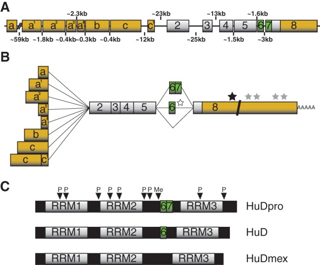
“The HuD protein, like all Hu members, has a molecular weight of ∼40 kDa and contains three RNA recognition motifs (RRMs) with a linker region separating the second and third RRM (Fig. 1C; Good 1995; Liu et al. 1995; Okano and Darnell 1997).”
§ Emerging complexity of the HuD/ELAVl4 gene; implications for neuronal development, function, and dysfunction > THE HuD GENE, mRNA, AND PROTEIN
What the sources say
- Not checked — no full text of the sourcecites Good 1995 source
- Not checked — no full text of the sourcecites Liu et al. 1995 source
- Supportedcites Okano and Darnell 1997 source
The Abstract describes HuD as encoding a target antigen among antigens of 35–40 kDa and states that Hu antigens have three RNA recognition motifs. The Cloning of mouse Hu gene family members section identifies a spacer region between RRM 2 and RRM 3. Page numbers are not provided in our copy. (LLM, conf=0.89)
plain- Asserted: passes — The authors put it forward as true.
- Checkable: passes — It can be checked against evidence.
- Load-bearing: fails — The paper’s conclusions may not depend on it.
- Based: passes — The paper gives a reason why it holds.
Probably not a substantial claim. The paper’s conclusions may not depend on it. It may state two separate claims as one.
The sources with a verdict support this claim; the rest weren’t checked yet, have no full text we could read, or their verdicts aren’t rated yet.
In the paper
“Nevertheless, there are a few known AUBPs that enhance mRNA stability, with Hu proteins being the most notable (Brennan and Steitz 2001; Hinman and Lou 2008; Pascale et al. 2008).”
§ Emerging complexity of the HuD/ELAVl4 gene; implications for neuronal development, function, and dysfunction > Cis-ELEMENT AND TARGET mRNAs OF HuD
What the sources say
- Not checked — no full text of the sourcecites Brennan and Steitz 2001 source
- Supportedcites Hinman and Lou 2008 source
In “Hu proteins and mRNA stability,” the paper says that most ARE-binding proteins decrease mRNA stability, whereas Hu proteins stabilize their target mRNAs. It also describes regulation of mRNA stability as the most well-known molecular function of Hu proteins, supporting the citing sentence’s characterization. (LLM, conf=0.91)
- Not checked — no full text of the sourcecites Pascale et al. 2008 source
plaindirect- Asserted: passes — The authors put it forward as true.
- Checkable: passes — It can be checked against evidence.
- Load-bearing: fails — The paper’s conclusions may not depend on it.
- Based: passes — The paper gives a reason why it holds.
Probably not a substantial claim. The paper’s conclusions may not depend on it.
The cited sources support only part of this claim — read each source’s verdict below.
In the paper
“ARE motifs are typically found in short-lived transcripts encoding proteins with diverse functions such as cellular proliferation, differentiation, transcription, RNA metabolism, inflammation, and stress-response (Bakheet et al. 2006; Khabar 2010).”
§ Emerging complexity of the HuD/ELAVl4 gene; implications for neuronal development, function, and dysfunction > Cis-ELEMENT AND TARGET mRNAs OF HuD
What the sources say
- Not matchedcites Bakheet et al. 2006
Couldn’t be matched to a paper in Crossref, so it can’t be checked.
- Supportedcites Khabar 2010 source
The Introduction describes ARE-bearing mRNAs as functionally diverse, listing proliferation, transcription, RNA metabolism, inflammatory responses, and development, and links these genes to rapid mRNA decay. It also identifies ARE-mediated regulation in external stress-mediated pathways; the sequence-characteristics section says ARE motifs occur preferentially in labile transcripts. The claim is a fair broad paraphrase, with differentiation corresponding to the paper’s broader category of development. (LLM, conf=0.88)
plaincorrelational- Asserted: passes — The authors put it forward as true.
- Checkable: passes — It can be checked against evidence.
- Load-bearing: fails — The paper’s conclusions may not depend on it.
- Based: passes — The paper gives a reason why it holds.
Probably not a substantial claim. The paper’s conclusions may not depend on it.
- Not matched
The cited sources support only part of this claim — read each source’s verdict below.
In the paper
“Out of the roughly 20 currently known AUBPs, the majority are expressed in neurons and promote mRNA decay including, for example, AUF1, TTP, and KSRP (Table 1; Wu and Brewer 2012).”
§ Emerging complexity of the HuD/ELAVl4 gene; implications for neuronal development, function, and dysfunction > Cis-ELEMENT AND TARGET mRNAs OF HuD
What the sources say
- Partly supportedcites Wu and Brewer 2012 sourceExperimental — accuracy not yet measured
Section 4.1 says there are about 20 known AUBPs, that most recruit mRNA-degradation machinery, and that AUF1, TTP, and KSRP promote degradation. It does not establish that a majority of AUBPs are expressed in neurons. (LLM, conf=0.90)
plaindirect- Asserted: passes — The authors put it forward as true.
- Checkable: passes — It can be checked against evidence.
- Load-bearing: fails — The paper’s conclusions may not depend on it.
- Based: passes — The paper gives a reason why it holds.
Probably not a substantial claim. The paper’s conclusions may not depend on it. It may state two separate claims as one.
- Partly supported
The cited sources support only part of this claim — read each source’s verdict below.
In the paper
“In developing neurons, HuD is found in growth cones of extending neurites, while in mature neurons it is present in axons and dendrites, including at the pre- and post-neurite terminals, respectively (Aranda-Abreu et al. 1999; Aronov et al. 2002).”
§ Emerging complexity of the HuD/ELAVl4 gene; implications for neuronal development, function, and dysfunction > TEMPORAL AND SPATIAL EXPRESSION OF HuD
What the sources say
- Partly supportedcites Aranda-Abreu et al. 1999 sourceExperimental — accuracy not yet measured
The Results section, “The ELAV-like protein–tau mRNA complex is associated with microtubules in vivo” (page not provided), reports ELAV-like protein staining extending into neurites and growth cones of NGF-treated PC12 cells. It does not establish that the staining is specifically HuD, or describe HuD in the axons, dendrites, or pre- and post-neurite terminals of mature neurons. (LLM, conf=0.85)
- Not checked — no full text of the sourcecites Aronov et al. 2002 source
plain- Asserted: passes — The authors put it forward as true.
- Checkable: passes — It can be checked against evidence.
- Load-bearing: unclear — It is unclear whether the paper’s conclusions depend on it.
- Based: passes — The paper gives a reason why it holds.
It may state two separate claims as one.
- Partly supported
Every source the paper cites for this claim was checked and supports it.
In the paper
“Since the 6.0-kb variant is less stable and not as efficiently translated as the 2.4-kb transcript, the authors postulated that the reduction of HuR expression prevents HuR’s ability to promote proliferation and thereby permits neurons to terminally differentiate (Mansfield and Keene 2011).”
§ Emerging complexity of the HuD/ELAVl4 gene; implications for neuronal development, function, and dysfunction > MULTILEVEL CONTROL OF mRNA METABOLISM BY HuD > Alternative polyadenylation
What the sources say
- Supportedcites Mansfield and Keene 2011 source
The Results sections on mRNA stability and translation (page not supplied) report that the 6.0-kb isoform is much less stable than the 2.4-kb isoform and that its 3′-UTR produces significantly less reporter expression. The Discussion explicitly presents reduced HuR expression permitting neuronal differentiation as a hypothesis, as the citing sentence does. (LLM, conf=0.91)
plaincorrelational- Asserted: passes — The authors put it forward as true.
- Checkable: passes — It can be checked against evidence.
- Load-bearing: passes — The paper’s conclusions depend on it.
- Based: passes — The paper gives a reason why it holds.
Probably not a substantial claim. It may state two separate claims as one.
- Supported
Every source the paper cites for this claim was checked and supports it.
In the paper
“The initial demonstration that Hu proteins regulate mRNA stability was based on the observation that HuB enhances GLUT1 mRNA half-life and, incidentally, translation (Jain et al. 1997).”
§ Emerging complexity of the HuD/ELAVl4 gene; implications for neuronal development, function, and dysfunction > MULTILEVEL CONTROL OF mRNA METABOLISM BY HuD > mRNA stability
What the sources say
- Supportedcites Jain et al. 1997 source
In the Results section (page not provided), GLUT1 mRNA half-life rose from 4.8 hours in untransfected cells to 7.0 hours in Hel-N1-expressing cells. The Discussion (page not provided) reports a Hel-N1-associated increase in GLUT1 translation, based on polysome profiles. The paper uses the name Hel-N1 rather than HuB. (LLM, conf=0.89)
plaindirect- Asserted: passes — The authors put it forward as true.
- Checkable: passes — It can be checked against evidence.
- Load-bearing: unclear — It is unclear whether the paper’s conclusions depend on it.
- Based: passes — The paper gives a reason why it holds.
It may state two separate claims as one.
- Supported
Every source the paper cites for this claim was checked and supports it.
In the paper
“In line with previous studies, nELAVs were found to enhance exon inclusion when bound to 5′ SSs in the downstream intron and inhibit exon inclusion when bound to adjacent 3′ and 5′ SSs of alternative exons.”
§ Emerging complexity of the HuD/ELAVl4 gene; implications for neuronal development, function, and dysfunction > MULTILEVEL CONTROL OF mRNA METABOLISM BY HuD > Alternative splicing
What the sources say
- Supportedcites Ince-Dunn et al. 2012 source
The Results section, “Generation of nElavl-RNA regulatory map” (page not provided), reports preferential binding at the downstream intron’s 5′ splice site where nElavl promotes inclusion, and at the immediate 3′ and 5′ splice sites where it promotes exclusion. (LLM, conf=0.95)
plaindirect- Asserted: passes — The authors put it forward as true.
- Checkable: passes — It can be checked against evidence.
- Load-bearing: passes — The paper’s conclusions depend on it.
- Based: passes — The paper gives a reason why it holds.
It may state two separate claims as one.
- Supported
The cited sources support only part of this claim — read each source’s verdict below.
In the paper
“Interestingly, HuD is more effective at stabilizing GAP-43 mRNAs that contain a longer poly(A) tail, indicating that HuD binds to both the ARE and poly(A) tail to increase GAP-43 mRNA half-life (Beckel-Mitchener et al. 2002).”
§ Emerging complexity of the HuD/ELAVl4 gene; implications for neuronal development, function, and dysfunction > MULTILEVEL CONTROL OF mRNA METABOLISM BY HuD > mRNA stability
What the sources say
- Partly supportedcites Beckel-Mitchener et al. 2002 sourceExperimental — accuracy not yet measured
The Results report that HuD preferentially stabilizes GAP-43 mRNA with long poly(A) tails and increases its half-life. The Discussion says HuD binding to the ARE was measured, but binding to the poly(A) tail was only a possible explanation consistent with the data—not a demonstrated mechanism. Page numbers are not provided in our copy. (LLM, conf=0.88)
hedgeddirect- Asserted: passes — The authors put it forward as true.
- Checkable: passes — It can be checked against evidence.
- Load-bearing: unclear — It is unclear whether the paper’s conclusions depend on it.
- Based: passes — The paper gives a reason why it holds.
It may state two separate claims as one.
- Partly supported
The cited sources that could be checked couldn’t confirm or dispute this claim.
In the paper
“In addition to these transcripts, HuD has been shown to bind and regulate several other mRNAs in vitro and in vivo, many of which encode proteins with important functions in neuronal differentiation such as cell cycle arrest (p21cip1/waf1) (Joseph et al. 1998), neuroblast proliferation (MSI-1) (Ratti et al. 2006), neuron migration (MARCKS) (Wein et al. 2003), neurite extension ([GAP-43] [Chung et”
§ Emerging complexity of the HuD/ELAVl4 gene; implications for neuronal development, function, and dysfunction > Cis-ELEMENT AND TARGET mRNAs OF HuD
What the sources say
- Not checked — no full text of the sourcecites Joseph et al. 1998 source
- Could not verifycites Ratti et al. 2006 sourceExperimental — accuracy not yet measured
The indexed copy contains a repository title indicating ELAV-related stabilization of Msi1 mRNA, but no article passage establishing that HuD binds or regulates it, or linking MSI-1 to neuroblast proliferation. Searches for HuD and Msi1 regulation returned only repository metadata and a PDF listing, not the paper’s substantive text. The other listed transcripts are attributed to separate citations in the citing sentence. (LLM, conf=0.72)
- Not matchedcites Wein et al. 2003
Couldn’t be matched to a paper in Crossref, so it can’t be checked.
boosteddirect- Asserted: passes — The authors put it forward as true.
- Checkable: passes — It can be checked against evidence.
- Load-bearing: passes — The paper’s conclusions depend on it.
- Based: passes — The paper gives a reason why it holds.
It may state two separate claims as one.
The cited sources support only part of this claim — read each source’s verdict below.
In the paper
“Multiple studies have shown that overexpression or down-regulation of HuD results in increased or decreased, respectively, GAP-43 mRNA levels and/or stability in cultured cells and the mouse nervous system (Anderson et al. 2000, 2001; Mobarak et al. 2000; Bolognani et al. 2006, 2007a).”
§ Emerging complexity of the HuD/ELAVl4 gene; implications for neuronal development, function, and dysfunction > MULTILEVEL CONTROL OF mRNA METABOLISM BY HuD > mRNA stability
What the sources say
- Not matchedcites Anderson et al. 2000, 2001
Couldn’t be matched to a paper in Crossref, so it can’t be checked.
- Supportedcites Mobarak et al. 2000 source
The Results report that HuD overexpression increased GAP-43 mRNA levels and extended its half-life from 5 to 9 hours in PC12 cells; HuD down-regulation shortened the half-life. This supports the cultured-cell portion for which this paper is cited. The sentence cites multiple studies for the broader statement about the mouse nervous system. (LLM, conf=0.92)
- Not matchedcites Bolognani et al. 2006, 2007a
Couldn’t be matched to a paper in Crossref, so it can’t be checked.
boosteddirect- Asserted: passes — The authors put it forward as true.
- Checkable: passes — It can be checked against evidence.
- Load-bearing: passes — The paper’s conclusions depend on it.
- Based: passes — The paper gives a reason why it holds.
It may state two separate claims as one.
- Not matched
Every source the paper cites for this claim was checked and supports it.
In the paper
“In addition, a recent report identified intronic nELAV binding sites flanking an alternative 3′ SS in the Gls1/Gls gene and demonstrated that nELAVs generate a longer Gls mRNA variant, indicating that they may enhance inclusion of a Gls 3′ exon(s) (Ince-Dunn et al. 2012).”
§ Emerging complexity of the HuD/ELAVl4 gene; implications for neuronal development, function, and dysfunction > MULTILEVEL CONTROL OF mRNA METABOLISM BY HuD > Alternative splicing
What the sources say
- Supportedcites Ince-Dunn et al. 2012 source
The Results section (‘A new role for nElavl in regulating glutamate in the brain,’ Fig. 6) reports intronic nElavl binding sites flanking the alternative Gls1 splice site and suggests that nElavl promotes use of the longer Gls1-l isoform. It also reports that Gls1-l mRNA falls to about 50% of wild-type levels when Elavl3 and Elavl4 are knocked out, supporting the citing sentence’s hedged interpretation. (LLM, conf=0.90)
hedgedconditional- Asserted: passes — The authors put it forward as true.
- Checkable: passes — It can be checked against evidence.
- Load-bearing: unclear — It is unclear whether the paper’s conclusions depend on it.
- Based: passes — The paper gives a reason why it holds.
It may state two separate claims as one.
- Supported
The cited sources support only part of this claim — read each source’s verdict below.
In the paper
“Employing mainly REMSAs and in vitro cleavage and polyadenylation assays, one study determined that Hu proteins bind U-rich sequences downstream from the exon 4 poly(A) signal in calcitonin/CGRP to block binding of cleavage stimulation factor 64 (CstF64) and cleavage-polyadenylation specificity factor (CPSF), essential components of the cleavage and polyadenylation machinery. Through this”
§ Emerging complexity of the HuD/ELAVl4 gene; implications for neuronal development, function, and dysfunction > MULTILEVEL CONTROL OF mRNA METABOLISM BY HuD > Alternative polyadenylation
What the sources say
- Partly supportedcites Zhu et al. 2006 sourceExperimental — accuracy not yet measured
Split into 2 atoms — 1 confirmed, 1 partially supported, 0 disputed, 0 misattributed, 0 unverifiable. Expand the claim to see per-atom verdicts.
boosteddirect- Asserted: passes — The authors put it forward as true.
- Checkable: passes — It can be checked against evidence.
- Load-bearing: passes — The paper’s conclusions depend on it.
- Based: passes — The paper gives a reason why it holds.
It may state two separate claims as one.
- Partly supported
The cited sources support only part of this claim — read each source’s verdict below.
In the paper
“In addition to GAP-43 transcripts, HuD has also been shown to promote the stability of several other ARE-containing mRNAs in neurons, such as AChE (Deschenes-Furry et al. 2003) and Nova1 (Ratti et al. 2008), both in vitro and in vivo (Table 2; Perrone-Bizzozero and Bird 2013).”
§ Emerging complexity of the HuD/ELAVl4 gene; implications for neuronal development, function, and dysfunction > MULTILEVEL CONTROL OF mRNA METABOLISM BY HuD > mRNA stability
What the sources say
- Supportedcites Deschenes-Furry et al. 2003 source
Deschênes-Furry et al. is cited for the AChE portion of the sentence; Nova1 and the broader in-vitro/in-vivo statement have separate citations. In the Discussion (page unavailable), the paper reports that AChE transcript abundance varied with HuD expression in PC12 cells and that HuD increased expression through the AChE 3′-UTR, supporting the attributed post-transcriptional effect. (LLM, conf=0.85)
- Partly supportedcites Ratti et al. 2008 sourceExperimental — accuracy not yet measured
The Nova1 mRNA degradation results (page unavailable) show that HuD re-expression prolonged Nova1 mRNA half-life in cultured NSC34 cells, supporting the Nova1 stability claim in cells. The Experimental Procedures identify NSC34 as a cell line; this paper does not establish Nova1 mRNA stabilization in an organism. AChE is attributed to a different citation in the citing sentence and is not judged here. (LLM, conf=0.88)
- Not matchedcites Perrone-Bizzozero and Bird 2013
Couldn’t be matched to a paper in Crossref, so it can’t be checked.
boosteddirect- Asserted: passes — The authors put it forward as true.
- Checkable: passes — It can be checked against evidence.
- Load-bearing: unclear — It is unclear whether the paper’s conclusions depend on it.
- Based: passes — The paper gives a reason why it holds.
It may state two separate claims as one.
- Supported
The cited sources support only part of this claim — read each source’s verdict below.
In the paper
“Of particular interest, this study also found that HuD binds mRNAs of several RBPs, including its own, HuB, HuR, Cugbp2, Musashi 2, and Staufen 2, indicating a complex regulatory network between HuD and other components of the ribonome (Pullmann et al. 2007; Mansfield and Keene 2009; Bolognani et al. 2010).”
§ Emerging complexity of the HuD/ELAVl4 gene; implications for neuronal development, function, and dysfunction > Cis-ELEMENT AND TARGET mRNAs OF HuD
What the sources say
- Could not verifycites Pullmann et al. 2007 sourceExperimental — accuracy not yet measured
The Abstract (page unavailable) says the study investigated AUF1, HuR, KSRP, NF90, TIA-1, and TIAR, and found binding among some of their mRNAs. Neither it nor the additional passages retrieved for HuD, Cugbp2, Musashi 2, and Staufen 2 reports the claimed HuD binding relationships. (LLM, conf=0.76)
- Partly supportedcites Mansfield and Keene 2009 sourceExperimental — accuracy not yet measured
The “Regulators of regulators” section (page unavailable) describes RBPs binding their own and one another’s mRNAs, supporting the idea of an interconnected ribonome. It discusses a group including HuR, not HuD; our copy does not report HuD binding the listed mRNAs (searched: HuD, Cugbp2, Musashi, Staufen). (LLM, conf=0.82)
- Supportedcites Bolognani et al. 2010 source
The Discussion lists HuD itself, HuB, HuR, Cugbp2, Musashi 2, and Staufen 2 among the RBP mRNAs identified as HuD targets. It also says these observations support a complex, integrated network of post-transcriptional regulators. (LLM, conf=0.94)
hedgedcorrelational- Asserted: passes — The authors put it forward as true.
- Checkable: passes — It can be checked against evidence.
- Load-bearing: passes — The paper’s conclusions depend on it.
- Based: passes — The paper gives a reason why it holds.
It may state two separate claims as one.
- Could not verify
The sources with a verdict support this claim; the rest weren’t checked yet, have no full text we could read, or their verdicts aren’t rated yet.
In the paper
“Using a variety of complementary techniques, including an in vitro protein methylation assay, CARM1 was demonstrated to interact with and methylate an amino acid located in the hinge region of rat (Arg236) (Fujiwara et al. 2006) and mouse (Arg248) (Hubers et al. 2010) HuD (Fig. 1C).”
§ Emerging complexity of the HuD/ELAVl4 gene; implications for neuronal development, function, and dysfunction > MOLECULAR MECHANISMS REGULATING HuD > Post-translational control of HuD expression and function
What the sources say
- Supportedcites Fujiwara et al. 2006 source
The Results identify HuD Arg236 as a CARM1 methylation target, and the Discussion locates Arg236 in the hinge region. The Results also show CARM1 and HuD colocalizing in PC12 cells. The mouse Arg248 statement is attached to a different citation, not Fujiwara et al. (LLM, conf=0.90)
- Not checked — no full text of the sourcecites Hubers et al. 2010 source
boosteddirect- Asserted: passes — The authors put it forward as true.
- Checkable: passes — It can be checked against evidence.
- Load-bearing: passes — The paper’s conclusions depend on it.
- Based: passes — The paper gives a reason why it holds.
It may state two separate claims as one.
- Supported
The cited sources support only part of this claim — read each source’s verdict below.
In the paper
“On the other hand, inhibition of HuD methylation or knockdown of CARM1 resulted in increased p21cip1/waf1 mRNA levels, cell-cycle withdrawal, and neuronal differentiation.”
§ Emerging complexity of the HuD/ELAVl4 gene; implications for neuronal development, function, and dysfunction > MOLECULAR MECHANISMS REGULATING HuD > Post-translational control of HuD expression and function
What the sources say
- Partly supportedcites Fujiwara et al. 2006 sourceExperimental — accuracy not yet measured
The Results report a 3.26-fold increase in p21cip1/waf1 mRNA in a CARM1-depleted PC12 clone, and slower proliferation and greater neurite outgrowth in depleted clones. The Discussion describes partially differentiated cells, while the Results show accelerated neuritogenesis mainly in response to NGF. The paper does not establish cell-cycle withdrawal: slower proliferation is not the same as cell-cycle exit. (LLM, conf=0.84)
- Not checked — no full text of the sourcecites Hubers et al. 2010 source
plaindirect- Asserted: passes — The authors put it forward as true.
- Checkable: passes — It can be checked against evidence.
- Load-bearing: passes — The paper’s conclusions depend on it.
- Based: passes — The paper gives a reason why it holds.
It may state two separate claims as one.
- Partly supported
Every source the paper cites for this claim was checked and supports it.
In the paper
“The classical PKCα isoform was shown to increase nELAV levels and provoke their nuclear export following treatment of SH-SY5Y NB cells with the PKC-activating compounds phorbol esters and bryostatin-1 (Pascale et al. 2005).”
§ Emerging complexity of the HuD/ELAVl4 gene; implications for neuronal development, function, and dysfunction > MOLECULAR MECHANISMS REGULATING HuD > Post-translational control of HuD expression and function
What the sources say
- Supportedcites Pascale et al. 2005 source
The Results report that PMA and bryostatin-1 treatment of SH-SY5Y cells reduced nuclear nELAV staining and increased total nELAV protein levels. The PKCα knock-down and inhibition experiments identify PKCα as mediating the PMA-induced increase; the Discussion places nuclear export and up-regulation in the PKCα-involving pathway. (LLM, conf=0.90)
plaindirect- Asserted: passes — The authors put it forward as true.
- Checkable: passes — It can be checked against evidence.
- Load-bearing: unclear — It is unclear whether the paper’s conclusions depend on it.
- Based: passes — The paper gives a reason why it holds.
It may state two separate claims as one.
- Supported
The cited sources support only part of this claim — read each source’s verdict below.
In the paper
“Moreover, stimulation of PKCα promotes its interaction with nELAV proteins resulting in their phosphorylation at threonine residues, redistribution to the neuronal cytoskeletal and membrane fractions, and stabilization of GAP-43 mRNA.”
§ Emerging complexity of the HuD/ELAVl4 gene; implications for neuronal development, function, and dysfunction > MOLECULAR MECHANISMS REGULATING HuD > Post-translational control of HuD expression and function
What the sources say
- Partly supportedcites Pascale et al. 2005 sourceExperimental — accuracy not yet measured
The Results report that PKC activation increases PKCα’s association with nELAV proteins and nELAV threonine phosphorylation; they also show PKCα-dependent stabilization of GAP-43 mRNA. However, the Results say the increase in nELAV proteins is confined to the cytoskeletal compartment. It is PKCα, not nELAV, that translocates to both membrane and cytoskeletal fractions (page unavailable in our copy). (LLM, conf=0.89)
plaindirect- Asserted: passes — The authors put it forward as true.
- Checkable: passes — It can be checked against evidence.
- Load-bearing: unclear — It is unclear whether the paper’s conclusions depend on it.
- Based: passes — The paper gives a reason why it holds.
It may state two separate claims as one.
- Partly supported
The cited sources support only part of this claim — read each source’s verdict below.
In the paper
“Moreover, methylation of HuD decreased p21cip1/waf1 mRNA stability and maintained neuronal precursor cells in a proliferative state.”
§ Emerging complexity of the HuD/ELAVl4 gene; implications for neuronal development, function, and dysfunction > MOLECULAR MECHANISMS REGULATING HuD > Post-translational control of HuD expression and function
What the sources say
- Partly supportedcites Fujiwara et al. 2006 sourceExperimental — accuracy not yet measured
The Results report that reducing HuD methylation lengthened p21cip1/waf1 mRNA half-life in PC12 cells, and the Abstract suggests that methylated HuD maintains those cells in a proliferative state by promoting mRNA decay. The claim extends this finding from PC12 cells to neuronal precursor cells generally, which the cited paper does not establish. (LLM, conf=0.86)
- Not checked — no full text of the sourcecites Hubers et al. 2010 source
plaindirect- Asserted: passes — The authors put it forward as true.
- Checkable: passes — It can be checked against evidence.
- Load-bearing: passes — The paper’s conclusions depend on it.
- Based: passes — The paper gives a reason why it holds.
It may state two separate claims as one.
- Partly supported
Every source the paper cites for this claim was checked and supports it.
In the paper
“Importantly, HuD is a focal downstream target of this neurite-extending pathway since nerve growth factor (NGF) or phorbol ester treatment of PC12 cells following HuD knockdown prevents neurite outgrowth (Mobarak et al. 2000).”
§ Emerging complexity of the HuD/ELAVl4 gene; implications for neuronal development, function, and dysfunction > MOLECULAR MECHANISMS REGULATING HuD > Post-translational control of HuD expression and function
What the sources say
- Supportedcites Mobarak et al. 2000 source
The Results report that PC12 cells with reduced HuD did not show increased neurite outgrowth after NGF treatment and that phorbol ester failed to induce neurite outgrowth, producing only short, stubby processes. The Discussion describes HuD reduction as selectively affecting the pathway activated by NGF and phorbol esters, while leaving the response to cAMP intact. The claim concerns outgrowth through that pathway, not all neurite outgrowth. (LLM, conf=0.91)
boosteddirect- Asserted: passes — The authors put it forward as true.
- Checkable: passes — It can be checked against evidence.
- Load-bearing: passes — The paper’s conclusions depend on it.
- Based: passes — The paper gives a reason why it holds.
It may state two separate claims as one.
- Supported
Every source the paper cites for this claim was checked and supports it.
In the paper
“The report by Kullman et al. suggests that inhibition of p27 translation by Hu proteins is an event that sustains cells in a proliferative state (Kullmann et al. 2002).”
§ Emerging complexity of the HuD/ELAVl4 gene; implications for neuronal development, function, and dysfunction > MULTILEVEL CONTROL OF mRNA METABOLISM BY HuD > mRNA localization and translation
What the sources say
- Supportedcites Kullmann et al. 2002 source
The Results (Fig. 7) report that HuR knockdown increased the proportion of G1-phase cells and reduced the S-phase proportion. The authors conclude that HuR helps regulate proliferation in part by inhibiting p27 translation, supporting the citing sentence’s qualified interpretation. (LLM, conf=0.92)
hedgeddirect- Asserted: passes — The authors put it forward as true.
- Checkable: passes — It can be checked against evidence.
- Load-bearing: fails — The paper’s conclusions may not depend on it.
- Based: passes — The paper gives a reason why it holds.
Probably not a substantial claim. The paper’s conclusions may not depend on it.
- Supported
The cited sources support only part of this claim — read each source’s verdict below.
In the paper
“Even though the mechanism of translational silencing is unclear, it was demonstrated to require HuD binding to an internal ribosome entry site (IRES) in the 5′ UTR of p27 mRNA (Kullmann et al. 2002) and a short ∼22-nt sequence in the 5′ UTR of Ins2 mRNA (Lee et al. 2012).”
§ Emerging complexity of the HuD/ELAVl4 gene; implications for neuronal development, function, and dysfunction > MULTILEVEL CONTROL OF mRNA METABOLISM BY HuD > mRNA localization and translation
What the sources say
- Partly supportedcites Kullmann et al. 2002 sourceExperimental — accuracy not yet measured
The Results (page not provided) show that HuD binds specifically to the p27 5′ UTR and that HuD overexpression inhibits p27 translation and IRES activity. They do not establish that HuD binds the IRES itself or that such binding is required for silencing. The Ins2 sequence is attributed to Lee et al. (2012), not to Kullmann et al. (LLM, conf=0.86)
- Supportedcites Lee et al. 2012 source
The Lee et al. citation supports the Ins2 portion of the sentence, not the separately cited p27 portion. In the Results section, the paper identifies positions 52–73 of the Ins2 5′ UTR as a HuD-binding segment and reports that this segment confers HuD-dependent repression in a translation reporter assay. Page numbers are not provided in our copy. (LLM, conf=0.92)
boosteddirect- Asserted: passes — The authors put it forward as true.
- Checkable: passes — It can be checked against evidence.
- Load-bearing: passes — The paper’s conclusions depend on it.
- Based: passes — The paper gives a reason why it holds.
It may state two separate claims as one.
- Partly supported
The cited sources support only part of this claim — read each source’s verdict below.
In the paper
“Various conditions, including KCl depolarization of primary hippocampal neurons, cocaine, or seizure treatment of mice and phorbol ester or bryostatin-1 application to human neuroblastoma (NB) cells, result in an augmented abundance of HuD, its redistribution to neurites, and its increased association with mRNAs and mRNP components (Pascale et al. 2005; Tiruchinapalli et al. 2008a,b).”
§ Emerging complexity of the HuD/ELAVl4 gene; implications for neuronal development, function, and dysfunction > MULTILEVEL CONTROL OF mRNA METABOLISM BY HuD > mRNA localization and translation
What the sources say
- Partly supportedcites Pascale et al. 2005 sourceExperimental — accuracy not yet measured
Split into 2 atoms — 0 confirmed, 2 partially supported, 0 disputed, 0 misattributed, 0 unverifiable. Expand the claim to see per-atom verdicts.
- Not matchedcites Tiruchinapalli et al. 2008a,b
Couldn’t be matched to a paper in Crossref, so it can’t be checked.
plaindirect- Asserted: passes — The authors put it forward as true.
- Checkable: passes — It can be checked against evidence.
- Load-bearing: unclear — It is unclear whether the paper’s conclusions depend on it.
- Based: passes — The paper gives a reason why it holds.
It may state two separate claims as one.
- Partly supported
Every source the paper cites for this claim was checked and supports it.
In the paper
“One functional consequence of this auto-regulatory binding is presumably in controlling alternative splicing of its own pre-mRNA (see above) (Wang et al. 2010).”
§ Emerging complexity of the HuD/ELAVl4 gene; implications for neuronal development, function, and dysfunction > MOLECULAR MECHANISMS REGULATING HuD > Post-transcriptional control of HuD expression
What the sources say
- Supportedcites Wang et al. 2010 source
The Discussion (‘A novel enhancer function of Hu proteins in splicing regulation’) states that Hu proteins bind intronic sequences to promote inclusion of HuD exon 6. Another Discussion passage (‘Biological function of alternative inclusion of HuD exon 6’) identifies HuD itself among the isoforms that promote that inclusion, supporting the citing sentence’s hedged description of autoregulation. (LLM, conf=0.90)
hedgedconditional- Asserted: passes — The authors put it forward as true.
- Checkable: passes — It can be checked against evidence.
- Load-bearing: fails — The paper’s conclusions may not depend on it.
- Based: passes — The paper gives a reason why it holds.
Probably not a substantial claim. The paper’s conclusions may not depend on it.
- Supported
The cited sources support only part of this claim — read each source’s verdict below.
In the paper
“Prior to our study, only two transcriptional mechanisms were known to regulate expression of the HuD gene—thyroid hormone (T3; presumably via activation of the thyroid hormone receptor) and TF forkhead box O1 (FoxO1)—and both of these factors were shown to negatively impact the abundance of HuD.”
§ Emerging complexity of the HuD/ELAVl4 gene; implications for neuronal development, function, and dysfunction > MOLECULAR MECHANISMS REGULATING HuD > Transcriptional control of HuD expression
What the sources say
- Partly supportedcites Cuadrado et al. 2003 sourceExperimental — accuracy not yet measured
In “HuD gene transcription is repressed by T3” (page not available), run-on assays show that T3 reduced HuD transcription by 60%; the paper also reports reduced HuD RNA in cells with appropriate thyroid hormone receptor levels. Our copy does not address FoxO1 or establish that these were the only two known transcriptional mechanisms. (LLM, conf=0.88)
- Partly supportedcites Lee et al. 2012 sourceExperimental — accuracy not yet measured
The Results section (‘HuD is expressed in pancreatic β cells’; page not provided) shows that FoxO1 binds the HuD promoter and that FoxO1 overexpression reduces HuD, supporting its role as a negative transcriptional regulator. The passages found do not address thyroid hormone (T3) or establish that these were the only two known transcriptional mechanisms. (LLM, conf=0.88)
plaindirect- Asserted: passes — The authors put it forward as true.
- Checkable: passes — It can be checked against evidence.
- Load-bearing: passes — The paper’s conclusions depend on it.
- Based: passes — The paper gives a reason why it holds.
It may state two separate claims as one.
- Partly supported
The cited sources support only part of this claim — read each source’s verdict below.
In the paper
“For example, neurosphere assays using neuronal stem/progenitor cells from the cerebral cortex of HuD knockout (−/−) mice revealed that HuD decreases self-renewal capacity and promotes cell cycle exit and differentiation of these cells.”
§ Emerging complexity of the HuD/ELAVl4 gene; implications for neuronal development, function, and dysfunction > FUNCTIONAL ROLES OF HuD IN THE NERVOUS SYSTEM > Neuronal development
What the sources say
- Partly supportedcites Akamatsu et al. 2005 sourceExperimental — accuracy not yet measured
The Results report that neurospheres from HuD-knockout embryonic cerebral cortex showed increased self-renewal, while a neurosphere differentiation assay found reduced production of neuronal progeny. The paper also concludes that HuD promotes cell-cycle exit, but that finding came from labeling experiments in the embryonic cerebral wall, not the neurosphere assays. No page numbers are available in our copy. (LLM, conf=0.86)
plaindirect- Asserted: passes — The authors put it forward as true.
- Checkable: passes — It can be checked against evidence.
- Load-bearing: passes — The paper’s conclusions depend on it.
- Based: passes — The paper gives a reason why it holds.
It may state two separate claims as one.
- Partly supported
The cited sources support only part of this claim — read each source’s verdict below.
In the paper
“T3 was demonstrated to decrease HuD mRNA synthesis in neuronal N2a cells, and T3 levels were inversely correlated with HuD mRNA levels in the rat brain (Cuadrado et al. 2003)”
§ Emerging complexity of the HuD/ELAVl4 gene; implications for neuronal development, function, and dysfunction > MOLECULAR MECHANISMS REGULATING HuD > Transcriptional control of HuD expression
What the sources say
- Partly supportedcites Cuadrado et al. 2003 sourceExperimental — accuracy not yet measured
The section ‘T3 inhibits HuD expression in PC12 and N2a cells’ reports reduced HuD RNA expression in N2a cells, while ‘HuD gene transcription is repressed by T3’ reports a reduced transcription rate without establishing that result specifically in N2a cells. The rat-brain results show higher HuD RNA under thyroid deficiency only in certain regions of the developing brain; they do not report a measured correlation between T3 and HuD mRNA levels across the rat brain. (LLM, conf=0.86)
boosteddirect- Asserted: passes — The authors put it forward as true.
- Checkable: passes — It can be checked against evidence.
- Load-bearing: unclear — It is unclear whether the paper’s conclusions depend on it.
- Based: passes — The paper gives a reason why it holds.
It may state two separate claims as one.
- Partly supported
The cited sources support only part of this claim — read each source’s verdict below.
In the paper
“Detailed analysis of these mice showed enhanced sprouting of GAP-43-containing mossy fibers to the CA3 region, ectopic expression of GAP-43 protein in the DG, and increased GAP-43 and calmodulin binding, possibly causing altered calcium signalling (Tanner et al. 2008).”
§ Emerging complexity of the HuD/ELAVl4 gene; implications for neuronal development, function, and dysfunction > FUNCTIONAL ROLES OF HuD IN THE NERVOUS SYSTEM > Neuronal plasticity
What the sources say
- Partly supportedcites Tanner et al. 2008 sourceExperimental — accuracy not yet measured
The Results report GAP-43-positive mossy fibers in CA3, increased GAP-43 staining in the dentate gyrus, and increased calmodulin binding to GAP-43; the Conclusions propose that calmodulin sequestration may alter calcium signaling (pages unavailable). But the Results do not establish enhanced mossy-fiber sprouting: the authors say the fibers interpreted as possible sprouts could instead have other origins. (LLM, conf=0.88)
hedgedconditional- Asserted: passes — The authors put it forward as true.
- Checkable: passes — It can be checked against evidence.
- Load-bearing: passes — The paper’s conclusions depend on it.
- Based: passes — The paper gives a reason why it holds.
It may state two separate claims as one.
- Partly supported
Every source the paper cites for this claim was checked and supports it.
In the paper
“In adult HuD−/− mice, an abnormal clasping reflex and poor performance on the rotarod test were observed, suggesting a sensory/motor defect in these animals and a role for HuD at the axon terminal (Akamatsu et al. 2005).”
§ Emerging complexity of the HuD/ELAVl4 gene; implications for neuronal development, function, and dysfunction > FUNCTIONAL ROLES OF HuD IN THE NERVOUS SYSTEM > Neuronal plasticity
What the sources say
- Supportedcites Akamatsu et al. 2005 source
The Results report that 70–80% of HuD−/− mice showed an abnormal hind-limb clasping reflex by 4–8 weeks of age. The same section reports that adult HuD−/− mice stayed on the rotarod for significantly less time than wild-type mice and continued to perform poorly across trials. (LLM, conf=0.94)
plain- Asserted: passes — The authors put it forward as true.
- Checkable: passes — It can be checked against evidence.
- Load-bearing: passes — The paper’s conclusions depend on it.
- Based: passes — The paper gives a reason why it holds.
It may state two separate claims as one.
- Supported
The cited sources support only part of this claim — read each source’s verdict below.
In the paper
“This function of HuD requires its shuttling to the cytoplasm and involves stabilization of various target mRNAs whose protein products promote microtubule assembly and growth cone development including GAP-43, Tau, and AChE (Chung et al. 1997; Aranda-Abreu et al. 1999; Kasashima et al. 1999; Deschenes-Furry et al. 2003).”
§ Emerging complexity of the HuD/ELAVl4 gene; implications for neuronal development, function, and dysfunction > FUNCTIONAL ROLES OF HuD IN THE NERVOUS SYSTEM > Neuronal development
What the sources say
- Partly supportedcites Chung et al. 1997 sourceExperimental — accuracy not yet measured
The GAP-43 binding-site results show that HuD binds a conserved element in GAP-43 mRNA. The Discussion proposes a possible role for Elav-like proteins in GAP-43 regulation, but treats their involvement in mRNA stabilization as a hypothesis requiring further investigation. This paper does not establish that HuD must shuttle to the cytoplasm or that it stabilizes the listed target mRNAs. Page numbers are unavailable in our copy. (LLM, conf=0.88)
- Partly supportedcites Aranda-Abreu et al. 1999 sourceExperimental — accuracy not yet measured
The ‘Characterization of HuD–tau mRNA complex formation’ section shows that HuD binds tau mRNA, and the antisense-treatment results link reduced HuD to reduced tau mRNA and protein in PC12 cells. The Discussion treats tau mRNA stabilization as a possible mechanism but says how ELAV-like proteins control mRNA half-life remains undetermined. These passages do not establish that HuD must shuttle to the cytoplasm or that this paper demonstrates stabilization of GAP-43 and AChE mRNAs. Page numbers were unavailable in the indexed passages. (LLM, conf=0.86)
- Not checked — no full text of the sourcecites Kasashima et al. 1999 source
plaindirect- Asserted: passes — The authors put it forward as true.
- Checkable: passes — It can be checked against evidence.
- Load-bearing: passes — The paper’s conclusions depend on it.
- Based: passes — The paper gives a reason why it holds.
It may state two separate claims as one.
- Partly supported
The cited sources support only part of this claim — read each source’s verdict below.
In the paper
“suggesting a sensory/motor defect in these animals and a role for HuD at the axon terminal (Akamatsu et al. 2005).”
§ Emerging complexity of the HuD/ELAVl4 gene; implications for neuronal development, function, and dysfunction > FUNCTIONAL ROLES OF HuD IN THE NERVOUS SYSTEM > Neuronal plasticity
What the sources say
- Partly supportedcites Akamatsu et al. 2005 sourceExperimental — accuracy not yet measured
The Results (page unavailable) report abnormal hind-limb clasping in adult HuD−/− mice, describe it as indicating motor/sensory defects, and report significantly poorer rotarod performance than in wild-type mice. Our copy does not connect these findings to a role for HuD at the axon terminal. (LLM, conf=0.86)
hedgedconditional- Asserted: passes — The authors put it forward as true.
- Checkable: passes — It can be checked against evidence.
- Load-bearing: passes — The paper’s conclusions depend on it.
- Based: passes — The paper gives a reason why it holds.
It may state two separate claims as one.
- Partly supported
Every source the paper cites for this claim was checked and supports it.
In the paper
“A study in our laboratory demonstrated that, between 1 and 4 d following axotomy of superior cervical ganglion (SCG) neurons, HuD protein and mRNA levels, its interaction with AChE mRNA, and AChE transcript levels decreased (Deschenes-Furry et al. 2007).”
§ Emerging complexity of the HuD/ELAVl4 gene; implications for neuronal development, function, and dysfunction > FUNCTIONAL ROLES OF HuD IN THE NERVOUS SYSTEM > Neuronal plasticity
What the sources say
- Supportedcites Deschenes-Furry et al. 2007 source
In “HuD expression is altered after SCG axotomy,” HuD transcripts decreased within 1 day and remained low through day 4; HuD protein was also reduced at day 2. “Axotomy alters AChE mRNA and protein interactions” reports reduced HuD binding to AChE RNA after axotomy, while “Axonal injury results in decreased AChE mRNA levels” reports declining AChE transcripts at days 1, 2, and 4. (LLM, conf=0.93)
boosteddirect- Asserted: passes — The authors put it forward as true.
- Checkable: passes — It can be checked against evidence.
- Load-bearing: passes — The paper’s conclusions depend on it.
- Based: passes — The paper gives a reason why it holds.
It may state two separate claims as one.
- Supported
The cited sources support only part of this claim — read each source’s verdict below.
In the paper
“In the hippocampus of AD patients, nELAV protein abundance was found to decrease in parallel with clinical dementia progression, and their expression was inversely correlated with Aβ peptide levels both in vitro and in vivo (Amadio et al. 2009).”
§ Emerging complexity of the HuD/ELAVl4 gene; implications for neuronal development, function, and dysfunction > FUNCTIONAL ROLES OF HuD IN THE NERVOUS SYSTEM > Neurological disorders
What the sources say
- Partly supportedcites Amadio et al. 2009 sourceExperimental — accuracy not yet measured
The Results section, “The nELAV are decreased in postmortem AD patients” (page unavailable), reports that hippocampal nELAV levels fell with dementia progression and were inversely correlated with hippocampal Aβ content. The Results section, “Aβ intracellular accumulation induces a reduction of nELAV in SH-SY5Y cells” (page unavailable), reports that Aβ exposure reduced nELAV in vitro, but does not establish an inverse correlation between their measured levels in vitro. (LLM, conf=0.86)
plaincorrelational- Asserted: passes — The authors put it forward as true.
- Checkable: passes — It can be checked against evidence.
- Load-bearing: passes — The paper’s conclusions depend on it.
- Based: passes — The paper gives a reason why it holds.
It may state two separate claims as one.
- Partly supported
In the paper
“Recent findings in our laboratory, however, have identified the expression of five additional HuD E1 variants, resulting in a total of eight (Fig. 1A,B; Bronicki et al. 2012).”
§ Emerging complexity of the HuD/ELAVl4 gene; implications for neuronal development, function, and dysfunction > THE HuD GENE, mRNA, AND PROTEIN
What the sources say
- Not matchedcites Bronicki et al. 2012
Couldn’t be matched to a paper in Crossref, so it can’t be checked.
plain- Asserted: passes — The authors put it forward as true.
- Checkable: passes — It can be checked against evidence.
- Load-bearing: unclear — It is unclear whether the paper’s conclusions depend on it.
- Based: passes — The paper gives a reason why it holds.
It may state two separate claims as one.
- Not matched
In the paper
“We also found that most E1 variants house an in-frame methionine codon (with the exception of E1a4, since it harbors two stop codons downstream from the AUG), suggesting that they may encode alternate HuD N termini.”
§ Emerging complexity of the HuD/ELAVl4 gene; implications for neuronal development, function, and dysfunction > THE HuD GENE, mRNA, AND PROTEIN
What the sources say
- Not matchedcites Bronicki et al. 2012
Couldn’t be matched to a paper in Crossref, so it can’t be checked.
plain- Asserted: passes — The authors put it forward as true.
- Checkable: passes — It can be checked against evidence.
- Load-bearing: passes — The paper’s conclusions depend on it.
- Based: passes — The paper gives a reason why it holds.
It may state two separate claims as one.
- Not matched
None of the sources cited for this claim have been checked yet.
In the paper
“As mentioned above, previous studies, along with data from our laboratory, revealed eight HuD E1 variants, indicating the existence of at least eight different 5′ UTRs (Fig. 1A,B), with most containing an in-frame translation start site (Abe et al. 1994; Inman et al. 1998; Bronicki et al. 2012).”
§ Emerging complexity of the HuD/ELAVl4 gene; implications for neuronal development, function, and dysfunction > MOLECULAR MECHANISMS REGULATING HuD > Post-transcriptional control of HuD expression
What the sources say
- Not matchedcites Abe et al. 1994
Couldn’t be matched to a paper in Crossref, so it can’t be checked.
- Not checked — no full text of the sourcecites Inman et al. 1998 source
- Not matchedcites Bronicki et al. 2012
Couldn’t be matched to a paper in Crossref, so it can’t be checked.
hedged- Asserted: passes — The authors put it forward as true.
- Checkable: passes — It can be checked against evidence.
- Load-bearing: unclear — It is unclear whether the paper’s conclusions depend on it.
- Based: passes — The paper gives a reason why it holds.
It may state two separate claims as one.
- Not matched
In the paper
“Remarkably, E1 and E′1 partially align with mouse E1b and E1c, respectively (L Bronicki and BJ Jasmin, unpubl.), implying that the developmental regulation and function of these two mammalian exons is evolutionarily conserved.”
§ Emerging complexity of the HuD/ELAVl4 gene; implications for neuronal development, function, and dysfunction > MOLECULAR MECHANISMS REGULATING HuD > Transcriptional control of HuD expression
What the sources say
- Not matchedcites L Bronicki and BJ Jasmin, unpubl.
Couldn’t be matched to a paper in Crossref, so it can’t be checked.
plaincorrelational- Asserted: passes — The authors put it forward as true.
- Checkable: passes — It can be checked against evidence.
- Load-bearing: passes — The paper’s conclusions depend on it.
- Based: passes — The paper gives a reason why it holds.
It may state two separate claims as one.
- Not matched
A result the paper reports from its own data — it cites no other source for it, so there is none to check. The quote shows it is in the paper.
In the paper
“RT-PCR analysis revealed that all eight exons are expressed in embryonic and adult mouse brain and are induced during neuronal differentiation.”
§ Emerging complexity of the HuD/ELAVl4 gene; implications for neuronal development, function, and dysfunction > MOLECULAR MECHANISMS REGULATING HuD > Post-transcriptional control of HuD expression
plaindirect- Asserted: passes — The authors put it forward as true.
- Checkable: passes — It can be checked against evidence.
- Load-bearing: unclear — It is unclear whether the paper’s conclusions depend on it.
- Based: passes — The paper gives a reason why it holds.
It may state two separate claims as one.
A result the paper reports from its own data — it cites no other source for it, so there is none to check. The quote shows it is in the paper.
In the paper
“Additionally, we demonstrated that a highly conserved 400-bp region upstream of the predominantly expressed E1 variant, E1c, houses several E-boxes, two of which are targeted by the basic helix-loop-helix transcription factor Neurogenin 2 (Ngn2).”
§ Emerging complexity of the HuD/ELAVl4 gene; implications for neuronal development, function, and dysfunction > MOLECULAR MECHANISMS REGULATING HuD > Transcriptional control of HuD expression
boosteddirect- Asserted: passes — The authors put it forward as true.
- Checkable: passes — It can be checked against evidence.
- Load-bearing: passes — The paper’s conclusions depend on it.
- Based: passes — The paper gives a reason why it holds.
It may state two separate claims as one.
The paper states this without a source or data behind it here, so there is nothing to check it against. The quote shows it is in the paper.
In the paper
“HuD is an RNA-binding protein that has been implicated in a wide variety of regulatory processes involving mRNA metabolism in neurons.”
§ Emerging complexity of the HuD/ELAVl4 gene; implications for neuronal development, function, and dysfunction > Abstract
plaincorrelational- Asserted: passes — The authors put it forward as true.
- Checkable: passes — It can be checked against evidence.
- Load-bearing: passes — The paper’s conclusions depend on it.
- Based: passes — The paper gives a reason why it holds.
Probably not a substantial claim.
In the paper
“In the HuD gene locus, E2 and E3, E4 and E5, and E8 encode the three RRMs, respectively, whereas the alternatively spliced E6 and E7 encode the linker region (Inman et al. 1998).”
§ Emerging complexity of the HuD/ELAVl4 gene; implications for neuronal development, function, and dysfunction > THE HuD GENE, mRNA, AND PROTEIN
What the sources say
- Not checked — no full text of the sourcecites Inman et al. 1998 source
plain- Asserted: passes — The authors put it forward as true.
- Checkable: passes — It can be checked against evidence.
- Load-bearing: fails — The paper’s conclusions may not depend on it.
- Based: passes — The paper gives a reason why it holds.
Probably not a substantial claim. The paper’s conclusions may not depend on it. It may state two separate claims as one.
None of the sources cited for this claim have been checked yet.
In the paper
“Overall, the HuD gene is well conserved among higher vertebrates and is located on chromosome 1 in humans and chromosome 4 in mice (Fig. 1A).”
§ Emerging complexity of the HuD/ELAVl4 gene; implications for neuronal development, function, and dysfunction > THE HuD GENE, mRNA, AND PROTEIN
What the sources say
- Not matchedcites Sekido et al. 1994
Couldn’t be matched to a paper in Crossref, so it can’t be checked.
- Not checked — no full text of the sourcecites Inman et al. 1998 source
boosted- Asserted: passes — The authors put it forward as true.
- Checkable: passes — It can be checked against evidence.
- Load-bearing: fails — The paper’s conclusions may not depend on it.
- Based: passes — The paper gives a reason why it holds.
Probably not a substantial claim. The paper’s conclusions may not depend on it. It may state two separate claims as one.
- Not matched
None of the sources cited for this claim have been checked yet.
In the paper
“The human and mouse HuD gene spans ∼146 kb of DNA and is divided into seven coding exons (E2 to E8) that encompass ∼44 kb of DNA (Sekido et al. 1994; Inman et al. 1998).”
§ Emerging complexity of the HuD/ELAVl4 gene; implications for neuronal development, function, and dysfunction > THE HuD GENE, mRNA, AND PROTEIN
What the sources say
- Not matchedcites Sekido et al. 1994
Couldn’t be matched to a paper in Crossref, so it can’t be checked.
- Not checked — no full text of the sourcecites Inman et al. 1998 source
plain- Asserted: passes — The authors put it forward as true.
- Checkable: passes — It can be checked against evidence.
- Load-bearing: unclear — It is unclear whether the paper’s conclusions depend on it.
- Based: passes — The paper gives a reason why it holds.
It may state two separate claims as one.
- Not matched
In the paper
“The HuD linker region contains multiple residues that are post-translationally modified and houses the nuclear export (NES) and putative nuclear localization (NLS) signals (Kasashima et al. 1999).”
§ Emerging complexity of the HuD/ELAVl4 gene; implications for neuronal development, function, and dysfunction > THE HuD GENE, mRNA, AND PROTEIN
What the sources say
- Not checked — no full text of the sourcecites Kasashima et al. 1999 source
hedged- Asserted: passes — The authors put it forward as true.
- Checkable: passes — It can be checked against evidence.
- Load-bearing: unclear — It is unclear whether the paper’s conclusions depend on it.
- Based: passes — The paper gives a reason why it holds.
It may state two separate claims as one.
In the paper
“In addition to the 5′ end, HuD pre-mRNA is subjected to alternative splicing of exons (E) 6 and 7, resulting in three additional transcript isoforms (HuD, HuDmex, and HuDpro) (Fig. 1B,C), two of which are predominantly expressed in the postnatal mouse brain (HuD and HuDpro) (Inman et al. 1998).”
§ Emerging complexity of the HuD/ELAVl4 gene; implications for neuronal development, function, and dysfunction > THE HuD GENE, mRNA, AND PROTEIN
What the sources say
- Not checked — no full text of the sourcecites Inman et al. 1998 source
plaindirect- Asserted: passes — The authors put it forward as true.
- Checkable: passes — It can be checked against evidence.
- Load-bearing: unclear — It is unclear whether the paper’s conclusions depend on it.
- Based: passes — The paper gives a reason why it holds.
It may state two separate claims as one.
The paper states this without a source or data behind it here, so there is nothing to check it against. The quote shows it is in the paper.
In the paper
“AREs usually range from 50 to 150 nt in size and, as their name indicates, consist of A- and U-rich stretches.”
§ Emerging complexity of the HuD/ELAVl4 gene; implications for neuronal development, function, and dysfunction > Cis-ELEMENT AND TARGET mRNAs OF HuD
plain- Asserted: passes — The authors put it forward as true.
- Checkable: passes — It can be checked against evidence.
- Load-bearing: fails — The paper’s conclusions may not depend on it.
- Based: passes — The paper gives a reason why it holds.
Probably not a substantial claim. The paper’s conclusions may not depend on it. It may state two separate claims as one.
In the paper
“However, it is important to point out that the presence of an AUUUA sequence is not always sufficient for HuD binding, indicating that HuD displays preference for specific ARE motifs (Toba et al. 2002).”
§ Emerging complexity of the HuD/ELAVl4 gene; implications for neuronal development, function, and dysfunction > Cis-ELEMENT AND TARGET mRNAs OF HuD
What the sources say
- Not checked — no full text of the sourcecites Toba et al. 2002 source
hedged- Asserted: passes — The authors put it forward as true.
- Checkable: passes — It can be checked against evidence.
- Load-bearing: unclear — It is unclear whether the paper’s conclusions depend on it.
- Based: passes — The paper gives a reason why it holds.
It may state two separate claims as one.
None of the sources cited for this claim have been checked yet.
In the paper
“Multiple laboratories have demonstrated via Northern blots and quantitative RT-PCR that HuD mRNA levels are detectable by day 10 of mouse and rat embryonic brain development, peak at E16, and slowly decline until birth (Clayton et al. 1998; Hambardzumyan et al. 2009; Abdelmohsen et al. 2010).”
§ Emerging complexity of the HuD/ELAVl4 gene; implications for neuronal development, function, and dysfunction > TEMPORAL AND SPATIAL EXPRESSION OF HuD
What the sources say
boostedcorrelational- Asserted: passes — The authors put it forward as true.
- Checkable: passes — It can be checked against evidence.
- Load-bearing: unclear — It is unclear whether the paper’s conclusions depend on it.
- Based: passes — The paper gives a reason why it holds.
It may state two separate claims as one.
In the paper
“The first motif consists of C- and U-rich stretches, with a prevalence of the former, and is located mostly in the 5′ and 3′ UTRs of mRNAs.”
§ Emerging complexity of the HuD/ELAVl4 gene; implications for neuronal development, function, and dysfunction > Cis-ELEMENT AND TARGET mRNAs OF HuD
What the sources say
- Not matchedcites Bolognani et al. 2010
Couldn’t be matched to a paper in Crossref, so it can’t be checked.
plain- Asserted: passes — The authors put it forward as true.
- Checkable: passes — It can be checked against evidence.
- Load-bearing: unclear — It is unclear whether the paper’s conclusions depend on it.
- Based: passes — The paper gives a reason why it holds.
It may state two separate claims as one.
- Not matched
In the paper
“The most extensively studied HuD-targeted cis-acting elements are adenosine/uridine (A/U)-rich elements (AREs) commonly found in the 3′ UTR of almost 10% of cellular mRNAs (Halees et al. 2008).”
§ Emerging complexity of the HuD/ELAVl4 gene; implications for neuronal development, function, and dysfunction > Cis-ELEMENT AND TARGET mRNAs OF HuD
What the sources say
- Not matchedcites Halees et al. 2008
Couldn’t be matched to a paper in Crossref, so it can’t be checked.
plain- Asserted: passes — The authors put it forward as true.
- Checkable: passes — It can be checked against evidence.
- Load-bearing: fails — The paper’s conclusions may not depend on it.
- Based: passes — The paper gives a reason why it holds.
Probably not a substantial claim. The paper’s conclusions may not depend on it.
- Not matched
None of the sources cited for this claim have been checked yet.
In the paper
“Deletion and mutational analysis of the HuD protein revealed that the first two RRMs bind to the cis-acting elements, typically in the 3′ UTR, while the third RRM interacts with the poly(A) tail of transcripts and stabilizes the RBP-mRNA complex (Chung et al. 1996, 1997; Ma et al. 1997; Ross et al. 1997; Anderson et al. 2000; Park et al. 2000; Wang and Tanaka Hall 2001; Beckel-Mitchener et al. 200”
§ Emerging complexity of the HuD/ELAVl4 gene; implications for neuronal development, function, and dysfunction > THE HuD GENE, mRNA, AND PROTEIN
What the sources say
- Not matchedcites Chung et al. 1996, 1997
Couldn’t be matched to a paper in Crossref, so it can’t be checked.
- Not matchedcites Ma et al. 1997
Couldn’t be matched to a paper in Crossref, so it can’t be checked.
- Not checked — no full text of the sourcecites Ross et al. 1997 source
plaindirect- Asserted: passes — The authors put it forward as true.
- Checkable: passes — It can be checked against evidence.
- Load-bearing: unclear — It is unclear whether the paper’s conclusions depend on it.
- Based: passes — The paper gives a reason why it holds.
It may state two separate claims as one.
- Not matched
In the paper
“These motifs vary considerably in sequence, and for simplification, they are categorized into three major classes; class I contains one to three AUUUA pentamers in a U-rich context, class II includes overlapping UUAUUUAUU nonamers, and class III consists of U-rich sequences without AUUUA pentamers (Bakheet et al. 2003, 2006).”
§ Emerging complexity of the HuD/ELAVl4 gene; implications for neuronal development, function, and dysfunction > Cis-ELEMENT AND TARGET mRNAs OF HuD
What the sources say
- Not matchedcites Bakheet et al. 2003, 2006
Couldn’t be matched to a paper in Crossref, so it can’t be checked.
plain- Asserted: passes — The authors put it forward as true.
- Checkable: passes — It can be checked against evidence.
- Load-bearing: fails — The paper’s conclusions may not depend on it.
- Based: passes — The paper gives a reason why it holds.
Probably not a substantial claim. The paper’s conclusions may not depend on it. It may state two separate claims as one.
- Not matched
In the paper
“The second and third motifs are U-rich with interspersed guanines (G) or adenosines (A), respectively. These latter two motifs are predominantly found in the 3′ UTR and occasionally in the 5′ UTR and coding regions of mRNAs.”
§ Emerging complexity of the HuD/ELAVl4 gene; implications for neuronal development, function, and dysfunction > Cis-ELEMENT AND TARGET mRNAs OF HuD
What the sources say
- Not matchedcites Bolognani et al. 2010
Couldn’t be matched to a paper in Crossref, so it can’t be checked.
plaincorrelational- Asserted: passes — The authors put it forward as true.
- Checkable: passes — It can be checked against evidence.
- Load-bearing: passes — The paper’s conclusions depend on it.
- Based: passes — The paper gives a reason why it holds.
It may state two separate claims as one.
- Not matched
In the paper
“Moreover, in vitro binding assays showed that the first two RRMs of HuD associate with the primary general mRNA export adaptor complex (TAP-p15; also known as NXF1/NXT1) in a RNA-independent manner and that this interaction may be necessary for shuttling of HuD (Saito et al. 2004).”
§ Emerging complexity of the HuD/ELAVl4 gene; implications for neuronal development, function, and dysfunction > MULTILEVEL CONTROL OF mRNA METABOLISM BY HuD > Nucleocytoplasmic shuttling
What the sources say
- Not checked — no full text of the sourcecites Saito et al. 2004 source
hedgedconditional- Asserted: passes — The authors put it forward as true.
- Checkable: passes — It can be checked against evidence.
- Load-bearing: unclear — It is unclear whether the paper’s conclusions depend on it.
- Based: passes — The paper gives a reason why it holds.
It may state two separate claims as one.
A conclusion the paper draws from its own results — it cites no other source for it, so there is none to check. The quote shows it is in the paper.
In the paper
“These results indicate that AKT1 increases the abundance of HuD protein and interacts with the HuD-mRNA complex to promote neurite elongation, possibly through regulation of HuD-mediated translation.”
§ Emerging complexity of the HuD/ELAVl4 gene; implications for neuronal development, function, and dysfunction > MOLECULAR MECHANISMS REGULATING HuD > Post-translational control of HuD expression and function
hedgeddirect- Asserted: passes — The authors put it forward as true.
- Checkable: passes — It can be checked against evidence.
- Load-bearing: passes — The paper’s conclusions depend on it.
- Based: passes — The paper gives a reason why it holds.
It may state two separate claims as one.
A conclusion the paper draws from its own results — it cites no other source for it, so there is none to check. The quote shows it is in the paper.
In the paper
“Based on these findings, an emerging model suggests that, through post-translational control of HuD expression and function, activation of different signalling pathways can maintain neuronal precursor/stem cells in a proliferative state (CARM1) or promote neuronal differentiation (PKC and AKT1) (Fig. 4A).”
§ Emerging complexity of the HuD/ELAVl4 gene; implications for neuronal development, function, and dysfunction > MOLECULAR MECHANISMS REGULATING HuD > Post-translational control of HuD expression and function
hedgeddirect- Asserted: passes — The authors put it forward as true.
- Checkable: passes — It can be checked against evidence.
- Load-bearing: passes — The paper’s conclusions depend on it.
- Based: passes — The paper gives a reason why it holds.
It may state two separate claims as one.
A conclusion the paper draws from its own results — it cites no other source for it, so there is none to check. The quote shows it is in the paper.
In the paper
“Altogether, these studies indicate that HuD recruits mRNAs to microtubules and may transport them to neurites to control their local translation in response to extracellular cues, particularly during neuronal development and function.”
§ Emerging complexity of the HuD/ELAVl4 gene; implications for neuronal development, function, and dysfunction > MULTILEVEL CONTROL OF mRNA METABOLISM BY HuD > mRNA localization and translation
hedgedconditional- Asserted: passes — The authors put it forward as true.
- Checkable: passes — It can be checked against evidence.
- Load-bearing: passes — The paper’s conclusions depend on it.
- Based: passes — The paper gives a reason why it holds.
It may state two separate claims as one.
The paper states this without a source or data behind it here, so there is nothing to check it against. The quote shows it is in the paper.
In the paper
“The mammalian HuD mRNA contains at least three alternate 5′ UTRs and a long 3′ UTR (∼2.6 kb), characteristics that are a strong indication of predisposition to post-transcriptional regulation.”
§ Emerging complexity of the HuD/ELAVl4 gene; implications for neuronal development, function, and dysfunction > MOLECULAR MECHANISMS REGULATING HuD > Post-transcriptional control of HuD expression
plain- Asserted: passes — The authors put it forward as true.
- Checkable: passes — It can be checked against evidence.
- Load-bearing: passes — The paper’s conclusions depend on it.
- Based: passes — The paper gives a reason why it holds.
It may state two separate claims as one.
In the paper
“In the study by Fujiwara et al., HuD mutants were used to demonstrate direct binding of active (phosphorylated) AKT1 to a sequence in the linker region of HuD and that this interaction is required for HuD-dependent neurite outgrowth (Fujiwara et al. 2011a).”
§ Emerging complexity of the HuD/ELAVl4 gene; implications for neuronal development, function, and dysfunction > MOLECULAR MECHANISMS REGULATING HuD > Post-translational control of HuD expression and function
What the sources say
- Not matchedcites Fujiwara et al. 2011a
Couldn’t be matched to a paper in Crossref, so it can’t be checked.
boosteddirect- Asserted: passes — The authors put it forward as true.
- Checkable: passes — It can be checked against evidence.
- Load-bearing: passes — The paper’s conclusions depend on it.
- Based: passes — The paper gives a reason why it holds.
It may state two separate claims as one.
- Not matched
In the paper
“In addition, a recent study demonstrated that PKCε also interacts with HuD, and activation of both PKC isoforms by bryostatin-1 results in HuD phosphorylation on nine residues (Lim and Alkon 2012).”
§ Emerging complexity of the HuD/ELAVl4 gene; implications for neuronal development, function, and dysfunction > MOLECULAR MECHANISMS REGULATING HuD > Post-translational control of HuD expression and function
What the sources say
- Not checked — no full text of the sourcecites Lim and Alkon 2012 source
boosteddirect- Asserted: passes — The authors put it forward as true.
- Checkable: passes — It can be checked against evidence.
- Load-bearing: unclear — It is unclear whether the paper’s conclusions depend on it.
- Based: passes — The paper gives a reason why it holds.
It may state two separate claims as one.
The paper states this without a source or data behind it here, so there is nothing to check it against. The quote shows it is in the paper.
In the paper
“HuD pre-mRNA undergoes alternative splicing, including inclusion of E6 mediated by Hu proteins.”
§ Emerging complexity of the HuD/ELAVl4 gene; implications for neuronal development, function, and dysfunction > MOLECULAR MECHANISMS REGULATING HuD
plaindirect- Asserted: passes — The authors put it forward as true.
- Checkable: passes — It can be checked against evidence.
- Load-bearing: fails — The paper’s conclusions may not depend on it.
- Based: fails — The paper may give no reason why it holds.
Probably not a substantial claim. The paper’s conclusions may not depend on it. The paper may give no reason why it holds. It may state two separate claims as one.
A conclusion the paper draws from its own results — it cites no other source for it, so there is none to check. The quote shows it is in the paper.
In the paper
“CARM1 methylation of HuD prevents its binding to specific mRNA targets and maintains neuronal precursors in an undifferentiated state.”
§ Emerging complexity of the HuD/ELAVl4 gene; implications for neuronal development, function, and dysfunction > MOLECULAR MECHANISMS REGULATING HuD
plaindirect- Asserted: passes — The authors put it forward as true.
- Checkable: passes — It can be checked against evidence.
- Load-bearing: passes — The paper’s conclusions depend on it.
- Based: passes — The paper gives a reason why it holds.
It may state two separate claims as one.
In the paper
“Further support of the notion that trans-acting factors regulate HuD mRNA metabolism surfaced when miR-375 was demonstrated to target the 3′ UTR of HuD and decrease its abundance by reducing both its mRNA stability and translation (Abdelmohsen et al. 2010).”
§ Emerging complexity of the HuD/ELAVl4 gene; implications for neuronal development, function, and dysfunction > MOLECULAR MECHANISMS REGULATING HuD > Post-transcriptional control of HuD expression
What the sources say
- Not checked — no full text of the sourcecites Abdelmohsen et al. 2010 source
boosteddirect- Asserted: passes — The authors put it forward as true.
- Checkable: passes — It can be checked against evidence.
- Load-bearing: passes — The paper’s conclusions depend on it.
- Based: passes — The paper gives a reason why it holds.
It may state two separate claims as one.
A conclusion the paper draws from its own results — it cites no other source for it, so there is none to check. The quote shows it is in the paper.
In the paper
“adding to the speculation that multiple promoters control HuD expression”
§ Emerging complexity of the HuD/ELAVl4 gene; implications for neuronal development, function, and dysfunction > MOLECULAR MECHANISMS REGULATING HuD > Transcriptional control of HuD expression
hedged- Asserted: passes — The authors put it forward as true.
- Checkable: passes — It can be checked against evidence.
- Load-bearing: passes — The paper’s conclusions depend on it.
- Based: passes — The paper gives a reason why it holds.
Probably not a substantial claim.
None of the sources cited for this claim have been checked yet.
In the paper
“Another possible significance of this interaction may be similar to the one found for the Drosophila ELAV homolog, which was demonstrated to negatively regulate its own transcript (Samson 1998; Borgeson and Samson 2005).”
§ Emerging complexity of the HuD/ELAVl4 gene; implications for neuronal development, function, and dysfunction > MOLECULAR MECHANISMS REGULATING HuD > Post-transcriptional control of HuD expression
What the sources say
- Not checked — no full text of the sourcecites Samson 1998 source
- Not matchedcites Borgeson and Samson 2005
Couldn’t be matched to a paper in Crossref, so it can’t be checked.
boosteddirect- Asserted: passes — The authors put it forward as true.
- Checkable: passes — It can be checked against evidence.
- Load-bearing: fails — The paper’s conclusions may not depend on it.
- Based: passes — The paper gives a reason why it holds.
Probably not a substantial claim. The paper’s conclusions may not depend on it.
A conclusion the paper draws from its own results — it cites no other source for it, so there is none to check. The quote shows it is in the paper.
In the paper
“implying that this fragment encompasses an important evolutionarily conserved regulatory structure and/or cis-element(s)”
§ Emerging complexity of the HuD/ELAVl4 gene; implications for neuronal development, function, and dysfunction > MOLECULAR MECHANISMS REGULATING HuD > Post-transcriptional control of HuD expression
hedgedcorrelational- Asserted: passes — The authors put it forward as true.
- Checkable: passes — It can be checked against evidence.
- Load-bearing: passes — The paper’s conclusions depend on it.
- Based: passes — The paper gives a reason why it holds.
It may state two separate claims as one.
A conclusion the paper draws from its own results — it cites no other source for it, so there is none to check. The quote shows it is in the paper.
In the paper
“On the other hand, an increase in unmethylated HuD protein levels, its post-translational modification (such as phosphorylation by PKC isoforms), and interaction with other proteins (e.g., AKT1) enable HuD to regulate mRNA metabolism of proneural genes and promote neuronal differentiation/neurite extension.”
§ Emerging complexity of the HuD/ELAVl4 gene; implications for neuronal development, function, and dysfunction > MOLECULAR MECHANISMS REGULATING HuD
plaindirect- Asserted: passes — The authors put it forward as true.
- Checkable: passes — It can be checked against evidence.
- Load-bearing: passes — The paper’s conclusions depend on it.
- Based: passes — The paper gives a reason why it holds.
It may state two separate claims as one.
In the paper
“As discussed above, a previous study suggested that at least three HuD E1 variants exist, pointing to the potential presence of multiple promoters (Inman et al. 1998).”
§ Emerging complexity of the HuD/ELAVl4 gene; implications for neuronal development, function, and dysfunction > MOLECULAR MECHANISMS REGULATING HuD > Transcriptional control of HuD expression
What the sources say
- Not checked — no full text of the sourcecites Inman et al. 1998 source
hedgedcorrelational- Asserted: passes — The authors put it forward as true.
- Checkable: passes — It can be checked against evidence.
- Load-bearing: fails — The paper’s conclusions may not depend on it.
- Based: passes — The paper gives a reason why it holds.
Probably not a substantial claim. The paper’s conclusions may not depend on it. It may state two separate claims as one.
The paper states this without a source or data behind it here, so there is nothing to check it against. The quote shows it is in the paper.
In the paper
“Synthesis of HuD mRNA is negatively regulated by thyroid hormone (presumably bound to thyroid hormone receptor) in neurons and FoxO1 in pancreatic β cells.”
§ Emerging complexity of the HuD/ELAVl4 gene; implications for neuronal development, function, and dysfunction > MOLECULAR MECHANISMS REGULATING HuD
hedgeddirect- Asserted: passes — The authors put it forward as true.
- Checkable: passes — It can be checked against evidence.
- Load-bearing: fails — The paper’s conclusions may not depend on it.
- Based: fails — The paper may give no reason why it holds.
Probably not a substantial claim. The paper’s conclusions may not depend on it. The paper may give no reason why it holds. It may state two separate claims as one.
A conclusion the paper draws from its own results — it cites no other source for it, so there is none to check. The quote shows it is in the paper.
In the paper
“These studies suggest that the decrease in nELAV protein levels leads to reduced ADAM10 expression which may be an important event in the development of AD.”
§ Emerging complexity of the HuD/ELAVl4 gene; implications for neuronal development, function, and dysfunction > FUNCTIONAL ROLES OF HuD IN THE NERVOUS SYSTEM > Neurological disorders
hedgeddirect- Asserted: passes — The authors put it forward as true.
- Checkable: passes — It can be checked against evidence.
- Load-bearing: passes — The paper’s conclusions depend on it.
- Based: passes — The paper gives a reason why it holds.
It may state two separate claims as one.
A conclusion the paper draws from its own results — it cites no other source for it, so there is none to check. The quote shows it is in the paper.
In the paper
“these studies indicate that HuD is important for various types of neuronal plasticity, potentially by directly acting at the synapse.”
§ Emerging complexity of the HuD/ELAVl4 gene; implications for neuronal development, function, and dysfunction > FUNCTIONAL ROLES OF HuD IN THE NERVOUS SYSTEM > Neuronal plasticity
hedgedconditional- Asserted: passes — The authors put it forward as true.
- Checkable: passes — It can be checked against evidence.
- Load-bearing: passes — The paper’s conclusions depend on it.
- Based: passes — The paper gives a reason why it holds.
It may state two separate claims as one.
In the paper
“Although these mice displayed physiological and molecular alterations including ectopic HuD protein expression throughout the hippocampus, they exhibited aberrant acquisition and retention of memories possibly due to the lack of strict temporal and spatial regulation of HuD protein (Bolognani et al. 2007b).”
§ Emerging complexity of the HuD/ELAVl4 gene; implications for neuronal development, function, and dysfunction > FUNCTIONAL ROLES OF HuD IN THE NERVOUS SYSTEM > Neuronal plasticity
What the sources say
- Not matchedcites Bolognani et al. 2007b
Couldn’t be matched to a paper in Crossref, so it can’t be checked.
hedgedconditional- Asserted: passes — The authors put it forward as true.
- Checkable: passes — It can be checked against evidence.
- Load-bearing: passes — The paper’s conclusions depend on it.
- Based: passes — The paper gives a reason why it holds.
It may state two separate claims as one.
- Not matched
In the paper
“A second study analyzed five SNPs in the HuD locus from Norwegian, United States, and Irish PD and control samples and found that two SNPs (rs967582 and rs3902720) are associated with AAO of PD but only in Irish patient samples, possibly due to a Celtic founder effect (Haugarvoll et al. 2007).”
§ Emerging complexity of the HuD/ELAVl4 gene; implications for neuronal development, function, and dysfunction > FUNCTIONAL ROLES OF HuD IN THE NERVOUS SYSTEM > Neurological disorders
What the sources say
- Not checked — no full text of the sourcecites Haugarvoll et al. 2007 source
hedgedcorrelational- Asserted: passes — The authors put it forward as true.
- Checkable: passes — It can be checked against evidence.
- Load-bearing: unclear — It is unclear whether the paper’s conclusions depend on it.
- Based: passes — The paper gives a reason why it holds.
It may state two separate claims as one.
In the paper
“Moreover, glutamate receptor activation following cocaine treatment was shown to increase HuD protein and target mRNA abundance (e.g., GAP-43) and promote HuD localization to dendritic spines (Tiruchinapalli et al. 2008a).”
§ Emerging complexity of the HuD/ELAVl4 gene; implications for neuronal development, function, and dysfunction > FUNCTIONAL ROLES OF HuD IN THE NERVOUS SYSTEM > Neuronal plasticity
What the sources say
- Not matchedcites Tiruchinapalli et al. 2008a
Couldn’t be matched to a paper in Crossref, so it can’t be checked.
plaindirect- Asserted: passes — The authors put it forward as true.
- Checkable: passes — It can be checked against evidence.
- Load-bearing: passes — The paper’s conclusions depend on it.
- Based: passes — The paper gives a reason why it holds.
It may state two separate claims as one.
- Not matched
The paper states this without a source or data behind it here, so there is nothing to check it against. The quote shows it is in the paper.
In the paper
“In addition to this function, increasing evidence implicates HuD in neuronal plasticity, nerve injury, learning and memory, and multiple neuronal diseases.”
§ Emerging complexity of the HuD/ELAVl4 gene; implications for neuronal development, function, and dysfunction > CONCLUSION AND PERSPECTIVE
hedgedcorrelational- Asserted: passes — The authors put it forward as true.
- Checkable: passes — It can be checked against evidence.
- Load-bearing: passes — The paper’s conclusions depend on it.
- Based: unclear — It is unclear why the paper says it holds.
It may state two separate claims as one.
The paper states this without a source or data behind it here, so there is nothing to check it against. The quote shows it is in the paper.
In the paper
“since the HuD gene may contain multiple promoters”
§ Emerging complexity of the HuD/ELAVl4 gene; implications for neuronal development, function, and dysfunction > CONCLUSION AND PERSPECTIVE
hedged- Asserted: passes — The authors put it forward as true.
- Checkable: passes — It can be checked against evidence.
- Load-bearing: passes — The paper’s conclusions depend on it.
- Based: fails — The paper may give no reason why it holds.
Probably not a substantial claim. The paper may give no reason why it holds.
In the paper
“indicating that increased levels of HuD in brains of schizophrenic patients may lead to altered post-transcriptional control and, consequently, an abundance of various HuD-targeted mRNA products (Perrone-Bizzozero and Bird 2013).”
§ Emerging complexity of the HuD/ELAVl4 gene; implications for neuronal development, function, and dysfunction > FUNCTIONAL ROLES OF HuD IN THE NERVOUS SYSTEM > Neurological disorders
What the sources say
- Not matchedcites Perrone-Bizzozero and Bird 2013
Couldn’t be matched to a paper in Crossref, so it can’t be checked.
hedgedconditional- Asserted: passes — The authors put it forward as true.
- Checkable: passes — It can be checked against evidence.
- Load-bearing: passes — The paper’s conclusions depend on it.
- Based: passes — The paper gives a reason why it holds.
It may state two separate claims as one.
- Not matched
The paper states this without a source or data behind it here, so there is nothing to check it against. The quote shows it is in the paper.
In the paper
“The Hu member HuD is one of the earliest markers of the neuronal phenotype and is broadly expressed in the mature nervous system.”
§ Emerging complexity of the HuD/ELAVl4 gene; implications for neuronal development, function, and dysfunction > CONCLUSION AND PERSPECTIVE
plain- Asserted: passes — The authors put it forward as true.
- Checkable: passes — It can be checked against evidence.
- Load-bearing: fails — The paper’s conclusions may not depend on it.
- Based: passes — The paper gives a reason why it holds.
Probably not a substantial claim. The paper’s conclusions may not depend on it.
In the paper
“In these cells, haploinsufficiency of HuD is thought to promote amplification of the N-myc gene and result in poor patient outcome (Grandinetti et al. 2006).”
§ Emerging complexity of the HuD/ELAVl4 gene; implications for neuronal development, function, and dysfunction > FUNCTIONAL ROLES OF HuD IN THE NERVOUS SYSTEM > Neurological disorders
What the sources say
- Not matchedcites Grandinetti et al. 2006
Couldn’t be matched to a paper in Crossref, so it can’t be checked.
hedgedconditional- Asserted: passes — The authors put it forward as true.
- Checkable: passes — It can be checked against evidence.
- Load-bearing: unclear — It is unclear whether the paper’s conclusions depend on it.
- Based: passes — The paper gives a reason why it holds.
It may state two separate claims as one.
- Not matched
The paper states this without a source or data behind it here, so there is nothing to check it against. The quote shows it is in the paper.
In the paper
“and harbors eight exon 1 variants and an extensive 3′ UTR.”
§ Emerging complexity of the HuD/ELAVl4 gene; implications for neuronal development, function, and dysfunction > CONCLUSION AND PERSPECTIVE
plain- Asserted: passes — The authors put it forward as true.
- Checkable: passes — It can be checked against evidence.
- Load-bearing: passes — The paper’s conclusions depend on it.
- Based: passes — The paper gives a reason why it holds.
It may state two separate claims as one.
In the paper
“In line with this, a recent study screened various neuroendocrine lung tumors and found 11 mutations of unidentified consequence(s) and two mutations that prevent full-length protein expression in the HuD gene (D’Alessandro et al. 2010).”
§ Emerging complexity of the HuD/ELAVl4 gene; implications for neuronal development, function, and dysfunction > FUNCTIONAL ROLES OF HuD IN THE NERVOUS SYSTEM > Neurological disorders
What the sources say
- Not matchedcites D’Alessandro et al. 2010
Couldn’t be matched to a paper in Crossref, so it can’t be checked.
plain- Asserted: passes — The authors put it forward as true.
- Checkable: passes — It can be checked against evidence.
- Load-bearing: passes — The paper’s conclusions depend on it.
- Based: passes — The paper gives a reason why it holds.
It may state two separate claims as one.
- Not matched
The paper relies on this without support.
HuD has key roles in neuronal development and plasticity and is implicated in neuronal diseases.
“The key roles of HuD in neuronal development and plasticity and its implications in neuronal diseases raise important questions regarding the molecular mechanisms that regulate its expression.”
Claims other papers make about this one
Each is checked against this paper’s own text: does it say what the citing paper says it does?
The cited source says what the claim attributes to it.
In the citing paper
“ELAVL4 is selectively expressed in neuronal populations vulnerable to cell death in tau-V337M organoids, consistent with its reported expression in the brain (Bronicki and Jasmin, 2013; DeBoer et al., 2014).”
From ELAVL4, splicing, and glutamatergic dysfunction precede neuron loss in MAPT mutation cerebral organoids (2021) · cites this paper as Bronicki and Jasmin, 2013
Citations
Cites
Ubiquitinated TDP-43 in Frontotemporal Lobar Degeneration and Amyotrophic Lateral Sclerosis
Neumann, M., Sampathu, D. M., Kwong, L. K., Truax, A. C., Micsenyi, M. C., Chou, T. T., Bruce, J., Schuck, T., Grossman, M., Clark, C. M., et al. (2006) · Science · doi:10.1126/science.1134108 · Cited by 6,576
Non-coding RNAs in human disease
Esteller, M. (2011) · Nature Reviews Genetics · doi:10.1038/nrg3074 · Cited by 4,825
Protein Arginine Methylation in Mammals: Who, What, and Why
Bedford, M. T., & Clarke, S. G. (2009) · Molecular Cell · doi:10.1016/j.molcel.2008.12.013 · Cited by 1,810
RNA regulons: coordination of post-transcriptional events
Keene, J. D. (2007) · Nature Reviews Genetics · doi:10.1038/nrg2111 · Cited by 1,321
RNA-binding proteins in human genetic disease
Gebauer, F., Schwarzl, T., Valcárcel, J., & Hentze, M. W. (2020) · Nature Reviews Genetics · doi:10.1038/s41576-020-00302-y · Cited by 1,208
Cooper, T. A., Wan, L., & Dreyfuss, G. (2009) · Cell · doi:10.1016/j.cell.2009.02.011 · Cited by 1,198
Hakak, Y., Walker, J. R., Li, C., Wong, W. H., Davis, K. L., Buxbaum, J. D., Haroutunian, V., & Fienberg, A. A. (2001) · Proceedings of the National Academy of Sciences · doi:10.1073/pnas.081071198 · Cited by 1,192
Feedback Inhibition of Macrophage Tumor Necrosis Factor-α Production by Tristetraprolin
Carballo, E., Lai, W. S., & Blackshear, P. J. (1998) · Science · doi:10.1126/science.281.5379.1001 · Cited by 1,162
Brennan, C. M., & Steitz*, J. A. (2001) · Cellular and Molecular Life Sciences · doi:10.1007/pl00000854 · Cited by 1,095
AU-rich elements and associated factors: are there unifying principles?
Barreau, C. (2005) · Nucleic Acids Research · doi:10.1093/nar/gki1012 · Cited by 907
Cited by
Sorted by citations
Items 101–126 of 126
Dicer is essential for proper maturation, composition, and function in the postnatal retina
Kang, S., Larbi, D., Bruns, E., Hahne, K., Khodadadi‐Jamayran, A., Sreenivasaiah, C., Carneiro, M. L., Andrade, M., Batsuuri, K., Chen, S., et al. (2025) · iScience · doi:10.1016/j.isci.2025.113794 · Cited by 1
“64,66,157 Recently, Elavl genes have been reported to be involved in AC subtype differentiation during retinogenesis to maintain normal retinal function.”
Yu, Y., Yang, J., HE, P.-A., & Zheng, X. (2026) · Briefings in Bioinformatics · doi:10.1093/bib/bbag074
MiR-129-5p: a novel therapeutic target for amyotrophic lateral sclerosis?
Hawley, Z. C. E., Campos‐Melo, D., & Strong, M. J. (2020) · Non-coding RNA Investigation · doi:10.21037/ncri-20-5 · Cited by 1
“These researchers reported that increased levels of miR-129-5p in both their in vitro and in vivo models correlates with decreased levels of ELAVL4, otherwise known as HuD—a protein critical for neuronal cell identity, maturation, axogenesis, dendrite growth, plasticity and survival (17).”
BackgroundAntibodies to Intracellular Antigens in CNS Disorders
Dalmau, J. O., & Graus, F. (2022) · Cambridge University Press eBooks · doi:10.1017/9781108696722.005 · Cited by 1
Stoffel, N. K., Sankaranarayanan, S., Müntjes, K., Busch, A., König, J., Zarnack, K., & Feldbrügge, M. (2026) · Nucleic Acids Research · doi:10.1093/nar/gkag210 · Cited by 2
“HuD, for example, is specifically implicated in neurogenesis in motor neuron function, as knock-out mice exhibit motor deficits and its loss leads to increased self-renewal of neuronal stem cells and progenitors [ 20 , 21 ].”
Glutamatergic dysfunction precedes neuron loss in cerebral organoids with MAPT mutationPreprint
Bowles, K. R., Silva, M., Whitney, K., Bertucci, T., Garza, J. C., Boles, N., Strang, K. H., Mahali, S. K., Marsh, J. A., Chen, C., et al. (2021) · bioRxiv · doi:10.1101/2021.02.03.429623 · Cited by 3
“The neuronal-specific RNA-binding gene ELAVL4 is selectively expressed in vulnerable neuronal populations in tau-V337M organoids, consistent with its observed expression in cortical glutamatergic neurons of layers V and VI in brain (Bronicki and Jasmin, 2013; DeBoer et al., 2014).”
BackgroundGarone, M. G., Birsa, N., Rosito, M., Salaris, F., Mochi, M., de Turris, V., Nair, R., Cunningham, T. J., Fisher, E., Morlando, M., et al. (2020) · bioRxiv (Cold Spring Harbor Laboratory) · doi:10.1101/2020.08.26.268631 · Cited by 1
Silvestri, B., Mochi, M., Mawrie, D., de Turris, V., Colantoni, A., Borhy, B., Medici, M., Anderson, E. N., Garone, M. G., Zammerilla, C. P., et al. (2023) · bioRxiv (Cold Spring Harbor Laboratory) · doi:10.1101/2023.08.22.554258 · Cited by 1
The neuronal RNA binding protein HuB as a potential tumor suppressor in glioblastoma
Tarter, M. (2013) · Unitn-eprints PhD (University of Trento) · doi:10.15168/11572_368942 · Cited by 1
Fabbri (2018) · Research Publications (Maastricht University) · doi:10.26481/dis.20181114cf
The effects of metabolic conditions relevant to Type 2 Diabetes on alternative splicing
Bulu, M. J. (2018) · Research Repository (Kingston University London)
Hawley, Z. C. E. (2020) · Scholarship@Western (Western University)
The Role of HuD in Alternative Splicing and Alternative Polyadenylation in the Brain
Sena, R. M. (2020) · UNM’s Digital Repository (University of New Mexico)
Switching of RNA splicing regulators in immature neuroblasts: a key step in adult neurogenesisPreprint
Bernou, C., Mouthon, M., Daynac, M., Demaille, B., Barroca, V., Couillard‐Després, S., Déchamps, N., Ménard, V., Bellenger, L., Antoniewski, C., et al. (2023) · eLife · doi:10.7554/elife.87083.1
Botezelli, V. S. (2021) · Digital Library of Theses and Dissertations (Universidade de São Paulo) · doi:10.11606/d.42.2021.tde-07072023-142542
Wolvetang, E. J., Balachandran, A., Dave, K. A., Tiwari, K., Gómez‐Inclán, C., Leeson, H. C., Su, S., Pietrogrande, G., Ovchinnikov, D. A., Hawksworth, O. A., et al. (2023) · Research Square · doi:10.21203/rs.3.rs-3324198/v1
Switching of RNA splicing regulators in immature neuroblasts: a key step in adult neurogenesisPreprint
Bernou, C., Mouthon, M., Daynac, M., Kortulewski, T., Demaille, B., Barroca, V., Couillard‐Després, S., Déchamps, N., Ménard, V., Bellenger, L., et al. (2024) · eLife · doi:10.7554/elife.87083.2
Skipper, W., Mehta, D. D., Adler, J., Rose, N., Johnson, I., Bressman, Z. J., Sheets, M. D., & Cooke, A. M. (2025) · bioRxiv (Cold Spring Harbor Laboratory) · doi:10.1101/2025.07.02.662812
“Post-transcriptional control permeates biological processes, and RNA-binding proteins (RBP) are major contributors of this control (Bronicki & Jasmin, 2013; Cooke et al., 2019; Corbett, 2018; Gerstberger et al., 2014; Halbeisen et al., 2008; Van Nostrand et al., 2020).”
MicroRNA Dysregulation in Neurodegeneration: Role, Biomarker Potential and Therapeutics
Dogra, S. R., Bhonsle, J., & Sharma, A. (2025) · Canadian Journal of Neurological Sciences / Journal Canadien des Sciences Neurologiques · doi:10.1017/cjn.2025.10361 · Cited by 1
“68,69 MiR-206 ’ s overexpression may reflect a compensatory response to muscle degeneration by promoting neuromuscular regeneration, potentially delaying disease progression.”
HuD and alpha-crystallin A axis protects neuro-retinal cells in early diabetes
Kim, C., Oh, S., & Park, Y. (2025) · Molecular and Cellular Biochemistry · doi:10.1007/s11010-025-05364-2
“The roles of HuD have been revealed in neuronal function, maintenance, and development, owing to its predominant expression in neuronal cells [9, 10].”
Mihaylov, S. R., Lopes, A. T., Silvestre, M., Bianchini, G., Flynn, H. R., Huseynova, A., Strohbuecker, S., Griffith, L., Militti, C., Baltussen, L. L., et al. (2026) · bioRxiv (Cold Spring Harbor Laboratory) · doi:10.64898/2026.04.07.716766
Protein Predictors of Worsening Cerebral Edema in Traumatic Brain Injury in Critically Ill PatientsPreprint
Radabaugh, H. L., Abdelhak, A., Ning, K., Jha, R. M., Rowell, S., Pollock, J. M., Mendoza, E., Rojas-Valencia, L. M., Ferguson, A. R., & Hinson, H. E. (2026) · bioRxiv (Cold Spring Harbor Laboratory) · doi:10.64898/2026.05.23.727416
“51–54 HOMER1 is a postsynaptic scaffold linking metabotropic glutamate receptors to intracellular calcium signaling pathways.”
Zhang, Y., Zhu, C., Xia, S., Zhang, M., Liu, Y.-R., Guan, J., Huang, Y., Zhao, K., Liao, J., & Fan, W. (2026) · World Journal of Gastroenterology · doi:10.3748/wjg.v32.i22.117194
Bishayee, K., Kaviarasan, V., Yoo, S. Y., Lee, S. H., & Park, Y. S. (2026) · International Journal of Molecular Sciences · doi:10.3390/ijms27177882
“HuD modulates gene levels and synaptic connection formation in motor neurons [47,48], highlighting its involvement in many neurodevelopmental processes and possible therapeutic target.”
InfluentialZhang, X.-L., Zhu, J.-Y., Yang, J.-L., et al. (2026) · World Journal of Clinical Oncology · doi:10.5306/wjco.124498
Metadata
- Authors
- Bronicki, L. M., & Jasmin, B. J.
- Affiliations
- University of Ottawa (CA)
- ORCID
- Jasmin, Bernard J.
- Venue
- RNA
- Publisher
- Cold Spring Harbor Laboratory
- Published
- 2013-07-16
- Volume
- 19
- Issue
- 8
- Pages
- 1019-1037
- ISSN
- 1355-8382 (ISSN-L)
- 1469-9001
- PMC
- PMC3708524
- OpenAlex
- W2152333262
- Semantic Scholar
- 2418f0f0ab2f119746d78856840839979745bea2
- Cited by
- 126
- References
- 221
- Citations by year
- 2026: 7 · 2025: 8 · 2024: 7 · 2023: 11 · 2022: 16 · 2021: 16 · 2020: 15 · 2019: 5 · 2018: 11 · 2017: 10 · 2016: 5 · 2015: 9 · 2014: 5 · 2013: 1
- FWCI
- 2.69
- Type
- journal article
- Language
- en
- Open access
- bronze
- doi.org
- Locations
- RNA (publishedVersion)
- PubMed (publishedVersion)
- Europe PMC (PubMed Central) (submittedVersion, cc-by-nc)
- PubMed Central (submittedVersion, cc-by-nc)
- HighWire Press Open Archive (submittedVersion)
- Funders
- Muscular Dystrophy Association
- Ontario Neurotrauma Foundation
- Muscular Dystrophy Canada
- Canadian Space Agency
- Canadian Institutes of Health Research
- Topics
- Life Sciences › Biochemistry, Genetics and Molecular Biology › Molecular Biology › RNA Research and Splicing
- Life Sciences › Biochemistry, Genetics and Molecular Biology › Genetics › Genetics and Neurodevelopmental Disorders
- Life Sciences › Neuroscience › Cellular and Molecular Neuroscience › Neuroscience and Neuropharmacology Research
- Keywords
- HUD, ELAVL4, RNA-binding proteins, mRNA metabolism, mRNA stability, pre-mRNA processing, translational regulation, neuronal development, neuroplasticity, learning and memory, neurological disorders, RNA localization, microRNA
- MeSH terms
- ELAV-Like Protein 4, Animals, Humans, Neurons, RNA, Messenger, Gene Expression Regulation, Developmental, Mice, ELAV Proteins
About these checks
Registry, retraction, Cites and statistics results are computed, not judged, with one exception: a printed reference that no index lists is read once by a model before it counts as a citation error. Claims are extracted by a model and kept only when their quote appears verbatim in the text; each cited source is then checked, with a model’s help, for whether it says what the claim relies on. Those verdicts can be wrong — read the claim’s quote and the cited source before relying on one.
A language model drew the claims from the paper on 2026-10-18, under version 3 of the claim definition (model deepseek-v4.1-flash, prompt extract-v3).
A second model labelled each claim on 2026-10-18 (model jev-1.13-20260917, definition revision gold-guideline-v0.2).
Cited sources are checked with a language model’s help (gpt-6-sol).
Statistics and arithmetic are recomputed, not judged.
Confidence bands are shown only for fields calibrated on our annotated test set. None is calibrated yet, so no band is shown.
What counts as a claim ›How we measure confidence ›How we check this ›
Are you an author, or do you see a problem with this page? Request a review
