

The DOI matches its Crossref record; 1 of the 1 references it cites were found in a registry or an index.
Circular RNAs Are the Predominant Transcript Isoform from Hundreds of Human Genes in Diverse Cell Types
Overview
The claims and statistics of this paper haven’t been checked yet.
Run the check — free, no account needed. It reads the open-access text and checks the sources it cites.
Integrity signals
Registry
Cites
Statistics
Figures
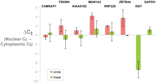
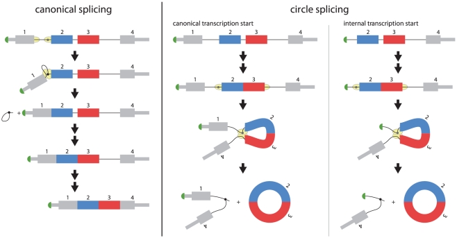
Verification
Claims the paper makes
The claims and statistics of this paper haven’t been checked yet.
Run the check — free, no account needed. It reads the open-access text and checks the sources it cites.
Claims other papers make about this one
Each is checked against this paper’s own text: does it say what the citing paper says it does?
In the citing paper
“It is also noteworthy that microRNAs can interact with other classes of non-coding RNAs as competing endogenous molecules.146,147”
From MiR-128-3p – a gray eminence of the human central nervous system (2024) · cites this paper as [146]
Self-check
Percentages, sums and reported differences re-computed from the paper’s own numbers.
The self-check needs the paper’s full text.
Citations
Cites
Electron microscopic evidence for the circular form of RNA in the cytoplasm of eukaryotic cells
HSU, M.-T., & COCA-PRADOS, M. (1979) · Nature · doi:10.1038/280339a0 · Cited by 1,021
Research works printed but not linked: 27
- JM Nigro, 1991, Scrambled exons., Cell, 64, 607, 10.1016/0092-8674(91)90244-S · DOI
- C Cocquerelle, 1992, Splicing with inverted order of exons occurs proximal to large introns., The EMBO journal, 11, 1095, 10.1002/j.1460-2075.1992.tb05148.x · DOI
- FA Saad, 1992, A 3′ consensus splice mutation in the human dystrophin gene detected by a screening for intra-exonic deletions., Human molecular genetics, 1, 345, 10.1093/hmg/1.5.345 · DOI
- B Capel, 1993, Circular transcripts of the testis-determining gene Sry in adult mouse testis., Cell, 73, 1019, 10.1016/0092-8674(93)90279-Y · DOI
- QP Cao, 1995, Expression of the mouse testis-determining gene Sry in male preimplantation embryos., Molecular reproduction and development, 40, 196, 10.1002/mrd.1080400208 · DOI
- A Hacker, 1995, Expression of Sry, the mouse sex determining gene., Development, 121, 1603, 10.1242/dev.121.6.1603 · DOI
- YW Jeske, 1995, Expression of a linear Sry transcript in the mouse genital ridge., Nature genetics, 10, 480, 10.1038/ng0895-480 · DOI
- X-FLaJ Lytton, 1999, A Circularized Sodium-Calcium Exchanger Exon 2 Transcript., The Journal of Biological Chemistry, 274, 8153, 10.1074/jbc.274.12.8153 · DOI
- PG Zaphiropoulos, 1996, Circular RNAs from transcripts of the rat cytochrome P450 2C24 gene: correlation with exon skipping., Proceedings of the National Academy of Sciences of the United States of America, 93, 6536, 10.1073/pnas.93.13.6536 · DOI
- PG Zaphiropoulos, 1997, Exon skipping and circular RNA formation in transcripts of the human cytochrome P-450 2C18 gene in epidermis and of the rat androgen binding protein gene in testis., Molecular and cellular biology, 17, 2985, 10.1128/MCB.17.6.2985 · DOI
- JM Houseley, 2006, Noncanonical RNAs from transcripts of the Drosophila muscleblind gene., The Journal of heredity, 97, 253, 10.1093/jhered/esj037 · DOI
- CWS Carlos Caldasa, 1998, Exon scrambling of MLL transcripts occur commonly and mimic partial genomic duplication of the gene., Gene, 208, 167, 10.1016/S0378-1119(97)00640-9 · DOI
- B Bailleul, 1996, During in vivo maturation of eukaryotic nuclear mRNA, splicing yields excised exon circles., Nucleic acids research, 24, 1015, 10.1093/nar/24.6.1015 · DOI
- A Surono, 1999, Circular dystrophin RNAs consisting of exons that were skipped by alternative splicing., Human molecular genetics, 8, 493, 10.1093/hmg/8.3.493 · DOI
- CE Burd, 2010, Expression of linear and novel circular forms of an INK4/ARF-associated non-coding RNA correlates with atherosclerosis risk., PLoS genetics, 6, e1001233, 10.1371/journal.pgen.1001233 · DOI
- C Cocquerelle, 1993, Mis-splicing yields circular RNA molecules., The FASEB journal : official publication of the Federation of American Societies for Experimental Biology, 7, 155, 10.1096/fasebj.7.1.7678559 · DOI
- RJ Dixon, 2005, A genome-wide survey demonstrates widespread non-linear mRNA in expressed sequences from multiple species., Nucleic acids research, 33, 5904, 10.1093/nar/gki893 · DOI
- X Shao, 2006, Bioinformatic analysis of exon repetition, exon scrambling and trans-splicing in humans., Bioinformatics, 22, 692, 10.1093/bioinformatics/bti795 · DOI
- HH Al-Balool, 2011, Post-transcriptional exon shuffling events in humans can be evolutionarily conserved and abundant., Genome research
- J Salzman, 2011, ESRRA-C11orf20 Is a Recurrent Gene Fusion in Serous Ovarian Carcinoma., PLoS biology, 9, e1001156, 10.1371/journal.pbio.1001156 · DOI
- L Yang, 2011, Genomewide characterization of non-polyadenylated RNAs., Genome biology, 12, R16, 10.1186/gb-2011-12-2-r16 · DOI
- H Suzuki, 2006, Characterization of RNase R-digested cellular RNA source that consists of lariat and circular RNAs from pre-mRNA splicing., Nucleic acids research, 34, e63, 10.1093/nar/gkl151 · DOI
- M de la Mata, 2010, First come, first served revisited: factors affecting the same alternative splicing event have different effects on the relative rates of intron removal., RNA, 16, 904, 10.1261/rna.1993510 · DOI
- C Schindewolf, 1996, In vitro generation of a circular exon from a linear pre-mRNA transcript., Nucleic acids research, 24, 1260, 10.1093/nar/24.7.1260 · DOI
- CY Chen, 1995, Initiation of protein synthesis by the eukaryotic translational apparatus on circular RNAs., Science, 268, 415, 10.1126/science.7536344 · DOI · Paper page
- TB Hansen, 2011, miRNA-dependent gene silencing involving Ago2-mediated cleavage of a circular antisense RNA., The EMBO journal, 30, 4414, 10.1038/emboj.2011.359 · DOI
- L Poliseno, 2010, A coding-independent function of gene and pseudogene mRNAs regulates tumour biology., Nature, 465, 1033, 10.1038/nature09144 · DOI
Cited by
Sorted by citations
Circular RNAs are a large class of animal RNAs with regulatory potency
Memczak, S., Jens, M., Elefsinioti, A., Torti, F., Krueger, J., Rybak, A., Maier, L., Mackowiak, S. D., Gregersen, L. H., Munschauer, M., et al. (2013) · Nature · doi:10.1038/nature11928 · Cited by 7,990
Natural RNA circles function as efficient microRNA sponges
Hansen, T. B., Jensen, T. I., Clausen, B. H., Bramsen, J. B., Finsen, B., Damgaard, C. K., & Kjems, J. (2013) · Nature · doi:10.1038/nature11993 · Cited by 7,985
The biogenesis, biology and characterization of circular RNAs
Kristensen, L. S., Andersen, M. S., Stagsted, L. V. W., Ebbesen, K. K., Hansen, T. B., & Kjems, J. (2019) · Nature Reviews Genetics · doi:10.1038/s41576-019-0158-7 · Cited by 4,823
Circular RNAs are abundant, conserved, and associated with ALU repeats
Jeck, W. R., Sorrentino, J. A., Wang, K., Slevin, M. K., Burd, C. E., Liu, J., Marzluff, W. F., & Sharpless, N. E. (2013) · RNA · doi:10.1261/rna.035667.112 · Cited by 4,388
circRNA biogenesis competes with pre-mRNA splicing
Ashwal-Fluss, R., Meyer, M., Pamudurti, N. R., Ivanov, A., Bartok, O., Hanan, M., Evantal, N., Memczak, S., Rajewsky, N., & Kadener, S. (2014) · Molecular Cell · doi:10.1016/j.molcel.2014.08.019 · Cited by 3,083
Circular RNAs in the Mammalian Brain Are Highly Abundant, Conserved, and Dynamically Expressed
Rybak-Wolf, A., Stottmeister, C., Glažar, P., Jens, M., Pino, N., Giusti, S., Hanan, M., Behm, M., Bartok, O., Ashwal-Fluss, R., et al. (2015) · Molecular Cell · doi:10.1016/j.molcel.2015.03.027 · Cited by 2,423
Circular intronic long noncoding RNAs
Zhang, Y., Zhang, X.-O., Chen, T., Xiang, J.-F., Yin, Q.-F., Xing, Y.-H., Zhu, S., Yang, L., & Chen, L.-L. (2013) · Molecular Cell · doi:10.1016/j.molcel.2013.08.017 · Cited by 2,221
Circ-ZNF609 is a circular RNA that can be translated and functions in myogenesis
Legnini, I., Di Timoteo, G., Rossi, F., Morlando, M., Briganti, F., Sthandier, O., Fatica, A., Santini, T., Andronache, A., Wade, M., et al. (2017) · Molecular Cell · doi:10.1016/j.molcel.2017.02.017 · Cited by 2,173
Complementary Sequence-Mediated Exon Circularization
Zhang, X.-O., Wang, H.-B., Zhang, Y., Lu, X., Chen, L.-L., & Yang, L. (2014) · Cell · doi:10.1016/j.cell.2014.09.001 · Cited by 1,999
Regulation of circRNA biogenesis
Chen, L.-L., & Yang, L. (2015) · RNA Biology · doi:10.1080/15476286.2015.1020271 · Cited by 1,953
Extensive translation of circular RNAs driven by N6-methyladenosine
Yang, Y., Fan, X., Mao, M., Song, X., Wu, P., Zhang, Y., Jin, Y., Yang, Y., Chen, L.-L., Wang, Y., et al. (2017) · Cell Research · doi:10.1038/cr.2017.31 · Cited by 1,855
Circular RNA: a new star of noncoding RNAs
Qu, S., Yang, X., Li, X., Wang, J., Gao, Y., Shang, R., Sun, W., Dou, K., & Li, H. (2015) · Cancer Letters · doi:10.1016/j.canlet.2015.06.003 · Cited by 1,808
The biogenesis and emerging roles of circular RNAs
Chen, L.-L. (2016) · Nature Reviews Molecular Cell Biology · doi:10.1038/nrm.2015.32 · Cited by 1,743
Foxo3 circular RNA retards cell cycle progression via forming ternary complexes with p21 and CDK2
Du, W. W., Yang, W., Liu, E., Yang, Z., Dhaliwal, P., & Yang, B. B. (2016) · Nucleic Acids Research · doi:10.1093/nar/gkw027 · Cited by 1,699
LNCcation: lncRNA localization and function
Bridges, M. C., Daulagala, A. C., & Kourtidis, A. (2021) · Journal of Cell Biology · doi:10.1083/jcb.202009045 · Cited by 1,556
Loss of a mammalian circular RNA locus causes miRNA deregulation and affects brain function
Piwecka, M., Glažar, P., Hernandez-Miranda, L. R., Memczak, S., Wolf, S. A., Rybak-Wolf, A., Filipchyk, A., Klironomos, F., Cerda Jara, C. A., Fenske, P., et al. (2017) · Science · doi:10.1126/science.aam8526 · Cited by 1,210
Diversifying microRNA sequence and function
Ameres, S. L., & Zamore, P. D. (2013) · Nature Reviews Molecular Cell Biology · doi:10.1038/nrm3611 · Cited by 1,175
Neural circular RNAs are derived from synaptic genes and regulated by development and plasticity
You, X., Vlatkovic, I., Babic, A., Will, T., Epstein, I., Tushev, G., Akbalik, G., Wang, M., Glock, C., Quedenau, C., et al. (2015) · Nature Neuroscience · doi:10.1038/nn.3975 · Cited by 1,149
Circular RNAs: analysis, expression and potential functions
Barrett, S. P., & Salzman, J. (2016) · Development · doi:10.1242/dev.128074 · Cited by 859
Self-citationEnuka, Y., Lauriola, M., Feldman, M. E., Sas-Chen, A., Ulitsky, I., & Yarden, Y. (2016) · Nucleic Acids Research · doi:10.1093/nar/gkv1367 · Cited by 757
The Biogenesis of Nascent Circular RNAs
Zhang, Y., Xue, W., Li, X., Zhang, J., Chen, S., Zhang, J.-L., Yang, L., & Chen, L.-L. (2016) · Cell Reports · doi:10.1016/j.celrep.2016.03.058 · Cited by 643
Circular RNA (circRNA) in Alzheimer’s disease (AD)
Lukiw, W. J. (2013) · Frontiers in Genetics · doi:10.3389/fgene.2013.00307 · Cited by 636
“Recently characterized, traditional methods of RNA detection and analysis requiring a free 5′ or 3′ ribonucleotide terminus may have significantly underestimated circRNA abundance and significance in eukaryotic cells (Salzman et al., 2012; Wilusz and Sharp, 2013; unpublished observations).”
Memczak, S., Papavasileiou, P., Peters, O., & Rajewsky, N. (2015) · PLoS ONE · doi:10.1371/journal.pone.0141214 · Cited by 639
Widespread and Functional RNA Circularization in Localized Prostate Cancer
Chen, S., Huang, V., Xu, X., Livingstone, J., Soares, F., Jeon, J., Zeng, Y., Hua, J. T., Petricca, J., Guo, H., et al. (2019) · Cell · doi:10.1016/j.cell.2019.01.025 · Cited by 544
Sensing Self and Foreign Circular RNAs by Intron Identity
Chen, Y. G., Kim, M. V., Chen, X., Batista, P. J., Aoyama, S., Wilusz, J. E., Iwasaki, A., & Chang, H. Y. (2017) · Molecular Cell · doi:10.1016/j.molcel.2017.05.022 · Cited by 475
Circular RNAs: splicing’s enigma variations
Hentze, M. W., & Preiss, T. (2013) · The EMBO Journal · doi:10.1038/emboj.2013.53 · Cited by 455
RNA circularization strategies in vivo and in vitro
Petkovic, S., & Müller, S. (2015) · Nucleic Acids Research · doi:10.1093/nar/gkv045 · Cited by 386
Dube, U., Del-Aguila, J. L., Li, Z., Budde, J. P., Jiang, S., Hsu, S., Ibanez, L., Fernandez, M. V., Farias, F., Norton, J., et al. (2019) · Nature Neuroscience · doi:10.1038/s41593-019-0501-5 · Cited by 345
Self-citationRole of circular RNAs in brain development and CNS diseases
Mehta, S. L., Dempsey, R. J., & Vemuganti, R. (2020) · Progress in Neurobiology · doi:10.1016/j.pneurobio.2020.101746 · Cited by 281
Hanan, M., Soreq, H., & Kadener, S. (2017) · RNA Biology · doi:10.1080/15476286.2016.1255398 · Cited by 229
Zona, L., Lupberger, J., Sidahmed-Adrar, N., Thumann, C., Harris, H. J., Barnes, A., Florentin, J., Tawar, R. G., Xiao, F., Turek, M., et al. (2013) · Cell Host & Microbe · doi:10.1016/j.chom.2013.02.006 · Cited by 182
Circular RNAs: biogenesis, function, and a role as possible cancer biomarkers
Bolha, L., Ravnik-Glavač, M., & Glavač, D. (2017) · International Journal of Genomics · doi:10.1155/2017/6218353 · Cited by 166
“EcircRNAs are the most abundant circRNA type, accounting for over 80% of identified circRNAs, and are predominantly located in the cytoplasm [3, 9, 12], though the exact process of nuclear export remains to be elucidated.”
BackgroundInfluentialNanomedicine-Based Approaches for mRNA Delivery
Uchida, S., Perche, F., Pichon, C., & Cabral, H. (2020) · Molecular Pharmaceutics · doi:10.1021/acs.molpharmaceut.0c00618 · Cited by 139
Insights into the biogenesis and potential functions of exonic circular RNA
Ragan, C., Goodall, G. J., Shirokikh, N. E., & Preiss, T. (2019) · Scientific Reports · doi:10.1038/s41598-018-37037-0 · Cited by 137
Closing the circle: current state and perspectives of circular RNA databases
Vromman, M., Vandesompele, J., & Volders, P.-J. (2021) · Briefings in Bioinformatics · doi:10.1093/bib/bbz175 · Cited by 111
The circular RNome of primary breast cancer
Smid, M., Wilting, S. M., Uhr, K., Rodríguez-González, F. G., de Weerd, V., Prager-Van der Smissen, W. J. C., van der Vlugt-Daane, M., van Galen, A., Nik-Zainal, S., Butler, A., et al. (2019) · Genome Research · doi:10.1101/gr.238121.118 · Cited by 105
Circular RNAs in Muscle Function and Disease
Greco, S., Cardinali, B., Falcone, G., & Martelli, F. (2018) · International Journal of Molecular Sciences · doi:10.3390/ijms19113454 · Cited by 89
Mahmoudi, E., Fitzsimmons, C., Geaghan, M. P., Shannon Weickert, C., Atkins, J. R., Wang, X., & Cairns, M. J. (2019) · Neuropsychopharmacology · doi:10.1038/s41386-019-0348-1 · Cited by 73
Role of RNA modifications in brain and behavior
Jung, Y., & Goldman, D. (2018) · Genes, Brain and Behavior · doi:10.1111/gbb.12444 · Cited by 66
Circular RNAs in depression: Biogenesis, function, expression, and therapeutic potential
Gan, H., Lei, Y., Yuan, N., Tang, K., Hao, W., Ma, Q., Wu, M., Zhou, X., Li, X., Huang, J., et al. (2021) · Biomedicine & Pharmacotherapy · doi:10.1016/j.biopha.2021.111244 · Cited by 41
Circular RNA: A novel and potential regulator in pathophysiology of schizophrenia
Singh, M., Dwibedy, S. L. L., Biswal, S. R., Muthuswamy, S., Kumar, A., & Kumar, S. (2022) · Metabolic Brain Disease · doi:10.1007/s11011-022-00978-7 · Cited by 15
MiR-128-3p – a gray eminence of the human central nervous system
Kiel, K., Król, S. K., Bronisz, A., & Godlewski, J. (2024) · Molecular Therapy Nucleic Acids · doi:10.1016/j.omtn.2024.102141 · Cited by 15
Metadata
- Authors
- Salzman, J., Gawad, C., Wang, P. L., Lacayo, N., & Brown, P. O.
- Venue
- PLoS ONE
- Publisher
- Public Library of Science (PLoS)
- Published
- 2012-02-01
- Volume
- 7
- Issue
- 2
- Pages
- e30733
- ISSN
- 1932-6203
- PMC
- PMC3270023
- Cited by
- 2,239
- References
- 1
- Language
- en
About these checks
Registry, retraction, Cites and statistics results are computed, not judged, with one exception: a printed reference that no index lists is read once by a model before it counts as a citation error. Claims are extracted by a model and kept only when their quote appears verbatim in the text; each cited source is then checked, with a model’s help, for whether it says what the claim relies on. Those verdicts can be wrong — read the claim’s quote and the cited source before relying on one.
Statistics and arithmetic are recomputed, not judged.
Confidence bands are shown only for fields calibrated on our annotated test set. None is calibrated yet, so no band is shown.
What counts as a claim ›How we measure confidence ›How we check this ›
Are you an author, or do you see a problem with this page? Request a review
