

The DOI matches its Crossref record; 184 of the 184 references it cites were found in a registry or an index; 10 of the 175 sources its claims cite were checked for support.
Mechanisms of DNA damage-mediated neurotoxicity in neurodegenerative disease
Open accessAbstract
Neurons are highly susceptible to DNA damage accumulation due to their large energy requirements, elevated transcriptional activity, and long lifespan. While newer research has shown that DNA breaks and mutations may facilitate neuron diversity during development and neuronal function throughout life, a wealth of evidence indicates deficient DNA damage repair underlies many neurological disorders, especially age-associated neurodegenerative diseases. Recently, efforts to clarify the molecular link between DNA damage and neurodegeneration have improved our understanding of how the genomic location of DNA damage and defunct repair proteins impact neuron health. Additionally, work establishing a role for senescence in the aging and diseased brain reveals DNA damage may play a central role in neuroinflammation associated with neurodegenerative disease.
Overview
AI snapshot
Written from the paper’s full text.
TL;DR
This review synthesizes evidence that DNA damage contributes mechanistically to neurodegeneration through two routes: the genomic location of lesions disrupting transcription of neuronal genes, and downstream lesion-detection signaling (DDR and cytosolic DNA sensing) driving apoptosis, senescence, and neuroinflammation. It argues that DNA repair syndromes serve as proxies for aging-related neurodegeneration and that senescence biology may link DNA damage to neuroinflammation.
Key findings
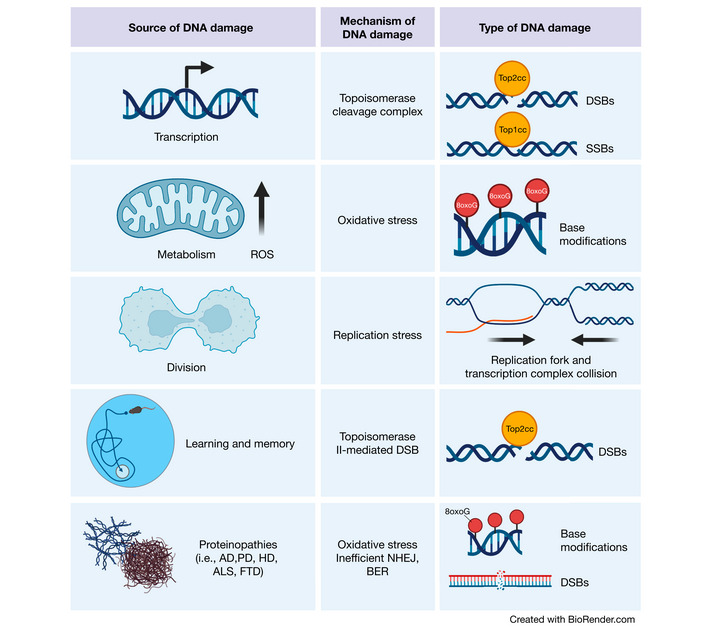
- Neurons are uniquely vulnerable because they are post-mitotic (excluding error-free homologous recombination), long-lived, and transcriptionally/energetically demanding — consuming approximately 20% of the body's available oxygen, with ROS-mediated damage greatest in the nervous system.
- DNA breaks have functional roles in neurons: activity-induced DSBs occur at immediate-early gene promoters (e.g., after bicuculline stimulation, fear learning, or novel environment exposure), but recurrence at these regulatory sequences makes them vulnerable to mutation and translocation; postmitotic neurons accumulate breaks in regulatory elements associated with neuronal function.
- SSBR mutations manifest exclusively as neurodegenerative syndromes (AOA1/AOA4 from APTX/PNKP, SCAN1 from TDP1, AOA5 from XRCC1); mutant XRCC1 causes PARP1 hyperactivity, excessive poly(ADP-ribose), NAD+/ATP depletion, Parthanatos, and in mouse brain progressive cerebellar degeneration, ataxia, seizure-like activity, and dysregulated presynaptic calcium signaling.
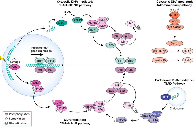
- DNA damage links to neuroinflammation via ATM–NF-κB signaling and cGAS-STING sensing of cytosolic self-DNA; removal of senescent cells mitigates NFT burden, amyloid burden, neuronal loss, and cognitive decline in mouse models of tauopathy, AD, PD, and normal aging.
Methods
- Narrative review; no primary data collection or new experiments.
- Synthesizes literature on DDR pathways (SSB/BER, NER, DSB/NHEJ, HR), DNA repair disorders, age-associated neurodegenerative disease, and senescence/neuroinflammation.
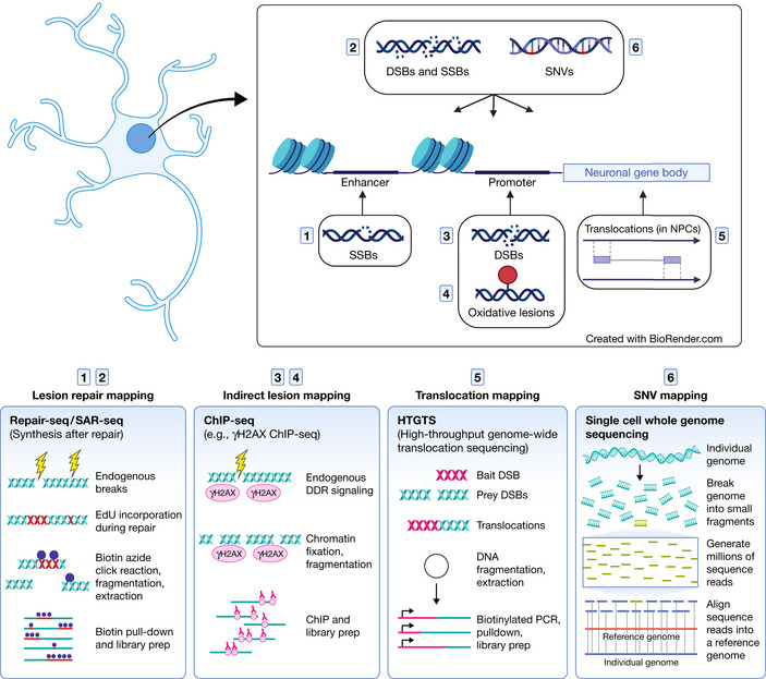
- Discusses DNA lesion-mapping technologies (γH2AX ChIP-seq, END-seq, BLISS, SSiNGLe, LAM-HTGTS, SAR-seq, Click-Code-seq, Nick-seq, ATAC-seq) and their application to neurons.
Limitations
- Stated by the paper: the mechanistic relationship between neuroinflammation and DNA damage "has yet to be fully realized"; the utility of newer break-mapping techniques (BLISS, SSiNGLe) in neurons has yet to be evaluated; it remains to be seen whether DNA damage itself is the main inducer of senescence in each brain cell type; the concept of senescent-like phenotypes in neurons is "particularly controversial" because senescence was initially defined in cycling cells; murine ATM knockout only mildly recapitulates AT ataxia and cerebellar atrophy, limiting mechanistic work.
- Evident from the text: as a review, all conclusions rest on cited primary studies rather than new data; several key claims (e.g., neuronal cGAS-STING in HD/ALS, neuronal senescence) are described as hypotheses or early observations requiring further dissection.
Model-written summary. Check it against the paper before relying on it.
Integrity signals
Registry
Cites
Statistics
Figures
Verification
Claims the paper makes
82 Not checked yet · 28 Own interpretation · 20 No basis given · 5 Partly supported · 2 Sources not found · 1 Supported · 1 Supported so far · accuracy not yet measured
The cited sources support only part of this claim — read each source’s verdict below.
In the paper
“reactive oxygen species (ROS), which attack DNA to form oxidized bases and abasic sites”
§ Mechanisms of DNA damage‐mediated neurotoxicity in neurodegenerative disease > The DNA damage response (DDR) > Single‐Strand Breaks (SSBs)
What the sources say
- Not checked yetcites Lindahl & Barnes, 2000 source
- Partly supportedcites Madabhushi et al, 2014 sourceExperimental — accuracy not yet measured
The cellular DNA damage response section says ROS generate oxidative base modifications, supporting the oxidized-bases part. It does not say ROS attack DNA to form abasic sites; it describes DNA glycosylases excising modified bases during repair. No page number is available in our copy. (LLM, conf=0.86)
- Partly supportedcites Tubbs & Nussenzweig, 2017 sourceExperimental — accuracy not yet measured
In “Stochastic Errors in DNA Replication or Spontaneous Deamination as a Source of Mutation” (page not provided), the paper identifies 8-oxoguanine as a DNA lesion arising from reactive oxygen, supporting the oxidized-bases portion. The passages found do not say that ROS form abasic sites. (LLM, conf=0.85)
plaindirect- Asserted: passes — The authors put it forward as true.
- Checkable: passes — It can be checked against evidence.
- Load-bearing: passes — The paper’s conclusions depend on it.
- Based: passes — The paper gives a reason why it holds.
The cited sources support only part of this claim — read each source’s verdict below.
In the paper
“In many cases, DNA damage seems to be one of the earliest indicators of neuropathology”
§ Mechanisms of DNA damage‐mediated neurotoxicity in neurodegenerative disease > Introduction
What the sources say
- Not checked yetcites Chow & Herrup, 2015 source
- Not matchedcites Simpson et al, 2015, 2016
Couldn’t be matched to a paper in Crossref, so it can’t be checked.
- Partly supportedcites Shanbhag et al, 2019 sourceExperimental — accuracy not yet measured
The Results report DNA double-strand breaks in mild cognitive impairment as well as Alzheimer’s disease, and the Discussion describes their accumulation at relatively early stages of the illness (page not provided). This supports an early association in Alzheimer’s disease, but the paper does not establish DNA damage as one of the earliest indicators of neuropathology across many conditions. (LLM, conf=0.84)
hedgedcorrelational- Asserted: passes — The authors put it forward as true.
- Checkable: passes — It can be checked against evidence.
- Load-bearing: passes — The paper’s conclusions depend on it.
- Based: passes — The paper gives a reason why it holds.
The sources with a verdict support this claim; the rest weren’t checked yet, have no full text we could read, or their verdicts aren’t rated yet.
In the paper
“Increased PARP1 activation and elevated poly(ADP‐ribose) has been observed in numerous neurodegenerative disorders and their associated animal models (Thapa et al, 2021; Mao & Zhang 2022).”
§ Mechanisms of DNA damage‐mediated neurotoxicity in neurodegenerative disease > Neurotoxicity associated with DNA damage > SSBs and DSBs
What the sources say
- Not checked yetcites Thapa et al, 2021 source
- Supportedcites Mao & Zhang 2022 source
The “PARP1 and neurodegenerative diseases” section reports excessive PARP1 activation in several disorders, including Huntington’s disease, ALS and multiple sclerosis, and increased PARylation in ataxia-telangiectasia mouse and worm models. The Alzheimer’s and Parkinson’s disease sections also describe PARP1 activation, supporting the broad paraphrase. (LLM, conf=0.87)
plaincorrelational- Asserted: passes — The authors put it forward as true.
- Checkable: passes — It can be checked against evidence.
- Load-bearing: passes — The paper’s conclusions depend on it.
- Based: passes — The paper gives a reason why it holds.
The cited sources support only part of this claim — read each source’s verdict below.
In the paper
“To this end, genome‐wide profiling of DNA breaks in post‐mitotic neurons have revealed that their locations are likely major mediators of their toxicity.”
§ Mechanisms of DNA damage‐mediated neurotoxicity in neurodegenerative disease > Neurotoxicity associated with DNA damage > SSBs and DSBs
What the sources say
- Partly supportedcites Reid et al, 2021 sourceExperimental — accuracy not yet measured
The paper’s Repair-seq results (section labeled Abstract; page unavailable) map genome-wide DNA repair hotspots in postmitotic human neurons and suggest that hotspots protect essential genomic elements. It profiles repair synthesis rather than DNA breaks themselves, and it does not establish that break locations are major mediators of toxicity. (LLM, conf=0.88)
- Not checked yetcites Wu et al, 2021
hedgedconditional- Asserted: passes — The authors put it forward as true.
- Checkable: passes — It can be checked against evidence.
- Load-bearing: passes — The paper’s conclusions depend on it.
- Based: passes — The paper gives a reason why it holds.
- Partly supported
Every source the paper cites for this claim was checked and supports it.
In the paper
“These data suggest that on top of its function as a pro‐survival and pro‐inflammatory transcription factor, NF‐kB is also critical for homeostatic neuronal function (Kaltschmidt & Kaltschmidt, 2009).”
§ Mechanisms of DNA damage‐mediated neurotoxicity in neurodegenerative disease > Neuroinflammation associated with DNA damage > The role of neuronal DNA damage in neuroinflammation
What the sources say
- Supportedcites Kaltschmidt & Kaltschmidt, 2009 source
The NEURONS section (page unavailable) describes constitutive NF-kB activity associated with physiological basal synaptic transmission. The LEARNING AND MEMORY section (page unavailable) reports impaired learning, synaptic plasticity, and synapse formation when neuronal NF-kB is inhibited or ablated. Together, these support its importance for normal neuronal function. (LLM, conf=0.88)
hedgeddirect- Asserted: passes — The authors put it forward as true.
- Checkable: passes — It can be checked against evidence.
- Load-bearing: passes — The paper’s conclusions depend on it.
- Based: passes — The paper gives a reason why it holds.
- Supported
None of the sources cited for this claim have been checked yet.
In the paper
“a wealth of evidence indicates deficient DNA damage repair underlies many neurological disorders, especially age‐associated neurodegenerative diseases.”
§ Mechanisms of DNA damage‐mediated neurotoxicity in neurodegenerative disease > Abstract
What the sources say
hedgeddirect- Asserted: passes — The authors put it forward as true.
- Checkable: passes — It can be checked against evidence.
- Load-bearing: passes — The paper’s conclusions depend on it.
- Based: passes — The paper gives a reason why it holds.
None of the sources cited for this claim have been checked yet.
In the paper
“work establishing a role for senescence in the aging and diseased brain reveals DNA damage may play a central role in neuroinflammation associated with neurodegenerative disease.”
§ Mechanisms of DNA damage‐mediated neurotoxicity in neurodegenerative disease > Abstract
What the sources say
hedgedconditional- Asserted: passes — The authors put it forward as true.
- Checkable: passes — It can be checked against evidence.
- Load-bearing: passes — The paper’s conclusions depend on it.
- Based: passes — The paper gives a reason why it holds.
None of the sources cited for this claim have been checked yet.
In the paper
“These insults ultimately push cellular fate toward apoptosis, senescence, or uncontrolled cell division”
§ Mechanisms of DNA damage‐mediated neurotoxicity in neurodegenerative disease > Introduction
What the sources say
plaindirect- Asserted: passes — The authors put it forward as true.
- Checkable: passes — It can be checked against evidence.
- Load-bearing: passes — The paper’s conclusions depend on it.
- Based: passes — The paper gives a reason why it holds.
None of the sources cited for this claim have been checked yet.
In the paper
“the majority ultimately manifest in the form of single‐strand breaks (SSBs)”
§ Mechanisms of DNA damage‐mediated neurotoxicity in neurodegenerative disease > The DNA damage response (DDR) > Single‐Strand Breaks (SSBs)
What the sources say
plaindirect- Asserted: passes — The authors put it forward as true.
- Checkable: passes — It can be checked against evidence.
- Load-bearing: unclear — It is unclear whether the paper’s conclusions depend on it.
- Based: passes — The paper gives a reason why it holds.
None of the sources cited for this claim have been checked yet.
In the paper
“we are just beginning to appreciate the significance of a lesion’s genomic location in relation to its effect on neuronal activity and, ultimately, degeneration”
§ Mechanisms of DNA damage‐mediated neurotoxicity in neurodegenerative disease > Introduction
What the sources say
hedgeddirect- Asserted: passes — The authors put it forward as true.
- Checkable: passes — It can be checked against evidence.
- Load-bearing: passes — The paper’s conclusions depend on it.
- Based: passes — The paper gives a reason why it holds.
In the paper
“neurons exhibit substantial mitochondrial respiration, consuming approximately 20% of the body’s available oxygen”
§ Mechanisms of DNA damage‐mediated neurotoxicity in neurodegenerative disease > The DNA damage response (DDR) > Single‐Strand Breaks (SSBs)
What the sources say
- Not checked yetcites Attwell & Laughlin, 2001 source
plain- Asserted: passes — The authors put it forward as true.
- Checkable: passes — It can be checked against evidence.
- Load-bearing: passes — The paper’s conclusions depend on it.
- Based: passes — The paper gives a reason why it holds.
A conclusion the paper draws from its own results — it cites no other source for it, so there is none to check. The quote shows it is in the paper.
In the paper
“suggesting it may be an initiating lesion of toxicity”
§ Mechanisms of DNA damage‐mediated neurotoxicity in neurodegenerative disease > Introduction
hedgedconditional- Asserted: passes — The authors put it forward as true.
- Checkable: passes — It can be checked against evidence.
- Load-bearing: passes — The paper’s conclusions depend on it.
- Based: passes — The paper gives a reason why it holds.
A conclusion the paper draws from its own results — it cites no other source for it, so there is none to check. The quote shows it is in the paper.
In the paper
“postmitotic neurons presumably incur the majority of their DSBs through transcriptional activity”
§ Mechanisms of DNA damage‐mediated neurotoxicity in neurodegenerative disease > The DNA damage response (DDR) > Double‐Strand Breaks (DSBs)
hedgedconditional- Asserted: passes — The authors put it forward as true.
- Checkable: passes — It can be checked against evidence.
- Load-bearing: unclear — It is unclear whether the paper’s conclusions depend on it.
- Based: unclear — It is unclear why the paper says it holds.
None of the sources cited for this claim have been checked yet.
In the paper
“A well‐established link exists between DNA damage and neurodegenerative diseases.”
§ Mechanisms of DNA damage‐mediated neurotoxicity in neurodegenerative disease > Introduction
What the sources say
boostedcorrelational- Asserted: passes — The authors put it forward as true.
- Checkable: passes — It can be checked against evidence.
- Load-bearing: passes — The paper’s conclusions depend on it.
- Based: passes — The paper gives a reason why it holds.
In the paper
“ROS‐mediated DNA damage is greatest in the nervous system”
§ Mechanisms of DNA damage‐mediated neurotoxicity in neurodegenerative disease > The DNA damage response (DDR) > Single‐Strand Breaks (SSBs)
What the sources say
- Not checked yetcites Nakamura & Swenberg, 1999
plain- Asserted: passes — The authors put it forward as true.
- Checkable: passes — It can be checked against evidence.
- Load-bearing: unclear — It is unclear whether the paper’s conclusions depend on it.
- Based: passes — The paper gives a reason why it holds.
A conclusion the paper draws from its own results — it cites no other source for it, so there is none to check. The quote shows it is in the paper.
In the paper
“The nervous system is particularly sensitive to loss‐of‐function mutations in DNA repair proteins.”
§ Mechanisms of DNA damage‐mediated neurotoxicity in neurodegenerative disease > Neurotoxicity associated with DNA damage
plaincorrelational- Asserted: passes — The authors put it forward as true.
- Checkable: passes — It can be checked against evidence.
- Load-bearing: passes — The paper’s conclusions depend on it.
- Based: passes — The paper gives a reason why it holds.
A conclusion the paper draws from its own results — it cites no other source for it, so there is none to check. The quote shows it is in the paper.
In the paper
“while DNA breaks may serve a physiological function in learning and memory, their recurrence in neuron regulatory sequences makes these regions extremely vulnerable to mutation and translocation.”
§ Mechanisms of DNA damage‐mediated neurotoxicity in neurodegenerative disease > Neurotoxicity associated with DNA damage > Functional roles for DNA damage in neuronal activity and development could lead to dysfunction later in life
plaindirect- Asserted: passes — The authors put it forward as true.
- Checkable: passes — It can be checked against evidence.
- Load-bearing: passes — The paper’s conclusions depend on it.
- Based: passes — The paper gives a reason why it holds.
A conclusion the paper draws from its own results — it cites no other source for it, so there is none to check. The quote shows it is in the paper.
In the paper
“Combined, these features reduce the burden of DNA damage toxicity in glia.”
§ Mechanisms of DNA damage‐mediated neurotoxicity in neurodegenerative disease > Neurotoxicity associated with DNA damage
plaindirect- Asserted: passes — The authors put it forward as true.
- Checkable: passes — It can be checked against evidence.
- Load-bearing: passes — The paper’s conclusions depend on it.
- Based: passes — The paper gives a reason why it holds.
In the paper
“Even simply introducing a mouse to a new environment is quickly followed by the induction of DSBs in neurons”
§ Mechanisms of DNA damage‐mediated neurotoxicity in neurodegenerative disease > Neurotoxicity associated with DNA damage > Functional roles for DNA damage in neuronal activity and development could lead to dysfunction later in life
What the sources say
- Not matchedcites Suberbielle et al, 2013, 2015
Couldn’t be matched to a paper in Crossref, so it can’t be checked.
plaindirect- Asserted: passes — The authors put it forward as true.
- Checkable: passes — It can be checked against evidence.
- Load-bearing: unclear — It is unclear whether the paper’s conclusions depend on it.
- Based: passes — The paper gives a reason why it holds.
- Not matched
None of the sources cited for this claim have been checked yet.
In the paper
“work has shown that some may underly neurodevelopmental and neurodegenerative disease.”
§ Mechanisms of DNA damage‐mediated neurotoxicity in neurodegenerative disease > Neurotoxicity associated with DNA damage > Functional roles for DNA damage in neuronal activity and development could lead to dysfunction later in life
What the sources say
hedgedconditional- Asserted: passes — The authors put it forward as true.
- Checkable: passes — It can be checked against evidence.
- Load-bearing: passes — The paper’s conclusions depend on it.
- Based: passes — The paper gives a reason why it holds.
The paper states this without a source or data behind it here, so there is nothing to check it against. The quote shows it is in the paper.
In the paper
“the protein kinase ATM phosphorylates numerous downstream substrates such as histone variant H2A.X, checkpoint kinase 2 (CHK2), and p53 to initiate DSB repair, cell cycle arrest, and apoptosis, respectively.”
§ Mechanisms of DNA damage‐mediated neurotoxicity in neurodegenerative disease > The DNA damage response (DDR) > SSB and DSB sensing
plaindirect- Asserted: passes — The authors put it forward as true.
- Checkable: passes — It can be checked against evidence.
- Load-bearing: unclear — It is unclear whether the paper’s conclusions depend on it.
- Based: passes — The paper gives a reason why it holds.
None of the sources cited for this claim have been checked yet.
In the paper
“DNA repair mapping reveals that postmitotic neurons do indeed accumulate breaks in regulatory elements associated with neuronal function”
§ Mechanisms of DNA damage‐mediated neurotoxicity in neurodegenerative disease > Neurotoxicity associated with DNA damage > Functional roles for DNA damage in neuronal activity and development could lead to dysfunction later in life
What the sources say
- Not checked yetcites Reid et al, 2021 source
- Not checked yetcites Wu et al, 2021
boostedcorrelational- Asserted: passes — The authors put it forward as true.
- Checkable: passes — It can be checked against evidence.
- Load-bearing: passes — The paper’s conclusions depend on it.
- Based: passes — The paper gives a reason why it holds.
None of the sources cited for this claim have been checked yet.
In the paper
“somatic mutations induced by erroneous DNA damage repair or transposable elements are a common feature of neural development, giving rise to neuron diversity through genomic mosaicism”
§ Mechanisms of DNA damage‐mediated neurotoxicity in neurodegenerative disease > Neurotoxicity associated with DNA damage > Functional roles for DNA damage in neuronal activity and development could lead to dysfunction later in life
What the sources say
plaindirect- Asserted: passes — The authors put it forward as true.
- Checkable: passes — It can be checked against evidence.
- Load-bearing: passes — The paper’s conclusions depend on it.
- Based: passes — The paper gives a reason why it holds.
None of the sources cited for this claim have been checked yet.
In the paper
“mutations in TDP2 result in a rare neurological disease termed spinocerebellar ataxia autosomal recessive 23 (SCAR23)”
§ Mechanisms of DNA damage‐mediated neurotoxicity in neurodegenerative disease > The DNA damage response (DDR) > Double‐Strand Breaks (DSBs)
What the sources say
- Not checked yetcites Zagnoli‐Vieira et al, 2018
- Not checked yetcites Gómez‐Herreros et al, 2014: 2
- Not checked yetcites Errichiello et al, 2020 source
plaindirect- Asserted: passes — The authors put it forward as true.
- Checkable: passes — It can be checked against evidence.
- Load-bearing: unclear — It is unclear whether the paper’s conclusions depend on it.
- Based: passes — The paper gives a reason why it holds.
None of the sources cited for this claim have been checked yet.
In the paper
“a DSB‐mapping technique known as END‐seq was recently used to identify strand breaks in human induced pluripotent stem cell (iPSC)‐derived neurons, resulting in the finding that enhancers are hotspots for SSBs”
§ Mechanisms of DNA damage‐mediated neurotoxicity in neurodegenerative disease > Neurotoxicity associated with DNA damage > Functional roles for DNA damage in neuronal activity and development could lead to dysfunction later in life
What the sources say
- Not checked yetcites Canela et al, 2016 source
- Not checked yetcites Wu et al, 2021
plaincorrelational- Asserted: passes — The authors put it forward as true.
- Checkable: passes — It can be checked against evidence.
- Load-bearing: unclear — It is unclear whether the paper’s conclusions depend on it.
- Based: passes — The paper gives a reason why it holds.
A conclusion the paper draws from its own results — it cites no other source for it, so there is none to check. The quote shows it is in the paper.
In the paper
“The explanation for this sensitivity may lie in the hallmarks of neuronal identity. That is, neurons perform transcriptionally and energetically demanding cellular functions and are post‐mitotic and long‐lived.”
§ Mechanisms of DNA damage‐mediated neurotoxicity in neurodegenerative disease > Neurotoxicity associated with DNA damage
hedgedconditional- Asserted: passes — The authors put it forward as true.
- Checkable: passes — It can be checked against evidence.
- Load-bearing: passes — The paper’s conclusions depend on it.
- Based: passes — The paper gives a reason why it holds.
- Rests on an assumption: Neurons are post-mitotic and long-lived.
The paper states this without a source or data behind it here, so there is nothing to check it against. The quote shows it is in the paper.
In the paper
“Because HR requires sister chromatids, this pathway can only occur in cycling cells during or following S phase.”
§ Mechanisms of DNA damage‐mediated neurotoxicity in neurodegenerative disease > The DNA damage response (DDR) > Double‐Strand Breaks (DSBs)
plaindirect- Asserted: passes — The authors put it forward as true.
- Checkable: passes — It can be checked against evidence.
- Load-bearing: passes — The paper’s conclusions depend on it.
- Based: passes — The paper gives a reason why it holds.
None of the sources cited for this claim have been checked yet.
In the paper
“DNA breaks seem to serve a functional role in neuronal activity”
§ Mechanisms of DNA damage‐mediated neurotoxicity in neurodegenerative disease > Neurotoxicity associated with DNA damage > Functional roles for DNA damage in neuronal activity and development could lead to dysfunction later in life
What the sources say
hedgeddirect- Asserted: passes — The authors put it forward as true.
- Checkable: passes — It can be checked against evidence.
- Load-bearing: passes — The paper’s conclusions depend on it.
- Based: passes — The paper gives a reason why it holds.
None of the sources cited for this claim have been checked yet.
In the paper
“Notably, CAG expansion has been found to be dependent upon DNA repair proteins such as OGG1, MLH1, and MSH2, whose detection of oxidized or mismatched bases in these repetitive elements could potentially elicit erroneous repair, leading to CAG expansion (Kovtun et al, 2007; Bettencourt et al, 2016; Pinto et al, 2013; Manley et al, 1999; Lee et al, 2017, 1).”
§ Mechanisms of DNA damage‐mediated neurotoxicity in neurodegenerative disease > Neurotoxicity associated with DNA damage > Oxidative DNA damage
What the sources say
plaindirect- Asserted: passes — The authors put it forward as true.
- Checkable: passes — It can be checked against evidence.
- Load-bearing: passes — The paper’s conclusions depend on it.
- Based: passes — The paper gives a reason why it holds.
- Rests on an assumption: Age-dependent CAG expansion occurs in the brains of individuals with HD, gradually increasing the toxicity of the Huntington protein in the striatum.
A conclusion the paper draws from its own results — it cites no other source for it, so there is none to check. The quote shows it is in the paper.
In the paper
“the pathogenic load of a loss‐of‐function mutation accelerates the development of age‐associated pathology”
§ Mechanisms of DNA damage‐mediated neurotoxicity in neurodegenerative disease > Neurotoxicity associated with DNA damage > DNA repair syndromes as proxies for aging and neurodegenerative disease
plaindirect- Asserted: passes — The authors put it forward as true.
- Checkable: passes — It can be checked against evidence.
- Load-bearing: passes — The paper’s conclusions depend on it.
- Based: passes — The paper gives a reason why it holds.
The paper states this without a source or data behind it here, so there is nothing to check it against. The quote shows it is in the paper.
In the paper
“To date, loss‐of‐function mutations in SSBR proteins have manifested exclusively as neurodegenerative syndromes.”
§ Mechanisms of DNA damage‐mediated neurotoxicity in neurodegenerative disease > Neurotoxicity associated with DNA damage > Neurological diseases caused by SSBR mutations
plaindirect- Asserted: passes — The authors put it forward as true.
- Checkable: passes — It can be checked against evidence.
- Load-bearing: unclear — It is unclear whether the paper’s conclusions depend on it.
- Based: passes — The paper gives a reason why it holds.
In the paper
“oxidative damage‐induced poly(ADP‐ribose) mediates α‐synuclein aggregation and consequent neurodegeneration in PD (Kam et al, 2018)”
§ Mechanisms of DNA damage‐mediated neurotoxicity in neurodegenerative disease > Neurotoxicity associated with DNA damage > SSBs and DSBs
What the sources say
- Not checked yetcites Kam et al, 2018 source
plaindirect- Asserted: passes — The authors put it forward as true.
- Checkable: passes — It can be checked against evidence.
- Load-bearing: passes — The paper’s conclusions depend on it.
- Based: passes — The paper gives a reason why it holds.
None of the sources cited for this claim have been checked yet.
In the paper
“With the exception of AOA5, which seems to have manifested exclusively in adults so far (Hoch et al, 2017: 1; O’Connor et al, 2018: 1), the majority of DNA damage disorders typically present in early childhood.”
§ Mechanisms of DNA damage‐mediated neurotoxicity in neurodegenerative disease > Neurotoxicity associated with DNA damage > DNA repair syndromes as proxies for aging and neurodegenerative disease
What the sources say
hedged- Asserted: passes — The authors put it forward as true.
- Checkable: passes — It can be checked against evidence.
- Load-bearing: unclear — It is unclear whether the paper’s conclusions depend on it.
- Based: passes — The paper gives a reason why it holds.
None of the sources cited for this claim have been checked yet.
In the paper
“Defective XRCC1 manifests in individuals as Ataxia with Oculomotor apraxia Type 5 (AOA5), another slow‐progressing neurodegenerative disease.”
§ Mechanisms of DNA damage‐mediated neurotoxicity in neurodegenerative disease > Neurotoxicity associated with DNA damage > Neurological diseases caused by SSBR mutations
What the sources say
plaindirect- Asserted: passes — The authors put it forward as true.
- Checkable: passes — It can be checked against evidence.
- Load-bearing: unclear — It is unclear whether the paper’s conclusions depend on it.
- Based: passes — The paper gives a reason why it holds.
None of the sources cited for this claim have been checked yet.
In the paper
“Gene‐wide association studies have further identified SNPs in DNA maintenance genes that influence the age of onset in this disease (Lee et al, 2015; Moss et al, 2017).”
§ Mechanisms of DNA damage‐mediated neurotoxicity in neurodegenerative disease > Neurotoxicity associated with DNA damage > Oxidative DNA damage
What the sources say
plaincorrelational- Asserted: passes — The authors put it forward as true.
- Checkable: passes — It can be checked against evidence.
- Load-bearing: passes — The paper’s conclusions depend on it.
- Based: passes — The paper gives a reason why it holds.
A conclusion the paper draws from its own results — it cites no other source for it, so there is none to check. The quote shows it is in the paper.
In the paper
“whether the resulting pathologies of a DNA repair deficit are developmental or age‐associated likely depends on both the brain cell type composition at the time and the selective vulnerabilities of different neuronal subtypes to different repair deficits.”
§ Mechanisms of DNA damage‐mediated neurotoxicity in neurodegenerative disease > Neurotoxicity associated with DNA damage > DNA repair syndromes as proxies for aging and neurodegenerative disease
hedgedconditional- Asserted: passes — The authors put it forward as true.
- Checkable: passes — It can be checked against evidence.
- Load-bearing: passes — The paper’s conclusions depend on it.
- Based: passes — The paper gives a reason why it holds.
In the paper
“facilitates the formation of cytoplasmic TDP‐43 foci in ALS (McGurk et al, 2018a, 2018b)”
§ Mechanisms of DNA damage‐mediated neurotoxicity in neurodegenerative disease > Neurotoxicity associated with DNA damage > SSBs and DSBs
What the sources say
- Not checked yetcites McGurk et al, 2018a, 2018b
plaindirect- Asserted: passes — The authors put it forward as true.
- Checkable: passes — It can be checked against evidence.
- Load-bearing: passes — The paper’s conclusions depend on it.
- Based: passes — The paper gives a reason why it holds.
The paper states this without a source or data behind it here, so there is nothing to check it against. The quote shows it is in the paper.
In the paper
“Deficient DNA damage repair in these vulnerable NPCs results in developmental abnormalities such as microcephaly”
§ Mechanisms of DNA damage‐mediated neurotoxicity in neurodegenerative disease > Neurotoxicity associated with DNA damage > DNA repair syndromes as proxies for aging and neurodegenerative disease
plaindirect- Asserted: passes — The authors put it forward as true.
- Checkable: passes — It can be checked against evidence.
- Load-bearing: passes — The paper’s conclusions depend on it.
- Based: passes — The paper gives a reason why it holds.
The paper states this without a source or data behind it here, so there is nothing to check it against. The quote shows it is in the paper.
In the paper
“inheritable DNA damage disorders, which frequently present with neurologic abnormalities”
§ Mechanisms of DNA damage‐mediated neurotoxicity in neurodegenerative disease > Neurotoxicity associated with DNA damage > DNA repair syndromes as proxies for aging and neurodegenerative disease
plaincorrelational- Asserted: passes — The authors put it forward as true.
- Checkable: passes — It can be checked against evidence.
- Load-bearing: passes — The paper’s conclusions depend on it.
- Based: passes — The paper gives a reason why it holds.
A conclusion the paper draws from its own results — it cites no other source for it, so there is none to check. The quote shows it is in the paper.
In the paper
“Aberrant DNA repair may also directly mediate the severity of neurodegenerative disorders like HD, whose toxicity is derived from the CAG trinucleotide expansion of the Huntingtin gene.”
§ Mechanisms of DNA damage‐mediated neurotoxicity in neurodegenerative disease > Neurotoxicity associated with DNA damage > Oxidative DNA damage
hedgedconditional- Asserted: passes — The authors put it forward as true.
- Checkable: passes — It can be checked against evidence.
- Load-bearing: passes — The paper’s conclusions depend on it.
- Based: passes — The paper gives a reason why it holds.
- Rests on an assumption: HD toxicity is derived from the CAG trinucleotide expansion of the Huntingtin gene.
In the paper
“Similarly, DSB marker proteins correlate with cognitive impairment in individuals with low levels of amyloid and tau pathology (Simpson et al, 2015).”
§ Mechanisms of DNA damage‐mediated neurotoxicity in neurodegenerative disease > Neurotoxicity associated with DNA damage > SSBs and DSBs
What the sources say
- Not checked yetcites Simpson et al, 2015 source
plaincorrelational- Asserted: passes — The authors put it forward as true.
- Checkable: passes — It can be checked against evidence.
- Load-bearing: unclear — It is unclear whether the paper’s conclusions depend on it.
- Based: passes — The paper gives a reason why it holds.
A conclusion the paper draws from its own results — it cites no other source for it, so there is none to check. The quote shows it is in the paper.
In the paper
“it is likely that post‐mitotic neurons are bearing the brunt of the DNA repair deficit.”
§ Mechanisms of DNA damage‐mediated neurotoxicity in neurodegenerative disease > Neurotoxicity associated with DNA damage > DNA repair syndromes as proxies for aging and neurodegenerative disease
hedgedconditional- Asserted: passes — The authors put it forward as true.
- Checkable: passes — It can be checked against evidence.
- Load-bearing: passes — The paper’s conclusions depend on it.
- Based: passes — The paper gives a reason why it holds.
A conclusion the paper draws from its own results — it cites no other source for it, so there is none to check. The quote shows it is in the paper.
In the paper
“Therefore, erroneous DNA repair mechanisms also directly influence HD severity.”
§ Mechanisms of DNA damage‐mediated neurotoxicity in neurodegenerative disease > Neurotoxicity associated with DNA damage > Oxidative DNA damage
plaindirect- Asserted: passes — The authors put it forward as true.
- Checkable: passes — It can be checked against evidence.
- Load-bearing: passes — The paper’s conclusions depend on it.
- Based: passes — The paper gives a reason why it holds.
- Rests on an assumption: HD toxicity is derived from the CAG trinucleotide expansion of the Huntingtin gene.
- Rests on an assumption: Age-dependent CAG expansion occurs in the brains of individuals with HD, gradually increasing the toxicity of the Huntington protein in the striatum.
The paper states this without a source or data behind it here, so there is nothing to check it against. The quote shows it is in the paper.
In the paper
“many DNA damage disorders are also defined by age‐associated pathologies such as progressive brain atrophy and peripheral neuropathy.”
§ Mechanisms of DNA damage‐mediated neurotoxicity in neurodegenerative disease > Neurotoxicity associated with DNA damage > DNA repair syndromes as proxies for aging and neurodegenerative disease
plaincorrelational- Asserted: passes — The authors put it forward as true.
- Checkable: passes — It can be checked against evidence.
- Load-bearing: passes — The paper’s conclusions depend on it.
- Based: passes — The paper gives a reason why it holds.
The paper states this without a source or data behind it here, so there is nothing to check it against. The quote shows it is in the paper.
In the paper
“we must acknowledge the pivotal role of DNA damage repair in neurodevelopment”
§ Mechanisms of DNA damage‐mediated neurotoxicity in neurodegenerative disease > Neurotoxicity associated with DNA damage > DNA repair syndromes as proxies for aging and neurodegenerative disease
boosteddirect- Asserted: passes — The authors put it forward as true.
- Checkable: passes — It can be checked against evidence.
- Load-bearing: passes — The paper’s conclusions depend on it.
- Based: passes — The paper gives a reason why it holds.
In the paper
“Specifically, increased DDR foci are observed in neurons from individuals with mild cognitive impairment (MCI) and AD compared to aged counterparts (Shanbhag et al, 2019).”
§ Mechanisms of DNA damage‐mediated neurotoxicity in neurodegenerative disease > Neurotoxicity associated with DNA damage > SSBs and DSBs
What the sources say
- Not checked yetcites Shanbhag et al, 2019 source
plaincorrelational- Asserted: passes — The authors put it forward as true.
- Checkable: passes — It can be checked against evidence.
- Load-bearing: passes — The paper’s conclusions depend on it.
- Based: passes — The paper gives a reason why it holds.
A conclusion the paper draws from its own results — it cites no other source for it, so there is none to check. The quote shows it is in the paper.
In the paper
“Accumulation of SNVs at neurodegeneration risk genes could potentially increase risk of disease development.”
§ Mechanisms of DNA damage‐mediated neurotoxicity in neurodegenerative disease > Neurotoxicity associated with DNA damage > Functional roles for DNA damage in neuronal activity and development could lead to dysfunction later in life
hedgedconditional- Asserted: passes — The authors put it forward as true.
- Checkable: passes — It can be checked against evidence.
- Load-bearing: passes — The paper’s conclusions depend on it.
- Based: passes — The paper gives a reason why it holds.
A conclusion the paper draws from its own results — it cites no other source for it, so there is none to check. The quote shows it is in the paper.
In the paper
“an individual devoid of DNA repair mutations must experience the progressive stress of aging (i.e. damage accumulation, oxidative stress, declining DNA repair enzyme efficiency) in order to recapitulate the pathogenic load of a DNA damage disorder.”
§ Mechanisms of DNA damage‐mediated neurotoxicity in neurodegenerative disease > Neurotoxicity associated with DNA damage > DNA repair syndromes as proxies for aging and neurodegenerative disease
plaindirect- Asserted: passes — The authors put it forward as true.
- Checkable: passes — It can be checked against evidence.
- Load-bearing: passes — The paper’s conclusions depend on it.
- Based: passes — The paper gives a reason why it holds.
None of the sources cited for this claim have been checked yet.
In the paper
“and FUS activates and recruits the XRCC1‐LIG3 complex to sites of oxidative DNA damage in a PARP1‐dependent manner (Naumann et al, 2018; Wang et al, 2018a).”
§ Mechanisms of DNA damage‐mediated neurotoxicity in neurodegenerative disease > Neurotoxicity associated with DNA damage > SSBs and DSBs
What the sources say
- Not checked yetcites Naumann et al, 2018 source
- Not checked yetcites Wang et al, 2018a
plaindirect- Asserted: passes — The authors put it forward as true.
- Checkable: passes — It can be checked against evidence.
- Load-bearing: unclear — It is unclear whether the paper’s conclusions depend on it.
- Based: passes — The paper gives a reason why it holds.
None of the sources cited for this claim have been checked yet.
In the paper
“However, toxic aggregation of α‐synuclein in the nucleus or cytosol elicits the accumulation of DSBs (Vasquez et al, 2017; Milanese et al, 2018).”
§ Mechanisms of DNA damage‐mediated neurotoxicity in neurodegenerative disease > Neurotoxicity associated with DNA damage > SSBs and DSBs
What the sources say
plaindirect- Asserted: passes — The authors put it forward as true.
- Checkable: passes — It can be checked against evidence.
- Load-bearing: passes — The paper’s conclusions depend on it.
- Based: passes — The paper gives a reason why it holds.
In the paper
“can mediate neuroinflammatory activity in AD (Kauppinen et al, 2011)”
§ Mechanisms of DNA damage‐mediated neurotoxicity in neurodegenerative disease > Neurotoxicity associated with DNA damage > SSBs and DSBs
What the sources say
- Not checked yetcites Kauppinen et al, 2011 source
plaindirect- Asserted: passes — The authors put it forward as true.
- Checkable: passes — It can be checked against evidence.
- Load-bearing: unclear — It is unclear whether the paper’s conclusions depend on it.
- Based: passes — The paper gives a reason why it holds.
None of the sources cited for this claim have been checked yet.
In the paper
“Tau may also play a role in DSB repair as well, as its deletion in the mouse brain leads to the accumulation of DSB foci in neurons (Violet et al, 2014; Mansuroglu et al, 2016).”
§ Mechanisms of DNA damage‐mediated neurotoxicity in neurodegenerative disease > Neurotoxicity associated with DNA damage > SSBs and DSBs
What the sources say
hedgeddirect- Asserted: passes — The authors put it forward as true.
- Checkable: passes — It can be checked against evidence.
- Load-bearing: unclear — It is unclear whether the paper’s conclusions depend on it.
- Based: passes — The paper gives a reason why it holds.
None of the sources cited for this claim have been checked yet.
In the paper
“Importantly, removal of senescent cells promotes healthy aging in mice, making senescence‐associated inflammatory signaling an attractive target of therapeutic intervention for aging and age‐related disease (Tilstra et al, 2012; Childs et al, 2015; Baker et al, 2016; Ogrodnik et al, 2021).”
§ Mechanisms of DNA damage‐mediated neurotoxicity in neurodegenerative disease > Neuroinflammation associated with DNA damage > DNA damage induces senescence
What the sources say
plaindirect- Asserted: passes — The authors put it forward as true.
- Checkable: passes — It can be checked against evidence.
- Load-bearing: passes — The paper’s conclusions depend on it.
- Based: passes — The paper gives a reason why it holds.
None of the sources cited for this claim have been checked yet.
In the paper
“Indeed, many of the risk genes associated with AD mediate their effects through microglia, the brain‐resident macrophages (Glass et al, 2010; Heneka et al, 2015; Nott et al, 2019).”
§ Mechanisms of DNA damage‐mediated neurotoxicity in neurodegenerative disease > Neuroinflammation associated with DNA damage
What the sources say
plaindirect- Asserted: passes — The authors put it forward as true.
- Checkable: passes — It can be checked against evidence.
- Load-bearing: unclear — It is unclear whether the paper’s conclusions depend on it.
- Based: passes — The paper gives a reason why it holds.
None of the sources cited for this claim have been checked yet.
In the paper
“Initially, induction of persistent DDR signaling in cell cultures including fibroblasts (Coppé et al, 2008; Rodier et al, 2009), bone marrow‐derived macrophages (BMDMs) (Härtlova et al, 2015), and primary monocytes (Brzostek‐Racine et al, 2011) was found to elicit the SASP (Coppé et al, 2008).”
§ Mechanisms of DNA damage‐mediated neurotoxicity in neurodegenerative disease > Neuroinflammation associated with DNA damage > DNA damage induces senescence
What the sources say
plaindirect- Asserted: passes — The authors put it forward as true.
- Checkable: passes — It can be checked against evidence.
- Load-bearing: passes — The paper’s conclusions depend on it.
- Based: passes — The paper gives a reason why it holds.
In the paper
“FUS is an RNA/DNA binding protein that has been found to facilitate DSB repair through its colocalization with HDAC1 (Wy et al, 2013, 1).”
§ Mechanisms of DNA damage‐mediated neurotoxicity in neurodegenerative disease > Neurotoxicity associated with DNA damage > SSBs and DSBs
What the sources say
- Not checked yetcites Wy et al, 2013, 1
plaindirect- Asserted: passes — The authors put it forward as true.
- Checkable: passes — It can be checked against evidence.
- Load-bearing: unclear — It is unclear whether the paper’s conclusions depend on it.
- Based: passes — The paper gives a reason why it holds.
None of the sources cited for this claim have been checked yet.
In the paper
“Furthermore, phosphorylation of FUS occurs in response to DSB detection by proteins ATM and DNA‐PK (Gardiner et al, 2008; Deng et al, 2014)”
§ Mechanisms of DNA damage‐mediated neurotoxicity in neurodegenerative disease > Neurotoxicity associated with DNA damage > SSBs and DSBs
What the sources say
plaindirect- Asserted: passes — The authors put it forward as true.
- Checkable: passes — It can be checked against evidence.
- Load-bearing: unclear — It is unclear whether the paper’s conclusions depend on it.
- Based: passes — The paper gives a reason why it holds.
A conclusion the paper draws from its own results — it cites no other source for it, so there is none to check. The quote shows it is in the paper.
In the paper
“It follows that while these breaks may be functionally relevant, over time, their erroneous repair could lead to disruptive translocations, mutations, and genomic instability.”
§ Mechanisms of DNA damage‐mediated neurotoxicity in neurodegenerative disease > Neurotoxicity associated with DNA damage > SSBs and DSBs
hedgedconditional- Asserted: passes — The authors put it forward as true.
- Checkable: passes — It can be checked against evidence.
- Load-bearing: passes — The paper’s conclusions depend on it.
- Based: passes — The paper gives a reason why it holds.
None of the sources cited for this claim have been checked yet.
In the paper
“This secretory profile was found to be at least partially ATM‐dependent, implying the importance of DSB repair specifically in immune activation (Rodier et al, 2009; Brzostek‐Racine et al, 2011).”
§ Mechanisms of DNA damage‐mediated neurotoxicity in neurodegenerative disease > Neuroinflammation associated with DNA damage > DNA damage induces senescence
What the sources say
- Not checked yetcites Rodier et al, 2009 source
- Not checked yetcites Brzostek‐Racine et al, 2011
plainconditional- Asserted: passes — The authors put it forward as true.
- Checkable: passes — It can be checked against evidence.
- Load-bearing: unclear — It is unclear whether the paper’s conclusions depend on it.
- Based: passes — The paper gives a reason why it holds.
A conclusion the paper draws from its own results — it cites no other source for it, so there is none to check. The quote shows it is in the paper.
In the paper
“Thus, senescence is now considered as a potential driver of neuroinflammation and neurodegeneration.”
§ Mechanisms of DNA damage‐mediated neurotoxicity in neurodegenerative disease > Neuroinflammation associated with DNA damage
hedgedconditional- Asserted: passes — The authors put it forward as true.
- Checkable: passes — It can be checked against evidence.
- Load-bearing: passes — The paper’s conclusions depend on it.
- Based: passes — The paper gives a reason why it holds.
None of the sources cited for this claim have been checked yet.
In the paper
“Previous work with AT fibroblasts had shown that the canonical immune transcription factor NF‐kB is activated in an ATM‐dependent manner following treatment with a radiomimetic, ionizing radiation, or topoisomerase inhibitor (Lee et al, 1998; Piret et al, 1999; Li et al, 2001).”
§ Mechanisms of DNA damage‐mediated neurotoxicity in neurodegenerative disease > Neuroinflammation associated with DNA damage > DNA damage induces senescence
What the sources say
plaindirect- Asserted: passes — The authors put it forward as true.
- Checkable: passes — It can be checked against evidence.
- Load-bearing: unclear — It is unclear whether the paper’s conclusions depend on it.
- Based: passes — The paper gives a reason why it holds.
None of the sources cited for this claim have been checked yet.
In the paper
“In addition to PARP1 activity, many genes that encode the building blocks of neurodegenerative proteinopathies have been found to play a role in DDR as well.”
§ Mechanisms of DNA damage‐mediated neurotoxicity in neurodegenerative disease > Neurotoxicity associated with DNA damage > SSBs and DSBs
plaincorrelational- Asserted: passes — The authors put it forward as true.
- Checkable: passes — It can be checked against evidence.
- Load-bearing: passes — The paper’s conclusions depend on it.
- Based: passes — The paper gives a reason why it holds.
A conclusion the paper draws from its own results — it cites no other source for it, so there is none to check. The quote shows it is in the paper.
In the paper
“While these findings have firmly established DNA breaks as a mechanism of neurodegenerative disease, the mechanisms by which break location may contribute to disease progression are not well described.”
§ Mechanisms of DNA damage‐mediated neurotoxicity in neurodegenerative disease > Neurotoxicity associated with DNA damage > SSBs and DSBs
boosteddirect- Asserted: passes — The authors put it forward as true.
- Checkable: passes — It can be checked against evidence.
- Load-bearing: passes — The paper’s conclusions depend on it.
- Based: passes — The paper gives a reason why it holds.
None of the sources cited for this claim have been checked yet.
In the paper
“In addition to DNA damage, neuroinflammation has emerged as a core feature and mechanism of neurodegenerative disease.”
§ Mechanisms of DNA damage‐mediated neurotoxicity in neurodegenerative disease > Neuroinflammation associated with DNA damage
plaindirect- Asserted: passes — The authors put it forward as true.
- Checkable: passes — It can be checked against evidence.
- Load-bearing: passes — The paper’s conclusions depend on it.
- Based: passes — The paper gives a reason why it holds.
None of the sources cited for this claim have been checked yet.
In the paper
“Recently, TDP‐43 was also found to be directly involved in NHEJ through its recruitment and stabilization of the XRCC4‐LIG4 complex at DSBs (Guerrero et al, 2019; Mitra et al, 2019).”
§ Mechanisms of DNA damage‐mediated neurotoxicity in neurodegenerative disease > Neurotoxicity associated with DNA damage > SSBs and DSBs
What the sources say
plaindirect- Asserted: passes — The authors put it forward as true.
- Checkable: passes — It can be checked against evidence.
- Load-bearing: unclear — It is unclear whether the paper’s conclusions depend on it.
- Based: passes — The paper gives a reason why it holds.
None of the sources cited for this claim have been checked yet.
In the paper
“As described earlier, the use of nucleoside analog incorporation to map DNA repair has led to the observation that postmitotic neurons harbor SSBs and other forms of breaks in the regulatory regions of genes that modulate synaptic plasticity and neuronal function (Reid et al, 2021; Wu et al, 2021).”
§ Mechanisms of DNA damage‐mediated neurotoxicity in neurodegenerative disease > Neurotoxicity associated with DNA damage > SSBs and DSBs
What the sources say
- Not checked yetcites Reid et al, 2021 source
- Not checked yetcites Wu et al, 2021
plain- Asserted: passes — The authors put it forward as true.
- Checkable: passes — It can be checked against evidence.
- Load-bearing: passes — The paper’s conclusions depend on it.
- Based: passes — The paper gives a reason why it holds.
In the paper
“Nevertheless, observations of senescent‐like phenotypes in neurons have been reported for quite some time.”
§ Mechanisms of DNA damage‐mediated neurotoxicity in neurodegenerative disease > Neuroinflammation associated with DNA damage > The role of neuronal DNA damage in neuroinflammation
What the sources say
- Not checked yetcites Jurk et al, 2012 source
plain- Asserted: passes — The authors put it forward as true.
- Checkable: passes — It can be checked against evidence.
- Load-bearing: unclear — It is unclear whether the paper’s conclusions depend on it.
- Based: passes — The paper gives a reason why it holds.
The paper states this without a source or data behind it here, so there is nothing to check it against. The quote shows it is in the paper.
In the paper
“An additional mechanism of cytosolic DNA‐mediated inflammation is through the NLRP3 inflammasome, which senses cell stressors (such as cytosolic DNA) to activate the cleavage of pro‐1L1b and pro‐IL18 via Caspase I to elicit their secretion.”
§ Mechanisms of DNA damage‐mediated neurotoxicity in neurodegenerative disease > Neuroinflammation associated with DNA damage > DNA damage induces senescence
plaindirect- Asserted: passes — The authors put it forward as true.
- Checkable: passes — It can be checked against evidence.
- Load-bearing: unclear — It is unclear whether the paper’s conclusions depend on it.
- Based: passes — The paper gives a reason why it holds.
In the paper
“Additionally, senescent astrocytes have been identified as modulators of PD pathology (Chinta et al, 2018).”
§ Mechanisms of DNA damage‐mediated neurotoxicity in neurodegenerative disease > Neuroinflammation associated with DNA damage > Senescence‐associated inflammation in brain aging and neurodegeneration
What the sources say
- Not checked yetcites Chinta et al, 2018 source
plaincorrelational- Asserted: passes — The authors put it forward as true.
- Checkable: passes — It can be checked against evidence.
- Load-bearing: unclear — It is unclear whether the paper’s conclusions depend on it.
- Based: passes — The paper gives a reason why it holds.
In the paper
“Here, DSBs were considered the major driver of these senescence‐associated phenotypes because knock‐out of p21, a protein activated by p53 to initiate DSB‐induced cell cycle arrest, was able to mitigate their enrichment.”
§ Mechanisms of DNA damage‐mediated neurotoxicity in neurodegenerative disease > Neuroinflammation associated with DNA damage > The role of neuronal DNA damage in neuroinflammation
What the sources say
- Not checked yetcites Jurk et al, 2012 source
plaindirect- Asserted: passes — The authors put it forward as true.
- Checkable: passes — It can be checked against evidence.
- Load-bearing: passes — The paper’s conclusions depend on it.
- Based: passes — The paper gives a reason why it holds.
In the paper
“AGS is caused by mutations in genes that regulate nucleic acid metabolism.”
§ Mechanisms of DNA damage‐mediated neurotoxicity in neurodegenerative disease > Neuroinflammation associated with DNA damage > DNA damage induces senescence
What the sources say
- Not checked yetcites Crow & Manel, 2015 source
plaindirect- Asserted: passes — The authors put it forward as true.
- Checkable: passes — It can be checked against evidence.
- Load-bearing: unclear — It is unclear whether the paper’s conclusions depend on it.
- Based: passes — The paper gives a reason why it holds.
In the paper
“Interestingly, sequencing the cytosolic DNA fragments from these microglia revealed that most were derived from the nuclear DNA as intergenic repetitive elements, suggesting that the source of cytosolic DNA may not be random (Song et al, 2021).”
§ Mechanisms of DNA damage‐mediated neurotoxicity in neurodegenerative disease > Neuroinflammation associated with DNA damage > Senescence‐associated inflammation in brain aging and neurodegeneration
What the sources say
- Not checked yetcites Song et al, 2021 source
hedgedcorrelational- Asserted: passes — The authors put it forward as true.
- Checkable: passes — It can be checked against evidence.
- Load-bearing: unclear — It is unclear whether the paper’s conclusions depend on it.
- Based: passes — The paper gives a reason why it holds.
In the paper
“Thus, the interplay between NF‐kB and p53 strongly influence cellular fate following DNA damage accumulation (Karin & Lin, 2002).”
§ Mechanisms of DNA damage‐mediated neurotoxicity in neurodegenerative disease > Neuroinflammation associated with DNA damage > DNA damage induces senescence
What the sources say
- Not checked yetcites Karin & Lin, 2002 source
boosteddirect- Asserted: passes — The authors put it forward as true.
- Checkable: passes — It can be checked against evidence.
- Load-bearing: unclear — It is unclear whether the paper’s conclusions depend on it.
- Based: passes — The paper gives a reason why it holds.
None of the sources cited for this claim have been checked yet.
In the paper
“Subsequent studies confirmed that following DSB induction, activated ATM couples with a number of protein intermediates leading to its cytoplasmic transport and downstream activation of NF‐kB (Hinz et al, 2010; Wu et al, 2010; Miyamoto, 2011).”
§ Mechanisms of DNA damage‐mediated neurotoxicity in neurodegenerative disease > Neuroinflammation associated with DNA damage > DNA damage induces senescence
What the sources say
boosteddirect- Asserted: passes — The authors put it forward as true.
- Checkable: passes — It can be checked against evidence.
- Load-bearing: passes — The paper’s conclusions depend on it.
- Based: passes — The paper gives a reason why it holds.
None of the sources cited for this claim have been checked yet.
In the paper
“In particular, microglial NLRP3 has been shown to be a major driver of amyloid beta and tau toxicity in AD, suggesting its activation has important implications for disease progression (Halle et al, 2008; Venegas et al, 2017; Ising et al, 2019).”
§ Mechanisms of DNA damage‐mediated neurotoxicity in neurodegenerative disease > Neuroinflammation associated with DNA damage > DNA damage induces senescence
What the sources say
boosteddirect- Asserted: passes — The authors put it forward as true.
- Checkable: passes — It can be checked against evidence.
- Load-bearing: unclear — It is unclear whether the paper’s conclusions depend on it.
- Based: passes — The paper gives a reason why it holds.
In the paper
“Finally, removal of senescent microglia from aged mice has been found to improve cognition (Ogrodnik et al, 2021).”
§ Mechanisms of DNA damage‐mediated neurotoxicity in neurodegenerative disease > Neuroinflammation associated with DNA damage > Senescence‐associated inflammation in brain aging and neurodegeneration
What the sources say
- Not checked yetcites Ogrodnik et al, 2021
plaindirect- Asserted: passes — The authors put it forward as true.
- Checkable: passes — It can be checked against evidence.
- Load-bearing: unclear — It is unclear whether the paper’s conclusions depend on it.
- Based: passes — The paper gives a reason why it holds.
The paper states this without a source or data behind it here, so there is nothing to check it against. The quote shows it is in the paper.
In the paper
“Because DNA damage accumulates in neurons early on in AD, and because it is also a primary driver of senescence, it is worth hypothesizing that DNA lesions mediate neurodegeneration at least in part through neuronal senescence, although senescence in other cell types clearly also plays important roles in neurodegeneration.”
§ Mechanisms of DNA damage‐mediated neurotoxicity in neurodegenerative disease > Neuroinflammation associated with DNA damage > The role of neuronal DNA damage in neuroinflammation
plain- Asserted: passes — The authors put it forward as true.
- Checkable: passes — It can be checked against evidence.
- Load-bearing: passes — The paper’s conclusions depend on it.
- Based: unclear — It is unclear why the paper says it holds.
None of the sources cited for this claim have been checked yet.
In the paper
“This dataset was later re‐analyzed to show that neurons with high levels of tau were enriched for signatures of DNA damage and senescence (Dunckley et al, 2006; Musi et al, 2018).”
§ Mechanisms of DNA damage‐mediated neurotoxicity in neurodegenerative disease > Neuroinflammation associated with DNA damage > The role of neuronal DNA damage in neuroinflammation
What the sources say
plaincorrelational- Asserted: passes — The authors put it forward as true.
- Checkable: passes — It can be checked against evidence.
- Load-bearing: passes — The paper’s conclusions depend on it.
- Based: passes — The paper gives a reason why it holds.
None of the sources cited for this claim have been checked yet.
In the paper
“For example, ATM‐deficient microglia have been shown to accumulate cytosolic DNA, thus activating the cGAS‐STING pathway to elicit pro‐inflammatory and neurotoxic signals (Quek et al, 2017; Song et al, 2019).”
§ Mechanisms of DNA damage‐mediated neurotoxicity in neurodegenerative disease > Neuroinflammation associated with DNA damage > Senescence‐associated inflammation in brain aging and neurodegeneration
What the sources say
- Not checked yetcites Quek et al, 2017
- Not checked yetcites Song et al, 2019 source
boosteddirect- Asserted: passes — The authors put it forward as true.
- Checkable: passes — It can be checked against evidence.
- Load-bearing: passes — The paper’s conclusions depend on it.
- Based: passes — The paper gives a reason why it holds.
In the paper
“As early as 2012, the term “senescence” was used to describe pathological features of neurons in the aged mouse brain, including increased DSBs, lipid peroxidation, and senescent‐associated β‐galactosidase staining (Jurk et al, 2012).”
§ Mechanisms of DNA damage‐mediated neurotoxicity in neurodegenerative disease > Neuroinflammation associated with DNA damage > The role of neuronal DNA damage in neuroinflammation
What the sources say
- Not checked yetcites Jurk et al, 2012 source
plaincorrelational- Asserted: passes — The authors put it forward as true.
- Checkable: passes — It can be checked against evidence.
- Load-bearing: unclear — It is unclear whether the paper’s conclusions depend on it.
- Based: passes — The paper gives a reason why it holds.
None of the sources cited for this claim have been checked yet.
In the paper
“Accordingly, RNASEH2 murine models of AGS reveal that CGAS‐STING sensing of micronuclei is the cause for type I IFN signaling in this disease (Mackenzie et al, 2016, 2017; Pokatayev et al, 2016).”
§ Mechanisms of DNA damage‐mediated neurotoxicity in neurodegenerative disease > Neuroinflammation associated with DNA damage > DNA damage induces senescence
What the sources say
- Not matchedcites Mackenzie et al, 2016, 2017
Couldn’t be matched to a paper in Crossref, so it can’t be checked.
- Not checked yetcites Pokatayev et al, 2016 source
plaindirect- Asserted: passes — The authors put it forward as true.
- Checkable: passes — It can be checked against evidence.
- Load-bearing: passes — The paper’s conclusions depend on it.
- Based: passes — The paper gives a reason why it holds.
- Not matched
None of the sources cited for this claim have been checked yet.
In the paper
“Senescent‐like phenotypes in neurons have also been noted in other models of aging and neurodegeneration (Chow et al, 2019; Moreno‐Blas et al, 2019).”
§ Mechanisms of DNA damage‐mediated neurotoxicity in neurodegenerative disease > Neuroinflammation associated with DNA damage > The role of neuronal DNA damage in neuroinflammation
What the sources say
- Not checked yetcites Chow et al, 2019 source
- Not checked yetcites Moreno‐Blas et al, 2019
plain- Asserted: passes — The authors put it forward as true.
- Checkable: passes — It can be checked against evidence.
- Load-bearing: unclear — It is unclear whether the paper’s conclusions depend on it.
- Based: passes — The paper gives a reason why it holds.
In the paper
“For example, deficits in RNASEH2, an enzyme responsible for removing ribonucleotides from DNA and resolving R‐loops, comprise more than half of AGS diagnoses (Crow & Manel, 2015).”
§ Mechanisms of DNA damage‐mediated neurotoxicity in neurodegenerative disease > Neuroinflammation associated with DNA damage > DNA damage induces senescence
What the sources say
- Not checked yetcites Crow & Manel, 2015 source
plaincorrelational- Asserted: passes — The authors put it forward as true.
- Checkable: passes — It can be checked against evidence.
- Load-bearing: unclear — It is unclear whether the paper’s conclusions depend on it.
- Based: passes — The paper gives a reason why it holds.
In the paper
“Recent single‐nucleus RNA sequencing of AT cerebella reveals that microglial activation likely precedes neuronal degeneration, further emphasizing the role of microglia in ATM‐mediated neuropathology (Lai et al, 2021).”
§ Mechanisms of DNA damage‐mediated neurotoxicity in neurodegenerative disease > Neuroinflammation associated with DNA damage > Senescence‐associated inflammation in brain aging and neurodegeneration
What the sources say
- Not checked yetcites Lai et al, 2021
hedgedconditional- Asserted: passes — The authors put it forward as true.
- Checkable: passes — It can be checked against evidence.
- Load-bearing: unclear — It is unclear whether the paper’s conclusions depend on it.
- Based: passes — The paper gives a reason why it holds.
A conclusion the paper draws from its own results — it cites no other source for it, so there is none to check. The quote shows it is in the paper.
In the paper
“Thus, dysregulated NF‐kB signaling in neurons could be a key mechanism linking DNA damage accumulation, altered synaptic function, and disease progression.”
§ Mechanisms of DNA damage‐mediated neurotoxicity in neurodegenerative disease > Neuroinflammation associated with DNA damage > The role of neuronal DNA damage in neuroinflammation
hedgedcorrelational- Asserted: passes — The authors put it forward as true.
- Checkable: passes — It can be checked against evidence.
- Load-bearing: passes — The paper’s conclusions depend on it.
- Based: passes — The paper gives a reason why it holds.
A conclusion the paper draws from its own results — it cites no other source for it, so there is none to check. The quote shows it is in the paper.
In the paper
“While these are mechanisms specific to neurons, emerging evidence also suggests that many cell types in the brain, including astrocytes, microglia, oligodendrocytes, and neurons, may mediate the cytotoxicity of DNA damage through senescence‐associated signaling.”
§ Mechanisms of DNA damage‐mediated neurotoxicity in neurodegenerative disease > Conclusions
hedgedconditional- Asserted: passes — The authors put it forward as true.
- Checkable: passes — It can be checked against evidence.
- Load-bearing: passes — The paper’s conclusions depend on it.
- Based: passes — The paper gives a reason why it holds.
None of the sources cited for this claim have been checked yet.
In the paper
“Indeed, observations of neuron‐derived immune signaling at early stages of neurodegenerative disease indicate that this may play a role in disease progression (Dutta et al, 2020; Welikovitch et al, 2020).”
§ Mechanisms of DNA damage‐mediated neurotoxicity in neurodegenerative disease > Neuroinflammation associated with DNA damage > The role of neuronal DNA damage in neuroinflammation
What the sources say
hedgedconditional- Asserted: passes — The authors put it forward as true.
- Checkable: passes — It can be checked against evidence.
- Load-bearing: unclear — It is unclear whether the paper’s conclusions depend on it.
- Based: passes — The paper gives a reason why it holds.
None of the sources cited for this claim have been checked yet.
In the paper
“Intriguingly, the inflammatory transcription factor NF‐kB also plays neuron‐specific roles in learning and memory.”
§ Mechanisms of DNA damage‐mediated neurotoxicity in neurodegenerative disease > Neuroinflammation associated with DNA damage > The role of neuronal DNA damage in neuroinflammation
What the sources say
plaindirect- Asserted: passes — The authors put it forward as true.
- Checkable: passes — It can be checked against evidence.
- Load-bearing: passes — The paper’s conclusions depend on it.
- Based: passes — The paper gives a reason why it holds.
A conclusion the paper draws from its own results — it cites no other source for it, so there is none to check. The quote shows it is in the paper.
In the paper
“The utilization of murine models of DNA damage and DNA break mapping techniques have allowed us to identify how DNA damage may regulate the expression of genes essential for neuronal function, and how this might lead to dysfunction later in life.”
§ Mechanisms of DNA damage‐mediated neurotoxicity in neurodegenerative disease > Conclusions
hedgedconditional- Asserted: passes — The authors put it forward as true.
- Checkable: passes — It can be checked against evidence.
- Load-bearing: passes — The paper’s conclusions depend on it.
- Based: passes — The paper gives a reason why it holds.
None of the sources cited for this claim have been checked yet.
In the paper
“Inhibition of NF‐kB signaling specifically in neurons impairs synaptic plasticity and synaptogenesis (O’Mahony et al, 2006; Boersma et al, 2011).”
§ Mechanisms of DNA damage‐mediated neurotoxicity in neurodegenerative disease > Neuroinflammation associated with DNA damage > The role of neuronal DNA damage in neuroinflammation
What the sources say
plaindirect- Asserted: passes — The authors put it forward as true.
- Checkable: passes — It can be checked against evidence.
- Load-bearing: passes — The paper’s conclusions depend on it.
- Based: passes — The paper gives a reason why it holds.
None of the sources cited for this claim have been checked yet.
In the paper
“However, work examining the immune response to neurotropic viral infections reveals neuron‐derived inflammatory signaling is a critical feature of the antiviral response (Klein et al, 2005; Chakraborty et al, 2010; Di Liberto et al, 2018).”
§ Mechanisms of DNA damage‐mediated neurotoxicity in neurodegenerative disease > Neuroinflammation associated with DNA damage > The role of neuronal DNA damage in neuroinflammation
What the sources say
plaindirect- Asserted: passes — The authors put it forward as true.
- Checkable: passes — It can be checked against evidence.
- Load-bearing: passes — The paper’s conclusions depend on it.
- Based: passes — The paper gives a reason why it holds.
In the paper
“Furthermore, suppression of neuronal NF‐kB in vivo renders neurons more susceptible to kainic acid‐induced neurotoxicity (Fridmacher et al, 2003).”
§ Mechanisms of DNA damage‐mediated neurotoxicity in neurodegenerative disease > Neuroinflammation associated with DNA damage > The role of neuronal DNA damage in neuroinflammation
What the sources say
- Not checked yetcites Fridmacher et al, 2003 source
plaindirect- Asserted: passes — The authors put it forward as true.
- Checkable: passes — It can be checked against evidence.
- Load-bearing: passes — The paper’s conclusions depend on it.
- Based: passes — The paper gives a reason why it holds.
None of the sources cited for this claim have been checked yet.
In the paper
“Indeed, the protein machinery required to detect cytosolic DNA such as STING is reported to be low in neurons compared to glial cells (Mathur et al, 2017; Song et al, 2019).”
§ Mechanisms of DNA damage‐mediated neurotoxicity in neurodegenerative disease > Neuroinflammation associated with DNA damage > The role of neuronal DNA damage in neuroinflammation
What the sources say
hedgedcorrelational- Asserted: passes — The authors put it forward as true.
- Checkable: passes — It can be checked against evidence.
- Load-bearing: passes — The paper’s conclusions depend on it.
- Based: passes — The paper gives a reason why it holds.
A conclusion the paper draws from its own results — it cites no other source for it, so there is none to check. The quote shows it is in the paper.
In the paper
“Regardless, it is clear that DNA damage can elicit neuron dysfunction broadly through two distinct mechanisms.”
§ Mechanisms of DNA damage‐mediated neurotoxicity in neurodegenerative disease > Conclusions
boosteddirect- Asserted: passes — The authors put it forward as true.
- Checkable: passes — It can be checked against evidence.
- Load-bearing: passes — The paper’s conclusions depend on it.
- Based: passes — The paper gives a reason why it holds.
A conclusion the paper draws from its own results — it cites no other source for it, so there is none to check. The quote shows it is in the paper.
In the paper
“First, the location of the lesion has a significant impact on transcriptional mechanisms required for normal cell function.”
§ Mechanisms of DNA damage‐mediated neurotoxicity in neurodegenerative disease > Conclusions
plaindirect- Asserted: passes — The authors put it forward as true.
- Checkable: passes — It can be checked against evidence.
- Load-bearing: passes — The paper’s conclusions depend on it.
- Based: passes — The paper gives a reason why it holds.
A conclusion the paper draws from its own results — it cites no other source for it, so there is none to check. The quote shows it is in the paper.
In the paper
“Second, the downstream signaling pathways of lesion detection, whether they be through DDR or cytosolic nucleic acid sensing, can elicit apoptotic or inflammatory signaling that lead to neurotoxicity.”
§ Mechanisms of DNA damage‐mediated neurotoxicity in neurodegenerative disease > Conclusions
plaindirect- Asserted: passes — The authors put it forward as true.
- Checkable: passes — It can be checked against evidence.
- Load-bearing: passes — The paper’s conclusions depend on it.
- Based: passes — The paper gives a reason why it holds.
- It may state two separate claims as one. 36
- Probably not a substantial claim. 9
The cited sources support only part of this claim — read each source’s verdict below.
In the paper
“all of which are hallmarks of age‐associated disease”
§ Mechanisms of DNA damage‐mediated neurotoxicity in neurodegenerative disease > Introduction
What the sources say
- Not checked yetcites Hoeijmakers, 2009 source
- Partly supportedcites Madabhushi et al, 2014 sourceExperimental — accuracy not yet measured
The Introduction (page unavailable) links DNA damage to cell death, senescence, aging, and cancer, which supports a general connection to the processes named. It does not identify apoptosis, senescence, and uncontrolled cell division collectively as hallmarks of age-associated disease; uncontrolled cell division is not stated in the relevant passage. (LLM, conf=0.82)
- Not checked yetcites Chow & Herrup, 2015 source
plaincorrelational- Asserted: passes — The authors put it forward as true.
- Checkable: passes — It can be checked against evidence.
- Load-bearing: unclear — It is unclear whether the paper’s conclusions depend on it.
- Based: passes — The paper gives a reason why it holds.
It may state two separate claims as one.
The cited sources support only part of this claim — read each source’s verdict below.
In the paper
“More recently, researchers have identified senescent‐like brain cells in murine models of AD and tauopathy, and their removal via senolytic drugs seems to mitigate pathology and improve cognition (Bussian et al, 2018; Musi et al, 2018; Zhang et al, 2019; Ogrodnik et al, 2021).”
§ Mechanisms of DNA damage‐mediated neurotoxicity in neurodegenerative disease > Neuroinflammation associated with DNA damage
What the sources say
- Partly supportedcites Bussian et al, 2018 sourceExperimental — accuracy not yet measured
The opening summary reports senescent astrocytes and microglia in a PS19 mouse model of tauopathy, and says genetic clearance prevented pathology and preserved cognitive function. The pharmacological-intervention passage reports that the senolytic ABT263 attenuated tau phosphorylation, but does not report improved cognition from drug treatment. This paper does not establish the claim for a murine AD model. (LLM, conf=0.88)
- Partly supportedcites Musi et al, 2018 sourceExperimental — accuracy not yet measured
The Introduction and Results report senescence-like features in tau-pathology mouse models and reduced NFT burden and brain atrophy after treatment with the senolytics dasatinib and quercetin. The Results describe MRI, histopathology, and cerebral blood-flow outcomes, but do not report a cognitive improvement; the paper only notes that blood flow is associated with cognitive impairment. (LLM, conf=0.88)
- Supportedcites Zhang et al, 2019 source
The Discussion reports senescent OPCs in APP/PS1 AD mice and says senolytic treatment reduced Aβ pathology and improved learning and memory. It also reports senescent glial cells in tau-mutant mice and describes the studies together as showing benefits from clearing senescent cells, while distinguishing genetic clearance from senolytic-drug treatment. (LLM, conf=0.90)
hedgedconditional- Asserted: passes — The authors put it forward as true.
- Checkable: passes — It can be checked against evidence.
- Load-bearing: passes — The paper’s conclusions depend on it.
- Based: passes — The paper gives a reason why it holds.
It may state two separate claims as one.
- Partly supported
A conclusion the paper draws from its own results — it cites no other source for it, so there is none to check. The quote shows it is in the paper.
In the paper
“Neurons are highly susceptible to DNA damage accumulation due to their large energy requirements, elevated transcriptional activity, and long lifespan.”
§ Mechanisms of DNA damage‐mediated neurotoxicity in neurodegenerative disease > Abstract
plaindirect- Asserted: passes — The authors put it forward as true.
- Checkable: passes — It can be checked against evidence.
- Load-bearing: passes — The paper’s conclusions depend on it.
- Based: fails — The paper may give no reason why it holds.
Probably not a substantial claim. The paper may give no reason why it holds.
- Rests on an assumption: Neurons are post-mitotic and long-lived.
In the paper
“it is popularly cited that just one DSB can induce cell cycle arrest and subsequent apoptosis”
§ Mechanisms of DNA damage‐mediated neurotoxicity in neurodegenerative disease > The DNA damage response (DDR) > Double‐Strand Breaks (DSBs)
What the sources say
- Not checked yetcites Huang et al, 1996 source
hedgeddirect- Asserted: passes — The authors put it forward as true.
- Checkable: passes — It can be checked against evidence.
- Load-bearing: fails — The paper’s conclusions may not depend on it.
- Based: passes — The paper gives a reason why it holds.
Probably not a substantial claim. The paper’s conclusions may not depend on it.
None of the sources cited for this claim have been checked yet.
In the paper
“Defective DDR pathways can result in toxic genomic rearrangements, transcriptional dysregulation, and the accumulation of unrepaired lesions”
§ Mechanisms of DNA damage‐mediated neurotoxicity in neurodegenerative disease > Introduction
What the sources say
plaindirect- Asserted: passes — The authors put it forward as true.
- Checkable: passes — It can be checked against evidence.
- Load-bearing: passes — The paper’s conclusions depend on it.
- Based: passes — The paper gives a reason why it holds.
It may state two separate claims as one.
In the paper
“SSBs may form DSBs through their collision with the transcriptional machinery and replication forks, or through their close proximity to another SSB.”
§ Mechanisms of DNA damage‐mediated neurotoxicity in neurodegenerative disease > The DNA damage response (DDR) > Double‐Strand Breaks (DSBs)
What the sources say
- Not checked yetcites Cannan & Pederson, 2016 source
hedgedconditional- Asserted: passes — The authors put it forward as true.
- Checkable: passes — It can be checked against evidence.
- Load-bearing: unclear — It is unclear whether the paper’s conclusions depend on it.
- Based: passes — The paper gives a reason why it holds.
It may state two separate claims as one.
The paper states this without a source or data behind it here, so there is nothing to check it against. The quote shows it is in the paper.
In the paper
“While SSBs are the more common form of DNA damage, double‐strand breaks (DSBs) have the higher potential for toxicity.”
§ Mechanisms of DNA damage‐mediated neurotoxicity in neurodegenerative disease > The DNA damage response (DDR) > Double‐Strand Breaks (DSBs)
plain- Asserted: passes — The authors put it forward as true.
- Checkable: passes — It can be checked against evidence.
- Load-bearing: unclear — It is unclear whether the paper’s conclusions depend on it.
- Based: passes — The paper gives a reason why it holds.
It may state two separate claims as one.
In the paper
“DSBs are also directly generated by transcription (Cannan & Pederson, 2016), whereby topoisomerase II (TOP2) induces transient DSBs to relieve torsional stress and facilitate gene expression.”
§ Mechanisms of DNA damage‐mediated neurotoxicity in neurodegenerative disease > The DNA damage response (DDR) > Double‐Strand Breaks (DSBs)
What the sources say
- Not checked yetcites Cannan & Pederson, 2016 source
plaindirect- Asserted: passes — The authors put it forward as true.
- Checkable: passes — It can be checked against evidence.
- Load-bearing: passes — The paper’s conclusions depend on it.
- Based: passes — The paper gives a reason why it holds.
It may state two separate claims as one.
None of the sources cited for this claim have been checked yet.
In the paper
“DSBs are required for T‐cell receptor and antibody diversity, chromosomal recombination during meiosis, and in the case of neurons, assist in the expression of immediate early genes”
§ Mechanisms of DNA damage‐mediated neurotoxicity in neurodegenerative disease > The DNA damage response (DDR) > Double‐Strand Breaks (DSBs)
What the sources say
plaindirect- Asserted: passes — The authors put it forward as true.
- Checkable: passes — It can be checked against evidence.
- Load-bearing: unclear — It is unclear whether the paper’s conclusions depend on it.
- Based: passes — The paper gives a reason why it holds.
It may state two separate claims as one.
The paper states this without a source or data behind it here, so there is nothing to check it against. The quote shows it is in the paper.
In the paper
“PARP1 senses both SSBs and DSBs, rapidly generating poly(ADP‐ribose) (PAR) chain scaffolds (PARylation) on itself and other target proteins to recruit DNA repair proteins and relax chromatin at break sites.”
§ Mechanisms of DNA damage‐mediated neurotoxicity in neurodegenerative disease > The DNA damage response (DDR) > SSB and DSB sensing
plaindirect- Asserted: passes — The authors put it forward as true.
- Checkable: passes — It can be checked against evidence.
- Load-bearing: unclear — It is unclear whether the paper’s conclusions depend on it.
- Based: passes — The paper gives a reason why it holds.
It may state two separate claims as one.
The paper states this without a source or data behind it here, so there is nothing to check it against. The quote shows it is in the paper.
In the paper
“NHEJ operates in all phases of the cell cycle and thus is the only DSB repair pathway available to post‐mitotic cells.”
§ Mechanisms of DNA damage‐mediated neurotoxicity in neurodegenerative disease > The DNA damage response (DDR) > Double‐Strand Breaks (DSBs)
plain- Asserted: passes — The authors put it forward as true.
- Checkable: passes — It can be checked against evidence.
- Load-bearing: passes — The paper’s conclusions depend on it.
- Based: passes — The paper gives a reason why it holds.
It may state two separate claims as one.
The paper states this without a source or data behind it here, so there is nothing to check it against. The quote shows it is in the paper.
In the paper
“Both alt‐NHEJ and SSA are inherently error‐prone”
§ Mechanisms of DNA damage‐mediated neurotoxicity in neurodegenerative disease > The DNA damage response (DDR) > Double‐Strand Breaks (DSBs)
plain- Asserted: passes — The authors put it forward as true.
- Checkable: passes — It can be checked against evidence.
- Load-bearing: passes — The paper’s conclusions depend on it.
- Based: passes — The paper gives a reason why it holds.
It may state two separate claims as one.
The paper states this without a source or data behind it here, so there is nothing to check it against. The quote shows it is in the paper.
In the paper
“phosphorylation of H2A.X (γH2AX) by ATM is a critical post‐translational modification, flanking DSB sites more than 500 kb upstream and downstream to form γH2AX foci”
§ Mechanisms of DNA damage‐mediated neurotoxicity in neurodegenerative disease > The DNA damage response (DDR) > SSB and DSB sensing
plaindirect- Asserted: passes — The authors put it forward as true.
- Checkable: passes — It can be checked against evidence.
- Load-bearing: fails — The paper’s conclusions may not depend on it.
- Based: fails — The paper may give no reason why it holds.
Probably not a substantial claim. The paper’s conclusions may not depend on it. The paper may give no reason why it holds.
None of the sources cited for this claim have been checked yet.
In the paper
“Stimulating primary neurons with bicuculline or subjecting mice to fear learning results in the induction of DSBs at the promoters of immediate‐early genes”
§ Mechanisms of DNA damage‐mediated neurotoxicity in neurodegenerative disease > Neurotoxicity associated with DNA damage > Functional roles for DNA damage in neuronal activity and development could lead to dysfunction later in life
What the sources say
plaindirect- Asserted: passes — The authors put it forward as true.
- Checkable: passes — It can be checked against evidence.
- Load-bearing: passes — The paper’s conclusions depend on it.
- Based: passes — The paper gives a reason why it holds.
It may state two separate claims as one.
The paper states this without a source or data behind it here, so there is nothing to check it against. The quote shows it is in the paper.
In the paper
“DNA damage in glia plays demonstrable roles in neurodegeneration, as we will discuss later.”
§ Mechanisms of DNA damage‐mediated neurotoxicity in neurodegenerative disease > Neurotoxicity associated with DNA damage
boosteddirect- Asserted: passes — The authors put it forward as true.
- Checkable: passes — It can be checked against evidence.
- Load-bearing: fails — The paper’s conclusions may not depend on it.
- Based: fails — The paper may give no reason why it holds.
Probably not a substantial claim. The paper’s conclusions may not depend on it. The paper may give no reason why it holds.
The paper states this without a source or data behind it here, so there is nothing to check it against. The quote shows it is in the paper.
In the paper
“Poly(ADP‐ribose) polymerase 1 (PARP1) and Ataxia telangectasia mutated (ATM) are two major sensors of SSBs and DSBs.”
§ Mechanisms of DNA damage‐mediated neurotoxicity in neurodegenerative disease > The DNA damage response (DDR) > SSB and DSB sensing
plain- Asserted: passes — The authors put it forward as true.
- Checkable: passes — It can be checked against evidence.
- Load-bearing: fails — The paper’s conclusions may not depend on it.
- Based: fails — The paper may give no reason why it holds.
Probably not a substantial claim. The paper’s conclusions may not depend on it. The paper may give no reason why it holds. It may state two separate claims as one.
The paper states this without a source or data behind it here, so there is nothing to check it against. The quote shows it is in the paper.
In the paper
“HR is considered an error‐free method of DSB repair by which resected DNA strands utilize their sister chromatid as a template for DNA synthesis.”
§ Mechanisms of DNA damage‐mediated neurotoxicity in neurodegenerative disease > The DNA damage response (DDR) > Double‐Strand Breaks (DSBs)
plaindirect- Asserted: passes — The authors put it forward as true.
- Checkable: passes — It can be checked against evidence.
- Load-bearing: fails — The paper’s conclusions may not depend on it.
- Based: passes — The paper gives a reason why it holds.
Probably not a substantial claim. The paper’s conclusions may not depend on it.
None of the sources cited for this claim have been checked yet.
In the paper
“DSB hotspots reside in long genes that are important for neuronal function and are risk factors for autism spectrum disorder and schizophrenia”
§ Mechanisms of DNA damage‐mediated neurotoxicity in neurodegenerative disease > Neurotoxicity associated with DNA damage > Functional roles for DNA damage in neuronal activity and development could lead to dysfunction later in life
What the sources say
- Not matchedcites Wei et al, 2016, 2018
Couldn’t be matched to a paper in Crossref, so it can’t be checked.
- Not checked yetcites Wang et al, 2020 source
plaincorrelational- Asserted: passes — The authors put it forward as true.
- Checkable: passes — It can be checked against evidence.
- Load-bearing: passes — The paper’s conclusions depend on it.
- Based: passes — The paper gives a reason why it holds.
It may state two separate claims as one.
- Not matched
In the paper
“a study performing whole exome sequencing of hippocampal tissue from individuals with Alzheimer’s disease (AD) revealed that somatic single nucleotide variations (SNVs) increase with age and are enriched in genes that regulate tau phosphorylation”
§ Mechanisms of DNA damage‐mediated neurotoxicity in neurodegenerative disease > Neurotoxicity associated with DNA damage > Functional roles for DNA damage in neuronal activity and development could lead to dysfunction later in life
What the sources say
- Not checked yetcites Park et al, 2019 source
plaindirect- Asserted: passes — The authors put it forward as true.
- Checkable: passes — It can be checked against evidence.
- Load-bearing: passes — The paper’s conclusions depend on it.
- Based: passes — The paper gives a reason why it holds.
It may state two separate claims as one.
The paper states this without a source or data behind it here, so there is nothing to check it against. The quote shows it is in the paper.
In the paper
“compared to neurons, glial cells are replaceable, have lower energy requirements, and, in some cases, are able to re‐enter the cell cycle, thus facilitating DNA repair.”
§ Mechanisms of DNA damage‐mediated neurotoxicity in neurodegenerative disease > Neurotoxicity associated with DNA damage
plaindirect- Asserted: passes — The authors put it forward as true.
- Checkable: passes — It can be checked against evidence.
- Load-bearing: passes — The paper’s conclusions depend on it.
- Based: passes — The paper gives a reason why it holds.
It may state two separate claims as one.
- Rests on an assumption: Glial cells are replaceable, have lower energy requirements, and can re-enter the cell cycle.
A conclusion the paper draws from its own results — it cites no other source for it, so there is none to check. The quote shows it is in the paper.
In the paper
“The consequence of these features is elevated ROS byproducts, exclusion of error‐free HR repair, age‐associated decline in DNA repair enzyme efficiency, and an overall increased chance of somatic mutation.”
§ Mechanisms of DNA damage‐mediated neurotoxicity in neurodegenerative disease > Neurotoxicity associated with DNA damage
plaindirect- Asserted: passes — The authors put it forward as true.
- Checkable: passes — It can be checked against evidence.
- Load-bearing: passes — The paper’s conclusions depend on it.
- Based: passes — The paper gives a reason why it holds.
It may state two separate claims as one.
A conclusion the paper draws from its own results — it cites no other source for it, so there is none to check. The quote shows it is in the paper.
In the paper
“erroneous DSB repair could lead to mutations that result in transcriptomic dysfunction, which could further manifest at the cellular level as impaired synaptic signaling.”
§ Mechanisms of DNA damage‐mediated neurotoxicity in neurodegenerative disease > Neurotoxicity associated with DNA damage > Functional roles for DNA damage in neuronal activity and development could lead to dysfunction later in life
hedgedconditional- Asserted: passes — The authors put it forward as true.
- Checkable: passes — It can be checked against evidence.
- Load-bearing: passes — The paper’s conclusions depend on it.
- Based: passes — The paper gives a reason why it holds.
Probably not a substantial claim. It may state two separate claims as one.
In the paper
“single‐cell whole genome sequencing has revealed that somatic SNVs located near neuronal genes increase with age in human neurons, presumably due to errors in BER, NER, and transcription‐associated repair (Fig 2) (Lodato et al, 2015, 2018).”
§ Mechanisms of DNA damage‐mediated neurotoxicity in neurodegenerative disease > Neurotoxicity associated with DNA damage > Oxidative DNA damage
What the sources say
- Not matchedcites Lodato et al, 2015, 2018
Couldn’t be matched to a paper in Crossref, so it can’t be checked.
hedgedconditional- Asserted: passes — The authors put it forward as true.
- Checkable: passes — It can be checked against evidence.
- Load-bearing: passes — The paper’s conclusions depend on it.
- Based: passes — The paper gives a reason why it holds.
It may state two separate claims as one.
- Not matched
The paper states this without a source or data behind it here, so there is nothing to check it against. The quote shows it is in the paper.
In the paper
“Ataxia with Oculomotor Apraxia types 1 and 4 (AOA1, AOA4) are caused by mutations in DNA end‐processing enzymes Aprataxin (APTX) and Polynucleotide Kinase 3′‐Phosphatase (PNKP) respectively.”
§ Mechanisms of DNA damage‐mediated neurotoxicity in neurodegenerative disease > Neurotoxicity associated with DNA damage > Neurological diseases caused by SSBR mutations
plaindirect- Asserted: passes — The authors put it forward as true.
- Checkable: passes — It can be checked against evidence.
- Load-bearing: unclear — It is unclear whether the paper’s conclusions depend on it.
- Based: passes — The paper gives a reason why it holds.
It may state two separate claims as one.
None of the sources cited for this claim have been checked yet.
In the paper
“SCAN1, caused by defective TDP1, is a similar neurodegenerative syndrome additionally characterized by peripheral neuropathy”
§ Mechanisms of DNA damage‐mediated neurotoxicity in neurodegenerative disease > Neurotoxicity associated with DNA damage > Neurological diseases caused by SSBR mutations
What the sources say
- Not checked yetcites Takashima et al, 2002 source
- Not checked yetcites El‐Khamisy et al, 2005
plaindirect- Asserted: passes — The authors put it forward as true.
- Checkable: passes — It can be checked against evidence.
- Load-bearing: unclear — It is unclear whether the paper’s conclusions depend on it.
- Based: passes — The paper gives a reason why it holds.
It may state two separate claims as one.
None of the sources cited for this claim have been checked yet.
In the paper
“8oxoG is capable of repressing gene transcription through many mechanisms, including blocking transcription factor binding (Ghosh & Mitchell, 1999; Moore et al, 2016), recruiting chromatin remodelers that result in the methylation of gene promoters(Wang et al, 2018b; Xia et al, 2017, 4), and erroneous repair through BER, which could result in single nucleotide variations (SNVs).”
§ Mechanisms of DNA damage‐mediated neurotoxicity in neurodegenerative disease > Neurotoxicity associated with DNA damage > Oxidative DNA damage
What the sources say
hedgeddirect- Asserted: passes — The authors put it forward as true.
- Checkable: passes — It can be checked against evidence.
- Load-bearing: passes — The paper’s conclusions depend on it.
- Based: passes — The paper gives a reason why it holds.
It may state two separate claims as one.
None of the sources cited for this claim have been checked yet.
In the paper
“A plethora of studies document increased levels of both SSBs and DSBs in AD, HD, PD, and ALS (Mullaart et al, 1990; Adamec et al, 1999; McKinnon, 2013, 2017; Madabhushi et al, 2014; Alt & Schwer, 2018; Shanbhag et al, 2019; Thadathil et al, 2021), and their accumulation seem to correlate with important milestones in disease progression.”
§ Mechanisms of DNA damage‐mediated neurotoxicity in neurodegenerative disease > Neurotoxicity associated with DNA damage > SSBs and DSBs
What the sources say
hedgedcorrelational- Asserted: passes — The authors put it forward as true.
- Checkable: passes — It can be checked against evidence.
- Load-bearing: passes — The paper’s conclusions depend on it.
- Based: passes — The paper gives a reason why it holds.
It may state two separate claims as one.
In the paper
“More recently, increased ROS and DSBs have also been documented in neurons derived directly from sporadic AD patient fibroblasts, indicating that age‐dependent features of genomic instability are recapitulated in human in vitro models of disease (Mertens et al, 2021).”
§ Mechanisms of DNA damage‐mediated neurotoxicity in neurodegenerative disease > Neurotoxicity associated with DNA damage > SSBs and DSBs
What the sources say
- Not checked yetcites Mertens et al, 2021 source
hedgedcorrelational- Asserted: passes — The authors put it forward as true.
- Checkable: passes — It can be checked against evidence.
- Load-bearing: unclear — It is unclear whether the paper’s conclusions depend on it.
- Based: passes — The paper gives a reason why it holds.
It may state two separate claims as one.
None of the sources cited for this claim have been checked yet.
In the paper
“DNA damage is a potent activator of inflammatory signaling and senescence (Coppé et al, 2008; Rodier et al, 2009; Brzostek‐Racine et al, 2011; Härtlova et al, 2015).”
§ Mechanisms of DNA damage‐mediated neurotoxicity in neurodegenerative disease > Neuroinflammation associated with DNA damage > DNA damage induces senescence
What the sources say
boosteddirect- Asserted: passes — The authors put it forward as true.
- Checkable: passes — It can be checked against evidence.
- Load-bearing: passes — The paper’s conclusions depend on it.
- Based: passes — The paper gives a reason why it holds.
It may state two separate claims as one.
In the paper
“Finally, α‐synuclein, whose cytoplasmic aggregation is a hallmark of PD, is able to bind double‐strand DNA and facilitate NHEJ in homeostasis (Schaser et al, 2019).”
§ Mechanisms of DNA damage‐mediated neurotoxicity in neurodegenerative disease > Neurotoxicity associated with DNA damage > SSBs and DSBs
What the sources say
- Not checked yetcites Schaser et al, 2019 source
plaindirect- Asserted: passes — The authors put it forward as true.
- Checkable: passes — It can be checked against evidence.
- Load-bearing: unclear — It is unclear whether the paper’s conclusions depend on it.
- Based: passes — The paper gives a reason why it holds.
It may state two separate claims as one.
None of the sources cited for this claim have been checked yet.
In the paper
“The Huntington protein may also play a role in DNA damage detection, potentially serving as a scaffold for ATM at sites of oxidized DNA (Maiuri et al, 2017), and ATXN3 and PNKP at sites of transcription‐coupled DNA break repair (Gao et al, 2019).”
§ Mechanisms of DNA damage‐mediated neurotoxicity in neurodegenerative disease > Neurotoxicity associated with DNA damage > SSBs and DSBs
What the sources say
- Not checked yetcites Maiuri et al, 2017
- Not checked yetcites Gao et al, 2019
hedgedconditional- Asserted: passes — The authors put it forward as true.
- Checkable: passes — It can be checked against evidence.
- Load-bearing: unclear — It is unclear whether the paper’s conclusions depend on it.
- Based: passes — The paper gives a reason why it holds.
It may state two separate claims as one.
None of the sources cited for this claim have been checked yet.
In the paper
“Importantly, while not well‐studied in the context of the degenerating brain, the mechanistic link between DNA damage and senescence has already been defined in great detail (d’Adda di Fagagna, 2008; Miyamoto, 2011; Li & Chen, 2018).”
§ Mechanisms of DNA damage‐mediated neurotoxicity in neurodegenerative disease > Neuroinflammation associated with DNA damage
What the sources say
boostedcorrelational- Asserted: passes — The authors put it forward as true.
- Checkable: passes — It can be checked against evidence.
- Load-bearing: fails — The paper’s conclusions may not depend on it.
- Based: passes — The paper gives a reason why it holds.
Probably not a substantial claim. The paper’s conclusions may not depend on it.
In the paper
“Interestingly, a subsequent study revealed that DNA damage can also evoke type‐I interferon signaling in AT cells due to the accumulation of cytosolic DNA fragments, suggesting activation of inflammatory signaling via DNA damage can occur independent of the DSB DDR (Härtlova et al, 2015).”
§ Mechanisms of DNA damage‐mediated neurotoxicity in neurodegenerative disease > Neuroinflammation associated with DNA damage > DNA damage induces senescence
What the sources say
- Not checked yetcites Härtlova et al, 2015 source
hedgeddirect- Asserted: passes — The authors put it forward as true.
- Checkable: passes — It can be checked against evidence.
- Load-bearing: passes — The paper’s conclusions depend on it.
- Based: passes — The paper gives a reason why it holds.
It may state two separate claims as one.
A conclusion the paper draws from its own results — it cites no other source for it, so there is none to check. The quote shows it is in the paper.
In the paper
“Therefore, DNA damage has direct molecular links to senescence‐associated inflammatory signaling, both through DDR pathway activation and the mis‐localization of DNA itself (Fig 3).”
§ Mechanisms of DNA damage‐mediated neurotoxicity in neurodegenerative disease > Neuroinflammation associated with DNA damage > DNA damage induces senescence
plaincorrelational- Asserted: passes — The authors put it forward as true.
- Checkable: passes — It can be checked against evidence.
- Load-bearing: passes — The paper’s conclusions depend on it.
- Based: passes — The paper gives a reason why it holds.
It may state two separate claims as one.
A conclusion the paper draws from its own results — it cites no other source for it, so there is none to check. The quote shows it is in the paper.
In the paper
“This indicates that different phenotypes of DNA damage response deficits are mediated by different pathways, which may have preferential function in different cell types.”
§ Mechanisms of DNA damage‐mediated neurotoxicity in neurodegenerative disease > Neuroinflammation associated with DNA damage > DNA damage induces senescence
hedgeddirect- Asserted: passes — The authors put it forward as true.
- Checkable: passes — It can be checked against evidence.
- Load-bearing: passes — The paper’s conclusions depend on it.
- Based: passes — The paper gives a reason why it holds.
It may state two separate claims as one.
In the paper
“This includes TLR9 (Toll‐like receptor 9), which binds to double‐stranded DNA engulfed in endosomes, particularly bacterial DNA or mtDNA. This recruits adaptor protein MYD88 (myeloid differentiation primary response 88) to activate NFκB and stimulate the expression of inflammatory cytokines.”
§ Mechanisms of DNA damage‐mediated neurotoxicity in neurodegenerative disease > Neuroinflammation associated with DNA damage > DNA damage induces senescence
What the sources say
- Not checked yetcites Miller et al, 2021 source
plaindirect- Asserted: passes — The authors put it forward as true.
- Checkable: passes — It can be checked against evidence.
- Load-bearing: unclear — It is unclear whether the paper’s conclusions depend on it.
- Based: passes — The paper gives a reason why it holds.
It may state two separate claims as one.
None of the sources cited for this claim have been checked yet.
In the paper
“It has since been shown that removal of senescent cells in the brain mitigates neurofibrillary tangle (NFT) burden, amyloid burden, neuronal loss, and cognitive decline in mouse models of tauopathy (Bussian et al, 2018; Musi et al, 2018), AD (Zhang et al, 2019), PD (Chinta et al, 2018), and normal aging (Ogrodnik et al, 2021).”
§ Mechanisms of DNA damage‐mediated neurotoxicity in neurodegenerative disease > Neuroinflammation associated with DNA damage > Senescence‐associated inflammation in brain aging and neurodegeneration
What the sources say
plaindirect- Asserted: passes — The authors put it forward as true.
- Checkable: passes — It can be checked against evidence.
- Load-bearing: passes — The paper’s conclusions depend on it.
- Based: passes — The paper gives a reason why it holds.
It may state two separate claims as one.
None of the sources cited for this claim have been checked yet.
In the paper
“For example both microglia (Bussian et al, 2018), astrocytes (Bussian et al, 2018), and neurons (Musi et al, 2018) have been found to display senescent‐like phenotypes in Tau P301S mice, and have been proposed as drivers of cognitive decline and neurodegeneration.”
§ Mechanisms of DNA damage‐mediated neurotoxicity in neurodegenerative disease > Neuroinflammation associated with DNA damage > Senescence‐associated inflammation in brain aging and neurodegeneration
What the sources say
plainconditional- Asserted: passes — The authors put it forward as true.
- Checkable: passes — It can be checked against evidence.
- Load-bearing: passes — The paper’s conclusions depend on it.
- Based: passes — The paper gives a reason why it holds.
It may state two separate claims as one.
In the paper
“Senescent‐like phenotypes have also been identified in oligodendrocyte precursor cells (OPCs) in APP/PS1 mice, and their removal has been shown to mitigate amyloid load and cognitive decline (Zhang et al, 2019).”
§ Mechanisms of DNA damage‐mediated neurotoxicity in neurodegenerative disease > Neuroinflammation associated with DNA damage > Senescence‐associated inflammation in brain aging and neurodegeneration
What the sources say
- Not checked yetcites Zhang et al, 2019 source
plaindirect- Asserted: passes — The authors put it forward as true.
- Checkable: passes — It can be checked against evidence.
- Load-bearing: unclear — It is unclear whether the paper’s conclusions depend on it.
- Based: passes — The paper gives a reason why it holds.
It may state two separate claims as one.
In the paper
“However, recent work investigating the cause of AGS neurotoxicity revealed that it is the DDR‐mediated activation of p53, not CGAS‐STING, that causes neuron cell death, although increased micronuclei and R‐loops were observed in astrocytes (Aditi et al, 2021).”
§ Mechanisms of DNA damage‐mediated neurotoxicity in neurodegenerative disease > Neuroinflammation associated with DNA damage > DNA damage induces senescence
What the sources say
- Not checked yetcites Aditi et al, 2021
plaindirect- Asserted: passes — The authors put it forward as true.
- Checkable: passes — It can be checked against evidence.
- Load-bearing: passes — The paper’s conclusions depend on it.
- Based: passes — The paper gives a reason why it holds.
It may state two separate claims as one.
The paper states this without a source or data behind it here, so there is nothing to check it against. The quote shows it is in the paper.
In the paper
“Importantly, as DNA damage is a well‐established source of inflammatory signaling, this uncovers a previously unstudied aspect of neuronal response to DNA damage.”
§ Mechanisms of DNA damage‐mediated neurotoxicity in neurodegenerative disease > Neuroinflammation associated with DNA damage > The role of neuronal DNA damage in neuroinflammation
boosteddirect- Asserted: passes — The authors put it forward as true.
- Checkable: passes — It can be checked against evidence.
- Load-bearing: passes — The paper’s conclusions depend on it.
- Based: fails — The paper may give no reason why it holds.
Probably not a substantial claim. The paper may give no reason why it holds.
None of the sources cited for this claim have been checked yet.
In the paper
“Notably, neuronal cGAS‐STING has been shown to be activated in models of HD and ALS, suggesting that toxic protein aggregates may stimulate DNA release (mitochondrial or genomic) to initiate NFκB activation in neurons (Sharma et al, 2020; Yu et al, 2020).”
§ Mechanisms of DNA damage‐mediated neurotoxicity in neurodegenerative disease > Neuroinflammation associated with DNA damage > The role of neuronal DNA damage in neuroinflammation
What the sources say
- Not checked yetcites Sharma et al, 2020 source
- Not checked yetcites Yu et al, 2020
hedgedconditional- Asserted: passes — The authors put it forward as true.
- Checkable: passes — It can be checked against evidence.
- Load-bearing: passes — The paper’s conclusions depend on it.
- Based: passes — The paper gives a reason why it holds.
It may state two separate claims as one.
A conclusion the paper draws from its own results — it cites no other source for it, so there is none to check. The quote shows it is in the paper.
In the paper
“The detection of viral nucleic acids utilizes the same cGAS‐STING pathway as that described for detection of self‐DNA within the cytosol of senescent cells, suggesting that the mechanism of innate immune signaling in DSB‐bearing neurons may be of some significance.”
§ Mechanisms of DNA damage‐mediated neurotoxicity in neurodegenerative disease > Neuroinflammation associated with DNA damage > The role of neuronal DNA damage in neuroinflammation
hedgedconditional- Asserted: passes — The authors put it forward as true.
- Checkable: passes — It can be checked against evidence.
- Load-bearing: passes — The paper’s conclusions depend on it.
- Based: passes — The paper gives a reason why it holds.
It may state two separate claims as one.
The paper relies on this without support.
Neurons are post-mitotic and long-lived.
“Post‐mitotic neurons are the basic cellular unit of the nervous system.”
“neurons perform transcriptionally and energetically demanding cellular functions and are post‐mitotic and long‐lived.”
The paper relies on this without support.
Glial cells are replaceable, have lower energy requirements, and can re-enter the cell cycle.
“compared to neurons, glial cells are replaceable, have lower energy requirements, and, in some cases, are able to re‐enter the cell cycle, thus facilitating DNA repair.”
The paper relies on this without support.
Most mutations are likely neutral.
“While most mutations are likely to be neutral”
The paper relies on this without support.
HD toxicity is derived from the CAG trinucleotide expansion of the Huntingtin gene.
“Aberrant DNA repair may also directly mediate the severity of neurodegenerative disorders like HD, whose toxicity is derived from the CAG trinucleotide expansion of the Huntingtin gene.”
The paper relies on this without support.
Age-dependent CAG expansion occurs in the brains of individuals with HD, gradually increasing the toxicity of the Huntington protein in the striatum.
“Specifically, age‐dependent CAG expansion occurs in the brains of individuals with HD, gradually increasing the toxicity of the Huntington protein in the striatum.”
Claims other papers make about this one
Each is checked against this paper’s own text: does it say what the citing paper says it does?
In the citing paper
“accumulation of DNA damage can lead to a senescence-like state in neurons and has been implicated in the progression of AD”
From Neuronal DNA double-strand breaks lead to genome structural variations and 3D genome disruption in neurodegeneration (2023) · cites this paper as [123]
Citations
Cites
Neuroinflammation in Alzheimer's disease
Heneka, M. T., Carson, M. J., Khoury, J. E., Landreth, G. E., Brosseron, F., Feinstein, D. L., Jacobs, A. H., Wyss-Coray, T., Vitorica, J., Ransohoff, R. M., et al. (2015) · The Lancet Neurology · doi:10.1016/s1474-4422(15)70016-5 · Cited by 6,226
Interplay between Cortical Spreading Depolarization and Seizures
Kramer, D. R., Fujii, T., Ohiorhenuan, I. E., & Liu, C. Y. (2017) · Stereotactic and Functional Neurosurgery · doi:10.1159/000452841 · Cited by 3,721
Mechanisms underlying inflammation in neurodegeneration
Glass, C. K., Saijo, K., Winner, B., Marchetto, M. C., & Gage, F. H. (2010) · Cell · doi:10.1016/j.cell.2010.02.016 · Cited by 3,580
An Energy Budget for Signaling in the Grey Matter of the Brain
Attwell, D., & Laughlin, S. B. (2001) · Journal of Cerebral Blood Flow & Metabolism · doi:10.1097/00004647-200110000-00001 · Cited by 3,543
Naturally occurring p16Ink4a-positive cells shorten healthy lifespan
Baker, D. J., Childs, B. G., Durik, M., Wijers, M. E., Sieben, C. J., Zhong, J., A. Saltness, R., Jeganathan, K. B., Verzosa, G. C., Pezeshki, A., et al. (2016) · Nature · doi:10.1038/nature16932 · Cited by 2,876
NF-κB at the crossroads of life and death
Karin, M., & Lin, A. (2002) · Nature Immunology · doi:10.1038/ni0302-221 · Cited by 2,709
The NALP3 inflammasome is involved in the innate immune response to amyloid-β
Halle, A., Hornung, V., Petzold, G. C., Stewart, C. R., Monks, B. G., Reinheckel, T., Fitzgerald, K. A., Latz, E., Moore, K. J., & Golenbock, D. T. (2008) · Nature Immunology · doi:10.1038/ni.1636 · Cited by 2,441
Cellular senescence in aging and age-related disease: from mechanisms to therapy
Childs, B. G., Durik, M., Baker, D. J., & van Deursen, J. M. (2015) · Nature Medicine · doi:10.1038/nm.4000 · Cited by 2,363
Cellular senescence in ageing: from mechanisms to therapeutic opportunities
Di Micco, R., Krizhanovsky, V., Baker, D., & d’Adda di Fagagna, F. (2021) · Nature Reviews Molecular Cell Biology · doi:10.1038/s41580-020-00314-w · Cited by 2,277 · cited 1×
“Initially defined by the permanent cessation of cell division and evasion of apoptosis, senescence is an age-associated cellular state that is thought to contribute to organismal aging, cancer, and more recently, neurodegeneration (Di Micco et al , 2021).”
Hoeijmakers, J. H. J. (2009) · New England Journal of Medicine · doi:10.1056/nejmra0804615 · Cited by 2,239
Research works printed but not linked: 1
- Lai J, Kim J, Jeffries AM, Tolles A, Chittenden TW, Buckley PG, Yu TW, Lodato MA, Lee EA (2021) Single‐nucleus transcriptomic analyses reveal microglial activation underlying cerebellar degeneration in Ataxia Telangiectasia. https://doi.org/10.1101/2021.09.09.459619 [PREPRINT] · DOI
Cited by
Sorted by citations
Mathys, H., Peng, Z., Boix, C. A., Victor, M. B., Leary, N., Babu, S., Abdelhady, G., Jiang, X., Ng, A. P., Ghafari, K., et al. (2023) · Cell · doi:10.1016/j.cell.2023.08.039 · Cited by 495
Self-citationCyclin-dependent protein kinases and cell cycle regulation in biology and disease
Pellarin, I., Dall’Acqua, A., Favero, A., Segatto, I., Rossi, V., Crestan, N., Karimbayli, J., Belletti, B., & Baldassarre, G. (2025) · Signal Transduction and Targeted Therapy · doi:10.1038/s41392-024-02080-z · Cited by 381
Inflammation and DNA damage: cause, effect or both
Pezone, A., Olivieri, F., Napoli, M. V., Procopio, A., Avvedimento, E. V., & Gabrielli, A. (2023) · Nature Reviews Rheumatology · doi:10.1038/s41584-022-00905-1 · Cited by 212
Insights into Alzheimer’s disease from single-cell genomic approaches
Murdock, M. H., & Tsai, L.-H. (2023) · Nature Neuroscience · doi:10.1038/s41593-022-01222-2 · Cited by 148
Self-citationDileep, V., Boix, C. A., Mathys, H., Marco, A., Welch, G. M., Meharena, H. S., Loon, A., Jeloka, R., Peng, Z., Bennett, D. A., et al. (2023) · Cell · doi:10.1016/j.cell.2023.08.038 · Cited by 130
Self-citation
Metadata
- Authors
- Welch, G., & Tsai, L.
- Affiliations
- Massachusetts Institute of Technology (US)
- Corresponding author
- Tsai, Li‐Huei
- Venue
- The EMBO Reports
- Published
- 2022-05-02
- Volume
- 23
- Issue
- 6
- Pages
- e54217-e54217
- ISSN
- 1469-221X (ISSN-L)
- 1469-3178
- PMC
- PMC9171412
- OpenAlex
- W4225288591
- Semantic Scholar
- 2d78ba2004dfe676ffd6931eb4f23964f7f7c921
- Cited by
- 129
- References
- 183
- Citations by year
- 2026: 22 · 2025: 36 · 2024: 42 · 2023: 25 · 2022: 4
- FWCI
- 2.78
- Type
- journal article
- Language
- en
- Open access
- hybrid
- doi.org
- Licence
- Locations
- EMBO Reports (publishedVersion, cc-by-nc-nd)
- PubMed Central (publishedVersion, cc-by-nc-nd)
- PubMed (publishedVersion)
- DSpace@MIT (Massachusetts Institute of Technology) (submittedVersion, cc-by-nc-nd)
- Topics
- Life Sciences › Biochemistry, Genetics and Molecular Biology › Molecular Biology › DNA Repair Mechanisms
- Life Sciences › Biochemistry, Genetics and Molecular Biology › Aging › Genetics, Aging, and Longevity in Model Organisms
- Life Sciences › Neuroscience › Cellular and Molecular Neuroscience › Alzheimer's disease research and treatments
- Keywords
- DNA damage, neurotoxicity, neurodegenerative diseases, DNA repair, DNA strand breaks, DNA mutations, neuronal aging, cellular senescence, neuroinflammation, age-related neurodegeneration
- MeSH terms
- Aging, DNA Damage, DNA Repair, Humans, Neurons, Neurodegenerative Diseases
About these checks
Registry, retraction, Cites and statistics results are computed, not judged, with one exception: a printed reference that no index lists is read once by a model before it counts as a citation error. Claims are extracted by a model and kept only when their quote appears verbatim in the text; each cited source is then checked, with a model’s help, for whether it says what the claim relies on. Those verdicts can be wrong — read the claim’s quote and the cited source before relying on one.
A language model drew the claims from the paper on 2026-10-10, under version 3 of the claim definition (model deepseek-v4.1-flash, prompt extract-v3).
A second model labelled each claim on 2026-10-10 (model jev-1.13-20260917, definition revision gold-guideline-v0.2).
Cited sources are checked with a language model’s help (gpt-6-sol).
Statistics and arithmetic are recomputed, not judged.
Confidence bands are shown only for fields calibrated on our annotated test set. None is calibrated yet, so no band is shown.
What counts as a claim ›How we measure confidence ›How we check this ›
Are you an author, or do you see a problem with this page? Request a review
