

The DOI matches its Crossref record; 464 of the 476 references it cites were found in a registry or an index.
Acylated Ghrelin as a Multi-Targeted Therapy for Alzheimer's and Parkinson's Disease
Open accessAbstract
Much thought has been given to the impact of Amyloid Beta, Tau and Alpha-Synuclein in the development of Alzheimer's disease (AD) and Parkinson's disease (PD), yet the clinical failures of the recent decades indicate that there are further pathological mechanisms at work. Indeed, besides amyloids, AD and PD are characterized by the culminative interplay of oxidative stress, mitochondrial dysfunction and hyperfission, defective autophagy and mitophagy, systemic inflammation, BBB and vascular damage, demyelination, cerebral insulin resistance, the loss of dopamine production in PD, impaired neurogenesis and, of course, widespread axonal, synaptic and neuronal degeneration that leads to cognitive and motor impediments. Interestingly, the acylated form of the hormone ghrelin has shown the potential to ameliorate the latter pathologic changes, although some studies indicate a few complications that need to be considered in the long-term administration of the hormone. As such, this review will illustrate the wide-ranging neuroprotective properties of acylated ghrelin and critically evaluate the hormone's therapeutic benefits for the treatment of AD and PD.
Overview
The claims and statistics of this paper haven’t been checked yet.
Run the check — free, no account needed. It reads the open-access text and checks the sources it cites.
Integrity signals
Registry
Cites
Statistics
Figures
Verification
Claims the paper makes
The claims and statistics of this paper haven’t been checked yet.
Run the check — free, no account needed. It reads the open-access text and checks the sources it cites.
Claims other papers make about this one
Each is checked against this paper’s own text: does it say what the citing paper says it does?
The cited source says what the claim attributes to it.
In the citing paper
“Moreover, the beneficial role of ghrelin in the central nervous system has been widely discussed, including enhanced cognition [48, 59–61].”
From Protective PLCG2 variants associate with a delayed onset of Alzheimer’s disease among heterozygous APOE ε4 carriers (2026) · cites this paper as [60]
Self-check
Percentages, sums and reported differences re-computed from the paper’s own numbers.
The self-check needs the paper’s full text.
Citations
Cites
Haleng, J., Pincemail, J., Defraigne, J., Charlier, C., & Chapelle, J. (2007) · Revue Medicale de Liege · doi:10.1007/springerreference_224261 · Cited by 7,347 · cited 1×
“, 2019), oxidative stress (Cenini et al., 2019), chronic systemic inflammation, pathological immune cell infiltration into the brain (Amor and Woodroofe, 2014; Anderson et al.”
BackgroundAMPK and mTOR regulate autophagy through direct phosphorylation of Ulk1
Kim, J., Kundu, M., Viollet, B., & Guan, K.-L. (2011) · Nature Cell Biology · doi:10.1038/ncb2152 · Cited by 7,227
Serum Immunoreactive-Leptin Concentrations in Normal-Weight and Obese Humans
Considine, R. V., Sinha, M. K., Heiman, M. L., Kriauciunas, A., Stephens, T. W., Nyce, M. R., Ohannesian, J. P., Marco, C. C., McKee, L. J., Bauer, T. L., et al. (1996) · New England Journal of Medicine · doi:10.1056/nejm199602013340503 · Cited by 6,326 · cited 1×
“Thus, obesity and T2DMassociated hyperleptinemia (Maffei et al., 1995; Considine et al., 1996; Okumura et al., 2003; Pandey et al., 2015) may contribute to the desensitization of AG in the hypothalamus.”
BackgroundAkt phosphorylation of BAD couples survival signals to the cell-intrinsic death machinery
Datta, S. R., Dudek, H., Tao, X., Masters, S., Fu, H., Gotoh, Y., & Greenberg, M. E. (1997) · Cell · doi:10.1016/s0092-8674(00)80405-5 · Cited by 5,689 · cited 1×
“Furthermore, in IGF1-positive brain areas, the increased phosphorylation of Akt, enhanced levels of the apoptosis-suppressing protein Bcl-2 as well as the inactivation of the apoptosis-mediator Bad were detected (Datta et al., 1997).”
BackgroundChronic Parkinsonism in Humans Due to a Product of Meperidine-Analog Synthesis
Langston, J. W., Ballard, P., Tetrud, J. W., & Irwin, I. (1983) · Science · doi:10.1126/science.6823561 · Cited by 4,914 · cited 1×
“Given the pivotal role of mitochondrial dysfunction in PD, toxins that are selectively taken up by dopaminergic neurons and impair mitochondrial complex 1, such as 1-methyl-4-phenyl-1,2,3,6-tetrahydropyridine (MPTP), kill SN-located, dopaminergic neurons and induce Parkinsonism in humans and rodents (see Figure 2) (La…”
BackgroundAMPK: guardian of metabolism and mitochondrial homeostasis
Herzig, S., & Shaw, R. J. (2017) · Nature Reviews Molecular Cell Biology · doi:10.1038/nrm.2017.95 · Cited by 4,192 · cited 1×
“, 2014), which is a master effector that upregulates ATP synthesis, curbs ATP utility, maintains the mitochondrial homeostasis and navigates mitophagy when the cellular energy stores are depleted (Herzig and Shaw, 2018).”
BackgroundLagouge, M., Argmann, C., Gerhart-Hines, Z., Meziane, H., Lerin, C., Daussin, F., Messadeq, N., Milne, J., Lambert, P., Elliott, P., et al. (2006) · Cell · doi:10.1016/j.cell.2006.11.013 · Cited by 4,057 · cited 1×
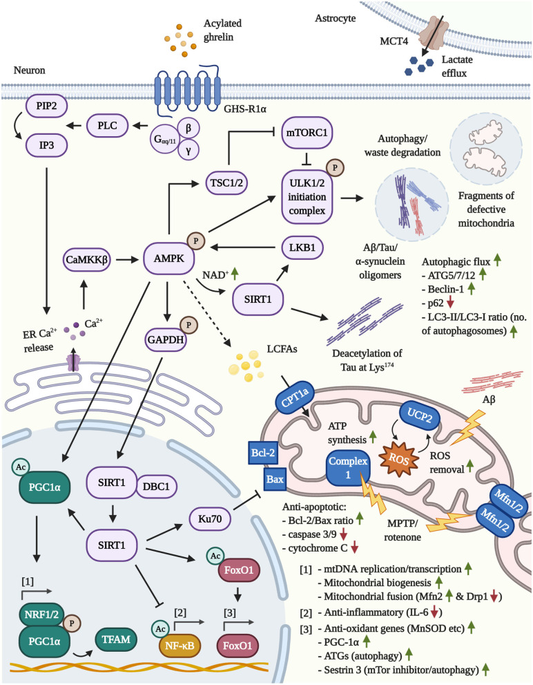
“, 2007) and deacetylates nuclear PGC1α to initiate mitochondrial biogenesis (Lagouge et al., 2006) as well as the transcription factor forkhead box protein O1 (FoxO1) to amplify the expression of PGC1α (Daitoku et al.”
BackgroundTSC2 Mediates Cellular Energy Response to Control Cell Growth and Survival
Inoki, K., Zhu, T., & Guan, K.-L. (2003) · Cell · doi:10.1016/s0092-8674(03)00929-2 · Cited by 3,914 · cited 1×
“autophagy and, typically in response to stressful cellular conditions and starvation, the activating phosphorylation of the cytoplasmic energy-sensor AMPK results in the AMPK-mediated phosphorylation of TSC1/2 and the inhibition of mTORC1 (Inoki et al., 2003;Manning andCantley, 2003; Demetriades et al., 2016).”
Suppression of basal autophagy in neural cells causes neurodegenerative disease in mice
Hara, T., Nakamura, K., Matsui, M., Yamamoto, A., Nakahara, Y., Suzuki-Migishima, R., Yokoyama, M., Mishima, K., Saito, I., Okano, H., et al. (2006) · Nature · doi:10.1038/nature04724 · Cited by 3,882 · cited 1×
“Thus, impairments in autophagy induce neurodegeneration independent of amyloid accumulation in affected brain areas (Hara et al., 2006; Komatsu et al., 2006).”
BackgroundMaffei, M., Halaas, J., Ravussin, E., Pratley, R. E., Lee, G. H., Zhang, Y., Fei, H., Kim, S., Lallone, R., Ranganathan, S., et al. (1995) · Nature Medicine · doi:10.1038/nm1195-1155 · Cited by 3,725 · cited 1×
“Thus, obesity and T2DMassociated hyperleptinemia (Maffei et al., 1995; Considine et al., 1996; Okumura et al., 2003; Pandey et al., 2015) may contribute to the desensitization of AG in the hypothalamus.”
BackgroundLoss of autophagy in the central nervous system causes neurodegeneration in mice
Komatsu, M., Waguri, S., Chiba, T., Murata, S., Iwata, J.-I., Tanida, I., Ueno, T., Koike, M., Uchiyama, Y., Kominami, E., et al. (2006) · Nature · doi:10.1038/nature04723 · Cited by 3,500
AMPK regulates energy expenditure by modulating NAD+ metabolism and SIRT1 activity
Cantó, C., Gerhart-Hines, Z., Feige, J. N., Lagouge, M., Noriega, L., Milne, J. C., Elliott, P. J., Puigserver, P., & Auwerx, J. (2009) · Nature · doi:10.1038/nature07813 · Cited by 3,300
Blood–brain barrier breakdown in Alzheimer disease and other neurodegenerative disorders
Sweeney, M. D., Sagare, A. P., & Zlokovic, B. V. (2018) · Nature Reviews Neurology · doi:10.1038/nrneurol.2017.188 · Cited by 3,076 · cited 3×
“, 2016), vascular abnormalities, reduced blood flow, blood brain barrier (BBB) damage (Kisler et al., 2017; Sweeney et al., 2018), dysfunctional autophagy and mitophagy (Kerr et al.”
BackgroundA preprandial rise in plasma ghrelin levels suggests a role in meal initiation in humans
Cummings, D. E., Purnell, J. Q., Frayo, R. S., Schmidova, K., Wisse, B. E., & Weigle, D. S. (2001) · Diabetes · doi:10.2337/diabetes.50.8.1714 · Cited by 2,940 · cited 1×
“Mature ghrelin is stored within secretory granules of X/A-like cells and released into the bloodstream upon fasting to stimulate appetite (Cummings et al., 2001; Yanagi et al., 2018).”
BackgroundDiagnosis and Treatment of Parkinson Disease: A Review
Armstrong, M. J., & Okun, M. S. (2020) · JAMA · doi:10.1001/jama.2019.22360 · Cited by 2,924
Mitochondrial TCA cycle metabolites control physiology and disease
Martínez-Reyes, I., & Chandel, N. S. (2020) · Nature Communications · doi:10.1038/s41467-019-13668-3 · Cited by 2,813 · cited 1×
“To recharge NAD and FAD and to resume ATP generation, the brain relies on glucose as major energy substrate as well as its metabolization in the tricarboxylic acid cycle (TCA) as main bioenergetic pathway (Penicaud et al., 2002; Arun et al., 2016; Martinez-Reyes and Chandel, 2020).”
BackgroundModulation of NF‐κB‐dependent transcription and cell survival by the SIRT1 deacetylase
Yeung, F., Hoberg, J. E., Ramsey, C. S., Keller, M. D., Jones, D. R., Frye, R. A., & Mayo, M. W. (2004) · The EMBO Journal · doi:10.1038/sj.emboj.7600244 · Cited by 2,750
How to Interpret LC3 Immunoblotting
Mizushima, N., & Yoshimori, T. (2007) · Autophagy · doi:10.4161/auto.4600 · Cited by 2,595 · cited 1×
“AG-driven autophagy is dependent on the stimulation of AMPK, leading to increased levels of ATG5, ATG7, ATG12, and Beclin-1, lessened p62 levels (an autophagy marker that is adversely correlated with autophagy), an elevated microtubule-associated protein light chain 3 (LC3)-II/LC3-I ratio, which serves as a marker to…”
BackgroundMolecular mechanisms of mTOR-mediated translational control
Ma, X. M., & Blenis, J. (2009) · Nature Reviews Molecular Cell Biology · doi:10.1038/nrm2672 · Cited by 2,558
AMP-activated protein kinase (AMPK) action in skeletal muscle via direct phosphorylation of PGC-1α
Jäger, S., Handschin, C., St.-Pierre, J., & Spiegelman, B. M. (2007) · Proceedings of the National Academy of Sciences · doi:10.1073/pnas.0705070104 · Cited by 2,491
The Roles of PINK1, Parkin, and Mitochondrial Fidelity in Parkinson’s Disease
Pickrell, A. M., & Youle, R. J. (2015) · Neuron · doi:10.1016/j.neuron.2014.12.007 · Cited by 2,310 · cited 1×
“Furthermore, the reduced expression of GHS-R1α was verified in induced pluripotent stem cells with a mutated or disrupted PARK2 gene, which is a mitochondrial gene implicated in mitochondrial quality control, mitophagy, and the development of early-onset PD (Pickrell and Youle, 2015), and in the SNpc of an in vivo mod…”
BackgroundSt-Pierre, J., Drori, S., Uldry, M., Silvaggi, J. M., Rhee, J., Jäger, S., Handschin, C., Zheng, K., Lin, J., Yang, W., et al. (2006) · Cell · doi:10.1016/j.cell.2006.09.024 · Cited by 2,233
Davson, H. (1977) · Nature · doi:10.1038/266661a0 · Cited by 2,229
A Receptor in Pituitary and Hypothalamus That Functions in Growth Hormone Release
Howard, A., Feighner, S., Cully, D., Arena, J. P., Liberator, P., Rosenblum, C., Hamelin, M., Hreniuk, D. L., Palyha, O., Anderson, J., et al. (1996) · Science · doi:10.1126/science.273.5277.974 · Cited by 2,106 · cited 1×
“Importantly, DAG is incapable of interacting with GHSR1α, yet the existence of distinct DAG-binding receptors has been postulated (Howard et al., 1996; Yanagi et al., 2018).”
BackgroundCirculating Ghrelin Levels Are Decreased in Human Obesity
Tschöp, M. H., Weyer, C., Tataranni, P. A., Devanarayan, V., Ravussin, É., & Heiman, M. L. (2001) · Diabetes · doi:10.2337/diabetes.50.4.707 · Cited by 2,031 · cited 1×
“Similarly, obese individuals and patients with metabolic syndrome displayed reduced plasma ghrelin pools (Tschop et al., 2001; Rigamonti et al., 2002; Shiiya et al., 2002; Tesauro et al., 2005).”
BackgroundDate, Y., Kojima, M., Hosoda, H., Sawaguchi, A., Mondal, M. S., Suganuma, T., Matsukura, S., Kangawa, K., & Nakazato, M. (2000) · Endocrinology · doi:10.1210/en.141.11.4255 · Cited by 1,943 · cited 1×
“Ghrelin is predominantly produced by gastric X/A-like cells that are located in the oxyntic gland of the stomach (Date et al., 2000), although a lower degree of the hormone is also expressed in various peripheral tissues, in lymphocytes and in the CNS (Ferrini et al.”
BackgroundCalorie restriction promotes mammalian cell survival by inducing the SIRT1 deacetylase
Cohen, H. Y., Miller, C., Bitterman, K. J., Wall, N. R., Hekking, B., Kessler, B., Howitz, K. T., Gorospe, M., de Cabo, R., & Sinclair, D. A. (2004) · Science · doi:10.1126/science.1099196 · Cited by 1,924 · cited 1×
“, 2004) or DNA repair factor Ku70, which was shown to scavenge proapoptotic Bax from mitochondria to support cellular survival (Cohen et al., 2004).”
BackgroundTalbot, K., Wang, H.-Y., Kazi, H., Han, L.-Y., Bakshi, K. P., Stucky, A., Fuino, R. L., Kawaguchi, K. R., Samoyedny, A. J., Wilson, R. S., et al. (2012) · Journal of Clinical Investigation · doi:10.1172/jci59903 · Cited by 1,871 · cited 1×
“Moreover, the rate of inhibitory serine phosphorylation of IRS1 (as a marker of insulin resistance), independent of even T2DM or the APOEε4 allele, rose gradually from previously healthy suspects to mild cognitive impairment (MCI) to AD patients, correlated with the quantity of Aß deposits and was inversely associated…”
Background(2014) · Nature Reviews Neurology · doi:10.1038/nrneurol.2014.114 · Cited by 1,832
Fang, E. F., Hou, Y., Palikaras, K., Adriaanse, B. A., Kerr, J. S., Yang, B., Lautrup, S., Hasan-Olive, M. M., Caponio, D., Dan, X., et al. (2019) · Nature Neuroscience · doi:10.1038/s41593-018-0332-9 · Cited by 1,740 · cited 2×
“As expected, mitophagy is widely impaired in respective models as well as in the brains of AD and PD patients, while the selective pharmacological enhancement of mitophagy can reverse several other pathological aspects, such as the generation of insoluble Aβ, Tau hyperphosphorylation, neuroinflammation, neuronal atrop…”
BackgroundSteen, E., Terry, B. M., J. Rivera, E., Cannon, J. L., Neely, T. R., Tavares, R., Xu, X. J., Wands, J. R., & de la Monte, S. M. (2005) · Journal of Alzheimer's Disease · doi:10.3233/jad-2005-7107 · Cited by 1,703 · cited 1×
“Such and related investigations led to the designation of AD as “type 3 diabetes” (Steen et al., 2005; Moloney et al., 2010).”
BackgroundDouble perovskites as a family of highly active catalysts for oxygen evolution in alkaline solution
Grimaud, A., May, K. J., Carlton, C. E., Lee, Y., Risch, M., Hong, W. T., Zhou, J., & Shao‐Horn, Y. (2013) · Nature Communications · doi:10.1038/ncomms3439 · Cited by 1,647
Hawley, S. A., Pan, D. A., Mustard, K. J., Ross, L., Bain, J., Edelman, A. M., Frenguelli, B. G., & Hardie, D. G. (2005) · Cell Metabolism · doi:10.1016/j.cmet.2005.05.009 · Cited by 1,589 · cited 1×
“Therefore, in general, AG induces intraneuronal Ca2+ accumulation in an IP3-mediated manner, leading to the activation of the AMPKphosphorylating calmodulin-dependent protein kinase kinase-β (CaMKKß) (Hawley et al., 2005; Anderson et al., 2008).”
BackgroundExtensive Involvement of Autophagy in Alzheimer Disease: An Immuno-Electron Microscopy Study
Nixon, R. A., Wegiel, J., Kumar, A., Yu, W. H., Peterhoff, C., Cataldo, A., & Cuervo, A. M. (2005) · Journal of Neuropathology & Experimental Neurology · doi:10.1093/jnen/64.2.113 · Cited by 1,588 · cited 1×
“This results in the intraneuronal accumulation of non-degraded and amyloid-containing autophagosomes, co-localizing strongly with neurons that display intracellular Tau pathology and the relative loss of mitochondria and other organelles (Cataldo et al., 1994; Nixon et al., 2005).”
BackgroundThe tissue distribution of the mRNA of ghrelin and subtypes of its receptor, GHS-R, in humans.
Gnanapavan, S., Kola, B., Bustin, S., Morris, D., McGee, P. J., Fairclough, P., Bhattacharya, S., Carpenter, R., Grossman, A., & Korbonits, M. (2002) · Journal of Clinical Endocrinology and Metabolism · doi:10.1210/jcem.87.6.8739 · Cited by 1,507 · cited 1×
“GHS-R1α is widely transcribed in multiple key areas of the brain, such as the hippocampus, hypothalamus, cortex, ventral tegmental area (VTA), SN, spinal cord, dorsal and median raphe nuclei, sympathetic preganglionic nerves and endothelial cells of the cerebral vasculature, yet it is also expressed by various immune…”
BackgroundNLRP3 inflammasome activation drives tau pathology
Ising, C., Venegas, C., Zhang, S., Scheiblich, H., Schmidt, S. V., Vieira-Saecker, A., Schwartz, S., Albasset, S., McManus, R. M., Tejera, D., et al. (2019) · Nature · doi:10.1038/s41586-019-1769-z · Cited by 1,487 · cited 3×
“Over time, prolonged neuroinflammation encourages various other secondary complications, such as impairments in protein degradation, amyloid misfolding, Tau hyperphosphorylation (in conjunction with the inflammationperpetuating activation of the inflammasome), permeabilization of the BBB, peripheral immune cell infilt…”
BackgroundPlasma Ghrelin Levels in Lean and Obese Humans and the Effect of Glucose on Ghrelin Secretion
Shiiya, T., Nakazato, M., Mizuta, M., Date, Y., Mondal, M. S., Tanaka, M., Nozoe, S.-I., Hosoda, H., Kangawa, K., & Matsukura, S. (2002) · The Journal of Clinical Endocrinology & Metabolism · doi:10.1210/jcem.87.1.8129 · Cited by 1,308
Cerebral blood flow regulation and neurovascular dysfunction in Alzheimer disease
Kisler, K., Nelson, A. R., Montagne, A., & Zlokovic, B. V. (2017) · Nature Reviews Neuroscience · doi:10.1038/nrn.2017.48 · Cited by 1,243
The TSC1–TSC2 complex: a molecular switchboard controlling cell growth
Huang, J., & Manning, B. D. (2008) · Biochemical Journal · doi:10.1042/bj20080281 · Cited by 1,237 · cited 1×
“Shut-down of the growthfacilitating mTORC1 pathway, however, promotes the activity of the unc-51 like autophagy activating kinase (ULK1/2) initiation complex, which launches autophagosome maturation and the cellular purging of waste products, such as Aß, Tau or αsynuclein (Huang and Manning, 2008; Ma and Blenis, 2009;…”
BackgroundImpaired Balance of Mitochondrial Fission and Fusion in Alzheimer's Disease
Wang, X., Su, B., Lee, H.-G., Li, X., Perry, G., Smith, M. A., & Zhu, X. (2009) · Journal of Neuroscience · doi:10.1523/jneurosci.1357-09.2009 · Cited by 1,224
Guan, X.-M., Yu, H., Palyha, O., McKee, K., Feighner, S., Sirinathsinghji, D., Smith, R. G., van der Ploeg, L., & Howard, A. (1997) · Brain Research. Molecular Brain Research · doi:10.1016/s0169-328x(97)00071-5 · Cited by 1,185 · cited 2×
“GHS-R1α is widely transcribed in multiple key areas of the brain, such as the hippocampus, hypothalamus, cortex, ventral tegmental area (VTA), SN, spinal cord, dorsal and median raphe nuclei, sympathetic preganglionic nerves and endothelial cells of the cerebral vasculature, yet it is also expressed by various immune…”
BackgroundBrochard, V., Combadière, B., Prigent, A., Laouar, Y., Perrin, A., Beray-Berthat, V., Bonduelle, O., Alvarez-Fischer, D., Callebert, J., Launay, J.-M., et al. (2008) · Journal of Clinical Investigation · doi:10.1172/jci36470 · Cited by 1,172 · cited 6×
“Regarding immune infiltration, CD4+ T-cell-deleted mice were shown to be protected from MPTP-triggered neurodegeneration, proposing that the adaptive immune system is heavily involved in PD pathology (Brochard et al., 2009).”
BackgroundInfluentialBoland, B., Kumar, A., Lee, S., Platt, F. M., Wegiel, J., Yu, W. H., & Nixon, R. A. (2008) · Journal of Neuroscience · doi:10.1523/jneurosci.0800-08.2008 · Cited by 1,119 · cited 1×
“In contrast, the inhibition of lysosome-associated proteolytic enzymes was capable of producing an AD-like phenotype (Boland et al., 2008).”
BackgroundBezzi, P., Domercq, M., Brambilla, L., Galli, R., Schols, D., De Clercq, E., Vescovi, A., Bagetta, G., Kollias, G., Meldolesi, J., et al. (2001) · Nature Neuroscience · doi:10.1038/89490 · Cited by 1,102 · cited 17×
“Moreover, AG downregulated the transcription of IL-1 and/or the inflammasome-activating cytokine TNF-α in face of MPTPinjury (PD), threohydroxyaspartate (THA)/kainic acid-assault (excitotoxicity), subarachnoid hemorrhage and SCI (Moon et al., 2009a; Ersahin et al., 2010; Lee J. et al., 2010; Lee et al., 2012, 2014b; A…”
BackgroundInfluentialS-Nitrosylation of Drp1 Mediates β-Amyloid-Related Mitochondrial Fission and Neuronal Injury
Cho, D.-H., Nakamura, T., Fang, J., Cieplak, P., Godzik, A., Gu, Z., & Lipton, S. A. (2009) · Science · doi:10.1126/science.1171091 · Cited by 1,092
AMP-activated protein kinase mediates mitochondrial fission in response to energy stress
Toyama, E. Q., Herzig, S., Courchet, J., Lewis, T. L., Losón, O. C., Hellberg, K., Young, N. P., Chen, H., Polleux, F., Chan, D. C., et al. (2016) · Science · doi:10.1126/science.aab4138 · Cited by 1,085 · cited 9×
“This is in line with the Mfn2-upregulating and Drp1-impeding function of PGC-1α (Peng et al., 2017), suggesting that AG stimulates the AMPK/PGC-1α axis to ameliorate mitochondrial fragmentation in response to cellular stress (Sirago et al., 2017).”
BackgroundInfluentialMüller, T. D., Nogueiras, R., Andermann, M. L., Andrews, Z. B., Anker, S. D., Argente, J., Batterham, R. L., Benoit, S. C., Bowers, C. Y., Broglio, F., et al. (2015) · Molecular Metabolism · doi:10.1016/j.molmet.2015.03.005 · Cited by 1,034
Spilman, P., Podlutskaya, N., Hart, M. J., Debnath, J., Gorostiza, O., Bredesen, D., Richardson, A., Strong, R., & Galvan, V. (2010) · PLoS ONE · doi:10.1371/journal.pone.0009979 · Cited by 1,033
Clinical progression in Parkinson disease and the neurobiology of axons
Cheng, H., Ulane, C. M., & Burke, R. E. (2010) · Annals of Neurology · doi:10.1002/ana.21995 · Cited by 1,012
Abizaid, A., Liu, Z.-W., Andrews, Z. B., Shanabrough, M., Borok, E., Elsworth, J. D., Roth, R. H., Sleeman, M. W., Picciotto, M. R., Tschöp, M. H., et al. (2006) · Journal of Clinical Investigation · doi:10.1172/jci29867 · Cited by 984 · cited 3×
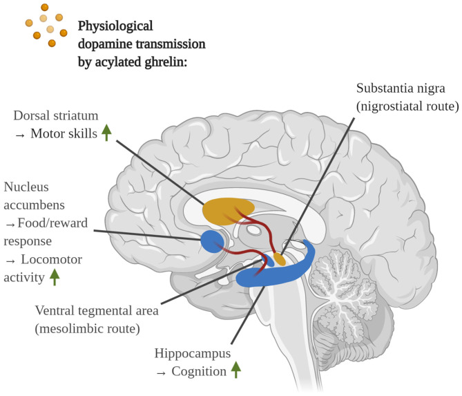
“Acylated Ghrelin Induces Dopamine Transmission in the Ventral Tegmental Area and Stimulates Locomotor Activity and Memory Noteworthy, GHS-R1α was found to be expressed by ∼60% of dopaminergic neurons in the VTA of rats (Abizaid et al., 2006).”
BackgroundInflammation in CNS neurodegenerative diseases
Stephenson, J., Nutma, E., van der Valk, P., & Amor, S. (2018) · Immunology · doi:10.1111/imm.12922 · Cited by 966 · cited 2×
“, 2019), chronic systemic inflammation, pathological immune cell infiltration into the brain (Amor and Woodroofe, 2014; Anderson et al., 2014; Stephenson et al., 2018), inflammasome activation (Ising et al.”
BackgroundRole of neuroinflammation in neurodegenerative diseases (Review)
Chen, W., Zhang, X., & Huang, W. (2016) · Molecular Medicine Reports · doi:10.3892/mmr.2016.4948 · Cited by 961
Review: Activation patterns of microglia and their identification in the human brain
Boche, D., Perry, V. H., & Nicoll, J. A. R. (2012) · Neuropathology and Applied Neurobiology · doi:10.1111/nan.12011 · Cited by 955 · cited 13×
“Moreover, the administration of LPS into older animals, which display lessened expression of GHS-R1α in the DVC, evoked the excessive release of TNF-α and IL-6, far greater than in younger littermates (Wu et al., 2009b).”
BackgroundInfluentialPARIS (ZNF746) Repression of PGC-1α Contributes to Neurodegeneration in Parkinson's Disease
Shin, J.-H., Ko, H. S., Kang, H., Lee, Y., Lee, Y.-I., Pletinkova, O., Troconso, J. C., Dawson, V. L., & Dawson, T. M. (2011) · Cell · doi:10.1016/j.cell.2011.02.010 · Cited by 953
Ghrelin and Des-acyl Ghrelin: Two Major Forms of Rat Ghrelin Peptide in Gastrointestinal Tissue
Hosoda, H., Kojima, M., Matsuo, H., & Kangawa, K. (2000) · Biochemical and Biophysical Research Communications · doi:10.1006/bbrc.2000.4039 · Cited by 943 · cited 14×
“Depending on the presence or absence of the acyl group at Ser3, mature ghrelin can be further distinguished into its active form, acylated ghrelin (AG), and desacylated ghrelin (DAG) (Hosoda et al., 2000; Yanagi et al., 2018).”
BackgroundInfluentialMitochondrial dysfunction in Parkinson's disease
Bose, A., & Beal, M. F. (2016) · Journal of Neurochemistry · doi:10.1111/jnc.13731 · Cited by 915 · cited 2×
“The mitochondrial pathology in AD and PD is further exacerbated by impaired mitochondrial biogenesis, a mechanism that leads to the generation of additional mitochondria to meet greater energetic demands in cells, and mitophagy, which is a form of autophagy that mediates the degradation of malfunctional, ROS-overprodu…”
BackgroundTwo defects contribute to hypothalamic leptin resistance in mice with diet-induced obesity
El-Haschimi, K., Pierroz, D. D., Hileman, S. M., Bjørbæk, C., & Flier, J. S. (2000) · Journal of Clinical Investigation · doi:10.1172/jci9842 · Cited by 887 · cited 1×
“Additionally, a high-fat diet, in an inflammation-mediated manner, promotes hypothalamic leptin resistance as well (El-Haschimi et al., 2000; Zhang et al., 2008).”
BackgroundDixit, V. D., Schaffer, E. M., Pyle, R. S., Collins, G. D., Sakthivel, S. K., Palaniappan, R., Lillard, J. W., & Taub, D. D. (2004) · Journal of Clinical Investigation · doi:10.1172/jci21134 · Cited by 882 · cited 18×
“Furthermore, in vitro studies have shown that the administration of AG downregulated the synthesis of IL-1β, IL-6, and TNFα in human peripheral blood mononuclear cells following irritation with the mitogen phytohemagglutinin (Dixit et al., 2004).”
BackgroundInfluentialLee, H.-J., Suk, J.-E., Patrick, C., Bae, E.-J., Cho, J.-H., Rho, S., Hwang, D., Masliah, E., & Lee, S.-J. (2010) · Journal of Biological Chemistry · doi:10.1074/jbc.m109.081125 · Cited by 877
Mitophagy and Alzheimer’s Disease: Cellular and Molecular Mechanisms
Kerr, J. S., Adriaanse, B. A., Greig, N. H., Mattson, M. P., Cader, M. Z., Bohr, V. A., & Fang, E. F. (2017) · Trends in Neurosciences · doi:10.1016/j.tins.2017.01.002 · Cited by 870
T cells from patients with Parkinson’s disease recognize α-synuclein peptides
Sulzer, D., Alcalay, R. N., Garretti, F., Cote, L., Kanter, E., Agin-Liebes, J., Liong, C., McMurtrey, C., Hildebrand, W. H., Mao, X., et al. (2017) · Nature · doi:10.1038/nature22815 · Cited by 866 · cited 1×
“In addition, heightened numbers of partially α-synuclein-reactive Th17 cells were discovered in the blood of PD patients (Sulzer et al., 2017); to the degree that PD has been postulated as an α-synuclein-reactive autoimmune disease (Benner et al., 2008; Hu, 2011).”
BackgroundKim, C., Ho, D.-H., Suk, J.-E., You, S., Michael, S., Kang, J., Joong Lee, S., Masliah, E., Hwang, D., Lee, H.-J., et al. (2013) · Nature Communications · doi:10.1038/ncomms2534 · Cited by 856
Wang, X., Su, B., Siedlak, S. L., Moreira, P. I., Fujioka, H., Wang, Y., Casadesus, G., & Zhu, X. (2008) · Proceedings of the National Academy of Sciences · doi:10.1073/pnas.0804871105 · Cited by 835
Ghrelin controls hippocampal spine synapse density and memory performance
Diano, S., Farr, S. A., Benoit, S. C., McNay, E. C., da Silva, I., Horvath, B., Gaskin, F. S., Nonaka, N., Jaeger, L. B., Banks, W. A., et al. (2006) · Nature Neuroscience · doi:10.1038/nn1656 · Cited by 828 · cited 29×
“Indeed, besides amyloids, AD and PD are characterized by the culminative interplay of oxidative stress, mitochondrial dysfunction and hyperfission, defective autophagy and mitophagy, systemic inflammation, BBB and vascular damage, demyelination, cerebral insulin resistance, the loss of dopamine production in PD, impai…”
BackgroundInfluentialSIRT1 Modulation of the Acetylation Status, Cytosolic Localization, and Activity of LKB1
Lan, F., Cacicedo, J. M., Ruderman, N., & Ido, Y. (2008) · Journal of Biological Chemistry · doi:10.1074/jbc.m805711200 · Cited by 825 · cited 14×
“Furthermore, AMPK phosphorylates glyceraldehyde 3- phosphate dehydrogenase (GAPDH) at Ser122 during starvation to encourage the nuclear trafficking of GAPDH and the displacement of SIRT1 from its repressor Deleted in Breast Cancer 1 (DBC1) (Chang et al., 2015).”
BackgroundInfluentialPathogenic Lysosomal Depletion in Parkinson's Disease
Dehay, B., Bové, J., Rodríguez-Muela, N., Perier, C., Recasens, A., Boya, P., & Vila, M. (2010) · The Journal of Neuroscience · doi:10.1523/jneurosci.1920-10.2010 · Cited by 825 · cited 1×
“This indicates that, similar to AD, autophagosomal efficiency is lost during PD, leading to the amassment of defective, waste-cluttered lysosomes and the failure of amyloid clearance (Chu et al., 2009; Alvarez-Erviti et al., 2010; Dehay et al., 2010).”
BackgroundLimiting inflammation—the negative regulation of NF-κB and the NLRP3 inflammasome
Afonina, I. S., Zhong, Z., Karin, M., & Beyaert, R. (2017) · Nature Immunology · doi:10.1038/ni.3772 · Cited by 785 · cited 1×
“, 2019), with NF-κB driving the synthesis of the inflammasome sensor NLRP3 as well as the pro-inflammatory cytokines pro-IL1/IL-1, pro-IL-18, TNF-α and many more (Afonina et al., 2017).”
BackgroundNucleocytoplasmic Shuttling of the NAD+-dependent Histone Deacetylase SIRT1*
Tanno, M., Sakamoto, J., Miura, T., Shimamoto, K., & Horio, Y. (2007) · Journal of Biological Chemistry · doi:10.1074/jbc.m609554200 · Cited by 784 · cited 1×
“Moreover, SIRT1 is capable of shuttling between the cytoplasm and nucleus (Tanno et al., 2007) and deacetylates nuclear PGC1α to initiate mitochondrial biogenesis (Lagouge et al.”
BackgroundCPT1A-mediated Fat Oxidation, Mechanisms, and Therapeutic Potential
Schlaepfer, I. R., & Joshi, M. (2020) · Endocrinology · doi:10.1210/endocr/bqz046 · Cited by 783
Stranahan, A. M., Norman, E. D., Lee, K. M., Cutler, R. G., Telljohann, R. S., Egan, J. M., & Mattson, M. P. (2008) · Hippocampus · doi:10.1002/hipo.20470 · Cited by 766 · cited 1×
“For instance, a highfat diet, which induces systemic insulin resistance, was shown to attenuate neuroprotective brain-derived neurotrophic factor levels, long-term potentiation (LTP) and dendritic spine density in the hippocampus of previously healthy mice, while accelerating Aβ plaque formation and memory loss in Aβt…”
BackgroundMoloney, A. M., Griffin, R. J., Timmons, S., O’Connor, R., Ravid, R., & O’Neill, C. (2010) · Neurobiology of Aging · doi:10.1016/j.neurobiolaging.2008.04.002 · Cited by 764 · cited 1×
“Such and related investigations led to the designation of AD as “type 3 diabetes” (Steen et al., 2005; Moloney et al., 2010).”
BackgroundBlood–brain barrier dysfunction in parkinsonian midbrain in vivo
Kortekaas, R., Leenders, K. L., van Oostrom, J. C. H., Vaalburg, W., Bart, J., Willemsen, A. T. M., & Hendrikse, N. H. (2005) · Annals of Neurology · doi:10.1002/ana.20369 · Cited by 733
SIRT1 and aging related signaling pathways
Chen, C., Zhou, M., Ge, Y., & Wang, X. (2020) · Mechanisms of Ageing and Development · doi:10.1016/j.mad.2020.111215 · Cited by 714
UCP2 mediates ghrelin’s action on NPY/AgRP neurons by lowering free radicals
Andrews, Z. B., Liu, Z.-W., Walllingford, N., Erion, D. M., Borok, E., Friedman, J. M., Tschöp, M. H., Shanabrough, M., Cline, G., Shulman, G. I., et al. (2008) · Nature · doi:10.1038/nature07181 · Cited by 710
Clinical trials of new drugs for Alzheimer disease
Huang, L.-K., Chao, S.-P., & Hu, C.-J. (2020) · Journal of Biomedical Science · doi:10.1186/s12929-019-0609-7 · Cited by 702 · cited 1×
“Therefore, monotherapies are presumably an ineffective way of approaching AD and PD and are more likely to fail, supporting the concept that multi-targeted therapies are more profitable (Iqbal and Grundke-Iqbal, 2010; Adams, 2020; Huang et al., 2020).”
BackgroundCalorie restriction induces mitochondrial biogenesis and bioenergetic efficiency
López-Lluch, G., Hunt, N., Jones, B., Zhu, M., Jamieson, H., Hilmer, S., Cascajo, M. V., Allard, J., Ingram, D. K., Navas, P., et al. (2006) · Proceedings of the National Academy of Sciences · doi:10.1073/pnas.0510452103 · Cited by 699 · cited 16×
“As such, the function of variousmitochondrial master modulators, including AMPK and PGC1α, is disturbed in AD and PD.”
BackgroundInfluentialLoh, Y. (2008) · doi:10.3410/f.1123102.580425 · Cited by 698 · cited 1×
“Additionally, a high-fat diet, in an inflammation-mediated manner, promotes hypothalamic leptin resistance as well (El-Haschimi et al., 2000; Zhang et al., 2008).”
Banks, W. A., Tschöp, M., Robinson, S. M., & Heiman, M. L. (2002) · The Journal of Pharmacology and Experimental Therapeutics · doi:10.1124/jpet.102.034827 · Cited by 691 · cited 27×
“Indeed, besides amyloids, AD and PD are characterized by the culminative interplay of oxidative stress, mitochondrial dysfunction and hyperfission, defective autophagy and mitophagy, systemic inflammation, BBB and vascular damage, demyelination, cerebral insulin resistance, the loss of dopamine production in PD, impai…”
BackgroundInfluentialRole for neuronal insulin resistance in neurodegenerative diseases
Schubert, M., Gautam, D., Surjo, D., Ueki, K., Baudler, S., Schubert, D., Kondo, T., Alber, J., Galldiks, N., Küstermann, E., et al. (2004) · Proceedings of the National Academy of Sciences · doi:10.1073/pnas.0308724101 · Cited by 659
The promise of low-dose interleukin-2 therapy for autoimmune and inflammatory diseases
Klatzmann, D., & Abbas, A. K. (2015) · Nature reviews. Immunology · doi:10.1038/nri3823 · Cited by 653 · cited 5×
“For instance, the administration of AG succeeded in the animal model of colitis, showing downregulated local and systemic release of pro-inflammatory modulators, reduced inflammatory Th 1 activity, elevated action of immunosuppressive regulatory T-cells (Tregs), diminished oxidative stress, ameliorated intestinal tiss…”
BackgroundInfluentialHo, L., Qin, W., Pompl, P. N., Xiang, Z., Wang, J., Zhao, Z., Peng, Y., Cambareri, G. M., Rocher, A. B., Mobbs, C. V., et al. (2004) · The FASEB Journal · doi:10.1096/fj.03-0978fje · Cited by 640 · cited 1×
“For instance, a highfat diet, which induces systemic insulin resistance, was shown to attenuate neuroprotective brain-derived neurotrophic factor levels, long-term potentiation (LTP) and dendritic spine density in the hippocampus of previously healthy mice, while accelerating Aβ plaque formation and memory loss in Aβt…”
BackgroundNakamura, K., Nemani, V. M., Azarbal, F., Skibinski, G., Levy, J. M., Egami, K., Munishkina, L. A., Zhang, J., Gardner, B. M., Wakabayashi, J., et al. (2011) · Journal of Biological Chemistry · doi:10.1074/jbc.m110.213538 · Cited by 624 · cited 1×
“While mutant α-synuclein enhances mitochondrial fragmentation, impairs the mitochondrial respiratory activity and induces neuronal death by inducing the displacement of wt α-synuclein from the inner-mitochondrial membrane (Kamp et al., 2010; Nakamura et al., 2011; Guardia-Laguarta et al., 2014), it must be noted that…”
BackgroundBrain Glucose Hypometabolism and Oxidative Stress in Preclinical Alzheimer's Disease
Mosconi, L., Pupi, A., & de Leon, M. J. (2008) · Annals of the New York Academy of Sciences · doi:10.1196/annals.1427.007 · Cited by 616 · cited 2×
“…(Muddapu et al., 2020), as evident in the brains of both AD (Lyingtunell et al., 1981; Hoyer et al., 1988; Ogawa et al., 1996; Drzezga et al., 2003; Mosconi et al., 2008) and PD patients (Huang et al., 2008; Hosokai et al., 2009; Liepelt et al., 2009; Borghammer et al., 2010; Berti et al., 2012).”
BackgroundSiebner, H. R., Riemenschneider, M. J., Willoch, F., Minoshima, S., Schwaiger, M., Kurz, A., Drzezga, A. E., & Lautenschlager, N. T. (2003) · European Journal of Nuclear Medicine and Molecular Imaging · doi:10.1007/s00259-003-1194-1 · Cited by 603 · cited 2×
“In accord with insulin resistance, glucose hypometabolism in the CNS, for instance within the cortex, has been linked to the transition from MCI to AD and cognitive dysfunction (Lyingtunell et al., 1981; Hoyer et al., 1988; Ogawa et al., 1996; Drzezga et al., 2003; Mosconi et al., 2008).”
BackgroundSpencer, B., Potkar, R., Trejo, M., Rockenstein, E., Patrick, C., Gindi, R., Adame, A., Wyss-Coray, T., & Masliah, E. (2009) · The Journal of Neuroscience · doi:10.1523/jneurosci.4390-09.2009 · Cited by 595
Lactate-supported synaptic function in the rat hippocampal slice preparation.
Schurr, A., West, C. A., & Rigor, B. (1988) · Science · doi:10.1126/science.3375817 · Cited by 594
CD14 and Toll-Like Receptors 2 and 4 Are Required for Fibrillar Aβ-Stimulated Microglial Activation
Reed-Geaghan, E. G., Savage, J. C., Hise, A. G., & Landreth, G. E. (2009) · The Journal of Neuroscience · doi:10.1523/jneurosci.3158-09.2009 · Cited by 591
Amyloid beta oligomers induce impairment of neuronal insulin receptors
Zhao, W., De Felice, F. G., Fernandez, S., Chen, H., Lambert, M. P., Quon, M. J., Krafft, G. A., & Klein, W. L. (2008) · The FASEB Journal · doi:10.1096/fj.06-7703com · Cited by 587 · cited 29×
“The NTS, on the other hand, further projects across the entirety of the CNS, ultimately leading to the intestinal return of immune-suppressing signals through the afferent ends of the vagal nerve.”
BackgroundInfluentialHemodynamic and hormonal effects of human ghrelin in healthy volunteers
Nagaya, N., Kojima, M., Uematsu, M., Yamagishi, M., Hosoda, H., Oya, H., Hayashi, Y., & Kangawa, K. (2001) · American Journal of Physiology-Regulatory, Integrative and Comparative Physiology · doi:10.1152/ajpregu.2001.280.5.r1483 · Cited by 586
Chaperone-Mediated Autophagy Markers in Parkinson Disease Brains
Alvarez‐Erviti, L., Rodríguez-Oroz, M. C., Cooper, J. A. D., Caballero, C., Ferrer, I., Obeso, J. A., & Schapira, A. H. V. (2010) · Archives of Neurology · doi:10.1001/archneurol.2010.198 · Cited by 577 · cited 1×
“This indicates that, similar to AD, autophagosomal efficiency is lost during PD, leading to the amassment of defective, waste-cluttered lysosomes and the failure of amyloid clearance (Chu et al., 2009; Alvarez-Erviti et al., 2010; Dehay et al., 2010).”
BackgroundSilent Information Regulator 1 Protects the Heart From Ischemia/Reperfusion
Hsu, C., Zhai, P., Yamamoto, T., Maejima, Y., Matsushima, S., Hariharan, N., Shao, D., Takagi, H., Oka, S., & Sadoshima, J. (2010) · Circulation · doi:10.1161/circulationaha.110.958033 · Cited by 573 · cited 1×
“, 2010) and, under conditions of cellular stress, FoxO1 drives the transcription of various anti-oxidant enzymes, such as the PGC1α-co-activated, mitochondrialMnSOD (St.-Pierre et al., 2006; Hsu et al., 2010; Tong et al., 2012).”
BackgroundBonaz, B., Sinniger, V., & Pellissier, S. (2016) · The Journal of Physiology · doi:10.1113/jp271539 · Cited by 561
The Role of Metabolic Disorders in Alzheimer Disease and Vascular Dementia: Two Roads Converged
Craft, S. (2009) · Archives of Neurology · doi:10.1001/archneurol.2009.27 · Cited by 547 · cited 1×
“The latter may be another consequence of insulin resistance, since insulin promotes the cerebral blood flow by enhancing NOdriven vasoconstriction and endothelin 1-mediated capillary recruitment (Craft, 2009).”
BackgroundFrescas, D., Valenti, L. V. C., & Accili, D. (2005) · Journal of Biological Chemistry · doi:10.1074/jbc.m412357200 · Cited by 541 · cited 16×
“As such, the function of variousmitochondrial master modulators, including AMPK and PGC1α, is disturbed in AD and PD.”
BackgroundInfluentialAn update on inflamm-aging: mechanisms, prevention, and treatment
Xia, S., Zhang, X., Zheng, S., Khanabdali, R., Kalionis, B., Wu, J., Wan, W., & Tai, X. (2016) · Journal of Immunology Research · doi:10.1155/2016/8426874 · Cited by 538
McKenzie, B., Mamik, M. K., Saito, L. B., Boghozian, R., Monaco, M. C., Major, E. O., Lu, J., Branton, W. G., & Power, C. (2018) · Proceedings of the National Academy of Sciences · doi:10.1073/pnas.1722041115 · Cited by 529 · cited 1×
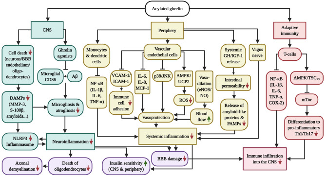
“Evidence that Acylated Ghrelin Opposes the Activation of the Inflammasome in the Brain The stimulation of the inflammasome and the associated pyroptosis, the “fiery death” of microglia, oligodendrocytes and other cells, a relatively recent upbringing, have been identified as major drivers of neuroinflammation, demyeli…”
BackgroundBlázquez, E., Velázquez, E., Hurtado-Carneiro, V., & Ruiz‐Albusac, J. M. (2014) · Frontiers in Endocrinology · doi:10.3389/fendo.2014.00161 · Cited by 523 · cited 1×
“Given the pivotal and neuroprotective role of insulin-signaling in the brain and that T2DM is one of the greatest known risk factors for AD and PD, it is no surprise that its early desensitization in the CNS, believed to predominantly occur in response to chronic inflammation, promotes the development of AD and PD (se…”
BackgroundNagaya, N., Moriya, J., Yasumura, Y., Uematsu, M., Ono, F., Shimizu, W., Ueno, K., Kitakaze, M., Miyatake, K., & Kangawa, K. (2004) · Circulation · doi:10.1161/01.cir.0000149746.62908.bb · Cited by 523
Insulin and Insulin-like Growth Factor-1 Regulate Tau Phosphorylation in Cultured Human Neurons
Hong, M., & Lee, V. M. ‐. (1997) · Journal of Biological Chemistry · doi:10.1074/jbc.272.31.19547 · Cited by 522 · cited 1×
“, 2011), hence the loss of insulin signaling initiates Tau hyperphosphorylation and aggregation in AD (Hong and Lee, 1997; Schubert et al., 2003, 2004).”
BackgroundOxidative Stress in Neurodegenerative Diseases: From a Mitochondrial Point of View
Cenini, G., Lloret, A., & Cascella, R. (2019) · Oxidative Medicine and Cellular Longevity · doi:10.1155/2019/2105607 · Cited by 521
α-Synuclein Is Localized to Mitochondria-Associated ER Membranes
Guardia-Laguarta, C., Area-Gomez, E., Rüb, C., Liu, Y., Magrané, J., Becker, D., Voos, W., Schon, E. A., & Przedborski, S. (2014) · The Journal of Neuroscience · doi:10.1523/jneurosci.2507-13.2014 · Cited by 515 · cited 2×
“While mutant α-synuclein enhances mitochondrial fragmentation, impairs the mitochondrial respiratory activity and induces neuronal death by inducing the displacement of wt α-synuclein from the inner-mitochondrial membrane (Kamp et al., 2010; Nakamura et al., 2011; Guardia-Laguarta et al., 2014), it must be noted that…”
BackgroundInhibition of mitochondrial fusion by α‐synuclein is rescued by PINK1, Parkin and DJ‐1
Kamp, F., Exner, N., Lutz, A. K., Wender, N., Hegermann, J., Brunner, B., Nuscher, B., Bartels, T., Giese, A., Beyer, K., et al. (2010) · The EMBO Journal · doi:10.1038/emboj.2010.223 · Cited by 504 · cited 1×
“While mutant α-synuclein enhances mitochondrial fragmentation, impairs the mitochondrial respiratory activity and induces neuronal death by inducing the displacement of wt α-synuclein from the inner-mitochondrial membrane (Kamp et al., 2010; Nakamura et al., 2011; Guardia-Laguarta et al., 2014), it must be noted that…”
BackgroundA Cell Surface Receptor Complex for Fibrillar β-Amyloid Mediates Microglial Activation
Bamberger, M. E., Harris, M. E., McDonald, D. R., Husemann, J., & Landreth, G. E. (2003) · The Journal of Neuroscience · doi:10.1523/jneurosci.23-07-02665.2003 · Cited by 501 · cited 11×
“This receptor has been reported to act as an inflammatory conductor for Aß, leading to the production of ROS and proinflammatory cytokines upon the interaction of fibrillar Aß with microglial or macrophage CD36 (Coraci et al., 2002; Bamberger et al., 2003; El Khoury et al., 2003; Demers et al., 2004).”
BackgroundInfluentialLi, W. G., Gavrila, D., Liu, X., Wang, L., Gunnlaugsson, S. T., Stoll, L. L., McCormick, M. L., Sigmund, C. D., Tang, C., & Weintraub, N. L. (2004) · Circulation · doi:10.1161/01.cir.0000127956.43874.f2 · Cited by 499 · cited 3×
“Since DAG failed to modify inflammatory reactions by HUVECs, it was implied that the described inflammation-dampening effects were reliant on GHS-R1α (Li et al., 2004).”
BackgroundMorales, I., Guzmán-MartÃnez, L., Cerda-Troncoso, C., FarÃas, G. A., & Maccioni, R. B. (2014) · Frontiers in Cellular Neuroscience · doi:10.3389/fncel.2014.00112 · Cited by 490 · cited 1×
“Furthermore, pathogen-associated molecular patterns (PAMPs) that originate from cerebral infections, such as LPS, or, in the case of AD, infections with members of the Herpesviridae family and Hepatitis C virus, may further potentiate neuroinflammation (Morales et al., 2014; Sochocka et al., 2017; Cortes et al., 2018;…”
BackgroundNuclear control of respiratory gene expression in mammalian cells
Scarpulla, R. C. (2006) · Journal of Cellular Biochemistry · doi:10.1002/jcb.20743 · Cited by 489 · cited 1×
“…in mitochondria-associated genes protect from MPTP oxidative assault in cell and animal models of PD, whereas the deletion of PGC1α exacerbates MPTP-induced injury and excitotoxicity (Scarpulla, 2002, 2006; Kang and Hamasaki, 2005; St.-Pierre et al., 2006; Mudo et al., 2012; Quan et al., 2020).”
BackgroundInsulin Receptor Substrate-2 Deficiency Impairs Brain Growth and Promotes Tau Phosphorylation
Schubert, M., Brazil, D. P., Burks, D. J., Kushner, J. A., Ye, J., Flint, C. L., Farhang-Fallah, J., Dikkes, P., Warot, X. M., Rio, C., et al. (2003) · The Journal of Neuroscience · doi:10.1523/jneurosci.23-18-07084.2003 · Cited by 488
Hypothalamic Fatty Acid Metabolism Mediates the Orexigenic Action of Ghrelin
López, M., Lage, R., Saha, A. K., Pérez–Tilve, D., Vazquez, M. J., Varela, L., Sangiao‐Alvarellos, S., Tovar, S. A., Raghay, K., Rodríguez‐Cuenca, S., et al. (2008) · Cell Metabolism · doi:10.1016/j.cmet.2008.03.006 · Cited by 486 · cited 1×
“In the other hand, AG does not modulate FAS in other brain areas, including the amygdala, striatum, hippocampus, several cortical regions and others (Lopez et al., 2008; Yanagi et al., 2018).”
BackgroundRheb fills a GAP between TSC and TOR
Manning, B. (2003) · Trends in Biochemical Sciences · doi:10.1016/j.tibs.2003.09.003 · Cited by 483
CD36 Mediates the Innate Host Response to β-Amyloid
El Khoury, J. B., Moore, K. J., Means, T. K., Leung, J., Terada, K., Toft, M., Freeman, M. W., & Luster, A. D. (2003) · The Journal of Experimental Medicine · doi:10.1084/jem.20021546 · Cited by 483
MPTP Mouse Models of Parkinson’s Disease: An Update
Meredith, G. E., & Rademacher, D. J. (2011) · Journal of Parkinson's Disease · doi:10.3233/jpd-2011-11023 · Cited by 471
Ghrelin action in the brain controls adipocyte metabolism
Theander-Carrillo, C. (2006) · Journal of Clinical Investigation · doi:10.1172/jci25811 · Cited by 465 · cited 1×
“In the periphery, for example, independent of the hormone’s orexigenic effects, AG stimulated the expression of lipogenic enzymes (fatty acid synthase (FAS), lipoprotein lipase and more) and lessened the transcription of carnitine palmitoyltransferase Ia (CPT1a), a ratelimiting effector necessary to induce fatty acid…”
BackgroundNeuronal Lipid Metabolism: Multiple Pathways Driving Functional Outcomes in Health and Disease
Tracey, T. J., Steyn, F. J., Wolvetang, E. J., & Ngo, S. T. (2018) · Frontiers in Molecular Neuroscience · doi:10.3389/fnmol.2018.00010 · Cited by 459 · cited 1×
“As a word of caution, while the β-oxidation of lipids has been proven in astrocytes, its utility by neurons is yet to be verified (Tracey et al., 2018).”
BackgroundChu, Y., Dodiya, H. B., Aebischer, P., Olanow, C. W., & Kordower, J. H. (2009) · Neurobiology of Disease · doi:10.1016/j.nbd.2009.05.023 · Cited by 447
Stancu, I.-C., Cremers, N., Vanrusselt, H., Couturier, J., Vanoosthuyse, A., Kessels, S., Lodder, C., Brône, B., Huaux, F., Octave, J.-N., et al. (2019) · Acta Neuropathologica · doi:10.1007/s00401-018-01957-y · Cited by 447
Jerlhag, E., Egecioglu, E., Dickson, S. L., Douhan, A., Svensson, L., & Engel, J. A. (2007) · Addiction Biology · doi:10.1111/j.1369-1600.2006.00041.x · Cited by 431
Reynolds, A. D., Stone, D. K., Hutter-Saunders, J. A. L., Benner, E. J., Mosley, R. L., & Gendelman, H. E. (2010) · The Journal of Immunology · doi:10.4049/jimmunol.0901852 · Cited by 417 · cited 2×
“Moreover, it has been strongly implied that CD4 T helper (Th)1 and Th17 cells, for instance immunoreactive against α-synuclein, are the main lymphocyte populations that contribute to the death of dopaminergic neurons (Brochard et al., 2009; Reynolds et al., 2010).”
BackgroundResultThe Infectious Etiology of Alzheimer’s Disease
Sochocka, M., Zwolińska, K., & Leszek, J. (2017) · Current Neuropharmacology · doi:10.2174/1570159x15666170313122937 · Cited by 399
Coraci, I. S., Husemann, J., Berman, J. W., Hulette, C., Dufour, J. H., Campanella, G. K., Luster, A. D., Silverstein, S. C., & El Khoury, J. B. (2002) · The American Journal of Pathology · doi:10.1016/s0002-9440(10)64354-4 · Cited by 398 · cited 12×
“This receptor has been reported to act as an inflammatory conductor for Aß, leading to the production of ROS and proinflammatory cytokines upon the interaction of fibrillar Aß with microglial or macrophage CD36 (Coraci et al., 2002; Bamberger et al., 2003; El Khoury et al., 2003; Demers et al., 2004).”
BackgroundInfluentialUCP2 mediates ghrelin’s action on NPY/AgRP neurons by lowering free radicals
Andrews, Z., Liu, Z.-W., Walllingford, N., Erion, D., Borók, E., Friedman, J., Tschöp, M., Shanabrough, M., Cline, G., Shulman, G., et al. (2009) · Nature · doi:10.1038/nature08132 · Cited by 392
Ghrelin Directly Interacts With Neuropeptide-Y-Containing Neurons in the Rat Arcuate Nucleus
Kohno, D., Gao, H., Muroya, S., Kikuyama, S., & Yada, T. (2003) · Diabetes · doi:10.2337/diabetes.52.4.948 · Cited by 389
Loss of Parkin or PINK1 Function Increases Drp1-dependent Mitochondrial Fragmentation
Lutz, A. K., Exner, N., Fett, M. E., Schlehe, J. S., Kloos, K., Lämmermann, K., Brunner, B., Kurz-Drexler, A., Vogel, F., Reichert, A. S., et al. (2009) · Journal of Biological Chemistry · doi:10.1074/jbc.m109.035774 · Cited by 388 · cited 2×
“and OPA1 ameliorated excessive mitochondrial fission and impaired ATP production in PINK1/Parkin-mutant cells (Lutz et al., 2009) and shielded against MPTP-driven mitochondrial fragmentation, the stimulation of the pro-apoptotic activity of p53, Bax and PUMA, dopaminergic neuron and nerve terminal loss as well as moto…”
BackgroundHypothalamic CaMKK2 Contributes to the Regulation of Energy Balance
Anderson, K. A., Ribar, T. J., Lin, F., Noeldner, P. K., Green, M. F., Muehlbauer, M. J., Witters, L. A., Kemp, B. E., & Means, A. R. (2008) · Cell Metabolism · doi:10.1016/j.cmet.2008.02.011 · Cited by 385
Matrix Metalloproteinases and Blood-Brain Barrier Disruption in Acute Ischemic Stroke
Lakhan, S., Kirchgessner, A., Tepper, D. E., & Leonard, A. (2013) · Frontiers in Neurology · doi:10.3389/fneur.2013.00032 · Cited by 379 · cited 1×
“, 2014a), the MMP family member that directly proteolyzes BBB components (Lakhan et al., 2013).”
BackgroundGhrelin increases anxiety-like behavior and memory retention in rats
Carlini, V. P., Monzon, M. E., Varas, M., Cragnolini, A. B., Schiöth, H. B., Scimonelli, T. N., & de Barioglio, S. R. (2002) · Biochemical and Biophysical Research Communications · doi:10.1016/s0006-291x(02)02740-7 · Cited by 373
Scarpulla, R. (2002) · Gene · doi:10.1016/s0378-1119(01)00809-5 · Cited by 371 · cited 1×
“…in mitochondria-associated genes protect from MPTP oxidative assault in cell and animal models of PD, whereas the deletion of PGC1α exacerbates MPTP-induced injury and excitotoxicity (Scarpulla, 2002, 2006; Kang and Hamasaki, 2005; St.-Pierre et al., 2006; Mudo et al., 2012; Quan et al., 2020).”
BackgroundNitrated α–Synuclein Immunity Accelerates Degeneration of Nigral Dopaminergic Neurons
Benner, E. J., Banerjee, R., Reynolds, A. D., Sherman, S., Писарев, В. М., Tsiperson, V., Nemachek, C., Ciborowski, P. S., Przedborski, S., Mosley, R. L., et al. (2008) · PLoS ONE · doi:10.1371/journal.pone.0001376 · Cited by 371 · cited 1×
“, 2017); to the degree that PD has been postulated as an α-synuclein-reactive autoimmune disease (Benner et al., 2008; Hu, 2011).”
BackgroundMorrow, V. A., Foufelle, F., Connell, J., Petrie, J. R., Gould, G. W., & Salt, I. P. (2003) · Journal of Biological Chemistry · doi:10.1074/jbc.m212831200 · Cited by 361 · cited 1×
“, 2013) and the stimulation of AMPK was proven to trigger the AMPKconveyed activating phosphorylation of eNOS at Ser1177 in cultured human and rat endothelial cells, thus strengthening vasodilation in vivo (Morrow et al., 2003; Suzuki et al., 2008), it is highly likely that the AG-evoked liberation of NO is”
BackgroundAblation of ghrelin improves the diabetic but not obese phenotype of ob/ob mice
Sun, Y., Asnicar, M., Saha, P., Chan, L., & Smith, R. G. (2006) · Cell Metabolism · doi:10.1016/j.cmet.2006.04.004 · Cited by 358 · cited 1×
“Furthermore, GHS-R1α antagonists, GOAT inhibitors and the genetic deletion of GHS-R1α enhanced the release of insulin, glucose tolerance, insulin sensitivity and weight loss in in vivo models of obesity (Sun et al., 2006; Esler et al., 2007; Longo et al., 2008; Barnett et al., 2010; Qi et al., 2011).”
BackgroundP1‐273: Changes in sleep/wake parameters in male PD‐APP transgenic mice
Shanks, E., Wafford, K., Hewes, N., McCarthy, A. P., Kinsley, P. J., & Edgar, D. M. (2012) · doi:10.1016/j.jalz.2012.05.553 · Cited by 358 · cited 2×
“Such findings indicate that autophagy-enhancing approaches must ensue early, since the mere upregulation of autophagy is insufficient at an advanced stage of AD, when insoluble and proteolysis-resistant aggregates have already formed in the brain (Majumder et al., 2011).”
BackgroundGonzález, H., Elgueta, D., Montoya, A., & Pacheco, R. (2014) · Journal of Neuroimmunology · doi:10.1016/j.jneuroim.2014.07.012 · Cited by 352 · cited 1×
“Over time, prolonged neuroinflammation encourages various other secondary complications, such as impairments in protein degradation, amyloid misfolding, Tau hyperphosphorylation (in conjunction with the inflammationperpetuating activation of the inflammasome), permeabilization of the BBB, peripheral immune cell infilt…”
BackgroundSankowski, R., Mader, S., & Valdés-Ferrer, S. I. (2015) · Frontiers in Cellular Neuroscience · doi:10.3389/fncel.2015.00028 · Cited by 351
Carlini, V., Varas, M., Cragnolini, A., Schiöth, H., Scimonelli, T., & Debarioglio, S. R. (2004) · Biochemical and Biophysical Research Communications - BBRC · doi:10.1016/j.bbrc.2003.11.150 · Cited by 347
Liu, Y. J., McIntyre, R. L., Janssens, G. E., & Houtkooper, R. H. (2020) · Mechanisms of Ageing and Development · doi:10.1016/j.mad.2020.111212 · Cited by 347 · cited 1×
“A Possible Implication of Acylated Ghrelin in the Enhancement of Mitochondrial Fusion and Fission Unsurprisingly, in accordance with general mitochondrial dysfunction, the efficiency of mitochondrial fusion and fission gradually declines during the aging process and is disturbed in neurodegenerative diseases (Liu et a…”
BackgroundGhrelin directly targets the ventral tegmental area to increase food motivation
Skibicka, K. P., Hansson, C., Álvarez-Crespo, M., Friberg, P. A., & Dickson, S. L. (2011) · Neuroscience · doi:10.1016/j.neuroscience.2011.02.016 · Cited by 346
Soriano, F. X., Liesa, M., Bach, D., Chan, D. C., Palacín, M., & Zorzano, A. (2006) · Diabetes · doi:10.2337/db05-0509 · Cited by 343
Insulin resistance and Parkinson’s disease: A new target for disease modification?
Athauda, D. S., & Foltynie, T. (2016) · Progress in Neurobiology · doi:10.1016/j.pneurobio.2016.10.001 · Cited by 340 · cited 1×
“Additionally, the co-emergence of insulin resistance, leading to reductions in PI3K/Akt/mTORC1-driven protein translation in dopaminergic neurons (Athauda and Foltynie, 2016; Anandhan et al., 2017; Holscher, 2019), presumably aggravates dopaminergic dysfunction and neuronal insensitivity toward AG.”
BackgroundCD4+ Regulatory and Effector/Memory T Cell Subsets Profile Motor Dysfunction in Parkinson’s Disease
Hutter-Saunders, J. A. L., Estes, K. A., Kosloski, L. M., Allen, H. E., Dempsey, K., Torres‐Russotto, D. R., Meza, J. L., Santamaria, P. M., Bertoni, J. M., Murman, D. L., et al. (2012) · Journal of Neuroimmune Pharmacology · doi:10.1007/s11481-012-9402-z · Cited by 339
Tong, J., Prigeon, R. L., Davis, H. W., Bidlingmaier, M., Kahn, S. E., Cummings, D. E., Tschöp, M. H., & D’Alessio, D. A. (2010) · Diabetes · doi:10.2337/db10-0504 · Cited by 334
The Homeostatic Force of Ghrelin
Yanagi, S., Sato, T., Kangawa, K., & Nakazato, M. (2018) · Cell Metabolism · doi:10.1016/j.cmet.2018.02.008 · Cited by 332
Browne, T. C., McQuillan, K., McManus, R. M., O’Reilly, J.-A., Mills, K. H. G., & Lynch, M. A. (2013) · The Journal of Immunology · doi:10.4049/jimmunol.1200947 · Cited by 330
Diet-Induced Obesity Causes Ghrelin Resistance in Arcuate NPY/AgRP Neurons
Briggs, D. I., Enriori, P. J., Lemus, M. B., Cowley, M. A., & Andrews, Z. B. (2010) · Endocrinology · doi:10.1210/en.2010-0556 · Cited by 320
Johnstone, M., Gearing, A. J. H., & Miller, K. (1999) · Journal of Neuroimmunology · doi:10.1016/s0165-5728(98)00226-4 · Cited by 316 · cited 17×
“Moreover, AG downregulated the transcription of IL-1 and/or the inflammasome-activating cytokine TNF-α in face of MPTPinjury (PD), threohydroxyaspartate (THA)/kainic acid-assault (excitotoxicity), subarachnoid hemorrhage and SCI (Moon et al., 2009a; Ersahin et al., 2010; Lee J. et al., 2010; Lee et al., 2012, 2014b; A…”
BackgroundInfluentialGhrelin Degradation by Serum and Tissue Homogenates: Identification of the Cleavage Sites
Vriese, C. D., Grégoire, F., Lema-Kisoka, R., Waelbroeck, M., Robberecht, P., & Delporte, C. (2004) · Endocrinology · doi:10.1210/en.2004-0569 · Cited by 316 · cited 2×
“…deacetylation halflife times (t1/2) of 4 h or 6.4 h, respectively, have been reported for AG in human plasma and∼27min in rat serum, whereas the degradation t1/2 of total circulatory ghrelin has been estimated to be as short as ∼9-11min (De Vriese et al., 2004; Liu et al., 2008; Tong et al., 2013).”
BackgroundInfluentialMudò, G., Mäkelä, J., Liberto, V. D., Tselykh, T. V., Olivieri, M., Piepponen, P. T., Eriksson, O., Mälkiä, A., Bonomo, A., Kairisalo, M., et al. (2011) · Cellular and Molecular Life Sciences · doi:10.1007/s00018-011-0850-z · Cited by 305 · cited 1×
“Jointly, the PGC1α-induced transcriptional changes in mitochondria-associated genes protect from MPTP oxidative assault in cell and animal models of PD, whereas the deletion of PGC1α exacerbates MPTP-induced injury and excitotoxicity (Scarpulla, 2002, 2006; Kang and Hamasaki, 2005; St.-Pierre et al., 2006; Mudo et al.…”
BackgroundPlasma ghrelin concentrations in elderly subjects: comparison with anorexic and obese patients
Rigamonti, A. E., Pincelli, A. I., Corrà, B., Viarengo, R., Bonomo, S., Galimberti, D., Scacchi, M., Scarpini, E. A., Cavagnini, F., & Ee, M. (2002) · Journal of Endocrinology · doi:10.1677/joe.0.175r001 · Cited by 302
Diabetes mellitus and Alzheimer's disease: GSK-3β as a potential link
Zhang, Y., Huang, N.-Q., Yan, F., Jin, H., Zhou, S.-Y., Shi, J.-S., & Jin, F. (2018) · Behavioural Brain Research · doi:10.1016/j.bbr.2017.11.015 · Cited by 301 · cited 1×
“GSK3β is a Tau-phosphorylating kinase whose activity is aberrantly enhanced in response to the desensitization of insulin and the associated loss of Akt-signaling during diabetes mellitus and AD (Jolivalt et al., 2008; Zhang et al., 2018).”
BackgroundJolivalt, C. G., Lee, C. A., Beiswenger, K. K., Smith, J. L. M., Orlov, M., Torrance, M., & Masliah, E. (2008) · Journal of Neuroscience Research · doi:10.1002/jnr.21787 · Cited by 295 · cited 1×
“GSK3β is a Tau-phosphorylating kinase whose activity is aberrantly enhanced in response to the desensitization of insulin and the associated loss of Akt-signaling during diabetes mellitus and AD (Jolivalt et al., 2008; Zhang et al., 2018).”
BackgroundJiang, H. F., Betancourt, L., & Smith, R. G. (2006) · Molecular Endocrinology · doi:10.1210/me.2005-0084 · Cited by 290 · cited 1×
“GHS-R1α is widely transcribed in multiple key areas of the brain, such as the hippocampus, hypothalamus, cortex, ventral tegmental area (VTA), SN, spinal cord, dorsal and median raphe nuclei, sympathetic preganglionic nerves and endothelial cells of the cerebral vasculature, yet it is also expressed by various immune…”
BackgroundAndrews, Z. B., Erion, D., Beiler, R., Liu, Z.-W., Abizaid, A., Zigman, J., Elsworth, J. D., Savitt, J. M., DiMarchi, R., Tschöp, M., et al. (2009) · The Journal of Neuroscience · doi:10.1523/jneurosci.3890-09.2009 · Cited by 289
Chang, C., Su, H., Zhang, D., Wang, Y., Shen, Q., Liu, B., Huang, R., Zhou, T., Peng, C., Wong, C. C. L., et al. (2015) · Molecular Cell · doi:10.1016/j.molcel.2015.10.037 · Cited by 287
Ortega‐Martínez, S. (2015) · Frontiers in Molecular Neuroscience · doi:10.3389/fnmol.2015.00046 · Cited by 285 · cited 2×
“In such AD in vivo models, AG rescued from hippocampal atrophy, synaptic damage and the degeneration of cholinergic projections, heightened brain glycogen levels, diminished Aß aggregation and deposition, blocked Aβimparted deficits in LTPs, normalized plasticity-associated p-CREB levels, improved insulin sensitivity,…”
BackgroundInsulin Resistance and Alzheimer’s Disease: Bioenergetic Linkages
Neth, B. J., & Craft, S. (2017) · Frontiers in Aging Neuroscience · doi:10.3389/fnagi.2017.00345 · Cited by 284 · cited 5×
“On the other hand, it must be noted that the prevalence of cerebral insulin resistance during AD leads to defects in the metabolism of glucose, which is believed to provoke the use of β-oxidation and lipids (ketones) as primary energy sources for neurons (Neth and Craft, 2017).”
BackgroundInfluential“Is dopamine involved in Alzheimer's disease?”
Martorana, A., & Koch, G. (2014) · Frontiers in Aging Neuroscience · doi:10.3389/fnagi.2014.00252 · Cited by 276 · cited 1×
“…insulin (Gault and Holscher, 2018; Holscher, 2019), as well as alterations of the dopaminergic system, including the extensive atrophy of substantia nigra pars compacta (SNpc)-located dopaminergic neurons and dopamine depletion in the striatum in PD (Martorana and Koch, 2014; Poewe et al., 2017).”
BackgroundPickford, F., Masliah, E., Britschgi, M., Lucin, K., Narasimhan, R., Jaeger, P. A., Small, S., Spencer, B., Rockenstein, E., Levine, B., et al. (2008) · Journal of Clinical Investigation · doi:10.1172/jci33585 · Cited by 268 · cited 1×
“Nonetheless, AD patients showed a massive decline in the transcriptional levels of the autophagy initiator Beclin1 during early stages of AD and strategies that have aimed to enhance the degree of autophagy, such as the lentivirusmediated expression of Beclin 1 or the autophagy-inducing blockage of mTor by rapamycin,…”
BackgroundSkibicka, K. P., Hansson, C., Egecioglu, E., & Dickson, S. L. (2011) · Addiction Biology · doi:10.1111/j.1369-1600.2010.00294.x · Cited by 267
Glucose and Weight Control in Mice with a Designed Ghrelin O-Acyltransferase Inhibitor
Barnett, B. P., Hwang, Y., Taylor, M. S., Kirchner, H., Pfluger, P. T., Bernard, V., Lin, Y.-Y., Bowers, E. M., Mukherjee, C., Song, W., et al. (2010) · Science · doi:10.1126/science.1196154 · Cited by 266 · cited 1×
“…GHS-R1α antagonists, GOAT inhibitors and the genetic deletion of GHS-R1α enhanced the release of insulin, glucose tolerance, insulin sensitivity and weight loss in in vivo models of obesity (Sun et al., 2006; Esler et al., 2007; Longo et al., 2008; Barnett et al., 2010; Qi et al., 2011).”
BackgroundRegulation of PGC-1 Promoter Activity by Protein Kinase B and the Forkhead Transcription Factor FKHR
Daitoku, H., Yamagata, K., Matsuzaki, H., Hatta, M., & Fukamizu, A. (2003) · Diabetes · doi:10.2337/diabetes.52.3.642 · Cited by 262 · cited 16×
“As such, the function of variousmitochondrial master modulators, including AMPK and PGC1α, is disturbed in AD and PD.”
BackgroundInfluentialGauna, C., Meyler, F. M., Janssen, J. A. M. J. L., Delhanty, P. J. D., Abribat, T., van Koetsveld, P. M., Hofland, L. J., Broglio, F., Ghigo, E., & van der Lely, A. J. (2004) · The Journal of Clinical Endocrinology & Metabolism · doi:10.1210/jc.2004-0363 · Cited by 261 · cited 2×
“, 2018), or DAG, to prevent AGencouraged glucose intolerance and hypoinsulinemia (Gauna et al., 2004; Kiewiet et al., 2009), may be profitable.”
BackgroundTherapeutic Action of Ghrelin in a Mouse Model of Colitis
González‐Rey, E., Chorny, A., & Delgado, M. (2006) · Gastroenterology · doi:10.1053/j.gastro.2006.01.041 · Cited by 261 · cited 1×
“For instance, the administration of AG succeeded in the animal model of colitis, showing downregulated local and systemic release of pro-inflammatory modulators, reduced inflammatory Th 1 activity, elevated action of immunosuppressive regulatory T-cells (Tregs), diminished oxidative stress, ameliorated intestinal tiss…”
BackgroundMitochondrial Dysfunction in Alzheimer’s Disease and the Rationale for Bioenergetics Based Therapies
Onyango, I. G., Dennis, J., & Khan, S. M. (2016) · Aging and Disease · doi:10.14336/ad.2015.1007 · Cited by 260 · cited 2×
“The mitochondrial pathology in AD and PD is further exacerbated by impaired mitochondrial biogenesis, a mechanism that leads to the generation of additional mitochondria to meet greater energetic demands in cells, and mitophagy, which is a form of autophagy that mediates the degradation of malfunctional, ROS-overprodu…”
BackgroundThe effects of ghrelin on inflammation and the immune system
Baatar, D., Patel, K. G., & Taub, D. D. (2011) · Molecular and Cellular Endocrinology · doi:10.1016/j.mce.2011.04.019 · Cited by 259 · cited 2×
“As concluded elsewhere in the context of colitis, the inflammation-ameliorating mechanisms of AG include (i) the attenuation of systemic innate and adaptive immune responses, which is dependent on the direct suppression of leukocytes; (ii) the AG-stimulated liberation of tissue-strengthening GH and insulin-like growth…”
BackgroundBaek, S. H., Park, S. J., Jeong, J. I., Kim, S. H., Han, J., Kyung, J. W., Baik, S.-H., Choi, Y., Choi, B. Y., Park, J. S., et al. (2017) · The Journal of Neuroscience · doi:10.1523/jneurosci.2385-16.2017 · Cited by 258
The Area Postrema: A Brain Monitor and Integrator of Systemic Autonomic State
Price, C. J., Hoyda, T. D., & Ferguson, A. V. (2008) · The Neuroscientist · doi:10.1177/1073858407311100 · Cited by 257 · cited 1×
“In concert, the area postrema, the nucleus tractus solitarius and the dorsal motor nucleus of the vagus (DMV) form the dorsal vagal complex (DVC) that serves as a commanding platform for the autonomic nervous system, navigating gastrointestinal motility, secretory activity and pancreatic hormone release (Price et al.,…”
BackgroundKim, Y. S., Kim, S. S., Cho, J. J., Choi, D. H., Hwang, O., Shin, D. H., Chun, H. S., Beal, M. F., & Joh, T. H. (2005) · Journal of Neuroscience · doi:10.1523/jneurosci.4346-04.2005 · Cited by 256
Nass, R., Pezzoli, S. S., Oliveri, M. C., Patrie, J. T., Harrell, F. E., Clasey, J. L., Heymsfield, S. B., Bach, M. A., Vance, M. L., & Thorner, M. O. (2008) · Annals of Internal Medicine · doi:10.7326/0003-4819-149-9-200811040-00003 · Cited by 255 · cited 2×
“A clinical trial, although slightly underpowered, has indicated that the injection of the ghrelin analog MK-677 proved to sustain IGF1 release in the long-term, which led to enhancements in the lean body mass of healthy, aged and non-obese subjects after a 1-year treatment period (Nass et al., 2008).”
BackgroundNeurodegenerative Diseases – Is Metabolic Deficiency the Root Cause?
Muddapu, V. R., Dharshini, S. A. P., Chakravarthy, V. S., & Gromiha, M. M. (2020) · Frontiers in Neuroscience · doi:10.3389/fnins.2020.00213 · Cited by 255 · cited 2×
“In any way, the desensitization of insulin in the CNS is linked to reduced cerebral glucose uptake, the diminished liberation of lactate, another pivotal energy source for neurons, by astrocytes and chronic glucose hypometabolism (Muddapu et al., 2020), as evident in the brains of both AD (Lyingtunell et al.”
BackgroundWaseem, T., Duxbury, M., Ito, H., Ashley, S., & Robinson, M. (2008) · Surgery · doi:10.1016/j.surg.2007.09.039 · Cited by 249
Liu, J., Prudom, C. E., Nass, R., Pezzoli, S. S., Oliveri, M. C., Johnson, M. L., Veldhuis, P., Gordon, D. A., Howard, A. D., Witcher, D. R., et al. (2008) · The Journal of Clinical Endocrinology & Metabolism · doi:10.1210/jc.2007-2235 · Cited by 248 · cited 1×
“4 h, respectively, have been reported for AG in human plasma and∼27min in rat serum, whereas the degradation t1/2 of total circulatory ghrelin has been estimated to be as short as ∼9-11min (De Vriese et al., 2004; Liu et al., 2008; Tong et al., 2013).”
BackgroundDysregulation of glucose metabolism is an early event in sporadic Parkinson's disease
Dunn, L., Allen, G. F. G., Mamais, A., Ling, H., Li, A., Duberley, K. E. C., Hargreaves, I. P., Pope, S. A. S., Holton, J. L., Lees, A. J., et al. (2013) · Neurobiology of Aging · doi:10.1016/j.neurobiolaging.2013.11.001 · Cited by 246 · cited 1×
“, 2015) as well as the reduced expression of glycolytic enzymes in the putamen and the cerebellum (Dunn et al., 2014).”
BackgroundZhang, J., Ke, K., Liu, Z., Qiu, Y., & Peng, Y.-P. (2013) · PLoS ONE · doi:10.1371/journal.pone.0075786 · Cited by 243
Esler, W. P., Rudolph, J., Claus, T. H., Tang, W., Barucci, N., Brown, S.-E., Bullock, W., Daly, M., DeCarr, L. B., Li, Y., et al. (2007) · Endocrinology · doi:10.1210/en.2007-0239 · Cited by 236 · cited 1×
“Furthermore, GHS-R1α antagonists, GOAT inhibitors and the genetic deletion of GHS-R1α enhanced the release of insulin, glucose tolerance, insulin sensitivity and weight loss in in vivo models of obesity (Sun et al., 2006; Esler et al., 2007; Longo et al., 2008; Barnett et al., 2010; Qi et al., 2011).”
BackgroundGurney, K. J., Estrada, E. Y., & Rosenberg, G. A. (2006) · Neurobiology of Disease · doi:10.1016/j.nbd.2006.02.006 · Cited by 231 · cited 1×
“Furthermore, the cerebral infusion of LPS, in a mostly MMP-3-conyeyed manner, injured the BBB through the upregulation of MMP-3, MMP9, and the MMPdriven degradation of various tight junction proteins in rodents (Gurney et al., 2006).”
BackgroundAlzheimer Disease, a Multifactorial Disorder Seeking Multi-therapies
Iqbal, K., & Grundke-Iqbal, I. (2010) · Alzheimer's & Dementia · doi:10.1016/j.jalz.2010.04.006 · Cited by 231 · cited 1×
“Therefore, monotherapies are presumably an ineffective way of approaching AD and PD and are more likely to fail, supporting the concept that multi-targeted therapies are more profitable (Iqbal and Grundke-Iqbal, 2010; Adams, 2020; Huang et al., 2020).”
BackgroundMetabolic abnormalities associated with mild cognitive impairment in Parkinson disease
Huang, C.-R., Mattis, P., Perrine, K., Brown, N., Dhawan, V., & Eidelberg, D. (2008) · Neurology · doi:10.1212/01.wnl.0000304050.05332.9c · Cited by 230 · cited 2×
“Interestingly, various studies have indicated that PD patients also exhibit cortical glucose hypometabolism and diminished blood flow in this area (Huang et al., 2008; Hosokai et al., 2009; Liepelt et al., 2009; Borghammer et al., 2010; Berti et al., 2012), a decreased rate of glucose consumption in the frontal lobe a…”
BackgroundMinguell, J., Erices, A., & Conget, P. (2001) · doi:10.1007/978-3-540-29678-2_3446 · Cited by 230 · cited 1×
“Further in vivo studies in the MPTP model support the idea that, in conjunction with BBB injury and the loss of tight junction proteins in the nigrostriatal area, the pathological infiltration of lymphocytes and other immune cells as well as the T-cell-driven induction of microglia occur in the SN (Kurkowska-Jastrzebs…”
ResultLoss of Mitochondrial Fission Depletes Axonal Mitochondria in Midbrain Dopamine Neurons
Berthet, A., Margolis, E. B., Zhang, J., Hsieh, I., Zhang, J., Hnasko, T. S., Ahmad, J., Edwards, R. H., Sesaki, H., Huang, E. J., et al. (2014) · Journal of Neuroscience · doi:10.1523/jneurosci.0930-14.2014 · Cited by 230 · cited 2×
“, 2014), it must be noted that wt α-synuclein, in fact, promotes fusion and its expression may be a compensatory and protective mechanism to prevent hyperfission in PD (Berthet et al., 2014; Guardia-Laguarta et al., 2014; Menges et al., 2017).”
BackgroundScreening of innate immune receptors in neurodegenerative diseases: A similar pattern
Letiembre, M., Liu, Y., Walter, S., Hao, W., Pfander, T., Wrede, A., Schulz-Schaeffer, W., & Fassbender, K. (2009) · Neurobiology of Aging · doi:10.1016/j.neurobiolaging.2007.08.018 · Cited by 228 · cited 2×
“, 2009), whereas the enhanced transcription of TLR2 and TLR4 have been detected in α-synuclein and MPTP mouse models of PD (Panaro et al., 2008; Letiembre et al., 2009), indicating that immune regulation is harmfully altered in AD and PD.”
BackgroundDifferential BBB interactions of three ingestive peptides: Obestatin, ghrelin, and adiponectin
Pan, W., Tu, H., & Kastin, A. J. (2006) · Peptides · doi:10.1016/j.peptides.2005.12.014 · Cited by 227
Mitochondrial ROS-Modulated mtDNA: A Potential Target for Cardiac Aging
Quan, Y., Xin, Y., Tian, G., Zhou, J., & Liu, X. (2020) · Oxidative Medicine and Cellular Longevity · doi:10.1155/2020/9423593 · Cited by 226 · cited 2×
“…in mitochondria-associated genes protect from MPTP oxidative assault in cell and animal models of PD, whereas the deletion of PGC1α exacerbates MPTP-induced injury and excitotoxicity (Scarpulla, 2002, 2006; Kang and Hamasaki, 2005; St.-Pierre et al., 2006; Mudo et al., 2012; Quan et al., 2020).”
BackgroundMitophagy in Parkinson’s Disease: From Pathogenesis to Treatment
Liu, J., Liu, W., Li, R., & Yang, H. (2019) · Cells · doi:10.3390/cells8070712 · Cited by 225
Glucose metabolism as the site of the primary abnormality in early-onset dementia of Alzheimer type?
Hoyer, S., Oesterreich, K., & Wagner, O. (1988) · Journal of Neurology · doi:10.1007/bf00314304 · Cited by 224 · cited 3×
“In accord with insulin resistance, glucose hypometabolism in the CNS, for instance within the cortex, has been linked to the transition from MCI to AD and cognitive dysfunction (Lyingtunell et al., 1981; Hoyer et al., 1988; Ogawa et al., 1996; Drzezga et al., 2003; Mosconi et al., 2008).”
BackgroundZilberter, Y., & Zilberter, M. Y. (2017) · Journal of Neuroscience Research · doi:10.1002/jnr.24064 · Cited by 222 · cited 3×
“These early impediments in the neuronal insulin and glucose metabolism have been proposed to trigger a detrimental bioenergetic shift from glucose to alternative and less efficient energy substrates (reviewed in Neth and Craft, 2017) and have been suggested to precede any other pathological alteration, including even…”
BackgroundKanaan, N. M., Pigino, G. F., Brady, S. T., Lazarov, O., Binder, L. I., & Morfini, G. A. (2012) · Experimental Neurology · doi:10.1016/j.expneurol.2012.06.003 · Cited by 215 · cited 1×
“, 2018), the degeneration of axons (Kandan et al., 2013), the development of type 2 diabetes mellitus (T2DM), the cerebral desensitization of growth and neurotrophic factors, in particular insulin (Gault and Holscher, 2018; Holscher, 2019), as well as alterations of the dopaminergic system, including the extensive atr…”
BackgroundNeuroprotection of rapamycin in lactacystin-induced neurodegeneration via autophagy enhancement
Pan, T., Kondo, S., Zhu, W., Xie, W., Jankovic, J. J., & Le, W. (2008) · Neurobiology of Disease · doi:10.1016/j.nbd.2008.06.003 · Cited by 214 · cited 2×
“…overexpression of Beclin 1 or the utility of the autophagy-activator and mTor-inhibitor rapamycin rescued the apoptosis of dopaminergic neurons in response to the loss of proteasomal function or the accumulation of α-synuclein in cells and animals (Pan et al., 2008; Spencer et al., 2009).”
BackgroundRecent advances in obesity-induced inflammation and insulin resistance
Tateya, S., Kim, F., & Tamori, Y. (2013) · Frontiers in Endocrinology · doi:10.3389/fendo.2013.00093 · Cited by 211
Pedrós, I., Petrov, D., Allgaier, M., Sureda, F. X., Barroso, E., Beas‐Zárate, C., Auladell, C., Pallàs, M., Vázquez‐Carrera, M., Casadesús, G., et al. (2014) · Biochimica et Biophysica Acta (BBA) - Molecular Basis of Disease · doi:10.1016/j.bbadis.2014.05.025 · Cited by 211 · cited 2×
“Additionally, the transcription of the biogenesis-mediators PGC1α and mitochondrial nuclear respiratory factor (NRF)1/2 were reduced in the hippocampi of the APPswe/PS1dE9 mice (Pedros et al., 2014).”
BackgroundRegulation of Amyloid Precursor Protein Processing by the Beclin 1 Complex
Jaeger, P. A., Pickford, F., Sun, C.-H. J., Lucin, K. M., Masliah, E., & Wyss‐Coray, T. (2010) · PLoS ONE · doi:10.1371/journal.pone.0011102 · Cited by 208 · cited 1×
“Nonetheless, AD patients showed a massive decline in the transcriptional levels of the autophagy initiator Beclin1 during early stages of AD and strategies that have aimed to enhance the degree of autophagy, such as the lentivirusmediated expression of Beclin 1 or the autophagy-inducing blockage of mTor by rapamycin,…”
BackgroundDramatic Shifts in Circulating CD4 but not CD8 T Cell Subsets in Mild Alzheimer's Disease
Larbi, A., Pawelec, G., Witkowski, J. M., Schipper, H. M., Derhovanessian, E., Goldeck, D., & Fülöp, T. (2009) · Journal of Alzheimer s Disease · doi:10.3233/jad-2009-1015 · Cited by 207 · cited 2×
“…responses, such as a total reduction in naive T-cells, a tendency of T-cells to differentiate into CD4+ subsets or the elevated activity of pro-inflammatory CD4+ Th17 cells (Shalit et al., 1995; Richartz-Salzburger et al., 2007; Speciale et al., 2007; Larbi et al., 2009; Saresella et al., 2011).”
BackgroundResultGhrelin Improves Endothelial Function in Patients With Metabolic Syndrome
Tesauro, M., Schinzari, F., Iantorno, M., Rizza, S., Melina, D., Lauro, D., & Cardillo, C. (2005) · Circulation · doi:10.1161/circulationaha.105.553883 · Cited by 205 · cited 2×
“Additionally, clinical studies indicate that AG, in an endothelial nitic oxide synthase (eNOS)-mediated and GH-independent manner, enhances the bioavailability of NO and elicits vasorelaxation, hence improving blood flow and decreasing blood pressure (Nagaya et al., 2001, 2004; Shimizu et al., 2003; Tesauro et al., 20…”
BackgroundBarcia, C., Ros, C. M., Annese, V, Gómez, A., Ros-Bernal, F., Aguado-Llera, D., Martínez-Pagán, M. E., de Pablos, V, Fernandez-Villalba, E., & Herrero, M. T. (2011) · Cell Death & Disease · doi:10.1038/cddis.2011.17 · Cited by 203 · cited 18×
“Moreover, AG downregulated the transcription of IL-1 and/or the inflammasome-activating cytokine TNF-α in face of MPTPinjury (PD), threohydroxyaspartate (THA)/kainic acid-assault (excitotoxicity), subarachnoid hemorrhage and SCI (Moon et al., 2009a; Ersahin et al., 2010; Lee J. et al., 2010; Lee et al., 2012, 2014b; A…”
BackgroundInfluentialMoon, M., Kim, H. G., Hwang, L., Seo, J.-H., Kim, S., Hwang, S., Kim, S., Lee, D., Chung, H., Oh, M. S., et al. (2009) · Neurotoxicity Research · doi:10.1007/s12640-009-9037-x · Cited by 203 · cited 13×
“…as exemplified in the previous pages, but also protected endothelial cells of the BBB and neurons from apoptosis in various contexts, leading to the reduced release of the microglial inflammatory activator MMP-3 (Kim et al., 2005; Moon et al., 2009a; Lee J. et al., 2010; Lee et al., 2015).”
BackgroundInfluentialGhrelin Inhibits Apoptosis in Hypothalamic Neuronal Cells during Oxygen-Glucose Deprivation
Chung, H., Kim, E., Lee, D. H., Seo, S., Ju, S., Lee, D., Kim, H., & Park, S. (2006) · Endocrinology · doi:10.1210/en.2006-0991 · Cited by 199 · cited 1×
“, 2018) and primary hypothalamic neurons during oxygen and glucose withdrawal (Chung et al., 2007).”
BackgroundAndrews, Z. B., Horvath, B., Barnstable, C. J., Elseworth, J., Yang, L., Beal, M. F., Roth, R. H., Matthews, R. T., & Horvath, T. L. (2005) · The Journal of Neuroscience · doi:10.1523/jneurosci.4269-04.2005 · Cited by 199
Histone deacetylase inhibitors induce autophagy through FOXO1-dependent pathways
Zhang, J., Ng, S., Wang, J., Zhou, J., Tan, S.-H., Yang, N., Lin, Q., Xia, D., & Shen, H. (2015) · Autophagy · doi:10.1080/15548627.2015.1023981 · Cited by 199
Ferrini, F., Salio, C., Lossi, L., & Merighi, A. (2009) · Current Neuropharmacology · doi:10.2174/157015909787602779 · Cited by 198 · cited 30×
“The NTS, on the other hand, further projects across the entirety of the CNS, ultimately leading to the intestinal return of immune-suppressing signals through the afferent ends of the vagal nerve.”
BackgroundInfluentialCerebral blood flow in Alzheimer’s disease
Roher, A. E., Debbins, Malek‐Ahmadi, M. H., Pipe, Maze, Belden, Maarouf, Hunter, Kokjohn, Walker, et al. (2012) · Vascular Health and Risk Management · doi:10.2147/vhrm.s34874 · Cited by 198
Targeting cAMP/PKA pathway for glycemic control and type 2 diabetes therapy
Yang, H., & Yang, L. (2016) · Journal of Molecular Endocrinology · doi:10.1530/jme-15-0316 · Cited by 198 · cited 1×
“Furthermore, cAMP/PKA-signaling is evoked by the physiological release of AG-counteracting and growth-promoting hormones that are associated with nutrient abundance and increased glucose metabolism, such as insulin and leptin (Yang and Yang, 2016).”
BackgroundAssociation Between Autophagy and Neurodegenerative Diseases
Fujikake, N., Shin, M., & Shimizu, S. (2018) · Frontiers in Neuroscience · doi:10.3389/fnins.2018.00255 · Cited by 197 · cited 1×
“, 2018), dysfunctional autophagy and mitophagy (Kerr et al., 2017; Fujikake et al., 2018; Liu J. et al., 2019), oxidative stress (Cenini et al.”
Background
Research works printed but not linked: 7
- Jerlhag, 2010, Ghrelin receptor antagonism attenuates cocaine- and amphetamine-induced locomotor stimulation, accumbal dopamine release, and conditioned place preference, Psychopharmacology, 211, 415, 10.1007/s00213-010-1907-7 · DOI
- Konturek, 2009, Ghrelin ameliorates colonic inflammation. role of nitric oxide and sensory nerves, J. Physiol. Pharmacol, 60, 41
- Majumder, 2011, Inducing autophagy by rapamycin before, but not after, the formation of plaques and tangles ameliorates cognitive deficits, PLoS ONE, 6, e25416, 10.1371/journal.pone.0025416 · DOI · Paper page
- Mao, 2015, Ghrelin attenuated lipotoxicity via autophagy induction and nuclear factor-kappa B inhibition, Cell. Physiol. Biochem., 37, 563, 10.1159/000430377 · DOI
- Poewe, 2017, Parkinson disease, Nat. Rev. Dis., 3, 17013, 10.1038/nrdp.2017.13 · DOI · Paper page
- Yokochi, 2007, Mesolimbic and mesocortical pathways in parkinson disease, Brain Nerve 59, 943, 10.11477/mf.1416100129 · DOI
- Zhang, 2008, Hypothalamic IKKbeta/NF-kappaB and ER stress link overnutrition to energy imbalance and obesity, Cell, 135, 61, 10.1016/j.cell.2008.07.043 · DOI · Paper page
Cited by
Sorted by citations
Reich, N., & Hölscher, C. (2022) · Frontiers in Neuroscience · doi:10.3389/fnins.2022.970925 · Cited by 248
Self-citationAcetyl-CoA Metabolism and Histone Acetylation in the Regulation of Aging and Lifespan
Bradshaw, P. C. (2021) · Antioxidants · doi:10.3390/antiox10040572 · Cited by 189
“Ghrelin has shown efficacy for the treatment of several mouse models of neurodegenerative disease consistent with the neuroprotective effects of AMPK signaling [208,209].”
Potential roles of gut microbiota and microbial metabolites in Parkinson’s disease
Zheng, S.-Y., Li, H.-X., Xu, R.-C., Miao, W.-T., Dai, M.-Y., Ding, S.-T., & Liu, H.-D. (2021) · Ageing Research Reviews · doi:10.1016/j.arr.2021.101347 · Cited by 101
Ruiz-Pozo, V. A., Tamayo-Trujillo, R., Cadena-Ullauri, S., Frias-Toral, E., Guevara-Ramírez, P., Paz-Cruz, E., Chapela, S., Montalván, M., Morales-López, T., Simancas-Racines, D., et al. (2023) · Nutrients · doi:10.3390/nu15163585 · Cited by 63
“Moreover, ghrelin can enhance several energy metabolic pathways like beta oxidation [133] and alleviate neuronal inflammation by reducing proinflammatory cytokines [134].”
BackgroundRusso, C., Valle, M. S., Russo, A., & Malaguarnera, L. (2022) · International Journal of Molecular Sciences · doi:10.3390/ijms232113432 · Cited by 40
Metadata
- Authors
- Reich, N., & Hölscher, C.
- Affiliations
- Lancaster University (GB)
- Henan University of Chinese Medicine (CN)
- Corresponding author
- Reich, Niklas
- Venue
- Frontiers in Neuroscience
- Publisher
- Frontiers Media SA
- Published
- 2020-12-14
- Volume
- 14
- Pages
- 614828-614828
- ISSN
- 1662-453X (ISSN-L)
- PMC
- PMC7767977
- OpenAlex
- W3111850577
- Cited by
- 48
- References
- 476
- Citations by year
- 2026: 7 · 2025: 6 · 2024: 7 · 2023: 9 · 2022: 13 · 2021: 6
- FWCI
- 0.92
- Type
- journal article
- Language
- en
- Open access
- gold
- doi.org
- Licence
- Locations
- Frontiers in Neuroscience (publishedVersion, cc-by)
- PubMed Central (publishedVersion, cc-by)
- PubMed (publishedVersion)
- DOAJ (DOAJ: Directory of Open Access Journals) (submittedVersion, cc-by-sa)
- Lancaster EPrints (Lancaster University) (submittedVersion)
- Topics
- Life Sciences › Neuroscience › Cellular and Molecular Neuroscience › Alzheimer's disease research and treatments
- Health Sciences › Medicine › Clinical Neurology › Parkinson's Disease Mechanisms and Treatments
- Life Sciences › Neuroscience › Neurology › Neuroinflammation and Neurodegeneration Mechanisms
- Keywords
- acylated ghrelin, Alzheimer's disease, Parkinson's disease, neuroprotection, oxidative stress, mitochondrial dysfunction, autophagy, mitophagy, systemic inflammation, blood-brain barrier, dopamine depletion, neurogenesis, multi-target therapy
About these checks
Registry, retraction, Cites and statistics results are computed, not judged, with one exception: a printed reference that no index lists is read once by a model before it counts as a citation error. Claims are extracted by a model and kept only when their quote appears verbatim in the text; each cited source is then checked, with a model’s help, for whether it says what the claim relies on. Those verdicts can be wrong — read the claim’s quote and the cited source before relying on one.
Statistics and arithmetic are recomputed, not judged.
Confidence bands are shown only for fields calibrated on our annotated test set. None is calibrated yet, so no band is shown.
What counts as a claim ›How we measure confidence ›How we check this ›
Are you an author, or do you see a problem with this page? Request a review
