

The DOI matches its Crossref record; 93 of the 93 references it cites were found in a registry or an index; 56 of the 129 sources its claims cite were checked for support.
Sebaceous-immunobiology is orchestrated by sebum lipids
Open accessAbstract
The major role of sebaceous glands in mammals is to produce sebum, which coats the epidermis and the hair providing waterproofing, thermoregulation and photoprotection. However, as the need for these functions decreased along the evolutionary changes in humans, a relevant question has been raised: are sebaceous glands and sebum the remnants of our mammalian heritage or do they have overtaken a far more complex role in human skin biology? Trying to provide answers to this question, this review introduces the evolving field of sebaceous immunobiology and puts into the focus the pathways that sebum lipids use to influence the immune milieu of the skin. By introducing possible modifiers of sebaceous lipogenesis and discussing the - human-specific - alterations in composition and amount of sebum, the attribute of sebum as a sensitive tool, which is capable of translating multiple signalling pathways into the dermal micro environment is presented. Further their interaction with macrophages and keratinocytes involves sebum lipid fractions into disease pathogenesis, which could lead - on the other side - to the development of novel sebum-based therapeutic strategies.
Overview
AI snapshot
Written from the paper’s full text.
TL;DR
This review argues that sebum lipids are not merely passive barrier components but active orchestrators of sebaceous-immunobiology, shaping skin immune responses and disease pathogenesis. It synthesizes evidence that sebum composition is altered in acne, seborrheic dermatitis, rosacea, psoriasis, and atopic dermatitis, and that sebum lipids modulate macrophages, keratinocytes, and the microbiome.
Key findings
- Human sebum is dominated by triglycerides/fatty acids (57.5% of total lipids), wax esters (26%), squalene (12%), and cholesterol (4.5%); unique sebaceous fatty acids include sapienic acid (C16:1Δ6) and sebaleic acid (18:2Δ5,8), with sapienic and oleic acid showing strong antibacterial/anti-inflammatory properties.
- Ex vivo Raman spectroscopy showed topically applied linoleic, stearic, oleic, palmitic acids and squalene penetrate epidermis into dermis; Oil Red staining revealed diffuse lipid accumulation in upper dermis of sebaceous gland-rich vs -poor skin.
- Sebum lipids modulate immune cells: linoleic and oleic acid induce alternative macrophage activation and enhance phagocytosis of particles and *P. acnes*; linoleic acid increases keratinocyte TSLP mRNA dose-dependently, and sebocyte supernatant increases TSLP.
- Disease-associated sebum alterations include acne (elevated sebum, altered saturated/unsaturated ratio, increased C16:0/C16:1 ratio improves acne, decreased linoleic acid, squalene peroxide/PPAR activation), rosacea (elevated myristic acid C14:0, reduced saturated long-chain fatty acids), seborrheic dermatitis (*Malassezia* lipases increase FFA), psoriasis (sebaceous atrophy without significant total sebum change), and AD (severely reduced sebum production).
Methods
- Narrative review; no systematic search or meta-analysis described.
- Synthesizes published literature and the authors’ prior studies, including an ex vivo Raman spectroscopy penetration study, Oil Red staining, macrophage/keratinocyte experiments, and one mention of own unpublished AD data.
- Main analysis is thematic: sebum lipid composition, dermal penetration, disease-associated alterations, immune functions, signaling pathways, and therapeutic perspectives.
Limitations
- Stated: exact mechanisms linking altered sebaceous gland function to AD remain unelucidated; it is still unknown how sebum lipids contribute to the skin inflammatory milieu; further research in sebocyte function and lipidomics is required.
- Evident: as a narrative review, it lacks systematic search, quality appraisal, or quantitative synthesis; some claims rely on the authors’ own unpublished data; no primary experimental data are reported in this paper.
Model-written summary. Check it against the paper before relying on it.
Integrity signals
Registry
Cites
Statistics
Figures
Verification
Claims the paper makes
27 No full text of the sources · 26 Partly supported · 11 Supported · 8 Supported so far · 7 Own interpretation · 4 Couldn’t confirm · 4 Not checked yet · 3 Not supported · 2 Own result · 1 No basis given · accuracy not yet measured
The cited sources support only part of this claim — read each source’s verdict below.
In the paper
“Ge et al. identified stearoyl-CoA Δ-6 desaturase as the major fatty acid desaturase in human sebaceous glands, which induces rapid oxidation and degradation of linoleic acid and its derivatives in sebaceous gland cells.11”
§ Sebaceous-immunobiology is orchestrated by sebum lipids > Lipid composition of human sebum
What the sources say
- Partly supportedcites [11] Ge L, Gordon JS, Hsuan C, Stenn K, Prouty SM Identification of the delta-6 desaturase of human sebaceous glands: expression and enzyme activity. J Invest Dermatol. 2003; 120: 707– 14. doi: 10.1046/j.1523-1747.2003.12123.x. PMID:12713571. 12713571 sourceExperimental — accuracy not yet measured
The Abstract (page unavailable) identifies Δ-6 desaturase/FADS2 as the major fatty acid desaturase in human sebaceous glands. The Discussion (page unavailable) instead suggests that linoleate catabolism may involve 15-lipoxygenase-2 and shield FADS2 from linoleate; it does not say FADS2 induces rapid oxidation or degradation of linoleic acid or its derivatives. The paper calls the enzyme FADS2, not stearoyl-CoA desaturase. (LLM, conf=0.90)
plaindirect- Asserted: passes — The authors put it forward as true.
- Checkable: passes — It can be checked against evidence.
- Load-bearing: unclear — It is unclear whether the paper’s conclusions depend on it.
- Based: passes — The paper gives a reason why it holds.
- Partly supported
The cited sources support only part of this claim — read each source’s verdict below.
In the paper
“Due to its prominent lubricant and penetration efficiency squalene is capable of neutralizing the formation of UV irradiation-induced reactive oxygen species in the skin.6,7”
§ Sebaceous-immunobiology is orchestrated by sebum lipids > Lipid composition of human sebum
What the sources say
- Partly supportedcites [6] Ohsawa K, Watanabe T, Matsukawa R, Yoshimura Y, Imaeda K The possible role of squalene and its peroxide of the sebum in the occurrence of sunburn and protection from the damage caused by U.V. irradiation. J Toxicol Sci. 1984; 9: 151– 9. doi: 10.2131/jts.9.151. PMID:6481825. 6481825 sourceExperimental — accuracy not yet measured
The Discussion (p. 8) proposes that squalene protects the skin surface by trapping UV energy through peroxidation. It does not say that squalene neutralizes the formation of reactive oxygen species; the Results (p. 4) instead report increased squalene oxidation after UV irradiation. The cited paper also does not attribute this effect to lubricant or penetration efficiency. (LLM, conf=0.84)
- Not checked — no full text of the sourcecites [7] Picardo M, Zompetta C, De Luca C, Cirone M, Faggioni A, Nazzaro-Porro M, Passi S, Prota G Role of skin surface lipids in UV-induced epidermal cell changes. Arch Dermatol Res. 1991; 283: 191– 7. doi: 10.1007/BF00372061. PMID:1867482. 1867482 source
plaindirect- Asserted: passes — The authors put it forward as true.
- Checkable: passes — It can be checked against evidence.
- Load-bearing: unclear — It is unclear whether the paper’s conclusions depend on it.
- Based: passes — The paper gives a reason why it holds.
- Partly supported
The sources with a verdict support this claim; the rest weren’t checked yet, have no full text we could read, or their verdicts aren’t rated yet.
In the paper
“In the recent years numerous studies have showed, that in addition to an elevated sebum production, acne patients have a markedly altered sebum composition.24,25”
§ Sebaceous-immunobiology is orchestrated by sebum lipids > Sebum alterations in various skin diseases > Acne
What the sources say
- Supportedcites [24] Ottaviani M, Camera E, Picardo M Lipid mediators in acne. Mediators of inflammation. 2010; 2010. doi: 10.1155/2010/858176. PMID: 20871834 20871834 PMC2943135 source
Section 3, “Sebum Alterations and Acne,” reports both quantitative and qualitative variations in sebum from acne patients. The Introduction also describes increased sebum secretion alongside qualitative modifications. Page numbers were not provided in our copy. (LLM, conf=0.93)
- Not checked — no full text of the sourcecites [25] Zouboulis CC, Jourdan E, Picardo M Acne is an inflammatory disease and alterations of sebum composition initiate acne lesions. J Eur Acad Dermatol Venereol. 2014; 28: 527– 32. doi: 10.1111/jdv.12298. PMID:24134468. 24134468 source
boostedcorrelational- Asserted: passes — The authors put it forward as true.
- Checkable: passes — It can be checked against evidence.
- Load-bearing: passes — The paper’s conclusions depend on it.
- Based: passes — The paper gives a reason why it holds.
- Supported
The sources with a verdict support this claim; the rest weren’t checked yet, have no full text we could read, or their verdicts aren’t rated yet.
In the paper
“In the recent years numerous studies have showed, that in addition to an elevated sebum production, acne patients have a markedly altered sebum composition.24,25”
§ Sebaceous-immunobiology is orchestrated by sebum lipids > Sebum alterations in various skin diseases > Acne
What the sources say
- Supportedcites [24] Ottaviani M, Camera E, Picardo M Lipid mediators in acne. Mediators of inflammation. 2010; 2010. doi: 10.1155/2010/858176. PMID: 20871834 20871834 PMC2943135 source
The Introduction explicitly states that increased sebum secretion is characteristic of acne patients. It also notes that seborrhea alone is not sufficient to cause acne, which does not limit the claim about elevated production. (LLM, conf=0.94)
- Not checked — no full text of the sourcecites [25] Zouboulis CC, Jourdan E, Picardo M Acne is an inflammatory disease and alterations of sebum composition initiate acne lesions. J Eur Acad Dermatol Venereol. 2014; 28: 527– 32. doi: 10.1111/jdv.12298. PMID:24134468. 24134468 source
boostedcorrelational- Asserted: passes — The authors put it forward as true.
- Checkable: passes — It can be checked against evidence.
- Load-bearing: unclear — It is unclear whether the paper’s conclusions depend on it.
- Based: passes — The paper gives a reason why it holds.
- Supported
The cited sources support only part of this claim — read each source’s verdict below.
In the paper
“Ge et al. identified stearoyl-CoA Δ-6 desaturase as the major fatty acid desaturase in human sebaceous glands, which induces rapid oxidation and degradation of linoleic acid and its derivatives in sebaceous gland cells.11”
§ Sebaceous-immunobiology is orchestrated by sebum lipids > Lipid composition of human sebum
What the sources say
- Partly supportedcites [11] Ge L, Gordon JS, Hsuan C, Stenn K, Prouty SM Identification of the delta-6 desaturase of human sebaceous glands: expression and enzyme activity. J Invest Dermatol. 2003; 120: 707– 14. doi: 10.1046/j.1523-1747.2003.12123.x. PMID:12713571. 12713571 sourceExperimental — accuracy not yet measured
The Abstract (page not provided) identifies Δ-6 desaturase/FADS2 as the major fatty acid desaturase in human sebaceous glands. But the Discussion (page not provided) distinguishes it from stearoyl-CoA desaturase (SCD), reporting very low SCD expression in those glands; the claim conflates the two enzymes. (LLM, conf=0.88)
plain- Asserted: passes — The authors put it forward as true.
- Checkable: passes — It can be checked against evidence.
- Load-bearing: unclear — It is unclear whether the paper’s conclusions depend on it.
- Based: passes — The paper gives a reason why it holds.
- Partly supported
The sources with a verdict support this claim; the rest weren’t checked yet, have no full text we could read, or their verdicts aren’t rated yet.
In the paper
“An altered ratio between saturated and unsaturated fatty acids has been identified, and particularly, an increase in the C16:0/C16:1 nutrition lipid ratio has been shown to improve acne, accompanied by diminishing levels of linoleic acid content.24,26”
§ Sebaceous-immunobiology is orchestrated by sebum lipids > Sebum alterations in various skin diseases > Acne
What the sources say
- Supportedcites [24] Ottaviani M, Camera E, Picardo M Lipid mediators in acne. Mediators of inflammation. 2010; 2010. doi: 10.1155/2010/858176. PMID: 20871834 20871834 PMC2943135 source
Section 3.2, “Fatty Acids: Saturated and Unsaturated Pattern,” reports a significantly different ratio between saturated and unsaturated fatty acids in acne. The supplied passage has no page number. (LLM, conf=0.95)
- Not checked — no full text of the sourcecites [26] Smith RN, Braue A, Varigos GA, Mann NJ The effect of a low glycemic load diet on acne vulgaris and the fatty acid composition of skin surface triglycerides. J Dermatol Sci. 2008; 50: 41– 52. doi: 10.1016/j.jdermsci.2007.11.005. PMID:18178063. 18178063 source
plaincorrelational- Asserted: passes — The authors put it forward as true.
- Checkable: passes — It can be checked against evidence.
- Load-bearing: passes — The paper’s conclusions depend on it.
- Based: passes — The paper gives a reason why it holds.
- Supported
Every source the paper cites for this claim was checked and supports it.
In the paper
“Sebum is primarily composed of cell debris and nonpolar (neutral) lipids, namely triglycerides, wax esters, squalene, cholesterol and free fatty acids.3,4”
§ Sebaceous-immunobiology is orchestrated by sebum lipids > Lipid composition of human sebum
What the sources say
- Supportedcites [3] Picardo M, Ottaviani M, Camera E, Mastrofrancesco A Sebaceous gland lipids. Dermato-endocrinology. 2009; 1: 68– 71. doi: 10.4161/derm.1.2.8472. PMID:20224686. 20224686 PMC2835893 source
The Sebum section (page not specified) describes sebum as a lipid mixture released through the disintegration of sebaceous cells, supporting the reference to cell debris. It also lists triglycerides, wax esters, squalene, cholesterol and free fatty acids as components of human sebum, with the first three groups accounting for most of its lipids. (LLM, conf=0.88)
- Supportedcites [4] Smith KR, Thiboutot DM Thematic review series: skin lipids. Sebaceous gland lipids: friend or foe? J Lipid Res. 2008; 49: 271– 81. doi: 10.1194/jlr.R700015-JLR200. PMID:17975220. 17975220 source
The “Lipids of the Sebaceous Gland” section lists the claimed lipid components, and Table 1 shows that they account for most sebum lipids. The “Sebaceous Gland Development” section explains that sebocytes disintegrate and release their contents into the follicle, supporting the reference to cellular material. No page numbers are available in our copy. (LLM, conf=0.86)
plain- Asserted: passes — The authors put it forward as true.
- Checkable: passes — It can be checked against evidence.
- Load-bearing: unclear — It is unclear whether the paper’s conclusions depend on it.
- Based: passes — The paper gives a reason why it holds.
- Supported
Every source the paper cites for this claim was checked and supports it.
In the paper
“Moreover, lipid staining with Oil Red further confirmed prominent differences in the lipid content of sebaceous gland-rich vs sebaceous gland-poor skin samples.23”
§ Sebaceous-immunobiology is orchestrated by sebum lipids > Sebum lipids in the dermis
What the sources say
- Supportedcites [23] Lovaszi M, Mattii M, Eyerich K, Gacsi A, Csanyi E, Kovacs D, et al. Sebum lipids influence macrophage polarization and activation. Br J Dermatol. 2017;[ Epub ahead of print]. doi: 10.1111/bjd.15754. PMID:28646583 28646583 source
The section “Sebaceous gland rich skin is enriched in its dermal lipid content” reports that Oil-Red-O staining was more intense in the dermis of sebaceous gland-rich samples than in gland-poor samples. Figure 1 describes the comparison as nose versus stomach skin. (LLM, conf=0.94)
boostedcorrelational- Asserted: passes — The authors put it forward as true.
- Checkable: passes — It can be checked against evidence.
- Load-bearing: passes — The paper’s conclusions depend on it.
- Based: passes — The paper gives a reason why it holds.
- Supported
The cited sources support only part of this claim — read each source’s verdict below.
In the paper
“Due to the rapid degradation of linoleic acid in the sebaceous cells, stearoyl-CoA Δ-6 desaturase is able to convert palmitic acid (C16:0) into sapienic acid (C16:1Δ6).3”
§ Sebaceous-immunobiology is orchestrated by sebum lipids > Lipid composition of human sebum
What the sources say
- Partly supportedcites [3] Picardo M, Ottaviani M, Camera E, Mastrofrancesco A Sebaceous gland lipids. Dermato-endocrinology. 2009; 1: 68– 71. doi: 10.4161/derm.1.2.8472. PMID:20224686. 20224686 PMC2835893 sourceExperimental — accuracy not yet measured
The supplied Abstract passages state that Δ6 desaturase (identified there as fatty acid desaturase-2) converts palmitic acid to sapienic acid, and that sebocytes preferentially metabolize linoleic acid through β-oxidation. They do not say that linoleic-acid degradation enables or causes that conversion, describe it as rapid, or identify the enzyme as stearoyl-CoA desaturase. No page number is provided in our copy. (LLM, conf=0.88)
plaindirect- Asserted: passes — The authors put it forward as true.
- Checkable: passes — It can be checked against evidence.
- Load-bearing: unclear — It is unclear whether the paper’s conclusions depend on it.
- Based: passes — The paper gives a reason why it holds.
- Partly supported
The cited sources support only part of this claim — read each source’s verdict below.
In the paper
“Remarkably, dietary palmitic acid directly activates Toll-like receptor (TLR)-2 in mouse monocytes15”
§ Sebaceous-immunobiology is orchestrated by sebum lipids > Lipid composition of human sebum
What the sources say
- Partly supportedcites [15] Snodgrass RG, Huang S, Choi IW, Rutledge JC, Hwang DH Inflammasome-mediated secretion of IL-1beta in human monocytes through TLR2 activation; modulation by dietary fatty acids. Journal of immunology (Baltimore, Md: 1950). 2013; 191: 4337– 47. doi: 10.4049/jimmunol.1300298. PMID:24043885. PMC3825708 24043885 sourceExperimental — accuracy not yet measured
The Abstract and Discussion (page numbers not provided) state that palmitic acid directly activates TLR2 in human monocytes. They do not establish the citing sentence’s claim about mouse monocytes. (LLM, conf=0.90)
plaindirect- Asserted: passes — The authors put it forward as true.
- Checkable: passes — It can be checked against evidence.
- Load-bearing: unclear — It is unclear whether the paper’s conclusions depend on it.
- Based: passes — The paper gives a reason why it holds.
- Partly supported
- It may state two separate claims as one. 31
- Probably not a substantial claim. 8
At least one cited source doesn’t support this claim or points to a different paper, and none confirmed it — see the verdicts below.
In the paper
“Another interesting aspect of the detected changes is the accumulation of lipid peroxides, specifically squalene peroxide, which activates the Peroxisome Proliferator Activated Receptors (PPARs), stimulate keratinocyte proliferation and lipoxygenase (LOX) activity, overall increasing the expression and secretion of proinflammatory cytokines in acne.27-29”
§ Sebaceous-immunobiology is orchestrated by sebum lipids > Sebum alterations in various skin diseases > Acne
What the sources say
- Not checked — no full text of the sourcecites [27] Jeremy AH, Holland DB, Roberts SG, Thomson KF, Cunliffe WJ Inflammatory events are involved in acne lesion initiation. J Invest Dermatol. 2003; 121: 20– 7. doi: 10.1046/j.1523-1747.2003.12321.x. PMID:12839559. 12839559 source
- Not checked — no full text of the sourcecites [28] Zouboulis CC Is acne vulgaris a genuine inflammatory disease? Dermatology (Basel, Switzerland). 2001; 203: 277– 9. doi: 10.1159/000051771. PMID:11752811. 11752811 source
- Disputedcites [29] Delerive P, Fruchart JC, Staels B Peroxisome proliferator-activated receptors in inflammation control. The Journal of endocrinology. 2001; 169: 453– 9. doi: 10.1677/joe.0.1690453. PMID:11375115. 11375115 sourceExperimental — accuracy not yet measured
The Abstract (page unavailable) says PPAR activators inhibit expression of proinflammatory genes, including cytokines. “Is there a role for PPAR in inflammation?” (page unavailable) likewise reports inhibition of several cytokines, contrary to the claimed overall increase. The cited paper does not address squalene peroxide, keratinocyte proliferation, or acne. (LLM, conf=0.87)
Checked by a reasoning model — may be wrong.
plaindirect- Asserted: passes — The authors put it forward as true.
- Checkable: passes — It can be checked against evidence.
- Load-bearing: passes — The paper’s conclusions depend on it.
- Based: passes — The paper gives a reason why it holds.
It may state two separate claims as one.
At least one cited source doesn’t support this claim or points to a different paper, and none confirmed it — see the verdicts below.
In the paper
“An altered ratio between saturated and unsaturated fatty acids has been identified, and particularly, an increase in the C16:0/C16:1 nutrition lipid ratio has been shown to improve acne, accompanied by diminishing levels of linoleic acid content.24,26”
§ Sebaceous-immunobiology is orchestrated by sebum lipids > Sebum alterations in various skin diseases > Acne
What the sources say
- Disputedcites [24] Ottaviani M, Camera E, Picardo M Lipid mediators in acne. Mediators of inflammation. 2010; 2010. doi: 10.1155/2010/858176. PMID: 20871834 20871834 PMC2943135 sourceExperimental — accuracy not yet measured
Section 3.2 reports that the C16:0/C16:1 ratio in skin-surface lipids is higher in acne, not that increasing it improves acne. Section 3.1 likewise describes diminished linoleic acid in affected patients and links low levels to processes involved in comedone formation. (LLM, conf=0.88)
Checked by a reasoning model — may be wrong.
- Not checked — no full text of the sourcecites [26] Smith RN, Braue A, Varigos GA, Mann NJ The effect of a low glycemic load diet on acne vulgaris and the fatty acid composition of skin surface triglycerides. J Dermatol Sci. 2008; 50: 41– 52. doi: 10.1016/j.jdermsci.2007.11.005. PMID:18178063. 18178063 source
plaindirect- Asserted: passes — The authors put it forward as true.
- Checkable: passes — It can be checked against evidence.
- Load-bearing: unclear — It is unclear whether the paper’s conclusions depend on it.
- Based: passes — The paper gives a reason why it holds.
It may state two separate claims as one.
- Disputed
At least one cited source doesn’t support this claim or points to a different paper, and none confirmed it — see the verdicts below.
In the paper
“However, an increase in the levels of phospholipids, triacylglycerols, and cholesterol were found in the epidermis of psoriatic patients, which correlated with the severity of psoriasis.36–38”
§ Sebaceous-immunobiology is orchestrated by sebum lipids > Sebum alterations in various skin diseases > Psoriasis
What the sources say
- Not checked — no full text of the sourcecites [36] Liakou AI, Nyengaard JR, Bonovas S, Knolle J, Makrantonaki E, Zouboulis CC Marked Reduction of the Number and Individual Volume of Sebaceous Glands in Psoriatic Lesions. Dermatology (Basel, Switzerland). 2016; 232: 415– 24. doi: 10.1159/000445942. PMID:27250651. 27250651 source
- Disputedcites [37] Rittie L, Tejasvi T, Harms PW, Xing X, Nair RP, Gudjonsson JE, Swindell WR, Elder JT Sebaceous Gland Atrophy in Psoriasis: An Explanation for Psoriatic Alopecia? J Invest Dermatol. 2016; 136: 1792– 800. doi: 10.1016/j.jid.2016.05.113. PMID:27312025. 27312025 PMC5112032 sourceExperimental — accuracy not yet measured
The Results report no apparent difference in neutral-lipid staining of the stratum corneum between lesional and nonlesional skin. The Discussion likewise reports no obvious stratum-corneum staining difference and markedly reduced staining in sebaceous glands, rather than the claimed lipid increase. The paper does not report a correlation of epidermal lipid levels with psoriasis severity. (LLM, conf=0.78)
Checked by a reasoning model — may be wrong.
- Not checked — no full text of the sourcecites [38] Tsambaos D, Kalofoutis A, Stratigos J, Miras C, Capetanakis J Thin-layer chromatography of phospholipid components of normal and psoriatic epidermis. Br J Dermatol. 1977; 97: 135– 8. doi: 10.1111/j.1365-2133.1977.tb15058.x. PMID:911675. 911675 source
plaincorrelational- Asserted: passes — The authors put it forward as true.
- Checkable: passes — It can be checked against evidence.
- Load-bearing: unclear — It is unclear whether the paper’s conclusions depend on it.
- Based: passes — The paper gives a reason why it holds.
It may state two separate claims as one.
The cited sources support only part of this claim — read each source’s verdict below.
In the paper
“Similarly to sapienic acid, oleic acid (18:1Δ9), a major ω9 monounsaturated fatty acid of sebum triglycerides, exerts the strongest antibacterial and anti-inflammatory properties among sebaceous fatty acids and strongly enhances the innate antibacterial defence of the skin.18,19”
§ Sebaceous-immunobiology is orchestrated by sebum lipids > Lipid composition of human sebum
What the sources say
- Partly supportedcites [18] Nakatsuji T, Kao MC, Zhang L, Zouboulis CC, Gallo RL, Huang C-M Sebum free fatty acids enhance the innate immune defense of human sebocytes by upregulating β-defensin-2 expression. J Invest Dermatol. 2010; 130: 985– 94. doi: 10.1038/jid.2009.384. PMID:20032992. 20032992 PMC3057125 sourceExperimental — accuracy not yet measured
Split into 2 atoms — 0 confirmed, 2 partially supported, 0 disputed, 0 misattributed, 0 unverifiable. Expand the claim to see per-atom verdicts.
- Not checked — no full text of the sourcecites [19] Chen CH, Wang Y, Nakatsuji T, Liu YT, Zouboulis CC, Gallo R, et al. An innate bactericidal oleic acid effective against skin infection of methicillin-resistant staphylococcus aureus: A therapy concordant with evolutionary medicine. J Microbiol Biotechnol. 2011; 21: 391– 9. PMID:21532323. 21532323 source
boosteddirect- Asserted: passes — The authors put it forward as true.
- Checkable: passes — It can be checked against evidence.
- Load-bearing: passes — The paper’s conclusions depend on it.
- Based: passes — The paper gives a reason why it holds.
It may state two separate claims as one.
- Partly supported
The cited sources support only part of this claim — read each source’s verdict below.
In the paper
“however, it provides direct antibacterial activities against Propionibacterium acnes (P. acnes) and enhances the skin innate antibacterial defence by inducing the expression of human β-defensin-2 (hBD2) in human sebocytes and mouse skin.18”
§ Sebaceous-immunobiology is orchestrated by sebum lipids > Lipid composition of human sebum
What the sources say
- Partly supportedcites [18] Nakatsuji T, Kao MC, Zhang L, Zouboulis CC, Gallo RL, Huang C-M Sebum free fatty acids enhance the innate immune defense of human sebocytes by upregulating β-defensin-2 expression. J Invest Dermatol. 2010; 130: 985– 94. doi: 10.1038/jid.2009.384. PMID:20032992. 20032992 PMC3057125 sourceExperimental — accuracy not yet measured
The Discussion reports that palmitic acid increased hBD-2 expression in human sebocytes, supporting the indirect-defense part of the claim. But it says palmitic acid did not have potent bactericidal activity against P. acnes. The mouse-skin Results tested oleic acid, not palmitic acid, and measured mouse β-defensin-4 rather than human hBD-2; page numbers are unavailable in our copy. (LLM, conf=0.91)
plaindirect- Asserted: passes — The authors put it forward as true.
- Checkable: passes — It can be checked against evidence.
- Load-bearing: passes — The paper’s conclusions depend on it.
- Based: passes — The paper gives a reason why it holds.
It may state two separate claims as one.
- Partly supported
The cited sources support only part of this claim — read each source’s verdict below.
In the paper
“On the other hand, peroxidated squalene along with unsaturated free fatty acids (FFA), has been reported to be comedogenic and therefore responsible for pathological skin conditions.8,9”
§ Sebaceous-immunobiology is orchestrated by sebum lipids > Lipid composition of human sebum
What the sources say
- Partly supportedcites [8] Chiba K, Yoshizawa K, Makino I, Kawakami K, Onoue M Comedogenicity of squalene monohydroperoxide in the skin after topical application. J Toxicol Sci. 2000; 25: 77– 83. doi: 10.2131/jts.25.77. PMID:10845185. 10845185 sourceExperimental — accuracy not yet measured
The Results (p. 4) report comedone formation after topical application of squalene monohydroperoxide to rabbit ears. The Discussion (p. 6) suggests a role in acne, but does not establish that it is responsible for pathological skin conditions in humans. The Introduction (p. 1) mentions fatty acids as compounds used in comedogenicity studies, without establishing the claim for unsaturated free fatty acids. (LLM, conf=0.87)
- Partly supportedcites [9] Leyden JJ New understandings of the pathogenesis of acne. J Am Acad Dermatol. 1995; 32: S15−25. doi: 10.1016/0190-9622(95)90416-6. PMID:7738222. 7738222 sourceExperimental — accuracy not yet measured
In “The follicular canal in acne” (page not provided), the paper reports that squalene and free fatty acids are comedogenic in a rabbit-ear model and discusses squalene peroxides as a possible contributor to comedogenesis. It does not specifically identify unsaturated free fatty acids as the comedogenic group, and its tentative account of acne does not establish that these lipids are responsible for pathological skin conditions generally. (LLM, conf=0.85)
hedgeddirect- Asserted: passes — The authors put it forward as true.
- Checkable: passes — It can be checked against evidence.
- Load-bearing: fails — The paper’s conclusions may not depend on it.
- Based: passes — The paper gives a reason why it holds.
Probably not a substantial claim. The paper’s conclusions may not depend on it. It may state two separate claims as one.
- Partly supported
The cited sources support only part of this claim — read each source’s verdict below.
In the paper
“Furthermore, studies also detected decreased level of vitamin E in acne subjects, which also leads to inflammation and underlines a general importance of lipid peroxidation in the development of acne.3”
§ Sebaceous-immunobiology is orchestrated by sebum lipids > Sebum alterations in various skin diseases > Acne
What the sources say
- Partly supportedcites [3] Picardo M, Ottaviani M, Camera E, Mastrofrancesco A Sebaceous gland lipids. Dermato-endocrinology. 2009; 1: 68– 71. doi: 10.4161/derm.1.2.8472. PMID:20224686. 20224686 PMC2835893 sourceExperimental — accuracy not yet measured
The passage labeled Abstract reports lower vitamin E levels and more squalene peroxide in acne subjects, and says these findings support a role for lipid peroxidation in acne development. Another passage labeled Abstract reports that squalene peroxide induces inflammatory mediators and that vitamin E helps limit its harmful effects. It does not establish that decreased vitamin E itself leads to inflammation. (LLM, conf=0.86)
plaindirect- Asserted: passes — The authors put it forward as true.
- Checkable: passes — It can be checked against evidence.
- Load-bearing: unclear — It is unclear whether the paper’s conclusions depend on it.
- Based: passes — The paper gives a reason why it holds.
It may state two separate claims as one.
- Partly supported
The cited sources support only part of this claim — read each source’s verdict below.
In the paper
“In relation to seborrheic dermatitis, studies suggest that the lipases and phosphatases produced by Malassezia hydrolyse sebaceous lipids, resulting in decreased triglycerides and a corresponding increase in FFA, which may cause irritation.30”
§ Sebaceous-immunobiology is orchestrated by sebum lipids > Sebum alterations in various skin diseases > Seborrheic dermatitis
What the sources say
- Partly supportedcites [30] DeAngelis YM, Saunders CW, Johnstone KR, Reeder NL, Coleman CG, Kaczvinsky JR Jr., Gale C, Walter R, Mekel M, Lacey MP, et al. Isolation and expression of a Malassezia globosa lipase gene, LIP1. J Invest Dermatol. 2007; 127: 2138– 46. doi: 10.1038/sj.jid.5700844. PMID:17460728. 17460728 sourceExperimental — accuracy not yet measured
The Discussion (page not specified in our copy) says Malassezia can lower triglyceride levels and increase free fatty acids, and presents lipase-mediated hydrolysis and irritation as a proposed model for dandruff and seborrheic dermatitis. The paper does not attribute this hydrolysis to phosphatases, so that part of the claim is unsupported. (LLM, conf=0.88)
hedgeddirect- Asserted: passes — The authors put it forward as true.
- Checkable: passes — It can be checked against evidence.
- Load-bearing: passes — The paper’s conclusions depend on it.
- Based: passes — The paper gives a reason why it holds.
It may state two separate claims as one.
- Partly supported
The cited sources support only part of this claim — read each source’s verdict below.
In the paper
“and activates both the NLRP3 inflammasome and mTORC116,17”
§ Sebaceous-immunobiology is orchestrated by sebum lipids > Lipid composition of human sebum
What the sources say
- Partly supportedcites [16] Yasuda M, Tanaka Y, Kume S, Morita Y, Chin-Kanasaki M, Araki H, Isshiki K, Araki S, Koya D, Haneda M, et al. Fatty acids are novel nutrient factors to regulate mTORC1 lysosomal localization and apoptosis in podocytes. Biochimica et biophysica acta. 2014; 1842: 1097– 108. doi: 10.1016/j.bbadis.2014.04.001. PMID:24726883. 24726883 sourceExperimental — accuracy not yet measured
Section 3.2 reports that palmitate activated mTORC1 signaling in cultured podocytes, and Section 3.4 links this to mTOR translocation onto lysosomal membranes (page numbers unavailable). The cited paper does not establish that dietary palmitic acid activates mTORC1, and the passages do not address NLRP3 inflammasome activation. (LLM, conf=0.88)
- Partly supportedcites [17] Legrand-Poels S, Esser N, L'Homme L, Scheen A, Paquot N, Piette J Free fatty acids as modulators of the NLRP3 inflammasome in obesity/type 2 diabetes. Biochemical pharmacology. 2014; 92: 131– 41. doi: 10.1016/j.bcp.2014.08.013. PMID:25175736. 25175736 sourceExperimental — accuracy not yet measured
Section 4 reports that palmitate activated the NLRP3 inflammasome in LPS-primed macrophages, supporting that part of the claim in a specific cell-culture setting. The passages found do not report that palmitate activates mTORC1 or establish the claim for dietary palmitic acid. (LLM, conf=0.87)
plaindirect- Asserted: passes — The authors put it forward as true.
- Checkable: passes — It can be checked against evidence.
- Load-bearing: unclear — It is unclear whether the paper’s conclusions depend on it.
- Based: passes — The paper gives a reason why it holds.
It may state two separate claims as one.
- Partly supported
Every source the paper cites for this claim was checked and supports it.
In the paper
“The elongation and further desaturation of sapienic acid yields another unique sebaceous gland fatty acid, sebaleic acid (18:2Δ5, 8).3”
§ Sebaceous-immunobiology is orchestrated by sebum lipids > Lipid composition of human sebum
What the sources say
- Supportedcites [3] Picardo M, Ottaviani M, Camera E, Mastrofrancesco A Sebaceous gland lipids. Dermato-endocrinology. 2009; 1: 68– 71. doi: 10.4161/derm.1.2.8472. PMID:20224686. 20224686 PMC2835893 source
The Sebum section (passage [1], indexed under Abstract; no page number provided) states that elongation of sapienic acid by two carbons and further unsaturation forms sebaleic acid (18:2, Δ5,8), which is peculiar to human sebum. This directly supports the citing sentence. (LLM, conf=0.95)
plaindirect- Asserted: passes — The authors put it forward as true.
- Checkable: passes — It can be checked against evidence.
- Load-bearing: fails — The paper’s conclusions may not depend on it.
- Based: passes — The paper gives a reason why it holds.
Probably not a substantial claim. The paper’s conclusions may not depend on it.
- Supported
Every source the paper cites for this claim was checked and supports it.
In the paper
“The healthy human skin is populated with a characteristic microbiome.54”
§ Sebaceous-immunobiology is orchestrated by sebum lipids > Immune functions of sebum lipids > Modulation of the microbiome
What the sources say
- Supportedcites [54] Grice EA, Segre JA The skin microbiome. Nat Rev Microbiol. 2011; 9: 244– 53. doi: 10.1038/nrmicro253710.1038/nrmicro2619. PMID:21407241. 21407241 PMC3535073 source
The Molecular analysis of skin microbiota section describes the bacteria found on human skin, including four dominant phyla. The paper also notes variation across skin sites, which does not undermine the general claim that healthy skin has a microbiome. (LLM, conf=0.92)
plain- Asserted: passes — The authors put it forward as true.
- Checkable: passes — It can be checked against evidence.
- Load-bearing: fails — The paper’s conclusions may not depend on it.
- Based: passes — The paper gives a reason why it holds.
Probably not a substantial claim. The paper’s conclusions may not depend on it.
- Supported
The cited sources support only part of this claim — read each source’s verdict below.
In the paper
“Therefore, individual sensitivity to the irritating FFA determines the consequent inflammatory response and may define susceptibility for seborrheic dermatitis.31”
§ Sebaceous-immunobiology is orchestrated by sebum lipids > Sebum alterations in various skin diseases > Seborrheic dermatitis
What the sources say
- Partly supportedcites [31] Thayikkannu AB, Kindo AJ, Veeraraghavan M Malassezia-Can it be Ignored? Indian journal of dermatology. 2015; 60: 332– 9. doi: 10.4103/0019-5154.160475. PMID:26288399. 26288399 PMC4533528 sourceExperimental — accuracy not yet measured
The Clinical scenario section says Malassezia breaks down sebum into irritating fatty acids that initiate inflammation. It also discusses host susceptibility to seborrheic dermatitis, but does not say that individual sensitivity to those fatty acids determines the inflammatory response or susceptibility. No page number is available in our copy. (LLM, conf=0.82)
hedgedconditional- Asserted: passes — The authors put it forward as true.
- Checkable: passes — It can be checked against evidence.
- Load-bearing: passes — The paper’s conclusions depend on it.
- Based: passes — The paper gives a reason why it holds.
It may state two separate claims as one.
- Partly supported
The sources with a verdict support this claim; the rest weren’t checked yet, have no full text we could read, or their verdicts aren’t rated yet.
In the paper
“Moreover, sebaceous lipids make an important contribution in maintaining the integrity of the skin barrier along with lipids of epidermal origin2,52 thus contributing to the body's first line of defence.53”
§ Sebaceous-immunobiology is orchestrated by sebum lipids > Immune functions of sebum lipids > Modulation of the microbiome
What the sources say
- Supportedcites [2] Zouboulis CC, Baron JM, Bohm M, Kippenberger S, Kurzen H, Reichrath J, Thielitz A Frontiers in sebaceous gland biology and pathology. Exp Dermatol. 2008; 17: 542– 51. doi: 10.1111/j.1600-0625.2008.00725.x. PMID:18474083. 18474083 source
The paper states that, after birth, sebaceous glands organize skin-surface lipids in a way that supports skin-barrier integrity (Abstract; page unavailable). Its Conclusion describes their responsibility for barrier integrity as partial, consistent with a contribution alongside other sources. The separate citation for the body's first line of defence is not the part attributed to this paper. (LLM, conf=0.90)
- Not checked — no full text of the sourcecites [52] Zouboulis CC Acne and sebaceous gland function. Clinics in dermatology. 2004; 22: 360– 6. doi: 10.1016/j.clindermatol.2004.03.004. PMID:15556719. 15556719 source
- Not checked — no full text of the sourcecites [53] Zouboulis CC, Picardo M, Ju Q, Kurokawa I, Torocsik D, Biro T, Schneider MR Beyond acne: Current aspects of sebaceous gland biology and function. Reviews in endocrine & metabolic disorders. 2016;17(3):319–334. doi: 10.1007/s11154-016-9389-5. PMID:27726049. 27726049 source
plaindirect- Asserted: passes — The authors put it forward as true.
- Checkable: passes — It can be checked against evidence.
- Load-bearing: unclear — It is unclear whether the paper’s conclusions depend on it.
- Based: passes — The paper gives a reason why it holds.
It may state two separate claims as one.
- Supported
The cited sources that could be checked couldn’t confirm or dispute this claim.
In the paper
“Both testosterone and 5α-DHT increase sebaceous proliferation, however, they do not affect lipid synthesis.68,69”
§ Sebaceous-immunobiology is orchestrated by sebum lipids > Signalling pathways affecting sebum lipid composition
What the sources say
- Could not verifycites [68] Fritsch M, Orfanos CE, Zouboulis CC Sebocytes are the key regulators of androgen homeostasis in human skin. J Invest Dermatol. 2001; 116: 793– 800. doi: 10.1046/j.1523-1747.2001.01312.x. PMID:11348472. 11348472 sourceExperimental — accuracy not yet measured
The introductory passage (p. 793) reports that testosterone stimulates sebum secretion, not sebaceous proliferation. The testosterone-metabolism results and Discussion (pp. 793–800) address conversion to 5α-DHT but do not report whether either androgen increases sebaceous proliferation. Searches for proliferation and cell growth found no passage establishing the claim. (LLM, conf=0.76)
- Not checked — no full text of the sourcecites [69] Akamatsu H, Zouboulis CC, Orfanos CE Control of human sebocyte proliferation in vitro by testosterone and 5-alpha-dihydrotestosterone is dependent on the localization of the sebaceous glands. J Invest Dermatol 1992; 99: 509– 11. doi: 10.1111/1523-1747.ep12616181. PMID:1402009. 1402009 source
plaindirect- Asserted: passes — The authors put it forward as true.
- Checkable: passes — It can be checked against evidence.
- Load-bearing: unclear — It is unclear whether the paper’s conclusions depend on it.
- Based: passes — The paper gives a reason why it holds.
It may state two separate claims as one.
- Could not verify
Every source the paper cites for this claim was checked and supports it.
In the paper
“These antimicrobials act as part of the innate immune system of the skin, and are generated through the action of ceramidases from the stratum corneum.63”
§ Sebaceous-immunobiology is orchestrated by sebum lipids > Immune functions of sebum lipids > Modulation of the microbiome
What the sources say
- Supportedcites [63] Fischer CL, Blanchette DR, Brogden KA, Dawson DV, Drake DR, Hill JR, Wertz PW The roles of cutaneous lipids in host defense. Biochimica et biophysica acta. 2014; 1841: 319– 22. doi: 10.1016/j.bbalip.2013.08.012. PMID:23994607. 23994607 PMC3943473 source
The Introduction says ceramidases release potent antibacterial long-chain bases from ceramides in the stratum corneum. It also describes antimicrobial lipids as contributors to the skin’s defense against microbial growth. (LLM, conf=0.93)
plaindirect- Asserted: passes — The authors put it forward as true.
- Checkable: passes — It can be checked against evidence.
- Load-bearing: unclear — It is unclear whether the paper’s conclusions depend on it.
- Based: passes — The paper gives a reason why it holds.
It may state two separate claims as one.
- Supported
Every source the paper cites for this claim was checked and supports it.
In the paper
“One of the major factors in humans are the hormonal changes taking place at puberty.67”
§ Sebaceous-immunobiology is orchestrated by sebum lipids > Signalling pathways affecting sebum lipid composition
What the sources say
- Supportedcites [67] Zouboulis CC, Chen WC, Thornton MJ, Qin K, Rosenfield R Sexual hormones in human skin. Hormone and metabolic research = Hormon- und Stoffwechselforschung = Hormones et metabolisme. 2007; 39: 85– 95. doi: 10.1055/s-2007-961807. PMID:17326004. 17326004 source
The “Androgens and the sebaceous gland” section (page not supplied) states that sebum production depends on androgens and describes a change in sebum production at puberty. The Introduction (page not supplied) notes that testosterone secretion in males begins at puberty, supporting the claimed hormonal link. (LLM, conf=0.88)
plaindirect- Asserted: passes — The authors put it forward as true.
- Checkable: passes — It can be checked against evidence.
- Load-bearing: fails — The paper’s conclusions may not depend on it.
- Based: passes — The paper gives a reason why it holds.
Probably not a substantial claim. The paper’s conclusions may not depend on it.
- Supported
The cited sources support only part of this claim — read each source’s verdict below.
In the paper
“The Liver X Receptor (LXR) receptors which have a well-defined role in cellular cholesterol homeostasis and lipid metabolism75,76”
§ Sebaceous-immunobiology is orchestrated by sebum lipids > Signalling pathways affecting sebum lipid composition
What the sources say
- Not checked — no full text of the sourcecites [75] Torocsik D, Szanto A, Nagy L Oxysterol signaling links cholesterol metabolism and inflammation via the liver X receptor in macrophages. Molecular aspects of medicine. 2009; 30: 134– 52. doi: 10.1016/j.mam.2009.02.002. PMID:19248804. 19248804 source
- Partly supportedcites [76] Zouboulis CC Sebaceous gland receptors. Dermato-endocrinology. 2009; 1: 77– 80. doi: 10.4161/derm.1.2.7804. PMID:20224688. 20224688 PMC2835895 sourceExperimental — accuracy not yet measured
The Sebocyte Receptors section (page not available) reports that LXR ligands increase lipogenesis in SZ95 sebocytes and induce lipid-synthesis targets, supporting a role in lipid metabolism. It does not describe a role in cellular cholesterol homeostasis, and its discussion is limited to sebocytes rather than cells generally. (LLM, conf=0.85)
boosted- Asserted: passes — The authors put it forward as true.
- Checkable: passes — It can be checked against evidence.
- Load-bearing: fails — The paper’s conclusions may not depend on it.
- Based: passes — The paper gives a reason why it holds.
Probably not a substantial claim. The paper’s conclusions may not depend on it.
The cited sources support only part of this claim — read each source’s verdict below.
In the paper
“Corticotropin-releasing hormone is expressed in human sebocytes and augments the synthesis of sebaceous lipids in vitro.85,79”
§ Sebaceous-immunobiology is orchestrated by sebum lipids > Signalling pathways affecting sebum lipid composition
What the sources say
- Supportedcites [85] Zouboulis CC, Seltmann H, Hiroi N, Chen W, Young M, Oeff M, Scherbaum WA, Orfanos CE, McCann SM, Bornstein SR Corticotropin-releasing hormone: an autocrine hormone that promotes lipogenesis in human sebocytes. Proceedings of the National Academy of Sciences of the United States of America. 2002; 99: 7148– 53. doi: 10.1073/pnas.102180999. PMID:12011471. 12011471 PMC124543 source
The Discussion (page not supplied) reports CRH expression in human sebocytes and states that CRH directly induces lipid synthesis. The Results (page not supplied) show increased lipid content in cultured SZ95 sebocytes at concentrations up to 10−7 M. (LLM, conf=0.94)
- Could not verifycites [79] Tsukada M, Schroder M, Roos TC, Chandraratna RA, Reichert U, Merk HF, Orfanos CE, Zouboulis CC 13-cis retinoic acid exerts its specific activity on human sebocytes through selective intracellular isomerization to all-trans retinoic acid and binding to retinoid acid receptors. J Invest Dermatol. 2000; 115: 321– 7. doi: 10.1046/j.1523-1747.2000.00066.x. PMID:10951254. 10951254 sourceExperimental — accuracy not yet measured
The Materials and Methods and Discussion passages address retinoid treatment of human sebocytes, not corticotropin-releasing hormone. Searches for “corticotropin-releasing hormone” and “CRH” found no passage in our copy addressing its expression in sebocytes or its effect on sebaceous lipid synthesis. Page numbers were not available in the indexed copy. (LLM, conf=0.76)
plaindirect- Asserted: passes — The authors put it forward as true.
- Checkable: passes — It can be checked against evidence.
- Load-bearing: unclear — It is unclear whether the paper’s conclusions depend on it.
- Based: passes — The paper gives a reason why it holds.
It may state two separate claims as one.
- Supported
The cited sources support only part of this claim — read each source’s verdict below.
In the paper
“Human sebocytes also express melanocortin 5 receptor (MC5R), which is involved in sebocyte differentiation and lipogenesis.87,88”
§ Sebaceous-immunobiology is orchestrated by sebum lipids > Signalling pathways affecting sebum lipid composition
What the sources say
- Partly supportedcites [87] Thiboutot D, Sivarajah A, Gilliland K, Cong Z, Clawson G The melanocortin 5 receptor is expressed in human sebaceous glands and rat preputial cells. J Invest Dermatol. 2000; 115: 614– 9. doi: 10.1046/j.1523-1747.2000.00094.x. PMID:10998132. 10998132 sourceExperimental — accuracy not yet measured
The Discussion reports MC5R expression in human sebaceous glands and cultured human sebocytes. It discusses differentiation as a subject for further study, but explicitly says that MC5R’s role in regulating human sebum production has not been established. It therefore does not establish that MC5R is involved in human sebocyte differentiation or lipogenesis. (LLM, conf=0.91)
- Not checked — no full text of the sourcecites [88] Zhang L, Li WH, Anthonavage M, Eisinger M Melanocortin-5 receptor: a marker of human sebocyte differentiation. Peptides. 2006; 27: 413– 20. doi: 10.1016/j.peptides.2005.05.030. PMID:16309786. 16309786 source
plaindirect- Asserted: passes — The authors put it forward as true.
- Checkable: passes — It can be checked against evidence.
- Load-bearing: unclear — It is unclear whether the paper’s conclusions depend on it.
- Based: passes — The paper gives a reason why it holds.
It may state two separate claims as one.
- Partly supported
The cited sources that could be checked couldn’t confirm or dispute this claim.
In the paper
“also affects lipogenesis by increasing triglyceride levels, thus increasing the ratio of monounsaturated fatty acids or polyunsaturated fatty acids to saturated fatty acids and by decreasing the levels of vitamin E”
§ Sebaceous-immunobiology is orchestrated by sebum lipids > Signalling pathways affecting sebum lipid composition
What the sources say
- Could not verifycites [24] Ottaviani M, Camera E, Picardo M Lipid mediators in acne. Mediators of inflammation. 2010; 2010. doi: 10.1155/2010/858176. PMID: 20871834 20871834 PMC2943135 sourceExperimental — accuracy not yet measured
Section 3.3 reports lower vitamin E in acne sebum, and section 3.2 reports altered saturated-to-unsaturated fatty-acid ratios in acne. Neither passage attributes these changes to leptin, and searches for leptin and the claimed triglyceride changes found no passage addressing that causal claim. (LLM, conf=0.76)
- Not checked — no full text of the sourcecites [25] Zouboulis CC, Jourdan E, Picardo M Acne is an inflammatory disease and alterations of sebum composition initiate acne lesions. J Eur Acad Dermatol Venereol. 2014; 28: 527– 32. doi: 10.1111/jdv.12298. PMID:24134468. 24134468 source
- Not checked — no full text of the sourcecites [44] Torocsik D, Kovacs D, Camera E, Lovaszi M, Cseri K, Nagy GG, Molinaro R, Ruhl R, Tax G, Szabo K, et al. Leptin promotes a proinflammatory lipid profile and induces inflammatory pathways in human SZ95 sebocytes. Br J Dermatol. 2014; 171: 1326– 35. doi: 10.1111/bjd.13229. PMID:24975960. 24975960 source
plaindirect- Asserted: passes — The authors put it forward as true.
- Checkable: passes — It can be checked against evidence.
- Load-bearing: unclear — It is unclear whether the paper’s conclusions depend on it.
- Based: passes — The paper gives a reason why it holds.
It may state two separate claims as one.
- Could not verify
A result the paper reports from its own data — it cites no other source for it, so there is none to check. The quote shows it is in the paper.
In the paper
“However, and despite the fact that the proportion of sebaceous lipids in the casual lipids is decreased and epidermal lipids (e.g. cholesterol) are increased, the total casual lipid amount is barely changed (own unpublished data).”
§ Sebaceous-immunobiology is orchestrated by sebum lipids > Sebum alterations in various skin diseases > Atopic Dermatitis (AD)
plaincorrelational- Asserted: passes — The authors put it forward as true.
- Checkable: passes — It can be checked against evidence.
- Load-bearing: unclear — It is unclear whether the paper’s conclusions depend on it.
- Based: passes — The paper gives a reason why it holds.
It may state two separate claims as one.
- Rests on an assumption: Atopic dermatitis patients have severely reduced sebum production.
A claim is checked only against the cited paper’s full text, never its abstract, and none is available for its sources.
In the paper
“Sapienic acid is a unique sebaceous monounsaturated fatty acid with a single double bond at the sixth position.12,13”
§ Sebaceous-immunobiology is orchestrated by sebum lipids > Lipid composition of human sebum
What the sources say
- Not checked — no full text of the sourcecites [12] Nicolaides N Skin lipids: their biochemical uniqueness. Science 1974; 186: 19– 26. doi: 10.1126/science.186.4158.19. PMID:4607408. 4607408 source
- Not checked — no full text of the sourcecites [13] Stewart ME, Owen Grahek M, Cambier LS, Wertz PW, Downing DT Dilutional effect of increased sebaceous gland activity on the proportion of linoleic acid in sebaceous wax esters and in epidermal acylceramides.] Invest Dermatol 1986; 87: 733– 6. 2946783 source
plain- Asserted: passes — The authors put it forward as true.
- Checkable: passes — It can be checked against evidence.
- Load-bearing: fails — The paper’s conclusions may not depend on it.
- Based: passes — The paper gives a reason why it holds.
Probably not a substantial claim. The paper’s conclusions may not depend on it.
In the paper
“Sapienic acid exerts strong antibacterial and antifungal activities.14”
§ Sebaceous-immunobiology is orchestrated by sebum lipids > Lipid composition of human sebum
What the sources say
- Not checked — no full text of the sourcecites [14] Wille JJ, Kydonieus A Palmitoleic acid isomer (C16:1Δ6) in human skin sebum is effective against gram-positive bacteria. Skin Pharmacol Physiol. 2003; 16: 176– 87. doi: 10.1159/000069757. 12677098 source
boosteddirect- Asserted: passes — The authors put it forward as true.
- Checkable: passes — It can be checked against evidence.
- Load-bearing: passes — The paper’s conclusions depend on it.
- Based: passes — The paper gives a reason why it holds.
It may state two separate claims as one.
A conclusion the paper draws from its own results — it cites no other source for it, so there is none to check. The quote shows it is in the paper.
In the paper
“Variations in the composition and amount of sebum in different skin diseases have been detected, which are suggested to not be simply disease markers but may also play a significant role in disease pathogenesis.”
§ Sebaceous-immunobiology is orchestrated by sebum lipids > Sebum alterations in various skin diseases
hedgedconditional- Asserted: passes — The authors put it forward as true.
- Checkable: passes — It can be checked against evidence.
- Load-bearing: passes — The paper’s conclusions depend on it.
- Based: fails — The paper may give no reason why it holds.
Probably not a substantial claim. The paper may give no reason why it holds.
In the paper
“Sebaceous glands secrete acids that form the acid mantle, a fine, slightly acidic (between pH 4.5 and pH 6.0) film on the surface of the skin that provides a barrier against pathogens, such as bacteria and viruses that might penetrate the skin and are primarily of alkaline nature.51”
§ Sebaceous-immunobiology is orchestrated by sebum lipids > Immune functions of sebum lipids > Modulation of the microbiome
What the sources say
- Not checked — no full text of the sourcecites [51] Schmid-Wendtner MH, Korting HC The pH of the skin surface and its impact on the barrier function. Skin pharmacology and physiology. 2006; 19: 296– 302. doi: 10.1159/000094670. PMID:16864974. 16864974 source
plaindirect- Asserted: passes — The authors put it forward as true.
- Checkable: passes — It can be checked against evidence.
- Load-bearing: unclear — It is unclear whether the paper’s conclusions depend on it.
- Based: passes — The paper gives a reason why it holds.
It may state two separate claims as one.
A claim is checked only against the cited paper’s full text, never its abstract, and none is available for its sources.
In the paper
“Therefore, a link between an altered sebaceous gland function and skin barrier dysfunction seem to contribute to the development of the disease, although the exact mechanisms have not been elucidated.40,41”
§ Sebaceous-immunobiology is orchestrated by sebum lipids > Sebum alterations in various skin diseases > Atopic Dermatitis (AD)
What the sources say
- Not checked — no full text of the sourcecites [40] Firooz A, Gorouhi F, Davari P, Atarod M, Hekmat S, Rashighi-Firoozabadi M, Solhpour A Comparison of hydration, sebum and pH values in clinically normal skin of patients with atopic dermatitis and healthy controls. Clinical and experimental dermatology. 2007; 32: 321– 2. doi: 10.1111/j.1365-2230.2007.02364.x. PMID:17335552. 17335552 source
- Not checked — no full text of the sourcecites [41] Wirth H, Gloor M, Stoika D Sebaceous glands in uninvolved skin of patients suffering from atopic dermatitis. Arch Dermatol Res. 1981; 270: 167– 9. doi: 10.1007/BF00408228. PMID:7247453. 7247453 source
hedgedconditional- Asserted: passes — The authors put it forward as true.
- Checkable: passes — It can be checked against evidence.
- Load-bearing: passes — The paper’s conclusions depend on it.
- Based: passes — The paper gives a reason why it holds.
It may state two separate claims as one.
- Rests on an assumption: Atopic dermatitis patients have severely reduced sebum production.
In the paper
“Affected individuals exhibit elevated levels of myristic acid (C14:0) and reduced levels of saturated long chain fatty acids.33”
§ Sebaceous-immunobiology is orchestrated by sebum lipids > Sebum alterations in various skin diseases > Rosacea
What the sources say
- Not checked — no full text of the sourcecites [33] Ni Raghallaigh S, Bender K, Lacey N, Brennan L, Powell FC The fatty acid profile of the skin surface lipid layer in papulopustular rosacea. Br J Dermatol. 2012; 166: 279– 87. doi: 10.1111/j.1365-2133.2011.10662.x. PMID:21967555. 21967555 source
plaincorrelational- Asserted: passes — The authors put it forward as true.
- Checkable: passes — It can be checked against evidence.
- Load-bearing: passes — The paper’s conclusions depend on it.
- Based: passes — The paper gives a reason why it holds.
It may state two separate claims as one.
In the paper
“This bacterial lipase activity not only leads to an increased presence of FFA on the surface of the skin, but also to an altered sebum composition.58”
§ Sebaceous-immunobiology is orchestrated by sebum lipids > Immune functions of sebum lipids > Modulation of the microbiome
What the sources say
- Not checked — no full text of the sourcecites [58] Drake DR, Brogden KA, Dawson DV, Wertz PW Thematic review series: skin lipids. Antimicrobial lipids at the skin surface. J Lipid Res. 2008; 49: 4– 11. doi: 10.1194/jlr.R700016-JLR200. PMID:17906220. 17906220 source
plaindirect- Asserted: passes — The authors put it forward as true.
- Checkable: passes — It can be checked against evidence.
- Load-bearing: passes — The paper’s conclusions depend on it.
- Based: passes — The paper gives a reason why it holds.
It may state two separate claims as one.
In the paper
“The presence of the IGF-1 receptor in sebocytes has been described and was shown to activate the PI3K/ Akt/FoxO1 pathway”
§ Sebaceous-immunobiology is orchestrated by sebum lipids > Signalling pathways affecting sebum lipid composition
What the sources say
- Not checked — no full text of the sourcecites [84] Mirdamadi Y, Thielitz A, Wiede A, Goihl A, Papakonstantinou E, Hartig R, Zouboulis CC, Reinhold D, Simeoni L, Bommhardt U, et al. Insulin and insulin-like growth factor-1 can modulate the phosphoinositide-3-kinase/Akt/FoxO1 pathway in SZ95 sebocytes in vitro. Molecular and cellular endocrinology. 2015; 415: 32– 44. doi: 10.1016/j.mce.2015.08.001. PMID:26257240. 26257240 source
plaindirect- Asserted: passes — The authors put it forward as true.
- Checkable: passes — It can be checked against evidence.
- Load-bearing: unclear — It is unclear whether the paper’s conclusions depend on it.
- Based: passes — The paper gives a reason why it holds.
It may state two separate claims as one.
In the paper
“Furthermore, PPAR-α seems to be related to β-oxidation of fatty acids and lipid catabolism.73”
§ Sebaceous-immunobiology is orchestrated by sebum lipids > Signalling pathways affecting sebum lipid composition
What the sources say
- Not checked — no full text of the sourcecites [73] Trivedi NR, Cong Z, Nelson AM, Albert AJ, Rosamilia LL, Sivarajah S, Gilliland KL, Liu W, Mauger DT, Gabbay RA, et al. Peroxisome proliferator-activated receptors increase human sebum production. J Invest Dermatol. 2006; 126: 2002– 9. doi: 10.1038/sj.jid.5700336. PMID:16675962. 16675962 source
hedgedcorrelational- Asserted: passes — The authors put it forward as true.
- Checkable: passes — It can be checked against evidence.
- Load-bearing: fails — The paper’s conclusions may not depend on it.
- Based: passes — The paper gives a reason why it holds.
Probably not a substantial claim. The paper’s conclusions may not depend on it.
In the paper
“These results shed light on how sebum composition plays a key role in regulating immune responses against bacteria, such as P. acnes, proving that sebum lipids not only can regulate macrophage function, but also integrate into disease-specific settings, such as acne.25”
§ Sebaceous-immunobiology is orchestrated by sebum lipids > Immune functions of sebum lipids > Immunoregulatory effect on macrophages
What the sources say
- Not checked — no full text of the sourcecites [25] Zouboulis CC, Jourdan E, Picardo M Acne is an inflammatory disease and alterations of sebum composition initiate acne lesions. J Eur Acad Dermatol Venereol. 2014; 28: 527– 32. doi: 10.1111/jdv.12298. PMID:24134468. 24134468 source
boosteddirect- Asserted: passes — The authors put it forward as true.
- Checkable: passes — It can be checked against evidence.
- Load-bearing: passes — The paper’s conclusions depend on it.
- Based: passes — The paper gives a reason why it holds.
It may state two separate claims as one.
None of the sources cited for this claim have been checked yet.
In the paper
“Sebaceous glands not only respond to androgens, but are also capable of producing them.”
§ Sebaceous-immunobiology is orchestrated by sebum lipids > Signalling pathways affecting sebum lipid composition
plaindirect- Asserted: passes — The authors put it forward as true.
- Checkable: passes — It can be checked against evidence.
- Load-bearing: unclear — It is unclear whether the paper’s conclusions depend on it.
- Based: passes — The paper gives a reason why it holds.
It may state two separate claims as one.
A conclusion the paper draws from its own results — it cites no other source for it, so there is none to check. The quote shows it is in the paper.
In the paper
“These changes lead to hyperkeratinisation and chronic inflammation, which further increases the pathological microbial colonization of the skin, thus sustaining acne formation.”
§ Sebaceous-immunobiology is orchestrated by sebum lipids > Immune functions of sebum lipids > Modulation of the microbiome
plaindirect- Asserted: passes — The authors put it forward as true.
- Checkable: passes — It can be checked against evidence.
- Load-bearing: passes — The paper’s conclusions depend on it.
- Based: passes — The paper gives a reason why it holds.
It may state two separate claims as one.
In the paper
“and their ligands all-trans retinoic acid (atRA) and 9-cis-retinoic acid (9cRA) inhibit sebocyte differentiation and lipid synthesis.80”
§ Sebaceous-immunobiology is orchestrated by sebum lipids > Signalling pathways affecting sebum lipid composition
What the sources say
- Not checked — no full text of the sourcecites [80] Zouboulis CC, Korge B, Akamatsu H, Xia LQ, Schiller S, Gollnick H, Orfanos CE Effects of 13-cis-retinoic acid, all-trans-retinoic acid, and acitretin on the proliferation, lipid synthesis and keratin expression of cultured human sebocytes in vitro. J Invest Dermatol. 1991; 96: 792– 7. doi: 10.1111/1523-1747.ep12471782. PMID:1708801. 1708801 source
plaindirect- Asserted: passes — The authors put it forward as true.
- Checkable: passes — It can be checked against evidence.
- Load-bearing: unclear — It is unclear whether the paper’s conclusions depend on it.
- Based: passes — The paper gives a reason why it holds.
It may state two separate claims as one.
In the paper
“Both retinoic acid receptors (RAR-α and -γ) and retinoid X receptors (RXR-α, -β, -γ) are expressed in human sebocytes”
§ Sebaceous-immunobiology is orchestrated by sebum lipids > Signalling pathways affecting sebum lipid composition
What the sources say
- Not checked — no full text of the sourcecites [80] Zouboulis CC, Korge B, Akamatsu H, Xia LQ, Schiller S, Gollnick H, Orfanos CE Effects of 13-cis-retinoic acid, all-trans-retinoic acid, and acitretin on the proliferation, lipid synthesis and keratin expression of cultured human sebocytes in vitro. J Invest Dermatol. 1991; 96: 792– 7. doi: 10.1111/1523-1747.ep12471782. PMID:1708801. 1708801 source
plain- Asserted: passes — The authors put it forward as true.
- Checkable: passes — It can be checked against evidence.
- Load-bearing: unclear — It is unclear whether the paper’s conclusions depend on it.
- Based: passes — The paper gives a reason why it holds.
It may state two separate claims as one.
In the paper
“PPARs with all three isoforms namely PPAR-α, -δ, -γ1 and -γ2 which are all expressed in sebocytes47”
§ Sebaceous-immunobiology is orchestrated by sebum lipids > Signalling pathways affecting sebum lipid composition
What the sources say
- Not checked — no full text of the sourcecites [47] Alestas T, Ganceviciene R, Fimmel S, Muller-Decker K, Zouboulis CC Enzymes involved in the biosynthesis of leukotriene B4 and prostaglandin E2 are active in sebaceous glands. Journal of molecular medicine (Berlin, Germany). 2006; 84: 75– 87. doi: 10.1007/s00109-005-0715-8. PMID:16388388. 16388388 source
plain- Asserted: passes — The authors put it forward as true.
- Checkable: passes — It can be checked against evidence.
- Load-bearing: unclear — It is unclear whether the paper’s conclusions depend on it.
- Based: passes — The paper gives a reason why it holds.
It may state two separate claims as one.
In the paper
“more interestingly arachidonoyl ethanolamide/anandamide, 2-arachidonoyl glycerol are also present in sebocytes and can induce lipid production and apoptosis.42”
§ Sebaceous-immunobiology is orchestrated by sebum lipids > Therapeutic perspectives in modulating sebum composition
What the sources say
- Not checked — no full text of the sourcecites [42] Dobrosi N, Toth BI, Nagy G, Dozsa A, Geczy T, Nagy L, Zouboulis CC, Paus R, Kovacs L, Biro T Endocannabinoids enhance lipid synthesis and apoptosis of human sebocytes via cannabinoid receptor-2-mediated signaling. FASEB journal: official publication of the Federation of American Societies for Experimental Biology. 2008; 22: 3685– 95. doi: 10.1096/fj.07-104877. PMID:18596221. 18596221 source
plaindirect- Asserted: passes — The authors put it forward as true.
- Checkable: passes — It can be checked against evidence.
- Load-bearing: unclear — It is unclear whether the paper’s conclusions depend on it.
- Based: passes — The paper gives a reason why it holds.
It may state two separate claims as one.
A conclusion the paper draws from its own results — it cites no other source for it, so there is none to check. The quote shows it is in the paper.
In the paper
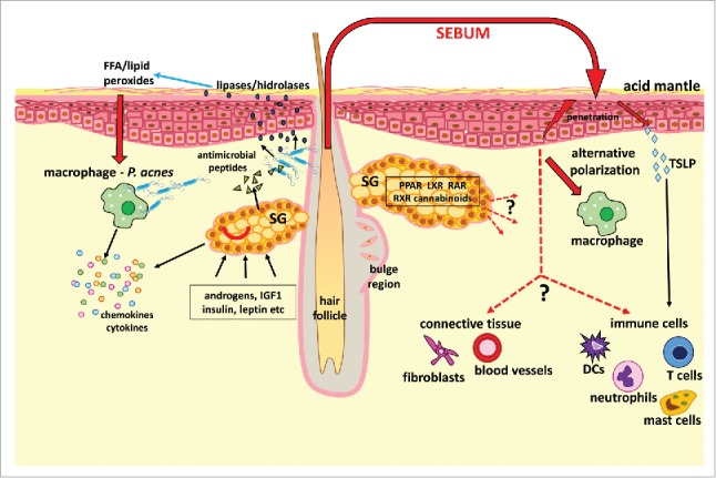
“Sebaceous gland lipids have been shown to exhibit pro-and anti-inflammatory properties, attributed with antimicrobial qualities and most recently they have been demonstrated to exert a modulating role on immune cells (Fig. 1).”
§ Sebaceous-immunobiology is orchestrated by sebum lipids > Conclusion
boosteddirect- Asserted: passes — The authors put it forward as true.
- Checkable: passes — It can be checked against evidence.
- Load-bearing: passes — The paper’s conclusions depend on it.
- Based: passes — The paper gives a reason why it holds.
It may state two separate claims as one.
The authors state this as an assumption.
The altered sebaceous fatty acid profile is an important contributor to skin barrier dysfunction leading to rosacea.
“These lipids have a role in maintaining the skin barrier integrity, therefore it is reasonable to assume that the altered sebaceous fatty acid profile is an important contributor to skin barrier dysfunction leading to rosacea.33”
The paper relies on this without support.
Atopic dermatitis patients have severely reduced sebum production.
“Although AD is mainly characterized by the dysfunctional synthesis of ceramides in keratinocytes39, AD patients seem to exert a severely reduced sebum production, which should contribute to diminished skin hydration.”
Citations
Cites
Alternative activation of macrophages: mechanism and functions
Gordon, S., & Martinez, F. O. (2010) · Immunity · doi:10.1016/j.immuni.2010.05.007 · Cited by 4,197 · cited 1×
“The sebum lipids linoleic and oleic acid were detected to be potent inducers of the so-called alternative macrophage activation, which is the characteristic activation pathway for macrophages involved in tissue homeostasis and repair functions.(64) Furthermore, these lipids also enhanced the ability of macrophages to…”
BackgroundGrice, E. A., & Segre, J. A. (2011) · Nature Reviews Microbiology · doi:10.1038/nrmicro2537 · Cited by 3,143
Kliewer, S. A., Lenhard, J. M., Willson, T. M., Patel, I., Morris, D. C., & Lehmann, J. M. (1995) · Cell · doi:10.1016/0092-8674(95)90194-9 · Cited by 2,028 · cited 2×
“Furthermore, PPAR-a seems to be related to b-oxidation of fatty acids and lipid catabolism.73 Interestingly, P. acnes and its soluble factors may also participate in the augmentation of sebaceous lipogenesis by increasing the production of the 15-deoxyD12,14-prostaglandin J2 (15d-PGJ2), an endogenous activator of PPAR…”
BackgroundSchweiger, D., Baufeld, C., Drescher, P., Oltrogge, B., Höpfner, S., Mess, A., Lüttke, J., Rippke, F., Filbry, A. W., & Max, H. (2013) · Skin Pharmacology and Physiology · doi:10.1159/000348473 · Cited by 1,349
The pH of the Skin Surface and Its Impact on the Barrier Function
Schmid-Wendtner, M.-H., & Korting, H. C. (2006) · Skin Pharmacology and Physiology · doi:10.1159/000094670 · Cited by 860
Peroxisome proliferator-activated receptors in inflammation control
Delerive, P., Fruchart, J., & Staels, B. (2001) · Journal of Endocrinology · doi:10.1677/joe.0.1690453 · Cited by 755
Antimicrobial psoriasin (S100A7) protects human skin from Escherichia coli infection
Gläser, R., Harder, J., Lange, H., Bartels, J., Christophers, E., & Schröder, J.-M. (2005) · Nature Immunology · doi:10.1038/ni1142 · Cited by 651
Nicolaides, N. (1974) · Science · doi:10.1126/science.186.4158.19 · Cited by 609 · cited 1×
“monounsaturated fatty acid with a single double bond at the sixth position.(12,13) The elongation and further desaturation of sapienic acid yields another unique sebaceous gland fatty acid, sebaleic acid (18:2D5, 8).”
BackgroundNew developments in our understanding of acne pathogenesis and treatment
Kurokawa, I., Danby, F. W., Ju, Q., Wang, X., Xiang, L. F., Xia, L., Chen, W., Nagy, I., Picardo, M., Suh, D. H., et al. (2009) · Experimental Dermatology · doi:10.1111/j.1600-0625.2009.00890.x · Cited by 584
Self-citationAcne and sebaceous gland function
Zouboulis, C. C. (2004) · Clinics in Dermatology · doi:10.1016/j.clindermatol.2004.03.004 · Cited by 561
Self-citation
Cited by
Sorted by citations
Items 101–151 of 151
Demodex Mites within Nevus Sebaceus Lesion of an Immune-Competent 7-Month-Old Infant
Lee, H., Park, H., Yoon, H., et al. (2019) · Annals of Dermatology · doi:10.5021/ad.2019.31.s.s34 · Cited by 2
Klas, K., Copic, D., Direder, M., Laggner, M., Prucksamas, P. S., Грубер, Ф., Ankersmit, H. J., & Mildner, M. (2021) · bioRxiv (Cold Spring Harbor Laboratory) · doi:10.1101/2021.10.07.463171 · Cited by 2
Walton‐Doyle, C., Sinclair, E., Begum, H., Hollywood, K. A., Trivedi, D. K., & Barran, P. E. (2023) · ChemRxiv · doi:10.26434/chemrxiv-2023-wxv2b · Cited by 2
M2 Macrophages in the Integument and in the Musculoskeletal System
Rőszer, T. (2020) · Progress in inflammation research · doi:10.1007/978-3-030-50480-9_7 · Cited by 1
Oláh, A., Szöllősi, A. G., & Tóth, B. I. (2024) · Elsevier eBooks · doi:10.1016/b978-0-443-18653-0.00004-6 · Cited by 1
Huang, P., Supan, O., Pak, C., Mehta, R., & Makino, E. T. (2024) · Journal of Cosmetic Dermatology · doi:10.1111/jocd.16568 · Cited by 3
Abidi, S. N. F., Chan, S. M., Seidel, K., et al. (2024) · eLife · doi:10.7554/elife.98747.3 · Cited by 1
Fernandes, B. A. P., da Silva, M. A. N. C. G. M. M., Alves, S. P., Schmidt, V. M., Bizarro, A. F., Pinto, M. S. D. N., Pereira, H. M. L., Marto, J. M., & Lourenço, A. M. (2025) · Veterinary Dermatology · doi:10.1111/vde.13363 · Cited by 1
Tsai, W., Huang, Y., & Fang, F.-M. (2026) · The Journal of Dermatology · doi:10.1111/1346-8138.70146 · Cited by 1
Retzinger, A. C. (2026) · Frontiers in Microbiology · doi:10.3389/fmicb.2026.1714798 · Cited by 1
“Among mammals, sebaceous glands are a key adaptation supporting epidermal barrier function and innate defense (Lovászi et al., 2017; Zouboulis et al., 2022).”
Development and applications of primary human sebocyte-derived sebaceous gland organoids
Tan, S. J., Law, D. C. L., Low, S. J. X., Tan, K. X. K., Lim, X., & Steensel, M. A. M. V. (2026) · Journal of Investigative Dermatology · doi:10.1016/j.jid.2026.04.007 · Cited by 1
Cell Death by Holocrine Secretion: The Final Step of Epithelial Differentiation in Sebaceous Glands
Eckhart, L., Sukseree, S., & Fischer, H. (2026) · Cells · doi:10.3390/cells15121058 · Cited by 1
“The synthesis and interactions of sebum lipids have been reviewed extensively [17,22,45–47].”
Effect of Tripeptide-85 (EVF) on Sebogenesis
Katekawa, E., Arruda Brasil, M. C. O., Princival, C. R., Padovani, G., Esteves-Pedro, N. M., Mussi, L., Camargo Junior, F. B., Magalhães, W. V., Guido, R. V. C., & Cilli, E. M. (2024) · Clinical & Experimental Dermatology and Therapies · doi:10.29011/2575-8268.100223 · Cited by 1
“However, a clinical trial has already been reported where a 5α reductase inhibitor alone was proved ineffective in the treatment of acne vulgaris [9].”
BackgroundChronological maturation of the skin immune barrier is topographically different
Kapitány, A., Soltész, L., Stercel, V., Szabó, L., Somogyi, O., Janka, E. A., Nagy, V., Póliska, S., Gáspár, K., Hendrik, Z., et al. (2025) · Mucosal Immunology · doi:10.1016/j.mucimm.2025.03.004 · Cited by 1
Self-citationClascoterone 1% cream in acne vulgaris: a profile of its use
Kim, E., & Keam, S. J. (2021) · Drugs & Therapy Perspectives · doi:10.1007/s40267-021-00831-4 · Cited by 1
Commentary on ‘Sebocyte differentiation as a new target for acne therapy: an in vivo experience’
Kemény, L. V., & Szabó, K. (2020) · Journal of the European Academy of Dermatology and Venereology · doi:10.1111/jdv.16788 · Cited by 1
Sarkar, D., Sinclair, E., Lim, S. H., et al. (2021) · doi:10.33774/chemrxiv-2021-vsjwj-v2 · Cited by 1
Sarkar, D., Trivedi, D. K., Sinclair, E., et al. (2020) · ChemRxiv · doi:10.26434/chemrxiv.12517385.v1 · Cited by 1
Comparison of canine and human immune system response to DemodicosisPreprint
Nooraei, S., & Mohseni, Z. (2024) · doi:10.22541/au.171919493.36798843/v1 · Cited by 1
ابراهیم, ا. (2020) · حولية الاتحاد العام للآثاريين العرب دراسات فى آثار الوطن العربى · doi:10.21608/cguaa.2020.23822.1029
Tissue repair and stress responses of aged skin epidermis
Wong, W. (2020) · Digital Repository Service (Northeastern University) · doi:10.17760/d20383670
Orbell, H. L., Cave, N., Parry, K., et al. (2020) · doi:10.21203/rs.3.rs-16633/v1
“The surface lipids, and in particular the sebaceous lipids, are also generating renewed interest as it emerges that, in addition to their role in barrier function, they may also have antimicrobial effects, they have pro- and anti-inflammatory properties, and also have a role in modulating immune cells [16, 18].”
BackgroundGordon, A. (2021) · doi:10.14293/s2199-1006.1.sor-.ppmhscs.v1
“[24][25][26][27][28][29][30][31][32][33][34][35] [14] [36][37] [38] [37][39][39][40][41][42][43][44] [45] [46] [46] [47] [48] [49]”
Dull, K., Deák, D., Fazekas, F., Kovács, D., Póliska, S., Szegedi, A., Zouboulis, C. C., & Törőcsik, D. (2020) · Research Square · doi:10.21203/rs.3.rs-125647/v1
Self-citationEtiologija i patogeneza vulgarne akne
Kovačević, M., & Massari, L. P. (2021) · Medicina Fluminensis · doi:10.21860/medflum2021_261185
Lipid Profile Disturbance in Acne Vulgaris of Iraqi patients
Hamood, A. A., & Mousa, H. M. (2022) · University of Thi-Qar Journal of Science · doi:10.32792/utq/utjsci.v9i1.882
Sebaceous-immunobiology in disease pathogenesis
Törőcsik, D., Dull, K., Fedor-Lénárt, K., Szegedi, A., & Kovács, D. (2023) · Bőrgyógyászati és Venerológiai Szemle · doi:10.7188/bvsz.2023.99.4.9
Self-citation100 ÉVES A DEBRECENI EGYETEM BŐRGYÓGYÁSZATI KLINIKÁJA
Szegedi, A. (2023) · Gerundium · doi:10.29116/gerundium/2023/3-4/6
Self-citationDermatoloji ve Kozmetolojide D Vitamini
Cenk, H., Rüstemoğlu, A., Akbulut, S., Uzdoğan, A., Yücel, Ç., Çifci, A., Yakut, H. İ., Oğuz, I. D., Türkmen, D., Demirbaş, A., et al. (2022) · Akademisyen Kitabevi eBooks · doi:10.37609/akya.1313
Hypothesis for COVID-19 Vaccine Detoxification
Pachanki, Y. (2023) · Associative Journal of Health Sciences · doi:10.31031/ajhs.2023.02.000544
Abidi, S. N. F., Chan, S., Seidel, K., et al. (2024) · doi:10.7554/elife.98747.1.sa1
Abidi, S. N. F., Chan, S., Seidel, K., et al. (2024) · doi:10.7554/elife.98747.1.sa0
Abidi, S. N. F., Chan, S., Seidel, K., et al. (2024) · doi:10.7554/elife.98747.1.sa2
Abidi, S. N. F., Chan, S. M., Seidel, K., et al. (2024) · eLife · doi:10.7554/elife.98747.1
Abidi, S. N. F., Chan, S. M., Seidel, K., et al. (2024) · eLife · doi:10.7554/elife.98747.2
(2000) · Time to knit · doi:10.1016/s0246-0319(24)44096-x
Secreciones sudorípara y sebácea
Aubert‐Pouëssel, A. (2025) · EMC - Dermatología · doi:10.1016/s1761-2896(24)50023-1
Goldschmidt‐Clermont, P. J., Montenegro, R., Rojas, K., et al. (2025) · Annals of Case Reports · doi:10.29011/2574-7754.102316
Role of a sebum-regulating complex in correcting epidermal barrier parameters in patients with acne
Zaslavsky, D. V., Таганов, А. В., Козлова, Д. В., et al. (2025) · Russian Journal of Skin and Venereal Diseases · doi:10.17816/dv642587
Characterization of double bond position in free fatty acids of human sebum
Telle, A., Mastromatteo, G., Maiellaro, M., Sciuto, L., Bottillo, G., & Camera, E. (2025) · Journal of Chromatography Open · doi:10.1016/j.jcoa.2025.100265
Chen, L., Xu, Y., Yang, S., Chen, N., Gao, J., & Lin, H. (2026) · Asian Journal of Pharmaceutical Sciences · doi:10.1016/j.ajps.2026.101163
“Moreover, skin as an immune organ, contains a dense network of immune cells and lymphatic vessels [ 40–42 ].”
Törőcsik, D., Tiszai, D., & Zouboulis, C. C. (2026) · Expert Opinion on Investigational Drugs · doi:10.1080/13543784.2026.2701814
Self-citationFrom sebocytes to skin engineering human sebaceous organoids within a reconstructed dermal matrix
Pusceddu, T., Ardondi, L., Cavaleri, M. P., et al. (2026) · Frontiers in Toxicology · doi:10.3389/ftox.2026.1874806
“…for the production of sebum, a lipid-rich substance that contributes to multiple cutaneous functions, including waterproofing, antimicrobial defense, immune modulation, and the maintenance of epidermal homeostasis (Pappas, 2009; Lovászi et al., 2017; Zouboulis et al., 2022; Mosca et al., 2025).”
Extraction and Quantification of Skin Immune Cells and Skin Lipids
Choa, R., Sun, L., & Kambayashi, T. (2026) · Methods in molecular biology · doi:10.1007/978-1-0716-5376-0_47
Na, H.-W., Cha, N., Kim, E., Kim, S., Park, W.-S., & Kim, H.-J. (2026) · Journal of Toxicology · doi:10.1155/jt/7925949
Briganti, S., Mosca, S., Ottaviani, M., & Flori, E. (2026) · Cells · doi:10.3390/cells15171591
“SGs contribute to skin barrier function through sebum production, which shapes microbial composition and modulates immune responses, while immune cells in turn regulate SG activity, establishing a bidirectional crosstalk essential for tissue homeostasis [124–126].”
Modulation of sebocyte function in acne: A pathway‐inclusive review
Zouboulis, C. C., Pappas, A., Martin, K., & Picardo, M. (2026) · Journal of the European Academy of Dermatology and Venereology · doi:10.1111/jdv.70752
Self-citationLi, Y., Qiu, Z., Pan, X., Xu, G., Peng, S., Zhu, R., Luo, Q., Fang, X., Yao, X., & Li, W. (2026) · Advanced Science · doi:10.1002/advs.78048
“Consequently, age-associated dysfunction of this niche may have implications extending beyond hair biology, potentially contributing to clinically relevant skin-aging phenotypes including xerosis [ 54 ], impaired wound healing, microbial imbalance, and chronic low-grade inflammation [ 55 ].”
Metadata
- Authors
- Lovászi, M., Szegedi, A., Zouboulis, C. C., & Törőcsik, D.
- Affiliations
- University of Debrecen (HU)
- Medizinische Hochschule Brandenburg Theodor Fontane (DE)
- Corresponding author
- Törőcsik, Dániel
- Venue
- Dermato-Endocrinology
- Publisher
- Informa UK Limited
- Published
- 2017-01-01
- Volume
- 9
- Issue
- 1
- Pages
- e1375636-e1375636
- ISSN
- 1938-1972 (ISSN-L)
- 1938-1980
- PMC
- PMC5821166
- OpenAlex
- W2762894008
- Semantic Scholar
- bdddc896532e6450af27244d27cd660c49a516f1
- Cited by
- 151
- References
- 93
- Citations by year
- 2026: 14 · 2025: 14 · 2024: 28 · 2023: 28 · 2022: 18 · 2021: 26 · 2020: 11 · 2019: 6 · 2018: 4
- FWCI
- 5.73
- Type
- journal article
- Language
- en
- Open access
- diamond
- doi.org
- Licence
- Locations
- Dermato-Endocrinology (publishedVersion, cc-by-nc)
- PubMed (publishedVersion)
- University of Debrecen Electronic Archive (University of Debrecen) (submittedVersion, cc-by-nc)
- Europe PMC (PubMed Central) (submittedVersion, cc-by-nc)
- PubMed Central (submittedVersion, cc-by-nc)
- University of Debrecen Electronic Archive (University of Debrecen) (submittedVersion)
- University of Debrecen Electronic Archive (University of Debrecen) (submittedVersion, other-oa)
- Funders
- Hungarian National Research Found (OTKA NN117020 NKFIH K108421)
- New National Excellence Program of the Ministry of Human Capacities (GINOP-2.3.2-15-2016-00050, GINOP-2.3.2-15-2016-00005)
- Topics
- Health Sciences › Medicine › Dermatology › Dermatology and Skin Diseases
- Health Sciences › Medicine › Dermatology › Acne and Rosacea Treatments and Effects
- Life Sciences › Biochemistry, Genetics and Molecular Biology › Biochemistry › Lipid metabolism and biosynthesis
- Keywords
- sebaceous glands, sebum lipids, macrophages, keratinocytes, skin disease pathogenesis
About these checks
Registry, retraction, Cites and statistics results are computed, not judged, with one exception: a printed reference that no index lists is read once by a model before it counts as a citation error. Claims are extracted by a model and kept only when their quote appears verbatim in the text; each cited source is then checked, with a model’s help, for whether it says what the claim relies on. Those verdicts can be wrong — read the claim’s quote and the cited source before relying on one.
A language model drew the claims from the paper on 2026-10-22, under version 3 of the claim definition (model deepseek-v4.1-flash, prompt extract-v3).
A second model labelled each claim on 2026-10-22 (model jev-1.13-20260917, definition revision gold-guideline-v0.2).
Cited sources are checked with a language model’s help (gpt-6-sol).
Statistics and arithmetic are recomputed, not judged.
Confidence bands are shown only for fields calibrated on our annotated test set. None is calibrated yet, so no band is shown.
What counts as a claim ›How we measure confidence ›How we check this ›
Are you an author, or do you see a problem with this page? Request a review
