

The DOI matches its Crossref record; 365 of the 367 references it cites were found in a registry or an index; 85 of the 158 sources its claims cite were checked for support.
Functional genomic screen and network analysis reveal novel modifiers of tauopathy dissociated from tau phosphorylation
Open accessAbstract
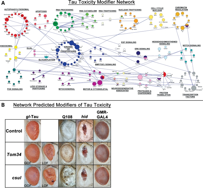
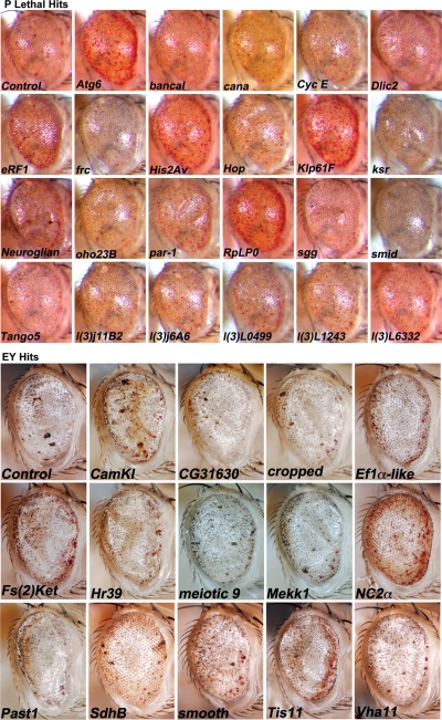
A functional genetic screen using loss-of-function and gain-of-function alleles was performed to identify modifiers of tau-induced neurotoxicity using the 2N/4R (full-length) isoform of wild-type human tau expressed in the fly retina. We previously reported eye pigment mutations, which create dysfunctional lysosomes, as potent modifiers; here, we report 37 additional genes identified from ∼1900 genes screened, including the kinases shaggy/GSK-3beta, par-1/MARK, CamKI and Mekk1. Tau acts synergistically with Mekk1 and p38 to down-regulate extracellular regulated kinase activity, with a corresponding decrease in AT8 immunoreactivity (pS202/T205), suggesting that tau can participate in signaling pathways to regulate its own kinases. Modifiers showed poor correlation with tau phosphorylation (using the AT8, 12E8 and AT270 epitopes); moreover, tested suppressors of wild-type tau were equally effective in suppressing toxicity of a phosphorylation-resistant S11A tau construct, demonstrating that changes in tau phosphorylation state are not required to suppress or enhance its toxicity. Genes related to autophagy, the cell cycle, RNA-associated proteins and chromatin-binding proteins constitute a large percentage of identified modifiers. Other functional categories identified include mitochondrial proteins, lipid trafficking, Golgi proteins, kinesins and dynein and the Hsp70/Hsp90-organizing protein (Hop). Network analysis uncovered several other genes highly associated with the functional modifiers, including genes related to the PI3K, Notch, BMP/TGF-β and Hedgehog pathways, and nuclear trafficking. Activity of GSK-3β is strongly upregulated due to TDP-43 expression, and reduced GSK-3β dosage is also a common suppressor of Aβ42 and TDP-43 toxicity. These findings suggest therapeutic targets other than mitigation of tau phosphorylation.
Overview
AI snapshot
Written from the paper’s full text.
TL;DR
Two Drosophila enhancer–suppressor screens identified 37 modifiers of wild-type human tau toxicity, with 40 total modifiers including previously reported pigment genes. The results dissociate tau toxicity from tau phosphorylation and implicate GSK-3β, ERK/p38 signaling, autophagy, RNA-associated processes, and other pathways as therapeutic targets.
Key findings
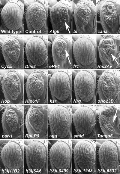
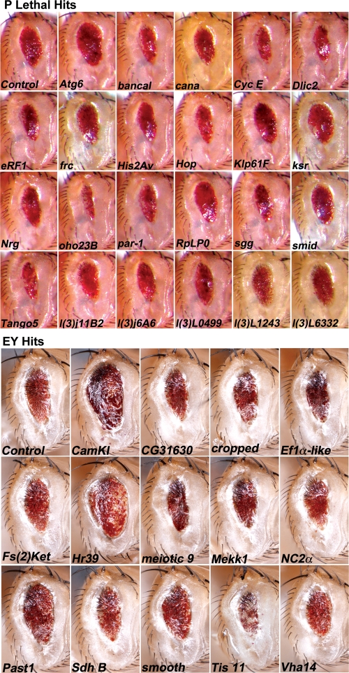
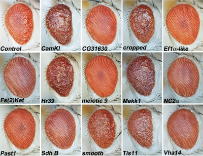
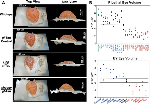
- From ∼1900 lines screened, 37 genes were identified: 23 modifiers from the P lethal LOF screen (11 suppressors, 12 enhancers) and 14 from the EY screen; adding white, brown, and rosy gave 40 total modifiers.
- Tau phosphorylation did not correlate with toxicity: 18 of 40 modifiers showed no significant AT8 difference, 22 of 40 showed significant changes but with no suppressor/enhancer pattern; suppressors of wild-type tau also suppressed the phosphorylation-resistant S11A isoform, while the non-toxic S2A isoform was resistant to all enhancers.
- Tau synergized with Mekk1/p38 to reduce ERK activity: co-expression of tau and Mekk1 produced a profound reduction (∼60%) in phospho-ERK, while Mekk1 overexpression increased p38 activity and decreased AT8.
- GSK-3β/shaggy was a common suppressor of tau, Aβ42, and TDP-43 toxicity; TDP-43Q331K strongly increased GSK-3β activity, indicated by reduced pGSK-3β[Ser9] (P = 0.004).
Methods
- The gl-tau Drosophila eye model expressed wild-type full-length 2N/4R human tau; the P lethal collection provided LOF alleles (920 stocks screened) and the EY collection provided overexpression/insertion alleles (895 annotated stocks screened), with blinded phenotypic scoring and validation with additional alleles where possible.
- Eye phenotypes were scored by roughness, necrosis, and eye volume using SEM, light microscopy, and 3D reconstructions; immunoblots measured total tau, phospho-tau (AT8, 12E8, AT270), phospho-p38, phospho-ERK, and phospho-GSK-3β[Ser9], with genetic tests against Q108, Aβ42, TDP-43Q331K, S11A, and S2A.
- Computational network analysis used Endeavor-HighFly on 35 annotated modifiers, compiled genes with P < 0.05, applied inclusion criteria based on recurrence across hit networks, and visualized the network with Cytoscape; human orthologs were assigned using PANTHER.
Limitations
- Stated in the paper: the screen was not saturating, covering less than 20% of the ∼12,000 fly genes; it was selective for dominant/haploinsufficiency effects and essential genes in the P lethal screen, and subtle modifiers may have been excluded by the robust-phenotype criterion.
- Stated in the paper: the eye/retina may not be the ideal tissue to observe effects of some genes, and the computational network neither represents a full genomic network nor includes all demonstrated or putative modifiers.
- Evident from the text: only 895 annotated EY stocks were screened, 17 of 40 modifier genes had only a single allele tested, and several modifiers were non-annotated; phosphorylation analysis was limited to selected epitopes, and many functional categories were not mechanistically resolved.
Model-written summary. Check it against the paper before relying on it.
Integrity signals
Registry
Cites
Statistics
Figures
Verification
Claims the paper makes
116 Own result · 79 Own interpretation · 26 Partly supported · 20 Supported · 18 No full text of the sources · 12 No basis given · 11 Own method · 7 Supported so far · 3 Couldn’t confirm · 2 Not supported · 2 Not checked yet · 1 Mixed support · accuracy not yet measured
Every source the paper cites for this claim was checked and supports it.
In the paper
“We incorporated genes white, brown and rosy into the network, which we identified as modifiers independent of the collections we screened (61).”
§ Functional genomic screen and network analysis reveal novel modifiers of tauopathy dissociated from tau phosphorylation > RESULTS > Computational network demonstrates wide range of functional categories
What the sources say
- Supportedcites [61] Ambegaokar, 2010, Interaction between eye pigment genes and tau-induced neurodegeneration in Drosophila melanogaster, Genetics source
The Abstract identifies white, brown, and rosy as genes that affect the tau-induced eye phenotype. The paper also describes testing these pigment genes after observing an effect during a P-element modifier screen. The reference to 'collections we screened' describes the citing authors’ own collections, not an additional finding attributed to this source. (LLM, conf=0.90)
plaindirect- Asserted: passes — The authors put it forward as true.
- Checkable: passes — It can be checked against evidence.
- Load-bearing: unclear — It is unclear whether the paper’s conclusions depend on it.
- Based: passes — The paper gives a reason why it holds.
- Supported
The cited sources support only part of this claim — read each source’s verdict below.
In the paper
“in vivo fly models of tau toxicity using phosphorylation-resistant tau constructs show equivalent or even increased toxicity when compared with wild-type tau (41,42).”
§ Functional genomic screen and network analysis reveal novel modifiers of tauopathy dissociated from tau phosphorylation > INTRODUCTION
What the sources say
- Partly supportedcites [41] Steinhilb, 2007, Tau phosphorylation sites work in concert to promote neurotoxicity in vivo, Mol. Biol. Cell sourceExperimental — accuracy not yet measured
The Results and Discussion report that fly tau constructs resistant to phosphorylation at individual sites, or at a selected group of five sites, caused toxicity similar to wild-type tau. They do not report increased toxicity for those constructs; the Results instead associate greater toxicity with a phosphomimetic construct and markedly reduced toxicity with mutation of all 14 SP/TP sites. Page numbers were not available in our copy. (LLM, conf=0.89)
- Supportedcites [42] Chatterjee, 2009, Dissociation of tau toxicity and phosphorylation: role of GSK-3βeta, MARK and Cdk5 in a Drosophila model, Hum. Mol. Genet. source
The Discussion section on S11A reports that this phosphorylation-resistant construct produced an external-eye phenotype as severe as wild-type tau in several fly-model comparisons. The paper does not report increased toxicity for S11A, and its Results find that a different resistant construct, S2A, was less toxic; the cited source supports the claim’s ‘equivalent’ alternative, not a generalization to all such constructs. (LLM, conf=0.86)
plain- Asserted: passes — The authors put it forward as true.
- Checkable: passes — It can be checked against evidence.
- Load-bearing: passes — The paper’s conclusions depend on it.
- Based: passes — The paper gives a reason why it holds.
- Partly supported
Every source the paper cites for this claim was checked and supports it.
In the paper
“Indeed, mature protein substrates of Tom70 were observed to be aggregate-prone in the absence of Tom70, suggesting a crucial role of Tom70 in preventing aggregation (69).”
§ Functional genomic screen and network analysis reveal novel modifiers of tauopathy dissociated from tau phosphorylation > RESULTS > Computational network demonstrates wide range of functional categories
What the sources say
- Supportedcites [69] Yamamoto, 2009, Roles of Tom70 in import of presequence-containing mitochondrial proteins, J. Biol. Chem. source
The Results (Fig. 7; page not provided) report that mature domains of Tom70-dependent proteins tend to aggregate and become more soluble when the cytosolic domain of Tom70 is added. The Discussion likewise states that their insoluble aggregates were suppressed by Tom70’s receptor domain. The experiment used a cell-free system lacking chaperones, rather than observing aggregation in Tom70-deleted cells. (LLM, conf=0.93)
plaindirect- Asserted: passes — The authors put it forward as true.
- Checkable: passes — It can be checked against evidence.
- Load-bearing: unclear — It is unclear whether the paper’s conclusions depend on it.
- Based: passes — The paper gives a reason why it holds.
- Supported
The cited sources that could be checked couldn’t confirm or dispute this claim.
In the paper
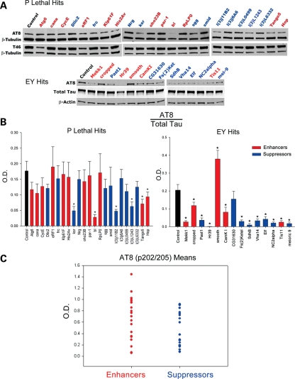
“No significant differences in phospho-tau levels relative to controls were observed (Supplementary Material, Fig. S4).”
§ Functional genomic screen and network analysis reveal novel modifiers of tauopathy dissociated from tau phosphorylation > RESULTS > Phosphorylation of tau does not correlate with enhancement or suppression of tau toxicity
What the sources say
- Could not verifycites [85] Seubert, 1995, Detection of phosphorylated Ser262 in fetal tau, adult tau, and paired helical filament tau, J. Biol. Chem. sourceExperimental — accuracy not yet measured
The immunoblot-analysis and 12E8 results passages describe tau phosphorylation and the 12E8 antibody, but do not report an EY modifier screen or a comparison with its controls. Searches for EY-screen modifiers and AT270 found no passage addressing the claimed absence of significant differences. Page numbers were not available in our copy. (LLM, conf=0.76)
- Could not verifycites [86] Goedert, 1994, Epitope mapping of monoclonal antibodies to the paired helical filaments of Alzheimer's disease: identification of phosphorylation sites in tau protein, Biochem. J. sourceExperimental — accuracy not yet measured
The indexed Abstract characterizes AT270 and reports phosphorylation in fetal, adult, and paired-helical-filament tau. It does not address an EY screen, 12E8, or statistical comparisons of phospho-tau levels with controls; searches for those terms found no relevant passage. The cited paper therefore cannot verify the reported lack of a significant difference. (LLM, conf=0.76)
plain- Asserted: passes — The authors put it forward as true.
- Checkable: passes — It can be checked against evidence.
- Load-bearing: passes — The paper’s conclusions depend on it.
- Based: passes — The paper gives a reason why it holds.
- Could not verify
The cited sources support only part of this claim — read each source’s verdict below.
In the paper
“Tau phosphorylation at S202/T205 as detected by the AT8 antibody (80) is commonly found in tauopathies (38,81–84).”
§ Functional genomic screen and network analysis reveal novel modifiers of tauopathy dissociated from tau phosphorylation > RESULTS > Phosphorylation of tau does not correlate with enhancement or suppression of tau toxicity
What the sources say
- Partly supportedcites [80] Biernat, 1992, The switch of tau protein to an Alzheimer-like state includes the phosphorylation of two serine-proline motifs upstream of the microtubule binding region, EMBO J. sourceExperimental — accuracy not yet measured
The indexed Abstract identifies AT8 as a phosphorylation-dependent antibody whose epitope is around residue 200, supporting the AT8 identification cited as (80). It specifies phosphorylation of serines 199 and/or 202, however, not T205. The separate claim that this phosphorylation is common in tauopathies is attached to other citations, not (80). (LLM, conf=0.88)
- Not checked — no full text of the sourcecites [38] Wada, 1998, Microtubule-stimulated phosphorylation of tau at Ser202 and Thr205 by Cdk5 decreases its microtubule nucleation activity, J. Biochem. source
- Not checked — no full text of the sourcecites [81] Wray, 2008, Direct analysis of tau from PSP brain identifies new phosphorylation sites and a major fragment of N-terminally cleaved tau containing four microtubule-binding repeats, J. Neurochem. source
- Partly supportedcites [82] Ferrer, 2003, Phosphorylated protein kinases associated with neuronal and glial tau deposits in argyrophilic grain disease, Brain Pathol. sourceExperimental — accuracy not yet measured
The Abstract reports using a Ser202 phosphospecific antibody to examine tau in argyrophilic grain disease and finding hyperphosphorylated tau in all cases. It does not establish that S202/T205 phosphorylation is commonly found across tauopathies. The AT8 antibody identification is attached to a separate citation in the citing sentence. (LLM, conf=0.81)
- Not checked — no full text of the sourcecites [83] Ikura, 1998, Levels of tau phosphorylation at different sites in Alzheimer disease brain, Neuroreport source
- Partly supportedcites [84] Han, 2009, Familial FTDP-17 missense mutations inhibit microtubule assembly-promoting activity of tau by increasing phosphorylation at Ser202 in vitro, J. Biol. Chem. sourceExperimental — accuracy not yet measured
The Discussion (page unavailable) reports strong AT8 staining in Alzheimer’s disease and Ser202 phosphorylation recognized by AT8 in FTDP-17 brains. This supports AT8-detected Ser202 phosphorylation in those conditions, but the copy does not establish Thr205 phosphorylation or its prevalence across tauopathies. (LLM, conf=0.86)
plain- Asserted: passes — The authors put it forward as true.
- Checkable: passes — It can be checked against evidence.
- Load-bearing: passes — The paper’s conclusions depend on it.
- Based: passes — The paper gives a reason why it holds.
- Partly supported
The cited sources support only part of this claim — read each source’s verdict below.
In the paper
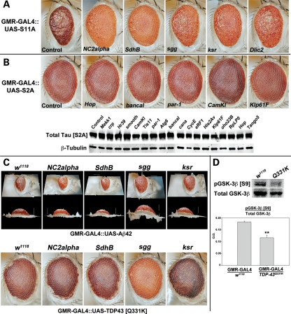
“Similar to tau and to polyglutamine proteins, expression of Aβ42 and TDP-43Q331K in the fly eye driven by GMR-GAL4 leads to eye phenotypes.”
§ Functional genomic screen and network analysis reveal novel modifiers of tauopathy dissociated from tau phosphorylation > RESULTS > Tau demonstrates synergy with Mekk1 and p38 to decrease ERK activity
What the sources say
- Partly supportedcites [89] Casas-Tinto, 2011, The ER stress factor XBP1s prevents amyloid-beta neurotoxicity, Hum. Mol. Genet. sourceExperimental — accuracy not yet measured
The “Generation of a new AD model in flies” section reports that Aβ42 expression with gmr-Gal4 produced small, disorganized eyes with necrotic spots. The cited paper does not report an eye phenotype for TDP-43Q331K, so it supports only the Aβ42 part of the claim. No page number is available in our copy. (LLM, conf=0.88)
plaindirect- Asserted: passes — The authors put it forward as true.
- Checkable: passes — It can be checked against evidence.
- Load-bearing: passes — The paper’s conclusions depend on it.
- Based: passes — The paper gives a reason why it holds.
- Rests on an assumption: TDP-43Q331K is an amyotrophic lateral sclerosis-causing mutation.
- Partly supported
The cited sources support only part of this claim — read each source’s verdict below.
In the paper
“Instead of alleviating toxicity, S11A demonstrates severe toxicity when expressed in the eye, producing an even smaller and rougher eye than wild-type tau (42).”
§ Functional genomic screen and network analysis reveal novel modifiers of tauopathy dissociated from tau phosphorylation > RESULTS > Tau demonstrates synergy with Mekk1 and p38 to decrease ERK activity
What the sources say
- Partly supportedcites [42] Chatterjee, 2009, Dissociation of tau toxicity and phosphorylation: role of GSK-3βeta, MARK and Cdk5 in a Drosophila model, Hum. Mol. Genet. sourceExperimental — accuracy not yet measured
The Results section describes S11A-expressing flies as having reduced, rough eyes with fused ommatidia and missing bristles. The Discussion says the S11A eye phenotype was, in several instances, as severe as that of wild-type tau; it does not support the claim that S11A produced an even smaller and rougher eye than wild-type tau. Page numbers were not available in our copy. (LLM, conf=0.89)
boosteddirect- Asserted: passes — The authors put it forward as true.
- Checkable: passes — It can be checked against evidence.
- Load-bearing: passes — The paper’s conclusions depend on it.
- Based: passes — The paper gives a reason why it holds.
- Rests on an assumption: The sites mutated in S11A are commonly hyperphosphorylated in tauopathic tissue and are known to be phosphorylated by GSK-3β and CDK-5.
- Partly supported
The cited sources support only part of this claim — read each source’s verdict below.
In the paper
“GSK-3β has not been identified as a kinase of TDP-43, even when specifically tested for TDP-43 phosphorylation (102)”
§ Functional genomic screen and network analysis reveal novel modifiers of tauopathy dissociated from tau phosphorylation > DISCUSSION > Kinases and dissociation of tau phosphorylation from tau toxicity
What the sources say
- Partly supportedcites [102] Hasegawa, 2008, Phosphorylated TDP-43 in frontotemporal lobar degeneration and amyotrophic lateral sclerosis, Ann. Neurol. sourceExperimental — accuracy not yet measured
The Methods describe an in vitro test of GSK3β on TDP-43. In the Results section, TDP-43 treated with GSK3β was negative with the study’s phosphorylation-specific antibodies. That supports a negative result for the epitopes tested, but not the broader assertion that GSK3β has not been identified as a TDP-43 kinase at any site. (LLM, conf=0.85)
plain- Asserted: passes — The authors put it forward as true.
- Checkable: passes — It can be checked against evidence.
- Load-bearing: passes — The paper’s conclusions depend on it.
- Based: passes — The paper gives a reason why it holds.
- Rests on an assumption: TDP-43Q331K is an amyotrophic lateral sclerosis-causing mutation.
- Partly supported
The cited sources support only part of this claim — read each source’s verdict below.
In the paper
“many investigators have shown that either inhibition or genetic reduction of GSK-3β ameliorates tau-related deficits (94–99).”
§ Functional genomic screen and network analysis reveal novel modifiers of tauopathy dissociated from tau phosphorylation > DISCUSSION > Kinases and dissociation of tau phosphorylation from tau toxicity
What the sources say
- Not checked — no full text of the sourcecites [94] Mudher, 2004, GSK-3beta inhibition reverses axonal transport defects and behavioural phenotypes in Drosophila, Mol. Psychiatry source
- Partly supportedcites [95] Noble, 2005, Inhibition of glycogen synthase kinase-3 by lithium correlates with reduced tauopathy and degeneration in vivo, Proc. Natl Acad. Sci. USA sourceExperimental — accuracy not yet measured
The Results and Discussion (page numbers unavailable) report that lithium reduced GSK-3β activity and that treatment with GSK-3 inhibitors was associated with less aggregated tau and degeneration in mice. The paper does not report genetic reduction of GSK-3β, so it supports the inhibition part of the claim but not the genetic-reduction part. (LLM, conf=0.87)
- Not checked — no full text of the sourcecites [96] Miyasaka, 2005, Progressive neurodegeneration in C. elegans model of tauopathy, Neurobiol. Dis. source
- Not checked — no full text of the sourcecites [97] Kraemer, 2006, Molecular pathways that influence human tau-induced pathology in Caenorhabditis elegans, Hum. Mol. Genet. source
- Partly supportedcites [98] Blard, 2007, Cytoskeleton proteins are modulators of mutant tau-induced neurodegeneration in Drosophila, Hum. Mol. Genet. sourceExperimental — accuracy not yet measured
The References (ref. 13) list a study reporting that GSK-3β inhibition reverses axonal-transport defects and behavioural phenotypes in Drosophila, supporting the inhibition part of the claim. But this paper’s Discussion (p. 565–566) says its screen did not identify GSK-3β modifiers; it does not report that genetic reduction of GSK-3β ameliorates tau-related deficits. (LLM, conf=0.78)
- Not checked — no full text of the sourcecites [99] Folwell, 2009, Abeta exacerbates the neuronal dysfunction caused by human tau expression in a Drosophila model of Alzheimer's disease, Exp. Neurol. source
boosteddirect- Asserted: passes — The authors put it forward as true.
- Checkable: passes — It can be checked against evidence.
- Load-bearing: unclear — It is unclear whether the paper’s conclusions depend on it.
- Based: passes — The paper gives a reason why it holds.
Every source the paper cites for this claim was checked and supports it.
In the paper
“when either 5 (41) or 11 (42) putative GSK-3β phosphorylation sites were mutated to alanine, tau toxicity either comparable to or more robust than the toxicity induced by wild-type tau was still observed.”
§ Functional genomic screen and network analysis reveal novel modifiers of tauopathy dissociated from tau phosphorylation > DISCUSSION > Kinases and dissociation of tau phosphorylation from tau toxicity
What the sources say
- Supportedcites [41] Steinhilb, 2007, Tau phosphorylation sites work in concert to promote neurotoxicity in vivo, Mol. Biol. Cell source
The Results report that tauAP5, with five phosphorylation sites mutated to alanine, had the same retinal toxicity as wild-type tau in Drosophila (Figure 6B). The five-site finding is the part of the citing sentence attached to citation (41); the 11-site finding is attached to a different citation (42). (LLM, conf=0.92)
- Supportedcites [42] Chatterjee, 2009, Dissociation of tau toxicity and phosphorylation: role of GSK-3βeta, MARK and Cdk5 in a Drosophila model, Hum. Mol. Genet. source
The citation to this paper is attached to the 11-site mutant, not the five-site mutant. In the Discussion section “S11A is resistant to GSK-3β/Shaggy phosphorylation but retains toxicity” (page unavailable), the authors report that S11A’s eye phenotype was, in several instances, as severe as wild-type tau’s despite its resistance to phosphorylation. (LLM, conf=0.91)
plaindirect- Asserted: passes — The authors put it forward as true.
- Checkable: passes — It can be checked against evidence.
- Load-bearing: passes — The paper’s conclusions depend on it.
- Based: passes — The paper gives a reason why it holds.
- Supported
- Probably not a substantial claim. 50
- It may state two separate claims as one. 85
- The quote only partly states this claim. 1
Some cited sources support this claim and some don’t — read each source’s verdict below.
In the paper
“as the CAG repeats in polyglutamine-encoding RNAs can induce toxicity (74,75)”
§ Functional genomic screen and network analysis reveal novel modifiers of tauopathy dissociated from tau phosphorylation > RESULTS > Computational network demonstrates wide range of functional categories
What the sources say
- Supportedcites [74] Li, 2008, RNA toxicity is a component of ataxin-3 degeneration in Drosophila, Nature source
The Abstract reports that interrupting CAG repeats within the polyglutamine-encoding region dramatically mitigated toxicity. It also reports that pathogenic-length CAG-repeat RNA induced neuronal degeneration even without encoding a toxic polyglutamine protein. No page number is supplied in our copy. (LLM, conf=0.93)
- Disputedcites [75] Daughters, 2009, RNA gain-of-function in spinocerebellar ataxia type 8, PLoS Genet. sourceExperimental — accuracy not yet measured
The Results section ('SCA8 CUGexp Not CAGexp Transcripts Misregulate GABT4') reports that CUG-expansion transcripts, but not CAG-expansion transcripts, altered GABT4 expression in the tested cells. The Introduction distinguishes the proposed toxic RNA effect of CUG transcripts from the polyglutamine protein encoded by CAG transcripts. This does not rule out every possible CAG-RNA toxicity, but the paper's finding runs contrary to the claim attributed to it. (LLM, conf=0.76)
Checked by a reasoning model — may be wrong.
plaindirect- Asserted: passes — The authors put it forward as true.
- Checkable: passes — It can be checked against evidence.
- Load-bearing: fails — The paper’s conclusions may not depend on it.
- Based: passes — The paper gives a reason why it holds.
Probably not a substantial claim. The paper’s conclusions may not depend on it.
- Supported
At least one cited source doesn’t support this claim or points to a different paper, and none confirmed it — see the verdicts below.
In the paper
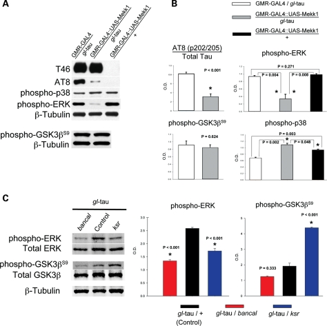
“However, reduced ERK activity has also been shown to enhance tau-induced toxicity (132)”
§ Functional genomic screen and network analysis reveal novel modifiers of tauopathy dissociated from tau phosphorylation > DISCUSSION > Signal transduction and tau signaling properties
What the sources say
- Disputedcites [132] Amadoro, 2006, NMDA receptor mediates tau-induced neurotoxicity by calpain and ERK/MAPK activation, Proc. Natl Acad. Sci. USA sourceExperimental — accuracy not yet measured
In “Tau Induces NMDAR-Dependent Activation of ERK1/2” (page not supplied), the paper reports that inhibiting ERK signaling or genetically removing ERK1 protects neurons from tau-induced cell death. It therefore finds the opposite of the attributed claim: reduced ERK activity lessens, rather than enhances, tau toxicity. (LLM, conf=0.93)
Checked by a reasoning model — may be wrong.
plaindirect- Asserted: passes — The authors put it forward as true.
- Checkable: passes — It can be checked against evidence.
- Load-bearing: fails — The paper’s conclusions may not depend on it.
- Based: passes — The paper gives a reason why it holds.
Probably not a substantial claim. The paper’s conclusions may not depend on it.
- Disputed
At least one cited source doesn’t support this claim or points to a different paper, and none confirmed it — see the verdicts below.
In the paper
“Interestingly, we also observe a strong decrease in GSK-3β activity in white and brown mutants and a strong increased activity in a rosy mutant (61).”
§ Functional genomic screen and network analysis reveal novel modifiers of tauopathy dissociated from tau phosphorylation > DISCUSSION > Autophagy/lysosomal pathways, proteases and chaperones
What the sources say
- Disputedcites [61] Ambegaokar, 2010, Interaction between eye pigment genes and tau-induced neurodegeneration in Drosophila melanogaster, Genetics sourceExperimental — accuracy not yet measured
The Results passage on GSK-3β activation and Figure 4B (page not provided in our copy) say the opposite for white and white-plus-brown flies: inhibitory Ser9 phosphorylation decreases, indicating increased GSK-3β activity. Figure 4B says a rosy mutation restores that inhibitory phosphorylation in this background, not that rosy increases activity. (LLM, conf=0.92)
Checked by a reasoning model — may be wrong.
boosteddirect- Asserted: passes — The authors put it forward as true.
- Checkable: passes — It can be checked against evidence.
- Load-bearing: unclear — It is unclear whether the paper’s conclusions depend on it.
- Based: passes — The paper gives a reason why it holds.
It may state two separate claims as one.
- Disputed
Every source the paper cites for this claim was checked and supports it.
In the paper
“The degree of roughness is intermediate and more pronounced in the anterior eye, making it useful for enhancer–suppressor modifier screens.”
§ Functional genomic screen and network analysis reveal novel modifiers of tauopathy dissociated from tau phosphorylation > INTRODUCTION
What the sources say
- Supportedcites [47] Jackson, 2002, Human wild-type tau interacts with wingless pathway components and produces neurofibrillary pathology in Drosophila, Neuron source
The description of gl-tau eyes accompanying Figure 1 says they are rough in anterior regions while posterior regions appear normal; the methods describe a line with a moderate phenotype. Figures 2 and 5 show that genetic changes can both suppress and enhance the rough-eye phenotype, supporting its usefulness for modifier screens. (LLM, conf=0.85)
plaincorrelational- Asserted: passes — The authors put it forward as true.
- Checkable: passes — It can be checked against evidence.
- Load-bearing: passes — The paper’s conclusions depend on it.
- Based: passes — The paper gives a reason why it holds.
It may state two separate claims as one.
- Supported
Every source the paper cites for this claim was checked and supports it.
In the paper
“Our laboratory generated a model of tauopathy in Drosophila melanogaster by expressing human wild-type full-length (2N/4R) tau in the eye using a direct fusion construct of the human tau cDNA to the eye-specific glass (gl) promoter (gl-tau fly). This misexpression causes a rough eye phenotype associated with abnormalities of photoreceptor neurons and other cell types in the underlying retina (47).”
§ Functional genomic screen and network analysis reveal novel modifiers of tauopathy dissociated from tau phosphorylation > INTRODUCTION
What the sources say
- Supportedcites [47] Jackson, 2002, Human wild-type tau interacts with wingless pathway components and produces neurofibrillary pathology in Drosophila, Neuron source
The gl-tau model description (page unavailable in our copy) says the longest four-repeat human tau isoform was placed in a modified GMR eye-expression vector and that the flies developed a rough eye. Figure 1 reports disorganized retinal architecture, photoreceptor loss, and abnormalities involving other eye structures. The vector description supports direct eye-specific expression, although it names a modified GMR vector rather than simply a glass promoter. (LLM, conf=0.88)
plaincorrelational- Asserted: passes — The authors put it forward as true.
- Checkable: passes — It can be checked against evidence.
- Load-bearing: passes — The paper’s conclusions depend on it.
- Based: passes — The paper gives a reason why it holds.
It may state two separate claims as one.
- Supported
The cited sources support only part of this claim — read each source’s verdict below.
In the paper
“Additionally, Tom20 and Tom70 have been associated to Parkinson's disease-linked parkin and ALS-linked SOD1 (64,65).”
§ Functional genomic screen and network analysis reveal novel modifiers of tauopathy dissociated from tau phosphorylation > RESULTS > Computational network demonstrates wide range of functional categories
What the sources say
- Supportedcites [64] Yoshii, 2011, Parkin mediates proteasome-dependent protein degradation and rupture of the outer mitochondrial membrane, J. Biol. Chem source
The paper identifies Parkin as Parkinson disease-related and reports that it mediates degradation of Tom20 and Tom70 (Abstract). This supports the Parkin association attributed to this source; the sentence's separate SOD1 association has another citation. (LLM, conf=0.91)
- Partly supportedcites [65] Li, 2010, ALS-linked mutant superoxide dismutase 1 (SOD1) alters mitochondrial protein composition and decreases protein import, Proc. Natl Acad. Sci. USA sourceExperimental — accuracy not yet measured
The Results report increased TOM20 in spinal-cord mitochondria from rats expressing ALS-linked SOD1 G93A, supporting an association between TOM20 and mutant SOD1. The cited paper's passages identify TOM20, TOM22, and TOM40, but do not establish an association between TOM70 and SOD1. They also do not establish the parkin association, which may be the subject of the sentence's other citation. (LLM, conf=0.88)
plaincorrelational- Asserted: passes — The authors put it forward as true.
- Checkable: passes — It can be checked against evidence.
- Load-bearing: fails — The paper’s conclusions may not depend on it.
- Based: passes — The paper gives a reason why it holds.
Probably not a substantial claim. The paper’s conclusions may not depend on it. It may state two separate claims as one.
- Supported
Every source the paper cites for this claim was checked and supports it.
In the paper
“Furthermore, tau phosphorylation at specific sites has been shown to ameliorate tau-induced deficits and toxicity (40)”
§ Functional genomic screen and network analysis reveal novel modifiers of tauopathy dissociated from tau phosphorylation > INTRODUCTION
What the sources say
- Supportedcites [40] Thies, 2007, Missorting of tau in neurons causes degeneration of synapses that can be rescued by the kinase MARK2/Par-1, J. Neurosci. source
In “Phosphorylation of tau by MARK2,” the authors report that MARK2 greatly increases phosphorylation at tau’s KXGS motifs and that this correlates with rescue of tau-induced synapse decay. The Discussion further states that MARK2 prevents tau-induced damage and cannot rescue traffic defects when those sites are made nonphosphorylatable. The claim is supported in the context of the neuronal cultures studied. (LLM, conf=0.90)
plaindirect- Asserted: passes — The authors put it forward as true.
- Checkable: passes — It can be checked against evidence.
- Load-bearing: fails — The paper’s conclusions may not depend on it.
- Based: passes — The paper gives a reason why it holds.
Probably not a substantial claim. The paper’s conclusions may not depend on it.
- Supported
Every source the paper cites for this claim was checked and supports it.
In the paper
“This approach was used to validate the in vivo protective effects of PSA, with genetic loss-of-function (LOF) mutations in Drosophila PSA (dPSA) enhancing the tau phenotype and overexpression of dPSA suppressing the roughness (48).”
§ Functional genomic screen and network analysis reveal novel modifiers of tauopathy dissociated from tau phosphorylation > INTRODUCTION
What the sources say
- Supportedcites [48] Karsten, 2006, A genomic screen for modifiers of tauopathy identifies puromycin-sensitive aminopeptidase as an inhibitor of tau-induced neurodegeneration, Neuron source
The Results report that a dPSA transposon insertion and a deletion of the PSA locus enhanced the tau rough-eye phenotype. They also report that dPSA overexpression suppressed the tau-induced eye phenotype (Results, Figures 4 and 6; page unavailable). (LLM, conf=0.93)
plaindirect- Asserted: passes — The authors put it forward as true.
- Checkable: passes — It can be checked against evidence.
- Load-bearing: passes — The paper’s conclusions depend on it.
- Based: passes — The paper gives a reason why it holds.
It may state two separate claims as one.
- Supported
The cited sources support only part of this claim — read each source’s verdict below.
In the paper
“Although associated with mitochondria, Tom34 is found more predominantly in the cytosol and binds to the chaperone proteins Hsp90 and Hsp70, as does Tom70 (66–68).”
§ Functional genomic screen and network analysis reveal novel modifiers of tauopathy dissociated from tau phosphorylation > RESULTS > Computational network demonstrates wide range of functional categories
What the sources say
- Not checked — no full text of the sourcecites [66] Yang, 2002, Yeast two-hybrid screening identifies binding partners of human Tom34 that have ATPase activity and form a complex with Tom34 in the cytosol, Arch. Biochem. Biophys. source
- Partly supportedcites [67] Tsaytler, 2009, Novel Hsp90 partners discovered using complementary proteomic approaches, Cell Stress Chaperones sourceExperimental — accuracy not yet measured
The Discussion (page unavailable) describes Tom34 as a suggested cytosolic protein and reports that it forms complexes with Hsp90. It does not establish that Tom34 is predominantly cytosolic or that it binds Hsp70. The paper discusses Tom70’s interaction with Hsp90, but its only mention connecting Tom70 to Hsp70 is the title of a cited work in the References. (LLM, conf=0.86)
- Partly supportedcites [68] Fan, 2011, Function of cytosolic chaperones in Tom70-mediated mitochondrial import, Protein Pept. Lett. sourceExperimental — accuracy not yet measured
The Tom70 Import Pathway section (page unavailable) says Tom70 interacts with Hsc70/Hsp70 and Hsp90. Our copy does not address Tom34’s localization or its binding to those chaperones (searched: Tom34 and predominantly cytosolic Tom34), so the Tom34 portion is unsupported. (LLM, conf=0.86)
plaincorrelational- Asserted: passes — The authors put it forward as true.
- Checkable: passes — It can be checked against evidence.
- Load-bearing: passes — The paper’s conclusions depend on it.
- Based: passes — The paper gives a reason why it holds.
It may state two separate claims as one.
The cited sources support only part of this claim — read each source’s verdict below.
In the paper
“Several invertebrate and vertebrate transgenic animal models have shown that tau can induce neurodegeneration and apoptosis (43–46).”
§ Functional genomic screen and network analysis reveal novel modifiers of tauopathy dissociated from tau phosphorylation > INTRODUCTION
What the sources say
- Not checked — no full text of the sourcecites [43] Ishihara, 1999, Age-dependent emergence and progression of a tauopathy in transgenic mice overexpressing the shortest human tau isoform, Neuron source
- Not checked — no full text of the sourcecites [44] Lewis, 2000, Neurofibrillary tangles, amyotrophy and progressive motor disturbance in mice expressing mutant (P301L) tau protein, Nat. Genet. source
- Not checked — no full text of the sourcecites [45] Wittmann, 2001, Tauopathy in Drosophila: neurodegeneration without neurofibrillary tangles, Science source
- Partly supportedcites [46] Kraemer, 2003, Neurodegeneration and defective neurotransmission in a Caenorhabditis elegans model of tauopathy, Proc. Natl Acad. Sci. USA sourceExperimental — accuracy not yet measured
The Abstract reports age-dependent loss of axons and neurons in transgenic C. elegans expressing tau. The Discussion also describes neurodegeneration in Drosophila and mouse tau models. Neither passage establishes apoptosis, so that part of the claim is unsupported. (LLM, conf=0.87)
plaindirect- Asserted: passes — The authors put it forward as true.
- Checkable: passes — It can be checked against evidence.
- Load-bearing: fails — The paper’s conclusions may not depend on it.
- Based: passes — The paper gives a reason why it holds.
Probably not a substantial claim. The paper’s conclusions may not depend on it.
Every source the paper cites for this claim was checked and supports it.
In the paper
“Most notably, Tomm40 has been reported in several publications to be a risk factor in AD (63).”
§ Functional genomic screen and network analysis reveal novel modifiers of tauopathy dissociated from tau phosphorylation > RESULTS > Computational network demonstrates wide range of functional categories
What the sources say
- Supportedcites [63] Grossman, 2010, Alzheimer's disease: diagnostics, prognostics and the road to prevention, EPMA J. source
The section on TOMM40 variable-length polymorphism reports that TOMM40-region variants were associated with Alzheimer’s disease in published genome-wide association studies. It also describes a TOMM40 variant as contributing to disease risk, supporting the claim that TOMM40 has been reported as a risk factor. (LLM, conf=0.92)
plaincorrelational- Asserted: passes — The authors put it forward as true.
- Checkable: passes — It can be checked against evidence.
- Load-bearing: fails — The paper’s conclusions may not depend on it.
- Based: passes — The paper gives a reason why it holds.
Probably not a substantial claim. The paper’s conclusions may not depend on it.
- Supported
The cited sources support only part of this claim — read each source’s verdict below.
In the paper
“The gene csul interacts with histones, splicesosomal proteins and other small ribonucleoproteins and can affect protein–RNA affinity and intracellular localization of certain mRNAs (70–72).”
§ Functional genomic screen and network analysis reveal novel modifiers of tauopathy dissociated from tau phosphorylation > RESULTS > Computational network demonstrates wide range of functional categories
What the sources say
- Partly supportedcites [70] Pollack, 1999, The human homologue of the yeast proteins Skb1 and Hsl7p interacts with Jak kinases and contains protein methyltransferase activity, J. Biol. Chem. sourceExperimental — accuracy not yet measured
The JBP1 Immunoprecipitates Contain Protein Methyltransferase Activity section (page unavailable) shows that JBP1 methylates histones H2A and H4. The Discussion (page unavailable) mentions that arginine methylation facilitates export of certain hnRNPs, but does not establish that JBP1 affects protein–RNA affinity or mRNA localization. The cited passages do not establish interactions with spliceosomal proteins or other small ribonucleoproteins. (LLM, conf=0.84)
- Not checked — no full text of the sourcecites [71] Anne, 2007, Arginine methyltransferase Capsuleen is essential for methylation of spliceosomal Sm proteins and germ cell formation in Drosophila, Development source
- Partly supportedcites [72] Nishida, 2009, Functional involvement of Tudor and dPRMT5 in the piRNA processing pathway in Drosophila germlines, EMBO J. sourceExperimental — accuracy not yet measured
The Introduction identifies Csul as dPRMT5 and discusses methylation of spliceosomal Sm proteins and snRNP assembly. The section ‘sDMA modification of Aub affects PIWI-piRNA loading’ reports effects on Aub’s association with piRNAs and Aub’s localization in nurse cells. Our copy does not establish Csul interactions with histones or other small ribonucleoproteins, or an effect on the intracellular localization of mRNAs; the localization result concerns a protein. (LLM, conf=0.85)
plaindirect- Asserted: passes — The authors put it forward as true.
- Checkable: passes — It can be checked against evidence.
- Load-bearing: fails — The paper’s conclusions may not depend on it.
- Based: passes — The paper gives a reason why it holds.
Probably not a substantial claim. The paper’s conclusions may not depend on it. It may state two separate claims as one.
- Partly supported
The cited sources support only part of this claim — read each source’s verdict below.
In the paper
“A recent report observed that mRNAs also associate with Tom70 (73), further establishing a link between RNA and RNA-associated proteins and tau toxicity.”
§ Functional genomic screen and network analysis reveal novel modifiers of tauopathy dissociated from tau phosphorylation > RESULTS > Computational network demonstrates wide range of functional categories
What the sources say
- Partly supportedcites [73] Gadir, 2011, Localization of mRNAs coding for mitochondrial proteins in the yeast Saccharomyces cerevisiae, RNA sourceExperimental — accuracy not yet measured
The Results report that deleting TOM70 significantly reduced the colocalization of ATP2 and OXA1 mRNAs with mitochondria. The Discussion interprets this as suggesting that TOM components may interact with different RNAs, but the paper does not report observing mRNAs associate directly with Tom70. (LLM, conf=0.85)
plaincorrelational- Asserted: passes — The authors put it forward as true.
- Checkable: passes — It can be checked against evidence.
- Load-bearing: fails — The paper’s conclusions may not depend on it.
- Based: passes — The paper gives a reason why it holds.
Probably not a substantial claim. The paper’s conclusions may not depend on it.
- Partly supported
The cited sources support only part of this claim — read each source’s verdict below.
In the paper
“RNA regulation may also play a significant role in polyQ toxicity as well, as the CAG repeats in polyglutamine-encoding RNAs can induce toxicity (74,75), and polyglutamine proteins can also bind RNA with an affinity dependent on the polyQ expansion length (76).”
§ Functional genomic screen and network analysis reveal novel modifiers of tauopathy dissociated from tau phosphorylation > RESULTS > Computational network demonstrates wide range of functional categories
What the sources say
- Supportedcites [74] Li, 2008, RNA toxicity is a component of ataxin-3 degeneration in Drosophila, Nature source
The supplied Abstract passages report that changing the CAG sequence while preserving the encoded polyglutamine protein mitigated toxicity, and that an untranslated CAG-repeat RNA caused neuronal degeneration. They support the cited point that CAG-repeat RNA can contribute to polyQ toxicity; the separate protein–RNA binding claim is attached to citation 76, not this source. (LLM, conf=0.94)
- Partly supportedcites [75] Daughters, 2009, RNA gain-of-function in spinocerebellar ataxia type 8, PLoS Genet. sourceExperimental — accuracy not yet measured
The Discussion (page unavailable) reports that CUG-expansion RNA disrupts splicing regulation and plays a significant role in SCA8, a disorder that also produces a polyglutamine protein. It does not show that the CAG-repeat RNAs encoding that protein themselves induce toxicity, as the citing sentence suggests; the Discussion says the relative contributions of RNA and protein effects remain to be assessed. (LLM, conf=0.86)
- Not checked — no full text of the sourcecites [76] Yue, 2001, The spinocerebellar ataxia type 1 protein, ataxin-1, has RNA-binding activity that is inversely affected by the length of its polyglutamine tract, Hum. Mol. Genet. source
hedgedconditional- Asserted: passes — The authors put it forward as true.
- Checkable: passes — It can be checked against evidence.
- Load-bearing: fails — The paper’s conclusions may not depend on it.
- Based: passes — The paper gives a reason why it holds.
Probably not a substantial claim. The paper’s conclusions may not depend on it.
- Supported
Every source the paper cites for this claim was checked and supports it.
In the paper
“Indeed, mature protein substrates of Tom70 were observed to be aggregate-prone in the absence of Tom70, suggesting a crucial role of Tom70 in preventing aggregation (69).”
§ Functional genomic screen and network analysis reveal novel modifiers of tauopathy dissociated from tau phosphorylation > RESULTS > Computational network demonstrates wide range of functional categories
What the sources say
- Supportedcites [69] Yamamoto, 2009, Roles of Tom70 in import of presequence-containing mitochondrial proteins, J. Biol. Chem. source
The Discussion reports that the mature parts of Tom70-dependent precursors formed insoluble aggregates in a cell-free system and that the receptor domain of Tom70 suppressed this aggregation. This supports the citing sentence’s conclusion that Tom70 helps prevent aggregation, within the tested conditions. (LLM, conf=0.93)
hedgeddirect- Asserted: passes — The authors put it forward as true.
- Checkable: passes — It can be checked against evidence.
- Load-bearing: fails — The paper’s conclusions may not depend on it.
- Based: passes — The paper gives a reason why it holds.
Probably not a substantial claim. The paper’s conclusions may not depend on it.
- Supported
Every source the paper cites for this claim was checked and supports it.
In the paper
“Mekk1 is a stress-induced MAP3K that leads to phosphorylation and activation of the stress-activated kinase, p38 MAPK (87).”
§ Functional genomic screen and network analysis reveal novel modifiers of tauopathy dissociated from tau phosphorylation > RESULTS > Tau demonstrates synergy with Mekk1 and p38 to decrease ERK activity
What the sources say
- Supportedcites [87] Inoue, 2001, A Drosophila MAPKKK, D-MEKK1, mediates stress responses through activation of p38 MAPK, EMBO J. source
The paper identifies Drosophila D-MEKK1 as a MAPKKK whose activity rises under environmental stress (Abstract; Discussion). In the section on environmental stresses, inducing D-MEKK1 increased phosphorylated D-p38, and mutants showed reduced stress-induced D-p38 activation. (LLM, conf=0.90)
plaindirect- Asserted: passes — The authors put it forward as true.
- Checkable: passes — It can be checked against evidence.
- Load-bearing: passes — The paper’s conclusions depend on it.
- Based: passes — The paper gives a reason why it holds.
Probably not a substantial claim.
- Supported
The cited sources support only part of this claim — read each source’s verdict below.
In the paper
“The Aβ42 eye produces a very rough and small eye, with increased roughness and ommatidial fusion toward the posterior of the eye (Fig. 8C; 89).”
§ Functional genomic screen and network analysis reveal novel modifiers of tauopathy dissociated from tau phosphorylation > RESULTS > Tau demonstrates synergy with Mekk1 and p38 to decrease ERK activity
What the sources say
- Partly supportedcites [89] Casas-Tinto, 2011, The ER stress factor XBP1s prevents amyloid-beta neurotoxicity, Hum. Mol. Genet. sourceExperimental — accuracy not yet measured
In “Generation of a new AD model in flies” and the Figure 1 caption (page not provided), the paper describes Aβ42-expressing eyes as small and highly disorganized, supporting the small, rough-eye description. It does not report increasing roughness or ommatidial fusion toward the posterior of the eye. (LLM, conf=0.86)
plaindirect- Asserted: passes — The authors put it forward as true.
- Checkable: passes — It can be checked against evidence.
- Load-bearing: unclear — It is unclear whether the paper’s conclusions depend on it.
- Based: passes — The paper gives a reason why it holds.
It may state two separate claims as one.
- Partly supported
The cited sources support only part of this claim — read each source’s verdict below.
In the paper
“GSK-3β has several downstream targets and is a regulator in many pathways, including Wnt, PI3K and Hedgehog signaling (104–106).”
§ Functional genomic screen and network analysis reveal novel modifiers of tauopathy dissociated from tau phosphorylation > DISCUSSION > Kinases and dissociation of tau phosphorylation from tau toxicity
What the sources say
- Not checked — no full text of the sourcecites [104] Cadigan, 2006, Wnt signaling: complexity at the surface, J. Cell Sci. source
- Partly supportedcites [105] Liang, 2003, Multiple roles of the PI3K/PKB (Akt) pathway in cell cycle progression, Cell Cycle sourceExperimental — accuracy not yet measured
The section on PKB in G1/S progression describes GSK-3β acting on cyclin D1 and c-Myc and being inhibited by PKB, supporting multiple targets and a role in PI3K/PKB signaling. The passages do not address a role for GSK-3β in Wnt or Hedgehog signaling (searched: Wnt, Hedgehog). Page numbers were not provided. (LLM, conf=0.88)
- Not checked — no full text of the sourcecites [106] Wang, 2007, Shifting paradigms in Hedgehog signaling, Curr. Opin. Cell Biol. source
plaindirect- Asserted: passes — The authors put it forward as true.
- Checkable: passes — It can be checked against evidence.
- Load-bearing: passes — The paper’s conclusions depend on it.
- Based: passes — The paper gives a reason why it holds.
Probably not a substantial claim. It may state two separate claims as one.
Every source the paper cites for this claim was checked and supports it.
In the paper
“The data presented here agree with these previous reports and replicate a previous report from our laboratory that LOF of one copy of shaggy suppresses tau toxicity (47).”
§ Functional genomic screen and network analysis reveal novel modifiers of tauopathy dissociated from tau phosphorylation > DISCUSSION > Kinases and dissociation of tau phosphorylation from tau toxicity
What the sources say
- Supportedcites [47] Jackson, 2002, Human wild-type tau interacts with wingless pathway components and produces neurofibrillary pathology in Drosophila, Neuron source
The passage describing Figure 2A–B reports that the severe eye phenotype caused by two copies of the tau transgene was dramatically suppressed in flies heterozygous for a shaggy loss-of-function mutation. The Figure 2 caption likewise identifies this as suppression of the tau rough-eye phenotype. Page numbers are not supplied in our copy. (LLM, conf=0.93)
plaindirect- Asserted: passes — The authors put it forward as true.
- Checkable: passes — It can be checked against evidence.
- Load-bearing: unclear — It is unclear whether the paper’s conclusions depend on it.
- Based: passes — The paper gives a reason why it holds.
It may state two separate claims as one.
- Supported
Every source the paper cites for this claim was checked and supports it.
In the paper
“GSK-3β, the mammalian homolog of sgg, is strongly implicated in AD (93)”
§ Functional genomic screen and network analysis reveal novel modifiers of tauopathy dissociated from tau phosphorylation > DISCUSSION > Kinases and dissociation of tau phosphorylation from tau toxicity
What the sources say
- Supportedcites [93] Hooper, 2008, The GSK3 hypothesis of Alzheimer's disease, J. Neurochem. source
The Concluding remarks state that substantial data strongly implicate GSK3 in AD pathogenesis. The GSK3 and tau phosphorylation section specifically links GSK3β to tau pathology and neurodegeneration, and identifies shaggy as the Drosophila form of GSK3. No page numbers are available in our copy. (LLM, conf=0.91)
boostedcorrelational- Asserted: passes — The authors put it forward as true.
- Checkable: passes — It can be checked against evidence.
- Load-bearing: fails — The paper’s conclusions may not depend on it.
- Based: passes — The paper gives a reason why it holds.
Probably not a substantial claim. The paper’s conclusions may not depend on it.
- Supported
The cited sources support only part of this claim — read each source’s verdict below.
In the paper
“Suppression of Aβ42 toxicity supports previous reports in cell, mouse and fly models in which Aβ42 induces GSK-3β activity and inhibition or reduction of GSK-3β ameliorates Aβ42-induced degeneration, either independent of or in concert with tau toxicity (99–101).”
§ Functional genomic screen and network analysis reveal novel modifiers of tauopathy dissociated from tau phosphorylation > DISCUSSION > Kinases and dissociation of tau phosphorylation from tau toxicity
What the sources say
- Not checked — no full text of the sourcecites [99] Folwell, 2009, Abeta exacerbates the neuronal dysfunction caused by human tau expression in a Drosophila model of Alzheimer's disease, Exp. Neurol. source
- Supportedcites [100] Sofola, 2010, Inhibition of GSK-3 ameliorates Abeta pathology in an adult-onset Drosophila model of Alzheimer's disease, PLoS Genet. source
The Abstract (page unavailable) reports that Aβ42 expression increased GSK-3 activity in the adult-onset fly model and that genetic or lithium-mediated GSK-3 inhibition rescued Aβ42 toxicity. The Discussion (page unavailable) reports protection in the presence or absence of tau and cites related observations in cells and mice. (LLM, conf=0.87)
- Partly supportedcites [101] Lei, 2011, GSK-3 in neurodegenerative diseases, Int. J. Alz. Dis. sourceExperimental — accuracy not yet measured
Section 2.2 (β-Amyloid; page unavailable) reports that Aβ activates GSK-3β in hippocampal cultures and that inhibiting GSK-3β prevents Aβ-induced neuronal toxicity; it also describes protective effects of GSK-3 deactivation in mouse models. The copy does not establish these findings specifically for Aβ42 across cell, mouse, and fly models, and it does not report a fly model. (LLM, conf=0.86)
plaindirect- Asserted: passes — The authors put it forward as true.
- Checkable: passes — It can be checked against evidence.
- Load-bearing: fails — The paper’s conclusions may not depend on it.
- Based: passes — The paper gives a reason why it holds.
Probably not a substantial claim. The paper’s conclusions may not depend on it. It may state two separate claims as one.
Every source the paper cites for this claim was checked and supports it.
In the paper
“TDP-43 can be phosphorylated, which is hypothesized to play a role in its toxicity (102,103).”
§ Functional genomic screen and network analysis reveal novel modifiers of tauopathy dissociated from tau phosphorylation > DISCUSSION > Kinases and dissociation of tau phosphorylation from tau toxicity
What the sources say
- Supportedcites [102] Hasegawa, 2008, Phosphorylated TDP-43 in frontotemporal lobar degeneration and amyotrophic lateral sclerosis, Ann. Neurol. source
The Discussion says that phosphorylation of TDP-43 facilitates oligomer and filament formation and proposes that these structures may be neurotoxic. It explicitly describes abnormal phosphorylation as potentially pathological through toxic gain-of-function, matching the claim’s tentative wording. (LLM, conf=0.93)
- Supportedcites [103] Geser, 2010, Amyotrophic lateral sclerosis and frontotemporal lobar degeneration: a spectrum of TDP-43 proteinopathies, Neuropathology source
The Biochemistry of Pathological TDP-43 section reports that pathological TDP-43 is phosphorylated. The Genetics of Pathological TDP-43 section suggests that aberrant phosphorylation could play a role in disease onset or progression, supporting the citing sentence’s hedged claim about toxicity. (LLM, conf=0.87)
plainconditional- Asserted: passes — The authors put it forward as true.
- Checkable: passes — It can be checked against evidence.
- Load-bearing: fails — The paper’s conclusions may not depend on it.
- Based: passes — The paper gives a reason why it holds.
Probably not a substantial claim. The paper’s conclusions may not depend on it. It may state two separate claims as one.
- Rests on an assumption: TDP-43Q331K is an amyotrophic lateral sclerosis-causing mutation.
- Supported
The cited sources support only part of this claim — read each source’s verdict below.
In the paper
“Additionally, in the process of conducting this screen, the background genes white, brown and rosy were also identified as modifiers (61), yielding a total of 40 modifier genes.”
§ Functional genomic screen and network analysis reveal novel modifiers of tauopathy dissociated from tau phosphorylation > DISCUSSION
What the sources say
- Partly supportedcites [61] Ambegaokar, 2010, Interaction between eye pigment genes and tau-induced neurodegeneration in Drosophila melanogaster, Genetics sourceExperimental — accuracy not yet measured
The Abstract (page not supplied) reports that white, brown, and rosy affect the tau-induced eye phenotype, supporting their identification as modifiers. Our copy does not say they were identified during the cited screen or give a total of 40 modifier genes. (LLM, conf=0.86)
plaincorrelational- Asserted: passes — The authors put it forward as true.
- Checkable: passes — It can be checked against evidence.
- Load-bearing: unclear — It is unclear whether the paper’s conclusions depend on it.
- Based: passes — The paper gives a reason why it holds.
It may state two separate claims as one.
- Partly supported
The cited sources that could be checked couldn’t confirm or dispute this claim.
In the paper
“Two modifiers identified in an unbiased manner from these screens, par-1 and shaggy, are known tau kinases (24,25,90–92)”
§ Functional genomic screen and network analysis reveal novel modifiers of tauopathy dissociated from tau phosphorylation > DISCUSSION > Kinases and dissociation of tau phosphorylation from tau toxicity
What the sources say
- Not checked — no full text of the sourcecites [24] Hanger, 1992, Glycogen synthase kinase-3 induces Alzheimer's disease-like phosphorylation of tau: generation of paired helical filament epitopes and neuronal localisation of the kinase, Neurosci. Lett. source
- Could not verifycites [25] Mandelkow, 1992, Glycogen synthase kinase-3 and the Alzheimer-like state of microtubule-associated protein tau, FEBS Lett. sourceExperimental — accuracy not yet measured
The Introduction and Results report that GSK3 phosphorylates tau, but our copy does not identify GSK3 as shaggy or discuss par-1 as a tau kinase. Searches for “shaggy,” “par-1,” and a Drosophila GSK3 homolog found no passage establishing either named modifier as a tau kinase. (LLM, conf=0.76)
- Not checked — no full text of the sourcecites [90] Drewes, 1995, Microtubule-associated protein/microtubule affinity-regulating kinase (p110MARK): a novel protein kinase that regulates tau-microtubule interactions and dynamic instability by phosphorylation at the Alzheimer-specific site serine 262, J. Biol. Chem. source
- Not checked — no full text of the sourcecites [91] Hernández, 2002, Spatial learning deficit in transgenic mice that conditionally over-express GSK-3βeta in the brain but do not form tau filaments, J. Neurochem. source
- Not checked — no full text of the sourcecites [92] Nishimura, 2004, PAR-1 kinase plays an initiator role in a temporally ordered phosphorylation process that confers tau toxicity in Drosophila, Cell source
plain- Asserted: passes — The authors put it forward as true.
- Checkable: passes — It can be checked against evidence.
- Load-bearing: passes — The paper’s conclusions depend on it.
- Based: passes — The paper gives a reason why it holds.
It may state two separate claims as one.
Every source the paper cites for this claim was checked and supports it.
In the paper
“GSK-3β has recently been shown to phosphorylate a key regulatory Ser in PAR-1; however, it remains unclear whether this activates (119) or inactivates PAR-1 (120,121).”
§ Functional genomic screen and network analysis reveal novel modifiers of tauopathy dissociated from tau phosphorylation > DISCUSSION > Kinases and dissociation of tau phosphorylation from tau toxicity
What the sources say
- Supportedcites [119] Kosuga, 2005, GSK-3βeta directly phosphorylates and activates MARK2/PAR-1, J. Biol. Chem. source
The Discussion identifies Ser-212 in MARK2/PAR-1’s activation loop as the site GSK-3β phosphorylates. In the section on Ser-212 and kinase activity (page unavailable), the authors say their findings suggest activation but acknowledge contrary evidence that Ser-212 phosphorylation is inhibitory. This supports the citing sentence’s phosphorylation claim and its stated uncertainty; citation 119 is attached to the activation possibility. (LLM, conf=0.90)
- Supportedcites [120] Timm, 2003, MARKK, a Ste20-like kinase, activates the polarity-inducing kinase MARK/PAR-1, EMBO J. source
This citation is attached to the possibility that phosphorylation inactivates PAR-1. In “Control of MARK2 by phosphorylation of the activation loop” (page unavailable), the paper says phosphorylation of the regulatory S212 in MARK2/PAR-1 appears inhibitory. (LLM, conf=0.90)
- Supportedcites [121] Timm, 2008, Glycogen synthase kinase (GSK) 3β directly phosphorylates Serine 212 in the regulatory loop and inhibits microtubule affinity-regulating kinase (MARK) 2, J. Biol. Chem. source
The citation to this paper is attached to the possibility that GSK-3β inactivates PAR-1. The Discussion states that GSK-3β phosphorylates MARK2/Par-1 at Ser-212 and that this produces an inactive kinase; it also acknowledges a contrasting report of activation. The paper itself concludes inactivation, rather than leaving its own result unresolved. (LLM, conf=0.91)
plainconditional- Asserted: passes — The authors put it forward as true.
- Checkable: passes — It can be checked against evidence.
- Load-bearing: fails — The paper’s conclusions may not depend on it.
- Based: passes — The paper gives a reason why it holds.
Probably not a substantial claim. The paper’s conclusions may not depend on it. It may state two separate claims as one.
- Supported
The cited sources support only part of this claim — read each source’s verdict below.
In the paper
“LKB-1 phosphorylates and activates PAR-1 (120,122,123), but also phosphorylates the inactivating Ser-9 on GSK-3β and may be the elusive GSK-3β kinase activated by Wnt signaling (124).”
§ Functional genomic screen and network analysis reveal novel modifiers of tauopathy dissociated from tau phosphorylation > DISCUSSION > Kinases and dissociation of tau phosphorylation from tau toxicity
What the sources say
- Could not verifycites [120] Timm, 2003, MARKK, a Ste20-like kinase, activates the polarity-inducing kinase MARK/PAR-1, EMBO J. sourceExperimental — accuracy not yet measured
This source is cited for the PAR-1 activation clause, not the separately cited GSK-3β clause. The Introduction identifies MARKK as a kinase that phosphorylates and activates MARK/PAR-1, but neither it nor the passages retrieved for LKB-1 say that LKB-1 does so. The copy does not rule out LKB-1 as another activating kinase. (LLM, conf=0.76)
- Could not verifycites [122] Lizcano, 2004, LKB1 is a master kinase that activates 13 kinases of the AMPK subfamily, including MARK/PAR-1, EMBO J. sourceExperimental — accuracy not yet measured
Citation (124) is attached to the GSK-3β clause. The paper’s ‘Phosphorylation and activation of the MARK kinases by LKB1’ section reports phosphorylation of MARK kinases, but no passage found for GSK-3β Ser-9 phosphorylation or an LKB1 kinase role activated by Wnt signaling (searched: GSK3, Ser-9, and Wnt signaling). (LLM, conf=0.75)
- Supportedcites [123] Wang, 2007, Activation of PAR-1 kinase and stimulation of tau phosphorylation by diverse signals require the tumor suppressor protein LKB1, J. Neurosci. source
This source is cited for the PAR-1 clause (citations 120, 122, 123), not the GSK-3β clause (citation 124). The section “LKB1 phosphorylates PAR-1 T408 site in vitro and in vivo” reports direct phosphorylation, and the Introduction identifies LKB1 as an activating kinase of PAR-1. (LLM, conf=0.94)
- Partly supportedcites [124] Green, 2004, Lkb1 and GSK3-beta: kinases at the center and poles of the action, Cell Cycle sourceExperimental — accuracy not yet measured
In “Kinases at the Center and Poles of the Action” (page not available), the paper says Lkb1 regulates inhibitory Ser-9 phosphorylation of GSK-3β, but does not say Lkb1 directly phosphorylates that site. It also says Ser-9 phosphorylation is not a step in Wnt signal transduction, so it does not support identifying Lkb1 as a GSK-3β kinase activated by Wnt. The PAR-1 clause has separate citations and is not assessed here. (LLM, conf=0.88)
hedgeddirect- Asserted: passes — The authors put it forward as true.
- Checkable: passes — It can be checked against evidence.
- Load-bearing: unclear — It is unclear whether the paper’s conclusions depend on it.
- Based: passes — The paper gives a reason why it holds.
It may state two separate claims as one.
- Could not verify
The sources with a verdict support this claim; the rest weren’t checked yet, have no full text we could read, or their verdicts aren’t rated yet.
In the paper
“Both Nishimura et al. (92) and Chatterjee et al. (42) reported that increased par-1 expression increases tau toxicity”
§ Functional genomic screen and network analysis reveal novel modifiers of tauopathy dissociated from tau phosphorylation > DISCUSSION > Kinases and dissociation of tau phosphorylation from tau toxicity
What the sources say
- Not checked — no full text of the sourcecites [92] Nishimura, 2004, PAR-1 kinase plays an initiator role in a temporally ordered phosphorylation process that confers tau toxicity in Drosophila, Cell source
- Supportedcites [42] Chatterjee, 2009, Dissociation of tau toxicity and phosphorylation: role of GSK-3βeta, MARK and Cdk5 in a Drosophila model, Hum. Mol. Genet. source
In the Results section on PAR-1-induced phosphorylation (Fig. 4), coexpression of PAR-1 made the rough-eye phenotype and retinal abnormalities caused by wild-type tau more severe. The authors caution that the combined effect may be additive rather than synergistic, but the citing claim does not assert synergy. (LLM, conf=0.92)
plaindirect- Asserted: passes — The authors put it forward as true.
- Checkable: passes — It can be checked against evidence.
- Load-bearing: fails — The paper’s conclusions may not depend on it.
- Based: passes — The paper gives a reason why it holds.
Probably not a substantial claim. The paper’s conclusions may not depend on it.
The sources with a verdict support this claim; the rest weren’t checked yet, have no full text we could read, or their verdicts aren’t rated yet.
In the paper
“Apart from its effects on microtubule binding, phosphorylation can alter tau subcellular localization to dendritic spines (127) or can alter its affinity for the plasma membrane (128), thereby causing alterations in synaptic function (127,129).”
§ Functional genomic screen and network analysis reveal novel modifiers of tauopathy dissociated from tau phosphorylation > DISCUSSION > Kinases and dissociation of tau phosphorylation from tau toxicity
What the sources say
- Supportedcites [127] Hoover, 2010, Tau mislocalization to dendritic spines mediates synaptic dysfunction independently of neurodegeneration, Neuron source
The Results section (“Role of phosphorylation in htau mislocalization”) states that tau mislocalization to dendritic spines and subsequent synaptic dysfunction depend on tau phosphorylation. The plasma-membrane-affinity clause is cited to reference 128, not this source. (LLM, conf=0.94)
- Not checked — no full text of the sourcecites [128] Pooler, 2011, Dynamic association of tau with neuronal membranes is regulated by phosphorylation, Neurobiol. Aging source
- Not checked yetcites [129] Ittner, 2010, Dendritic function of tau mediates amyloid-beta toxicity in Alzheimer's disease mouse models, Cell source
Checked before — its earlier verdict is held back until a new check runs.
plaindirect- Asserted: passes — The authors put it forward as true.
- Checkable: passes — It can be checked against evidence.
- Load-bearing: unclear — It is unclear whether the paper’s conclusions depend on it.
- Based: passes — The paper gives a reason why it holds.
It may state two separate claims as one.
- Supported
The cited sources support only part of this claim — read each source’s verdict below.
In the paper
“ERK can phosphorylate tau (29), and elevated activity of MAPK/ERK has been reported in several tauopathies and may play a role in early stages of neurofibriliary tangle (NFT) formation (82,130,131).”
§ Functional genomic screen and network analysis reveal novel modifiers of tauopathy dissociated from tau phosphorylation > DISCUSSION > Signal transduction and tau signaling properties
What the sources say
- Supportedcites [29] Drewes, 1992, Mitogen activated protein (MAP) kinase transforms tau protein into an Alzheimer-like state, EMBO J. source
Citation (29) is attached only to the claim that ERK can phosphorylate tau; the later claims have separate citations. The Abstract identifies the tau-phosphorylating 42 kDa kinase as a MAP kinase and states that it phosphorylates tau. No page number is supplied for the Abstract. (LLM, conf=0.90)
- Partly supportedcites [82] Ferrer, 2003, Phosphorylated protein kinases associated with neuronal and glial tau deposits in argyrophilic grain disease, Brain Pathol. sourceExperimental — accuracy not yet measured
The Abstract reports increased phosphorylated MAPK/ERK in argyrophilic grain disease and its presence in neurons with pre-tangles, supporting an association with early tangle formation. The paper's own findings are limited to that disease; its reference list names research on other tauopathies, but does not itself establish elevated MAPK/ERK activity across several tauopathies. (LLM, conf=0.84)
- Not checked — no full text of the sourcecites [130] Pei, 2002, Up-regulation of mitogen-activated protein kinases ERK1/2 and MEK1/2 is associated with the progression of neurofibrillary degeneration in Alzheimer's disease, Brain Res. Mol. Brain Res. source
- Supportedcites [131] Ferrer, 2001, Phosphorylated map kinase (ERK1, ERK2) expression is associated with early tau deposition in neurones and glial cells, but not with increased nuclear DNA vulnerability and cell death, in Alzheimer disease, Pick's disease, progressive supranuclear palsy and corticobasal degeneration, Brain Pathol. source
The Abstract reports markedly increased phosphorylated (active) ERK in Alzheimer disease, Pick’s disease, progressive supranuclear palsy and corticobasal degeneration. It also associates active ERK with early tau deposition and notes less frequent staining in neurons with developed tangles, supporting the hedged claim about early NFT formation. The separate statement that ERK can phosphorylate tau is attached to citation (29), not this source. (LLM, conf=0.93)
hedgedconditional- Asserted: passes — The authors put it forward as true.
- Checkable: passes — It can be checked against evidence.
- Load-bearing: fails — The paper’s conclusions may not depend on it.
- Based: passes — The paper gives a reason why it holds.
Probably not a substantial claim. The paper’s conclusions may not depend on it. It may state two separate claims as one.
- Supported
The cited sources support only part of this claim — read each source’s verdict below.
In the paper
“AKT phosphorylates and enhances PAR-1 activity (125).”
§ Functional genomic screen and network analysis reveal novel modifiers of tauopathy dissociated from tau phosphorylation > DISCUSSION > Kinases and dissociation of tau phosphorylation from tau toxicity
What the sources say
- Partly supportedcites [125] Dickey, 2008, Akt and CHIP coregulate tau degradation through coordinated interactions, Proc. Natl Acad. Sci. USA sourceExperimental — accuracy not yet measured
The Results report that Akt binds PAR1/MARK2 and increases PAR1-associated phosphorylation of tau; the Discussion describes possible regulation of PAR1/MARK2 activity. Neither section shows that Akt phosphorylates PAR1 itself, and the Results leave open whether Akt activates PAR1 or instead makes tau more accessible to it (page numbers unavailable). (LLM, conf=0.88)
plaindirect- Asserted: passes — The authors put it forward as true.
- Checkable: passes — It can be checked against evidence.
- Load-bearing: unclear — It is unclear whether the paper’s conclusions depend on it.
- Based: passes — The paper gives a reason why it holds.
It may state two separate claims as one.
- Partly supported
Every source the paper cites for this claim was checked and supports it.
In the paper
“Two modifiers of tau toxicity—Atg6 and Dlic2 (dynein light chain 2)—were also recently identified as modifiers of lysosomal/autophagic vacuole trafficking (193)”
§ Functional genomic screen and network analysis reveal novel modifiers of tauopathy dissociated from tau phosphorylation > DISCUSSION > Autophagy/lysosomal pathways, proteases and chaperones
What the sources say
- Supportedcites [193] Simonsen, 2007, Genetic modifiers of the Drosophila blue cheese gene link defects in lysosomal transport with decreased life span and altered ubiquitinated-protein profiles, Genetics source
The paper identifies Atg6 mutations as enhancers of the Blue Cheese eye phenotype in “Modifying autophagy genes” (page unavailable). In “Lysosomal-autophagic trafficking defects” (page unavailable), it lists dlic2 as a dynein light-chain modifier and discusses dynein light-chain mutations among trafficking-related modifiers. The cited finding concerns the Blue Cheese phenotype; the sentence does not attribute the tau-toxicity finding to this source. (LLM, conf=0.88)
plaincorrelational- Asserted: passes — The authors put it forward as true.
- Checkable: passes — It can be checked against evidence.
- Load-bearing: fails — The paper’s conclusions may not depend on it.
- Based: passes — The paper gives a reason why it holds.
Probably not a substantial claim. The paper’s conclusions may not depend on it.
- Supported
The sources with a verdict support this claim; the rest weren’t checked yet, have no full text we could read, or their verdicts aren’t rated yet.
In the paper
“It is interesting to note that both KSR and NRG (137–139) are involved in scaffolding membrane-associated proteins”
§ Functional genomic screen and network analysis reveal novel modifiers of tauopathy dissociated from tau phosphorylation > DISCUSSION > Signal transduction and tau signaling properties
What the sources say
- Not checked — no full text of the sourcecites [137] Whittard, 2006, MAP kinase pathway-dependent phosphorylation of the L1-CAM ankyrin binding site regulates neuronal growth, Mol. Biol. Cell source
- Supportedcites [138] Dubreuil, 1996, Neuroglian-mediated cell adhesion induces assembly of the membrane skeleton at cell contact sites, J. Cell Biol. source
The discussion (p. 653) says neuroglian recruits ankyrin to the plasma membrane, where ankyrin provides an attachment site for spectrin and other membrane-skeleton proteins. This supports the NRG portion of the claim for which this paper is cited. (LLM, conf=0.88)
- Not checked — no full text of the sourcecites [139] Bennett, 1999, Physiological roles of axonal ankyrins in survival of premyelinated axons and localization of voltage-gated sodium channels, J. Neurocytol. source
plaincorrelational- Asserted: passes — The authors put it forward as true.
- Checkable: passes — It can be checked against evidence.
- Load-bearing: passes — The paper’s conclusions depend on it.
- Based: passes — The paper gives a reason why it holds.
It may state two separate claims as one.
Every source the paper cites for this claim was checked and supports it.
In the paper
“previous efforts have suggested that tau toxicity may be partially due to ‘hyperstabilization’ of microtubules (42)”
§ Functional genomic screen and network analysis reveal novel modifiers of tauopathy dissociated from tau phosphorylation > DISCUSSION > Signal transduction and tau signaling properties
What the sources say
- Supportedcites [42] Chatterjee, 2009, Dissociation of tau toxicity and phosphorylation: role of GSK-3βeta, MARK and Cdk5 in a Drosophila model, Hum. Mol. Genet. source
The Discussion section says that tau may bind irreversibly to microtubules, overstabilizing them and blocking transport-dependent cellular processes. It identifies this as a possible form of tau pathology, matching the citing sentence’s qualified claim. No page number is provided in the supplied passages. (LLM, conf=0.93)
hedgedconditional- Asserted: passes — The authors put it forward as true.
- Checkable: passes — It can be checked against evidence.
- Load-bearing: passes — The paper’s conclusions depend on it.
- Based: passes — The paper gives a reason why it holds.
Probably not a substantial claim.
- Supported
The cited sources support only part of this claim — read each source’s verdict below.
In the paper
“Mekk1 is a MAP3K that leads to the phosphorylation and activation of p38/MAPK (87), which is activated in response to cell stress (142–144).”
§ Functional genomic screen and network analysis reveal novel modifiers of tauopathy dissociated from tau phosphorylation > DISCUSSION > Signal transduction and tau signaling properties
What the sources say
- Supportedcites [87] Inoue, 2001, A Drosophila MAPKKK, D-MEKK1, mediates stress responses through activation of p38 MAPK, EMBO J. source
The “D-MEKK1 is a MAPKKK” section reports that D-MEKK1 activates the MKK6–p38 pathway. The “Environmental stresses activate the D-MEKK1–D-p38 pathway” section also reports increased phosphorylated D-p38 after D-MEKK1 induction. Citation (87) is attached to this phosphorylation-and-activation claim; the stress-response clause has separate citations. (LLM, conf=0.94)
- Partly supportedcites [142] Brun, 2006, The MAPKKK Mekk1 regulates the expression of Turandot stress genes in response to septic injury in Drosophila, Genes Cells sourceExperimental — accuracy not yet measured
The Introduction identifies Drosophila Mekk1 as a MAPKKK and reports reduced p38 activation in Mekk1 mutants; the Discussion likewise says Mekk1 inhibition reduces p38 activation. Our copy does not specifically state that Mekk1 leads to p38 phosphorylation. The cell-stress clause has separate citations in the citing sentence, so it is not assessed against this source. (LLM, conf=0.84)
- Not checked — no full text of the sourcecites [143] Sano, 2005, Drosophila activating transcription factor-2 is involved in stress response via activation by p38, but not c-Jun NH(2)-terminal kinase, Mol. Biol. Cell. source
- Not checked — no full text of the sourcecites [144] Zhuang, 2006, Regulation of Drosophila p38 activation by specific MAP2 kinase and MAP3 kinase in response to different stimuli, Cell Signal. source
plaindirect- Asserted: passes — The authors put it forward as true.
- Checkable: passes — It can be checked against evidence.
- Load-bearing: fails — The paper’s conclusions may not depend on it.
- Based: passes — The paper gives a reason why it holds.
Probably not a substantial claim. The paper’s conclusions may not depend on it. It may state two separate claims as one.
- Supported
The cited sources support only part of this claim — read each source’s verdict below.
In the paper
“both of which promote ERK signaling (133–136)”
§ Functional genomic screen and network analysis reveal novel modifiers of tauopathy dissociated from tau phosphorylation > DISCUSSION > Signal transduction and tau signaling properties
What the sources say
- Partly supportedcites [133] Therrien, 1996, KSR modulates signal propagation within the MAPK cascade, Gen. Dev. sourceExperimental — accuracy not yet measured
The Discussion (page not provided) says KSR accelerates MEK and MAPK activation, supporting the KSR part of the claim. Our copy does not address whether Neuroglian (Nrg) promotes ERK signaling, so this source does not support that part. (LLM, conf=0.86)
- Supportedcites [134] Roy, 2002, KSR is a scaffold required for activation of the ERK/MAPK module, Gen. Dev. source
The Abstract reports that KSR promotes signal propagation through the ERK/MAPK module, and the Discussion says it is required for transmission through that pathway. This supports the KSR part of the statement; the paper is not offered as evidence for Neuroglian specifically within the citation group. (LLM, conf=0.90)
- Could not verifycites [135] García-Alonso, 2000, The EGF and FGF receptors mediate neuroglian function to control growth cone decisions during sensory axon guidance in Drosophila, Neuron sourceExperimental — accuracy not yet measured
The passage labeled Abstract (page unavailable) describes Neuroglian acting through the FGF and EGF receptors during axon guidance, and separately says that L1 can signal through the MAP kinase pathway. It does not establish that Neuroglian promotes ERK signaling. A search for ksr and ERK signaling found no passage addressing ksr’s role either. (LLM, conf=0.72)
- Partly supportedcites [136] Islam, 2004, Activation of EGF receptor kinase by L1-mediated homophilic cell interactions, Mol. Biol. Cell sourceExperimental — accuracy not yet measured
The Results report that Neuroglian functions through EGFR, and the Discussion links L1-type proteins to activation of the neuronal MAPK pathway. The paper does not establish that Neuroglian specifically promotes ERK signaling, and the passages found do not address ksr (searched: ksr, ERK, and MAPK). (LLM, conf=0.80)
plaindirect- Asserted: passes — The authors put it forward as true.
- Checkable: passes — It can be checked against evidence.
- Load-bearing: passes — The paper’s conclusions depend on it.
- Based: passes — The paper gives a reason why it holds.
It may state two separate claims as one.
- Partly supported
The cited sources support only part of this claim — read each source’s verdict below.
In the paper
“Elevated p38 activity is found in brains from patients with tauopathies (145–147) and in transgenic AD mice models expressing human tau (148–150) or mutant APP (151), both of which correlate with tau aggregation.”
§ Functional genomic screen and network analysis reveal novel modifiers of tauopathy dissociated from tau phosphorylation > DISCUSSION > Signal transduction and tau signaling properties
What the sources say
- Not checked — no full text of the sourcecites [145] Hensley, 1999, p38 kinase is activated in the Alzheimer's disease brain, J. Neurochem. source
- Not checked — no full text of the sourcecites [146] Ferrer, 2003, Tau phosphorylation and kinase activation in familial tauopathy linked to deln296 mutation, Neuropathol. Appl. Neurobiol. source
- Not checked — no full text of the sourcecites [147] Hartzler, 2002, The p38 pathway is activated in Pick disease and progressive supranuclear palsy: a mechanistic link between mitogenic pathways, oxidative stress, and tau, Neurobiol. Aging source
- Not checked — no full text of the sourcecites [148] Sahara, 2004, Phosphorylated p38MAPK specific antibodies cross-react with sarkosyl-insoluble hyperphosphorylated tau proteins, J. Neurochem. source
- Not checked — no full text of the sourcecites [149] Kelleher, 2007, Kinase activities increase during the development of tauopathy in htau mice, J. Neurochem. source
- Partly supportedcites [150] Oddo, 2005, Chronic nicotine administration exacerbates tau pathology in a transgenic model of Alzheimer's disease, Proc. Natl Acad. Sci. USA sourceExperimental — accuracy not yet measured
The Materials and Methods identify the 3xTg-AD mice as expressing mutant human APP. The Results report increased activated p38 and tau aggregation in these mice after chronic nicotine treatment, but do not establish elevated p38 as a general finding in mutant-APP mice without that treatment. (LLM, conf=0.86)
- Not checked — no full text of the sourcecites [151] Hwang, 2004, Aberrant expressions of pathogenic phenotype in Alzheimer's diseased transgenic mice carrying NSE-controlled APPsw, Exp. Neurol. source
plaincorrelational- Asserted: passes — The authors put it forward as true.
- Checkable: passes — It can be checked against evidence.
- Load-bearing: unclear — It is unclear whether the paper’s conclusions depend on it.
- Based: passes — The paper gives a reason why it holds.
It may state two separate claims as one.
The sources with a verdict support this claim; the rest weren’t checked yet, have no full text we could read, or their verdicts aren’t rated yet.
In the paper
“LOF alleles of VCP/ter94 in Drosophila suppress polyglutamine toxicity (298) and retinal degeneration (299), similar to our finding of LOF of smid suppressing tau toxicity.”
§ Functional genomic screen and network analysis reveal novel modifiers of tauopathy dissociated from tau phosphorylation > DISCUSSION > Cell cycle/nuclear/chromatin binding
What the sources say
- Not checked — no full text of the sourcecites [298] Higashiyama, 2002, Identification of ter94, Drosophila VCP, as a modulator of polyglutamine-induced neurodegeneration, Cell Death Differ. source
- Supportedcites [299] Griciuc, 2010, Inactivation of VCP/ter94 suppresses retinal pathology caused by misfolded rhodopsin in Drosophila, PLoS Genet. source
The citation to this paper (299) is attached to the retinal-degeneration part of the sentence, not the polyglutamine-toxicity part (298). The Results, in “Suppression of Rh1P37H-Induced Retinal Degeneration by VCP Loss-of-Function Alleles” (Figure 5), report that decreasing VCP function suppresses retinal degeneration in Rh1P37H flies. (LLM, conf=0.93)
plaindirect- Asserted: passes — The authors put it forward as true.
- Checkable: passes — It can be checked against evidence.
- Load-bearing: fails — The paper’s conclusions may not depend on it.
- Based: passes — The paper gives a reason why it holds.
Probably not a substantial claim. The paper’s conclusions may not depend on it. It may state two separate claims as one.
The cited sources support only part of this claim — read each source’s verdict below.
In the paper
“Indeed, calpain A has been shown to cleave tau and is reported to be responsible for producing a 17 kDa fragment that may be a highly toxic tau derivative (211,212).”
§ Functional genomic screen and network analysis reveal novel modifiers of tauopathy dissociated from tau phosphorylation > DISCUSSION > Autophagy/lysosomal pathways, proteases and chaperones
What the sources say
- Supportedcites [211] Park, 2005, The generation of a 17 kDa neurotoxic fragment: an alternative mechanism by which tau mediates beta-amyloid-induced neurodegeneration, J. Neurosci. source
The Introduction reports that calpain-1 activation generated a 17 kDa tau fragment in hippocampal neurons and that expression of the fragment led to neurite degeneration and cell death. The in vitro cleavage section also reports that calpain-1 cleaved full-length tau to produce a roughly 17 kDa fragment. (LLM, conf=0.91)
- Partly supportedcites [212] Canu, 1998, Tau cleavage and dephosphorylation in cerebellar granule neurons undergoing apoptosis, J. Neurosci. sourceExperimental — accuracy not yet measured
The section 'Identification of the proteases responsible for producing the 17 kDa fragment' reports that purified calpain cleaves tau to produce a 17 kDa band matching the one seen during neuronal apoptosis (page not indexed). Our copy does not identify the protease specifically as 'calpain A' or describe the fragment as highly toxic; the Discussion instead suggests a possible role for tau cleavage in cytoskeletal breakdown. (LLM, conf=0.88)
hedgedconditional- Asserted: passes — The authors put it forward as true.
- Checkable: passes — It can be checked against evidence.
- Load-bearing: fails — The paper’s conclusions may not depend on it.
- Based: passes — The paper gives a reason why it holds.
Probably not a substantial claim. The paper’s conclusions may not depend on it. It may state two separate claims as one.
- Supported
The sources with a verdict support this claim; the rest weren’t checked yet, have no full text we could read, or their verdicts aren’t rated yet.
In the paper
“Tau has the capacity to bind DNA (281,304) and may protect it from oxidative damage (305).”
§ Functional genomic screen and network analysis reveal novel modifiers of tauopathy dissociated from tau phosphorylation > DISCUSSION > Cell cycle/nuclear/chromatin binding
What the sources say
- Not checked — no full text of the sourcecites [281] Hua, 2002, Effect of phosphorylation and aggregation on tau binding to DNA, Protein Pept. Lett. source
- Not checked — no full text of the sourcecites [304] Krylova, 2005, Tau protein binds single-stranded DNA sequence specifically—the proof obtained in vitro with non-equilibrium capillary electrophoresis of equilibrium mixtures, FEBS Lett. source
- Supportedcites [305] Wei, 2008, Binding to the minor groove of the double-strand, tau protein prevents DNA from damage by peroxidation, PloS ONE source
The Results section reports that DNA damage from hydroxyl free radicals decreased in the presence of tau in an in vitro experiment. The Discussion interprets this as protection from radical attack while noting that protection in vivo remains untested; the citing sentence’s “may protect” is consistent with that limitation. (LLM, conf=0.93)
hedgedconditional- Asserted: passes — The authors put it forward as true.
- Checkable: passes — It can be checked against evidence.
- Load-bearing: fails — The paper’s conclusions may not depend on it.
- Based: passes — The paper gives a reason why it holds.
Probably not a substantial claim. The paper’s conclusions may not depend on it. It may state two separate claims as one.
The paper states this without a source or data behind it here, so there is nothing to check it against. The quote shows it is in the paper.
In the paper
“Of these, 19 were putative modifiers.”
- Also stated in: § P lethal screen reveals 23 modifiers and EY screen reveals 14 modifiers · plain
“they were included as tau modifiers making for a total of 14 EY modifiers.”
§ Functional genomic screen and network analysis reveal novel modifiers of tauopathy dissociated from tau phosphorylation > RESULTS > P lethal screen reveals 23 modifiers and EY screen reveals 14 modifiers
plain- Asserted: passes — The authors put it forward as true.
- Checkable: passes — It can be checked against evidence.
- Load-bearing: passes — The paper’s conclusions depend on it.
- Based: passes — The paper gives a reason why it holds.
It may state two separate claims as one. The quote only partly states this claim.
- Also stated in: § P lethal screen reveals 23 modifiers and EY screen reveals 14 modifiers · plain
A result the paper reports from its own data — it cites no other source for it, so there is none to check. The quote shows it is in the paper.
In the paper
“Additionally, the ERK signaling pathway appears to be relevant, with ksr and Nrg identified in the screen, but other signaling pathways were also predicted to be relevant, including the BMP/TGF-β, epidermal growth factor (EGF) and Hedgehog/Smoothened pathways”
§ Functional genomic screen and network analysis reveal novel modifiers of tauopathy dissociated from tau phosphorylation > RESULTS > Computational network demonstrates wide range of functional categories
hedgedcorrelational- Asserted: passes — The authors put it forward as true.
- Checkable: passes — It can be checked against evidence.
- Load-bearing: unclear — It is unclear whether the paper’s conclusions depend on it.
- Based: passes — The paper gives a reason why it holds.
It may state two separate claims as one.
A result the paper reports from its own data — it cites no other source for it, so there is none to check. The quote shows it is in the paper.
In the paper
“however, csul overexpression increases Q108 toxicity, as evidenced by a smaller and more collapsed eye and increased pigmentation abnormalities.”
§ Functional genomic screen and network analysis reveal novel modifiers of tauopathy dissociated from tau phosphorylation > RESULTS > Computational network demonstrates wide range of functional categories
plaindirect- Asserted: passes — The authors put it forward as true.
- Checkable: passes — It can be checked against evidence.
- Load-bearing: passes — The paper’s conclusions depend on it.
- Based: passes — The paper gives a reason why it holds.
It may state two separate claims as one.
A result the paper reports from its own data — it cites no other source for it, so there is none to check. The quote shows it is in the paper.
In the paper
“However, both genes also modify polyglutamine toxicity.”
§ Functional genomic screen and network analysis reveal novel modifiers of tauopathy dissociated from tau phosphorylation > RESULTS > Computational network demonstrates wide range of functional categories
plaindirect- Asserted: passes — The authors put it forward as true.
- Checkable: passes — It can be checked against evidence.
- Load-bearing: unclear — It is unclear whether the paper’s conclusions depend on it.
- Based: passes — The paper gives a reason why it holds.
It may state two separate claims as one.
A result the paper reports from its own data — it cites no other source for it, so there is none to check. The quote shows it is in the paper.
In the paper
“Overxpression of Tom34 suppresses Q108 toxicity, as evidenced by reduced ommatidial fusion and increased pigment retention”
§ Functional genomic screen and network analysis reveal novel modifiers of tauopathy dissociated from tau phosphorylation > RESULTS > Computational network demonstrates wide range of functional categories
plaindirect- Asserted: passes — The authors put it forward as true.
- Checkable: passes — It can be checked against evidence.
- Load-bearing: passes — The paper’s conclusions depend on it.
- Based: passes — The paper gives a reason why it holds.
It may state two separate claims as one.
A result the paper reports from its own data — it cites no other source for it, so there is none to check. The quote shows it is in the paper.
In the paper
“However, autophagy/lysosomal genes and genes related to RNA processing were also surprisingly highly represented, along with protein translation or ribosomal-associated genes.”
§ Functional genomic screen and network analysis reveal novel modifiers of tauopathy dissociated from tau phosphorylation > RESULTS > Computational network demonstrates wide range of functional categories
plain- Asserted: passes — The authors put it forward as true.
- Checkable: passes — It can be checked against evidence.
- Load-bearing: passes — The paper’s conclusions depend on it.
- Based: passes — The paper gives a reason why it holds.
It may state two separate claims as one.
A result the paper reports from its own data — it cites no other source for it, so there is none to check. The quote shows it is in the paper.
In the paper
“As expected, kinases and phosphatases occupy the largest functional group, and motor or cytoskeletal genes show a high degree of connectivity to many genes in the network.”
§ Functional genomic screen and network analysis reveal novel modifiers of tauopathy dissociated from tau phosphorylation > RESULTS > Computational network demonstrates wide range of functional categories
plaincorrelational- Asserted: passes — The authors put it forward as true.
- Checkable: passes — It can be checked against evidence.
- Load-bearing: unclear — It is unclear whether the paper’s conclusions depend on it.
- Based: passes — The paper gives a reason why it holds.
It may state two separate claims as one.
A result the paper reports from its own data — it cites no other source for it, so there is none to check. The quote shows it is in the paper.
In the paper
“No hits from the P lethal screened showed any robust effect on the Q108 phenotype; similarly, almost none of the modifiers from the EY screen was appreciably different from the control (Fig. 2).”
§ Functional genomic screen and network analysis reveal novel modifiers of tauopathy dissociated from tau phosphorylation > RESULTS > Modifiers show little to no effect on polyglutamine toxicity
plain- Asserted: passes — The authors put it forward as true.
- Checkable: passes — It can be checked against evidence.
- Load-bearing: passes — The paper’s conclusions depend on it.
- Based: passes — The paper gives a reason why it holds.
It may state two separate claims as one.
A result the paper reports from its own data — it cites no other source for it, so there is none to check. The quote shows it is in the paper.
In the paper
“The kinase shaggy/GSK-3β is one of the most highly connected genes in the network and is regulated by several pathways, including PI3K and Wnt, although no other Wnt pathway components were identified functionally or computationally.”
§ Functional genomic screen and network analysis reveal novel modifiers of tauopathy dissociated from tau phosphorylation > RESULTS > Computational network demonstrates wide range of functional categories
plaincorrelational- Asserted: passes — The authors put it forward as true.
- Checkable: passes — It can be checked against evidence.
- Load-bearing: unclear — It is unclear whether the paper’s conclusions depend on it.
- Based: passes — The paper gives a reason why it holds.
It may state two separate claims as one.
A result the paper reports from its own data — it cites no other source for it, so there is none to check. The quote shows it is in the paper.
In the paper
“suppressors show consistently higher volumes than control samples, whereas enhancers show consistently lower volumes.”
§ Functional genomic screen and network analysis reveal novel modifiers of tauopathy dissociated from tau phosphorylation > RESULTS > P lethal screen reveals 23 modifiers and EY screen reveals 14 modifiers
plaincorrelational- Asserted: passes — The authors put it forward as true.
- Checkable: passes — It can be checked against evidence.
- Load-bearing: passes — The paper’s conclusions depend on it.
- Based: passes — The paper gives a reason why it holds.
It may state two separate claims as one.
A result the paper reports from its own data — it cites no other source for it, so there is none to check. The quote shows it is in the paper.
In the paper
“The PI3K is the largest represented and the most highly connected signaling pathway in the network.”
§ Functional genomic screen and network analysis reveal novel modifiers of tauopathy dissociated from tau phosphorylation > RESULTS > Computational network demonstrates wide range of functional categories
plain- Asserted: passes — The authors put it forward as true.
- Checkable: passes — It can be checked against evidence.
- Load-bearing: unclear — It is unclear whether the paper’s conclusions depend on it.
- Based: passes — The paper gives a reason why it holds.
It may state two separate claims as one.
A result the paper reports from its own data — it cites no other source for it, so there is none to check. The quote shows it is in the paper.
In the paper
“These alleles neither appear to mitigate general apoptosis (assayed with GMR-hid) nor have any intrinsic effects on eye morphology (assayed with GMR-GAL4 alone).”
§ Functional genomic screen and network analysis reveal novel modifiers of tauopathy dissociated from tau phosphorylation > RESULTS > Computational network demonstrates wide range of functional categories
hedged- Asserted: passes — The authors put it forward as true.
- Checkable: passes — It can be checked against evidence.
- Load-bearing: passes — The paper’s conclusions depend on it.
- Based: passes — The paper gives a reason why it holds.
It may state two separate claims as one.
A result the paper reports from its own data — it cites no other source for it, so there is none to check. The quote shows it is in the paper.
In the paper
“Using both GOF and LOF approaches, we observed that both Tom 34 and csul suppress tau toxicity when overexpressed and enhance toxicity when expression is reduced.”
§ Functional genomic screen and network analysis reveal novel modifiers of tauopathy dissociated from tau phosphorylation > RESULTS > Computational network demonstrates wide range of functional categories
plaindirect- Asserted: passes — The authors put it forward as true.
- Checkable: passes — It can be checked against evidence.
- Load-bearing: passes — The paper’s conclusions depend on it.
- Based: passes — The paper gives a reason why it holds.
It may state two separate claims as one.
A result the paper reports from its own data — it cites no other source for it, so there is none to check. The quote shows it is in the paper.
In the paper
“Thirty-two of the 37 hits were annotated with a described function.”
§ Functional genomic screen and network analysis reveal novel modifiers of tauopathy dissociated from tau phosphorylation > RESULTS > Computational network demonstrates wide range of functional categories
plain- Asserted: passes — The authors put it forward as true.
- Checkable: passes — It can be checked against evidence.
- Load-bearing: fails — The paper’s conclusions may not depend on it.
- Based: passes — The paper gives a reason why it holds.
Probably not a substantial claim. The paper’s conclusions may not depend on it.
A result the paper reports from its own data — it cites no other source for it, so there is none to check. The quote shows it is in the paper.
In the paper
“Suppressors and enhancers of the gl-tau eye phenotype show strong changes in morphological eye volume, which can be used as a quantitative metric of modification.”
§ Functional genomic screen and network analysis reveal novel modifiers of tauopathy dissociated from tau phosphorylation > RESULTS > P lethal screen reveals 23 modifiers and EY screen reveals 14 modifiers
boostedcorrelational- Asserted: passes — The authors put it forward as true.
- Checkable: passes — It can be checked against evidence.
- Load-bearing: passes — The paper’s conclusions depend on it.
- Based: passes — The paper gives a reason why it holds.
It may state two separate claims as one.
A result the paper reports from its own data — it cites no other source for it, so there is none to check. The quote shows it is in the paper.
In the paper
“However, sgg and par-1 mutations did not show any significant effect on AT8 levels in trans to gl-tau, whereas CamKI and Mekk1, both enhancers, showed significant decreases in AT8 levels.”
§ Functional genomic screen and network analysis reveal novel modifiers of tauopathy dissociated from tau phosphorylation > RESULTS > Tau demonstrates synergy with Mekk1 and p38 to decrease ERK activity
plaindirect- Asserted: passes — The authors put it forward as true.
- Checkable: passes — It can be checked against evidence.
- Load-bearing: passes — The paper’s conclusions depend on it.
- Based: passes — The paper gives a reason why it holds.
It may state two separate claims as one.
A result the paper reports from its own data — it cites no other source for it, so there is none to check. The quote shows it is in the paper.
In the paper
“The other half (22 of 40) showed a significant difference in AT8; however, no pattern of phosphorylation correlated with enhancement or suppression of the tau phenotype.”
§ Functional genomic screen and network analysis reveal novel modifiers of tauopathy dissociated from tau phosphorylation > RESULTS > Phosphorylation of tau does not correlate with enhancement or suppression of tau toxicity
plain- Asserted: passes — The authors put it forward as true.
- Checkable: passes — It can be checked against evidence.
- Load-bearing: passes — The paper’s conclusions depend on it.
- Based: passes — The paper gives a reason why it holds.
It may state two separate claims as one.
A result the paper reports from its own data — it cites no other source for it, so there is none to check. The quote shows it is in the paper.
In the paper
“Both enhancer bancal and suppressor ksr decrease ERK activity, but ksr also strongly reduced GSK-3β activity, while bancal trended to increase GSK-3β activity.”
§ Functional genomic screen and network analysis reveal novel modifiers of tauopathy dissociated from tau phosphorylation > RESULTS > Tau demonstrates synergy with Mekk1 and p38 to decrease ERK activity
plaindirect- Asserted: passes — The authors put it forward as true.
- Checkable: passes — It can be checked against evidence.
- Load-bearing: passes — The paper’s conclusions depend on it.
- Based: passes — The paper gives a reason why it holds.
It may state two separate claims as one.
A result the paper reports from its own data — it cites no other source for it, so there is none to check. The quote shows it is in the paper.
In the paper
“Six modifiers showed a significant decrease in phosphorylation: three were enhancers (bl, Hop and Tango5) and three were suppressors (ksr, l(3)j11B2 and l(3)L1243).”
§ Functional genomic screen and network analysis reveal novel modifiers of tauopathy dissociated from tau phosphorylation > RESULTS > Phosphorylation of tau does not correlate with enhancement or suppression of tau toxicity
plain- Asserted: passes — The authors put it forward as true.
- Checkable: passes — It can be checked against evidence.
- Load-bearing: passes — The paper’s conclusions depend on it.
- Based: passes — The paper gives a reason why it holds.
It may state two separate claims as one.
A result the paper reports from its own data — it cites no other source for it, so there is none to check. The quote shows it is in the paper.
In the paper
“Only l(3)L6332 showed a significant increase in tau expression but suppressed toxicity; thus, its modifying effect is not attributable to any effect on tau expression.”
§ Functional genomic screen and network analysis reveal novel modifiers of tauopathy dissociated from tau phosphorylation > RESULTS > Phosphorylation of tau does not correlate with enhancement or suppression of tau toxicity
plaindirectSingle case- Asserted: passes — The authors put it forward as true.
- Checkable: passes — It can be checked against evidence.
- Load-bearing: passes — The paper’s conclusions depend on it.
- Based: passes — The paper gives a reason why it holds.
It may state two separate claims as one.
The paper states this without a source or data behind it here, so there is nothing to check it against. The quote shows it is in the paper.
In the paper
“However, no other genes that are directly involved with RNA and protein localization or transport were screened, which suggests a specific role of RNA trafficking or localization in polyQ toxicity.”
§ Functional genomic screen and network analysis reveal novel modifiers of tauopathy dissociated from tau phosphorylation > RESULTS > Computational network demonstrates wide range of functional categories
plain- Asserted: passes — The authors put it forward as true.
- Checkable: passes — It can be checked against evidence.
- Load-bearing: passes — The paper’s conclusions depend on it.
- Based: passes — The paper gives a reason why it holds.
Probably not a substantial claim.
A result the paper reports from its own data — it cites no other source for it, so there is none to check. The quote shows it is in the paper.
In the paper
“Overexpression of Mekk1 also strongly reduced S202/T205 tau phosphorylation levels (AT8), but did not alter total tau levels (T46).”
§ Functional genomic screen and network analysis reveal novel modifiers of tauopathy dissociated from tau phosphorylation > RESULTS > Tau demonstrates synergy with Mekk1 and p38 to decrease ERK activity
boosteddirect- Asserted: passes — The authors put it forward as true.
- Checkable: passes — It can be checked against evidence.
- Load-bearing: passes — The paper’s conclusions depend on it.
- Based: passes — The paper gives a reason why it holds.
It may state two separate claims as one.
A result the paper reports from its own data — it cites no other source for it, so there is none to check. The quote shows it is in the paper.
In the paper
“As expected, both ksr and bancal showed significantly decreased phospho-ERK levels, indicating reduced ERK activity (Fig. 7B).”
§ Functional genomic screen and network analysis reveal novel modifiers of tauopathy dissociated from tau phosphorylation > RESULTS > Tau demonstrates synergy with Mekk1 and p38 to decrease ERK activity
hedgeddirect- Asserted: passes — The authors put it forward as true.
- Checkable: passes — It can be checked against evidence.
- Load-bearing: passes — The paper’s conclusions depend on it.
- Based: passes — The paper gives a reason why it holds.
It may state two separate claims as one.
A result the paper reports from its own data — it cites no other source for it, so there is none to check. The quote shows it is in the paper.
In the paper
“38 of the 40 showed no effect on polyglutamine toxicity, with NC2α and SdhB being the only two exceptions (Fig. 9).”
§ Functional genomic screen and network analysis reveal novel modifiers of tauopathy dissociated from tau phosphorylation > DISCUSSION
plain- Asserted: passes — The authors put it forward as true.
- Checkable: passes — It can be checked against evidence.
- Load-bearing: passes — The paper’s conclusions depend on it.
- Based: passes — The paper gives a reason why it holds.
It may state two separate claims as one.
A result the paper reports from its own data — it cites no other source for it, so there is none to check. The quote shows it is in the paper.
In the paper
“this report is the first to demonstrate that mutant TDP-43 expression strongly induces GSK-3β activity (Fig. 8D) and that reduction in GSK-3β/shaggy can suppress TDP-43 toxicity (Fig. 8C).”
§ Functional genomic screen and network analysis reveal novel modifiers of tauopathy dissociated from tau phosphorylation > DISCUSSION > Kinases and dissociation of tau phosphorylation from tau toxicity
boosteddirect- Asserted: passes — The authors put it forward as true.
- Checkable: passes — It can be checked against evidence.
- Load-bearing: passes — The paper’s conclusions depend on it.
- Based: passes — The paper gives a reason why it holds.
It may state two separate claims as one.
- Rests on an assumption: TDP-43Q331K is an amyotrophic lateral sclerosis-causing mutation.
A result the paper reports from its own data — it cites no other source for it, so there is none to check. The quote shows it is in the paper.
In the paper
“The TDP-43 phenotype is more subtle, with no obvious ommatidial fusion or roughness, but with characteristic discoloration to a darker brown-like color, and loss of pigmentation, starting in the periphery of the eye and leading to a mosaic-like pattern with aging (Fig. 8C).”
§ Functional genomic screen and network analysis reveal novel modifiers of tauopathy dissociated from tau phosphorylation > RESULTS > Tau demonstrates synergy with Mekk1 and p38 to decrease ERK activity
plaindirect- Asserted: passes — The authors put it forward as true.
- Checkable: passes — It can be checked against evidence.
- Load-bearing: unclear — It is unclear whether the paper’s conclusions depend on it.
- Based: passes — The paper gives a reason why it holds.
It may state two separate claims as one.
- Rests on an assumption: TDP-43Q331K is an amyotrophic lateral sclerosis-causing mutation.
A result the paper reports from its own data — it cites no other source for it, so there is none to check. The quote shows it is in the paper.
In the paper
“We were intrigued by the result that both ksr and bancal showed significant reductions in pS202/T205 (AT8) levels, although ksr is a strong suppressor, whereas bancal is a strong enhancer.”
§ Functional genomic screen and network analysis reveal novel modifiers of tauopathy dissociated from tau phosphorylation > RESULTS > Tau demonstrates synergy with Mekk1 and p38 to decrease ERK activity
plaindirect- Asserted: passes — The authors put it forward as true.
- Checkable: passes — It can be checked against evidence.
- Load-bearing: passes — The paper’s conclusions depend on it.
- Based: passes — The paper gives a reason why it holds.
It may state two separate claims as one.
A result the paper reports from its own data — it cites no other source for it, so there is none to check. The quote shows it is in the paper.
In the paper
“Moreover, neither sgg nor par-1 loss of one copy produced any change in AT8 signal (Fig. 6).”
§ Functional genomic screen and network analysis reveal novel modifiers of tauopathy dissociated from tau phosphorylation > DISCUSSION > Kinases and dissociation of tau phosphorylation from tau toxicity
plain- Asserted: passes — The authors put it forward as true.
- Checkable: passes — It can be checked against evidence.
- Load-bearing: passes — The paper’s conclusions depend on it.
- Based: passes — The paper gives a reason why it holds.
It may state two separate claims as one.
A result the paper reports from its own data — it cites no other source for it, so there is none to check. The quote shows it is in the paper.
In the paper
“the opposing effects on tau toxicity exerted by reductions in par-1, an enhancer, and sgg, a suppressor”
§ Functional genomic screen and network analysis reveal novel modifiers of tauopathy dissociated from tau phosphorylation > DISCUSSION > Kinases and dissociation of tau phosphorylation from tau toxicity
plaindirect- Asserted: passes — The authors put it forward as true.
- Checkable: passes — It can be checked against evidence.
- Load-bearing: passes — The paper’s conclusions depend on it.
- Based: passes — The paper gives a reason why it holds.
It may state two separate claims as one.
A result the paper reports from its own data — it cites no other source for it, so there is none to check. The quote shows it is in the paper.
In the paper
“Both enhancer bancal and suppressor ksr decrease ERK activity, but ksr also strongly reduced GSK-3β activity, while bancal trended to increase GSK-3β activity.”
§ Functional genomic screen and network analysis reveal novel modifiers of tauopathy dissociated from tau phosphorylation > RESULTS > Tau demonstrates synergy with Mekk1 and p38 to decrease ERK activity
boosteddirect- Asserted: passes — The authors put it forward as true.
- Checkable: passes — It can be checked against evidence.
- Load-bearing: passes — The paper’s conclusions depend on it.
- Based: passes — The paper gives a reason why it holds.
It may state two separate claims as one.
A result the paper reports from its own data — it cites no other source for it, so there is none to check. The quote shows it is in the paper.
In the paper
“Although NC2α and SdhB suppressed tau and polyglutamine toxicity, neither gene had a significant effect on the Aβ42 or the TDP-43Q331K phenotype.”
§ Functional genomic screen and network analysis reveal novel modifiers of tauopathy dissociated from tau phosphorylation > RESULTS > Tau demonstrates synergy with Mekk1 and p38 to decrease ERK activity
plain- Asserted: passes — The authors put it forward as true.
- Checkable: passes — It can be checked against evidence.
- Load-bearing: passes — The paper’s conclusions depend on it.
- Based: passes — The paper gives a reason why it holds.
It may state two separate claims as one.
- Rests on an assumption: TDP-43Q331K is an amyotrophic lateral sclerosis-causing mutation.
A result the paper reports from its own data — it cites no other source for it, so there is none to check. The quote shows it is in the paper.
In the paper
“the modifiers Mekk1 and CamKI, both kinases that enhance tau toxicity despite producing reductions in AT8 levels (phospho-tau at S202/T205).”
§ Functional genomic screen and network analysis reveal novel modifiers of tauopathy dissociated from tau phosphorylation > DISCUSSION > Kinases and dissociation of tau phosphorylation from tau toxicity
plaindirect- Asserted: passes — The authors put it forward as true.
- Checkable: passes — It can be checked against evidence.
- Load-bearing: passes — The paper’s conclusions depend on it.
- Based: passes — The paper gives a reason why it holds.
It may state two separate claims as one.
The paper states this without a source or data behind it here, so there is nothing to check it against. The quote shows it is in the paper.
In the paper
“We were intrigued by the result that both ksr and bancal showed significant reductions in pS202/T205 (AT8) levels, although ksr is a strong suppressor, whereas bancal is a strong enhancer.”
§ Functional genomic screen and network analysis reveal novel modifiers of tauopathy dissociated from tau phosphorylation > RESULTS > Tau demonstrates synergy with Mekk1 and p38 to decrease ERK activity
boosteddirect- Asserted: passes — The authors put it forward as true.
- Checkable: passes — It can be checked against evidence.
- Load-bearing: passes — The paper’s conclusions depend on it.
- Based: passes — The paper gives a reason why it holds.
It may state two separate claims as one.
A result the paper reports from its own data — it cites no other source for it, so there is none to check. The quote shows it is in the paper.
In the paper
“The suppressor ksr also showed moderate suppression of the Aβ42 phenotype, but not as robust as seen with sgg.”
§ Functional genomic screen and network analysis reveal novel modifiers of tauopathy dissociated from tau phosphorylation > RESULTS > Tau demonstrates synergy with Mekk1 and p38 to decrease ERK activity
plaindirect- Asserted: passes — The authors put it forward as true.
- Checkable: passes — It can be checked against evidence.
- Load-bearing: unclear — It is unclear whether the paper’s conclusions depend on it.
- Based: passes — The paper gives a reason why it holds.
It may state two separate claims as one.
A result the paper reports from its own data — it cites no other source for it, so there is none to check. The quote shows it is in the paper.
In the paper
“When assayed for pGSK-3β[Ser9] (inactivated GSK-3β), we did indeed see a reduction of inactivated GSK-3β in bancal mutants when compared with control, although the result was not statistically significant (Fig. 7B).”
§ Functional genomic screen and network analysis reveal novel modifiers of tauopathy dissociated from tau phosphorylation > RESULTS > Tau demonstrates synergy with Mekk1 and p38 to decrease ERK activity
plaindirect- Asserted: passes — The authors put it forward as true.
- Checkable: passes — It can be checked against evidence.
- Load-bearing: unclear — It is unclear whether the paper’s conclusions depend on it.
- Based: passes — The paper gives a reason why it holds.
It may state two separate claims as one.
A result the paper reports from its own data — it cites no other source for it, so there is none to check. The quote shows it is in the paper.
In the paper
“the S2A mutant tau construct, with only two residues (S262 and S356) made resistant to phosphorylation, exhibits no toxicity at all in the eye and furthermore cannot be rendered toxic by genetic enhancers of wild-type tau toxicity (Fig. 8B).”
§ Functional genomic screen and network analysis reveal novel modifiers of tauopathy dissociated from tau phosphorylation > DISCUSSION > Kinases and dissociation of tau phosphorylation from tau toxicity
plain- Asserted: passes — The authors put it forward as true.
- Checkable: passes — It can be checked against evidence.
- Load-bearing: passes — The paper’s conclusions depend on it.
- Based: passes — The paper gives a reason why it holds.
It may state two separate claims as one.
A result the paper reports from its own data — it cites no other source for it, so there is none to check. The quote shows it is in the paper.
In the paper
“Neither Mekk1 expression nor tau expression alone affects ERK activity, whereas co-expression leads to marked reduction of phospho-ERK levels (Fig. 6).”
§ Functional genomic screen and network analysis reveal novel modifiers of tauopathy dissociated from tau phosphorylation > DISCUSSION > Signal transduction and tau signaling properties
plaindirect- Asserted: passes — The authors put it forward as true.
- Checkable: passes — It can be checked against evidence.
- Load-bearing: passes — The paper’s conclusions depend on it.
- Based: passes — The paper gives a reason why it holds.
It may state two separate claims as one.
A result the paper reports from its own data — it cites no other source for it, so there is none to check. The quote shows it is in the paper.
In the paper
“Both are involved in the ERK pathway and, as expected, both bancal and ksr showed significantly reduced levels of pERK (Fig. 7B), which correlates well with the reduced AT8 levels seen with both genes.”
§ Functional genomic screen and network analysis reveal novel modifiers of tauopathy dissociated from tau phosphorylation > DISCUSSION > Signal transduction and tau signaling properties
boostedcorrelational- Asserted: passes — The authors put it forward as true.
- Checkable: passes — It can be checked against evidence.
- Load-bearing: unclear — It is unclear whether the paper’s conclusions depend on it.
- Based: passes — The paper gives a reason why it holds.
It may state two separate claims as one.
A result the paper reports from its own data — it cites no other source for it, so there is none to check. The quote shows it is in the paper.
In the paper
“However, ksr showed a significantly higher level of inactivated GSK-3β, whereas bancal trended to lower levels of inactivated GSK-3β (Fig. 7B).”
§ Functional genomic screen and network analysis reveal novel modifiers of tauopathy dissociated from tau phosphorylation > DISCUSSION > Signal transduction and tau signaling properties
plaincorrelational- Asserted: passes — The authors put it forward as true.
- Checkable: passes — It can be checked against evidence.
- Load-bearing: passes — The paper’s conclusions depend on it.
- Based: passes — The paper gives a reason why it holds.
It may state two separate claims as one.
A result the paper reports from its own data — it cites no other source for it, so there is none to check. The quote shows it is in the paper.
In the paper
“Overexpression of Mekk1 was found to strongly enhance tau toxicity in this screen (Fig. 3) and increases p38 activity in our model (Fig. 6).”
§ Functional genomic screen and network analysis reveal novel modifiers of tauopathy dissociated from tau phosphorylation > DISCUSSION > Signal transduction and tau signaling properties
boosteddirect- Asserted: passes — The authors put it forward as true.
- Checkable: passes — It can be checked against evidence.
- Load-bearing: passes — The paper’s conclusions depend on it.
- Based: passes — The paper gives a reason why it holds.
It may state two separate claims as one.
A result the paper reports from its own data — it cites no other source for it, so there is none to check. The quote shows it is in the paper.
In the paper
“Two genes—bancal and ksr—intrigued us as they showed similar reductions in AT8 levels (Fig. 6) but had opposite effects on toxicity.”
§ Functional genomic screen and network analysis reveal novel modifiers of tauopathy dissociated from tau phosphorylation > DISCUSSION > Signal transduction and tau signaling properties
plaincorrelational- Asserted: passes — The authors put it forward as true.
- Checkable: passes — It can be checked against evidence.
- Load-bearing: passes — The paper’s conclusions depend on it.
- Based: passes — The paper gives a reason why it holds.
It may state two separate claims as one.
A result the paper reports from its own data — it cites no other source for it, so there is none to check. The quote shows it is in the paper.
In the paper
“at S202/T205 (AT8 epitope), S262/S356 (12E8 epitope) or T181 (AT270 epitope) did not correlate with toxicity”
§ Functional genomic screen and network analysis reveal novel modifiers of tauopathy dissociated from tau phosphorylation > CONCLUSION
plain- Asserted: passes — The authors put it forward as true.
- Checkable: passes — It can be checked against evidence.
- Load-bearing: passes — The paper’s conclusions depend on it.
- Based: passes — The paper gives a reason why it holds.
It may state two separate claims as one.
A result the paper reports from its own data — it cites no other source for it, so there is none to check. The quote shows it is in the paper.
In the paper
“Smid is predicted to be a serine-type endopeptidase, although no substrates have been identified, and total tau protein levels and AT8 signal show no differences in trans to smid when compared with control, indicating that tau is not a substrate.”
§ Functional genomic screen and network analysis reveal novel modifiers of tauopathy dissociated from tau phosphorylation > DISCUSSION > Cell cycle/nuclear/chromatin binding
hedged- Asserted: passes — The authors put it forward as true.
- Checkable: passes — It can be checked against evidence.
- Load-bearing: passes — The paper’s conclusions depend on it.
- Based: passes — The paper gives a reason why it holds.
It may state two separate claims as one.
A result the paper reports from its own data — it cites no other source for it, so there is none to check. The quote shows it is in the paper.
In the paper
“We observe that tau functions synergistically with Mekk1 and p38 to down-regulate ERK activity, with a corresponding decrease in AT8-positive phosphorylation.”
§ Functional genomic screen and network analysis reveal novel modifiers of tauopathy dissociated from tau phosphorylation > CONCLUSION
plaindirect- Asserted: passes — The authors put it forward as true.
- Checkable: passes — It can be checked against evidence.
- Load-bearing: passes — The paper’s conclusions depend on it.
- Based: passes — The paper gives a reason why it holds.
It may state two separate claims as one.
What the paper did — it cites no source for this, so there is none to check. The quote shows it is in the paper.
In the paper
“The screen described here also differs from previously reported screens in that (i) tau expression and the phenotype of gl-tau eye are not GAL4-dependent, thus eliminating potential confounds of modifiers of GAL4 function and not tau toxicity”
§ Functional genomic screen and network analysis reveal novel modifiers of tauopathy dissociated from tau phosphorylation > INTRODUCTION
plaindirect- Asserted: passes — The authors put it forward as true.
- Checkable: passes — It can be checked against evidence.
- Load-bearing: passes — The paper’s conclusions depend on it.
- Based: passes — The paper gives a reason why it holds.
It may state two separate claims as one.
What the paper did — it cites no source for this, so there is none to check. The quote shows it is in the paper.
In the paper
“Our screen was selective for dominant effects/haploinsufficiency and for essential genes in the case of the P lethal screen.”
§ Functional genomic screen and network analysis reveal novel modifiers of tauopathy dissociated from tau phosphorylation > RESULTS > Computational network demonstrates wide range of functional categories
plain- Asserted: passes — The authors put it forward as true.
- Checkable: passes — It can be checked against evidence.
- Load-bearing: passes — The paper’s conclusions depend on it.
- Based: passes — The paper gives a reason why it holds.
It may state two separate claims as one.
What the paper did — it cites no source for this, so there is none to check. The quote shows it is in the paper.
In the paper
“Our screen was carried out with blinded phenotypic scoring, as P lethal lines were initially known only by their stock numbers; only after the F1 phenotypes were scored for modifying effect on the tau eye phenotype was information on P element location and the affected gene obtained.”
§ Functional genomic screen and network analysis reveal novel modifiers of tauopathy dissociated from tau phosphorylation > RESULTS > P lethal screen reveals 23 modifiers and EY screen reveals 14 modifiers
plain- Asserted: passes — The authors put it forward as true.
- Checkable: passes — It can be checked against evidence.
- Load-bearing: passes — The paper’s conclusions depend on it.
- Based: passes — The paper gives a reason why it holds.
Probably not a substantial claim.
A conclusion the paper draws from its own results — it cites no other source for it, so there is none to check. The quote shows it is in the paper.
In the paper
“This may simply indicate that the eye and retina may not be the ideal tissue to induce or observe effects of these genes on tau toxicity.”
§ Functional genomic screen and network analysis reveal novel modifiers of tauopathy dissociated from tau phosphorylation > RESULTS > Computational network demonstrates wide range of functional categories
hedged- Asserted: passes — The authors put it forward as true.
- Checkable: fails — It may be too vague to check against evidence.
- Load-bearing: fails — The paper’s conclusions may not depend on it.
- Based: passes — The paper gives a reason why it holds.
Probably not a substantial claim. It may be too vague to check against evidence. The paper’s conclusions may not depend on it.
A conclusion the paper draws from its own results — it cites no other source for it, so there is none to check. The quote shows it is in the paper.
In the paper
“However, no other genes that are directly involved with RNA and protein localization or transport were screened, which suggests a specific role of RNA trafficking or localization in polyQ toxicity.”
§ Functional genomic screen and network analysis reveal novel modifiers of tauopathy dissociated from tau phosphorylation > RESULTS > Computational network demonstrates wide range of functional categories
hedgeddirect- Asserted: passes — The authors put it forward as true.
- Checkable: passes — It can be checked against evidence.
- Load-bearing: fails — The paper’s conclusions may not depend on it.
- Based: fails — The paper may give no reason why it holds.
Probably not a substantial claim. The paper’s conclusions may not depend on it. The paper may give no reason why it holds.
In the paper
“The p38 pathway has been reported to interfere with ERK activity during apoptosis (see 88 for review); thus, ERK activity was assayed in these genotypes by measuring phospho-ERK levels.”
§ Functional genomic screen and network analysis reveal novel modifiers of tauopathy dissociated from tau phosphorylation > RESULTS > Tau demonstrates synergy with Mekk1 and p38 to decrease ERK activity
What the sources say
- Not checked — no full text of the sourcecites [88] Junttila, 2008, Phosphatase-mediated crosstalk between MAPK signaling pathways in the regulation of cell survival, FASEB J. source
plaindirect- Asserted: passes — The authors put it forward as true.
- Checkable: passes — It can be checked against evidence.
- Load-bearing: passes — The paper’s conclusions depend on it.
- Based: passes — The paper gives a reason why it holds.
Probably not a substantial claim.
In the paper
“and polyglutamine proteins can also bind RNA with an affinity dependent on the polyQ expansion length (76).”
§ Functional genomic screen and network analysis reveal novel modifiers of tauopathy dissociated from tau phosphorylation > RESULTS > Computational network demonstrates wide range of functional categories
What the sources say
- Not checked — no full text of the sourcecites [76] Yue, 2001, The spinocerebellar ataxia type 1 protein, ataxin-1, has RNA-binding activity that is inversely affected by the length of its polyglutamine tract, Hum. Mol. Genet. source
plaincorrelational- Asserted: passes — The authors put it forward as true.
- Checkable: passes — It can be checked against evidence.
- Load-bearing: fails — The paper’s conclusions may not depend on it.
- Based: passes — The paper gives a reason why it holds.
Probably not a substantial claim. The paper’s conclusions may not depend on it.
A claim is checked only against the cited paper’s full text, never its abstract, and none is available for its sources.
In the paper
“Other genetic screens of polyQ toxicity have also uncovered RNA-binding proteins (77–79), although those genes identified do not overlap with the modifiers presented in this study.”
§ Functional genomic screen and network analysis reveal novel modifiers of tauopathy dissociated from tau phosphorylation > RESULTS > Computational network demonstrates wide range of functional categories
What the sources say
- Not checked — no full text of the sourcecites [77] Fernandez-Funez, 2000, Identification of genes that modify ataxin-1-induced neurodegeneration, Nature source
- Not checked — no full text of the sourcecites [78] Mutsuddi, 2004, The spinocerebellar ataxia 8 noncoding RNA causes neurodegeneration and associates with staufen in Drosophila, Curr. Biol. source
- Not checked — no full text of the sourcecites [79] Murata, 2008, RNA-binding protein hoip accelerates polyQ-induced neurodegeneration in Drosophila, Biosci. Biotechnol. Biochem. source
plain- Asserted: passes — The authors put it forward as true.
- Checkable: passes — It can be checked against evidence.
- Load-bearing: fails — The paper’s conclusions may not depend on it.
- Based: passes — The paper gives a reason why it holds.
Probably not a substantial claim. The paper’s conclusions may not depend on it. It may state two separate claims as one.
A conclusion the paper draws from its own results — it cites no other source for it, so there is none to check. The quote shows it is in the paper.
In the paper
“This result helps to explain that the sgg mutation might suppress toxicity by reducing levels of Sgg/GSK-3β and that ksr might exert its suppressive effects by increasing inactivated GSK-3β, as observed in gl-tau flies (Fig. 7B).”
§ Functional genomic screen and network analysis reveal novel modifiers of tauopathy dissociated from tau phosphorylation > RESULTS > Tau demonstrates synergy with Mekk1 and p38 to decrease ERK activity
hedgedconditional- Asserted: passes — The authors put it forward as true.
- Checkable: passes — It can be checked against evidence.
- Load-bearing: passes — The paper’s conclusions depend on it.
- Based: passes — The paper gives a reason why it holds.
It may state two separate claims as one.
- Rests on an assumption: TDP-43Q331K is an amyotrophic lateral sclerosis-causing mutation.
What the paper did — it cites no source for this, so there is none to check. The quote shows it is in the paper.
In the paper
“These collections allowed for an unbiased, genome-spanning, blinded genetic screen.”
§ Functional genomic screen and network analysis reveal novel modifiers of tauopathy dissociated from tau phosphorylation > DISCUSSION
plaindirect- Asserted: passes — The authors put it forward as true.
- Checkable: passes — It can be checked against evidence.
- Load-bearing: passes — The paper’s conclusions depend on it.
- Based: passes — The paper gives a reason why it holds.
Probably not a substantial claim.
A conclusion the paper draws from its own results — it cites no other source for it, so there is none to check. The quote shows it is in the paper.
In the paper
“This suggests that reduced GSK-3β activity correlates with reduced degeneration, but through mechanisms other than direct tau phosphorylation at S202/T205.”
§ Functional genomic screen and network analysis reveal novel modifiers of tauopathy dissociated from tau phosphorylation > DISCUSSION > Kinases and dissociation of tau phosphorylation from tau toxicity
hedgedcorrelational- Asserted: passes — The authors put it forward as true.
- Checkable: passes — It can be checked against evidence.
- Load-bearing: passes — The paper’s conclusions depend on it.
- Based: passes — The paper gives a reason why it holds.
It may state two separate claims as one.
A conclusion the paper draws from its own results — it cites no other source for it, so there is none to check. The quote shows it is in the paper.
In the paper
“Together, we conclude from these results that (1) ERK activity correlates with S202/T205 phosphorylation, but not toxicity;”
§ Functional genomic screen and network analysis reveal novel modifiers of tauopathy dissociated from tau phosphorylation > RESULTS > Tau demonstrates synergy with Mekk1 and p38 to decrease ERK activity
plaincorrelational- Asserted: passes — The authors put it forward as true.
- Checkable: passes — It can be checked against evidence.
- Load-bearing: passes — The paper’s conclusions depend on it.
- Based: passes — The paper gives a reason why it holds.
It may state two separate claims as one.
A conclusion the paper draws from its own results — it cites no other source for it, so there is none to check. The quote shows it is in the paper.
In the paper
“(2) ksr not only participates in the MAPK/ERK pathway, but can also interact in the GSK-3β pathway;”
§ Functional genomic screen and network analysis reveal novel modifiers of tauopathy dissociated from tau phosphorylation > RESULTS > Tau demonstrates synergy with Mekk1 and p38 to decrease ERK activity
plaincorrelational- Asserted: passes — The authors put it forward as true.
- Checkable: passes — It can be checked against evidence.
- Load-bearing: passes — The paper’s conclusions depend on it.
- Based: passes — The paper gives a reason why it holds.
It may state two separate claims as one.
None of the sources cited for this claim have been checked yet.
In the paper
“However, studies of the role of PAR-1 in tau toxicity have yielded, in part, contradictory results.”
§ Functional genomic screen and network analysis reveal novel modifiers of tauopathy dissociated from tau phosphorylation > DISCUSSION > Kinases and dissociation of tau phosphorylation from tau toxicity
plain- Asserted: passes — The authors put it forward as true.
- Checkable: passes — It can be checked against evidence.
- Load-bearing: fails — The paper’s conclusions may not depend on it.
- Based: passes — The paper gives a reason why it holds.
Probably not a substantial claim. The paper’s conclusions may not depend on it.
A claim is checked only against the cited paper’s full text, never its abstract, and none is available for its sources.
In the paper
“there is much cross-talk with the Wnt pathway through GSK-3β and casein kinase I (115–118)”
§ Functional genomic screen and network analysis reveal novel modifiers of tauopathy dissociated from tau phosphorylation > DISCUSSION > Kinases and dissociation of tau phosphorylation from tau toxicity
What the sources say
- Not checked — no full text of the sourcecites [115] Cavallo, 1998, Drosophila Tcf and Groucho interact to repress Wingless signalling activity, Nature source
- Not checked — no full text of the sourcecites [116] Jia, 2002, Shaggy/GSK3 antagonizes Hedgehog signalling by regulating Cubitus interruptus, Nature source
- Not checked — no full text of the sourcecites [117] Price, 2002, Proteolysis of the Hedgehog signaling effector Cubitus interruptus requires phosphorylation by Glycogen Synthase Kinase 3 and Casein Kinase 1, Cell source
- Not checked — no full text of the sourcecites [118] Kalderon, 2002, Similarities between the Hedgehog and Wnt signaling pathways, Trends Cell Biol. source
plaincorrelational- Asserted: passes — The authors put it forward as true.
- Checkable: passes — It can be checked against evidence.
- Load-bearing: passes — The paper’s conclusions depend on it.
- Based: passes — The paper gives a reason why it holds.
Probably not a substantial claim.
A claim is checked only against the cited paper’s full text, never its abstract, and none is available for its sources.
In the paper
“The PI3K pathway is activated by insulin, which leads to the inhibition of GSK-3β activity via phosphorylation of its serine-9 residue by AKT/PKB (108,109).”
§ Functional genomic screen and network analysis reveal novel modifiers of tauopathy dissociated from tau phosphorylation > DISCUSSION > Kinases and dissociation of tau phosphorylation from tau toxicity
What the sources say
- Not checked — no full text of the sourcecites [108] Cross, 1995, Inhibition of glycogen synthase kinase-3 by insulin mediated by protein kinase B, Nature source
- Not checked — no full text of the sourcecites [109] Cohen, 1997, PDK1, one of the missing links in insulin signal transduction, FEBS Lett. source
plaindirect- Asserted: passes — The authors put it forward as true.
- Checkable: passes — It can be checked against evidence.
- Load-bearing: fails — The paper’s conclusions may not depend on it.
- Based: passes — The paper gives a reason why it holds.
Probably not a substantial claim. The paper’s conclusions may not depend on it. It may state two separate claims as one.
In the paper
“PAR-1/MARK-2 (mammalian homolog microtubule-associated protein/microtubule affinity-regulating kinase 2) has been shown to phosphorylate tau at the S262/S356 sites, causing a significant reduction in binding affinity of tau for microtubules (26).”
§ Functional genomic screen and network analysis reveal novel modifiers of tauopathy dissociated from tau phosphorylation > DISCUSSION > Kinases and dissociation of tau phosphorylation from tau toxicity
What the sources say
- Not checked — no full text of the sourcecites [26] Drewes, 1997, MARK, a novel family of protein kinases that phosphorylate microtubule-associated proteins and trigger microtubule disruption, Cell source
plaindirect- Asserted: passes — The authors put it forward as true.
- Checkable: passes — It can be checked against evidence.
- Load-bearing: fails — The paper’s conclusions may not depend on it.
- Based: passes — The paper gives a reason why it holds.
Probably not a substantial claim. The paper’s conclusions may not depend on it. It may state two separate claims as one.
A conclusion the paper draws from its own results — it cites no other source for it, so there is none to check. The quote shows it is in the paper.
In the paper
“This finding would support the hyperphosphorylation/microtubule instability hypothesis.”
§ Functional genomic screen and network analysis reveal novel modifiers of tauopathy dissociated from tau phosphorylation > DISCUSSION > Kinases and dissociation of tau phosphorylation from tau toxicity
hedgeddirect- Asserted: passes — The authors put it forward as true.
- Checkable: passes — It can be checked against evidence.
- Load-bearing: fails — The paper’s conclusions may not depend on it.
- Based: passes — The paper gives a reason why it holds.
Probably not a substantial claim. The paper’s conclusions may not depend on it.
A conclusion the paper draws from its own results — it cites no other source for it, so there is none to check. The quote shows it is in the paper.
In the paper
“supporting a GSK-3β-mediated inactivation of PAR-1 model, i.e. reduced sgg leads to increased active par-1.”
§ Functional genomic screen and network analysis reveal novel modifiers of tauopathy dissociated from tau phosphorylation > DISCUSSION > Kinases and dissociation of tau phosphorylation from tau toxicity
plaindirect- Asserted: passes — The authors put it forward as true.
- Checkable: passes — It can be checked against evidence.
- Load-bearing: passes — The paper’s conclusions depend on it.
- Based: passes — The paper gives a reason why it holds.
It may state two separate claims as one.
In the paper
“GSK-3β phosphorylates the transcription factor Armadillo/β-catenin, which targets it for degradation (107)”
§ Functional genomic screen and network analysis reveal novel modifiers of tauopathy dissociated from tau phosphorylation > DISCUSSION > Kinases and dissociation of tau phosphorylation from tau toxicity
What the sources say
- Not checked — no full text of the sourcecites [107] Willert, 1998, Beta-catenin: a key mediator of Wnt signaling, Curr. Opin. Genet. Dev. source
plaindirect- Asserted: passes — The authors put it forward as true.
- Checkable: passes — It can be checked against evidence.
- Load-bearing: passes — The paper’s conclusions depend on it.
- Based: passes — The paper gives a reason why it holds.
Probably not a substantial claim. It may state two separate claims as one.
In the paper
“whereas Shulman and Feany (52) found that increased par-1 expression suppressed mutant tau-induced toxicity”
§ Functional genomic screen and network analysis reveal novel modifiers of tauopathy dissociated from tau phosphorylation > DISCUSSION > Kinases and dissociation of tau phosphorylation from tau toxicity
What the sources say
- Not checked — no full text of the sourcecites [52] Shulman, 2003, Genetic modifiers of tauopathy in Drosophila, Genetics source
plaindirect- Asserted: passes — The authors put it forward as true.
- Checkable: passes — It can be checked against evidence.
- Load-bearing: passes — The paper’s conclusions depend on it.
- Based: passes — The paper gives a reason why it holds.
Probably not a substantial claim.
In the paper
“Tau impedes kinesin and dynein motors on microtubules via competition for microtubule binding (198); thus tau may also block proper trafficking and fusion of autophagosomes to lysosomes, causing impaired autophagy.”
§ Functional genomic screen and network analysis reveal novel modifiers of tauopathy dissociated from tau phosphorylation > DISCUSSION > Autophagy/lysosomal pathways, proteases and chaperones
What the sources say
- Not checked — no full text of the sourcecites [198] Dixit, 2008, Differential regulation of dynein and kinesin motor proteins by tau, Science source
hedgedconditional- Asserted: passes — The authors put it forward as true.
- Checkable: passes — It can be checked against evidence.
- Load-bearing: fails — The paper’s conclusions may not depend on it.
- Based: passes — The paper gives a reason why it holds.
Probably not a substantial claim. The paper’s conclusions may not depend on it.
A conclusion the paper draws from its own results — it cites no other source for it, so there is none to check. The quote shows it is in the paper.
In the paper
“This difference in GSK-3β activity correlates well with the difference in toxicity modification, with reduced GSK-3β activity induced by ksr suppressing toxicity and marginal increased GSK-3β activity induced by bancal enhancing toxicity.”
§ Functional genomic screen and network analysis reveal novel modifiers of tauopathy dissociated from tau phosphorylation > DISCUSSION > Signal transduction and tau signaling properties
boostedcorrelational- Asserted: passes — The authors put it forward as true.
- Checkable: passes — It can be checked against evidence.
- Load-bearing: passes — The paper’s conclusions depend on it.
- Based: passes — The paper gives a reason why it holds.
It may state two separate claims as one.
A claim is checked only against the cited paper’s full text, never its abstract, and none is available for its sources.
In the paper
“Furthermore, tau can be phosphorylated by p38, which can induce microtubule assembly in vitro (33,39).”
§ Functional genomic screen and network analysis reveal novel modifiers of tauopathy dissociated from tau phosphorylation > DISCUSSION > Signal transduction and tau signaling properties
What the sources say
- Not checked — no full text of the sourcecites [33] Reynolds, 1997, Reactivating kinase/p38 phosphorylates tau protein in vitro, J. Neurochem. source
- Not checked — no full text of the sourcecites [39] Feijoo, 2005, Evidence that phosphorylation of the microtubule-associated protein Tau by SAPK4/p38delta at Thr50 promotes microtubule assembly, J. Cell Sci. source
plaindirect- Asserted: passes — The authors put it forward as true.
- Checkable: passes — It can be checked against evidence.
- Load-bearing: fails — The paper’s conclusions may not depend on it.
- Based: passes — The paper gives a reason why it holds.
Probably not a substantial claim. The paper’s conclusions may not depend on it. It may state two separate claims as one.
None of the sources cited for this claim have been checked yet.
In the paper
“Furthermore, there is abundant evidence linking p38 to tau pathology.”
§ Functional genomic screen and network analysis reveal novel modifiers of tauopathy dissociated from tau phosphorylation > DISCUSSION > Signal transduction and tau signaling properties
boostedcorrelational- Asserted: passes — The authors put it forward as true.
- Checkable: passes — It can be checked against evidence.
- Load-bearing: fails — The paper’s conclusions may not depend on it.
- Based: passes — The paper gives a reason why it holds.
Probably not a substantial claim. The paper’s conclusions may not depend on it.
A conclusion the paper draws from its own results — it cites no other source for it, so there is none to check. The quote shows it is in the paper.
In the paper
“thus, the role of ERK in neurodegeneration remains incompletely understood.”
§ Functional genomic screen and network analysis reveal novel modifiers of tauopathy dissociated from tau phosphorylation > DISCUSSION > Signal transduction and tau signaling properties
plain- Asserted: passes — The authors put it forward as true.
- Checkable: passes — It can be checked against evidence.
- Load-bearing: fails — The paper’s conclusions may not depend on it.
- Based: passes — The paper gives a reason why it holds.
Probably not a substantial claim. The paper’s conclusions may not depend on it.
A claim is checked only against the cited paper’s full text, never its abstract, and none is available for its sources.
In the paper
“Phospho-p38 is found exclusively in cells with NFT or tau aggregates in AD cases (152,153), co-localizes with tau aggregates in tauopathic brains (145,154) and co-precipitates with insoluble tau in both human cases and transgenic AD mouse models (145,148).”
§ Functional genomic screen and network analysis reveal novel modifiers of tauopathy dissociated from tau phosphorylation > DISCUSSION > Signal transduction and tau signaling properties
What the sources say
- Not checked — no full text of the sourcecites [152] Zhu, 2000, Activation of p38 kinase links tau phosphorylation, oxidative stress, and cell cycle-related events in Alzheimer disease, J. Neuropathol. Exp. Neurol. source
- Not checked — no full text of the sourcecites [153] Sun, 2003, P38 MAP kinase is activated at early stages in Alzheimer's disease brain, Exp. Neurol. source
- Not checked — no full text of the sourcecites [145] Hensley, 1999, p38 kinase is activated in the Alzheimer's disease brain, J. Neurochem. source
- Not checked — no full text of the sourcecites [154] Puig, 2004, Active stress kinase p38 enhances and perpetuates abnormal tau phosphorylation and deposition in Pick's disease, Acta Neuropathol. source
- Not checked — no full text of the sourcecites [148] Sahara, 2004, Phosphorylated p38MAPK specific antibodies cross-react with sarkosyl-insoluble hyperphosphorylated tau proteins, J. Neurochem. source
plaincorrelational- Asserted: passes — The authors put it forward as true.
- Checkable: passes — It can be checked against evidence.
- Load-bearing: unclear — It is unclear whether the paper’s conclusions depend on it.
- Based: passes — The paper gives a reason why it holds.
It may state two separate claims as one.
A conclusion the paper draws from its own results — it cites no other source for it, so there is none to check. The quote shows it is in the paper.
In the paper
“This observation suggests that tau can be induced to regulate its kinases, providing evidence for signaling properties of tau.”
§ Functional genomic screen and network analysis reveal novel modifiers of tauopathy dissociated from tau phosphorylation > CONCLUSION
hedgeddirect- Asserted: passes — The authors put it forward as true.
- Checkable: passes — It can be checked against evidence.
- Load-bearing: passes — The paper’s conclusions depend on it.
- Based: passes — The paper gives a reason why it holds.
It may state two separate claims as one.
What the paper did — it cites no source for this, so there is none to check. The quote shows it is in the paper.
In the paper
“Enhancers were scored when compared with the GMR-GAL4/+;gl-tau/+ control, ensuring that only the most robust and reliable modifiers of toxicity were included.”
§ Functional genomic screen and network analysis reveal novel modifiers of tauopathy dissociated from tau phosphorylation > MATERIALS AND METHODS > Stocks and genetics
plaindirect- Asserted: passes — The authors put it forward as true.
- Checkable: passes — It can be checked against evidence.
- Load-bearing: passes — The paper’s conclusions depend on it.
- Based: passes — The paper gives a reason why it holds.
Probably not a substantial claim.
A conclusion the paper draws from its own results — it cites no other source for it, so there is none to check. The quote shows it is in the paper.
In the paper
“These observations would suggest dietary cholesterol also influences tauopathies.”
§ Functional genomic screen and network analysis reveal novel modifiers of tauopathy dissociated from tau phosphorylation > DISCUSSION > Microtubule, endosomal and lipid trafficking proteins
hedgedconditional- Asserted: passes — The authors put it forward as true.
- Checkable: passes — It can be checked against evidence.
- Load-bearing: fails — The paper’s conclusions may not depend on it.
- Based: passes — The paper gives a reason why it holds.
Probably not a substantial claim. The paper’s conclusions may not depend on it.
A result the paper reports from its own data — it cites no other source for it, so there is none to check. The quote shows it is in the paper.
In the paper
“Reductions in VCP expression appear to suppress neurodegenerative effects, while increased torsin A expression is beneficial, suggesting different functional pathways, of which our data suggest smid may be more closely related to VCP functions.”
§ Functional genomic screen and network analysis reveal novel modifiers of tauopathy dissociated from tau phosphorylation > DISCUSSION > Cell cycle/nuclear/chromatin binding
hedgedconditional- Asserted: passes — The authors put it forward as true.
- Checkable: passes — It can be checked against evidence.
- Load-bearing: passes — The paper’s conclusions depend on it.
- Based: passes — The paper gives a reason why it holds.
It may state two separate claims as one.
A conclusion the paper draws from its own results — it cites no other source for it, so there is none to check. The quote shows it is in the paper.
In the paper
“As tau, dynein and kinesin compete for the same tubulin-binding sites, and as tau preferentially outcompetes kinesin, a reduction in dynein may reduce the competition for microtubule binding and increase kinesin availability for microtubule binding in a milieu of abundant tau, which could improve axonal transport and suppress toxicity.”
§ Functional genomic screen and network analysis reveal novel modifiers of tauopathy dissociated from tau phosphorylation > DISCUSSION > Microtubule, endosomal and lipid trafficking proteins
hedgedconditional- Asserted: passes — The authors put it forward as true.
- Checkable: passes — It can be checked against evidence.
- Load-bearing: passes — The paper’s conclusions depend on it.
- Based: passes — The paper gives a reason why it holds.
Probably not a substantial claim. It may state two separate claims as one.
In the paper
“Two modifiers identified from this study, Transport and Golgi organization 5 (Tango5) and Klp61F, were also recently identified as key regulators of Golgi apparatus (GA) structure and organization (229).”
§ Functional genomic screen and network analysis reveal novel modifiers of tauopathy dissociated from tau phosphorylation > DISCUSSION > Microtubule, endosomal and lipid trafficking proteins
What the sources say
- Not checked — no full text of the sourcecites [229] Bard, 2006, Functional genomics reveals genes involved in protein secretion and Golgi organization, Nature source
plaindirect- Asserted: passes — The authors put it forward as true.
- Checkable: passes — It can be checked against evidence.
- Load-bearing: fails — The paper’s conclusions may not depend on it.
- Based: passes — The paper gives a reason why it holds.
Probably not a substantial claim. The paper’s conclusions may not depend on it.
What the paper did — it cites no source for this, so there is none to check. The quote shows it is in the paper.
In the paper
“thus, a candidate modifier was identified as a suppressor if it suppressed when compared with the GMR-GAL4/y1w67c23;gl-tau/+ control.”
§ Functional genomic screen and network analysis reveal novel modifiers of tauopathy dissociated from tau phosphorylation > MATERIALS AND METHODS > Stocks and genetics
plaindirect- Asserted: passes — The authors put it forward as true.
- Checkable: passes — It can be checked against evidence.
- Load-bearing: passes — The paper’s conclusions depend on it.
- Based: passes — The paper gives a reason why it holds.
Probably not a substantial claim.
- Rests on an assumption: The GMR-GAL4/y1w67c23;gl-tau/+ genotype has a suppressed phenotype compared to GMR-GAL4/+;gl-tau/+.
What the paper did — it cites no source for this, so there is none to check. The quote shows it is in the paper.
In the paper
“To match w+ copy number between control and experimental crosses, the F1 gl-tau control was on a white heterozygous background: w1118/+;gl-tau/+.”
§ Functional genomic screen and network analysis reveal novel modifiers of tauopathy dissociated from tau phosphorylation > MATERIALS AND METHODS > Stocks and genetics
plain- Asserted: passes — The authors put it forward as true.
- Checkable: passes — It can be checked against evidence.
- Load-bearing: passes — The paper’s conclusions depend on it.
- Based: passes — The paper gives a reason why it holds.
Probably not a substantial claim.
The paper relies on this without support.
Essential genes in Drosophila are more likely to have a homolog in vertebrates and thus may correspond to relevant human modifier genes in tauopathies.
“Also, essential genes in the fly are more likely to have a homolog in vertebrates and thus may correspond to relevant human modifier genes in tauopathies.”
The paper relies on this without support.
The UAS insertion can block transcription of the downstream gene if it is inserted in the opposite orientation.
“however, the UAS insertion can also block transcription of the downstream gene if it is inserted in the opposite orientation.”
The paper relies on this without support.
No LOF screen with a tauopathic animal model has previously been reported.
“The P lethal collection represents solely LOF alleles, and no LOF screen with a tauopathic animal model has previously been reported.”
The paper relies on this without support.
The large black plaques observed in gl-tau/Atg6 progeny indicate enhanced tau-induced toxicity.
“Atg6 was included as an enhancer due to large black plaques in the anterior region of the eye that were commonly found in gl-tau/Atg6 progeny (see arrows in Fig. 1) that were not present in controls.”
The paper relies on this without support.
Most tauopathies are idiopathic and lack causative mutations in MAPT.
“However, most tauopathies are idiopathic and lack causative mutations in MAPT”
The paper relies on this without support.
PHFs are primarily composed of hyperphosphorylated tau.
“PHFs are primarily composed of ‘hyperphosphorylated’ tau; thus, increased tau phosphorylation is regarded as a key component in tau-related pathogenesis.”
The paper relies on this without support.
Increased tau phosphorylation is a key component in tau-related pathogenesis.
“PHFs are primarily composed of ‘hyperphosphorylated’ tau; thus, increased tau phosphorylation is regarded as a key component in tau-related pathogenesis.”
The paper relies on this without support.
The sites mutated in S11A are commonly hyperphosphorylated in tauopathic tissue and are known to be phosphorylated by GSK-3β and CDK-5.
“The sites mutated are ones commonly observed as hyperphosphorylated in tauopathic tissue and are known to be phosphorylated by key kinases such as GSK-3β and CDK-5.”
The paper relies on this without support.
TDP-43Q331K is an amyotrophic lateral sclerosis-causing mutation.
“an amyotrophic lateral sclerosis-causing mutation in TAR DNA Binding Protein-43, TDP-43Q331K”
The paper relies on this without support.
sgg/GSK-3β and par-1/MARK-2 are known tau kinases.
“of these, sgg/GSK-3β and par-1/MARK-2 are known tau kinases, validating the design of the screen to identify modifiers of tau or tau toxicity.”
The paper relies on this without support.
The GMR-GAL4/y1w67c23;gl-tau/+ genotype has a suppressed phenotype compared to GMR-GAL4/+;gl-tau/+.
“GMR-GAL4/y1w67c23;gl-tau/+ has a suppressed phenotype versus GMR-GAL4/+;gl-tau/+ (not shown)”
Self-check
Percentages, sums and reported differences re-computed from the paper’s own numbers.
Each contradiction between two passages listed here was confirmed by an independent check.
Rated minor: it would not touch a result — a value no result rests on, a p-value that stays on the same side of the significance level, or a conflict that is clerical, resolved elsewhere in the paper, or outside its own findings.
In the paper
§ Functional genomic screen and network analysis reveal novel modifiers of tauopathy dissociated from tau phosphorylation > RESULTS > Computational network demonstrates wide range of functional categories
“although no other Wnt pathway components were identified functionally or computationally.”
§ Functional genomic screen and network analysis reveal novel modifiers of tauopathy dissociated from tau phosphorylation > DISCUSSION > Signal transduction and tau signaling properties
“Several other transcription factors were network-associated, including β-catenin/armadillo (arm).”
Rated minor: it would not touch a result — a value no result rests on, a p-value that stays on the same side of the significance level, or a conflict that is clerical, resolved elsewhere in the paper, or outside its own findings.
In the paper
§ Functional genomic screen and network analysis reveal novel modifiers of tauopathy dissociated from tau phosphorylation > DISCUSSION > Autophagy/lysosomal pathways, proteases and chaperones
“we previously identified mutations in the pigment-associated genes white, brown and rosy that enhance tau toxicity (61).”
§ Functional genomic screen and network analysis reveal novel modifiers of tauopathy dissociated from tau phosphorylation > RESULTS > P lethal screen reveals 23 modifiers and EY screen reveals 14 modifiers
“| ry | rosy | Su; LOF | n/a | Xanthine dehydrogenase/oxireductase; aldehyde oxidase | AOX1 | Autophagy/lysosomal |”
Rated minor: it would not touch a result — a value no result rests on, a p-value that stays on the same side of the significance level, or a conflict that is clerical, resolved elsewhere in the paper, or outside its own findings.
In the paper
§ Functional genomic screen and network analysis reveal novel modifiers of tauopathy dissociated from tau phosphorylation > DISCUSSION > Autophagy/lysosomal pathways, proteases and chaperones
“Our results suggest that disruption of lysosomal function leads to increased GSK-3β activity.”
§ Functional genomic screen and network analysis reveal novel modifiers of tauopathy dissociated from tau phosphorylation > DISCUSSION > Autophagy/lysosomal pathways, proteases and chaperones
“Interestingly, we also observe a strong decrease in GSK-3β activity in white and brown mutants and a strong increased activity in a rosy mutant (61).”
Rated minor: it would not touch a result — a value no result rests on, a p-value that stays on the same side of the significance level, or a conflict that is clerical, resolved elsewhere in the paper, or outside its own findings.
In the paper
§ Functional genomic screen and network analysis reveal novel modifiers of tauopathy dissociated from tau phosphorylation > MATERIALS AND METHODS > Stocks and genetics
“The S11A line has the following sites mutated from serine/threonine to alanine: S46, S50, S199, S202, S205, S212, T214, T231, S235, S396 and S404.”
§ Functional genomic screen and network analysis reveal novel modifiers of tauopathy dissociated from tau phosphorylation > RESULTS > Phosphorylation of tau does not correlate with enhancement or suppression of tau toxicity
“Tau phosphorylation at S202/T205 as detected by the AT8 antibody (80) is commonly found in tauopathies (38,81–84).”
Rated minor: it would not touch a result — a value no result rests on, a p-value that stays on the same side of the significance level, or a conflict that is clerical, resolved elsewhere in the paper, or outside its own findings.
In the paper
§ Functional genomic screen and network analysis reveal novel modifiers of tauopathy dissociated from tau phosphorylation > MATERIALS AND METHODS > Stocks and genetics
“The S11A line has the following sites mutated from serine/threonine to alanine: S46, S50, S199, S202, S205, S212, T214, T231, S235, S396 and S404.”
§ Functional genomic screen and network analysis reveal novel modifiers of tauopathy dissociated from tau phosphorylation > INTRODUCTION
“phosphorylation at the Ser235, Ser404 and Thr50 sites may in fact promote microtubule binding, formation and stability at specific sites (38,39).”
Cited works that were retracted
Works this paper cites that were later retracted, from the retraction notices publishers deposit. Major when the notice came out before this paper was published.
A note, not an issue: for example, a cited work that was retracted only after this paper was published, which its authors could not have known.
In the paper
“CamKI is also involved in regulating the cell cycle, particularly the G1 phase (284,285), and in transcriptional regulation (286–288).”
Cited work 10.1016/j.canlet.2008.12.018 · retracted 2013-02-24 · notice 10.1016/j.canlet.2013.02.026 · this paper published 2011-09-23
A note, not an issue: for example, a cited work that was retracted only after this paper was published, which its authors could not have known.
In the paper
“Ghribi, 2008, Potential mechanisms linking cholesterol to Alzheimer's disease-like pathology in rabbit brain, hippocampal organotypic slices, and skeletal muscle, J. Alz. Dis., 15, 673, 10.3233/JAD-2008-15412”
Cited work 10.3233/jad-2008-15412 · retracted 2024-03-01 · notice 10.3233/jad-239016 · this paper published 2011-09-23
Citations
Cites
2 retracted
Ghribi, O. (2008) · Journal of Alzheimer's Disease · doi:10.3233/jad-2008-15412 · Cited by 68
RetractedTakai, N., Ueda, T., Nasu, K., et al. (2009) · Cancer Letters · doi:10.1016/j.canlet.2008.12.018 · Cited by 21 · cited 1×
“CamKI is also involved in regulating the cell cycle, particularly the G1 phase (284,285), and in transcriptional regulation (286–288).”
BackgroundRetractedCytoscape: A software Environment for integrated models of biomolecular interaction networks
Shannon, P., Markiel, A., Ozier, O., Baliga, N. S., Wang, J. T., Ramage, D., Amin, N., Schwikowski, B., & Ideker, T. (2003) · Genome Research · doi:10.1101/gr.1239303 · Cited by 51,888
Mitochondrial Dysfunction and Oxidative Stress in Neurodegenerative Diseases
Lin, M. T., & Beal, M. F. (2006) · Nature · doi:10.1038/nature05292 · Cited by 6,455
Inhibition of glycogen synthase kinase-3 by insulin mediated by protein kinase B
Cross, D. A. E., Alessi, D. R., Cohen, P., Andjelkovich, M., & Hemmings, B. A. (1995) · Nature · doi:10.1038/378785a0 · Cited by 5,244
Suppression of basal autophagy in neural cells causes neurodegenerative disease in mice
Hara, T., Nakamura, K., Matsui, M., Yamamoto, A., Nakahara, Y., Suzuki-Migishima, R., Yokoyama, M., Mishima, K., Saito, I., Okano, H., et al. (2006) · Nature · doi:10.1038/nature04724 · Cited by 3,882
Grundke-Iqbal, I., Iqbal, K., Tung, Y. C., Quinlan, M., Wisniewski, H. M., & Binder, L. I. (1986) · Proceedings of the National Academy of Sciences · doi:10.1073/pnas.83.13.4913 · Cited by 3,676 · cited 9×
“There are several neurodegenerative diseases in which PHFs are the sole or primary neuropathological hallmark, including frontotemporal lobar degeneration (FTLD), corticobasal degeneration (CBD) and progressive supranuclear palsy (PSP).”
BackgroundInfluentialAutophagy as a Regulated Pathway of Cellular Degradation
Klionsky, D. J., & Emr, S. D. (2000) · Science · doi:10.1126/science.290.5497.1717 · Cited by 3,630 · cited 1×
“Autophagosomes are trafficked through the cell along microtubules for fusion with lysosomes (196).”
BackgroundLoss of autophagy in the central nervous system causes neurodegeneration in mice
Komatsu, M., Waguri, S., Chiba, T., Murata, S., Iwata, J.-I., Tanida, I., Ueno, T., Koike, M., Uchiyama, Y., Kominami, E., et al. (2006) · Nature · doi:10.1038/nature04723 · Cited by 3,500
Association of missense and 5′-splice-site mutations in tau with the inherited dementia FTDP-17
Hutton, M., Lendon, C. L., Rizzu, P., Baker, M., Froelich, S., Houlden, H., Pickering-Brown, S., Chakraverty, S., Isaacs, A., Grover, A., et al. (1998) · Nature · doi:10.1038/31508 · Cited by 3,498 · cited 5×
“However, for the following genes, only a single allele was obtainable for screening through donated lines or lines obtained from stock centers: Atg6, CamKI, Fs(2)Ket, Hr39, mei-9, Mi-2/l(3)L1243, NC2a, par-1, Past1, smooth, smid, Tango5; and the five non-annotated genes, l(3)j11B2, l(3)j6A6, l(3)L0499, l(3)L6332 and C…”
BackgroundInfluential
Cited by
Sorted by citations
Items 101–114 of 114
Chen, M., Kretzschmar, D., Verdile, G., & Lardelli, M. T. (2013) · Elsevier eBooks · doi:10.1016/b978-0-12-415894-8.00025-7 · Cited by 2
Neurodegeneration models in Drosophila
Roy, B., & Jackson, G. R. (2014) · Cambridge University Press eBooks · doi:10.1017/cbo9780511920585.012 · Cited by 1
Self-citationExperimental Models of Tau Aggregation
Buck, K., Jahn, T. R., & Gasparini, L. (2018) · Elsevier eBooks · doi:10.1016/b978-0-12-811353-0.00070-1 · Cited by 1
Drosophila as a Neurotoxicological Model
Lawal, H. O., & Krantz, D. E. (2018) · Methods in pharmacology and toxicology · doi:10.1007/7653_2018_12 · Cited by 1
Alzheimer’s Disease: What Do We Know About It and How Can We Tackle It?
Mirpuri, B. (2022) · International journal of high school research · doi:10.36838/v4i2.13
White, M. A., Massenzio, F., Li, X., Coleman, M. P., Barmada, S. J., & Sreedharan, J. (2021) · bioRxiv (Cold Spring Harbor Laboratory) · doi:10.1101/2021.02.03.429569 · Cited by 2
“observations are in keeping with previous studies demonstrating that TDP-43 activates GSK3[31].”
BackgroundResultFlying with Flies: Decoding Human Neurodegenerative Disorders in
Sarkar, S., Singh, M. D., Yadav, R., et al. (2012)
Altered tau in rTg4510 mice after a single interfaced CHIMERA traumatic brain injuryPreprint
Cheng, W. H., Cheung, H., Kang, A., Fan, J., Cooper, J. G., Anwer, M., Barrón, C., Wilkinson, A., Hu, G., Yue, J., et al. (2022) · bioRxiv (Cold Spring Harbor Laboratory) · doi:10.1101/2022.09.01.506145
Identification of Tau Toxicity Modifiers in the Drosophila Eye
Dourlen, P. (2024) · Methods in molecular biology · doi:10.1007/978-1-0716-3629-9_27
Additional file 1 of Integrated analysis of the aging brain transcriptome and proteome in tauopathy
Mangleburg, C. G., Wu, T., Yalamanchili, H. K., Guo, C., Hsieh, Y.-C., Duong, D. M., Dammer, E. B., Jager, P. L. D., Seyfried, N. T., Liu, Z., et al. (2020) · Figshare · doi:10.6084/m9.figshare.13024037
Grabinska-Rogala, A., & Gestwicki, J. E. (2026) · Topics in medicinal chemistry · doi:10.1007/7355_2026_190
Blair, K., Gosset, P., Martínez-Serra, R., Mórotz, G. M., Martin-Guerrero, S. M., Gómez‐Suaga, P., Atherton, J., Mitchell, J. C., Noble, W. J., Miller, C. C. J., et al. (2026) · Cells · doi:10.3390/cells15171536
“Several studies have shown that the toxicity associated with mutant TDP43 is at least partly mediated through increased GSK3 β activity [16,41,43,45,47,48].”
Banerjee, S., Wang, K. Z. Q., DeChellis-Marks, M. R., Ross, M. R., Oury, N. H., & Chu, C. T. (2026) · Neurobiology of Disease · doi:10.1016/j.nbd.2026.107592
TMEM55B downregulation promotes neurodegeneration in Alzheimer’s disease models
Roy, B., Contu, V. R., Jeziorek, M., Friedman, W. J., & Wong, C.-O. (2026) · doi:10.64898/2026.10.21.760809
Metadata
- Authors
- Ambegaokar, S. S., & Jackson, G. R.
- Affiliations
- The University of Texas Medical Branch at Galveston (US)
- Corresponding author
- Jackson, George R.
- Venue
- Human Molecular Genetics
- Publisher
- Oxford University Press (OUP)
- Published
- 2011-09-23
- Volume
- 20
- Issue
- 24
- Pages
- 4947-4977
- ISSN
- 0964-6906 (ISSN-L)
- 1460-2083
- PMC
- PMC3221533
- OpenAlex
- W2106473141
- Semantic Scholar
- b42c429b8f349687e9705329b14b7eed4275758f
- Cited by
- 113
- References
- 367
- Citations by year
- 2026: 5 · 2025: 4 · 2024: 5 · 2023: 6 · 2022: 8 · 2021: 11 · 2020: 6 · 2019: 10 · 2018: 6 · 2017: 8 · 2016: 8 · 2015: 10 · 2014: 10 · 2013: 8 · 2012: 8
- FWCI
- 5.01
- Type
- journal article
- Language
- en
- Open access
- hybrid
- doi.org
- Licence
- other-oa
- Locations
- Human Molecular Genetics (publishedVersion, other-oa)
- PubMed (publishedVersion)
- Europe PMC (PubMed Central) (submittedVersion, cc-by-nc)
- PubMed Central (submittedVersion, cc-by-nc)
- Funders
- American Health Assistance Foundation
- National Institutes of Health (5t32ns007449-15, 5p50ag016570-15)
- University of California, Irvine
- Mitchell Center for Neurodegenerative Diseases, University of Texas Medical Branch
- National Institute on Aging (AG 16570)
- National Institute of Mental Health (T32MH073526)
- National Institute of Neurological Disorders and Stroke (NS040648, R01 NS046489, T32-NS07449)
- National Institute of Environmental Health Sciences (P01ES016732, ES016732)
- Topics
- Life Sciences › Neuroscience › Cellular and Molecular Neuroscience › Alzheimer's disease research and treatments
- Life Sciences › Neuroscience › Cellular and Molecular Neuroscience › Neurobiology and Insect Physiology Research
- Health Sciences › Medicine › Clinical Neurology › Amyotrophic Lateral Sclerosis Research
- Keywords
- tauopathy, genetic modifiers, tau phosphorylation, ERK signaling, autophagy, TDP-43, amyloid-beta, Drosophila retina, network analysis, therapeutic target
- MeSH terms
- Glycogen Synthase Kinase 3 beta, Animals, DNA-Binding Proteins, Drosophila melanogaster, Enzyme Activation, Eye, Genetic Testing, Humans, Neurotoxins, Peptides, Phosphorylation, Genes, Suppressor, Amyloid beta-Peptides, tau Proteins, Computational Biology, Genomics, Tauopathies, Glycogen Synthase Kinase 3, Extracellular Signal-Regulated MAP Kinases, p38 Mitogen-Activated Protein Kinases, MAP Kinase Kinase Kinase 1, Mutant Proteins, Gene Regulatory Networks, Genes, Modifier
About these checks
Registry, retraction, Cites and statistics results are computed, not judged, with one exception: a printed reference that no index lists is read once by a model before it counts as a citation error. Claims are extracted by a model and kept only when their quote appears verbatim in the text; each cited source is then checked, with a model’s help, for whether it says what the claim relies on. Those verdicts can be wrong — read the claim’s quote and the cited source before relying on one.
A language model drew the claims from the paper on 2026-10-18, under version 3 of the claim definition (model deepseek-v4.1-flash, prompt extract-v3).
A second model labelled each claim on 2026-10-18 (model jev-1.13-20260917, definition revision gold-guideline-v0.2).
Cited sources are checked with a language model’s help (gpt-6-sol).
Statistics and arithmetic are recomputed, not judged.
Confidence bands are shown only for fields calibrated on our annotated test set. None is calibrated yet, so no band is shown.
What counts as a claim ›How we measure confidence ›How we check this ›
Are you an author, or do you see a problem with this page? Request a review
