

The DOI matches its Crossref record; 246 of the 246 references it cites were found in a registry or an index; 24 of the 331 sources its claims cite were checked for support.
The connection between the dynamic remodeling of the mitochondrial network and the regulation of muscle mass
Open accessAbstract
The dynamic coordination of processes controlling the quality of the mitochondrial network is crucial to maintain the function of mitochondria in skeletal muscle. Changes of mitochondrial proteolytic system, dynamics (fusion/fission), and mitophagy induce pathways that affect muscle mass and performance. When muscle mass is lost, the risk of disease onset and premature death is dramatically increased. For instance, poor quality of muscles correlates with the onset progression of several age-related disorders such as diabetes, obesity, cancer, and aging sarcopenia. To date, there are no drug therapies to reverse muscle loss, and exercise remains the best approach to improve mitochondrial health and to slow atrophy in several diseases. This review will describe the principal mechanisms that control mitochondrial quality and the pathways that link mitochondrial dysfunction to muscle mass regulation.
Overview
AI snapshot
Written from the paper’s full text.
TL;DR
This review synthesizes how mitochondrial quality-control pathways—proteostasis, fusion/fission dynamics, and mitophagy—are mechanistically coupled to the regulation of skeletal muscle mass. It argues that dysregulation of any of these interconnected systems triggers catabolic signaling (notably via AMPK–FoxO3, ATF4, and FGF21) that feeds forward to muscle atrophy, while balanced mitochondrial remodeling preserves muscle and whole-body homeostasis.
Key findings
- Mitochondrial quality control operates through coordinated systems: mitochondrial proteases (LONP1, ClpP, m-AAA, YME1L1, OMA1, PARL), the UPS/MAD pathway (p97/VCP), UPRmt, mitochondrial dynamics (MFN1/2, OPA1 for fusion; DRP1, MFF, MiD49/51 for fission), MDVs, and mitophagy (PINK1/Parkin-dependent and Parkin-independent receptors BNIP3, NIX, FUNDC1, Bcl2-L-13). A Drosophila proteomic study cited found ~70% of mitochondrial protein turnover occurs via non-autophagic processes, with mitophagy accounting for the remaining ~one third.
- Muscle-specific deletion of fusion proteins causes severe phenotypes: MFN1/MFN2 double knockout mice die within 6–8 weeks with mtDNA depletion and point mutations; OPA1 deletion in adult mice (3–5 months) reduces mtDNA, respiratory complexes/supercomplexes, and membrane potential, causing atrophy, weakness, systemic inflammation, epithelial senescence, and premature death (rescued by FGF21 co-deletion). OPA1 overexpression protects against denervation- and COX15-deletion-induced muscle loss.
- Fission is also required: DRP1 overexpression activates atrophy via AMPK–FoxO3 and the eIF2α–ATF4–FGF21 axis; DRP1 ablation in adult muscle causes muscle loss with mitophagy impairment, MCU upregulation, calcium overload, ER stress, and FGF21 induction. PUM2-mediated repression of MFF increases with age; PUM2 deletion in old mice raises MFF, enhances fission/mitophagy, and improves mitochondrial function and lifespan.
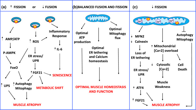
- Unbalanced dynamics are more deleterious than simultaneous reduction of both: OPA1/DRP1 double knockout mice show a less severe phenotype than OPA1 knockout alone, with transient FGF21 elevation and rescued lethality. FGF21 is commonly induced across OPA1-null, DRP1-null, and DKO models but with different magnitudes and outcomes (dose-dependent: moderate = adaptive, dramatic = detrimental).
Methods
- Narrative review; no primary experimental data. Synthesizes mechanistic cell-biology literature and mouse/human genetic models of mitochondrial quality control and muscle atrophy.
- Covers human disease genetics (CMT2A from MFN2 mutations, DOA from OPA1 mutations, DRP1/MFF/MiD49 mutations, VCP-associated myopathy) and rodent conditional knockout/transgenic models (muscle-specific MFN1/2, OPA1, DRP1, DKO, PUM2, ATG5/7, VPS15, etc.).
- Includes a summary table (Table 1) comparing signaling and phenotype of muscle-specific Opa1-, Drp1-, and Drp1/Opa1-null mice at early (70 days) and late (365 days) post-deletion.
Limitations
- The paper is a review and states no formal limitations of its own; it repeatedly notes open questions, e.g., that IMJ molecular composition and function "have to be defined," that the IMM fission machinery players remain largely unidentified, and that MDV biogenesis machinery is unknown.
- Evident from the text: several mechanistic conclusions rest on in vitro data whose in vivo physiological relevance "needs to be validated" (e.g., p62/optineurin/NDP52 mitophagy receptor data), and some findings are described as controversial (e.g., MARCHV/MITOL effects on DRP1 vary by cell context).
- Evident from the text: many causal inferences about muscle mass come from constitutive or conditional knockout mouse models with embryonic lethality or early death, which may not fully represent adult physiological remodeling.
Model-written summary. Check it against the paper before relying on it.
Integrity signals
Registry
Cites
Statistics
Figures
Verification
Claims the paper makes
181 Not checked yet · 46 No basis given · 30 Own interpretation · 9 Partly supported · 5 Supported · 1 Supported so far · accuracy not yet measured
The cited sources support only part of this claim — read each source’s verdict below.
In the paper
“Moreover, at least half of the atrogenes require FoxOs for their up- or downregulation [16].”
§ The connection between the dynamic remodeling of the mitochondrial network and the regulation of muscle mass > Signaling pathways regulating muscle mass
What the sources say
- Partly supportedcites [16] Milan G, Romanello V, Pescatore F et al (2015) Regulation of autophagy and the ubiquitin-proteasome system by the FoxO transcriptional network during muscle atrophy. Nat Commun 6:6670. https://doi.org/10.1038/ncomms7670 sourceExperimental — accuracy not yet measured
The Results section, “Half of the atrophy-related genes are under FoxO regulation” (page unavailable), reports that 29 of 63 atrophy-related genes require FoxO for normal induction during fasting. That supports a substantial FoxO-dependent share, but 29 of 63 is less than half, and the reported count concerns induction rather than both up- and downregulation. (LLM, conf=0.88)
plaindirect- Asserted: passes — The authors put it forward as true.
- Checkable: passes — It can be checked against evidence.
- Load-bearing: unclear — It is unclear whether the paper’s conclusions depend on it.
- Based: passes — The paper gives a reason why it holds.
- Partly supported
The cited sources support only part of this claim — read each source’s verdict below.
In the paper
“GTP hydrolysis of MFN1 causes a conformational change that allows the OMMs of opposing mitochondria to contact and fuse [61, 62].”
§ The connection between the dynamic remodeling of the mitochondrial network and the regulation of muscle mass > Quality control pathways finely tune mitochondrial function > Mitochondrial dynamics
What the sources say
- Not checked yetcites [61] Ishihara N, Eura Y, Mihara K (2004) Mitofusin 1 and 2 play distinct roles in mitochondrial fusion reactions via GTPase activity. J Cell Sci 117:6535–6546. https://doi.org/10.1242/jcs.01565 source
- Partly supportedcites [62] Cao Y-L, Meng S, Chen Y et al (2017) MFN1 structures reveal nucleotide-triggered dimerization critical for mitochondrial fusion. Nature 542:372–376. https://doi.org/10.1038/nature21077 sourceExperimental — accuracy not yet measured
The discussion passage labeled Abstract proposes that, during GTP hydrolysis, MFN1’s HD2 may fold back and bring opposing outer mitochondrial membranes close together. Extended Data Figure 9 presents this as a model and says subsequent membrane merger may depend on local curvature. The claim overstates a proposed mechanism as an established causal sequence through fusion. (LLM, conf=0.86)
plaindirect- Asserted: passes — The authors put it forward as true.
- Checkable: passes — It can be checked against evidence.
- Load-bearing: unclear — It is unclear whether the paper’s conclusions depend on it.
- Based: passes — The paper gives a reason why it holds.
Every source the paper cites for this claim was checked and supports it.
In the paper
“Genetic loss of MFN1 induces a higher degree of mitochondrial fragmentation than MFN2 deletion [60].”
§ The connection between the dynamic remodeling of the mitochondrial network and the regulation of muscle mass > Quality control pathways finely tune mitochondrial function > Mitochondrial dynamics
What the sources say
- Supportedcites [60] Chen H, Detmer SA, Ewald AJ et al (2003) Mitofusins Mfn1 and Mfn2 coordinately regulate mitochondrial fusion and are essential for embryonic development. J Cell Biol 160:189–200. https://doi.org/10.1083/jcb.200211046 source
The Results section, under “Both Mfn1- and Mfn2-deficient cells have aberrant mitochondrial morphology,” explicitly states that loss of Mfn1 leads to a greater degree of fragmentation than loss of Mfn2. This directly supports the comparison in the citing sentence. (LLM, conf=0.95)
plaindirect- Asserted: passes — The authors put it forward as true.
- Checkable: passes — It can be checked against evidence.
- Load-bearing: unclear — It is unclear whether the paper’s conclusions depend on it.
- Based: passes — The paper gives a reason why it holds.
- Supported
Every source the paper cites for this claim was checked and supports it.
In the paper
“OMA1 deficiency causes the lack of processing of OPA1L, which results in the shift of mitochondrial dynamics towards fusion.”
§ The connection between the dynamic remodeling of the mitochondrial network and the regulation of muscle mass > Mitochondria quality pathways are essential for skeletal muscle physiology > Mitochondrial proteostasis is critical for skeletal muscle function
What the sources say
- Supportedcites [167] Quirós PM, Ramsay AJ, Sala D et al (2012) Loss of mitochondrial protease OMA1 alters processing of the GTPase OPA1 and causes obesity and defective thermogenesis in mice. EMBO J 31:2117–2133. https://doi.org/10.1038/emboj.2012.70 source
The Discussion states that Oma1-deficient cells lack processing of L-OPA1 and that this shifts mitochondrial dynamics toward fusion. This directly supports the claim. (LLM, conf=0.95)
plaindirect- Asserted: passes — The authors put it forward as true.
- Checkable: passes — It can be checked against evidence.
- Load-bearing: unclear — It is unclear whether the paper’s conclusions depend on it.
- Based: passes — The paper gives a reason why it holds.
- Supported
The cited sources support only part of this claim — read each source’s verdict below.
In the paper
“When OPA1 was deleted during fetal development, animals died a few days after birth.”
§ The connection between the dynamic remodeling of the mitochondrial network and the regulation of muscle mass > Mitochondria quality pathways are essential for skeletal muscle physiology > Dysregulation of mitochondrial fusion in skeletal muscle leads to muscle atrophy, weakness, and have severe consequences in whole-body physiology
What the sources say
- Partly supportedcites [187] Tezze C, Romanello V, Desbats MA et al (2017) Age-associated loss of OPA1 in muscle impacts muscle mass, metabolic homeostasis, systemic inflammation, and epithelial senescence. Cell Metab 25:1374-1389.e6. https://doi.org/10.1016/j.cmet.2017.04.021 sourceExperimental — accuracy not yet measured
The Results section, “Muscle-Specific Opa1 Deletion in Newborns Blocks Animal Growth and Is Lethal” (page unavailable), reports that muscle-specific Opa1 knockout mice died within nine days of birth. The Methods describe a constitutive muscle-specific knockout, but our copy does not establish that deletion occurred during fetal development; the claim also omits the muscle-specific qualifier. (LLM, conf=0.83)
- Partly supportedcites [192] Rodríguez-Nuevo A, Díaz-Ramos A, Noguera E et al (2018) Mitochondrial DNA and TLR9 drive muscle inflammation upon Opa1 deficiency. EMBO J. https://doi.org/10.15252/embj.201796553 sourceExperimental — accuracy not yet measured
The section ‘Skeletal muscle-specific Opa1 ablation causes reduced body growth and premature death’ reports that Opa1 knockout mice had a dramatically reduced lifespan. It does not report death a few days after birth; the same section describes knockout mice alive at 9 weeks of age. Our copy also does not specify that deletion occurred during fetal development. (LLM, conf=0.88)
- Could not verifycites [193] Pereira RO, Tadinada SM, Zasadny FM et al (2017) OPA1 deficiency promotes secretion of FGF21 from muscle that prevents obesity and insulin resistance. EMBO J 36:2126–2145. https://doi.org/10.15252/embj.201696179 sourceExperimental — accuracy not yet measured
The section on OPA1 deficiency and mitochondrial respiratory capacity says OPA1 was reduced in mice by administering tamoxifen at four weeks of age, not during fetal development. Our copy does not report whether fetal OPA1 deletion caused death after birth; searches for embryonic deletion, neonatal lethality, and birth found no such passage. (LLM, conf=0.74)
plaindirect- Asserted: passes — The authors put it forward as true.
- Checkable: passes — It can be checked against evidence.
- Load-bearing: unclear — It is unclear whether the paper’s conclusions depend on it.
- Based: passes — The paper gives a reason why it holds.
- Partly supported
The cited sources support only part of this claim — read each source’s verdict below.
In the paper
“Interestingly, a mild inhibition of OPA1, which does not alter mitochondrial complex and supercomplex formation as well as mitochondrial DNA content and citrate synthase activity, in muscle during neonatal growth results in several beneficial metabolic changes in terms of resistance to obesity when the animals were challenged with a high-fat diet [193].”
§ The connection between the dynamic remodeling of the mitochondrial network and the regulation of muscle mass > Mitochondria quality pathways are essential for skeletal muscle physiology > Dysregulation of mitochondrial fusion in skeletal muscle leads to muscle atrophy, weakness, and have severe consequences in whole-body physiology
What the sources say
- Partly supportedcites [193] Pereira RO, Tadinada SM, Zasadny FM et al (2017) OPA1 deficiency promotes secretion of FGF21 from muscle that prevents obesity and insulin resistance. EMBO J 36:2126–2145. https://doi.org/10.15252/embj.201696179 sourceExperimental — accuracy not yet measured
Split into 2 atoms — 0 confirmed, 2 partially supported, 0 disputed, 0 misattributed, 0 unverifiable. Expand the claim to see per-atom verdicts.
plaindirect- Asserted: passes — The authors put it forward as true.
- Checkable: passes — It can be checked against evidence.
- Load-bearing: passes — The paper’s conclusions depend on it.
- Based: passes — The paper gives a reason why it holds.
- Partly supported
The cited sources support only part of this claim — read each source’s verdict below.
In the paper
“Interestingly, the stress-response myokine FGF21 is dramatically increased in muscle and serum of all these three models, even if with a different degree.”
§ The connection between the dynamic remodeling of the mitochondrial network and the regulation of muscle mass > Mitochondria quality pathways are essential for skeletal muscle physiology > FGF21 and the inhibition of fusion, fission, or both in skeletal muscle—a common factor with different outcomes
What the sources say
- Partly supportedcites [187] Tezze C, Romanello V, Desbats MA et al (2017) Age-associated loss of OPA1 in muscle impacts muscle mass, metabolic homeostasis, systemic inflammation, and epithelial senescence. Cell Metab 25:1374-1389.e6. https://doi.org/10.1016/j.cmet.2017.04.021 sourceExperimental — accuracy not yet measured
The Results section on Opa1 deletion reports that FGF21 was massively upregulated in skeletal muscle and rose in circulating blood. Our copy does not report corresponding muscle and serum increases in DRP1 or OPA1/DRP1 double-knockout models, so it does not support the claim about all three models or their differing degrees of increase (page unavailable). (LLM, conf=0.90)
- Partly supportedcites [196] Favaro G, Romanello V, Varanita T et al (2019) DRP1-mediated mitochondrial shape controls calcium homeostasis and muscle mass. Nat Commun 10:2576. https://doi.org/10.1038/s41467-019-10226-9 sourceExperimental — accuracy not yet measured
The Results sections report increased muscle FGF21 expression and blood FGF21 in DRP1-knockout mice, including a 20-fold blood increase after acute deletion. The Discussion describes FGF21 induction after OPA1 ablation and says serum levels are higher in OPA1-knockout than DRP1-knockout mice. Our copy does not report FGF21 levels in an OPA1/DRP1 double-knockout (DKO) model, so it does not support the claim for all three models. (LLM, conf=0.87)
- Partly supportedcites [197] Romanello V, Scalabrin M, Albiero M et al (2019) Inhibition of the fission machinery mitigates OPA1 impairment in adult skeletal muscles. Cells. https://doi.org/10.3390/cells8060597 sourceExperimental — accuracy not yet measured
Results §3.4 reports that FGF21 in DKO muscle and blood peaked at 70 days but returned to control levels by 365 days. The Discussion also describes elevated circulating FGF21 in Opa1-deleted mice. The paper does not establish increased FGF21 in both muscle and serum of a DRP1-only model, so it does not support the claim for all three models. (LLM, conf=0.87)
plaindirect- Asserted: passes — The authors put it forward as true.
- Checkable: passes — It can be checked against evidence.
- Load-bearing: passes — The paper’s conclusions depend on it.
- Based: passes — The paper gives a reason why it holds.
- Partly supported
The cited sources support only part of this claim — read each source’s verdict below.
In the paper
“Several reports have shown that rebalancing of mitochondrial dynamics rescues the phenotype of certain diseases associated with alterations of mitochondrial fusion or fission [119, 194, 195, 214]”
§ The connection between the dynamic remodeling of the mitochondrial network and the regulation of muscle mass > Mitochondria quality pathways are essential for skeletal muscle physiology > The balance between fusion and fission events is critical for muscle mass and whole-body homeostasis.
What the sources say
- Partly supportedcites [119] D’Amico D, Mottis A, Potenza F et al (2019) The RNA-binding protein PUM2 impairs mitochondrial dynamics and mitophagy during aging. Mol Cell 73:775-787.e10. https://doi.org/10.1016/j.molcel.2018.11.034 sourceExperimental — accuracy not yet measured
The Discussion (page unavailable) reports that increasing fission through PUM2 loss rescues abnormal mitochondrial networks in old worms and mice. That supports rescue of an age-associated phenotype, but the paper does not establish rescue of phenotypes in diseases associated with altered mitochondrial fusion or fission. (LLM, conf=0.84)
- Partly supportedcites [194] Varanita T, Soriano ME, Romanello V et al (2015) The OPA1-dependent mitochondrial cristae remodeling pathway controls atrophic, apoptotic, and ischemic tissue damage. Cell Metab 21:834–844. https://doi.org/10.1016/j.cmet.2015.05.007 sourceExperimental — accuracy not yet measured
The Discussion (page not provided) reports that OPA1 overexpression protects against several forms of tissue damage and counteracts myopathy caused by a genetic mitochondrial defect. The paper identifies OPA1 as a fusion protein, but attributes protection chiefly to cristae maintenance; it does not establish that rebalancing fusion and fission rescues diseases caused by alterations in those processes. (LLM, conf=0.86)
- Partly supportedcites [195] Civiletto G, Varanita T, Cerutti R et al (2015) Opa1 overexpression ameliorates the phenotype of two mitochondrial disease mouse models. Cell Metab 21:845–854. https://doi.org/10.1016/j.cmet.2015.04.016 sourceExperimental — accuracy not yet measured
The Results report that Opa1 overexpression improved disease phenotypes in two mouse models, supporting the rescue component of the claim. But the Introduction identifies these as models of respiratory-chain complex I and IV deficiency, not diseases caused by altered mitochondrial fusion or fission. The Discussion attributes the improvement to cristae and respiratory-chain stabilization; it does not establish that rebalancing fusion and fission rescued the phenotypes. Page numbers are unavailable in our copy. (LLM, conf=0.88)
- Partly supportedcites [214] Rana A, Oliveira MP, Khamoui AV et al (2017) Promoting Drp1-mediated mitochondrial fission in midlife prolongs healthy lifespan of Drosophila melanogaster. Nat Commun 8:448. https://doi.org/10.1038/s41467-017-00525-4 sourceExperimental — accuracy not yet measured
The Discussion reports that promoting mitochondrial fission rescues several degenerative phenotypes in PINK1 and Parkin mutant flies. This supports rescue in disease-associated fly models, but the paper does not establish that rebalancing mitochondrial dynamics rescues the phenotypes of diseases themselves more generally. (LLM, conf=0.82)
plaindirect- Asserted: passes — The authors put it forward as true.
- Checkable: passes — It can be checked against evidence.
- Load-bearing: passes — The paper’s conclusions depend on it.
- Based: passes — The paper gives a reason why it holds.
- Partly supported
A conclusion the paper draws from its own results — it cites no other source for it, so there is none to check. The quote shows it is in the paper.
In the paper
“Therefore, by controlling FoxOs and mTOR, AKT is a regulatory node between catabolic and anabolic processes.”
§ The connection between the dynamic remodeling of the mitochondrial network and the regulation of muscle mass > Signaling pathways regulating muscle mass
plaindirect- Asserted: passes — The authors put it forward as true.
- Checkable: passes — It can be checked against evidence.
- Load-bearing: unclear — It is unclear whether the paper’s conclusions depend on it.
- Based: passes — The paper gives a reason why it holds.
In the paper
“The mitochondrial muscle reticulum forms a conductive pathway that can rapidly transfer energy from the oxygen source, the capillary, to the contractile apparatus [24].”
§ The connection between the dynamic remodeling of the mitochondrial network and the regulation of muscle mass > Mitochondrial communication is essential for optimal mitochondrial function in skeletal muscle > Mitochondria populations are connected to support energy distribution in skeletal muscle
What the sources say
- Not checked yetcites [24] Glancy B, Hartnell LM, Malide D et al (2015) Mitochondrial reticulum for cellular energy distribution in muscle. Nature 523:617–620. https://doi.org/10.1038/nature14614 source
plaindirect- Asserted: passes — The authors put it forward as true.
- Checkable: passes — It can be checked against evidence.
- Load-bearing: passes — The paper’s conclusions depend on it.
- Based: passes — The paper gives a reason why it holds.
- It may state two separate claims as one. 116
- Probably not a substantial claim. 30
Every source the paper cites for this claim was checked and supports it.
In the paper
“Conversely, the formation of a Lys63–polyubiquitin chain by the E3 ligase MARCHV/MITOLl does not induce MFN2 degradation but increases its activity and MAM formation and function [71].”
§ The connection between the dynamic remodeling of the mitochondrial network and the regulation of muscle mass > Quality control pathways finely tune mitochondrial function > Mitochondrial dynamics
What the sources say
- Supportedcites [71] Sugiura A, Nagashima S, Tokuyama T et al (2013) MITOL regulates endoplasmic reticulum-mitochondria contacts via Mitofusin2. Mol Cell 51:20–34. https://doi.org/10.1016/j.molcel.2013.04.023 source
The Results identify MITOL-mediated K63-linked polyubiquitination of Mfn2 and report that MITOL does not promote Mfn2 degradation. The Discussion states that this ubiquitination activates Mfn2 and leads to oligomerization for MAM formation; the paper’s Summary also reports MAM dysfunction after MITOL knockdown. (LLM, conf=0.93)
plaindirect- Asserted: passes — The authors put it forward as true.
- Checkable: passes — It can be checked against evidence.
- Load-bearing: unclear — It is unclear whether the paper’s conclusions depend on it.
- Based: passes — The paper gives a reason why it holds.
It may state two separate claims as one.
- Supported
The sources with a verdict support this claim; the rest weren’t checked yet, have no full text we could read, or their verdicts aren’t rated yet.
In the paper
“In contrast to Yme1L1, the germline deletion of OMA1 does not impair embryogenesis, which indicates that OMA1-induced OPA1 processing is dispensable for embryonic and adult mouse development [166, 167].”
§ The connection between the dynamic remodeling of the mitochondrial network and the regulation of muscle mass > Mitochondria quality pathways are essential for skeletal muscle physiology > Mitochondrial proteostasis is critical for skeletal muscle function
What the sources say
- Not checked yetcites [166] Wai T, García-Prieto J, Baker MJ et al (2015) Imbalanced OPA1 processing and mitochondrial fragmentation cause heart failure in mice. Science 350:aad0116. https://doi.org/10.1126/science.aad0116 source
- Supportedcites [167] Quirós PM, Ramsay AJ, Sala D et al (2012) Loss of mitochondrial protease OMA1 alters processing of the GTPase OPA1 and causes obesity and defective thermogenesis in mice. EMBO J 31:2117–2133. https://doi.org/10.1038/emboj.2012.70 source
In “Generation of mice deficient in OMA1” (page not provided), the paper reports that Oma1-null mice were born at the expected Mendelian ratio and developed normally, and explicitly concludes that OMA1 is dispensable for embryonic and adult mouse development. Its OPA1-processing results show altered processing in the deficient mice, supporting the claim about OMA1-induced processing. (LLM, conf=0.92)
hedged- Asserted: passes — The authors put it forward as true.
- Checkable: passes — It can be checked against evidence.
- Load-bearing: unclear — It is unclear whether the paper’s conclusions depend on it.
- Based: passes — The paper gives a reason why it holds.
It may state two separate claims as one.
The cited sources support only part of this claim — read each source’s verdict below.
In the paper
“p97/VCP is critical in the retrotranslocation and degradation of endoplasmic reticulum-associated proteins (ERAD) [47] and mitochondrial proteins ( MAD) [49, 50].”
§ The connection between the dynamic remodeling of the mitochondrial network and the regulation of muscle mass > Mitochondria quality pathways are essential for skeletal muscle physiology > Mitochondrial proteostasis is critical for skeletal muscle function
What the sources say
- Not checked yetcites [47] Ye Y, Meyer HH, Rapoport TA (2001) The AAA ATPase Cdc48/p97 and its partners transport proteins from the ER into the cytosol. Nature 414:652–656. https://doi.org/10.1038/414652a source
- Partly supportedcites [49] Tanaka A, Cleland MM, Xu S et al (2010) Proteasome and p97 mediate mitophagy and degradation of mitofusins induced by Parkin. J Cell Biol 191:1367–1380. https://doi.org/10.1083/jcb.201007013 sourceExperimental — accuracy not yet measured
The Results section (‘Role of p97 in Mfn proteasomal degradation’; page unavailable) reports that inhibiting p97 largely prevented Parkin-mediated degradation of the mitochondrial proteins Mfn1 and Mfn2. It explicitly says how these proteins are extracted from the outer mitochondrial membrane is unknown, so it does not establish p97’s role in their retrotranslocation. The ERAD portion of the citing sentence is attached to a different citation, [47]. (LLM, conf=0.88)
- Partly supportedcites [50] Xu S, Peng G, Wang Y et al (2011) The AAA-ATPase p97 is essential for outer mitochondrial membrane protein turnover. Mol Biol Cell 22:291–300. https://doi.org/10.1091/mbc.E10-09-0748 sourceExperimental — accuracy not yet measured
The Introduction and Discussion report that p97 drives retrotranslocation and proteasomal degradation of the outer mitochondrial membrane proteins Mcl1 and Mfn1, supporting the mitochondrial portion of the citation. The claim says “mitochondrial proteins” more broadly, while the Discussion says the extent of p97’s role in mitochondrial proteostasis remains unclear. The ERAD portion is attached to a different citation [47]. (LLM, conf=0.82)
plaindirect- Asserted: passes — The authors put it forward as true.
- Checkable: passes — It can be checked against evidence.
- Load-bearing: passes — The paper’s conclusions depend on it.
- Based: passes — The paper gives a reason why it holds.
It may state two separate claims as one.
The cited sources support only part of this claim — read each source’s verdict below.
In the paper
“In mice, muscle knockdown of PARL results in an alteration in mitochondrial cristae morphology, mitochondrial dysfunction, oxidative stress, and altered insulin signaling, which probably can be explained by the reduction in PARL-mediated OPA1 processing [168].”
§ The connection between the dynamic remodeling of the mitochondrial network and the regulation of muscle mass > Mitochondria quality pathways are essential for skeletal muscle physiology > Mitochondrial proteostasis is critical for skeletal muscle function
What the sources say
- Partly supportedcites [168] Civitarese AE, MacLean PS, Carling S et al (2010) Regulation of skeletal muscle oxidative capacity and insulin signaling by the mitochondrial rhomboid protease PARL. Cell Metab 11:412–426. https://doi.org/10.1016/j.cmet.2010.04.004 sourceExperimental — accuracy not yet measured
The Results (mCK-CAR-PARL(−) mice and impaired insulin-signaling sections; page unavailable) report disorganized cristae, elevated oxidative damage, and impaired insulin signaling after muscle PARL knockdown. The Discussion proposes PARL-mediated OPA1 cleavage as an explanation for the cristae changes and for reduced energetic capacity and elevated ROS in PARL-depleted myotubes. It does not establish reduced OPA1 processing as the explanation for impaired insulin signaling in mouse muscle. (LLM, conf=0.86)
hedgeddirect- Asserted: passes — The authors put it forward as true.
- Checkable: passes — It can be checked against evidence.
- Load-bearing: passes — The paper’s conclusions depend on it.
- Based: passes — The paper gives a reason why it holds.
It may state two separate claims as one.
- Partly supported
Every source the paper cites for this claim was checked and supports it.
In the paper
“In mice, MFF deletion leads to smaller animals that display neuromuscular defects, kyphosis, and premature death at 13 weeks due to dilated cardiomyopathy [207].”
§ The connection between the dynamic remodeling of the mitochondrial network and the regulation of muscle mass > Mitochondria quality pathways are essential for skeletal muscle physiology > Mitochondrial fission controls muscle development, maintenance, and function; the link with ER stress and calcium homeostasis.
What the sources say
- Supportedcites [207] Chen H, Ren S, Clish C et al (2015) Titration of mitochondrial fusion rescues Mff-deficient cardiomyopathy. J Cell Biol 211:795–805. https://doi.org/10.1083/jcb.201507035 source
In Results and discussion, “Mff-deficient mice exhibit pleiotropic phenotypes” reports that Mff gene-trap mutants are smaller, have kyphosis and neuromuscular defects, and die at a mean age of 13 weeks from dilated cardiomyopathy. The “Dilated cardiomyopathy leads to heart failure” section confirms the cause of early death. No page numbers are provided in our copy. (LLM, conf=0.95)
plaindirect- Asserted: passes — The authors put it forward as true.
- Checkable: passes — It can be checked against evidence.
- Load-bearing: unclear — It is unclear whether the paper’s conclusions depend on it.
- Based: passes — The paper gives a reason why it holds.
It may state two separate claims as one.
- Supported
The cited sources support only part of this claim — read each source’s verdict below.
In the paper
“MFN2 deletion in young muscle causes extensive mitochondrial fragmentation, mitochondrial dysfunction, ROS production, ER stress, and autophagy inhibition resulting in muscle atrophy [67, 186].”
§ The connection between the dynamic remodeling of the mitochondrial network and the regulation of muscle mass > Mitochondria quality pathways are essential for skeletal muscle physiology > Dysregulation of mitochondrial fusion in skeletal muscle leads to muscle atrophy, weakness, and have severe consequences in whole-body physiology
What the sources say
- Partly supportedcites [67] Sebastián D, Hernández-Alvarez MI, Segalés J et al (2012) Mitofusin 2 (Mfn2) links mitochondrial and endoplasmic reticulum function with insulin signaling and is essential for normal glucose homeostasis. Proc Natl Acad Sci USA 109:5523–5528. https://doi.org/10.1073/pnas.1108220109 sourceExperimental — accuracy not yet measured
The Results report mitochondrial dysfunction, increased hydrogen peroxide, and ER stress in Mfn2-deficient muscle. The Discussion describes larger mitochondria in skeletal muscle, not extensive fragmentation; our copy does not establish that deletion in young muscle inhibits autophagy or results in muscle atrophy. (LLM, conf=0.90)
- Partly supportedcites [186] Sebastián D, Sorianello E, Segalés J et al (2016) Mfn2 deficiency links age-related sarcopenia and impaired autophagy to activation of an adaptive mitophagy pathway. EMBO J 35:1677–1693. https://doi.org/10.15252/embj.201593084 sourceExperimental — accuracy not yet measured
The Results report muscle atrophy in young Mfn2KO mice, reduced oxidative capacity, increased oxidative stress, and impaired mitochondrial autophagy; rescue experiments link the autophagy defect to atrophy. The copy does not show extensive mitochondrial fragmentation or ER stress in young muscle (searched for fragmentation, ER stress, and unfolded protein response). (LLM, conf=0.88)
plaindirect- Asserted: passes — The authors put it forward as true.
- Checkable: passes — It can be checked against evidence.
- Load-bearing: passes — The paper’s conclusions depend on it.
- Based: passes — The paper gives a reason why it holds.
It may state two separate claims as one.
- Partly supported
Every source the paper cites for this claim was checked and supports it.
In the paper
“Moreover, muscle denervation, oxidative stress, and inflammation are mitigated in DKO muscles, rescuing the lethal phenotype of OPA1 knockout mice [197].”
§ The connection between the dynamic remodeling of the mitochondrial network and the regulation of muscle mass > Mitochondria quality pathways are essential for skeletal muscle physiology > The balance between fusion and fission events is critical for muscle mass and whole-body homeostasis.
What the sources say
- Supportedcites [197] Romanello V, Scalabrin M, Albiero M et al (2019) Inhibition of the fission machinery mitigates OPA1 impairment in adult skeletal muscles. Cells. https://doi.org/10.3390/cells8060597 source
The Discussion states the claim directly. Results §3.4 also reports mitigated denervation, oxidative stress, and inflammation in DKO muscles, with survival matching controls rather than the premature death seen after Opa1 deletion alone. (LLM, conf=0.95)
plaindirect- Asserted: passes — The authors put it forward as true.
- Checkable: passes — It can be checked against evidence.
- Load-bearing: passes — The paper’s conclusions depend on it.
- Based: passes — The paper gives a reason why it holds.
It may state two separate claims as one.
- Supported
In the paper
“Subsarcolemmal (SS) mitochondria have a globular shape and are located just beneath the plasma membrane (sarcolemma). A fraction of these surrounds capillaries and nuclei (perivascular and perinuclear mitochondria, respectively) [21].”
§ The connection between the dynamic remodeling of the mitochondrial network and the regulation of muscle mass > Mitochondrial communication is essential for optimal mitochondrial function in skeletal muscle > Mitochondria populations are connected to support energy distribution in skeletal muscle
What the sources say
- Not checked yetcites [21] Vincent AE, White K, Davey T et al (2019) Quantitative 3D mapping of the human skeletal muscle mitochondrial network. Cell Rep 26:996-1009.e4. https://doi.org/10.1016/j.celrep.2019.01.010 source
plain- Asserted: passes — The authors put it forward as true.
- Checkable: passes — It can be checked against evidence.
- Load-bearing: unclear — It is unclear whether the paper’s conclusions depend on it.
- Based: passes — The paper gives a reason why it holds.
It may state two separate claims as one.
The paper states this without a source or data behind it here, so there is nothing to check it against. The quote shows it is in the paper.
In the paper
“Two central ATP-dependent proteolytic systems are activated during muscle atrophy. The ubiquitin–proteasome system degrades predominantly myofibrillar proteins, whereas the autophagy–lysosome system removes dysfunctional organelles, protein aggregates, and unfolded and toxic proteins.”
§ The connection between the dynamic remodeling of the mitochondrial network and the regulation of muscle mass > Signaling pathways regulating muscle mass
plaindirect- Asserted: passes — The authors put it forward as true.
- Checkable: passes — It can be checked against evidence.
- Load-bearing: unclear — It is unclear whether the paper’s conclusions depend on it.
- Based: passes — The paper gives a reason why it holds.
It may state two separate claims as one.
The paper states this without a source or data behind it here, so there is nothing to check it against. The quote shows it is in the paper.
In the paper
“Moreover, mitochondrial function and, thus, energy production are generally reduced during muscle wasting.”
§ The connection between the dynamic remodeling of the mitochondrial network and the regulation of muscle mass > Signaling pathways regulating muscle mass
plaincorrelational- Asserted: passes — The authors put it forward as true.
- Checkable: passes — It can be checked against evidence.
- Load-bearing: passes — The paper’s conclusions depend on it.
- Based: passes — The paper gives a reason why it holds.
It may state two separate claims as one.
The paper states this without a source or data behind it here, so there is nothing to check it against. The quote shows it is in the paper.
In the paper
“Conversely, in catabolic conditions, protein degradation exceeds protein synthesis leading to muscle weakness and muscle atrophy.”
§ The connection between the dynamic remodeling of the mitochondrial network and the regulation of muscle mass > Signaling pathways regulating muscle mass
plaindirect- Asserted: passes — The authors put it forward as true.
- Checkable: passes — It can be checked against evidence.
- Load-bearing: fails — The paper’s conclusions may not depend on it.
- Based: passes — The paper gives a reason why it holds.
Probably not a substantial claim. The paper’s conclusions may not depend on it. It may state two separate claims as one.
None of the sources cited for this claim have been checked yet.
In the paper
“BMP pathway is activated in myostatin knockout and is sufficient, when induced, to promote muscle growth and to counteract muscle loss after denervation and in the absence of nutrients [6–8].”
§ The connection between the dynamic remodeling of the mitochondrial network and the regulation of muscle mass > Signaling pathways regulating muscle mass
What the sources say
- Not checked yetcites [6] Sartori R, Schirwis E, Blaauw B et al (2013) BMP signaling controls muscle mass. Nat Genet 45:1309–1318. https://doi.org/10.1038/ng.2772 source
- Not checked yetcites [7] Winbanks CE, Chen JL, Qian H et al (2013) The bone morphogenetic protein axis is a positive regulator of skeletal muscle mass. J Cell Biol 203:345–357. https://doi.org/10.1083/jcb.201211134 source
- Not checked yetcites [8] Sartori R, Sandri M (2015) Bone and morphogenetic protein signalling and muscle mass. Curr Opin Clin Nutr Metab Care 18:215–220. https://doi.org/10.1097/MCO.0000000000000172 source
plaindirect- Asserted: passes — The authors put it forward as true.
- Checkable: passes — It can be checked against evidence.
- Load-bearing: unclear — It is unclear whether the paper’s conclusions depend on it.
- Based: passes — The paper gives a reason why it holds.
It may state two separate claims as one.
None of the sources cited for this claim have been checked yet.
In the paper
“Induction of BMP signaling activates Smad1/5/8, which, together with SMAD 4, leads to the suppression of the MUSA1 [6] and the activation of mTOR-dependent protein synthesis [7].”
§ The connection between the dynamic remodeling of the mitochondrial network and the regulation of muscle mass > Signaling pathways regulating muscle mass
What the sources say
- Not checked yetcites [6] Sartori R, Schirwis E, Blaauw B et al (2013) BMP signaling controls muscle mass. Nat Genet 45:1309–1318. https://doi.org/10.1038/ng.2772 source
- Not checked yetcites [7] Winbanks CE, Chen JL, Qian H et al (2013) The bone morphogenetic protein axis is a positive regulator of skeletal muscle mass. J Cell Biol 203:345–357. https://doi.org/10.1083/jcb.201211134 source
plaindirect- Asserted: passes — The authors put it forward as true.
- Checkable: passes — It can be checked against evidence.
- Load-bearing: unclear — It is unclear whether the paper’s conclusions depend on it.
- Based: passes — The paper gives a reason why it holds.
It may state two separate claims as one.
In the paper
“Based on myosin heavy chain (MHC) expression, mouse muscles contain four major fiber types, slow type 1 and fast 2A, 2X and 2B; while human muscles contain three major fiber types, slow type 1 and fast 2A and 2X [30].”
§ The connection between the dynamic remodeling of the mitochondrial network and the regulation of muscle mass > The conformation and connectivity of the mitochondrial network are tailored to the metabolism and contractility of each fiber type
What the sources say
- Not checked yetcites [30] Schiaffino S, Reggiani C (2011) Fiber types in mammalian skeletal muscles. Physiol Rev 91:1447–1531. https://doi.org/10.1152/physrev.00031.2010 source
plain- Asserted: passes — The authors put it forward as true.
- Checkable: passes — It can be checked against evidence.
- Load-bearing: unclear — It is unclear whether the paper’s conclusions depend on it.
- Based: passes — The paper gives a reason why it holds.
It may state two separate claims as one.
None of the sources cited for this claim have been checked yet.
In the paper
“The intermyofibrillar (IMF) mitochondria are elongated with a tubular shape [21] and are inserted among myofibrils arranged in pairs at the z-line of each sarcomere [22].”
§ The connection between the dynamic remodeling of the mitochondrial network and the regulation of muscle mass > Mitochondrial communication is essential for optimal mitochondrial function in skeletal muscle > Mitochondria populations are connected to support energy distribution in skeletal muscle
What the sources say
- Not checked yetcites [21] Vincent AE, White K, Davey T et al (2019) Quantitative 3D mapping of the human skeletal muscle mitochondrial network. Cell Rep 26:996-1009.e4. https://doi.org/10.1016/j.celrep.2019.01.010 source
- Not checked yetcites [22] Vendelin M, Béraud N, Guerrero K et al (2005) Mitochondrial regular arrangement in muscle cells: a “crystal-like” pattern. Am J Physiol, Cell Physiol 288:C757-767. https://doi.org/10.1152/ajpcell.00281.2004 source
plain- Asserted: passes — The authors put it forward as true.
- Checkable: passes — It can be checked against evidence.
- Load-bearing: unclear — It is unclear whether the paper’s conclusions depend on it.
- Based: passes — The paper gives a reason why it holds.
It may state two separate claims as one.
In the paper
“The IGF1–AKT-–mTOR axis increases protein synthesis by stimulating the translational machinery and simultaneously blocks FoxOs transcription factors and protein degradation [9].”
§ The connection between the dynamic remodeling of the mitochondrial network and the regulation of muscle mass > Signaling pathways regulating muscle mass
What the sources say
- Not checked yetcites [9] Sandri M, Sandri C, Gilbert A et al (2004) Foxo transcription factors induce the atrophy-related ubiquitin ligase atrogin-1 and cause skeletal muscle atrophy. Cell 117:399–412. https://doi.org/10.1016/s0092-8674(04)00400-3 source
plaindirect- Asserted: passes — The authors put it forward as true.
- Checkable: passes — It can be checked against evidence.
- Load-bearing: unclear — It is unclear whether the paper’s conclusions depend on it.
- Based: passes — The paper gives a reason why it holds.
It may state two separate claims as one.
In the paper
“To prevent the spread of dysfunction, IMJs are detached in dysfunctional mitochondria, promoting the electrical isolation and further segregation from the network of the malfunctioning organelle [28].”
§ The connection between the dynamic remodeling of the mitochondrial network and the regulation of muscle mass > Mitochondrial communication is essential for optimal mitochondrial function in skeletal muscle > Interorganelle communication relies on intermitochondrial junctions and nanotunnels
What the sources say
- Not checked yetcites [28] Glancy B, Hartnell LM, Combs CA et al (2017) Power grid protection of the muscle mitochondrial reticulum. Cell Rep 19:487–496. https://doi.org/10.1016/j.celrep.2017.03.063 source
plaindirect- Asserted: passes — The authors put it forward as true.
- Checkable: passes — It can be checked against evidence.
- Load-bearing: unclear — It is unclear whether the paper’s conclusions depend on it.
- Based: passes — The paper gives a reason why it holds.
It may state two separate claims as one.
The paper states this without a source or data behind it here, so there is nothing to check it against. The quote shows it is in the paper.
In the paper
“The muscle mass in adulthood is defined by the dynamic balance between protein synthesis and protein degradation.”
§ The connection between the dynamic remodeling of the mitochondrial network and the regulation of muscle mass > Signaling pathways regulating muscle mass
plaindirect- Asserted: passes — The authors put it forward as true.
- Checkable: passes — It can be checked against evidence.
- Load-bearing: fails — The paper’s conclusions may not depend on it.
- Based: passes — The paper gives a reason why it holds.
Probably not a substantial claim. The paper’s conclusions may not depend on it.
None of the sources cited for this claim have been checked yet.
In the paper
“On the other hand, BMP signaling is a positive regulator of muscle mass and is dominant over the myostatin pathway [6, 7].”
§ The connection between the dynamic remodeling of the mitochondrial network and the regulation of muscle mass > Signaling pathways regulating muscle mass
What the sources say
- Not checked yetcites [6] Sartori R, Schirwis E, Blaauw B et al (2013) BMP signaling controls muscle mass. Nat Genet 45:1309–1318. https://doi.org/10.1038/ng.2772 source
- Not checked yetcites [7] Winbanks CE, Chen JL, Qian H et al (2013) The bone morphogenetic protein axis is a positive regulator of skeletal muscle mass. J Cell Biol 203:345–357. https://doi.org/10.1083/jcb.201211134 source
plaindirect- Asserted: passes — The authors put it forward as true.
- Checkable: passes — It can be checked against evidence.
- Load-bearing: unclear — It is unclear whether the paper’s conclusions depend on it.
- Based: passes — The paper gives a reason why it holds.
It may state two separate claims as one.
None of the sources cited for this claim have been checked yet.
In the paper
“IMJs are highly specific and regulated electron-dense structures, defined by the close contact of the inner and outer mitochondrial membrane.”
§ The connection between the dynamic remodeling of the mitochondrial network and the regulation of muscle mass > Mitochondrial communication is essential for optimal mitochondrial function in skeletal muscle > Interorganelle communication relies on intermitochondrial junctions and nanotunnels
plain- Asserted: passes — The authors put it forward as true.
- Checkable: passes — It can be checked against evidence.
- Load-bearing: fails — The paper’s conclusions may not depend on it.
- Based: passes — The paper gives a reason why it holds.
Probably not a substantial claim. The paper’s conclusions may not depend on it.
The paper states this without a source or data behind it here, so there is nothing to check it against. The quote shows it is in the paper.
In the paper
“Instead, muscles that generate a high-intensity activity for short periods (e.g., jumping, kicking), have a high representation of fast glycolytic fibers, which have poor mitochondrial content, decreased reliance on OXPHOS and are fatigable.”
§ The connection between the dynamic remodeling of the mitochondrial network and the regulation of muscle mass > The conformation and connectivity of the mitochondrial network are tailored to the metabolism and contractility of each fiber type
plaincorrelational- Asserted: passes — The authors put it forward as true.
- Checkable: passes — It can be checked against evidence.
- Load-bearing: unclear — It is unclear whether the paper’s conclusions depend on it.
- Based: passes — The paper gives a reason why it holds.
It may state two separate claims as one.
The paper states this without a source or data behind it here, so there is nothing to check it against. The quote shows it is in the paper.
In the paper
“Skeletal muscles are composed of specialized fiber types which differ in their mitochondrial content, metabolic properties, and myosin composition.”
§ The connection between the dynamic remodeling of the mitochondrial network and the regulation of muscle mass > The conformation and connectivity of the mitochondrial network are tailored to the metabolism and contractility of each fiber type
plaincorrelational- Asserted: passes — The authors put it forward as true.
- Checkable: passes — It can be checked against evidence.
- Load-bearing: passes — The paper’s conclusions depend on it.
- Based: passes — The paper gives a reason why it holds.
Probably not a substantial claim.
A conclusion the paper draws from its own results — it cites no other source for it, so there is none to check. The quote shows it is in the paper.
In the paper
“Therefore, maintaining a functional mitochondrial network in this tissue is fundamental to sustain the metabolic demands imposed by contraction, ultimately regulating fuel utilization, energy expenditure, and general metabolism.”
§ The connection between the dynamic remodeling of the mitochondrial network and the regulation of muscle mass > Introduction
plaindirect- Asserted: passes — The authors put it forward as true.
- Checkable: passes — It can be checked against evidence.
- Load-bearing: passes — The paper’s conclusions depend on it.
- Based: passes — The paper gives a reason why it holds.
It may state two separate claims as one.
None of the sources cited for this claim have been checked yet.
In the paper
“Recent findings on high-resolution three-dimensional microscopy challenged the existence of two separated mitochondrial pools and strengthened the concept of a physically and functionally highly interconnected mitochondrial network in skeletal muscle [21, 24, 26].”
§ The connection between the dynamic remodeling of the mitochondrial network and the regulation of muscle mass > Mitochondrial communication is essential for optimal mitochondrial function in skeletal muscle > Mitochondria populations are connected to support energy distribution in skeletal muscle
What the sources say
- Not checked yetcites [21] Vincent AE, White K, Davey T et al (2019) Quantitative 3D mapping of the human skeletal muscle mitochondrial network. Cell Rep 26:996-1009.e4. https://doi.org/10.1016/j.celrep.2019.01.010 source
- Not checked yetcites [24] Glancy B, Hartnell LM, Malide D et al (2015) Mitochondrial reticulum for cellular energy distribution in muscle. Nature 523:617–620. https://doi.org/10.1038/nature14614 source
- Not checked yetcites [26] Dahl R, Larsen S, Dohlmann TL et al (2015) Three-dimensional reconstruction of the human skeletal muscle mitochondrial network as a tool to assess mitochondrial content and structural organization. Acta Physiol 213:145–155. https://doi.org/10.1111/apha.12289 source
boosteddirect- Asserted: passes — The authors put it forward as true.
- Checkable: passes — It can be checked against evidence.
- Load-bearing: passes — The paper’s conclusions depend on it.
- Based: passes — The paper gives a reason why it holds.
It may state two separate claims as one.
None of the sources cited for this claim have been checked yet.
In the paper
“Instead, maintaining a healthy skeletal muscle mass is associated with a lower risk of mortality [1, 2]”
§ The connection between the dynamic remodeling of the mitochondrial network and the regulation of muscle mass > Introduction
What the sources say
- Not checked yetcites [1] Srikanthan P, Karlamangla AS (2014) Muscle mass index as a predictor of longevity in older adults. Am J Med 127:547–553. https://doi.org/10.1016/j.amjmed.2014.02.007 source
- Not checked yetcites [2] Newman AB, Kupelian V, Visser M et al (2006) Strength, but not muscle mass, is associated with mortality in the health, aging and body composition study cohort. J Gerontol A Biol Sci Med Sci 61:72–77. https://doi.org/10.1093/gerona/61.1.72 source
plaincorrelational- Asserted: passes — The authors put it forward as true.
- Checkable: passes — It can be checked against evidence.
- Load-bearing: fails — The paper’s conclusions may not depend on it.
- Based: passes — The paper gives a reason why it holds.
Probably not a substantial claim. The paper’s conclusions may not depend on it.
The paper states this without a source or data behind it here, so there is nothing to check it against. The quote shows it is in the paper.
In the paper
“The ubiquitin–proteasome system degrades predominantly myofibrillar proteins, whereas the autophagy–lysosome system removes dysfunctional organelles, protein aggregates, and unfolded and toxic proteins.”
§ The connection between the dynamic remodeling of the mitochondrial network and the regulation of muscle mass > Signaling pathways regulating muscle mass
plaindirect- Asserted: passes — The authors put it forward as true.
- Checkable: passes — It can be checked against evidence.
- Load-bearing: fails — The paper’s conclusions may not depend on it.
- Based: passes — The paper gives a reason why it holds.
Probably not a substantial claim. The paper’s conclusions may not depend on it. It may state two separate claims as one.
None of the sources cited for this claim have been checked yet.
In the paper
“Nanotunnels are conserved OMM and IMM double-membrane projections, connecting two non-adjacent mitochondria [29], particularly under pathological conditions [21].”
§ The connection between the dynamic remodeling of the mitochondrial network and the regulation of muscle mass > Mitochondrial communication is essential for optimal mitochondrial function in skeletal muscle > Interorganelle communication relies on intermitochondrial junctions and nanotunnels
What the sources say
- Not checked yetcites [29] Vincent AE, Turnbull DM, Eisner V et al (2017) Mitochondrial nanotunnels. Trends Cell Biol 27:787–799. https://doi.org/10.1016/j.tcb.2017.08.009 source
- Not checked yetcites [21] Vincent AE, White K, Davey T et al (2019) Quantitative 3D mapping of the human skeletal muscle mitochondrial network. Cell Rep 26:996-1009.e4. https://doi.org/10.1016/j.celrep.2019.01.010 source
plain- Asserted: passes — The authors put it forward as true.
- Checkable: passes — It can be checked against evidence.
- Load-bearing: unclear — It is unclear whether the paper’s conclusions depend on it.
- Based: passes — The paper gives a reason why it holds.
It may state two separate claims as one.
The paper states this without a source or data behind it here, so there is nothing to check it against. The quote shows it is in the paper.
In the paper
“The main pathways controlling muscle size are IGF1–AKT–mTOR–FoxO and TGFβ/myostatin/BMP signaling.”
§ The connection between the dynamic remodeling of the mitochondrial network and the regulation of muscle mass > Signaling pathways regulating muscle mass
plaindirect- Asserted: passes — The authors put it forward as true.
- Checkable: passes — It can be checked against evidence.
- Load-bearing: fails — The paper’s conclusions may not depend on it.
- Based: passes — The paper gives a reason why it holds.
Probably not a substantial claim. The paper’s conclusions may not depend on it. It may state two separate claims as one.
None of the sources cited for this claim have been checked yet.
In the paper
“MFN2, and not MFN1, is expressed on the mitochondria-associated endoplasmic reticulum membranes (MAM) and, to a lesser extent, to the endoplasmic/sarcoplasmic reticulum (ER/SR) [63, 64].”
§ The connection between the dynamic remodeling of the mitochondrial network and the regulation of muscle mass > Quality control pathways finely tune mitochondrial function > Mitochondrial dynamics
What the sources say
- Not checked yetcites [63] de Brito OM, Scorrano L (2008) Mitofusin 2 tethers endoplasmic reticulum to mitochondria. Nature 456:605–610. https://doi.org/10.1038/nature07534 source
- Not checked yetcites [64] Naon D, Zaninello M, Giacomello M et al (2016) Critical reappraisal confirms that Mitofusin 2 is an endoplasmic reticulum-mitochondria tether. Proc Natl Acad Sci USA 113:11249–11254. https://doi.org/10.1073/pnas.1606786113 source
plain- Asserted: passes — The authors put it forward as true.
- Checkable: passes — It can be checked against evidence.
- Load-bearing: unclear — It is unclear whether the paper’s conclusions depend on it.
- Based: passes — The paper gives a reason why it holds.
It may state two separate claims as one.
The paper states this without a source or data behind it here, so there is nothing to check it against. The quote shows it is in the paper.
In the paper
“Mitochondria are continuously challenged by reactive oxygen species (ROS), an inexorable by-product of oxidative phosphorylation.”
§ The connection between the dynamic remodeling of the mitochondrial network and the regulation of muscle mass > Quality control pathways finely tune mitochondrial function
plaindirect- Asserted: passes — The authors put it forward as true.
- Checkable: passes — It can be checked against evidence.
- Load-bearing: fails — The paper’s conclusions may not depend on it.
- Based: fails — The paper may give no reason why it holds.
Probably not a substantial claim. The paper’s conclusions may not depend on it. The paper may give no reason why it holds.
The paper states this without a source or data behind it here, so there is nothing to check it against. The quote shows it is in the paper.
In the paper
“This pathway's activation improves protein folding, inhibits protein synthesis to alleviate ER stress, and removes damaged proteins.”
§ The connection between the dynamic remodeling of the mitochondrial network and the regulation of muscle mass > Quality control pathways finely tune mitochondrial function > Mitochondrial proteostasis—a mitochondrial protein quality control mechanism
plaindirect- Asserted: passes — The authors put it forward as true.
- Checkable: passes — It can be checked against evidence.
- Load-bearing: unclear — It is unclear whether the paper’s conclusions depend on it.
- Based: passes — The paper gives a reason why it holds.
It may state two separate claims as one.
The paper states this without a source or data behind it here, so there is nothing to check it against. The quote shows it is in the paper.
In the paper
“MFN1 and MFN2 have a high degree of homology, but they do not have the same functions.”
§ The connection between the dynamic remodeling of the mitochondrial network and the regulation of muscle mass > Quality control pathways finely tune mitochondrial function > Mitochondrial dynamics
plain- Asserted: passes — The authors put it forward as true.
- Checkable: passes — It can be checked against evidence.
- Load-bearing: unclear — It is unclear whether the paper’s conclusions depend on it.
- Based: passes — The paper gives a reason why it holds.
It may state two separate claims as one.
In the paper
“This difference can be explained because MFN1 has a greater GTPase membrane tethering activity [61].”
§ The connection between the dynamic remodeling of the mitochondrial network and the regulation of muscle mass > Quality control pathways finely tune mitochondrial function > Mitochondrial dynamics
What the sources say
- Not checked yetcites [61] Ishihara N, Eura Y, Mihara K (2004) Mitofusin 1 and 2 play distinct roles in mitochondrial fusion reactions via GTPase activity. J Cell Sci 117:6535–6546. https://doi.org/10.1242/jcs.01565 source
plaindirect- Asserted: passes — The authors put it forward as true.
- Checkable: passes — It can be checked against evidence.
- Load-bearing: unclear — It is unclear whether the paper’s conclusions depend on it.
- Based: passes — The paper gives a reason why it holds.
It may state two separate claims as one.
The paper states this without a source or data behind it here, so there is nothing to check it against. The quote shows it is in the paper.
In the paper
“Mammalian cells contain several mitochondria quality control systems to preserve the organelle homeostasis.”
§ The connection between the dynamic remodeling of the mitochondrial network and the regulation of muscle mass > Quality control pathways finely tune mitochondrial function
plain- Asserted: passes — The authors put it forward as true.
- Checkable: passes — It can be checked against evidence.
- Load-bearing: fails — The paper’s conclusions may not depend on it.
- Based: passes — The paper gives a reason why it holds.
Probably not a substantial claim. The paper’s conclusions may not depend on it.
The paper states this without a source or data behind it here, so there is nothing to check it against. The quote shows it is in the paper.
In the paper
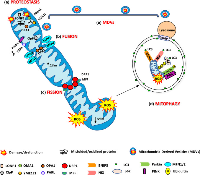
“Mitochondrial homeostasis is ensured by the coordination of pathways like mitochondrial biogenesis, mitochondrial dynamics, and degradative pathways like the activation of mitochondrial proteases, mitochondrial-derived vesicles, and mitophagy, the selective degradation of mitochondria via autophagy (Fig. 1).”
§ The connection between the dynamic remodeling of the mitochondrial network and the regulation of muscle mass > Quality control pathways finely tune mitochondrial function
plaindirect- Asserted: passes — The authors put it forward as true.
- Checkable: passes — It can be checked against evidence.
- Load-bearing: fails — The paper’s conclusions may not depend on it.
- Based: passes — The paper gives a reason why it holds.
Probably not a substantial claim. The paper’s conclusions may not depend on it.
The paper states this without a source or data behind it here, so there is nothing to check it against. The quote shows it is in the paper.
In the paper
“In contrast, the mitochondrial network in glycolytic fibers is fragmented and perpendicularly oriented to the muscle contraction axis and the I bands.”
§ The connection between the dynamic remodeling of the mitochondrial network and the regulation of muscle mass > The conformation and connectivity of the mitochondrial network are tailored to the metabolism and contractility of each fiber type
plaincorrelational- Asserted: passes — The authors put it forward as true.
- Checkable: passes — It can be checked against evidence.
- Load-bearing: unclear — It is unclear whether the paper’s conclusions depend on it.
- Based: passes — The paper gives a reason why it holds.
It may state two separate claims as one.
None of the sources cited for this claim have been checked yet.
In the paper
“Under stress conditions, when the degradation pathways are insufficient to repair the consequences of the alteration in mitochondrial translation [52] or the accumulation of oxidatively damaged or misfolded proteins, a retrograde signal is activated which coordinates nuclear gene expression. This mitochondria-to- nucleus response is named mitochondrial unfolding protein response (UPRmt) [53].”
§ The connection between the dynamic remodeling of the mitochondrial network and the regulation of muscle mass > Quality control pathways finely tune mitochondrial function > Mitochondrial proteostasis—a mitochondrial protein quality control mechanism
What the sources say
- Not checked yetcites [52] Callegari S, Dennerlein S (2018) Sensing the stress: a role for the UPRmt and UPRam in the quality control of mitochondria. Front Cell Dev Biol 6:31. https://doi.org/10.3389/fcell.2018.00031 source
- Not checked yetcites [53] Zhao Q, Wang J, Levichkin IV et al (2002) A mitochondrial specific stress response in mammalian cells. EMBO J 21:4411–4419. https://doi.org/10.1093/emboj/cdf445 source
plaindirect- Asserted: passes — The authors put it forward as true.
- Checkable: passes — It can be checked against evidence.
- Load-bearing: unclear — It is unclear whether the paper’s conclusions depend on it.
- Based: passes — The paper gives a reason why it holds.
It may state two separate claims as one.
The paper states this without a source or data behind it here, so there is nothing to check it against. The quote shows it is in the paper.
In the paper
“In mammals, mitochondrial fusion is independently regulated at the OMM and the IMM.”
§ The connection between the dynamic remodeling of the mitochondrial network and the regulation of muscle mass > Quality control pathways finely tune mitochondrial function > Mitochondrial dynamics
plain- Asserted: passes — The authors put it forward as true.
- Checkable: passes — It can be checked against evidence.
- Load-bearing: fails — The paper’s conclusions may not depend on it.
- Based: fails — The paper may give no reason why it holds.
Probably not a substantial claim. The paper’s conclusions may not depend on it. The paper may give no reason why it holds.
A conclusion the paper draws from its own results — it cites no other source for it, so there is none to check. The quote shows it is in the paper.
In the paper
“Thus, mitochondrial integrity and function need to be highly regulated.”
§ The connection between the dynamic remodeling of the mitochondrial network and the regulation of muscle mass > Quality control pathways finely tune mitochondrial function
plain- Asserted: passes — The authors put it forward as true.
- Checkable: passes — It can be checked against evidence.
- Load-bearing: passes — The paper’s conclusions depend on it.
- Based: passes — The paper gives a reason why it holds.
Probably not a substantial claim.
In the paper
“For example, the mitochondrial matrix protein OSCP, a subunit of OXPHOS complex V, can be retrotranslocated to the OMM, where it can be ubiquitinated and degraded [51].”
§ The connection between the dynamic remodeling of the mitochondrial network and the regulation of muscle mass > Quality control pathways finely tune mitochondrial function > Mitochondrial proteostasis—a mitochondrial protein quality control mechanism
What the sources say
- Not checked yetcites [51] Margineantu DH, Emerson CB, Diaz D, Hockenbery DM (2007) Hsp90 inhibition decreases mitochondrial protein turnover. PLoS ONE 2:e1066. https://doi.org/10.1371/journal.pone.0001066 source
plaindirect- Asserted: passes — The authors put it forward as true.
- Checkable: passes — It can be checked against evidence.
- Load-bearing: unclear — It is unclear whether the paper’s conclusions depend on it.
- Based: passes — The paper gives a reason why it holds.
It may state two separate claims as one.
The paper states this without a source or data behind it here, so there is nothing to check it against. The quote shows it is in the paper.
In the paper
“Mitoproteases do not only monitor mitochondrial protein quality, but also they can decide mitochondrial fate.”
§ The connection between the dynamic remodeling of the mitochondrial network and the regulation of muscle mass > Quality control pathways finely tune mitochondrial function > Mitochondrial proteostasis—a mitochondrial protein quality control mechanism
plaindirect- Asserted: passes — The authors put it forward as true.
- Checkable: passes — It can be checked against evidence.
- Load-bearing: unclear — It is unclear whether the paper’s conclusions depend on it.
- Based: passes — The paper gives a reason why it holds.
It may state two separate claims as one.
None of the sources cited for this claim have been checked yet.
In the paper
“Mitochondria have an ERAD-like mechanism, the mitochondria-associated degradation (MAD) pathway, and share with ERAD some key components.”
§ The connection between the dynamic remodeling of the mitochondrial network and the regulation of muscle mass > Quality control pathways finely tune mitochondrial function > Mitochondrial proteostasis—a mitochondrial protein quality control mechanism
plaincorrelational- Asserted: passes — The authors put it forward as true.
- Checkable: passes — It can be checked against evidence.
- Load-bearing: unclear — It is unclear whether the paper’s conclusions depend on it.
- Based: passes — The paper gives a reason why it holds.
It may state two separate claims as one.
In the paper
“However, only 13 proteins among the approximately 1200 proteins from the mitochondrial proteome, are encoded by mitochondrial DNA (mtDNA) and synthesized inside the organelle [34].”
§ The connection between the dynamic remodeling of the mitochondrial network and the regulation of muscle mass > Quality control pathways finely tune mitochondrial function > Mitochondrial proteostasis—a mitochondrial protein quality control mechanism
What the sources say
- Not checked yetcites [34] Calvo SE, Clauser KR, Mootha VK (2016) MitoCarta2.0: an updated inventory of mammalian mitochondrial proteins. Nucleic Acids Res 44:D1251-1257. https://doi.org/10.1093/nar/gkv1003 source
plain- Asserted: passes — The authors put it forward as true.
- Checkable: passes — It can be checked against evidence.
- Load-bearing: fails — The paper’s conclusions may not depend on it.
- Based: passes — The paper gives a reason why it holds.
Probably not a substantial claim. The paper’s conclusions may not depend on it.
The paper states this without a source or data behind it here, so there is nothing to check it against. The quote shows it is in the paper.
In the paper
“Mitochondrial dysfunction has been linked to several human diseases, like specific genetic defects, neurodegenerative and age-related diseases like aging sarcopenia, diabetes, and obesity.”
§ The connection between the dynamic remodeling of the mitochondrial network and the regulation of muscle mass > Quality control pathways finely tune mitochondrial function
plaincorrelational- Asserted: passes — The authors put it forward as true.
- Checkable: passes — It can be checked against evidence.
- Load-bearing: fails — The paper’s conclusions may not depend on it.
- Based: passes — The paper gives a reason why it holds.
Probably not a substantial claim. The paper’s conclusions may not depend on it.
None of the sources cited for this claim have been checked yet.
In the paper
“This process enables the redistribution of energy in the form of mitochondrial potential, metabolites, proteins, and mtDNA, improving calcium handling, and ATP synthesis [24, 54–57].”
§ The connection between the dynamic remodeling of the mitochondrial network and the regulation of muscle mass > Quality control pathways finely tune mitochondrial function > Mitochondrial dynamics
What the sources say
- Not checked yetcites [24] Glancy B, Hartnell LM, Malide D et al (2015) Mitochondrial reticulum for cellular energy distribution in muscle. Nature 523:617–620. https://doi.org/10.1038/nature14614 source
- Not checked yetcites [54] Eisner V, Lenaers G, Hajnóczky G (2014) Mitochondrial fusion is frequent in skeletal muscle and supports excitation-contraction coupling. J Cell Biol 205:179–195. https://doi.org/10.1083/jcb.201312066 source
- Not checked yetcites [55] Gomes LC, Di Benedetto G, Scorrano L (2011) During autophagy mitochondria elongate, are spared from degradation and sustain cell viability. Nat Cell Biol 13:589–598. https://doi.org/10.1038/ncb2220 source
- Not checked yetcites [56] Skulachev VP (2001) Mitochondrial filaments and clusters as intracellular power-transmitting cables. Trends Biochem Sci 26:23–29. https://doi.org/10.1016/s0968-0004(00)01735-7 source
- Not checked yetcites [57] Ono T, Isobe K, Nakada K, Hayashi JI (2001) Human cells are protected from mitochondrial dysfunction by complementation of DNA products in fused mitochondria. Nat Genet 28:272–275. https://doi.org/10.1038/90116 source
plaindirect- Asserted: passes — The authors put it forward as true.
- Checkable: passes — It can be checked against evidence.
- Load-bearing: passes — The paper’s conclusions depend on it.
- Based: passes — The paper gives a reason why it holds.
It may state two separate claims as one.
The paper states this without a source or data behind it here, so there is nothing to check it against. The quote shows it is in the paper.
In the paper
“In the mitochondrial matrix, protein turnover is controlled by 3 AAA proteases: the soluble LONP1 and ClpP and the membrane-bound m-AAA.”
§ The connection between the dynamic remodeling of the mitochondrial network and the regulation of muscle mass > Quality control pathways finely tune mitochondrial function > Mitochondrial proteostasis—a mitochondrial protein quality control mechanism
plaindirect- Asserted: passes — The authors put it forward as true.
- Checkable: passes — It can be checked against evidence.
- Load-bearing: fails — The paper’s conclusions may not depend on it.
- Based: passes — The paper gives a reason why it holds.
Probably not a substantial claim. The paper’s conclusions may not depend on it.
The paper states this without a source or data behind it here, so there is nothing to check it against. The quote shows it is in the paper.
In the paper
“According to the degree of mitochondrial damage, different pathways can be activated, ranging from the segmental repair of the damage to the whole degradation of the dysfunctional organelle.”
§ The connection between the dynamic remodeling of the mitochondrial network and the regulation of muscle mass > Quality control pathways finely tune mitochondrial function
plaindirect- Asserted: passes — The authors put it forward as true.
- Checkable: passes — It can be checked against evidence.
- Load-bearing: fails — The paper’s conclusions may not depend on it.
- Based: passes — The paper gives a reason why it holds.
Probably not a substantial claim. The paper’s conclusions may not depend on it.
In the paper
“Besides, the fusion between healthy and damaged organelles allows to dilute the damaged material into the healthy network, avoids the accumulation of dysfunctional mitochondria, and maintains their overall function [58].”
§ The connection between the dynamic remodeling of the mitochondrial network and the regulation of muscle mass > Quality control pathways finely tune mitochondrial function > Mitochondrial dynamics
What the sources say
- Not checked yetcites [58] Twig G, Elorza A, Molina AJA et al (2008) Fission and selective fusion govern mitochondrial segregation and elimination by autophagy. EMBO J 27:433–446. https://doi.org/10.1038/sj.emboj.7601963 source
plaindirect- Asserted: passes — The authors put it forward as true.
- Checkable: passes — It can be checked against evidence.
- Load-bearing: passes — The paper’s conclusions depend on it.
- Based: passes — The paper gives a reason why it holds.
It may state two separate claims as one.
In the paper
“The incorporation of these precursors into specific mitochondria subcompartments such as the outer mitochondrial membrane (OMM), the intermembrane space (IMS), the inner mitochondrial membrane (IMM) and the mitochondrial matrix requires specific and highly regulated import machinery [35].”
§ The connection between the dynamic remodeling of the mitochondrial network and the regulation of muscle mass > Quality control pathways finely tune mitochondrial function > Mitochondrial proteostasis—a mitochondrial protein quality control mechanism
What the sources say
- Not checked yetcites [35] Grevel A, Pfanner N, Becker T (2019) Coupling of import and assembly pathways in mitochondrial protein biogenesis. Biol Chem 401:117–129. https://doi.org/10.1515/hsz-2019-0310 source
plaindirect- Asserted: passes — The authors put it forward as true.
- Checkable: passes — It can be checked against evidence.
- Load-bearing: fails — The paper’s conclusions may not depend on it.
- Based: passes — The paper gives a reason why it holds.
Probably not a substantial claim. The paper’s conclusions may not depend on it.
The paper states this without a source or data behind it here, so there is nothing to check it against. The quote shows it is in the paper.
In the paper
“The nuclear genome encodes the remaining proteins, which are synthesized by ribosomes in precursor forms.”
§ The connection between the dynamic remodeling of the mitochondrial network and the regulation of muscle mass > Quality control pathways finely tune mitochondrial function > Mitochondrial proteostasis—a mitochondrial protein quality control mechanism
plain- Asserted: passes — The authors put it forward as true.
- Checkable: passes — It can be checked against evidence.
- Load-bearing: fails — The paper’s conclusions may not depend on it.
- Based: passes — The paper gives a reason why it holds.
Probably not a substantial claim. The paper’s conclusions may not depend on it. It may state two separate claims as one.
None of the sources cited for this claim have been checked yet.
In the paper
“Indeed, MFN2 bridges mitochondria to ER/SR, facilitating critical processes linked to ER–mitochondria interactions like calcium homeostasis and the modulation of the UPR during ER stress via PERK [63, 65–68].”
§ The connection between the dynamic remodeling of the mitochondrial network and the regulation of muscle mass > Quality control pathways finely tune mitochondrial function > Mitochondrial dynamics
What the sources say
- Not checked yetcites [63] de Brito OM, Scorrano L (2008) Mitofusin 2 tethers endoplasmic reticulum to mitochondria. Nature 456:605–610. https://doi.org/10.1038/nature07534 source
- Not checked yetcites [65] Muñoz JP, Ivanova S, Sánchez-Wandelmer J et al (2013) Mfn2 modulates the UPR and mitochondrial function via repression of PERK. EMBO J 32:2348–2361. https://doi.org/10.1038/emboj.2013.168 source
- Not checked yetcites [66] Ngoh GA, Papanicolaou KN, Walsh K (2012) Loss of mitofusin 2 promotes endoplasmic reticulum stress. J Biol Chem 287:20321–20332. https://doi.org/10.1074/jbc.M112.359174 source
- Not checked yetcites [67] Sebastián D, Hernández-Alvarez MI, Segalés J et al (2012) Mitofusin 2 (Mfn2) links mitochondrial and endoplasmic reticulum function with insulin signaling and is essential for normal glucose homeostasis. Proc Natl Acad Sci USA 109:5523–5528. https://doi.org/10.1073/pnas.1108220109 source
- Not checked yetcites [68] Ainbinder A, Boncompagni S, Protasi F, Dirksen RT (2015) Role of Mitofusin-2 in mitochondrial localization and calcium uptake in skeletal muscle. Cell Calcium 57:14–24. https://doi.org/10.1016/j.ceca.2014.11.002 source
plaindirect- Asserted: passes — The authors put it forward as true.
- Checkable: passes — It can be checked against evidence.
- Load-bearing: unclear — It is unclear whether the paper’s conclusions depend on it.
- Based: passes — The paper gives a reason why it holds.
It may state two separate claims as one.
In the paper
“Mitochondrial fusion leads to elongated organelles, which expand the mitochondrial network and its interconnectivity [32].”
§ The connection between the dynamic remodeling of the mitochondrial network and the regulation of muscle mass > Quality control pathways finely tune mitochondrial function > Mitochondrial dynamics
What the sources say
- Not checked yetcites [32] Bleck CKE, Kim Y, Willingham TB, Glancy B (2018) Subcellular connectomic analyses of energy networks in striated muscle. Nat Commun 9:5111. https://doi.org/10.1038/s41467-018-07676-y source
plaindirect- Asserted: passes — The authors put it forward as true.
- Checkable: passes — It can be checked against evidence.
- Load-bearing: unclear — It is unclear whether the paper’s conclusions depend on it.
- Based: passes — The paper gives a reason why it holds.
It may state two separate claims as one.
The paper states this without a source or data behind it here, so there is nothing to check it against. The quote shows it is in the paper.
In the paper
“It starts with the tethering of two adjacent mitochondria, continues with the fusion of OMM, and concludes with IMM fusion.”
§ The connection between the dynamic remodeling of the mitochondrial network and the regulation of muscle mass > Quality control pathways finely tune mitochondrial function > Mitochondrial dynamics
plaindirect- Asserted: passes — The authors put it forward as true.
- Checkable: passes — It can be checked against evidence.
- Load-bearing: fails — The paper’s conclusions may not depend on it.
- Based: passes — The paper gives a reason why it holds.
Probably not a substantial claim. The paper’s conclusions may not depend on it. It may state two separate claims as one.
In the paper
“Mitochondria have their genome, transcription, and translation machinery [33].”
§ The connection between the dynamic remodeling of the mitochondrial network and the regulation of muscle mass > Quality control pathways finely tune mitochondrial function > Mitochondrial proteostasis—a mitochondrial protein quality control mechanism
What the sources say
- Not checked yetcites [33] Lightowlers RN, Rozanska A, Chrzanowska-Lightowlers ZM (2014) Mitochondrial protein synthesis: figuring the fundamentals, complexities and complications, of mammalian mitochondrial translation. FEBS Lett 588:2496–2503. https://doi.org/10.1016/j.febslet.2014.05.054 source
plain- Asserted: passes — The authors put it forward as true.
- Checkable: passes — It can be checked against evidence.
- Load-bearing: fails — The paper’s conclusions may not depend on it.
- Based: passes — The paper gives a reason why it holds.
Probably not a substantial claim. The paper’s conclusions may not depend on it. It may state two separate claims as one.
The paper states this without a source or data behind it here, so there is nothing to check it against. The quote shows it is in the paper.
In the paper
“On the contrary, fission separates the dysfunctional or damaged components from the network. The resulting unconnected shorter mitochondria will be further removed via mitophagy.”
§ The connection between the dynamic remodeling of the mitochondrial network and the regulation of muscle mass > Quality control pathways finely tune mitochondrial function > Mitochondrial dynamics
plaindirect- Asserted: passes — The authors put it forward as true.
- Checkable: passes — It can be checked against evidence.
- Load-bearing: unclear — It is unclear whether the paper’s conclusions depend on it.
- Based: passes — The paper gives a reason why it holds.
It may state two separate claims as one.
In the paper
“During ER stress, misfolded proteins accumulate into the ER lumen. They need to be retrotranslocated into the cytosol, where they are flagged with ubiquitin and degraded by the proteasome in a process called ER-associated protein degradation (ERAD) [47].”
§ The connection between the dynamic remodeling of the mitochondrial network and the regulation of muscle mass > Quality control pathways finely tune mitochondrial function > Mitochondrial proteostasis—a mitochondrial protein quality control mechanism
What the sources say
- Not checked yetcites [47] Ye Y, Meyer HH, Rapoport TA (2001) The AAA ATPase Cdc48/p97 and its partners transport proteins from the ER into the cytosol. Nature 414:652–656. https://doi.org/10.1038/414652a source
plaindirect- Asserted: passes — The authors put it forward as true.
- Checkable: passes — It can be checked against evidence.
- Load-bearing: fails — The paper’s conclusions may not depend on it.
- Based: passes — The paper gives a reason why it holds.
Probably not a substantial claim. The paper’s conclusions may not depend on it. It may state two separate claims as one.
In the paper
“Moreover, unfolded IMS proteins can exit the IMS through the TOM translocase, which is the same route used for import [40].”
§ The connection between the dynamic remodeling of the mitochondrial network and the regulation of muscle mass > Quality control pathways finely tune mitochondrial function > Mitochondrial proteostasis—a mitochondrial protein quality control mechanism
What the sources say
- Not checked yetcites [40] Bragoszewski P, Gornicka A, Sztolsztener ME, Chacinska A (2013) The ubiquitin-proteasome system regulates mitochondrial intermembrane space proteins. Mol Cell Biol 33:2136–2148. https://doi.org/10.1128/MCB.01579-12 source
plaindirect- Asserted: passes — The authors put it forward as true.
- Checkable: passes — It can be checked against evidence.
- Load-bearing: fails — The paper’s conclusions may not depend on it.
- Based: passes — The paper gives a reason why it holds.
Probably not a substantial claim. The paper’s conclusions may not depend on it.
The paper states this without a source or data behind it here, so there is nothing to check it against. The quote shows it is in the paper.
In the paper
“Mitochondrial fission is a multi-step process that depends primarily on the cytosolic GTPase dynamin-related protein 1 (DRP1).”
§ The connection between the dynamic remodeling of the mitochondrial network and the regulation of muscle mass > Quality control pathways finely tune mitochondrial function > Mitochondrial dynamics
plaindirect- Asserted: passes — The authors put it forward as true.
- Checkable: passes — It can be checked against evidence.
- Load-bearing: fails — The paper’s conclusions may not depend on it.
- Based: passes — The paper gives a reason why it holds.
Probably not a substantial claim. The paper’s conclusions may not depend on it.
In the paper
“Although DRP1 can tubulate the membranes, it cannot complete membrane scission [84].”
§ The connection between the dynamic remodeling of the mitochondrial network and the regulation of muscle mass > Quality control pathways finely tune mitochondrial function > Mitochondrial dynamics
What the sources say
- Not checked yetcites [84] Yoon Y, Pitts KR, McNiven MA (2001) Mammalian dynamin-like protein DLP1 tubulates membranes. Mol Biol Cell 12:2894–2905. https://doi.org/10.1091/mbc.12.9.2894 source
plain- Asserted: passes — The authors put it forward as true.
- Checkable: passes — It can be checked against evidence.
- Load-bearing: unclear — It is unclear whether the paper’s conclusions depend on it.
- Based: passes — The paper gives a reason why it holds.
It may state two separate claims as one.
The paper states this without a source or data behind it here, so there is nothing to check it against. The quote shows it is in the paper.
In the paper
“DRP1 lacks hydrophobic membrane-binding domains, and so, its recruitment depends on integral OMM proteins that act as receptors/adaptors.”
§ The connection between the dynamic remodeling of the mitochondrial network and the regulation of muscle mass > Quality control pathways finely tune mitochondrial function > Mitochondrial dynamics
plaindirect- Asserted: passes — The authors put it forward as true.
- Checkable: passes — It can be checked against evidence.
- Load-bearing: unclear — It is unclear whether the paper’s conclusions depend on it.
- Based: passes — The paper gives a reason why it holds.
It may state two separate claims as one.
None of the sources cited for this claim have been checked yet.
In the paper
“The inner membrane constriction occurs at ER–mitochondria contact sites before DRP1 recruitment and is thus independent of DRP1 action and OMM division [102, 103].”
§ The connection between the dynamic remodeling of the mitochondrial network and the regulation of muscle mass > Quality control pathways finely tune mitochondrial function > Mitochondrial dynamics
What the sources say
- Not checked yetcites [102] Cho B, Cho HM, Jo Y et al (2017) Constriction of the mitochondrial inner compartment is a priming event for mitochondrial division. Nat Commun 8:15754. https://doi.org/10.1038/ncomms15754 source
- Not checked yetcites [103] Chakrabarti R, Ji W-K, Stan RV et al (2018) INF2-mediated actin polymerization at the ER stimulates mitochondrial calcium uptake, inner membrane constriction, and division. J Cell Biol 217:251–268. https://doi.org/10.1083/jcb.201709111 source
plain- Asserted: passes — The authors put it forward as true.
- Checkable: passes — It can be checked against evidence.
- Load-bearing: unclear — It is unclear whether the paper’s conclusions depend on it.
- Based: passes — The paper gives a reason why it holds.
It may state two separate claims as one.
In the paper
“OPA1-dependent mitochondrial fusion needs Mfn1 [73] as well as balanced OPA1S and OPA1L forms.”
§ The connection between the dynamic remodeling of the mitochondrial network and the regulation of muscle mass > Quality control pathways finely tune mitochondrial function > Mitochondrial dynamics
What the sources say
- Not checked yetcites [73] Cipolat S, Martins de Brito O, Dal Zilio B, Scorrano L (2004) OPA1 requires mitofusin 1 to promote mitochondrial fusion. Proc Natl Acad Sci USA 101:15927–15932. https://doi.org/10.1073/pnas.0407043101 source
plaindirect- Asserted: passes — The authors put it forward as true.
- Checkable: passes — It can be checked against evidence.
- Load-bearing: unclear — It is unclear whether the paper’s conclusions depend on it.
- Based: passes — The paper gives a reason why it holds.
It may state two separate claims as one.
A conclusion the paper draws from its own results — it cites no other source for it, so there is none to check. The quote shows it is in the paper.
In the paper
“The DRP1 affinity for MFF is higher than for Fis1, suggesting that MFF preferentially functions as a DRP1 receptor.”
§ The connection between the dynamic remodeling of the mitochondrial network and the regulation of muscle mass > Quality control pathways finely tune mitochondrial function > Mitochondrial dynamics
hedgedcorrelational- Asserted: passes — The authors put it forward as true.
- Checkable: passes — It can be checked against evidence.
- Load-bearing: unclear — It is unclear whether the paper’s conclusions depend on it.
- Based: passes — The paper gives a reason why it holds.
It may state two separate claims as one.
In the paper
“However, mammals lack a homolog of the fission protein FtsZ [99].”
§ The connection between the dynamic remodeling of the mitochondrial network and the regulation of muscle mass > Quality control pathways finely tune mitochondrial function > Mitochondrial dynamics
What the sources say
- Not checked yetcites [99] Leger MM, Petrů M, Žárský V et al (2015) An ancestral bacterial division system is widespread in eukaryotic mitochondria. Proc Natl Acad Sci USA 112:10239–10246. https://doi.org/10.1073/pnas.1421392112 source
plain- Asserted: passes — The authors put it forward as true.
- Checkable: passes — It can be checked against evidence.
- Load-bearing: fails — The paper’s conclusions may not depend on it.
- Based: passes — The paper gives a reason why it holds.
Probably not a substantial claim. The paper’s conclusions may not depend on it.
In the paper
“OPA1 is a pleiotropic protein that, by forming oligomers of soluble and membrane-bound OPA1 isoforms, also controls cristae remodeling and the assembly of respiratory chain complexes into supercomplexes, a structure that enhances mitochondrial respiration [78].”
§ The connection between the dynamic remodeling of the mitochondrial network and the regulation of muscle mass > Quality control pathways finely tune mitochondrial function > Mitochondrial dynamics
What the sources say
- Not checked yetcites [78] Cogliati S, Frezza C, Soriano ME et al (2013) Mitochondrial cristae shape determines respiratory chain supercomplexes assembly and respiratory efficiency. Cell 155:160–171. https://doi.org/10.1016/j.cell.2013.08.032 source
plaindirect- Asserted: passes — The authors put it forward as true.
- Checkable: passes — It can be checked against evidence.
- Load-bearing: unclear — It is unclear whether the paper’s conclusions depend on it.
- Based: passes — The paper gives a reason why it holds.
It may state two separate claims as one.
None of the sources cited for this claim have been checked yet.
In the paper
“MIEF1/MiD51 and the variant MIEF2/MiD49 recruit DRP1 to mitochondria independent of MFF, but in contrast to MFF, inhibit DRP1 GTPase activity leading to mitochondrial elongation when MiD49/51 is overexpressed [92, 95, 96].”
§ The connection between the dynamic remodeling of the mitochondrial network and the regulation of muscle mass > Quality control pathways finely tune mitochondrial function > Mitochondrial dynamics
What the sources say
- Not checked yetcites [92] Palmer CS, Elgass KD, Parton RG et al (2013) Adaptor proteins MiD49 and MiD51 can act independently of Mff and Fis1 in Drp1 recruitment and are specific for mitochondrial fission. J Biol Chem 288:27584–27593. https://doi.org/10.1074/jbc.M113.479873 source
- Not checked yetcites [95] Zhao J, Liu T, Jin S et al (2011) Human MIEF1 recruits Drp1 to mitochondrial outer membranes and promotes mitochondrial fusion rather than fission. EMBO J 30:2762–2778. https://doi.org/10.1038/emboj.2011.198 source
- Not checked yetcites [96] Losón OC, Song Z, Chen H, Chan DC (2013) Fis1, Mff, MiD49, and MiD51 mediate Drp1 recruitment in mitochondrial fission. Mol Biol Cell 24:659–667. https://doi.org/10.1091/mbc.E12-10-0721 source
plaindirect- Asserted: passes — The authors put it forward as true.
- Checkable: passes — It can be checked against evidence.
- Load-bearing: unclear — It is unclear whether the paper’s conclusions depend on it.
- Based: passes — The paper gives a reason why it holds.
It may state two separate claims as one.
In the paper
“Subsequently, DRP1 is recruited to the marked division sites to bind to its OMM receptors/adaptors. This binding facilitates DRP1 oligomerization, forming a ring-like structure that favors an additional narrowing of the membranes [83].”
§ The connection between the dynamic remodeling of the mitochondrial network and the regulation of muscle mass > Quality control pathways finely tune mitochondrial function > Mitochondrial dynamics
What the sources say
- Not checked yetcites [83] Kraus F, Ryan MT (2017) The constriction and scission machineries involved in mitochondrial fission. J Cell Sci 130:2953–2960. https://doi.org/10.1242/jcs.199562 source
plaindirect- Asserted: passes — The authors put it forward as true.
- Checkable: passes — It can be checked against evidence.
- Load-bearing: fails — The paper’s conclusions may not depend on it.
- Based: passes — The paper gives a reason why it holds.
Probably not a substantial claim. The paper’s conclusions may not depend on it. It may state two separate claims as one.
None of the sources cited for this claim have been checked yet.
In the paper
“On the contrary, silencing MFF generates elongated mitochondria and DRP1 cytosolic distribution [90, 93].”
§ The connection between the dynamic remodeling of the mitochondrial network and the regulation of muscle mass > Quality control pathways finely tune mitochondrial function > Mitochondrial dynamics
What the sources say
- Not checked yetcites [90] Otera H, Wang C, Cleland MM et al (2010) Mff is an essential factor for mitochondrial recruitment of Drp1 during mitochondrial fission in mammalian cells. J Cell Biol 191:1141–1158. https://doi.org/10.1083/jcb.201007152 source
- Not checked yetcites [93] Gandre-Babbe S, van der Bliek AM (2008) The novel tail-anchored membrane protein Mff controls mitochondrial and peroxisomal fission in mammalian cells. Mol Biol Cell 19:2402–2412. https://doi.org/10.1091/mbc.e07-12-1287 source
plaindirect- Asserted: passes — The authors put it forward as true.
- Checkable: passes — It can be checked against evidence.
- Load-bearing: unclear — It is unclear whether the paper’s conclusions depend on it.
- Based: passes — The paper gives a reason why it holds.
It may state two separate claims as one.
In the paper
“Under stress conditions, hyperacetylation of OPA1 reduces its GTPase activity while SIRT3-dependent deacetylation increases OPA1 GTPase activity [79].”
§ The connection between the dynamic remodeling of the mitochondrial network and the regulation of muscle mass > Quality control pathways finely tune mitochondrial function > Mitochondrial dynamics
What the sources say
- Not checked yetcites [79] Samant SA, Zhang HJ, Hong Z et al (2014) SIRT3 deacetylates and activates OPA1 to regulate mitochondrial dynamics during stress. Mol Cell Biol 34:807–819. https://doi.org/10.1128/MCB.01483-13 source
plaindirect- Asserted: passes — The authors put it forward as true.
- Checkable: passes — It can be checked against evidence.
- Load-bearing: unclear — It is unclear whether the paper’s conclusions depend on it.
- Based: passes — The paper gives a reason why it holds.
It may state two separate claims as one.
In the paper
“Importantly, ERMD increases in the presence of mtDNA. Thus, mtDNA synthesis is coupled to mitochondrial division to ensure the distribution of newly replicated mtDNA to daughter mitochondria [80].”
§ The connection between the dynamic remodeling of the mitochondrial network and the regulation of muscle mass > Quality control pathways finely tune mitochondrial function > Mitochondrial dynamics
What the sources say
- Not checked yetcites [80] Lewis SC, Uchiyama LF, Nunnari J (2016) ER-mitochondria contacts couple mtDNA synthesis with mitochondrial division in human cells. Science 353:aff5549. https://doi.org/10.1126/science.aaf5549 source
plaindirect- Asserted: passes — The authors put it forward as true.
- Checkable: passes — It can be checked against evidence.
- Load-bearing: unclear — It is unclear whether the paper’s conclusions depend on it.
- Based: passes — The paper gives a reason why it holds.
It may state two separate claims as one.
In the paper
“Indeed, the GTPase Dynamin 2 (DYN2) assembles in the DRP1-mediated mitochondrial constriction neck to drive mitochondrial scission [85].”
§ The connection between the dynamic remodeling of the mitochondrial network and the regulation of muscle mass > Quality control pathways finely tune mitochondrial function > Mitochondrial dynamics
What the sources say
- Not checked yetcites [85] Lee JE, Westrate LM, Wu H et al (2016) Multiple dynamin family members collaborate to drive mitochondrial division. Nature 540:139–143. https://doi.org/10.1038/nature20555 source
plaindirect- Asserted: passes — The authors put it forward as true.
- Checkable: passes — It can be checked against evidence.
- Load-bearing: fails — The paper’s conclusions may not depend on it.
- Based: passes — The paper gives a reason why it holds.
Probably not a substantial claim. The paper’s conclusions may not depend on it.
None of the sources cited for this claim have been checked yet.
In the paper
“The mechanism which drives IMM constriction are not elucidated, but the requirement of MCU and OMA1-induced OPA1S has been shown [102, 103].”
§ The connection between the dynamic remodeling of the mitochondrial network and the regulation of muscle mass > Quality control pathways finely tune mitochondrial function > Mitochondrial dynamics
What the sources say
- Not checked yetcites [102] Cho B, Cho HM, Jo Y et al (2017) Constriction of the mitochondrial inner compartment is a priming event for mitochondrial division. Nat Commun 8:15754. https://doi.org/10.1038/ncomms15754 source
- Not checked yetcites [103] Chakrabarti R, Ji W-K, Stan RV et al (2018) INF2-mediated actin polymerization at the ER stimulates mitochondrial calcium uptake, inner membrane constriction, and division. J Cell Biol 217:251–268. https://doi.org/10.1083/jcb.201709111 source
plaindirect- Asserted: passes — The authors put it forward as true.
- Checkable: passes — It can be checked against evidence.
- Load-bearing: unclear — It is unclear whether the paper’s conclusions depend on it.
- Based: passes — The paper gives a reason why it holds.
It may state two separate claims as one.
In the paper
“OPA1 deletion leads to mitochondrial fragmentation, whereas OPA1 overexpression induces mitochondrial elongation [73].”
§ The connection between the dynamic remodeling of the mitochondrial network and the regulation of muscle mass > Quality control pathways finely tune mitochondrial function > Mitochondrial dynamics
What the sources say
- Not checked yetcites [73] Cipolat S, Martins de Brito O, Dal Zilio B, Scorrano L (2004) OPA1 requires mitofusin 1 to promote mitochondrial fusion. Proc Natl Acad Sci USA 101:15927–15932. https://doi.org/10.1073/pnas.0407043101 source
plaindirect- Asserted: passes — The authors put it forward as true.
- Checkable: passes — It can be checked against evidence.
- Load-bearing: unclear — It is unclear whether the paper’s conclusions depend on it.
- Based: passes — The paper gives a reason why it holds.
It may state two separate claims as one.
None of the sources cited for this claim have been checked yet.
In the paper
“In mammals, there are no orthologues for MDV1 and Cav 4, and recent evidence suggests that Fis1 is not required for fission [89, 90].”
§ The connection between the dynamic remodeling of the mitochondrial network and the regulation of muscle mass > Quality control pathways finely tune mitochondrial function > Mitochondrial dynamics
What the sources say
- Not checked yetcites [89] Osellame LD, Singh AP, Stroud DA et al (2016) Cooperative and independent roles of the Drp1 adaptors Mff, MiD49 and MiD51 in mitochondrial fission. J Cell Sci 129:2170–2181. https://doi.org/10.1242/jcs.185165 source
- Not checked yetcites [90] Otera H, Wang C, Cleland MM et al (2010) Mff is an essential factor for mitochondrial recruitment of Drp1 during mitochondrial fission in mammalian cells. J Cell Biol 191:1141–1158. https://doi.org/10.1083/jcb.201007152 source
hedged- Asserted: passes — The authors put it forward as true.
- Checkable: passes — It can be checked against evidence.
- Load-bearing: unclear — It is unclear whether the paper’s conclusions depend on it.
- Based: passes — The paper gives a reason why it holds.
It may state two separate claims as one.
None of the sources cited for this claim have been checked yet.
In the paper
“On the contrary, the complete conversion of OPA1L into OPA1S inhibits fusion and increases mitochondrial fission [74, 76].”
§ The connection between the dynamic remodeling of the mitochondrial network and the regulation of muscle mass > Quality control pathways finely tune mitochondrial function > Mitochondrial dynamics
What the sources say
- Not checked yetcites [74] Ishihara N, Fujita Y, Oka T, Mihara K (2006) Regulation of mitochondrial morphology through proteolytic cleavage of OPA1. EMBO J 25:2966–2977. https://doi.org/10.1038/sj.emboj.7601184 source
- Not checked yetcites [76] Anand R, Wai T, Baker MJ et al (2014) The i-AAA protease YME1L and OMA1 cleave OPA1 to balance mitochondrial fusion and fission. J Cell Biol 204:919–929. https://doi.org/10.1083/jcb.201308006 source
plaindirect- Asserted: passes — The authors put it forward as true.
- Checkable: passes — It can be checked against evidence.
- Load-bearing: unclear — It is unclear whether the paper’s conclusions depend on it.
- Based: passes — The paper gives a reason why it holds.
It may state two separate claims as one.
The paper states this without a source or data behind it here, so there is nothing to check it against. The quote shows it is in the paper.
In the paper
“Mitochondrial division requires the division of both the inner and outer mitochondrial membranes.”
§ The connection between the dynamic remodeling of the mitochondrial network and the regulation of muscle mass > Quality control pathways finely tune mitochondrial function > Mitochondrial dynamics
plaindirect- Asserted: passes — The authors put it forward as true.
- Checkable: passes — It can be checked against evidence.
- Load-bearing: fails — The paper’s conclusions may not depend on it.
- Based: passes — The paper gives a reason why it holds.
Probably not a substantial claim. The paper’s conclusions may not depend on it.
In the paper
“In prokaryotic cells and certain primitive eukaryotic species, FtsZ is a self-assembly GTPase that forms a calcium-mediated constricting ring on IMM's matrix side, suggesting that specific mechanisms for IMM constriction must exist [98].”
§ The connection between the dynamic remodeling of the mitochondrial network and the regulation of muscle mass > Quality control pathways finely tune mitochondrial function > Mitochondrial dynamics
What the sources say
- Not checked yetcites [98] Nishida K, Takahara M, Miyagishima S et al (2003) Dynamic recruitment of dynamin for final mitochondrial severance in a primitive red alga. Proc Natl Acad Sci USA 100:2146–2151. https://doi.org/10.1073/pnas.0436886100 source
hedgeddirect- Asserted: passes — The authors put it forward as true.
- Checkable: passes — It can be checked against evidence.
- Load-bearing: unclear — It is unclear whether the paper’s conclusions depend on it.
- Based: passes — The paper gives a reason why it holds.
It may state two separate claims as one.
None of the sources cited for this claim have been checked yet.
In the paper
“The full knockout mice of either Mfn1, Mfn2, or OPA1 result in embryonic lethality of mice, demonstrating the biological importance of mitochondrial fusion in early development [60, 178].”
§ The connection between the dynamic remodeling of the mitochondrial network and the regulation of muscle mass > Mitochondria quality pathways are essential for skeletal muscle physiology > Dysregulation of mitochondrial fusion in skeletal muscle leads to muscle atrophy, weakness, and have severe consequences in whole-body physiology
What the sources say
- Not checked yetcites [60] Chen H, Detmer SA, Ewald AJ et al (2003) Mitofusins Mfn1 and Mfn2 coordinately regulate mitochondrial fusion and are essential for embryonic development. J Cell Biol 160:189–200. https://doi.org/10.1083/jcb.200211046 source
- Not checked yetcites [178] Davies VJ, Hollins AJ, Piechota MJ et al (2007) Opa1 deficiency in a mouse model of autosomal dominant optic atrophy impairs mitochondrial morphology, optic nerve structure and visual function. Hum Mol Genet 16:1307–1318. https://doi.org/10.1093/hmg/ddm079 source
boosteddirect- Asserted: passes — The authors put it forward as true.
- Checkable: passes — It can be checked against evidence.
- Load-bearing: unclear — It is unclear whether the paper’s conclusions depend on it.
- Based: passes — The paper gives a reason why it holds.
It may state two separate claims as one.
A conclusion the paper draws from its own results — it cites no other source for it, so there is none to check. The quote shows it is in the paper.
In the paper
“The central role of mitochondria in skeletal muscle homeostasis depends not only on energy production, but also on the buffering of intracellular calcium, and the signaling pathways that control nuclear gene programs which regulate muscle mass.”
§ The connection between the dynamic remodeling of the mitochondrial network and the regulation of muscle mass > Mitochondria quality pathways are essential for skeletal muscle physiology
plaindirect- Asserted: passes — The authors put it forward as true.
- Checkable: passes — It can be checked against evidence.
- Load-bearing: passes — The paper’s conclusions depend on it.
- Based: passes — The paper gives a reason why it holds.
It may state two separate claims as one.
In the paper
“Moreover, p97/VCP deficiency resulted in the accumulation of numerous vesicular bodies, autophagy impairment, and altered myofibrillar organization, causing a severe myopathy.”
§ The connection between the dynamic remodeling of the mitochondrial network and the regulation of muscle mass > Mitochondria quality pathways are essential for skeletal muscle physiology > Mitochondrial proteostasis is critical for skeletal muscle function
What the sources say
- Not checked yetcites [175] Kustermann M, Manta L, Paone C et al (2018) Loss of the novel Vcp (valosin containing protein) interactor Washc4 interferes with autophagy-mediated proteostasis in striated muscle and leads to myopathy in vivo. Autophagy 14:1911–1927. https://doi.org/10.1080/15548627.2018.1491491 source
plaindirect- Asserted: passes — The authors put it forward as true.
- Checkable: passes — It can be checked against evidence.
- Load-bearing: passes — The paper’s conclusions depend on it.
- Based: passes — The paper gives a reason why it holds.
It may state two separate claims as one.
In the paper
“Interestingly, cardiac function and lifespan of mice lacking YME1L in cardiomyocytes are normalized without restoring mitochondrial morphology, when the deletion of YME1L happens in both cardiomyocytes and skeletal muscle fibers (median life span 125 weeks).”
§ The connection between the dynamic remodeling of the mitochondrial network and the regulation of muscle mass > Mitochondria quality pathways are essential for skeletal muscle physiology > Mitochondrial proteostasis is critical for skeletal muscle function
What the sources say
- Not checked yetcites [166] Wai T, García-Prieto J, Baker MJ et al (2015) Imbalanced OPA1 processing and mitochondrial fragmentation cause heart failure in mice. Science 350:aad0116. https://doi.org/10.1126/science.aad0116 source
plaindirect- Asserted: passes — The authors put it forward as true.
- Checkable: passes — It can be checked against evidence.
- Load-bearing: passes — The paper’s conclusions depend on it.
- Based: passes — The paper gives a reason why it holds.
It may state two separate claims as one.
None of the sources cited for this claim have been checked yet.
In the paper
“Loss-of-function mutations in MFN2 and OPA1 genes cause two neurodegenerative diseases, Charcot–Marie–Tooth type 2A (CMT2A) [179] and dominant optic atrophy (DOA) [180, 181], respectively.”
§ The connection between the dynamic remodeling of the mitochondrial network and the regulation of muscle mass > Mitochondria quality pathways are essential for skeletal muscle physiology > Dysregulation of mitochondrial fusion in skeletal muscle leads to muscle atrophy, weakness, and have severe consequences in whole-body physiology
What the sources say
- Not checked yetcites [179] Züchner S, Mersiyanova IV, Muglia M et al (2004) Mutations in the mitochondrial GTPase mitofusin 2 cause Charcot-Marie-Tooth neuropathy type 2A. Nat Genet 36:449–451. https://doi.org/10.1038/ng1341 source
- Not checked yetcites [180] Alexander C, Votruba M, Pesch UE et al (2000) OPA1, encoding a dynamin-related GTPase, is mutated in autosomal dominant optic atrophy linked to chromosome 3q28. Nat Genet 26:211–215. https://doi.org/10.1038/79944 source
- Not checked yetcites [181] Delettre C, Lenaers G, Griffoin JM et al (2000) Nuclear gene OPA1, encoding a mitochondrial dynamin-related protein, is mutated in dominant optic atrophy. Nat Genet 26:207–210. https://doi.org/10.1038/79936 source
plaindirect- Asserted: passes — The authors put it forward as true.
- Checkable: passes — It can be checked against evidence.
- Load-bearing: unclear — It is unclear whether the paper’s conclusions depend on it.
- Based: passes — The paper gives a reason why it holds.
It may state two separate claims as one.
In the paper
“mitochondrial fusion in skeletal muscle is necessary to adapt to the cell's specific functional needs and support skeletal muscle myofibers contractile function [54].”
§ The connection between the dynamic remodeling of the mitochondrial network and the regulation of muscle mass > Mitochondria quality pathways are essential for skeletal muscle physiology > Dysregulation of mitochondrial fusion in skeletal muscle leads to muscle atrophy, weakness, and have severe consequences in whole-body physiology
What the sources say
- Not checked yetcites [54] Eisner V, Lenaers G, Hajnóczky G (2014) Mitochondrial fusion is frequent in skeletal muscle and supports excitation-contraction coupling. J Cell Biol 205:179–195. https://doi.org/10.1083/jcb.201312066 source
plaindirect- Asserted: passes — The authors put it forward as true.
- Checkable: passes — It can be checked against evidence.
- Load-bearing: passes — The paper’s conclusions depend on it.
- Based: passes — The paper gives a reason why it holds.
It may state two separate claims as one.
In the paper
“In agreement, double deletion of Yme1L1 and OMA1, specifically in the heart, restored mitochondrial morphology and rescued cardiac function [166].”
§ The connection between the dynamic remodeling of the mitochondrial network and the regulation of muscle mass > Mitochondria quality pathways are essential for skeletal muscle physiology > Mitochondrial proteostasis is critical for skeletal muscle function
What the sources say
- Not checked yetcites [166] Wai T, García-Prieto J, Baker MJ et al (2015) Imbalanced OPA1 processing and mitochondrial fragmentation cause heart failure in mice. Science 350:aad0116. https://doi.org/10.1126/science.aad0116 source
plaindirect- Asserted: passes — The authors put it forward as true.
- Checkable: passes — It can be checked against evidence.
- Load-bearing: passes — The paper’s conclusions depend on it.
- Based: passes — The paper gives a reason why it holds.
It may state two separate claims as one.
None of the sources cited for this claim have been checked yet.
In the paper
“Mutations in p97 in mice [173] and humans [174] cause mitochondrial swelling, cristae disruption, oxidative stress, and decreased ATP, resulting in skeletal muscle atrophy and weakness.”
§ The connection between the dynamic remodeling of the mitochondrial network and the regulation of muscle mass > Mitochondria quality pathways are essential for skeletal muscle physiology > Mitochondrial proteostasis is critical for skeletal muscle function
What the sources say
- Not checked yetcites [173] Nalbandian A, Llewellyn KJ, Kitazawa M et al (2012) The homozygote VCPR155H/R155H mouse model exhibits accelerated human VCP-associated disease pathology. PLoS ONE 7:e46308. https://doi.org/10.1371/journal.pone.0046308 source
- Not checked yetcites [174] Bartolome F, Wu H-C, Burchell VS et al (2013) Pathogenic VCP mutations induce mitochondrial uncoupling and reduced ATP levels. Neuron 78:57–64. https://doi.org/10.1016/j.neuron.2013.02.028 source
plaindirect- Asserted: passes — The authors put it forward as true.
- Checkable: passes — It can be checked against evidence.
- Load-bearing: passes — The paper’s conclusions depend on it.
- Based: passes — The paper gives a reason why it holds.
It may state two separate claims as one.
In the paper
“In mice, total genetic ablation of Yme1L1 leads to embryonic death, while the specific inhibition of the gene in the heart causes dilated cardiomyopathy that shortens lifespan (median life span:46 weeks).”
§ The connection between the dynamic remodeling of the mitochondrial network and the regulation of muscle mass > Mitochondria quality pathways are essential for skeletal muscle physiology > Mitochondrial proteostasis is critical for skeletal muscle function
What the sources say
- Not checked yetcites [166] Wai T, García-Prieto J, Baker MJ et al (2015) Imbalanced OPA1 processing and mitochondrial fragmentation cause heart failure in mice. Science 350:aad0116. https://doi.org/10.1126/science.aad0116 source
plaindirect- Asserted: passes — The authors put it forward as true.
- Checkable: passes — It can be checked against evidence.
- Load-bearing: unclear — It is unclear whether the paper’s conclusions depend on it.
- Based: passes — The paper gives a reason why it holds.
It may state two separate claims as one.
In the paper
“Mechanistically, YME1L ablation in cardiomyocytes activates OMA1 and promotes OPA1 processing and mitochondrial fragmentation, which causes heart failure [166].”
§ The connection between the dynamic remodeling of the mitochondrial network and the regulation of muscle mass > Mitochondria quality pathways are essential for skeletal muscle physiology > Mitochondrial proteostasis is critical for skeletal muscle function
What the sources say
- Not checked yetcites [166] Wai T, García-Prieto J, Baker MJ et al (2015) Imbalanced OPA1 processing and mitochondrial fragmentation cause heart failure in mice. Science 350:aad0116. https://doi.org/10.1126/science.aad0116 source
plaindirect- Asserted: passes — The authors put it forward as true.
- Checkable: passes — It can be checked against evidence.
- Load-bearing: passes — The paper’s conclusions depend on it.
- Based: passes — The paper gives a reason why it holds.
It may state two separate claims as one.
None of the sources cited for this claim have been checked yet.
In the paper
“In human skeletal muscle, the reduction of LonP1 activity results in electron-dense intra-mitochondrial accumulations, mitochondrial dysfunction, oxidative stress, and muscle weakness [162–164].”
§ The connection between the dynamic remodeling of the mitochondrial network and the regulation of muscle mass > Mitochondria quality pathways are essential for skeletal muscle physiology > Mitochondrial proteostasis is critical for skeletal muscle function
What the sources say
- Not checked yetcites [162] Peter B, Waddington CL, Oláhová M et al (2018) Defective mitochondrial protease LonP1 can cause classical mitochondrial disease. Hum Mol Genet 27:1743–1753. https://doi.org/10.1093/hmg/ddy080 source
- Not checked yetcites [163] Hannah-Shmouni F, MacNeil L, Brady L et al (2019) Expanding the clinical spectrum of LONP1-related mitochondrial cytopathy. Front Neurol 10:981. https://doi.org/10.3389/fneur.2019.00981 source
- Not checked yetcites [164] Nimmo GAM, Venkatesh S, Pandey AK et al (2019) Bi-allelic mutations of LONP1 encoding the mitochondrial LonP1 protease cause pyruvate dehydrogenase deficiency and profound neurodegeneration with progressive cerebellar atrophy. Hum Mol Genet 28:290–306. https://doi.org/10.1093/hmg/ddy351 source
plaindirect- Asserted: passes — The authors put it forward as true.
- Checkable: passes — It can be checked against evidence.
- Load-bearing: passes — The paper’s conclusions depend on it.
- Based: passes — The paper gives a reason why it holds.
It may state two separate claims as one.
In the paper
“OMA1-deficient mice display a diet-induced obesity phenotype, with increased hepatic steatosis and alteration of glucose metabolism, in addition to defective thermogenesis, suggesting a role for OMA1 in energy metabolism [167].”
§ The connection between the dynamic remodeling of the mitochondrial network and the regulation of muscle mass > Mitochondria quality pathways are essential for skeletal muscle physiology > Mitochondrial proteostasis is critical for skeletal muscle function
What the sources say
- Not checked yetcites [167] Quirós PM, Ramsay AJ, Sala D et al (2012) Loss of mitochondrial protease OMA1 alters processing of the GTPase OPA1 and causes obesity and defective thermogenesis in mice. EMBO J 31:2117–2133. https://doi.org/10.1038/emboj.2012.70 source
hedgedcorrelational- Asserted: passes — The authors put it forward as true.
- Checkable: passes — It can be checked against evidence.
- Load-bearing: unclear — It is unclear whether the paper’s conclusions depend on it.
- Based: passes — The paper gives a reason why it holds.
It may state two separate claims as one.
In the paper
“Knockout mice are viable at birth, but they display muscle growth defects characterized by mitochondrial dysfunction, reduction of mitochondrial DNA (mtDNA) in skeletal muscle, and accumulation of point mutations deletions in the mitochondrial genome that cause death within 6–8 weeks of age [190].”
§ The connection between the dynamic remodeling of the mitochondrial network and the regulation of muscle mass > Mitochondria quality pathways are essential for skeletal muscle physiology > Dysregulation of mitochondrial fusion in skeletal muscle leads to muscle atrophy, weakness, and have severe consequences in whole-body physiology
What the sources say
- Not checked yetcites [190] Chen H, Vermulst M, Wang YE et al (2010) Mitochondrial fusion is required for mtDNA stability in skeletal muscle and tolerance of mtDNA mutations. Cell 141:280–289. https://doi.org/10.1016/j.cell.2010.02.026 source
plaindirect- Asserted: passes — The authors put it forward as true.
- Checkable: passes — It can be checked against evidence.
- Load-bearing: passes — The paper’s conclusions depend on it.
- Based: passes — The paper gives a reason why it holds.
It may state two separate claims as one.
In the paper
“Loss-of-function mutations in MiD49 cause severe myopathy in humans, with Complex I and Complex IV deficiency in muscle [206].”
§ The connection between the dynamic remodeling of the mitochondrial network and the regulation of muscle mass > Mitochondria quality pathways are essential for skeletal muscle physiology > Mitochondrial fission controls muscle development, maintenance, and function; the link with ER stress and calcium homeostasis.
What the sources say
- Not checked yetcites [206] Bartsakoulia M, Pyle A, Troncoso-Chandía D et al (2018) A novel mechanism causing imbalance of mitochondrial fusion and fission in human myopathies. Hum Mol Genet 27:1186–1195. https://doi.org/10.1093/hmg/ddy033 source
plaindirect- Asserted: passes — The authors put it forward as true.
- Checkable: passes — It can be checked against evidence.
- Load-bearing: unclear — It is unclear whether the paper’s conclusions depend on it.
- Based: passes — The paper gives a reason why it holds.
It may state two separate claims as one.
None of the sources cited for this claim have been checked yet.
In the paper
“Impairment of fission leads to the disruption of selective mitochondrial autophagic degradation, followed by dysfunctional organelles accumulation [58, 196–200].”
§ The connection between the dynamic remodeling of the mitochondrial network and the regulation of muscle mass > Mitochondria quality pathways are essential for skeletal muscle physiology > Mitochondrial fission controls muscle development, maintenance, and function; the link with ER stress and calcium homeostasis.
What the sources say
- Not checked yetcites [58] Twig G, Elorza A, Molina AJA et al (2008) Fission and selective fusion govern mitochondrial segregation and elimination by autophagy. EMBO J 27:433–446. https://doi.org/10.1038/sj.emboj.7601963 source
- Not checked yetcites [196] Favaro G, Romanello V, Varanita T et al (2019) DRP1-mediated mitochondrial shape controls calcium homeostasis and muscle mass. Nat Commun 10:2576. https://doi.org/10.1038/s41467-019-10226-9 source
- Not checked yetcites [197] Romanello V, Scalabrin M, Albiero M et al (2019) Inhibition of the fission machinery mitigates OPA1 impairment in adult skeletal muscles. Cells. https://doi.org/10.3390/cells8060597 source
- Not checked yetcites [198] Ikeda Y, Shirakabe A, Maejima Y et al (2015) Endogenous Drp1 mediates mitochondrial autophagy and protects the heart against energy stress. Circ Res 116:264–278. https://doi.org/10.1161/CIRCRESAHA.116.303356 source
- Not checked yetcites [199] Kageyama Y, Hoshijima M, Seo K, et al (2014) Parkin-independent mitophagy requires Drp1 and maintains the integrity of mammalian heart and brain. EMBO J 33:2798–2813. https://doi.org/https://doi.org/10.15252/embj.201488658 source
- Not checked yetcites [200] Song M, Mihara K, Chen Y et al (2015) Mitochondrial fission and fusion factors reciprocally orchestrate mitophagic culling in mouse hearts and cultured fibroblasts. Cell Metab 21:273–286. https://doi.org/10.1016/j.cmet.2014.12.011 source
plaindirect- Asserted: passes — The authors put it forward as true.
- Checkable: passes — It can be checked against evidence.
- Load-bearing: passes — The paper’s conclusions depend on it.
- Based: passes — The paper gives a reason why it holds.
It may state two separate claims as one.
None of the sources cited for this claim have been checked yet.
In the paper
“Constitutive DRP1 knockout animals are embryonically lethal, demonstrating that the fission machinery is critical for tissue development and function [208, 209].”
§ The connection between the dynamic remodeling of the mitochondrial network and the regulation of muscle mass > Mitochondria quality pathways are essential for skeletal muscle physiology > Mitochondrial fission controls muscle development, maintenance, and function; the link with ER stress and calcium homeostasis.
What the sources say
- Not checked yetcites [208] Ishihara N, Nomura M, Jofuku A et al (2009) Mitochondrial fission factor Drp1 is essential for embryonic development and synapse formation in mice. Nat Cell Biol 11:958–966. https://doi.org/10.1038/ncb1907 source
- Not checked yetcites [209] Wakabayashi J, Zhang Z, Wakabayashi N et al (2009) The dynamin-related GTPase Drp1 is required for embryonic and brain development in mice. J Cell Biol 186:805–816. https://doi.org/10.1083/jcb.200903065 source
boosteddirect- Asserted: passes — The authors put it forward as true.
- Checkable: passes — It can be checked against evidence.
- Load-bearing: passes — The paper’s conclusions depend on it.
- Based: passes — The paper gives a reason why it holds.
It may state two separate claims as one.
None of the sources cited for this claim have been checked yet.
In the paper
“OPA1 transgenic mice are protected from acute muscle loss induced by denervation [194] as well as from chronic muscle loss in a model of myopathy caused by muscle-specific deletion of the mitochondrial subunit COX15 [195].”
§ The connection between the dynamic remodeling of the mitochondrial network and the regulation of muscle mass > Mitochondria quality pathways are essential for skeletal muscle physiology > Dysregulation of mitochondrial fusion in skeletal muscle leads to muscle atrophy, weakness, and have severe consequences in whole-body physiology
What the sources say
- Not checked yetcites [194] Varanita T, Soriano ME, Romanello V et al (2015) The OPA1-dependent mitochondrial cristae remodeling pathway controls atrophic, apoptotic, and ischemic tissue damage. Cell Metab 21:834–844. https://doi.org/10.1016/j.cmet.2015.05.007 source
- Not checked yetcites [195] Civiletto G, Varanita T, Cerutti R et al (2015) Opa1 overexpression ameliorates the phenotype of two mitochondrial disease mouse models. Cell Metab 21:845–854. https://doi.org/10.1016/j.cmet.2015.04.016 source
plaindirect- Asserted: passes — The authors put it forward as true.
- Checkable: passes — It can be checked against evidence.
- Load-bearing: passes — The paper’s conclusions depend on it.
- Based: passes — The paper gives a reason why it holds.
It may state two separate claims as one.
In the paper
“Altogether, these transcriptional-dependent programs enhance ubiquitin–proteasome and autophagy–lysosome protein breakdown, hypoglycemia, lipolysis, liver steatosis, inflammation, and a pro-senescent phenotype.”
§ The connection between the dynamic remodeling of the mitochondrial network and the regulation of muscle mass > Mitochondria quality pathways are essential for skeletal muscle physiology > Dysregulation of mitochondrial fusion in skeletal muscle leads to muscle atrophy, weakness, and have severe consequences in whole-body physiology
What the sources say
- Not checked yetcites [187] Tezze C, Romanello V, Desbats MA et al (2017) Age-associated loss of OPA1 in muscle impacts muscle mass, metabolic homeostasis, systemic inflammation, and epithelial senescence. Cell Metab 25:1374-1389.e6. https://doi.org/10.1016/j.cmet.2017.04.021 source
plaindirect- Asserted: passes — The authors put it forward as true.
- Checkable: passes — It can be checked against evidence.
- Load-bearing: passes — The paper’s conclusions depend on it.
- Based: passes — The paper gives a reason why it holds.
It may state two separate claims as one.
A conclusion the paper draws from its own results — it cites no other source for it, so there is none to check. The quote shows it is in the paper.
In the paper
“Altogether, the maintenance of mitochondrial shape and function, through fusion events, is required not only to modulate nuclear gene expression programs and, thus, muscle mass but also to control the crosstalk of muscle with distant organs influencing the whole-body metabolic homeostasis.”
§ The connection between the dynamic remodeling of the mitochondrial network and the regulation of muscle mass > Mitochondria quality pathways are essential for skeletal muscle physiology > Dysregulation of mitochondrial fusion in skeletal muscle leads to muscle atrophy, weakness, and have severe consequences in whole-body physiology
plaindirect- Asserted: passes — The authors put it forward as true.
- Checkable: passes — It can be checked against evidence.
- Load-bearing: passes — The paper’s conclusions depend on it.
- Based: passes — The paper gives a reason why it holds.
It may state two separate claims as one.
In the paper
“Moreover, OPA1-deficient muscles reverberate to the whole body, causing a systemic inflammatory response, senescence of epithelial tissues, and premature death.”
§ The connection between the dynamic remodeling of the mitochondrial network and the regulation of muscle mass > Mitochondria quality pathways are essential for skeletal muscle physiology > Dysregulation of mitochondrial fusion in skeletal muscle leads to muscle atrophy, weakness, and have severe consequences in whole-body physiology
What the sources say
- Not checked yetcites [187] Tezze C, Romanello V, Desbats MA et al (2017) Age-associated loss of OPA1 in muscle impacts muscle mass, metabolic homeostasis, systemic inflammation, and epithelial senescence. Cell Metab 25:1374-1389.e6. https://doi.org/10.1016/j.cmet.2017.04.021 source
plaindirect- Asserted: passes — The authors put it forward as true.
- Checkable: passes — It can be checked against evidence.
- Load-bearing: passes — The paper’s conclusions depend on it.
- Based: passes — The paper gives a reason why it holds.
It may state two separate claims as one.
None of the sources cited for this claim have been checked yet.
In the paper
“Deletion of OPA1 in adult mice (3–5 months of age) resulted in a decrease of mtDNA, reduced respiratory complexes and supercomplexes content, and complex activity, respiration, and mitochondrial membrane potential.”
§ The connection between the dynamic remodeling of the mitochondrial network and the regulation of muscle mass > Mitochondria quality pathways are essential for skeletal muscle physiology > Dysregulation of mitochondrial fusion in skeletal muscle leads to muscle atrophy, weakness, and have severe consequences in whole-body physiology
What the sources say
- Not checked yetcites [187] Tezze C, Romanello V, Desbats MA et al (2017) Age-associated loss of OPA1 in muscle impacts muscle mass, metabolic homeostasis, systemic inflammation, and epithelial senescence. Cell Metab 25:1374-1389.e6. https://doi.org/10.1016/j.cmet.2017.04.021 source
- Not checked yetcites [192] Rodríguez-Nuevo A, Díaz-Ramos A, Noguera E et al (2018) Mitochondrial DNA and TLR9 drive muscle inflammation upon Opa1 deficiency. EMBO J. https://doi.org/10.15252/embj.201796553 source
- Not checked yetcites [193] Pereira RO, Tadinada SM, Zasadny FM et al (2017) OPA1 deficiency promotes secretion of FGF21 from muscle that prevents obesity and insulin resistance. EMBO J 36:2126–2145. https://doi.org/10.15252/embj.201696179 source
plaindirect- Asserted: passes — The authors put it forward as true.
- Checkable: passes — It can be checked against evidence.
- Load-bearing: passes — The paper’s conclusions depend on it.
- Based: passes — The paper gives a reason why it holds.
It may state two separate claims as one.
In the paper
“Mechanistically, OPA1 inhibition causes mitochondrial dysfunction, ROS production, and mitochondrial DNA release that trigger different transcription factors such as FoxO3, NFkB, and ATF4 that coordinate the upregulation of atrophy-related genes, FGF21 and inflammatory cytokines like IL6.”
§ The connection between the dynamic remodeling of the mitochondrial network and the regulation of muscle mass > Mitochondria quality pathways are essential for skeletal muscle physiology > Dysregulation of mitochondrial fusion in skeletal muscle leads to muscle atrophy, weakness, and have severe consequences in whole-body physiology
What the sources say
- Not checked yetcites [187] Tezze C, Romanello V, Desbats MA et al (2017) Age-associated loss of OPA1 in muscle impacts muscle mass, metabolic homeostasis, systemic inflammation, and epithelial senescence. Cell Metab 25:1374-1389.e6. https://doi.org/10.1016/j.cmet.2017.04.021 source
plaindirect- Asserted: passes — The authors put it forward as true.
- Checkable: passes — It can be checked against evidence.
- Load-bearing: passes — The paper’s conclusions depend on it.
- Based: passes — The paper gives a reason why it holds.
It may state two separate claims as one.
In the paper
“Accordingly, acute OPA1 inhibition in adult animals' skeletal muscles triggers oxidative stress, ER stress, muscle atrophy, and weakness, recapitulating several features of the conditional model [187].”
§ The connection between the dynamic remodeling of the mitochondrial network and the regulation of muscle mass > Mitochondria quality pathways are essential for skeletal muscle physiology > Dysregulation of mitochondrial fusion in skeletal muscle leads to muscle atrophy, weakness, and have severe consequences in whole-body physiology
What the sources say
- Not checked yetcites [187] Tezze C, Romanello V, Desbats MA et al (2017) Age-associated loss of OPA1 in muscle impacts muscle mass, metabolic homeostasis, systemic inflammation, and epithelial senescence. Cell Metab 25:1374-1389.e6. https://doi.org/10.1016/j.cmet.2017.04.021 source
plaindirect- Asserted: passes — The authors put it forward as true.
- Checkable: passes — It can be checked against evidence.
- Load-bearing: passes — The paper’s conclusions depend on it.
- Based: passes — The paper gives a reason why it holds.
It may state two separate claims as one.
In the paper
“FGF21 mediates these pro-aging effects because the deletion of both OPA1 and FGF21 reverts precocious senescence and mortality [187].”
§ The connection between the dynamic remodeling of the mitochondrial network and the regulation of muscle mass > Mitochondria quality pathways are essential for skeletal muscle physiology > Dysregulation of mitochondrial fusion in skeletal muscle leads to muscle atrophy, weakness, and have severe consequences in whole-body physiology
What the sources say
- Not checked yetcites [187] Tezze C, Romanello V, Desbats MA et al (2017) Age-associated loss of OPA1 in muscle impacts muscle mass, metabolic homeostasis, systemic inflammation, and epithelial senescence. Cell Metab 25:1374-1389.e6. https://doi.org/10.1016/j.cmet.2017.04.021 source
plaindirect- Asserted: passes — The authors put it forward as true.
- Checkable: passes — It can be checked against evidence.
- Load-bearing: passes — The paper’s conclusions depend on it.
- Based: passes — The paper gives a reason why it holds.
It may state two separate claims as one.
In the paper
“Accordingly, the specific deletion of PUM2 in old mice's skeletal muscle increases MFF levels, enhances mitochondrial fission and mitophagy, and improves mitochondrial function and lifespan [119].”
§ The connection between the dynamic remodeling of the mitochondrial network and the regulation of muscle mass > Mitochondria quality pathways are essential for skeletal muscle physiology > Mitochondrial fission controls muscle development, maintenance, and function; the link with ER stress and calcium homeostasis.
What the sources say
- Not checked yetcites [119] D’Amico D, Mottis A, Potenza F et al (2019) The RNA-binding protein PUM2 impairs mitochondrial dynamics and mitophagy during aging. Mol Cell 73:775-787.e10. https://doi.org/10.1016/j.molcel.2018.11.034 source
plaindirect- Asserted: passes — The authors put it forward as true.
- Checkable: passes — It can be checked against evidence.
- Load-bearing: passes — The paper’s conclusions depend on it.
- Based: passes — The paper gives a reason why it holds.
It may state two separate claims as one.
The paper states this without a source or data behind it here, so there is nothing to check it against. The quote shows it is in the paper.
In the paper
“As stated before, muscle-specific OPA1, DRP1, and the double OPA1/DRP1 (DKO) knockout mice have common alterations in mitochondrial function, ER stress, and UPS activation but different phenotypes in terms of muscle atrophy, weakness, senescence and animal survival (Table 1).”
§ The connection between the dynamic remodeling of the mitochondrial network and the regulation of muscle mass > Mitochondria quality pathways are essential for skeletal muscle physiology > FGF21 and the inhibition of fusion, fission, or both in skeletal muscle—a common factor with different outcomes
plaincorrelational- Asserted: passes — The authors put it forward as true.
- Checkable: passes — It can be checked against evidence.
- Load-bearing: passes — The paper’s conclusions depend on it.
- Based: passes — The paper gives a reason why it holds.
It may state two separate claims as one.
A conclusion the paper draws from its own results — it cites no other source for it, so there is none to check. The quote shows it is in the paper.
In the paper
“Therefore, it is possible that in case of a profound inhibition of either fusion or fission machinery, muscle cells downregulate the other one to mitigate the detrimental effects of an unbalanced mitochondrial dynamics.”
§ The connection between the dynamic remodeling of the mitochondrial network and the regulation of muscle mass > Mitochondria quality pathways are essential for skeletal muscle physiology > The balance between fusion and fission events is critical for muscle mass and whole-body homeostasis.
hedgedconditional- Asserted: passes — The authors put it forward as true.
- Checkable: passes — It can be checked against evidence.
- Load-bearing: passes — The paper’s conclusions depend on it.
- Based: fails — The paper may give no reason why it holds.
Probably not a substantial claim. The paper may give no reason why it holds.
- Rests on an assumption: Under physiological conditions, mitochondrial fusion and fission are balanced to control mitochondrial morphology, size, and number.
- Rests on an assumption: Excessive activation or impairment of either fusion or fission alters the balance and compromises mitochondrial function and cell health.
In the paper
“Another study using muscle-specific DRP1 heterozygote mice showed reduced muscle endurance and running performance, and altered muscle adaptations in response to exercise training [211].”
§ The connection between the dynamic remodeling of the mitochondrial network and the regulation of muscle mass > Mitochondria quality pathways are essential for skeletal muscle physiology > Mitochondrial fission controls muscle development, maintenance, and function; the link with ER stress and calcium homeostasis.
What the sources say
- Not checked yetcites [211] Moore TM, Zhou Z, Cohn W et al (2019) The impact of exercise on mitochondrial dynamics and the role of Drp1 in exercise performance and training adaptations in skeletal muscle. Mol Metab 21:51–67. https://doi.org/10.1016/j.molmet.2018.11.012 source
plaindirect- Asserted: passes — The authors put it forward as true.
- Checkable: passes — It can be checked against evidence.
- Load-bearing: unclear — It is unclear whether the paper’s conclusions depend on it.
- Based: passes — The paper gives a reason why it holds.
It may state two separate claims as one.
In the paper
“The atrophy program's initial activation and the induction of FGF21 resolve over time in DKO muscles despite persistent mitochondrial dysfunction.”
§ The connection between the dynamic remodeling of the mitochondrial network and the regulation of muscle mass > Mitochondria quality pathways are essential for skeletal muscle physiology > The balance between fusion and fission events is critical for muscle mass and whole-body homeostasis.
What the sources say
- Not checked yetcites [197] Romanello V, Scalabrin M, Albiero M et al (2019) Inhibition of the fission machinery mitigates OPA1 impairment in adult skeletal muscles. Cells. https://doi.org/10.3390/cells8060597 source
plain- Asserted: passes — The authors put it forward as true.
- Checkable: passes — It can be checked against evidence.
- Load-bearing: passes — The paper’s conclusions depend on it.
- Based: passes — The paper gives a reason why it holds.
It may state two separate claims as one.
In the paper
“According to the combination of these variables, FGF21 can be both a therapeutic agent and a biomarker of disease [219].”
§ The connection between the dynamic remodeling of the mitochondrial network and the regulation of muscle mass > Mitochondria quality pathways are essential for skeletal muscle physiology > FGF21 and the inhibition of fusion, fission, or both in skeletal muscle—a common factor with different outcomes
What the sources say
- Not checked yetcites [219] Suomalainen A, Elo JM, Pietiläinen KH et al (2011) FGF-21 as a biomarker for muscle-manifesting mitochondrial respiratory chain deficiencies: a diagnostic study. Lancet Neurol 10:806–818. https://doi.org/10.1016/S1474-4422(11)70155-7 source
plaincorrelational- Asserted: passes — The authors put it forward as true.
- Checkable: passes — It can be checked against evidence.
- Load-bearing: unclear — It is unclear whether the paper’s conclusions depend on it.
- Based: passes — The paper gives a reason why it holds.
It may state two separate claims as one.
In the paper
“DKO showed muscle loss and weakness due to FoxO-dependent activation of the ubiquitin–proteasome system and general autophagy impairment.”
§ The connection between the dynamic remodeling of the mitochondrial network and the regulation of muscle mass > Mitochondria quality pathways are essential for skeletal muscle physiology > The balance between fusion and fission events is critical for muscle mass and whole-body homeostasis.
What the sources say
- Not checked yetcites [197] Romanello V, Scalabrin M, Albiero M et al (2019) Inhibition of the fission machinery mitigates OPA1 impairment in adult skeletal muscles. Cells. https://doi.org/10.3390/cells8060597 source
plaindirectSingle case- Asserted: passes — The authors put it forward as true.
- Checkable: passes — It can be checked against evidence.
- Load-bearing: passes — The paper’s conclusions depend on it.
- Based: passes — The paper gives a reason why it holds.
It may state two separate claims as one.
A conclusion the paper draws from its own results — it cites no other source for it, so there is none to check. The quote shows it is in the paper.
In the paper
“Thus, mitochondrial fission is critical for muscle mass maintenance and homeostasis and can be protective or detrimental according to the degree of induction and the physiological context.”
§ The connection between the dynamic remodeling of the mitochondrial network and the regulation of muscle mass > Mitochondria quality pathways are essential for skeletal muscle physiology > Mitochondrial fission controls muscle development, maintenance, and function; the link with ER stress and calcium homeostasis.
plaindirect- Asserted: passes — The authors put it forward as true.
- Checkable: passes — It can be checked against evidence.
- Load-bearing: passes — The paper’s conclusions depend on it.
- Based: passes — The paper gives a reason why it holds.
It may state two separate claims as one.
In the paper
“Since FGF21 effects are dose dependent, a moderate induction results in adaptive responses to stress, while a dramatic increase is detrimental [218].”
§ The connection between the dynamic remodeling of the mitochondrial network and the regulation of muscle mass > Mitochondria quality pathways are essential for skeletal muscle physiology > FGF21 and the inhibition of fusion, fission, or both in skeletal muscle—a common factor with different outcomes
What the sources say
- Not checked yetcites [218] Conte M, Ostan R, Fabbri C et al (2019) Human aging and longevity are characterized by high levels of mitokines. J Gerontol A Biol Sci Med Sci 74:600–607. https://doi.org/10.1093/gerona/gly153 source
plaindirect- Asserted: passes — The authors put it forward as true.
- Checkable: passes — It can be checked against evidence.
- Load-bearing: passes — The paper’s conclusions depend on it.
- Based: passes — The paper gives a reason why it holds.
It may state two separate claims as one.
In the paper
“Early deletion of DRP1 in skeletal muscle during embryogenesis resulted in reduced postnatal growth and premature lethality, while its acute ablation in adulthood causes muscle loss and degeneration [196].”
§ The connection between the dynamic remodeling of the mitochondrial network and the regulation of muscle mass > Mitochondria quality pathways are essential for skeletal muscle physiology > Mitochondrial fission controls muscle development, maintenance, and function; the link with ER stress and calcium homeostasis.
What the sources say
- Not checked yetcites [196] Favaro G, Romanello V, Varanita T et al (2019) DRP1-mediated mitochondrial shape controls calcium homeostasis and muscle mass. Nat Commun 10:2576. https://doi.org/10.1038/s41467-019-10226-9 source
plaindirect- Asserted: passes — The authors put it forward as true.
- Checkable: passes — It can be checked against evidence.
- Load-bearing: passes — The paper’s conclusions depend on it.
- Based: passes — The paper gives a reason why it holds.
It may state two separate claims as one.
In the paper
“For instance, the specific deletion of MFN1, MFN2, and DRP1 leads to less severe cardiomyopathy and delays mortality with respect to unopposed fission or fusion [217].”
§ The connection between the dynamic remodeling of the mitochondrial network and the regulation of muscle mass > Mitochondria quality pathways are essential for skeletal muscle physiology > The balance between fusion and fission events is critical for muscle mass and whole-body homeostasis.
What the sources say
- Not checked yetcites [217] Song M, Franco A, Fleischer JA et al (2017) Abrogating mitochondrial dynamics in mouse hearts accelerates mitochondrial senescence. Cell Metab 26:872-883.e5. https://doi.org/10.1016/j.cmet.2017.09.023 source
plaindirect- Asserted: passes — The authors put it forward as true.
- Checkable: passes — It can be checked against evidence.
- Load-bearing: passes — The paper’s conclusions depend on it.
- Based: passes — The paper gives a reason why it holds.
It may state two separate claims as one.
In the paper
“PUM2 levels increase with age in worms, mice and humans, while MFF is reduced upon aging, suggesting that abnormal mitochondrial fission and mitophagy contribute to age-related sarcopenia.”
§ The connection between the dynamic remodeling of the mitochondrial network and the regulation of muscle mass > Mitochondria quality pathways are essential for skeletal muscle physiology > Mitochondrial fission controls muscle development, maintenance, and function; the link with ER stress and calcium homeostasis.
What the sources say
- Not checked yetcites [119] D’Amico D, Mottis A, Potenza F et al (2019) The RNA-binding protein PUM2 impairs mitochondrial dynamics and mitophagy during aging. Mol Cell 73:775-787.e10. https://doi.org/10.1016/j.molcel.2018.11.034 source
hedgeddirect- Asserted: passes — The authors put it forward as true.
- Checkable: passes — It can be checked against evidence.
- Load-bearing: passes — The paper’s conclusions depend on it.
- Based: passes — The paper gives a reason why it holds.
It may state two separate claims as one.
In the paper
“Accordingly, the constitutive overexpression of DRP1 in skeletal muscle caused muscle loss and decreased exercise performance.”
§ The connection between the dynamic remodeling of the mitochondrial network and the regulation of muscle mass > Mitochondria quality pathways are essential for skeletal muscle physiology > Mitochondrial fission controls muscle development, maintenance, and function; the link with ER stress and calcium homeostasis.
What the sources say
- Not checked yetcites [210] Touvier T, De Palma C, Rigamonti E et al (2015) Muscle-specific Drp1 overexpression impairs skeletal muscle growth via translational attenuation. Cell Death Dis 6:e1663. https://doi.org/10.1038/cddis.2014.595 source
plaindirect- Asserted: passes — The authors put it forward as true.
- Checkable: passes — It can be checked against evidence.
- Load-bearing: passes — The paper’s conclusions depend on it.
- Based: passes — The paper gives a reason why it holds.
It may state two separate claims as one.
In the paper
“In this mouse model, stress-induced mitochondria-dependent signals activate both the UPRmt and the eIF2α–ATF4– FGF21 axis, causing a reduction in protein synthesis and a blockade of growth hormones actions that prevent muscle growth [210].”
§ The connection between the dynamic remodeling of the mitochondrial network and the regulation of muscle mass > Mitochondria quality pathways are essential for skeletal muscle physiology > Mitochondrial fission controls muscle development, maintenance, and function; the link with ER stress and calcium homeostasis.
What the sources say
- Not checked yetcites [210] Touvier T, De Palma C, Rigamonti E et al (2015) Muscle-specific Drp1 overexpression impairs skeletal muscle growth via translational attenuation. Cell Death Dis 6:e1663. https://doi.org/10.1038/cddis.2014.595 source
plaindirectSingle case- Asserted: passes — The authors put it forward as true.
- Checkable: passes — It can be checked against evidence.
- Load-bearing: passes — The paper’s conclusions depend on it.
- Based: passes — The paper gives a reason why it holds.
It may state two separate claims as one.
In the paper
“Mechanistically, DRP1 inhibition induced autophagy and mitophagy impairment, MCU upregulation and mitochondrial calcium overload, ER stress, UPR activation, and FGF21 induction.”
§ The connection between the dynamic remodeling of the mitochondrial network and the regulation of muscle mass > Mitochondria quality pathways are essential for skeletal muscle physiology > Mitochondrial fission controls muscle development, maintenance, and function; the link with ER stress and calcium homeostasis.
What the sources say
- Not checked yetcites [196] Favaro G, Romanello V, Varanita T et al (2019) DRP1-mediated mitochondrial shape controls calcium homeostasis and muscle mass. Nat Commun 10:2576. https://doi.org/10.1038/s41467-019-10226-9 source
plaindirect- Asserted: passes — The authors put it forward as true.
- Checkable: passes — It can be checked against evidence.
- Load-bearing: passes — The paper’s conclusions depend on it.
- Based: passes — The paper gives a reason why it holds.
It may state two separate claims as one.
In the paper
“In DRP1-null muscles, the sarcoplasmic reticulum's calcium stores are unchanged, while abnormal elongated mitochondria display increased MCU-dependent mitochondrial Ca2 + uptake capacity, leading to myofiber death and muscle regeneration [196].”
§ The connection between the dynamic remodeling of the mitochondrial network and the regulation of muscle mass > Mitochondria quality pathways are essential for skeletal muscle physiology > Mitochondrial fission controls muscle development, maintenance, and function; the link with ER stress and calcium homeostasis.
What the sources say
- Not checked yetcites [196] Favaro G, Romanello V, Varanita T et al (2019) DRP1-mediated mitochondrial shape controls calcium homeostasis and muscle mass. Nat Commun 10:2576. https://doi.org/10.1038/s41467-019-10226-9 source
plaindirect- Asserted: passes — The authors put it forward as true.
- Checkable: passes — It can be checked against evidence.
- Load-bearing: passes — The paper’s conclusions depend on it.
- Based: passes — The paper gives a reason why it holds.
It may state two separate claims as one.
In the paper
“Acute overexpression of DRP1 is sufficient to activate mitochondrial dysfunction, mitophagy, and energy stress, which result in the activation of an atrophy program via the AMPK–FoxO3 axis [152].”
§ The connection between the dynamic remodeling of the mitochondrial network and the regulation of muscle mass > Mitochondria quality pathways are essential for skeletal muscle physiology > Mitochondrial fission controls muscle development, maintenance, and function; the link with ER stress and calcium homeostasis.
What the sources say
- Not checked yetcites [152] Romanello V, Guadagnin E, Gomes L et al (2010) Mitochondrial fission and remodelling contributes to muscle atrophy. EMBO J 29:1774–1785. https://doi.org/10.1038/emboj.2010.60 source
plaindirect- Asserted: passes — The authors put it forward as true.
- Checkable: passes — It can be checked against evidence.
- Load-bearing: passes — The paper’s conclusions depend on it.
- Based: passes — The paper gives a reason why it holds.
It may state two separate claims as one.
In the paper
“Similarly, the simultaneous deletion of MFN1 and MFF in mice rescues mitochondrial function, heart dysfunction, and lifespan of both lethal MFN1 and MFF knockout mice [207].”
§ The connection between the dynamic remodeling of the mitochondrial network and the regulation of muscle mass > Mitochondria quality pathways are essential for skeletal muscle physiology > The balance between fusion and fission events is critical for muscle mass and whole-body homeostasis.
What the sources say
- Not checked yetcites [207] Chen H, Ren S, Clish C et al (2015) Titration of mitochondrial fusion rescues Mff-deficient cardiomyopathy. J Cell Biol 211:795–805. https://doi.org/10.1083/jcb.201507035 source
plaindirect- Asserted: passes — The authors put it forward as true.
- Checkable: passes — It can be checked against evidence.
- Load-bearing: passes — The paper’s conclusions depend on it.
- Based: passes — The paper gives a reason why it holds.
It may state two separate claims as one.
In the paper
“As a consequence of mitochondrial dysfunction, ER stress, UPR, and FGF21 pathways are activated and further contribute to muscle atrophy.”
§ The connection between the dynamic remodeling of the mitochondrial network and the regulation of muscle mass > Mitochondria quality pathways are essential for skeletal muscle physiology > The balance between fusion and fission events is critical for muscle mass and whole-body homeostasis.
What the sources say
- Not checked yetcites [197] Romanello V, Scalabrin M, Albiero M et al (2019) Inhibition of the fission machinery mitigates OPA1 impairment in adult skeletal muscles. Cells. https://doi.org/10.3390/cells8060597 source
plaindirectSingle case- Asserted: passes — The authors put it forward as true.
- Checkable: passes — It can be checked against evidence.
- Load-bearing: passes — The paper’s conclusions depend on it.
- Based: passes — The paper gives a reason why it holds.
It may state two separate claims as one.
A conclusion the paper draws from its own results — it cites no other source for it, so there is none to check. The quote shows it is in the paper.
In the paper
“Thus, FGF21 is commonly induced when mitochondrial function is impaired and dictates healthy or unhealthy skeletal muscle outcomes.”
§ The connection between the dynamic remodeling of the mitochondrial network and the regulation of muscle mass > Mitochondria quality pathways are essential for skeletal muscle physiology > FGF21 and the inhibition of fusion, fission, or both in skeletal muscle—a common factor with different outcomes
plaindirect- Asserted: passes — The authors put it forward as true.
- Checkable: passes — It can be checked against evidence.
- Load-bearing: passes — The paper’s conclusions depend on it.
- Based: passes — The paper gives a reason why it holds.
It may state two separate claims as one.
In the paper
“Consistently, the simultaneous inhibition of OPA1 and DRP1 rescues mortality [197] and FGF21 serum levels are only transiently elevated.”
§ The connection between the dynamic remodeling of the mitochondrial network and the regulation of muscle mass > Mitochondria quality pathways are essential for skeletal muscle physiology > FGF21 and the inhibition of fusion, fission, or both in skeletal muscle—a common factor with different outcomes
What the sources say
- Not checked yetcites [197] Romanello V, Scalabrin M, Albiero M et al (2019) Inhibition of the fission machinery mitigates OPA1 impairment in adult skeletal muscles. Cells. https://doi.org/10.3390/cells8060597 source
plaindirect- Asserted: passes — The authors put it forward as true.
- Checkable: passes — It can be checked against evidence.
- Load-bearing: passes — The paper’s conclusions depend on it.
- Based: passes — The paper gives a reason why it holds.
It may state two separate claims as one.
None of the sources cited for this claim have been checked yet.
In the paper
“Both excessive autophagy [140, 186] and the deficient degradation of cytosolic components [225, 226] contribute to muscle atrophy.”
§ The connection between the dynamic remodeling of the mitochondrial network and the regulation of muscle mass > Mitochondria quality pathways are essential for skeletal muscle physiology > Balanced mitophagy flux is a crucial regulator of muscle mass and homeostasis
What the sources say
- Not checked yetcites [140] Sandoval H, Thiagarajan P, Dasgupta SK et al (2008) Essential role for Nix in autophagic maturation of erythroid cells. Nature 454:232–235. https://doi.org/10.1038/nature07006 source
- Not checked yetcites [186] Sebastián D, Sorianello E, Segalés J et al (2016) Mfn2 deficiency links age-related sarcopenia and impaired autophagy to activation of an adaptive mitophagy pathway. EMBO J 35:1677–1693. https://doi.org/10.15252/embj.201593084 source
- Not checked yetcites [225] Greene JC, Whitworth AJ, Kuo I et al (2003) Mitochondrial pathology and apoptotic muscle degeneration in Drosophila parkin mutants. Proc Natl Acad Sci USA 100:4078–4083. https://doi.org/10.1073/pnas.0737556100 source
- Not checked yetcites [226] Park J, Lee SB, Lee S et al (2006) Mitochondrial dysfunction in Drosophila PINK1 mutants is complemented by parkin. Nature 441:1157–1161. https://doi.org/10.1038/nature04788 source
plaindirect- Asserted: passes — The authors put it forward as true.
- Checkable: passes — It can be checked against evidence.
- Load-bearing: passes — The paper’s conclusions depend on it.
- Based: passes — The paper gives a reason why it holds.
It may state two separate claims as one.
A conclusion the paper draws from its own results — it cites no other source for it, so there is none to check. The quote shows it is in the paper.
In the paper
“Importantly, the protective effects of autophagy and mitophagy rely on the fine-tuning of the flux; otherwise, it can become detrimental instead of being protective.”
§ The connection between the dynamic remodeling of the mitochondrial network and the regulation of muscle mass > Mitochondria quality pathways are essential for skeletal muscle physiology > Balanced mitophagy flux is a crucial regulator of muscle mass and homeostasis
plaindirect- Asserted: passes — The authors put it forward as true.
- Checkable: passes — It can be checked against evidence.
- Load-bearing: passes — The paper’s conclusions depend on it.
- Based: passes — The paper gives a reason why it holds.
It may state two separate claims as one.
None of the sources cited for this claim have been checked yet.
In the paper
“Moreover, exercise is the best non-pharmacological strategy to reactivate mitophagy, which reduces the accumulation of ROS and thus, improves mitochondrial health and delays the loss of muscle mass that accompanies various pathologies, including aging sarcopenia, heart failure and neurogenic myopathy [224, 238–240].”
§ The connection between the dynamic remodeling of the mitochondrial network and the regulation of muscle mass > Mitochondria quality pathways are essential for skeletal muscle physiology > Balanced mitophagy flux is a crucial regulator of muscle mass and homeostasis
What the sources say
- Not checked yetcites [224] Memme JM, Erlich AT, Phukan G, Hood DA (2019) Exercise and mitochondrial health. J Physiol (Lond). https://doi.org/10.1113/JP278853 source
- Not checked yetcites [238] Campos JC, Baehr LM, Gomes KMS et al (2018) Exercise prevents impaired autophagy and proteostasis in a model of neurogenic myopathy. Sci Rep 8:11818. https://doi.org/10.1038/s41598-018-30365-1 source
- Not checked yetcites [239] Campos JC, Queliconi BB, Bozi LHM et al (2017) Exercise reestablishes autophagic flux and mitochondrial quality control in heart failure. Autophagy 13:1304–1317. https://doi.org/10.1080/15548627.2017.1325062 source
- Not checked yetcites [240] Cunha TF, Bechara LRG, Bacurau AVN et al (2017) Exercise training decreases NADPH oxidase activity and restores skeletal muscle mass in heart failure rats. J Appl Physiol 122:817–827. https://doi.org/10.1152/japplphysiol.00182.2016 source
boosteddirect- Asserted: passes — The authors put it forward as true.
- Checkable: passes — It can be checked against evidence.
- Load-bearing: passes — The paper’s conclusions depend on it.
- Based: passes — The paper gives a reason why it holds.
It may state two separate claims as one.
In the paper
“Consequently, there is an increase in autophagosome formation on mitochondria, that however, cannot fuse with the lysosome resulting in mitophagy impairment and mitochondrial dysfunction during the time [229].”
§ The connection between the dynamic remodeling of the mitochondrial network and the regulation of muscle mass > Mitochondria quality pathways are essential for skeletal muscle physiology > Balanced mitophagy flux is a crucial regulator of muscle mass and homeostasis
What the sources say
- Not checked yetcites [229] Kravic B, Harbauer AB, Romanello V et al (2018) In mammalian skeletal muscle, phosphorylation of TOMM22 by protein kinase CSNK2/CK2 controls mitophagy. Autophagy 14:311–335. https://doi.org/10.1080/15548627.2017.1403716 source
plaindirect- Asserted: passes — The authors put it forward as true.
- Checkable: passes — It can be checked against evidence.
- Load-bearing: passes — The paper’s conclusions depend on it.
- Based: passes — The paper gives a reason why it holds.
It may state two separate claims as one.
A conclusion the paper draws from its own results — it cites no other source for it, so there is none to check. The quote shows it is in the paper.
In the paper
“Thus, mitochondria are signaling platforms that not only control muscle mass, but also mediate systemically the communication of the muscle with distant tissues influencing the whole-body homeostasis.”
§ The connection between the dynamic remodeling of the mitochondrial network and the regulation of muscle mass > Conclusions and perspectives
plaindirect- Asserted: passes — The authors put it forward as true.
- Checkable: passes — It can be checked against evidence.
- Load-bearing: passes — The paper’s conclusions depend on it.
- Based: passes — The paper gives a reason why it holds.
It may state two separate claims as one.
A conclusion the paper draws from its own results — it cites no other source for it, so there is none to check. The quote shows it is in the paper.
In the paper
“Multiple mechanisms have evolved to couple the constant reshaping of the mitochondrial network to the regulation of mitochondrial function, including the activation of proteolytic cascades, mitochondrial fusion, and fission mitophagy.”
§ The connection between the dynamic remodeling of the mitochondrial network and the regulation of muscle mass > Conclusions and perspectives
plaindirect- Asserted: passes — The authors put it forward as true.
- Checkable: passes — It can be checked against evidence.
- Load-bearing: passes — The paper’s conclusions depend on it.
- Based: fails — The paper may give no reason why it holds.
Probably not a substantial claim. The paper may give no reason why it holds.
In the paper
“Long-term mTORC1 inhibition in muscles results in myopathy, muscle weakness, and alterations in neuromuscular junctions reducing general autophagy and mitophagy.”
§ The connection between the dynamic remodeling of the mitochondrial network and the regulation of muscle mass > Mitochondria quality pathways are essential for skeletal muscle physiology > Balanced mitophagy flux is a crucial regulator of muscle mass and homeostasis
What the sources say
- Not checked yetcites [235] Baraldo M, Geremia A, Pirazzini M et al (2020) Skeletal muscle mTORC1 regulates neuromuscular junction stability. J Cachexia Sarcopenia Muscle 11:208–225. https://doi.org/10.1002/jcsm.12496 source
plaindirect- Asserted: passes — The authors put it forward as true.
- Checkable: passes — It can be checked against evidence.
- Load-bearing: passes — The paper’s conclusions depend on it.
- Based: passes — The paper gives a reason why it holds.
It may state two separate claims as one.
A conclusion the paper draws from its own results — it cites no other source for it, so there is none to check. The quote shows it is in the paper.
In the paper
“These mechanisms collectively constitute an interconnected mitochondrial quality control system that recognizes and resolves mitochondrial dysfunction, crucial for skeletal muscle mass maintenance.”
§ The connection between the dynamic remodeling of the mitochondrial network and the regulation of muscle mass > Conclusions and perspectives
plaindirect- Asserted: passes — The authors put it forward as true.
- Checkable: passes — It can be checked against evidence.
- Load-bearing: passes — The paper’s conclusions depend on it.
- Based: passes — The paper gives a reason why it holds.
It may state two separate claims as one.
A conclusion the paper draws from its own results — it cites no other source for it, so there is none to check. The quote shows it is in the paper.
In the paper
“Moreover, skeletal muscle is an endocrine organ that releases myokines in response to mitochondrial stress, such as FGF21, regulating the physiology of the organism, and influencing disease progression in other tissues.”
§ The connection between the dynamic remodeling of the mitochondrial network and the regulation of muscle mass > Conclusions and perspectives
plaindirect- Asserted: passes — The authors put it forward as true.
- Checkable: passes — It can be checked against evidence.
- Load-bearing: passes — The paper’s conclusions depend on it.
- Based: unclear — It is unclear why the paper says it holds.
It may state two separate claims as one.
None of the sources cited for this claim have been checked yet.
In the paper
“Likewise, impairment of autophagy by muscle-specific ablation of ATG5 and ATG7 induces accumulation of abnormal mitochondria, induction of oxidative stress, apoptosis, muscle atrophy, weakness, several features of myopathy [230, 231].”
§ The connection between the dynamic remodeling of the mitochondrial network and the regulation of muscle mass > Mitochondria quality pathways are essential for skeletal muscle physiology > Balanced mitophagy flux is a crucial regulator of muscle mass and homeostasis
What the sources say
- Not checked yetcites [230] Raben N, Hill V, Shea L et al (2008) Suppression of autophagy in skeletal muscle uncovers the accumulation of ubiquitinated proteins and their potential role in muscle damage in Pompe disease. Hum Mol Genet 17:3897–3908. https://doi.org/10.1093/hmg/ddn292 source
- Not checked yetcites [231] Masiero E, Agatea L, Mammucari C et al (2009) Autophagy is required to maintain muscle mass. Cell Metab 10:507–515. https://doi.org/10.1016/j.cmet.2009.10.008 source
plaindirect- Asserted: passes — The authors put it forward as true.
- Checkable: passes — It can be checked against evidence.
- Load-bearing: passes — The paper’s conclusions depend on it.
- Based: passes — The paper gives a reason why it holds.
It may state two separate claims as one.
A conclusion the paper draws from its own results — it cites no other source for it, so there is none to check. The quote shows it is in the paper.
In the paper
“Mitochondria undergo adaptive structural and functional remodeling to meet the dynamic changes in the cell's metabolic demands.”
§ The connection between the dynamic remodeling of the mitochondrial network and the regulation of muscle mass > Conclusions and perspectives
plaindirect- Asserted: passes — The authors put it forward as true.
- Checkable: passes — It can be checked against evidence.
- Load-bearing: passes — The paper’s conclusions depend on it.
- Based: fails — The paper may give no reason why it holds.
Probably not a substantial claim. The paper may give no reason why it holds.
None of the sources cited for this claim have been checked yet.
In the paper
“Boosting mitophagy by overexpressing PINK1, Parkin, DRP1, or p62 in Drosophila muscles improves the age-dependent muscle function deterioration and extends lifespan [214, 236, 237].”
§ The connection between the dynamic remodeling of the mitochondrial network and the regulation of muscle mass > Mitochondria quality pathways are essential for skeletal muscle physiology > Balanced mitophagy flux is a crucial regulator of muscle mass and homeostasis
What the sources say
- Not checked yetcites [214] Rana A, Oliveira MP, Khamoui AV et al (2017) Promoting Drp1-mediated mitochondrial fission in midlife prolongs healthy lifespan of Drosophila melanogaster. Nat Commun 8:448. https://doi.org/10.1038/s41467-017-00525-4 source
- Not checked yetcites [236] Si H, Ma P, Liang Q et al (2019) Overexpression of pink1 or parkin in indirect flight muscles promotes mitochondrial proteostasis and extends lifespan in Drosophila melanogaster. PLoS ONE. https://doi.org/10.1371/journal.pone.0225214 source
- Not checked yetcites [237] Aparicio R, Rana A, Walker DW (2019) Upregulation of the autophagy adaptor p62/SQSTM1 prolongs health and lifespan in middle-aged drosophila. Cell Rep 28:1029-1040.e5. https://doi.org/10.1016/j.celrep.2019.06.070 source
plaindirect- Asserted: passes — The authors put it forward as true.
- Checkable: passes — It can be checked against evidence.
- Load-bearing: passes — The paper’s conclusions depend on it.
- Based: passes — The paper gives a reason why it holds.
It may state two separate claims as one.
In the paper
“The reduced CK2-mediated TOMM22 phosphorylation weakens the binding between PINK1 and TOMM22, leading to decreased IMM PINK1 import and further OMM PINK1 accumulation.”
§ The connection between the dynamic remodeling of the mitochondrial network and the regulation of muscle mass > Mitochondria quality pathways are essential for skeletal muscle physiology > Balanced mitophagy flux is a crucial regulator of muscle mass and homeostasis
What the sources say
- Not checked yetcites [229] Kravic B, Harbauer AB, Romanello V et al (2018) In mammalian skeletal muscle, phosphorylation of TOMM22 by protein kinase CSNK2/CK2 controls mitophagy. Autophagy 14:311–335. https://doi.org/10.1080/15548627.2017.1403716 source
plaindirect- Asserted: passes — The authors put it forward as true.
- Checkable: passes — It can be checked against evidence.
- Load-bearing: passes — The paper’s conclusions depend on it.
- Based: passes — The paper gives a reason why it holds.
It may state two separate claims as one.
None of the sources cited for this claim have been checked yet.
In the paper
“The acute deletion of ATG7, specifically in skeletal muscle, does not affect exercise performance suggesting that autophagy is not required to sustain muscle contraction during exercise training [222, 223].”
§ The connection between the dynamic remodeling of the mitochondrial network and the regulation of muscle mass > Mitochondria quality pathways are essential for skeletal muscle physiology > Balanced mitophagy flux is a crucial regulator of muscle mass and homeostasis
What the sources say
- Not checked yetcites [222] Lo Verso F, Carnio S, Vainshtein A, Sandri M (2014) Autophagy is not required to sustain exercise and PRKAA1/AMPK activity but is important to prevent mitochondrial damage during physical activity. Autophagy 10:1883–1894. https://doi.org/10.4161/auto.32154 source
- Not checked yetcites [223] Lira VA, Okutsu M, Zhang M et al (2013) Autophagy is required for exercise training-induced skeletal muscle adaptation and improvement of physical performance. FASEB J 27:4184–4193. https://doi.org/10.1096/fj.13-228486 source
hedged- Asserted: passes — The authors put it forward as true.
- Checkable: passes — It can be checked against evidence.
- Load-bearing: unclear — It is unclear whether the paper’s conclusions depend on it.
- Based: passes — The paper gives a reason why it holds.
It may state two separate claims as one.
In the paper
“Dysregulation of the autophagy flux is detrimental for myofiber health and is a common feature of several myopathies [241].”
§ The connection between the dynamic remodeling of the mitochondrial network and the regulation of muscle mass > Mitochondria quality pathways are essential for skeletal muscle physiology > Balanced mitophagy flux is a crucial regulator of muscle mass and homeostasis
What the sources say
- Not checked yetcites [241] Castets P, Frank S, Sinnreich M, Rüegg MA (2016) “Get the balance right”: pathological significance of autophagy perturbation in neuromuscular disorders. J Neuromuscul Dis 3:127–155. https://doi.org/10.3233/JND-160153 source
plaindirect- Asserted: passes — The authors put it forward as true.
- Checkable: passes — It can be checked against evidence.
- Load-bearing: passes — The paper’s conclusions depend on it.
- Based: passes — The paper gives a reason why it holds.
It may state two separate claims as one.
In the paper
“Accordingly, muscle-specific ATG7 null mice display premature aging characterized by increased oxidative stress, mitochondrial dysfunction, muscle loss and weakness, and degeneration of neuromuscular junctions [234].”
§ The connection between the dynamic remodeling of the mitochondrial network and the regulation of muscle mass > Mitochondria quality pathways are essential for skeletal muscle physiology > Balanced mitophagy flux is a crucial regulator of muscle mass and homeostasis
What the sources say
- Not checked yetcites [234] Carnio S, LoVerso F, Baraibar MA et al (2014) Autophagy impairment in muscle induces neuromuscular junction degeneration and precocious aging. Cell Rep 8:1509–1521. https://doi.org/10.1016/j.celrep.2014.07.061 source
plaindirect- Asserted: passes — The authors put it forward as true.
- Checkable: passes — It can be checked against evidence.
- Load-bearing: passes — The paper’s conclusions depend on it.
- Based: passes — The paper gives a reason why it holds.
It may state two separate claims as one.
The paper relies on this without support.
Mitochondrial dysfunction is linked to muscle function loss in age-related metabolic disorders such as diabetes, obesity, cancer, and aging sarcopenia.
“Mitochondrial dysfunction has been linked to muscle function loss that occurs in several age-related metabolic disorders such as diabetes, obesity, cancer, and aging sarcopenia.”
The paper relies on this without support.
Loss of muscle mass increases the risk of disease onset and premature death.
“When muscle mass is lost, the risk of disease onset and premature death is dramatically increased.”
The paper relies on this without support.
Skeletal muscle is a post-mitotic tissue whose cells do not divide, so damaged or dysfunctional mitochondria cannot be diluted through cellular division.
“Skeletal muscle is a post-mitotic tissue. Its cells do not divide, and consequently, damaged/dysfunctional mitochondria cannot be diluted through cellular division.”
The paper relies on this without support.
Under physiological conditions, mitochondrial fusion and fission are balanced to control mitochondrial morphology, size, and number.
“Under physiological conditions, fusion and fission processes are balanced to control mitochondrial morphology, size, and number.”
The paper relies on this without support.
Excessive activation or impairment of either fusion or fission alters the balance and compromises mitochondrial function and cell health.
“However, an excessive activation or the impairment of either fusion or fission alter the balance and compromise mitochondrial function and cell health.”
Claims other papers make about this one
Each is checked against this paper’s own text: does it say what the citing paper says it does?
In the citing paper
“Subsequently, DRP1 is recruited to these marked division sites, binding to its OMM receptors and adaptors, namely mitochondrial fission factor (Mff) and fission protein 1 (Fis1). This binding event facilitates the oligomerization of DRP1, leading to the formation of a ring-like structure that promotes complete division of the mitochondria [15].”
From Mitochondrial Adaptation in Skeletal Muscle: Impact of Obesity, Caloric Restriction, and Dietary Compounds (2024) · cites this paper as [15]
Self-check
Percentages, sums and reported differences re-computed from the paper’s own numbers.
Each contradiction between two passages listed here was confirmed by an independent check.
Rated minor: it would not touch a result — a value no result rests on, a p-value that stays on the same side of the significance level, or a conflict that is clerical, resolved elsewhere in the paper, or outside its own findings.
In the paper
§ The connection between the dynamic remodeling of the mitochondrial network and the regulation of muscle mass > Quality control pathways finely tune mitochondrial function > Mitochondrial dynamics
“Conversely, the RNA-binding protein Pumilio 2 (PUM2) mediates a transcriptional inhibition of MFF, leading to impairment of mitochondrial fission, mitochondrial function, and mitophagy [119].”
§ The connection between the dynamic remodeling of the mitochondrial network and the regulation of muscle mass > Mitochondria quality pathways are essential for skeletal muscle physiology > Mitochondrial fission controls muscle development, maintenance, and function; the link with ER stress and calcium homeostasis.
“The RNA-binding protein PUM2 binds and represses, specifically the translation of MFF mRNA [119].”
Rated minor: it would not touch a result — a value no result rests on, a p-value that stays on the same side of the significance level, or a conflict that is clerical, resolved elsewhere in the paper, or outside its own findings.
In the paper
§ The connection between the dynamic remodeling of the mitochondrial network and the regulation of muscle mass > Mitochondria quality pathways are essential for skeletal muscle physiology
“A reduction of mitochondrial fusion results in the accumulation of elongated dysfunctional mitochondria”
§ The connection between the dynamic remodeling of the mitochondrial network and the regulation of muscle mass > Quality control pathways finely tune mitochondrial function > Mitochondrial dynamics
“OPA1 deletion leads to mitochondrial fragmentation, whereas OPA1 overexpression induces mitochondrial elongation [73].”
Rated minor: it would not touch a result — a value no result rests on, a p-value that stays on the same side of the significance level, or a conflict that is clerical, resolved elsewhere in the paper, or outside its own findings.
In the paper
§ The connection between the dynamic remodeling of the mitochondrial network and the regulation of muscle mass > Quality control pathways finely tune mitochondrial function > Mitochondrial proteostasis—a mitochondrial protein quality control mechanism
“The ultimate purpose of UPRmt is to maintain proteostasis by promoting the expression of chaperones and m-AAA proteases ClpP and YME1L1, inside mitochondria.”
§ The connection between the dynamic remodeling of the mitochondrial network and the regulation of muscle mass > Quality control pathways finely tune mitochondrial function > Mitochondrial proteostasis—a mitochondrial protein quality control mechanism
“In the mitochondrial matrix, protein turnover is controlled by 3 AAA proteases: the soluble LONP1 and ClpP and the membrane-bound m-AAA. Protein degradation in the IMS is controlled by the membrane-bound i-AAA YME1L1”
Citations
Cites
Parkin is recruited selectively to impaired mitochondria and promotes their autophagy
Narendra, D., Tanaka, A., Suen, D.-F., & Youle, R. J. (2008) · The Journal of Cell Biology · doi:10.1083/jcb.200809125 · Cited by 4,026
Identification of ubiquitin ligases required for skeletal muscle atrophy.
Bodine, S. C., Latres, E., Baumhueter, S., Lai, V. K.-M., Nunez, L., Clarke, B. A., Poueymirou, W. T., Panaro, F. J., Na, E., Dharmarajan, K., et al. (2001) · Science · doi:10.1126/science.1065874 · Cited by 3,521
Fission and selective fusion govern mitochondrial segregation and elimination by autophagy
Twig, G., Elorza, A., Molina, A. J. A., Mohamed, H., Wikstrom, J. D., Walzer, G., Stiles, L., Haigh, S. E., Katz, S., Las, G., et al. (2008) · The EMBO Journal · doi:10.1038/sj.emboj.7601963 · Cited by 3,082
Fiber Types in Mammalian Skeletal Muscles
Schiaffino, S., & Reggiani, C. (2011) · Physiological Reviews · doi:10.1152/physrev.00031.2010 · Cited by 3,022
Sandri, M., Sandri, C., Gilbert, A., Skurk, C., Calabria, E., Picard, A., Walsh, K., Schiaffino, S., Lecker, S. H., & Goldberg, A. L. (2004) · Cell · doi:10.1016/s0092-8674(04)00400-3 · Cited by 3,012
Self-citationThe role of autophagy during the early neonatal starvation period
Kuma, A., Hatano, M., Matsui, M., Yamamoto, A., Nakaya, H., Yoshimori, T., Ohsumi, Y., Tokuhisa, T., & Mizushima, N. (2004) · Nature · doi:10.1038/nature03029 · Cited by 2,865
The ubiquitin kinase PINK1 recruits autophagy receptors to induce mitophagy
Lazarou, M., Sliter, D. A., Kane, L. A., Sarraf, S. A., Wang, C., Burman, J. L., Sideris, D. P., Fogel, A. I., & Youle, R. J. (2015) · Nature · doi:10.1038/nature14893 · Cited by 2,774
Chen, H., Detmer, S. A., Ewald, A. J., Griffin, E. E., Fraser, S. E., & Chan, D. C. (2003) · The Journal of Cell Biology · doi:10.1083/jcb.200211046 · Cited by 2,601
Mitofusin 2 tethers endoplasmic reticulum to mitochondria
de Brito, O. M., & Scorrano, L. (2008) · Nature · doi:10.1038/nature07534 · Cited by 2,442
Impairment of starvation-induced and constitutive autophagy in Atg7 -deficient mice
Komatsu, M., Waguri, S., Ueno, T., Iwata, J., Murata, S., Tanida, I., Ezaki, J., Mizushima, N., Ohsumi, Y., Uchiyama, Y., et al. (2005) · The Journal of Cell Biology · doi:10.1083/jcb.200412022 · Cited by 2,331
Cited by
Sorted by citations
Mitochondrial dysfunction: roles in skeletal muscle atrophy
Chen, X., Ji, Y., Liu, R., Zhu, X., Wang, K., Yang, X., Liu, B., Gao, Z., Huang, Y. H., Shen, Y., et al. (2023) · Journal of Translational Medicine · doi:10.1186/s12967-023-04369-z · Cited by 362
Advances in muscle health and nutrition: A toolkit for healthcare professionals
Prado, C. M., Landi, F., Chew, S. T. H., Atherton, P. J., Molinger, J., Ruck, T., & Gonzalez, M. C. (2022) · Clinical Nutrition · doi:10.1016/j.clnu.2022.07.041 · Cited by 237
Skeletal muscle alterations in patients with acute Covid‐19 and post‐acute sequelae of Covid‐19
Soares, M. N., Eggelbusch, M., Naddaf, E., Gerrits, K. H. L., van der Schaaf, M., van den Borst, B., Wiersinga, W. J., van Vugt, M., Weijs, P. J. M., Murray, A. J., et al. (2022) · Journal of Cachexia Sarcopenia and Muscle · doi:10.1002/jcsm.12896 · Cited by 247
The molecular athlete: exercise physiology from mechanisms to medals
Furrer, R., Hawley, J. A., & Handschin, C. (2023) · Physiological Reviews · doi:10.1152/physrev.00017.2022 · Cited by 253
Mitochondrial quality control in human health and disease
Liu, B., Xu, C.-Z., Liu, Y., Lu, Z., Fu, T.-L., Li, G., Deng, Y., Luo, G., Ding, S. Z., Li, N., et al. (2024) · Military Medical Research · doi:10.1186/s40779-024-00536-5 · Cited by 211
Pathophysiological mechanisms leading to muscle loss in chronic kidney disease
Wang, X. H., Mitch, W. E., & Price, S. R. (2021) · Nature Reviews Nephrology · doi:10.1038/s41581-021-00498-0 · Cited by 182
Burtscher, J., Millet, G. P., Place, N., Kayser, B., & Zanou, N. (2021) · International Journal of Molecular Sciences · doi:10.3390/ijms22126479 · Cited by 164
Mitochondrial dysfunction and skeletal muscle atrophy: Causes, mechanisms, and treatment strategies
Kubat, G. B., Bouhamida, E., Ülger, Ö., Türkel, İ., Pedriali, G., Ramaccini, D., Ekinci, Ö., Özerkliğ, B., Atalay, Ö., Patergnani, S., et al. (2023) · Mitochondrion · doi:10.1016/j.mito.2023.07.003 · Cited by 134
Hyatt, H. W., & Powers, S. K. (2021) · Antioxidants · doi:10.3390/antiox10040588 · Cited by 134
OPA1 regulation of mitochondrial dynamics in skeletal and cardiac muscle
Noone, J., O’Gorman, D. J., & Kenny, H. (2022) · Trends in Endocrinology and Metabolism · doi:10.1016/j.tem.2022.07.003 · Cited by 120
The Interplay between Mitochondrial Morphology and Myomitokines in Aging Sarcopenia
Romanello, V. (2020) · International Journal of Molecular Sciences · doi:10.3390/ijms22010091 · Cited by 101
Self-citationLei, Y., Gan, M., Qiu, Y., Chen, Q., Wang, X., Liao, T., Zhao, M., Chen, L., Zhang, S., Zhao, Y., et al. (2024) · Cellular & Molecular Biology Letters · doi:10.1186/s11658-024-00572-y · Cited by 111
p21 induces a senescence program and skeletal muscle dysfunction
Englund, D. A., Jolliffe, A., Aversa, Z., Zhang, X., Sturmlechner, I., Sakamoto, A. E., Zeidler, J. D., Warner, G. M., McNinch, C., White, T. A., et al. (2023) · Molecular Metabolism · doi:10.1016/j.molmet.2022.101652 · Cited by 90
FoxO transcription factors in mitochondrial homeostasis
Cheng, Z. (2022) · Biochemical Journal · doi:10.1042/bcj20210777 · Cited by 81
Yan, Y., Li, M., Lin, J., Ji, Y., Wang, K., Yan, D., Shen, Y., Wang, W., Huang, Z., Jiang, H., et al. (2022) · Frontiers in Pharmacology · doi:10.3389/fphar.2022.947387 · Cited by 84
Paoli, A., & Cerullo, G. (2023) · Antioxidants · doi:10.3390/antiox12051065 · Cited by 79
Rahman, F. A., & Quadrilatero, J. (2021) · Cellular and Molecular Life Sciences · doi:10.1007/s00018-021-03807-9 · Cited by 77
Decoding the transcriptome of denervated muscle at single‐nucleus resolution
Lin, H., Ma, X., Sun, Y., Peng, H., Wang, Y., Thomas, S. S., & Hu, Z. (2022) · Journal of Cachexia Sarcopenia and Muscle · doi:10.1002/jcsm.13023 · Cited by 72
Powers, S. K., & Schrager, M. A. (2022) · Redox Biology · doi:10.1016/j.redox.2022.102374 · Cited by 72
Burtscher, J., Burtscher, M., & Millet, G. P. (2021) · Redox Biology · doi:10.1016/j.redox.2021.101976 · Cited by 67
Colosio, M., Brocca, L., Gatti, M. F., Neri, M., Crea, E., Cadile, F., Canepari, M., Pellegrino, M. A., Polla, B., Porcelli, S., et al. (2023) · Journal of Applied Physiology · doi:10.1152/japplphysiol.00158.2023 · Cited by 65
Adams, V., Schauer, A., Augstein, A., Kirchhoff, V., Draskowski, R., Jannasch, A., Goto, K., Lyall, G. K., Männel, A., Barthel, P., et al. (2022) · Journal of Cachexia Sarcopenia and Muscle · doi:10.1002/jcsm.12968 · Cited by 60
Liu, Q., Wang, X., Hu, Y., Zhao, J.-N., Huang, C.-H., Li, T., Zhang, B.-G., He, Y., Wu, Y.-Q., Zhang, Z.-J., et al. (2023) · Redox Biology · doi:10.1016/j.redox.2023.102697 · Cited by 58
Manifestations of Age on Autophagy, Mitophagy and Lysosomes in Skeletal Muscle
Triolo, M., & Hood, D. A. (2021) · Cells · doi:10.3390/cells10051054 · Cited by 60
Livshits, G., & Kalinkovich, A. (2023) · Ageing Research Reviews · doi:10.1016/j.arr.2023.101852 · Cited by 56
Kerr, H. L., Krumm, K., Anderson, B., Christiani, A. J., Strait‐Bodey, L., Li, T., Irwin, B., Jiang, S., Rybachok, A., Chen, A. L., et al. (2024) · Journal of Clinical Investigation · doi:10.1172/jci172890 · Cited by 57
The Role of Mitophagy in Skeletal Muscle Damage and Regeneration
Chatzinikita, E., Maridaki, M. D., Palikaras, K., Koutsilieris, M., & Philippou, Α. (2023) · Cells · doi:10.3390/cells12050716 · Cited by 63
Signatures of muscle disuse in spaceflight and bed rest revealed by single muscle fiber proteomics
Murgia, M., Ciciliot, S., Nagaraj, N., Reggiani, C., Schiaffino, S., Franchi, M. V., Pišot, R., Šimunič, B., Toniolo, L., Blaauw, B., et al. (2022) · PNAS Nexus · doi:10.1093/pnasnexus/pgac086 · Cited by 53
Self-citationDiabetes Mellitus, Energy Metabolism, and COVID-19
Conte, C., Cipponeri, E., & Roden, M. (2023) · Endocrine Reviews · doi:10.1210/endrev/bnad032 · Cited by 51
Li, Q., Wu, J., Huang, J., Hu, R., You, H., Liu, L., Wang, D., & Wei, L.-B. (2022) · Frontiers in Pharmacology · doi:10.3389/fphar.2022.859723 · Cited by 48
Intracellular to Interorgan Mitochondrial Communication in Striated Muscle in Health and Disease
Boardman, N. T., Trani, G., Scalabrin, M., Romanello, V., & Wüst, R. C. I. (2023) · Endocrine Reviews · doi:10.1210/endrev/bnad004 · Cited by 51
Self-citationMuscle aging and sarcopenia: The pathology, etiology, and most promising therapeutic targets
Grima-Terrén, M., Campanario, S., Ramírez‐Pardo, I., Cisneros, A., Hong, X., Perdiguero, E., Serrano, A. L., Isern, J., & Muñoz‐Cánoves, P. (2024) · Molecular Aspects of Medicine · doi:10.1016/j.mam.2024.101319 · Cited by 52
Miao, Y., Xie, L., Song, J., Cai, X., Yang, J., Ma, X., Chen, S., & Xie, P. (2024) · Physiological Reports · doi:10.14814/phy2.15917 · Cited by 50
“Moreover, regulation of the mitochondrial network plays a key role in determining muscle size (Herbst et al., 2007; Rahman & Quadrilatero, 2021; Romanello & Sandri, 2021; Xu et al., 2022).”
BackgroundImplications of mitochondrial fusion and fission in skeletal muscle mass and health
Romanello, V., & Sandri, M. (2022) · Seminars in Cell and Developmental Biology · doi:10.1016/j.semcdb.2022.02.011 · Cited by 46
Self-citationMitochondria: At the crossroads between mechanobiology and cell metabolism
Su, É., Villard, C., & Manneville, J. (2023) · Biology of the Cell · doi:10.1111/boc.202300010 · Cited by 44
Regulatory networks coordinating mitochondrial quality control in skeletal muscle
Slavin, M. B., Memme, J. M., Oliveira, A. N., Moradi, N., & Hood, D. A. (2022) · American Journal of Physiology-Cell Physiology · doi:10.1152/ajpcell.00065.2022 · Cited by 45
Ambra1 deficiency impairs mitophagy in skeletal muscle
Gambarotto, L., Metti, S., Chrisam, M., Cerqua, C., Sabatelli, P., Armani, A., Zanon, C., Spizzotin, M., Castagnaro, S., Strappazzon, F., et al. (2022) · Journal of Cachexia Sarcopenia and Muscle · doi:10.1002/jcsm.13010 · Cited by 40
Muscle wasting in cancer: opportunities and challenges for exercise in clinical cancer trials
Fairman, C. M., Lønbro, S., Cardaci, T. D., VanderVeen, B. N., Nilsen, T. S., & Murphy, A. E. (2021) · JCSM Rapid Communications · doi:10.1002/rco2.56 · Cited by 42
ATF5 is a regulator of exercise-induced mitochondrial quality control in skeletal muscle
Slavin, M. B., Kumari, R., & Hood, D. A. (2022) · Molecular Metabolism · doi:10.1016/j.molmet.2022.101623 · Cited by 33
Kunz, H., Port, J. D., Kaufman, K. R., Jatoi, A., Hart, C. R., Gries, K. J., Lanza, I. R., & Kumar, R. (2021) · Journal of Applied Physiology · doi:10.1152/japplphysiol.00746.2021 · Cited by 32
Neurohr, J. M., Paulson, E. T., & Kinsey, S. T. (2021) · Journal of Experimental Biology · doi:10.1242/jeb.242462 · Cited by 32
Master Regulators of Muscle Atrophy: Role of Costamere Components
Gorza, L., Sorge, M., Seclì, L., & Brancaccio, M. (2021) · Cells · doi:10.3390/cells10010061 · Cited by 31
Structural functionality of skeletal muscle mitochondria and its correlation with metabolic diseases
Swalsingh, G., Pani, P., & Bal, N. C. (2022) · Clinical Science · doi:10.1042/cs20220636 · Cited by 35
Marco, G. D., Gherardi, G., Mario, A. D., Piazza, I., Baraldo, M., Mattarei, A., Blaauw, B., Rizzuto, R., Stefani, D. D., & Mammucari, C. (2024) · Cell Death and Disease · doi:10.1038/s41419-024-06426-x · Cited by 31
Mitochondrial Dysfunction and Sarcopenic Obesity: The Role of Exercise
Hadjispyrou, S., Giannopoulos, A., Philippou, Α., & Theos, A. (2023) · Journal of Clinical Medicine · doi:10.3390/jcm12175628 · Cited by 30
Dynamic Changes to the Skeletal Muscle Proteome and Ubiquitinome Induced by the E3 Ligase, ASB2β
Goodman, C. A., Davey, J. R., Hagg, A., Parker, B. L., & Gregorevic, P. (2021) · Molecular & Cellular Proteomics · doi:10.1016/j.mcpro.2021.100050 · Cited by 30
Firearms-related skeletal muscle trauma: pathophysiology and novel approaches for regeneration
Moriscot, A. S., Miyabara, E. H., Langeani, B., Belli, A., Egginton, S., & Bowen, T. S. (2021) · npj Regenerative Medicine · doi:10.1038/s41536-021-00127-1 · Cited by 36
Mao, X., Gu, Y., Sui, X., Shen, L., Han, J. S., Wang, H., Xi, Q., Zhuang, Q., Meng, Q., & Wu, G. (2021) · Frontiers in Cell and Developmental Biology · doi:10.3389/fcell.2021.673618 · Cited by 35
Sun, C., Yang, D. K., Chen, Z., Xiao, J., Xiao, Q., Li, C., Zhou, Z., Peng, X., Tang, C., & Zheng, L. (2022) · FEBS Journal · doi:10.1111/febs.16637 · Cited by 34
Cabrera, A. R., Deaver, J. W., Lim, S., da Silva, F. M., Schrems, E. R., Saling, L. W., Tsitkanou, S., Rosa‐Caldwell, M. E., Wiggs, M. P., Washington, T. A., et al. (2023) · Journal of Applied Physiology · doi:10.1152/japplphysiol.00196.2023 · Cited by 29
Jun, L., Tao, Y.-X., Geetha, T., & Babu, J. R. (2024) · Current Nutrition Reports · doi:10.1007/s13668-024-00555-7 · Cited by 27
Clemente‐Suárez, V. J., Rubio-Zarapuz, A., Belinchón-deMiguel, P., Beltrán-Velasco, A. I., Martín-Rodríguez, A., & Tornero-Aguilera, J. F. (2024) · Cells · doi:10.3390/cells13231940 · Cited by 30
Hosotani, M., Kametani, K., Ohno, N., Hiramatsu, K., Kawasaki, T., Hasegawa, Y., Iwasaki, T., & Watanabe, T. (2021) · Journal of Veterinary Medical Science · doi:10.1292/jvms.21-0408 · Cited by 29
Redox Control of Signalling Responses to Contractile Activity and Ageing in Skeletal Muscle
Jackson, M. J., Pollock, N., Staunton, C. A., Jones, S., & McArdle, A. (2022) · Cells · doi:10.3390/cells11101698 · Cited by 28
Pan, X., Zhao, Y., Li, Y., Chen, J., Zhang, W., Yang, L., Xiong, Y. Z., Ying, Y., Xu, H., Zhang, Y., et al. (2024) · Nature Communications · doi:10.1038/s41467-024-54720-1 · Cited by 27
Bosutti, A., Ganse, B., Maffiuletti, N. A., Wüst, R. C. I., Strijkers, G. J., Sanderson, A. J. R., & Degens, H. (2025) · Experimental Physiology · doi:10.1113/ep092345 · Cited by 31
Li, L., Huang, T., Yang, J., Yang, P., Lan, H., Liang, J., Cai, D., Zhong, H., Jiao, W., & Song, Y. (2023) · Biomedicine & Pharmacotherapy · doi:10.1016/j.biopha.2023.114533 · Cited by 24
Mitochondrial transplantation as a possible therapeutic option for sarcopenia
Türkel, İ., Özerkliğ, B., Yılmaz, M., Ülger, Ö., Kubat, G. B., & Tuncer, M. (2023) · Journal of Molecular Medicine · doi:10.1007/s00109-023-02326-3 · Cited by 25
Aging-Related Sarcopenia: Metabolic Characteristics and Therapeutic Strategies
Chen, Y., & Wu, J. (2024) · Aging and Disease · doi:10.14336/ad.2024.0407 · Cited by 24
Resveratrol and Vitamin D: Eclectic Molecules Promoting Mitochondrial Health in Sarcopenia
Russo, C., Valle, M. S., D’Angeli, F., Surdo, S., & Malaguarnera, L. (2024) · International Journal of Molecular Sciences · doi:10.3390/ijms25147503 · Cited by 27
Li, Y., Zhao, W., & Yang, Q. (2025) · Frontiers in Physiology · doi:10.3389/fphys.2025.1554222 · Cited by 23
Dietary zinc deficiency disrupts skeletal muscle proteostasis and mitochondrial biology in rats
Reddy, S. S., Addi, U. R., Pullakhandam, R., & Reddy, G. B. (2022) · Nutrition · doi:10.1016/j.nut.2022.111625 · Cited by 23
Charrier, D., Cerullo, G., Carpenito, R., Vindigni, V., Bassetto, F., Simoni, L., Moro, T., & Paoli, A. (2024) · Antioxidants · doi:10.3390/antiox13091027 · Cited by 23
Angelino, E., Bodo, L., Sartori, R., Malacarne, V., D’Anna, B., Formaggio, N., Barua, S., Raiteri, T., Lauria, A., Reano, S., et al. (2025) · Nature Metabolism · doi:10.1038/s42255-025-01397-5 · Cited by 22
Self-citationYu, J., Zheng, C., Guo, Q., Yin, Y., Duan, Y., & Li, F. (2024) · Science China Life Sciences · doi:10.1007/s11427-023-2552-7 · Cited by 22
Pham, T. C. P., Raun, S. H., Havula, E., Henriquez-Olguín, C., Rubalcava-Gracia, D., Frank, E., Fritzen, A. M., Jannig, P. R., Andersen, N. R., Kruse, R., et al. (2024) · Nature Communications · doi:10.1038/s41467-024-54183-4 · Cited by 19
Kim, J., Kim, H. M., Kim, J. H., Guo, S., Lee, D. H., Lim, G. M., Kim, W., & Kim, C. Y. (2023) · International Journal of Molecular Sciences · doi:10.3390/ijms24031876 · Cited by 20
Protein Turnover in Skeletal Muscle: Looking at Molecular Regulation towards an Active Lifestyle
Ferreira, R. P., & Duarte, J. A. (2023) · International Journal of Sports Medicine · doi:10.1055/a-2044-8277 · Cited by 23
Fibroblast Growth Factor 21: A Fascinating Perspective on the Regulation of Muscle Metabolism
Li, S., Chen, J., Wei, P., Zou, T., & You, J. (2023) · International Journal of Molecular Sciences · doi:10.3390/ijms242316951 · Cited by 19
Kim, K. W., Baek, M., Yoon, M., & Son, K. H. (2021) · Clinical Nutrition · doi:10.1016/j.clnu.2021.03.009 · Cited by 20
Marshall, R. N., Smeuninx, B., Seabright, A. P., Morgan, P. T., Atherton, P. J., Philp, A., & Breen, L. (2022) · Physiological Reports · doi:10.14814/phy2.15345 · Cited by 17
Passive heat stress induces mitochondrial adaptations in skeletal muscle
Marchant, E. D., Nelson, W. B., Hyldahl, R. D., Gifford, J. R., & Hancock, C. R. (2023) · International Journal of Hyperthermia · doi:10.1080/02656736.2023.2205066 · Cited by 19
Salagre, D., Álvarez, E. R., Cendán, C. M., Aouichat, S., & Agil, A. (2023) · Antioxidants · doi:10.3390/antiox12081499 · Cited by 20
Interactions of mitochondrial and skeletal muscle biology in mitochondrial myopathy
Leo, V. D., Gomes, T. M. B., & Vincent, A. E. (2023) · Biochemical Journal · doi:10.1042/bcj20220233 · Cited by 18
Han, D. C., Chen, Y.-B., Zhao, K., Li, H.-Z., Chen, X., Zhu, G.-Z., Tu, C., Gao, J.-W., Zhuang, J., Wu, Z., et al. (2024) · Archives of Biochemistry and Biophysics · doi:10.1016/j.abb.2024.110215 · Cited by 17
CPNE1 regulates myogenesis through the PERK-eIF2α pathway mediated by endoplasmic reticulum stress
Chen, L., Pan, L., Zeng, Y., Zhu, X., & Li, Y. (2022) · Cell and Tissue Research · doi:10.1007/s00441-022-03720-y · Cited by 17
Chen, Q., Zhang, Q., Amrouche, A. T., Huang, W., & Lu, B. (2023) · Food & Function · doi:10.1039/d3fo01024d · Cited by 18
Integrating Mechanisms of Exacerbated Atrophy and Other Adverse Skeletal Muscle Impact in COPD
Taivassalo, T., & Hepple, R. T. (2022) · Frontiers in Physiology · doi:10.3389/fphys.2022.861617 · Cited by 16
Yeon, M., Seo, E., Lee, J., & Jun, H. (2023) · Antioxidants · doi:10.3390/antiox12010137 · Cited by 17
Cui, H., Hu, D., Liu, Y., & Zhao, J. (2023) · Gene · doi:10.1016/j.gene.2023.148053 · Cited by 16
Liu, H., Wang, K., Shang, T., Cai, Z., Lu, C., Shen, M., Yu, S., Yao, X., Shen, Y., Chen, X., et al. (2024) · Molecular Neurobiology · doi:10.1007/s12035-024-04590-x · Cited by 16
Huang, D., Yang, L., Guo, Y., Weng, M., Ye, H., Zhang, T., Lin, F., Zhang, K., & Fang, X. (2025) · Communications Biology · doi:10.1038/s42003-025-07654-3 · Cited by 15
Triolo, M., Baker, N., Agarwal, S., Larionov, N., Podinić, T., & Khacho, M. (2024) · iScience · doi:10.1016/j.isci.2024.109164 · Cited by 15
An Overview of Mitochondrial Protein Defects in Neuromuscular Diseases
Marra, F., Lunetti, P., Curcio, R., Lasorsa, F. M., Capobianco, L., Porcelli, V., Dolce, V., Fiermonte, G., & Scarcia, P. (2021) · Biomolecules · doi:10.3390/biom11111633 · Cited by 15
Cheng, K. Y.-K., Bao, Z., Long, Y., Liu, C., Huang, T., Cui, C., Chow, S. K.-H., Wong, R. M. Y., & Cheung, W.-H. (2023) · Subcellular Biochemistry · doi:10.1007/978-3-031-26576-1_6 · Cited by 14
Ginsenosides Rh1, Rg2, and Rg3 ameliorate dexamethasone-induced muscle atrophy in C2C12 myotubes
Men, X., Han, X., Lee, S.-J., Oh, G., Im, J., Bae, K. S., Seong, G.-S., La, I.-J., Lee, D., Choi, S., et al. (2023) · Food Science and Biotechnology · doi:10.1007/s10068-023-01407-w · Cited by 14
Kim, J., Kim, H. M., Kim, J. H., Lee, J.-H., Zhang, K., Guo, S., Lee, D. H., Gao, E. M., Son, R. H., Kim, S., et al. (2022) · Heliyon · doi:10.1016/j.heliyon.2022.e11597 · Cited by 15
Heme oxygenase-1: A potential therapeutic target for improving skeletal muscle atrophy
Xiao, Q., Sun, C., & Tang, C. (2023) · Experimental Gerontology · doi:10.1016/j.exger.2023.112335 · Cited by 14
Negro, M., Cerullo, G., Parimbelli, M., Ravazzani, A., Feletti, F., Berardinelli, A. L., Cena, H., & D’Antona, G. (2021) · Frontiers in Physiology · doi:10.3389/fphys.2021.704290 · Cited by 15
Edwards, S. J., Shad, B. J., Marshall, R. N., Morgan, P. T., Wallis, G. A., & Breen, L. (2021) · Journal of Applied Physiology · doi:10.1152/japplphysiol.00650.2021 · Cited by 14
Mitochondria-Shaping Proteins and Chemotherapy
Xie, L., Zhou, T., Xie, Y., Bode, A. M., & Cao, Y. (2021) · Frontiers in Oncology · doi:10.3389/fonc.2021.769036 · Cited by 13
Ellagic Acid Attenuates Muscle Atrophy in STZ-Induced Diabetic Mice
Liu, X., CHENG, C., Deng, B.-Q., & LIU, M. (2022) · Physiological Research · doi:10.33549/physiolres.934918 · Cited by 14
The novel circular RNA CircMef2c is positively associated with muscle growth in Nile tilapia
Rbbani, G., Nedoluzhko, A., Siriyappagouder, P., Сергеевич, Ш. Ф., Galindo‐Villegas, J., Raeymaekers, J. A. M., Joshi, R., & Fernandes, J. M. O. (2023) · Genomics · doi:10.1016/j.ygeno.2023.110598 · Cited by 14
Tanaka, M., Kaji, K., Nishimura, N., Asada, S., Koizumi, A., Matsuda, T., Yorioka, N., Tsuji, Y., Fujinaga, Y., Sato, S., et al. (2023) · Biochimica et Biophysica Acta (BBA) - Molecular Cell Research · doi:10.1016/j.bbamcr.2023.119649 · Cited by 13
Beckett, L., Williams, P. M., Toh, L. S., Hessel, V., Gerstweiler, L., Fisk, I. D., Toronjo‐Urquiza, L., & Chauhan, V. M. (2024) · npj Microgravity · doi:10.1038/s41526-024-00418-z · Cited by 14
Guan, K., Li, H., Liu, D., Liu, M., & He, C. (2023) · Journal of Dairy Science · doi:10.3168/jds.2022-22745 · Cited by 12
Chen, Q., Huang, W., Çapanoğlu, E., Amrouche, A. T., & Lu, B. (2023) · Food Frontiers · doi:10.1002/fft2.261 · Cited by 12
Potential of Lycii Radicis Cortex as an Ameliorative Agent for Skeletal Muscle Atrophy
Son, R. H., Kim, M. I., Kim, H. M., Guo, S., Lee, D. H., Lim, G. M., Kim, S.-M., Kim, J., & Kim, C. Y. (2024) · Pharmaceuticals · doi:10.3390/ph17040462 · Cited by 14
The “Sunshine Vitamin” and Its Antioxidant Benefits for Enhancing Muscle Function
Russo, C., Santangelo, R., Malaguarnera, L., & Valle, M. S. (2024) · Nutrients · doi:10.3390/nu16142195 · Cited by 12
Ferroptosis: An Energetic Villain of Age-Related Macular Degeneration
Zhao, N. R., Li, S., Wu, H., Dong, W., Pu, N., Wang, K., Liu, Y., Tao, Y., & Song, Z. (2025) · Biomedicines · doi:10.3390/biomedicines13040986 · Cited by 14
Metadata
- Authors
- Romanello, V., & Sandri, M.
- Affiliations
- University of Padua (IT)
- Corresponding author
- Romanello, Vanina
- Sandri, Marco
- Venue
- Cellular and Molecular Life Sciences
- Published
- 2020-10-19
- Volume
- 78
- Issue
- 4
- Pages
- 1305-1328
- ISSN
- 1420-682X (ISSN-L)
- 1420-9071
- PMC
- PMC7904552
- OpenAlex
- W3093074122
- Semantic Scholar
- 6cf3598c4058c9e10455aa048d5f3688e336a1cb
- Cited by
- 197
- References
- 246
- Citations by year
- 2026: 23 · 2025: 28 · 2024: 40 · 2023: 44 · 2022: 36 · 2021: 25 · 2020: 1
- FWCI
- 2.54
- Type
- journal article
- Language
- en
- Open access
- hybrid
- doi.org
- Locations
- Cellular and Molecular Life Sciences (publishedVersion, cc-by)
- PubMed Central (publishedVersion, cc-by)
- PubMed (publishedVersion)
- Padua Research Archive (University of Padova) (submittedVersion, cc-by)
- Funders
- AFM-Téléthon (22457)
- STARSCOG
- Associazione Italiana per la Ricerca sul Cancro
- Università degli Studi di Padova
- Topics
- Life Sciences › Biochemistry, Genetics and Molecular Biology › Molecular Biology › Mitochondrial Function and Pathology
- Life Sciences › Biochemistry, Genetics and Molecular Biology › Molecular Biology › Muscle Physiology and Disorders
- Health Sciences › Medicine › Physiology (medical) › Adipose Tissue and Metabolism
- Keywords
- mitochondrial network dynamics, mitochondrial fission, mitochondrial fusion, mitophagy, mitochondrial proteolysis, skeletal muscle mass, muscle atrophy, sarcopenia, mitochondrial quality control, physical activity, mitochondrial dysfunction, age-related muscle loss
- MeSH terms
- Aging, Humans, Mitochondria, Muscular Diseases, Muscle, Skeletal, Proteolysis, Mitochondrial Dynamics, Mitophagy
About these checks
Registry, retraction, Cites and statistics results are computed, not judged, with one exception: a printed reference that no index lists is read once by a model before it counts as a citation error. Claims are extracted by a model and kept only when their quote appears verbatim in the text; each cited source is then checked, with a model’s help, for whether it says what the claim relies on. Those verdicts can be wrong — read the claim’s quote and the cited source before relying on one.
A language model drew the claims from the paper on 2026-10-06, under version 3 of the claim definition (model deepseek-v4.1-flash, prompt extract-v3).
A second model labelled each claim on 2026-10-06 (model jev-1.13-20260917, definition revision gold-guideline-v0.2).
Cited sources are checked with a language model’s help (gpt-6-sol).
Statistics and arithmetic are recomputed, not judged.
Confidence bands are shown only for fields calibrated on our annotated test set. None is calibrated yet, so no band is shown.
What counts as a claim ›How we measure confidence ›How we check this ›
Are you an author, or do you see a problem with this page? Request a review
