

Bruch’s Membrane: A Key Consideration with Complement-Based Therapies for Age-Related Macular Degeneration
Open accessThe DOI matches its Crossref record; 315 of the 321 references it cites were found in a registry or an index; 69 of the 243 sources its claims cite were checked for support.
Abstract
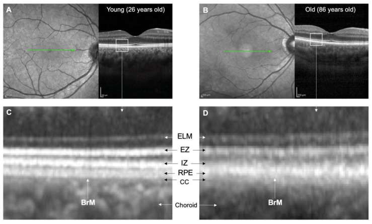
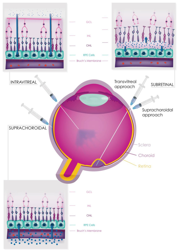
The complement system is crucial for immune surveillance, providing the body’s first line of defence against pathogens. However, an imbalance in its regulators can lead to inappropriate overactivation, resulting in diseases such as age-related macular degeneration (AMD), a leading cause of irreversible blindness globally affecting around 200 million people. Complement activation in AMD is believed to begin in the choriocapillaris, but it also plays a critical role in the subretinal and retinal pigment epithelium (RPE) spaces. Bruch’s membrane (BrM) acts as a barrier between the retina/RPE and choroid, hindering complement protein diffusion. This impediment increases with age and AMD, leading to compartmentalisation of complement activation. In this review, we comprehensively examine the structure and function of BrM, including its age-related changes visible through in vivo imaging, and the consequences of complement dysfunction on AMD pathogenesis. We also explore the potential and limitations of various delivery routes (systemic, intravitreal, subretinal, and suprachoroidal) for safe and effective delivery of conventional and gene therapy-based complement inhibitors to treat AMD. Further research is needed to understand the diffusion of complement proteins across BrM and optimise therapeutic delivery to the retina.
Overview
CoChat Summary
TL;DR
This review argues that Bruch’s membrane (BrM) is a selectively permeable, age-changing barrier that compartmentalises complement activation in age-related macular degeneration (AMD) and complicates drug delivery to the retina. It evaluates systemic, intravitreal, subretinal, and suprachoroidal routes for complement inhibitors and gene therapies, concluding that further work is needed to understand and optimise transit across BrM.
Key findings
- BrM is a 2–4 μm five-layer extracellular matrix between the RPE and choroid; its permeability declines with age, with one study reporting an almost 100% reduction in serum protein permeability from the first to ninth decades of life (from 3.5 × 10⁻⁶ to 0.2 × 10⁻⁶ cm/s), and a permeability threshold falling from >200 kDa in younger donors to 100 kDa in elderly donors.
- Complement protein passage across BrM is selective: FHL-1, Factor D, and C5a can cross, whereas C3a is not able to despite its small molecular weight; C3/C3b, FH, FB, and FI are unable to penetrate BrM even at small quantities. FHL-1 (49 kDa) has better tissue penetration than full-length glycosylated FH (155 kDa).
- AMD affects around 200 million people globally; 95% of cases are non-exudative. Genetic risk includes CFH p.Y402H, conferring approximately two-fold higher risk of late-stage AMD per allele, and GWASs have detected 52 independent and rare variants across 34 loci. Smoking is associated with an odds ratio of 1.78 (95% CI 1.52–2.09) for current versus ex-smokers.
- Intravitreal complement inhibitors pegcetacoplan and avacincaptad pegol showed anatomical benefits for geographic atrophy at one year in Phase 3 studies, but visual outcome benefit remains unknown and both have safety concerns regarding induction of exudative AMD. Systemic eculizumab completed a randomised controlled trial in AMD but failed to show significant anatomical or functional benefit.
Methods
- Narrative review synthesising literature on BrM structure and function, age-related changes, AMD pathogenesis, complement dysfunction, in vivo imaging, and therapeutic delivery routes.
- Evidence base includes human tissue studies, animal and cell models, genetic and imaging studies, and clinical trial reports; no systematic search strategy, meta-analysis, or primary data collection is described.
- Main analysis is qualitative synthesis, with tables and figures summarising age-related BrM changes and delivery approaches.
Limitations
- Stated: further research is needed to understand diffusion of complement proteins across BrM and to optimise therapeutic delivery to the retina; diffusion chamber models may not fully capture human physiology; murine models have limitations for studying AMD; the effects of complete complement inhibition in the eye remain unclear.
- Stated: the eculizumab AMD trial had limited treatment duration (6 months), a small sample size, and several methodological biases.
- Evident: as a narrative review, it does not describe a systematic literature search or formal risk-of-bias assessment, and many conclusions rely on preclinical/animal models or early-phase and interim clinical data.
Model-written summary. Check it against the paper before relying on it.
Integrity signals
Registry
Cites
Statistics
Figures



Verification
Claims the paper makes
32 Valid · 5 Not valid · 95 Not checked · 20 Own results and interpretations
Some cited sources support this claim and some don’t — read each source’s verdict below.
In the paper
“This may exacerbate the effect of the FH p.Y402H polymorphism (a major genetic risk factor for AMD) as it has been proposed that the variant protein binds less well to HSPGs than the wild-type protein [56,57].”
§ Bruch’s Membrane: A Key Consideration with Complement-Based Therapies for Age-Related Macular Degeneration > 3. Ageing Processes in BrM > 3.1. Anatomical Changes
What the sources say
- Disputedcites [56] Clark S.J. Higman V.A. Mulloy B. Perkins S.J. Lea S.M. Sim R.B. Day A.J. His-384 allotypic variant of factor H associated with age-related macular degeneration has different heparin binding properties from the non-disease-associated form J. Biol. Chem. 2006 281 24713 24720 10.1074/jbc.M605083200 16787919 sourceExperimental — accuracy not yet measured
In Determining Whether His-384/Tyr-384 Bind HS (page unavailable), the authors report that neither variant bound the heparan sulfate preparations tested, so they did not find weaker binding by the risk variant to HS. In Characterization of the His-384/Tyr-384-Heparin Interaction (page unavailable), the risk-associated His-384 variant bound more strongly to several unfractionated heparin preparations, although binding depended on the preparation. The citing authors’ suggestion about the consequences of reduced HSPGs is not itself assessed here. (LLM, conf=0.84)
Checked by a reasoning model — may be wrong.
- Partly supportedcites [57] Prosser B.E. Johnson S. Roversi P. Herbert A.P. Blaum B.S. Tyrrell J. Jowitt T.A. Clark S.J. Tarelli E. Uhrin D. Structural basis for complement factor H linked age-related macular degeneration J. Exp. Med. 2007 204 2277 2283 10.1084/jem.20071069 17893204 PMC2118454 sourceExperimental — accuracy not yet measured
The Results and Discussion section ('Direct role for AMD-related polymorphic residue in glycan binding'; page unavailable) says the His402 and Tyr402 variants have different affinities and specificities for differently sulfated GAGs. It does not establish that the variant binds HSPGs less well than the wild type in general. The claim that reduced HSPGs may exacerbate the variant's effect is therefore not established by this source. (LLM, conf=0.84)
hedgedstates a possible cause- Asserted: passes — The authors put it forward as true.
- Checkable: passes — It can be checked against evidence.
- Load-bearing: unclear — It is unclear whether the paper’s conclusions depend on it.
- Based: passes — The paper gives a reason why it holds.
- Disputed
Some cited sources support this claim and some don’t — read each source’s verdict below.
In the paper
“Several experiments using mouse models of laser-induced CNV have demonstrated that all complement pathways, particularly the alternative and terminal pathways, play a significant role in angiogenesis [150,152,153,154,155,156,157,158,159,160,161,162,163].”
§ Bruch’s Membrane: A Key Consideration with Complement-Based Therapies for Age-Related Macular Degeneration > 7. Complement Dysfunction in AMD > 7.4. Complement Dysfunction in Macular Neovascularisation
What the sources say
- Partly supportedcites [150] Cashman S.M. Ramo K. Kumar-Singh R. A non membrane-targeted human soluble CD59 attenuates choroidal neovascularization in a model of age related macular degeneration PLoS ONE 2011 6 e19078 10.1371/journal.pone.0019078 21552568 PMC3084256 sourceExperimental — accuracy not yet measured
The Abstract and Results report that inhibiting membrane attack complex (MAC) formation reduced laser-induced CNV in mice, supporting a role for the terminal complement pathway. This paper does not demonstrate that all complement pathways, including the alternative pathway, play a significant role; it tests inhibition at the terminal step. (LLM, conf=0.87)
- Partly supportedcites [152] Schnabolk G. Coughlin B. Joseph K. Kunchithapautham K. Bandyopadhyay M. O’Quinn E.C. Nowling T. Rohrer B. Local production of the alternative pathway component factor B is sufficient to promote laser-induced choroidal neovascularization Investig. Ophthalmol. Vis. Sci. 2015 56 1850 1863 10.1167/iovs.14-15910 25593023 PMC4364641 sourceExperimental — accuracy not yet measured
In “RPE-Derived CFB Promotes Choroidal Neovascularization” (page not available), the paper reports that factor B knockout or alternative-pathway inhibition reduced laser-induced CNV lesions in mice. It also detects the terminal-pathway marker C5b-9 in lesions, but does not establish a significant role for the terminal pathway in angiogenesis or demonstrate that all complement pathways play such a role. (LLM, conf=0.88)
- Not checked yetcites [153] Rohrer B. Coughlin B. Kunchithapautham K. Long Q. Tomlinson S. Takahashi K. Holers V.M. The alternative pathway is required, but not alone sufficient, for retinal pathology in mouse laser-induced choroidal neovascularization Mol. Immunol. 2011 48 e1 e8 10.1016/j.molimm.2010.12.016 21257205 PMC3063365 source
- Not checked yetcites [154] Bora N.S. Kaliappan S. Jha P. Xu Q. Sohn J.H. Dhaulakhandi D.B. Kaplan H.J. Bora P.S. Complement activation via alternative pathway is critical in the development of laser-induced choroidal neovascularization: Role of factor B and factor H J. Immunol. 2006 177 1872 1878 10.4049/jimmunol.177.3.1872 16849499 source
- Not checked yetcites [155] Bora P.S. Sohn J.H. Cruz J.M. Jha P. Nishihori H. Wang Y. Kaliappan S. Kaplan H.J. Bora N.S. Role of complement and complement membrane attack complex in laser-induced choroidal neovascularization J. Immunol. 2005 174 491 497 10.4049/jimmunol.174.1.491 15611275 source
- Not checked yetcites [156] Nozaki M. Raisler B.J. Sakurai E. Sarma J.V. Barnum S.R. Lambris J.D. Chen Y. Zhang K. Ambati B.K. Baffi J.Z. Drusen complement components C3a and C5a promote choroidal neovascularization Proc. Natl. Acad. Sci. USA 2006 103 2328 2333 10.1073/pnas.0408835103 16452172 PMC1413680 source
- Not checked yetcites [157] Ramo K. Cashman S.M. Kumar-Singh R. Evaluation of adenovirus-delivered human CD59 as a potential therapy for AMD in a model of human membrane attack complex formation on murine RPE Investig. Ophthalmol. Vis. Sci. 2008 49 4126 4136 10.1167/iovs.08-2025 18487376 PMC4139013 source
- Partly supportedcites [158] Gandhi J. Cashman S.M. Kumar-Singh R. Soluble CD59 expressed from an adenovirus in vivo is a potent inhibitor of complement deposition on murine liver vascular endothelium PLoS ONE 2011 6 e21621 10.1371/journal.pone.0021621 21720565 PMC3123367 sourceExperimental — accuracy not yet measured
The Introduction and Discussion (page unavailable) report that soluble CD59 reduced membrane attack complex deposition and laser-induced choroidal neovascularization in a mouse model, supporting a role for terminal complement activity. The cited paper’s own experiments concern murine liver endothelium; it does not establish that all complement pathways, particularly the alternative pathway, contribute to angiogenesis. (LLM, conf=0.85)
- Partly supportedcites [159] Bora N.S. Jha P. Lyzogubov V.V. Kaliappan S. Liu J. Tytarenko R.G. Fraser D.A. Morgan B.P. Bora P.S. Recombinant membrane-targeted form of CD59 inhibits the growth of choroidal neovascular complex in mice J. Biol. Chem. 2010 285 33826 33833 10.1074/jbc.M110.153130 20736175 PMC2962482 sourceExperimental — accuracy not yet measured
The Introduction links alternative-pathway activation and membrane attack complex (MAC) formation to angiogenic growth factors in mouse CNV. The Discussion also reports that inhibiting the alternative pathway or blocking MAC reduced those factors in laser-induced CNV. These passages support the role of the alternative and terminal pathways, but do not establish that all complement pathways play a significant role or rank their relative importance. Page numbers are not available in our copy. (LLM, conf=0.87)
- Disputedcites [160] Rohrer B. Long Q. Coughlin B. Wilson R.B. Huang Y. Qiao F. Tang P.H. Kunchithapautham K. Gilkeson G.S. Tomlinson S. A targeted inhibitor of the alternative complement pathway reduces angiogenesis in a mouse model of age-related macular degeneration Investig. Ophthalmol. Vis. Sci. 2009 50 3056 3064 10.1167/iovs.08-2222 19264882 PMC2908507 sourceExperimental — accuracy not yet measured
The Discussion supports roles for the alternative and terminal pathways in mouse CNV, but directly contradicts the claim that all complement pathways play a significant role: it says deficiency or inhibition of classical and lectin pathway proteins has no impact on murine CNV. (LLM, conf=0.88)
Checked by a reasoning model — may be wrong.
boostedstates an association- Asserted: passes — The authors put it forward as true.
- Checkable: passes — It can be checked against evidence.
- Load-bearing: passes — The paper’s conclusions depend on it.
- Based: passes — The paper gives a reason why it holds.
- Partly supported
The cited sources support only part of this claim — read each source’s verdict below.
In the paper
“This observation may reflect the higher positive net charge of C3a at neutral pH [35], causing it to be trapped within the BrM, which is negatively charged due to the presence of glycosaminoglycans [35,37,38].”
§ Bruch’s Membrane: A Key Consideration with Complement-Based Therapies for Age-Related Macular Degeneration > 2. The Structure and Function of BrM
What the sources say
- Supportedcites [35] Clark S.J. McHarg S. Tilakaratna V. Brace N. Bishop P.N. Bruch’s Membrane Compartmentalizes Complement Regulation in the Eye with Implications for Therapeutic Design in Age-Related Macular Degeneration Front. Immunol. 2017 8 1778 10.3389/fimmu.2017.01778 29312308 PMC5742201 source
The Discussion (p. of 'Diffusion of the anaphylatoxins C3a and C5a through BrM' / Discussion section) states that although C3a and C5a are small with almost identical hydrodynamic radii, 'C3a is calculated to have a significantly greater net positive charge that C5a (Table 2)' and 'Given BrM is loaded with negatively charged glycosaminoglycans [such as heparan sulfate (22,23)], it is plausible that C3a becomes bound and collects on/within BrM.' The Results section confirms the premise that 'no detectable levels of recombinant C3a crossed the enriched macula BrM in our test system,' and Table 2 gives C3a net charge at pH 7 as 9.2 versus 5.2 for C5a. The citing sentence's hedge ('may reflect') matches the source's own hedging ('it is plausible'), so the paraphrase carries the qualifiers intact. (LLM, conf=0.92)
- Not checked yetcites [37] Langford-Smith A. Day A.J. Bishop P.N. Clark S.J. Complementing the Sugar Code: Role of GAGs and Sialic Acid in Complement Regulation Front. Immunol. 2015 6 25 10.3389/fimmu.2015.00025 25699044 PMC4313701
- Partly supportedcites [38] Clark S.J. Bishop P.N. Day A.J. The proteoglycan glycomatrix: A sugar microenvironment essential for complement regulation Front. Immunol. 2013 4 412 10.3389/fimmu.2013.00412 24324472 PMC3840399 sourceExperimental — accuracy not yet measured
Our copy of Clark et al. (2013) supports only the matrix side of the sentence: the 'Modulation of the Complement System by Proteoglycans' section states that Bruch's membrane carries a sulfated glycomatrix ('CFH-binding sites in Bruch's membrane are comprised mainly of HS, but with dermatan sulfate also playing a minor role'; Table 1 shows 402Y binding BrM '+++' and requiring 2- and 6-O sulfation). What it does not support is the C3a-specific assertion: no passage mentions C3a, anaphylatoxins, or a net positive charge at neutral pH (searched: 'C3a anaphylatoxin positive net charge', 'anaphylatoxin C3a C5a complement fragment diffusion', 'electrostatic interactions basic residues positively charged'), and the paper never describes Bruch's membrane as 'negatively charged' — it frames GAG recognition as structure/sulfation-specific 'postcodes' rather than generic charge trapping. The charge-based trapping of C3a is therefore not this paper's point; only the GAG-rich, sulfated nature of BrM is. (LLM, conf=0.68)
hedgedstates a possible cause- Asserted: passes — The authors put it forward as true.
- Checkable: passes — It can be checked against evidence.
- Load-bearing: unclear — It is unclear whether the paper’s conclusions depend on it.
- Based: passes — The paper gives a reason why it holds.
- Supported
The cited sources support only part of this claim — read each source’s verdict below.
In the paper
“Some studies have shown that FB has a pro-angiogenic effect mediated through increased VEGF and VEGFR2 expression by endothelial cells in mouse models of laser-induced CNV and retinal hypoxia [81,153,171].”
§ Bruch’s Membrane: A Key Consideration with Complement-Based Therapies for Age-Related Macular Degeneration > 7. Complement Dysfunction in AMD > 7.4. Complement Dysfunction in Macular Neovascularisation
What the sources say
- Could not verifycites [81] Tzoumas N. Kavanagh D. Cordell H.J. Lotery A.J. Patel P.J. Steel D.H. Rare complement factor I variants associated with reduced macular thickness and age-related macular degeneration in the UK Biobank Hum. Mol. Genet. 2022 31 2678 2692 10.1093/hmg/ddac060 35285476 PMC9402241 sourceExperimental — accuracy not yet measured
The Introduction and Discussion discuss complement Factor I variants, macular thickness, and AMD, but do not address a pro-angiogenic effect of Factor B. Searches for VEGF, VEGFR2, endothelial cells, laser-induced CNV, and retinal hypoxia found no passage supporting the attributed mechanism. (LLM, conf=0.76)
- Not checked yetcites [153] Rohrer B. Coughlin B. Kunchithapautham K. Long Q. Tomlinson S. Takahashi K. Holers V.M. The alternative pathway is required, but not alone sufficient, for retinal pathology in mouse laser-induced choroidal neovascularization Mol. Immunol. 2011 48 e1 e8 10.1016/j.molimm.2010.12.016 21257205 PMC3063365 source
- Partly supportedcites [171] Murray H. Qiu B. Ho S.Y. Wang X. Complement Factor B Mediates Ocular Angiogenesis through Regulating the VEGF Signaling Pathway Int. J. Mol. Sci. 2021 22 9580 10.3390/ijms22179580 34502486 PMC8431595 sourceExperimental — accuracy not yet measured
Results §2.1 reports increased CFB expression in mouse models of laser-induced CNV and oxygen-induced retinopathy, while Results §2.4 shows that CFB promotes angiogenesis through VEGF signaling in cultured human retinal endothelial cells. The paper does not show that increased VEGF and VEGFR2 expression by endothelial cells mediates the effect in those mouse models: it reports correlated CFB and VEGF expression in the hypoxic retina and reduced VEGFR2 expression after CFB knockdown in cultured cells. Page numbers were not supplied. (LLM, conf=0.88)
hedgedstates a cause- Asserted: passes — The authors put it forward as true.
- Checkable: passes — It can be checked against evidence.
- Load-bearing: unclear — It is unclear whether the paper’s conclusions depend on it.
- Based: passes — The paper gives a reason why it holds.
- Could not verify
Every source the paper cites for this claim was checked and supports it.
In the paper
“the common FH risk variant in AMD, CFH p.Y402H, resulted in even poorer microglial elimination [203].”
§ Bruch’s Membrane: A Key Consideration with Complement-Based Therapies for Age-Related Macular Degeneration > 7. Complement Dysfunction in AMD > 7.5. Immune Cell Regulation
What the sources say
- Supportedcites [203] Calippe B. Augustin S. Beguier F. Charles-Messance H. Poupel L. Conart J.-B. Hu S.J. Lavalette S. Fauvet A. Rayes J. Complement Factor H Inhibits CD47-Mediated Resolution of Inflammation Immunity 2017 46 261 272 10.1016/j.immuni.2017.01.006 28228282 source
The Results section (p. ~ 'The CFH(H402) Variant Inhibits Subretinal MC Elimination More Potently than the Common CFH(Y402)') states that the Y402H substitution accounts for a major part of AMD genetic risk and that CFH(H402) limited microglial (MC) clearance by 37% relative to CFH(Y402), and was 50% more potent at inhibiting TSP-1-driven MC elimination. The Summary (p. 2, Florian Sennlaub) likewise states the AMD-associated CFH(H402) variant markedly increased this inhibitory effect on microglial cells. Both bound the claim to the AMD risk variant and to microglial elimination, as the citing sentence does. (LLM, conf=0.92)
plainstates a cause- Asserted: passes — The authors put it forward as true.
- Checkable: passes — It can be checked against evidence.
- Load-bearing: passes — The paper’s conclusions depend on it.
- Based: passes — The paper gives a reason why it holds.
- Supported
95 more claims · 19 with their cited sources checked
Create a free account to see all 152 claims and to verify the 174 references still open.
- Probably not a substantial claim. 16
- It may state two separate claims as one. 40
Claims other papers make about this one
Each is checked against this paper’s own text: does it say what the citing paper says it does?
In the citing paper
“This could be due to the structural differences between species that result in different transduction cell types, including Bruch’s membrane (BrM) thickness, the thickness or number of cells within the retinal layers73,74.”
From An adeno-associated virus variant enabling efficient ocular-directed gene delivery across species (2024) · cites this paper as [73]
Self-check
Percentages, sums and reported differences re-computed from the paper’s own numbers.
Each contradiction between two passages listed here was confirmed by an independent check.
Rated minor: it would not touch a result — a value no result rests on, a p-value that stays on the same side of the significance level, or a conflict that is clerical, resolved elsewhere in the paper, or outside its own findings.
In the paper
§ Bruch’s Membrane: A Key Consideration with Complement-Based Therapies for Age-Related Macular Degeneration > 2. The Structure and Function of BrM
“BrM is a 2–4 μm thin layer of extracellular matrix between the retina and choroid in the eye serving as a selectively permeable membrane between the RPE and choriocapillaris (CC).”
§ Bruch’s Membrane: A Key Consideration with Complement-Based Therapies for Age-Related Macular Degeneration > 2. The Structure and Function of BrM
“The OCL’s thickness ranges from 1 to 5 μm [29]”
Citations
Cites
1 retracted
Items 201–315 of 315
Complement activation levels are related to disease stage in AMD
Heesterbeek, T. J., Lechanteur, Y. T. E., Lorés-Motta, L., Schick, T., Daha, M. R., Altay, L., Liakopoulos, S., Smailhodzic, D., den Hollander, A. I., Hoyng, C. B., et al. (2020) · Investigative Opthalmology & Visual Science · doi:10.1167/iovs.61.3.18 · Cited by 76
Hallam, D., Collin, J., Bojic, S., Chichagova, V., Buskin, A., Xu, Y., Lafage, L., Otten, E. G., Anyfantis, G., Mellough, C., et al. (2017) · Stem Cells · doi:10.1002/stem.2708 · Cited by 75
Self-citationA local complement response by RPE causes early-stage macular degeneration
Fernandez-Godino, R., Garland, D. L., & Pierce, E. A. (2015) · Human Molecular Genetics · doi:10.1093/hmg/ddv287 · Cited by 73
Loss of Complement Factor H impairs antioxidant capacity and energy metabolism of human RPE cells
Armento, A., Honisch, S., Panagiotakopoulou, V., Sonntag, I., Jacob, A., Bolz, S., Kilger, E., Deleidi, M., Clark, S., & Ueffing, M. (2020) · Scientific Reports · doi:10.1038/s41598-020-67292-z · Cited by 73 · cited 1×
“A lack of functional FH may also impair the clearance of lipid peroxidation products [131], leading to oxidative stress [139].”
BackgroundKunchithapautham, K., & Rohrer, B. (2011) · Journal of Biological Chemistry · doi:10.1074/jbc.m110.214593 · Cited by 71 · cited 1×
“For example, sublytic levels of MAC have been shown to stimulate the release of growth factors from nucleated cells [99,100], including VEGF from RPE in cell culture studies [146,165].”
BackgroundVogt, S. D., Barnum, S. R., Curcio, C. A., & Read, R. W. (2006) · Experimental Eye Research · doi:10.1016/j.exer.2006.04.002 · Cited by 71 · cited 2×
“To protect themselves from harmful complement activation, RPE cells secrete FH and express several regulators of the complement system, such as CD59 on their apical side [113,127,128] and CD46 (also known as membrane cofactor protein or MCP) on their basal side [113,129,130].”
BackgroundClark, S. J., McHarg, S., Tilakaratna, V., Brace, N., & Bishop, P. N. (2017) · Frontiers in Immunology · doi:10.3389/fimmu.2017.01778 · Cited by 71 · cited 9×
“This observation may reflect the higher positive net charge of C3a at neutral pH [35], causing it to be trapped within the BrM, which is negatively charged due to the presence of glycosaminoglycans [35,37,38].”
BackgroundInfluentialImplications of genetic variation in the complement system in age-related macular degeneration
de Jong, S., Gagliardi, G., Garanto, A., de Breuk, A., Lechanteur, Y. T. E., Katti, S., van den Heuvel, L. P., Volokhina, E. B., & den Hollander, A. I. (2021) · Progress in Retinal and Eye Research · doi:10.1016/j.preteyeres.2021.100952 · Cited by 71
Paun (2018) · doi:10.1016/j.ophtha.2017.12.023 · Cited by 70
Progression of subclinical choroidal neovascularization in age-related macular degeneration
Heiferman, M. J., & Fawzi, A. A. (2019) · PLOS ONE · doi:10.1371/journal.pone.0217805 · Cited by 70
Gene therapy for inherited retinal disease: Long-term durability of effect
Leroy, B. P., Fischer, M. D., Flannery, J. G., MacLaren, R. E., Dalkara, D., Scholl, H. P. N., Chung, D. C., Spera, C., Viriato, D., & Banhazi, J. (2023) · Ophthalmic Research · doi:10.1159/000526317 · Cited by 70
Complement-Associated Deposits in the Human Retina
Seth, A., Cui, J., To, E., Kwee, M., & Matsubara, J. (2008) · Investigative Opthalmology & Visual Science · doi:10.1167/iovs.07-1072 · Cited by 67
Long-term Structural Outcomes of Late-stage RPE65 Gene Therapy
Gardiner, K. L., Cideciyan, A. V., Swider, M., Dufour, V. L., Sumaroka, A., Komáromy, A. M., Hauswirth, W. W., Iwabe, S., Jacobson, S. G., Beltran, W. A., et al. (2020) · Molecular Therapy · doi:10.1016/j.ymthe.2019.08.013 · Cited by 67
The future of retinal gene therapy: evolving from subretinal to intravitreal vector delivery
Ross, M., & Ofri, R. (2021) · Neural Regeneration Research · doi:10.4103/1673-5374.306063 · Cited by 67 · cited 2×
“However, this approach may require higher vector concentrations to compensate for the long diffusion distance within the vitreous [247].”
BackgroundProtective responses to sublytic complement in the retinal pigment epithelium
Tan, L. X., Toops, K. A., & Lakkaraju, A. (2016) · Proceedings of the National Academy of Sciences · doi:10.1073/pnas.1523061113 · Cited by 66 · cited 1×
“RPE cells with impaired lysosomal function are thought to be susceptible to MAC deposition due to their inability to adequately recycle and traffic complement inhibitors such as CD59 to the cell surface [138].”
BackgroundA Perspective of AMD Through the Eyes of Immunology
Copland, D. A., Theodoropoulou, S., Liu, J., & Dick, A. D. (2018) · Investigative Opthalmology & Visual Science · doi:10.1167/iovs.18-23893 · Cited by 66
Keenan, T. D. L., Pickford, C. E., Holley, R. J., Clark, S. J., Lin, W., Dowsey, A. W., Merry, C. L., Day, A. J., & Bishop, P. N. (2014) · Investigative Opthalmology & Visual Science · doi:10.1167/iovs.14-14126 · Cited by 65 · cited 2×
“H (FH), have been shown to be reduced with age [55] and may lead to increased comple-”
BackgroundCoughlin, B., Schnabolk, G., Joseph, K., Raikwar, H., Kunchithapautham, K., Johnson, K., Moore, K., Wang, Y., & Rohrer, B. (2016) · Scientific Reports · doi:10.1038/srep23794 · Cited by 65 · cited 2×
“C5a signalling generally leads to inflammation through the protection of CD4+ T-cells against apoptosis and the upregulation of pro-inflammatory cytokines such as IL-17 and IL-22 [187].”
BackgroundYe, G.-J., Budzynski, E., Sonnentag, P., Nork, T. M., Miller, P. E., Sharma, A. K., Ver Hoeve, J. N., Smith, L. M., Arndt, T., Calcedo, R., et al. (2016) · Human Gene Therapy Clinical Development · doi:10.1089/humc.2015.164 · Cited by 64
Peden, M. C., Min, J., Meyers, C., Lukowski, Z., Li, Q., Boye, S. L., Levine, M., Hauswirth, W. W., Ratnakaram, R., Dawson, W., et al. (2011) · PLoS ONE · doi:10.1371/journal.pone.0017140 · Cited by 63 · cited 1×
“Furthermore, the suprachoroidal route offers faster diffusion for smaller, lipophilic agents [307] and, as an ab externo approach, avoids the intraocular complications associated with intravitreal and subretinal administration [308].”
BackgroundMultiplex analysis of age-related protein and lipid modifications in human Bruch's membrane
Beattie, J. R., Pawlak, A. M., Boulton, M. E., Zhang, J., Monnier, V. M., McGarvey, J. J., & Stitt, A. W. (2010) · The FASEB Journal · doi:10.1096/fj.10-166090 · Cited by 63
Expanding horizons in complement drug discovery: challenges and emerging strategies
Harris, C. L. (2018) · Seminars in Immunopathology · doi:10.1007/s00281-017-0655-8 · Cited by 63 · cited 3×
“Homing agents that bind to neoepitopes on complement proteins or stress-related antigens on apoptotic or necrotic cells may offer an additional way of restricting drug action to inflammation hotspots and reducing the risk of widespread systemic effects [225], though it is unclear how such a therapy would specifically…”
BackgroundInfluentialSelf-citationSelective accumulation of the complement membrane attack complex in aging choriocapillaris
Chirco, K. R., Tucker, B. A., Stone, E. M., & Mullins, R. F. (2016) · Experimental Eye Research · doi:10.1016/j.exer.2015.09.003 · Cited by 62
Rohrer, B., Coughlin, B., Kunchithapautham, K., Long, Q., Tomlinson, S., Takahashi, K., & Holers, V. M. (2011) · Molecular Immunology · doi:10.1016/j.molimm.2010.12.016 · Cited by 62 · cited 1×
“Some studies have shown that FB has a pro-angiogenic effect mediated through increased VEGF and VEGFR2 expression by endothelial cells in mouse models of laser-induced CNV and retinal hypoxia [81,153,171].”
BackgroundDiscrete expression and distribution pattern of TIMP-3 in the human retina and choroid
Vranka, J. A., Johnson, E., Zhu, X., Shepardson, A., Alexander, J. P., Bradley, J. M. B., Wirtz, M. K., Weleber, R. G., Klein, M. L., & Acott, T. S. (1997) · Current Eye Research · doi:10.1076/ceyr.16.2.102.5086 · Cited by 62 · cited 1×
“TIMP-1 and TIMP-2 are thought to move freely within the BrM, while TIMP-3 is thought to be bound to the RPE and CEC basement membrane [44].”
BackgroundC5aR2 Activation Broadly Modulates the Signaling and Function of Primary Human Macrophages
Li, X. X., Clark, R. J., & Woodruff, T. M. (2020) · The Journal of Immunology · doi:10.4049/jimmunol.2000407 · Cited by 62
Chiang, B., Venugopal, N., Edelhauser, H. F., & Prausnitz, M. R. (2016) · Experimental Eye Research · doi:10.1016/j.exer.2016.10.011 · Cited by 60 · cited 1×
“Co-localisation with a polymer may improve particle durability and spread [309].”
BackgroundWoodard, K. T., Liang, K. J., Bennett, W. C., & Samulski, R. J. (2016) · Journal of Virology · doi:10.1128/jvi.01568-16 · Cited by 59 · cited 6×
“The inner limiting membrane (ILM) and glial processes of the neural retina act as barriers to the diffusion of naturally occurring AAV serotypes in humans [244,245].”
InfluentialDevelopment of a Genotype Assay for Age-Related Macular Degeneration
de Breuk, A., Acar, I. E., Kersten, E., Schijvenaars, M. M. V. A. P., Colijn, J. M., Haer-Wigman, L., Bakker, B., de Jong, S., Meester-Smoor, M. A., Verzijden, T., et al. (2021) · Ophthalmology · doi:10.1016/j.ophtha.2020.07.037 · Cited by 59
Immunohistological study of subretinal membranes in age-related macular degeneration.
Baudouin, C., Peyman, G. A., Fredj-Reygrobellet, D., Gordon, W. C., Lapalus, P., Gastaud, P., & Bazan, N. G. (1992) · PubMed · Cited by 58
Terminal complexes of the complement system: New structural insights and their relevance to function
Morgan, B. P., Walters, D., Serna, M., & Bubeck, D. (2016) · Immunological Reviews · doi:10.1111/imr.12461 · Cited by 58 · cited 17×
“Complement may play a constitutive role in the choroid and retina decades before the onset of AMD, as indicated in human tissue studies by higher MAC deposition [108,112], CD59 expression [113], and FH production relative to other retinal tissues.”
BackgroundInfluentialMashal, M., Attia, N., Martínez-Navarrete, G., Soto-Sánchez, C., Fernández, E., Grijalvo, S., Eritja, R., Puras, G., & Pedraz, J. L. (2019) · Journal of Controlled Release · doi:10.1016/j.jconrel.2019.05.010 · Cited by 58 · cited 1×
“Strategies such as PEGylation [271], charge modification [272], and coating with hyaluronate or chloroquine [273,274] have been shown to improve retinal transduction in animal models when administered intravitreally.”
BackgroundCerniauskas, E., Kurzawa-Akanbi, M., Xie, L., Hallam, D., Moya-Molina, M., White, K., Steel, D., Doherty, M., Whitfield, P., Al-Aama, J., et al. (2020) · Stem Cells Translational Medicine · doi:10.1002/sctm.20-0211 · Cited by 58
Self-citationLamin, A., Oakley, J. D., Dubis, A. M., Russakoff, D. B., & Sivaprasad, S. (2019) · Eye · doi:10.1038/s41433-018-0234-9 · Cited by 57 · cited 2×
“healthy eyes according to global and/or low-density OCT spatial analysis [92,93].”
BackgroundHallam, T. M., Marchbank, K. J., Harris, C. L., Osmond, C., Shuttleworth, V. G., Griffiths, H., Cree, A. J., Kavanagh, D., & Lotery, A. J. (2020) · Investigative Opthalmology & Visual Science · doi:10.1167/iovs.61.6.18 · Cited by 57 · cited 1×
“Beyond FHL-1, it has also been shown that FI is found at significantly lower levels in the aqueous fluid of the eye compared to FI levels found in simultaneously sampled serum [211].”
BackgroundSelf-citationHost immune responses after suprachoroidal delivery of AAV8 in nonhuman primate eyes
Chung, S. H., Mollhoff, I. N., Mishra, A., Sin, T.-N., Ngo, T., Ciulla, T., Sieving, P., Thomasy, S. M., & Yiu, G. (2021) · Human Gene Therapy · doi:10.1089/hum.2020.281 · Cited by 57
Zeng, S., Whitmore, S. S., Sohn, E. H., Riker, M. J., Wiley, L. A., Scheetz, T. E., Stone, E. M., Tucker, B. A., & Mullins, R. F. (2016) · The Journal of Pathology · doi:10.1002/path.4669 · Cited by 56 · cited 1×
“MMP expression in choroidal endothelial cells has also been found to increase with MAC-dependent complement activation [172], suggesting that complement may promote angiogenesis through multiple pathways.”
BackgroundClusterin is Present in Drusen in Age-related Macular Degeneration
Sakaguchi, H., Miyagi, M., Shadrach, K. G., Rayborn, M. E., Crabb, J. W., & Hollyfield, J. G. (2002) · Experimental Eye Research · doi:10.1006/exer.2002.1186 · Cited by 54
The suprachoroidal pathway: a new drug delivery route to the back of the eye
Rai, U. D. J. P., Young, S. A., Thrimawithana, T. R., Abdelkader, H., Alani, A. W. G., Pierscionek, B., & Alany, R. G. (2015) · Drug Discovery Today · doi:10.1016/j.drudis.2014.10.010 · Cited by 53 · cited 1×
“Furthermore, the suprachoroidal route offers faster diffusion for smaller, lipophilic agents [307] and, as an ab externo approach, avoids the intraocular complications associated with intravitreal and subretinal administration [308].”
BackgroundBandyopadhyay, M., & Rohrer, B. (2012) · Investigative Opthalmology & Visual Science · doi:10.1167/iovs.11-8638 · Cited by 53 · cited 17×
“This method has been particularly successful in the treatment of vascular retinal disorders such as neovascular AMD through the use of anti-VEGF agents.”
BackgroundInfluentialBora, N. S., Jha, P., Lyzogubov, V. V., Kaliappan, S., Liu, J., Tytarenko, R. G., Fraser, D. A., Morgan, B. P., & Bora, P. S. (2010) · Journal of Biological Chemistry · doi:10.1074/jbc.m110.153130 · Cited by 53
Cabrera, A. P., Bhaskaran, A., Xu, J., Yang, X., Scott, H. A., Mohideen, U., & Ghosh, K. (2016) · Investigative Opthalmology & Visual Science · doi:10.1167/iovs.16-19727 · Cited by 53 · cited 1×
“With increasing age, endothelial cells may become more sensitive to MAC injury due to higher cytoskeletal Rho kinase activity [202].”
BackgroundEbeling, M. C., Geng, Z., Kapphahn, R. J., Roehrich, H., Montezuma, S. R., Dutton, J. R., & Ferrington, D. A. (2021) · Cells · doi:10.3390/cells10040789 · Cited by 51
Hillenkamp, J. (2004) · Investigative Ophthalmology & Visual Science · doi:10.1167/iovs.03-0765 · Cited by 49
Prediction of Ocular Drug Distribution from Systemic Blood Circulation
Vellonen, K.-S., Soini, E.-M., del Amo, E. M., & Urtti, A. (2016) · Molecular Pharmaceutics · doi:10.1021/acs.molpharmaceut.5b00729 · Cited by 49 · cited 1×
“Studies have simulated drug distribution from the plasma to the vitreous in rabbits [230], but it is unclear how these pharmacokinetics translate to humans and whether higher rates of systemic drug clearance may be a factor [228].”
BackgroundEmerging therapies and their delivery for treating age‐related macular degeneration
Thomas, C. N., Sim, D. A., Lee, W. H., Alfahad, N., Dick, A. D., Denniston, A. K., & Hill, L. J. (2021) · British Journal of Pharmacology · doi:10.1111/bph.15459 · Cited by 49 · cited 1×
“Early therapeutic intervention in the disease course may offer the greatest benefit [279], and there are several ongoing intravitreal gene therapy trials for advanced AMD [280], including JNJ-1887 (formerly AAVCAGsCD59), an intravitreal AAV-based vector expressing a soluble form of the MAC inhibitory protein CD59 that…”
BackgroundNovel Role for a Complement Regulatory Protein (CD46) in Retinal Pigment Epithelial Adhesion
McLaughlin, B. J., Fan, W., Zheng, J. J., Cai, H., Del Priore, L. V., Bora, N. S., & Kaplan, H. J. (2003) · Investigative Opthalmology & Visual Science · doi:10.1167/iovs.02-0813 · Cited by 48 · cited 1×
“To protect themselves from harmful complement activation, RPE cells secrete FH and express several regulators of the complement system, such as CD59 on their apical side [113,127,128] and CD46 (also known as membrane cofactor protein or MCP) on their basal side [113,129,130].”
BackgroundSynthesis and Secretion of Hemoglobin by Retinal Pigment Epithelium
Tezel, T. H., Geng, L., Bodek, E., Schaal, S., Liu, Y., Dean, D. C., Klein, J. B., & Kaplan, H. J. (2009) · Investigative Ophthalmology & Visual Science · doi:10.1167/iovs.07-1372 · Cited by 48
Khandhadia, S., Hakobyan, S., Heng, L. Z., Gibson, J., Adams, D. H., Alexander, G. J., Gibson, J. M., Martin, K. R., Menon, G., Nash, K., et al. (2013) · Ophthalmology · doi:10.1016/j.ophtha.2013.01.004 · Cited by 47 · cited 1×
“Y402H status, suggesting that systemic complement production by the liver may not contribute to retinal disease [224].”
BackgroundDifferential regulation of endothelial cell activation by complement and interleukin 1α
Brunn, G. J., Saadi, S., & Platt, J. L. (2006) · Circulation Research · doi:10.1161/01.res.0000216071.87981.16 · Cited by 47
Chiang, B., Kim, Y. C., Edelhauser, H. F., & Prausnitz, M. R. (2016) · Experimental Eye Research · doi:10.1016/j.exer.2016.03.008 · Cited by 46 · cited 1×
“It is bound anteriorly by the scleral spur and posteriorly by the optic nerve and posterior ciliary arteries [304].”
BackgroundSubILM Injection of AAV for Gene Delivery to the Retina
Gamlin, P. D., Alexander, J. J., Boye, S. L., Witherspoon, C. D., & Boye, S. E. (2019) · Methods in Molecular Biology · doi:10.1007/978-1-4939-9139-6_14 · Cited by 46 · cited 1×
“Other experimental techniques, such as ILM peeling or vitrectomy [252,253], sub-ILM injection [254,255], and ILM photo-disruption with indocyanine green [256], have also been proposed, but their safety is uncertain.”
BackgroundLyzogubov, V. V., Bora, P. S., Wu, X., Horn, L. E., de Roque, R., Rudolf, X. V., Atkinson, J. P., & Bora, N. S. (2016) · The American Journal of Pathology · doi:10.1016/j.ajpath.2016.03.021 · Cited by 45 · cited 1×
“In addition, the inhibition of CD46 by gene knockout in mouse models led to increased VEGF production in the retina and choroid, increased MAC deposition on these tissues, increased susceptibility to laser-induced CNV, and degenerative changes in retinal tissues resembling an AMD phenotype [166,167].”
BackgroundC5a anaphylatoxin and its role in critical illness-induced organ dysfunction
Wood, A. J. T., Vassallo, A., Summers, C., Chilvers, E. R., & Conway‐Morris, A. (2018) · European Journal of Clinical Investigation · doi:10.1111/eci.13028 · Cited by 45
Minassian, D. C., & Reidy, A. (2009) · Cited by 44
Lynch, A. M., Mandava, N., Patnaik, J. L., Frazer-Abel, A. A., Wagner, B. D., Palestine, A. G., Mathias, M. T., Siringo, F. S., Cathcart, J. N., & Holers, V. M. (2020) · European Journal of Ophthalmology · doi:10.1177/1120672119857896 · Cited by 44
Fibulin-5 distribution in human eyes: Relevance to age-related macular degeneration
Mullins, R. F., Olvera, M. A., Clark, A. F., & Stone, E. M. (2006) · Experimental Eye Research · doi:10.1016/j.exer.2006.09.021 · Cited by 43
Yang, P., Baciu, P. C., Kerrigan, B. C. P., Etheridge, M., Sung, E., Toimil, B. A., Berchuck, J. E., & Jaffe, G. J. (2014) · Investigative Ophthalmology & Visual Science · doi:10.1167/iovs.13-13554 · Cited by 41
Complement Regulatory Protein CD46 Protects against Choroidal Neovascularization in Mice
Lyzogubov, V., Wu, X., Jha, P., Tytarenko, R., Triebwasser, M., Kolar, G., Bertram, P., Bora, P. S., Atkinson, J. P., & Bora, N. S. (2014) · The American Journal of Pathology · doi:10.1016/j.ajpath.2014.06.001 · Cited by 41 · cited 1×
“In addition, the inhibition of CD46 by gene knockout in mouse models led to increased VEGF production in the retina and choroid, increased MAC deposition on these tissues, increased susceptibility to laser-induced CNV, and degenerative changes in retinal tissues resembling an AMD phenotype [166,167].”
BackgroundNonclinical Safety Evaluation of scAAV8-RLBP1 for Treatment of RLBP1 Retinitis Pigmentosa
MacLachlan, T. K., Milton, M. N., Turner, O., Tukov, F., Choi, V. W., Penraat, J., Hanks, S. M., Vrouvlianis, J., Leehy, B., Li, H., et al. (2018) · Molecular Therapy - Methods & Clinical Development · doi:10.1016/j.omtm.2017.12.001 · Cited by 40
The proteoglycan glycomatrix: A sugar microenvironment essential for complement regulation
Clark, S. J., Bishop, P. N., & Day, A. J. (2013) · Frontiers in Immunology · doi:10.3389/fimmu.2013.00412 · Cited by 39 · cited 2×
“charged due to the presence of glycosaminoglycans [35,37,38].”
BackgroundCorrection: Systemic Complement Activation in Age-Related Macular Degeneration
Scholl, H. P. N., Charbel Issa, P., Walier, M., Janzer, S., Pollok-Kopp, B., Börncke, F., Fritsche, L. G., Chong, N. V., Fimmers, R., Wienker, T., et al. (2008) · PLoS ONE · doi:10.1371/annotation/32b9bc31-ed6d-4d31-9ce0-480407017bad · Cited by 39
Ramo, K., Cashman, S. M., & Kumar-Singh, R. (2008) · Investigative Opthalmology & Visual Science · doi:10.1167/iovs.08-2025 · Cited by 38
The alternative complement pathway regulates pathological angiogenesis in the retina
Sweigard, J. H., Yanai, R., Gaissert, P., Saint‐Geniez, M., Kataoka, K., Thanos, A., Stahl, G. L., Lambris, J. D., & Connor, K. M. (2014) · The FASEB Journal · doi:10.1096/fj.14-251041 · Cited by 38
Complement inhibitors for age-related macular degeneration
Tzoumas, N., Riding, G., Williams, M. A., & Steel, D. H. (2023) · Cochrane Database of Systematic Reviews · doi:10.1002/14651858.cd009300.pub3 · Cited by 38 · cited 5×
“, the PEGylation of agents such as pegcetacoplan and avacincaptad pegol) or target [173].”
BackgroundInfluentialSelf-citationSchnabolk, G., Coughlin, B., Joseph, K., Kunchithapautham, K., Bandyopadhyay, M., O'Quinn, E. C., Nowling, T., & Rohrer, B. (2015) · Investigative Opthalmology & Visual Science · doi:10.1167/iovs.14-15910 · Cited by 37
Ebrahimi, K. B., Callaway, N. F., Cano, M., & Handa, J. T. (2014) · Advances in experimental medicine and biology · doi:10.1007/978-1-4614-3209-8_33 · Cited by 36 · cited 6×
“Complement may play a constitutive role in the choroid and retina decades before the onset of AMD, as indicated in human tissue studies by higher MAC deposition [108,112], CD59 expression [113], and FH production relative to other retinal tissues.”
BackgroundInfluentialSelf versus nonself discrimination by the soluble complement regulators factor H and FHL-1
Dopler, A., Guntau, L., Harder, M. J., Palmer, A., Höchsmann, B., Schrezenmeier, H., Simmet, T., Huber-Lang, M., & Schmidt, C. Q. (2019) · The Journal of Immunology · doi:10.4049/jimmunol.1801545 · Cited by 35 · cited 2×
“Another clue that FHL-1 may have evolved specifically to control complement activation locally is that the genetic sequences consistent with FHL-1 expression are found in old world monkeys, but not in their new world ancestors [210].”
BackgroundDavis, W. L., Jones, R. G., & Hagler, H. K. (1981) · Journal of Histochemistry & Cytochemistry · doi:10.1177/29.5.7252127 · Cited by 34 · cited 2×
“ing [58], causing BrM to become stiffer.”
BackgroundZaal, A., Lissenberg-Thunnissen, S. N., van Schijndel, G., Wouters, D., Ham, S. M. V., & ten Brinke, A. (2013) · Immunobiology · doi:10.1016/j.imbio.2012.02.014 · Cited by 33
Teo, K. Y. C., Lee, S. Y., Barathi, A. V., Tun, S. B. B., Tan, L., & Constable, I. J. (2018) · Investigative Opthalmology & Visual Science · doi:10.1167/iovs.18-24333 · Cited by 33 · cited 1×
“Other experimental techniques, such as ILM peeling or vitrectomy [252,253], sub-ILM injection [254,255], and ILM photo-disruption with indocyanine green [256], have also been proposed, but their safety is uncertain.”
BackgroundTzoumas, N., Hallam, D., Harris, C. L., Lako, M., Kavanagh, D., & Steel, D. H. W. (2021) · Survey of Ophthalmology · doi:10.1016/j.survophthal.2020.10.008 · Cited by 33 · cited 21×
“Complement may play a constitutive role in the choroid and retina decades before the onset of AMD, as indicated in human tissue studies by higher MAC deposition [108,112], CD59 expression [113], and FH production relative to other retinal tissues.”
BackgroundInfluentialSelf-citationChen, L., Yang, P., & Curcio, C. A. (2022) · Eye · doi:10.1038/s41433-022-02016-3 · Cited by 33 · cited 4×
“healthy eyes according to global and/or low-density OCT spatial analysis [92,93].”
BackgroundInfluentialICG-mediated photodisruption of the inner limiting membrane enhances retinal drug delivery
Peynshaert, K., Vanluchene, H., Clerck, K. D., Minnaert, A., Verhoeven, M., Gouspillou, N., Bostan, N., Hisatomi, T., Accou, G., Sauvage, F., et al. (2022) · Journal of Controlled Release · doi:10.1016/j.jconrel.2022.07.002 · Cited by 33
Molecular Basis of Factor H R1210C Association with Ocular and Renal Diseases
Recalde, S., Tortajada, A., Subias, M., Anter, J., Blasco, M., Maranta, R., Coco, R., Pinto, S., Noris, M., García-Layana, A., et al. (2016) · Journal of the American Society of Nephrology · doi:10.1681/asn.2015050580 · Cited by 32
γδT-cell function in sepsis is modulated by C5a receptor signalling
Han, G., Geng, S., Li, Y., Chen, G., Wang, R., Li, X., Ma, Y., Shen, B., & Li, Y. (2011) · Immunology · doi:10.1111/j.1365-2567.2011.03445.x · Cited by 32
Bruch’s Membrane and the Choroid in Age-Related Macular Degeneration
Edwards, M., & Lutty, G. A. (2021) · Advances in Experimental Medicine and Biology · doi:10.1007/978-3-030-66014-7_4 · Cited by 32 · cited 1×
“BrM is a continuous membrane posterior to the ora serrata, deficient only at the point of optic nerve insertion [11].”
BackgroundDenard, J., Marolleau, B., Jenny, C., Rao, T. N., Fehling, H. J., Voit, T., & Svinartchouk, F. (2013) · Journal of Virology · doi:10.1128/jvi.01813-13 · Cited by 31 · cited 1×
“Additionally, triggering a systemic immune response against an AAV-based vector can result in unintended side effects such as transgene expression in lymphoid organs [263,264] or systemic complement activation [265,266].”
BackgroundMulfaul, K., Mullin, N. K., Giacalone, J. C., Voigt, A. P., DeVore, M., Stone, E. M., Tucker, B. A., & Mullins, R. F. (2022) · The Journal of Pathology · doi:10.1002/path.5867 · Cited by 31
Tzoumas, N., Kavanagh, D., Cordell, H. J., Lotery, A. J., Patel, P. J., & Steel, D. H. (2022) · Human Molecular Genetics · doi:10.1093/hmg/ddac060 · Cited by 31 · cited 3×
“Some studies have shown that FB has a pro-angiogenic effect mediated through increased VEGF and VEGFR2 expression by endothelial cells in mouse models of laser-induced CNV and retinal hypoxia [81,153,171].”
BackgroundSelf-citationTsiftsoglou, S. A., Arnold, J. N., Roversi, P., Crispin, M. D., Radcliffe, C., Lea, S. M., Dwek, R. A., Rudd, P. M., & Sim, R. B. (2006) · Biochimica et Biophysica Acta (BBA) - Proteins and Proteomics · doi:10.1016/j.bbapap.2006.09.007 · Cited by 29 · cited 2×
“explain the lack of diffusion of Factor H (FH) and Factor B (FB) [35,39].”
BackgroundCall, T. W., & Hollyfield, J. G. (1990) · Experimental Eye Research · doi:10.1016/0014-4835(90)90158-q · Cited by 29
Trinh, M., Kalloniatis, M., Alonso-Caneiro, D., & Nivison-Smith, L. (2022) · Investigative Opthalmology & Visual Science · doi:10.1167/iovs.63.5.36 · Cited by 29 · cited 3×
“early AMD, likely due to the build-up of lipoprotein-related lipids [95].”
BackgroundInfluentialA short history of the retinal pigment epithelium
Wolfensberger, T. J. (1998) · Documenta Ophthalmologica Proceedings Series · doi:10.1007/978-94-011-5137-5_1 · Cited by 28
Boyer, D. D., Ko, Y.-P., Podos, S. D., Cartwright, M. E., Gao, X., Wiles, J. A., & Huang, M. (2022) · Translational Vision Science & Technology · doi:10.1167/tvst.11.10.37 · Cited by 27 · cited 1×
“The results of Phase 2 clinical trials of several systemic agents are eagerly awaited and may provide more insight into the potential of systemic complement inhibitors for AMD [236].”
BackgroundPauleikhoff, D., Wojteki, S., Müller, D., Bornfeld, N., & Heiligenhaus, A. (2000) · Der Ophthalmologe · doi:10.1007/s003470050520 · Cited by 26 · cited 2×
“This phenomenon is likely due to the accumulation of lipids, including esterified cholesterol, fatty acids, and triglycerides [48,49], which reduces its permeability [50,51].”
BackgroundMcKinley, C. E., Richards, S. J., Munir, T., Griffin, M., Mitchell, L. D., Arnold, L., Riley, K., Copeland, N., Newton, D. J., Hill, A., et al. (2017) · White Rose Research Online (University of Leeds, The University of Sheffield, University of York) · doi:10.1182/blood.v130.suppl_1.3471.3471 · Cited by 25 · cited 1×
“Complement inhibition, in particular, has been associated with an increased risk of encapsulated bacterial infection and the loading of host cells with complement proteins upstream of the inhibited molecule [231,232], resulting in a need for vaccination and the exclusion of patients who are immunocompromised.”
BackgroundBirke, K., Lipo, E., Birke, M. T., & Kumar‐Singh, R. (2013) · PLoS ONE · doi:10.1371/journal.pone.0076766 · Cited by 24
Khan, K. N., Borooah, S., Landó, L., Dans, K., Mahroo, O. A., Meshi, A., Kalitzeos, A. A., Agorogiannis, G., Moghimi, S., Freeman, W. R., et al. (2020) · Translational Vision Science & Technology · doi:10.1167/tvst.9.6.26 · Cited by 23 · cited 4×
“The extent of separation of the RPE–BrM complex has been suggested to correspond with the degree of visual symptoms in DD and SFD [97].”
BackgroundInfluentialOCT-OCTA segmentation: combining structural and blood flow information to segment Bruch’s membrane
Schottenhamml, J., Moult, E. M., Ploner, S. B., Chen, S., Novais, E. A., Husvogt, L., Duker, J. S., Waheed, N. K., Fujimoto, J. G., & Maier, A. (2020) · Biomedical Optics Express · doi:10.1364/boe.398222 · Cited by 23 · cited 5×
“can sometimes cause large retinal vessels to appear at the level of the BrM [90].”
BackgroundInfluentialAccumulation of immune complexes in glomerular disease is independent of locally synthesized c3
Sheerin, N. S., Abe, K., Risley, P., & Sacks, S. H. (2006) · Journal of the American Society of Nephrology · doi:10.1681/asn.2004070515 · Cited by 22 · cited 1×
“For example, in experimental mouse models, the renal tubular epithelium has been found to be more susceptible to donor kidney-derived C3, while the fenestrated capillaries of the renal glomerulus are more prone to systemic C3 activation [204,207].”
BackgroundCrowley, M. A., Garland, D. L., Sellner, H., Banks, A., Fan, L., Rejtar, T., Buchanan, N., Delgado, O., Xu, Y. Y., Jose, S., et al. (2023) · Human Molecular Genetics · doi:10.1093/hmg/ddac187 · Cited by 20
Lin, W. L., Essner, E. S., McCarthy, K. J., & Couchman, J. (1992) · PubMed · Cited by 19
[Receptors for advanced glycation end products and their physiological and clinical significance].
Pietkiewicz, J., Seweryn, E., Bartyś, A., & Gamian, A. (2008) · PubMed · Cited by 18
Sastry, A., Li, J. D., Raynor, W., Viehland, C., Song, Z., Xu, L., Farsiu, S., Izatt, J. A., Toth, C. A., & Vajzovic, L. M. (2021) · Translational Vision Science & Technology · doi:10.1167/tvst.10.7.24 · Cited by 17 · cited 1×
“These issues may be partly addressed through the use of novel surgical delivery systems [295].”
BackgroundGandhi, J., Cashman, S. M., & Kumar-Singh, R. (2011) · PLoS ONE · doi:10.1371/journal.pone.0021621 · Cited by 16
Ross, M., Obolensky, A., Averbukh, E., Ezra-Elia, R., Yamin, E., Honig, H., Dvir, H., Rosov, A., Hauswirth, W. W., Gootwine, E., et al. (2020) · Human Gene Therapy · doi:10.1089/hum.2020.023 · Cited by 16 · cited 1×
“There have been attempts to manipulate the ILM to reduce the necessary viral load for effective transduction, including the use of proteases to digest the ILM [244,248,249], electric microcurrents [250], and non-penetrating AAV vectors to saturate heparan binding sites in the vitreous [251].”
Annamalai, B., Parsons, N., Nicholson, C., Obert, E., Jones, B., & Rohrer, B. (2021) · Investigative Opthalmology & Visual Science · doi:10.1167/iovs.62.4.11 · Cited by 16 · cited 2×
“Subretinal gene delivery has been shown to be more effective and have a faster onset than intravitreal delivery in animal models [260,285,286].”
BackgroundComplement Factor B Mediates Ocular Angiogenesis through Regulating the VEGF Signaling Pathway
Murray, H., Qiu, B., Ho, S. Y., & Wang, X. (2021) · International Journal of Molecular Sciences · doi:10.3390/ijms22179580 · Cited by 16 · cited 1×
“Some studies have shown that FB has a pro-angiogenic effect mediated through increased VEGF and VEGFR2 expression by endothelial cells in mouse models of laser-induced CNV and retinal hypoxia [81,153,171].”
BackgroundYoungs Modulus of Bruchs Membrane: Implications for AMD
Chan, W. H. R., Hussain, A. A., & Marshall, J. (2007) · Investigative Ophthalmology & Visual Science · Cited by 14
Complement C5 is not critical for the formation of sub-RPE deposits in Efemp1 mutant mice
Garland, D. L., Pierce, E. A., & Fernandez-Godino, R. (2021) · Scientific Reports · doi:10.1038/s41598-021-89978-8 · Cited by 14
Prevalence and phenotype associations of complement factor I mutations in geographic atrophy
Khan, A. H., Sutton, J., Cree, A. J., Khandhadia, S., Salvo, G. D., Tobin, J., Prakash, P., Arora, R., Amoaku, W. M. K., Issa, P. C., et al. (2021) · Human Mutation · doi:10.1002/humu.24242 · Cited by 14 · cited 1×
“Both common and rare variants have been found to contribute to AMD risk, with rare variants in CFI associated with particularly high risk [81,82].”
BackgroundSelf-citationTrinh, M., Khou, V., Kalloniatis, M., & Nivison-Smith, L. (2021) · Investigative Opthalmology & Visual Science · doi:10.1167/iovs.62.13.13 · Cited by 14 · cited 2×
“tral macula shows the most significant thickening [94].”
BackgroundSweigard, J. H., Cashman, S. M., & Kumar‐Singh, R. (2011) · Gene Therapy · doi:10.1038/gt.2011.6 · Cited by 12
Complement halts angiogenesis gone wild
Kahr, W. H. A. (2010) · Blood · doi:10.1182/blood-2010-08-297648 · Cited by 11 · cited 17×
“This method has been particularly successful in the treatment of vascular retinal disorders such as neovascular AMD through the use of anti-VEGF agents.”
BackgroundInfluentialAhmad, A., Mandwie, M., Dreismann, A. K., Smyth, C. M., Doyle, H. J., Malik, T. H., Pickering, M. C., Lachmann, P. J., Alexander, I. E., & Logan, G. J. (2021) · Human Gene Therapy · doi:10.1089/hum.2021.022 · Cited by 11
Song, H., Zeng, Y., Sardar Pasha, S. P. B., Bush, R. A., Vijayasarathy, C., Qian, H., Wei, L., Wiley, H. E., Wu, Z., & Sieving, P. A. (2020) · Translational Vision Science & Technology · doi:10.1167/tvst.9.7.28 · Cited by 9 · cited 1×
“There have been attempts to manipulate the ILM to reduce the necessary viral load for effective transduction, including the use of proteases to digest the ILM [244,248,249], electric microcurrents [250], and non-penetrating AAV vectors to saturate heparan binding sites in the vitreous [251].”
Hancock, S. E., Phadke, A. S., Kansara, V. S., Boyer, D. S., Rivera, J., Marlor, C. W., Podos, S. D., Wiles, J. A., McElheny, R., Ciulla, T. A., et al. (2020) · Investigative Ophthalmology & Visual Science · Cited by 9
Systemic Complement Activation Profiles in Nonexudative Age-Related Macular Degeneration
Lin, J. B., Serghiou, S., Miller, J. W., & Vavvas, D. G. (2022) · Ophthalmology Science · doi:10.1016/j.xops.2022.100118 · Cited by 9
Improving outcomes in retinal detachment: the potential role of rho-kinase inhibitors
Halász, É., Townes‐Anderson, E., & Zarbin, M. A. (2020) · Current Opinion in Ophthalmology · doi:10.1097/icu.0000000000000658 · Cited by 8
Leaderer, D., Cashman, S. M., & Kumar‐Singh, R. (2015) · The Journal of Gene Medicine · doi:10.1002/jgm.2829 · Cited by 7
Complement Inhibition in Age-Related Macular Degeneration—Treat Early!
Scholl, H. P. N. (2022) · JAMA Ophthalmology · doi:10.1001/jamaophthalmol.2021.6068 · Cited by 6
Cozzi, M., Viola, F., Belotti, M., Cigada, M., Cherepanoff, S., Staurenghi, G., & Invernizzi, A. (2020) · Ophthalmology Retina · doi:10.1016/j.oret.2020.07.029 · Cited by 3 · cited 1×
“The increased thickness of the RPE–BrM complex with age may also correlate positively with quantitative autofluorescence [91].”
BackgroundOptical Coherence Tomography Image Retinal Database
Gholami, P., & Lakshminarayanan, V. (2019) · ICPSR Data Holdings · doi:10.3886/e108503 · Cited by 2
The suprachoroidal pathway: a new drug delivery route to the back of the eye
Rai, U. D. J. P., Young, S. A., Thrimawithana, T. R., Abdelkader, H., Alani, A. W. G., Pierscionek, B. K., & Alany, R. G. (2015) · Anglia Ruskin Research Online (Anglia Ruskin University) · Cited by 2
Research works printed but not linked: 2
- Hogan M.J. Alvarado A.J. Weddell J.E. Histology of the Human. Eye: An. Atlas and Textbook Saunders Philadelphia, PA, USA 1971
- Johnson M. Curcio C.A. Structure, Function, and Pathology of Bruch’s Membrane Elsevier Amsterdam, The Netherlands 2012 Volume 1
Cited by
Kurzawa‐Akanbi, M., Tzoumas, N., Corral-Serrano, J. C., Guarascio, R., STEEL, D. H., Cheetham, M. E., Armstrong, L., & Lako, M. (2024) · Progress in Retinal and Eye Research · doi:10.1016/j.preteyeres.2024.101248 · Cited by 44
Self-citationAn adeno-associated virus variant enabling efficient ocular-directed gene delivery across species
Luo, S., Jiang, H., Li, Q., Qin, Y., Yang, S., Li, J., Xu, L., Gou, Y., Zhang, Y., Liu, F., et al. (2024) · Nature Communications · doi:10.1038/s41467-024-48221-4 · Cited by 39
“This could be due to the structural differences between species that result in different transduction cell types, including Bruch ’ s membrane (BrM) thickness, the thickness or number of cells within the retinal layers 73,74 .”
Antioxidants in Age-Related Macular Degeneration: Lights and Shadows
Parmar, U. P. S., Surico, P. L., Mori, T., Singh, R. B., Cutrupi, F., Premkishore, P., Afflitto, G. G., Zazzo, A. D., Coassin, M., & Romano, F. (2025) · Antioxidants · doi:10.3390/antiox14020152 · Cited by 37
“A clinical trial (NCT03144999) [165] demonstrated that a single intravitreal injection of JNJ-81201887 was well tolerated and led to a sustained reduction in GA lesion growth, with the high-dose cohort showing a decline in lesion progression over a 24-month period [164].”
Therapeutic targeting of the complement system in ocular disease
Rathi, S., Hasan, R., Ueffing, M., & Clark, S. J. (2023) · Drug Discovery Today · doi:10.1016/j.drudis.2023.103757 · Cited by 24
Alhelaly, M., Soylu, C., Corradetti, G., Corvi, F., Mahmoudi, A., Abbasgholizadeh, R., Bellisario, G., He, Y., Quarta, A., Nittala, M. G., et al. (2025) · Investigative Ophthalmology & Visual Science · doi:10.1167/iovs.66.9.19 · Cited by 17
“11, 13 Furthermore, BM calcification may predispose the Postmortem histological studies have revealed multiple breaks and fragmentation of BM in AMD, with significantly higher occurrences of BM breaks in exudative compared to non-exudative AMD.”
Metadata
- Authors
- Hammadi, S., Tzoumas, N., Ferrara, M., Meschede, I. P., Lo, K., Harris, C., Lako, M., & Steel, D. H.
- Affiliations
- Newcastle University (GB)
- Sunderland Eye Infirmary (GB)
- Manchester Royal Eye Hospital (GB)
- Gyroscope Therapeutics Limited, a Novartis Company, Rolling Stock Yard, 6th Floor, 188 York Way, London N7 9AS, UK
- Corresponding author
- Steel, David H.
- Venue
- Journal of Clinical Medicine
- Publisher
- MDPI AG
- Published
- 2023-04-14
- Volume
- 12
- Issue
- 8
- Pages
- 2870
- ISSN
- 2077-0383 (ISSN-L)
- PMC
- PMC10145879
- OpenAlex
- W4365514215
- Semantic Scholar
- 8936880dfad5b2eafe433ef7ed04c4ff1ddb3b9f
- Cited by
- 44
- References
- 321
- Citations by year
- 2026: 15 · 2025: 16 · 2024: 8 · 2023: 5
- FWCI
- 2.24
- Type
- journal article
- Language
- en
- Open access
- gold
- doi.org
- Licence
- Locations
- Journal of Clinical Medicine (publishedVersion, cc-by)
- PubMed Central (publishedVersion, cc-by)
- PubMed (publishedVersion)
- Institutional Research Information System (Università degli Studi di Brescia) (submittedVersion)
- Newcastle University ePrints (Newcastle Univesity) (cc-by)
- Funders
- Newcastle University (ACF-2021-01-008, #GR602, #GR601)
- Gyroscope Therapeutics, a Novartis company (ACF-2021-01-008, #GR602, #GR601)
- NIHR Academic Clinical Fellowship (ACF-2021-01-008, #GR602, #GR601)
- Retina UK (ACF-2021-01-008, #GR602, #GR601)
- Macular Society UK (ACF-2021-01-008, #GR602, #GR601)
- companies developing complement therapeutics (ACF-2021-01-008, #GR602, #GR601)
- Gyroscope Therapeutics (a Novartis company) (relevant to work under consideration), Alcon, Boehringer Ingelheim, Bayer, DORC, and Roche (ACF-2021-01-008, #GR602, #GR601)
- Topics
- Health Sciences › Medicine › Ophthalmology › Retinal Diseases and Treatments
- Life Sciences › Immunology and Microbiology › Immunology › Complement system in diseases
- Health Sciences › Medicine › Ophthalmology › Ocular Diseases and Behçet’s Syndrome
- Keywords
- Bruch's membrane, age-related macular degeneration, complement system, complement activation, complement inhibitors, retinal pigment epithelium, choriocapillaris, gene therapy, drug delivery routes, intravitreal delivery, subretinal delivery, systemic drug delivery, in vivo imaging, AMD pathogenesis
About these checks
Registry, retraction, Cites and statistics results are computed, not judged, with one exception: a printed reference that no index lists is read once by a model before it counts as a citation error. Claims are extracted by a model and kept only when their quote appears verbatim in the text; each cited source is then checked, with a model’s help, for whether it says what the claim relies on. Those verdicts can be wrong — read the claim’s quote and the cited source before relying on one.
The claim verdicts this report counts are experimental: their accuracy is not yet measured.
A language model drew the claims from the paper on 2026-10-04, under version 3 of the claim definition (model deepseek-v4.1-flash, prompt extract-v3).
A second model labelled each claim on 2026-10-04 (model jev-1.13-20260917, definition revision gold-guideline-v0.2).
Cited sources are checked with a language model’s help (gpt-6-sol and qwen3.8-omni-flash).
Statistics and arithmetic are recomputed, not judged.
Confidence bands are shown only for fields calibrated on our annotated test set. None is calibrated yet, so no band is shown.
What counts as a claim ›How we measure confidence ›How we check this ›
Are you an author, or do you see a problem with this page? Request a review
Data sources
Bibliographic data: Semantic Scholar, OpenAlex, Crossref.
Contains information from the Semantic Scholar Academic Graph Datasets, made available under the ODC Attribution License (ODC-BY 1.0).
Semantic Scholar’s data is described in: Kinney, R., et al. (2023). The Semantic Scholar Open Data Platform. arXiv:2301.10140.
