

The DOI matches its Crossref record; 213 of the 215 references it cites were found in a registry or an index; 9 of the 205 sources its claims cite were checked for support.
Regulation and function of the mammalian tricarboxylic acid cycle
Open accessAbstract
The tricarboxylic acid (TCA) cycle, otherwise known as the Krebs cycle, is a central metabolic pathway that performs the essential function of oxidizing nutrients to support cellular bioenergetics. More recently, it has become evident that TCA cycle behavior is dynamic, and products of the TCA cycle can be co-opted in cancer and other pathologic states. In this review, we revisit the TCA cycle, including its potential origins and the history of its discovery. We provide a detailed accounting of the requirements for sustained TCA cycle function and the critical regulatory nodes that can stimulate or constrain TCA cycle activity. We also discuss recent advances in our understanding of the flexibility of TCA cycle wiring and the increasingly appreciated heterogeneity in TCA cycle activity exhibited by mammalian cells. Deeper insight into how the TCA cycle can be differentially regulated and, consequently, configured in different contexts will shed light on how this pathway is primed to meet the requirements of distinct mammalian cell states.
Overview
AI snapshot
Written from the paper’s full text.
TL;DR
This review revisits the mammalian tricarboxylic acid (TCA) cycle, covering its evolutionary origins, historical discovery, biochemical wiring, regulation, and context-dependent heterogeneity. It argues that TCA cycle activity is dynamic and can be rewired or co-opted in cancer, immune cells, stem cells, and other mammalian cell states.
Key findings
- Each complete turn of the TCA cycle oxidizes acetyl-CoA, releases two CO2 molecules, generates three NADH and one FADH2, and produces one GTP or ATP; oxaloacetate (OAA) is regenerated and acts catalytically. The cycle is amphibolic, supporting both catabolism and biosynthesis.
- Sustained TCA cycle function requires continuous acetyl-CoA supply and anaplerosis to replace intermediates lost through cataplerosis. Acetyl-CoA sources include glucose/pyruvate, circulating lactate, fatty acids, amino acids, ketone bodies, and acetate; anaplerotic routes include glutamine oxidation, pyruvate carboxylase, propionyl-CoA/succinyl-CoA, and fumarate entry.
- TCA cycle wiring is flexible: reductive carboxylation occurs under hypoxia or impaired respiration; reverse SDH reduces fumarate to succinate when ETC is inhibited; reverse citrate synthase can occur in SDH deficiency; and the citrate–malate shuttle can act as a noncanonical TCA cycle in cancer cells and embryonic stem cells.
- Regulation occurs through allosteric control (NADH inhibits CS, IDH, and OGDH; ATP/ADP inhibits IDH; succinyl-CoA inhibits CS and OGDH; OAA inhibits SDH), covalent PDHC phosphorylation by PDKs/PDPs, and ion-mediated control (Ca2+ activates IDH, OGDH, and PDHC; iron affects aconitase). TCA metabolites also influence chromatin and cell fate, with oncometabolites succinate, fumarate, and 2HG inhibiting αKG-dependent dioxygenases. Reported examples include branched-chain amino acid catabolism accounting for almost a third of cellular acetyl-CoA pools in differentiated adipocytes, up to half of intramitochondrial acetyl-CoA derived from circulating acetate in glioblastoma tumors, and 2HG accumulating to millimolar levels in CD8+ T cells.
Methods
- Narrative review; no primary experimental data are presented.
- Synthesizes historical and recent literature on TCA cycle evolution, discovery, wiring, regulation, and heterogeneity across mammalian cell types and disease contexts.
- Main analysis is conceptual and integrative, with figures summarizing the oxidative TCA cycle, reductive TCA cycle, inputs/outputs, electron shuttles, PDHC regulation, and chromatin regulation.
Limitations
- The paper does not include a formal limitations section. It states many open questions and future directions, including how TCA cycle diversity is achieved, the extent to which cells transit between metabolic states, how intermediates are partitioned across compartments, what signals control anaplerotic fluxes, and whether core TCA cycle enzymes are regulated by phosphorylation.
- Evident from the text: as a narrative review, it lacks systematic search criteria or quantitative synthesis, so its conclusions depend on the selected cited literature.
- The authors declare a conflict of interest: they are authors on patent applications covering links between cellular metabolism and cell fate control.
Model-written summary. Check it against the paper before relying on it.
Integrity signals
Registry
Cites
Statistics
Figures
Verification
Claims the paper makes
132 Not checked yet · 26 Own interpretation · 13 No basis given · 3 Supported · 3 Partly supported · 1 Own result · accuracy not yet measured
The cited sources support only part of this claim — read each source’s verdict below.
In the paper
“The preferred anaplerotic substrate in most proliferating cells growing in culture is glutamine, the most abundant circulating amino acid in mammals (30, 72, 73, 74).”
§ Regulation and function of the mammalian tricarboxylic acid cycle > Wiring of the TCA cycle > TCA cycle anaplerosis
What the sources say
- Partly supportedcites [30] Intlekofer, 2019, Metabolic signatures of cancer cells and stem cells, Nat. Metab. sourceExperimental — accuracy not yet measured
The section ‘Glucose and glutamine are fundamental substrates for mammalian cells growing in culture’ describes glutamine as central to cultured-cell proliferation, and ‘Beyond glucose and glutamine: alternative metabolic strategies’ calls glutamine oxidation nearly ubiquitous in cultured cancer cells. Neither passage establishes that glutamine is the preferred anaplerotic substrate in most proliferating cultured cells; the latter addresses cancer cells specifically. Page numbers were not provided in our copy. (LLM, conf=0.82)
- Not checked yetcites [72] DeBerardinis, 2008, The biology of cancer: metabolic reprogramming fuels cell growth and proliferation, Cell Metab. source
- Partly supportedcites [73] DeBerardinis, 2007, Beyond aerobic glycolysis: transformed cells can engage in glutamine metabolism that exceeds the requirement for protein and nucleotide synthesis, Proc. Natl. Acad. Sci. U. S. A. sourceExperimental — accuracy not yet measured
The Discussion reports that most oxaloacetate in the studied proliferating glioblastoma cells was derived from glutamine. It says this may reflect a general phenomenon during rapid cell growth, but does not establish that glutamine is the preferred anaplerotic substrate in most proliferating cells grown in culture. (LLM, conf=0.88)
plain- Asserted: passes — The authors put it forward as true.
- Checkable: passes — It can be checked against evidence.
- Load-bearing: unclear — It is unclear whether the paper’s conclusions depend on it.
- Based: passes — The paper gives a reason why it holds.
- Partly supported
The cited sources support only part of this claim — read each source’s verdict below.
In the paper
“For example, liver tumors with aberrant overexpression of MYC display enhanced glutamine oxidation and sensitivity to therapies that target glutamine catabolism relative to liver tumors driven by overexpression of MET (196).”
§ Regulation and function of the mammalian tricarboxylic acid cycle > TCA cycle heterogeneity > TCA cycle heterogeneity in cancer
What the sources say
- Partly supportedcites [196] Yuneva, 2012, The metabolic profile of tumors depends on both the responsible genetic lesion and tissue type, Cell Metab. sourceExperimental — accuracy not yet measured
The Results section, “MYC and MET Differently Affect Glutamine Metabolism in the Liver” (page unavailable), reports increased glutamine catabolism in MYC-induced liver tumors, whereas MET-induced tumors produce glutamine rather than catabolize it. The Results section, “MYC Sensitizes Cells to Inhibition of Glutaminase” (page unavailable), shows greater sensitivity to GLS1 inhibition in MYC-overexpressing cell lines, but does not demonstrate greater treatment sensitivity in MYC-driven liver tumors than in MET-driven liver tumors. (LLM, conf=0.88)
plaindirect- Asserted: passes — The authors put it forward as true.
- Checkable: passes — It can be checked against evidence.
- Load-bearing: unclear — It is unclear whether the paper’s conclusions depend on it.
- Based: passes — The paper gives a reason why it holds.
- Partly supported
In the paper
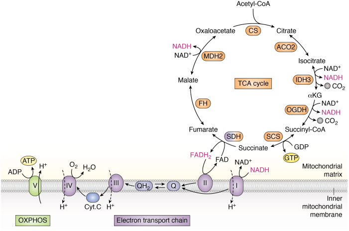
“Electron funneling through the complexes of the ETC is coupled to generation of the mitochondrial membrane potential that is ultimately used to power the production of the cellular currency (ATP) in a process known as oxidative phosphorylation (OXPHOS), as oxygen serves as the terminal acceptor for electrons that transit through the ETC (Fig. 1) (3).”
§ Regulation and function of the mammalian tricarboxylic acid cycle > Abstract
What the sources say
- Not checked yetcites [3] Ryan, 2020, Krebs cycle Reborn in macrophage immunometabolism, Annu. Rev. Immunol. source
plaindirect- Asserted: passes — The authors put it forward as true.
- Checkable: passes — It can be checked against evidence.
- Load-bearing: unclear — It is unclear whether the paper’s conclusions depend on it.
- Based: passes — The paper gives a reason why it holds.
A conclusion the paper draws from its own results — it cites no other source for it, so there is none to check. The quote shows it is in the paper.
In the paper
“These observations support the notion that the TCA cycle plays a significant role in sustaining mammalian tissue function.”
§ Regulation and function of the mammalian tricarboxylic acid cycle > Abstract
plaindirect- Asserted: passes — The authors put it forward as true.
- Checkable: passes — It can be checked against evidence.
- Load-bearing: passes — The paper’s conclusions depend on it.
- Based: passes — The paper gives a reason why it holds.
None of the sources cited for this claim have been checked yet.
In the paper
“In 1937, Hans Krebs (1, 32) showed that succinate could be synthesized by animal tissues in the presence of pyruvate, leading him to speculate that succinate may arise from citrate oxidation downstream of pyruvate.”
§ Regulation and function of the mammalian tricarboxylic acid cycle > Discovery of the TCA cycle
What the sources say
plaindirect- Asserted: passes — The authors put it forward as true.
- Checkable: passes — It can be checked against evidence.
- Load-bearing: unclear — It is unclear whether the paper’s conclusions depend on it.
- Based: passes — The paper gives a reason why it holds.
None of the sources cited for this claim have been checked yet.
In the paper
“However, several chemical intermediates of the TCA cycle also serve as critical precursors for biosynthetic reactions, which we will discuss in detail later (5, 6).”
§ Regulation and function of the mammalian tricarboxylic acid cycle > Abstract
What the sources say
plaindirect- Asserted: passes — The authors put it forward as true.
- Checkable: passes — It can be checked against evidence.
- Load-bearing: passes — The paper’s conclusions depend on it.
- Based: passes — The paper gives a reason why it holds.
In the paper
“Support for this idea came the following year when Stare (33) and Baumann found that very small quantities of dicarboxylic acids were sufficient to cause an increase in oxygen consumption and that this increase was beyond what was necessary for the oxidation of the quantities of substrate added.”
§ Regulation and function of the mammalian tricarboxylic acid cycle > Discovery of the TCA cycle
What the sources say
- Not checked yetcites [33] Stare, 1936, The effect of fumarate on the respiration of liver and kidney tissue, Biochem. J. source
plaindirect- Asserted: passes — The authors put it forward as true.
- Checkable: passes — It can be checked against evidence.
- Load-bearing: unclear — It is unclear whether the paper’s conclusions depend on it.
- Based: passes — The paper gives a reason why it holds.
The paper states this without a source or data behind it here, so there is nothing to check it against. The quote shows it is in the paper.
In the paper
“Multiple metabolic signals control both TCA cycle inputs and directionality, and increasing work demonstrates the importance of such TCA cycle flexibility for optimizing cellular fitness both under physiological conditions and in the context of disease.”
§ Regulation and function of the mammalian tricarboxylic acid cycle > Abstract
boostedcorrelational- Asserted: passes — The authors put it forward as true.
- Checkable: passes — It can be checked against evidence.
- Load-bearing: passes — The paper’s conclusions depend on it.
- Based: passes — The paper gives a reason why it holds.
The paper states this without a source or data behind it here, so there is nothing to check it against. The quote shows it is in the paper.
In the paper
“Given the central role of the TCA cycle in both provisioning key anabolic substrates and in maintaining energy production, it is not surprising that TCA cycle activity is under tight physiologic regulation.”
§ Regulation and function of the mammalian tricarboxylic acid cycle > Abstract
plain- Asserted: passes — The authors put it forward as true.
- Checkable: passes — It can be checked against evidence.
- Load-bearing: passes — The paper’s conclusions depend on it.
- Based: passes — The paper gives a reason why it holds.
None of the sources cited for this claim have been checked yet.
In the paper
“By adding malonate, an inhibitor of succinate oxidation, they found that succinate accumulated when OAA was added to muscle, implying that this series of reactions was cyclical in nature (2, 32).”
§ Regulation and function of the mammalian tricarboxylic acid cycle > Discovery of the TCA cycle
What the sources say
plaindirect- Asserted: passes — The authors put it forward as true.
- Checkable: passes — It can be checked against evidence.
- Load-bearing: unclear — It is unclear whether the paper’s conclusions depend on it.
- Based: passes — The paper gives a reason why it holds.
- Probably not a substantial claim. 15
- It may state two separate claims as one. 73
Every source the paper cites for this claim was checked and supports it.
In the paper
“Distinct isoforms of ACSS localize specifically to the cytosol and mitochondria, but how conversion of acetate to acetyl-CoA is coordinated between these two compartments remains poorly understood (71).”
§ Regulation and function of the mammalian tricarboxylic acid cycle > Wiring of the TCA cycle > Sources of acetyl-CoA
What the sources say
- Supportedcites [71] Schug, 2016, The metabolic fate of acetate in cancer, Nat. Rev. Cancer source
The “Acetyl-CoA synthetases” section identifies ACSS1 as mitochondrial and ACSS2 as nucleocytosolic. The “Acetate contribution to fatty acid synthesis” section describes a route between the compartments but says the interplay of ACSS1, ACSS2 and ACLY in controlling acetyl-CoA metabolism remains an area for future research. Page numbers are unavailable in our copy. (LLM, conf=0.89)
plain- Asserted: passes — The authors put it forward as true.
- Checkable: passes — It can be checked against evidence.
- Load-bearing: fails — The paper’s conclusions may not depend on it.
- Based: passes — The paper gives a reason why it holds.
Probably not a substantial claim. The paper’s conclusions may not depend on it. It may state two separate claims as one.
- Supported
The cited sources support only part of this claim — read each source’s verdict below.
In the paper
“Krebs found that GDH operates at close to equilibrium in vitro, and some evidence indicates that GDH may be reversible in some tissues in vivo (112, 126).”
§ Regulation and function of the mammalian tricarboxylic acid cycle > Flexibility of TCA cycle wiring
What the sources say
- Partly supportedcites [112] Williamson, 1967, The redox state of free nicotinamide-adenine dinucleotide in the cytoplasm and mitochondria of rat liver, Biochem. J. sourceExperimental — accuracy not yet measured
In the section comparing the glutamate- and β-hydroxybutyrate-dehydrogenase systems (page unavailable), the paper presents evidence that glutamate dehydrogenase is in equilibrium in rat liver in vivo. It does not report that the authors found GDH operating close to equilibrium in vitro; its separately measured equilibrium constant is not such a finding. (LLM, conf=0.85)
- Not checked yetcites [126] Treberg, 2010, On the reversibility of glutamate dehydrogenase and the source of hyperammonemia in the hyperinsulinism/hyperammonemia syndrome, Adv. Enzyme Regul. source
hedgedconditional- Asserted: passes — The authors put it forward as true.
- Checkable: passes — It can be checked against evidence.
- Load-bearing: unclear — It is unclear whether the paper’s conclusions depend on it.
- Based: passes — The paper gives a reason why it holds.
It may state two separate claims as one.
- Partly supported
Every source the paper cites for this claim was checked and supports it.
In the paper
“Notably, while NAD+ itself can be imported into mitochondria through SLC25A51–MCART1 (98, 99, 100), NADH is not efficiently transported.”
§ Regulation and function of the mammalian tricarboxylic acid cycle > Wiring of the TCA cycle > Electron shuttles
What the sources say
- Supportedcites [98] Girardi, 2020, Epistasis-driven identification of SLC25A51 as a regulator of human mitochondrial NAD import, Nat. Commun. source
The citation in the printed sentence attaches to NAD+ import, not to the subsequent statement about NADH. The Discussion identifies SLC25A51 as required for mitochondrial NAD+ uptake in human cells, while noting that direct transport assays are still needed to establish its mechanism definitively. (LLM, conf=0.90)
- Supportedcites [99] Kory, 2020, MCART1/SLC25A51 is required for mitochondrial NAD transport, Sci. Adv. source
The citation is attached to the statement that NAD+ can be imported into mitochondria through SLC25A51/MCART1. The Discussion identifies MCART1 as a putative mitochondrial NAD importer in humans, and the Results report MCART1-dependent NAD+ uptake. The sentence's separate statement about inefficient NADH transport is not the part attributed to this citation. (LLM, conf=0.93)
- Supportedcites [100] Luongo, 2020, SLC25A51 is a mammalian mitochondrial NAD(+) transporter, Nature source
In the printed sentence, the citations attach to the claim that NAD+ enters mitochondria through SLC25A51, rather than to the subsequent statement about NADH. The paper’s “SLC25A51 drives mitochondrial NAD+ uptake” section reports SLC25A51-dependent uptake of NAD+ into the mitochondrial matrix. The paper does not establish the additional electron-shuttle explanation in the supplied claim. (LLM, conf=0.91)
plaindirect- Asserted: passes — The authors put it forward as true.
- Checkable: passes — It can be checked against evidence.
- Load-bearing: passes — The paper’s conclusions depend on it.
- Based: passes — The paper gives a reason why it holds.
It may state two separate claims as one.
- Supported
Every source the paper cites for this claim was checked and supports it.
In the paper
“For example, intestinal stem cells (ISCs) express lower levels of MPC than their differentiated progeny, and blocking mitochondrial pyruvate entry via MPC inhibition drives increased ISC numbers and proliferative potential (206).”
§ Regulation and function of the mammalian tricarboxylic acid cycle > TCA cycle heterogeneity > TCA cycle heterogeneity in stem cells
What the sources say
- Supportedcites [206] Schell, 2017, Control of intestinal stem cell function and proliferation by mitochondrial pyruvate metabolism, Nat. Cell Biol. source
The Results section on MPC expression reports that MPC1 is nearly absent at the crypt base, where LGR5+ intestinal stem cells reside, but strongly expressed in the upper crypt and villus; the Discussion says MPC expression increases upon differentiation. The Drosophila Results report approximately twice as many stem cells and increased proliferation after MPC disruption, while the organoid Results report enhanced stem-cell function and markers after MPC inhibition. (LLM, conf=0.92)
plaindirect- Asserted: passes — The authors put it forward as true.
- Checkable: passes — It can be checked against evidence.
- Load-bearing: unclear — It is unclear whether the paper’s conclusions depend on it.
- Based: passes — The paper gives a reason why it holds.
It may state two separate claims as one.
- Supported
None of the sources cited for this claim have been checked yet.
In the paper
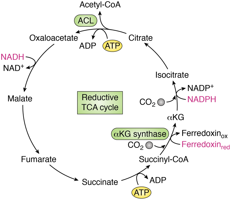
“However, demonstration of the C-C bond forming reactions of the rTCA cycle has yet to be achieved efficiently and nonenzymatically under experimental conditions, raising the possibility that this portion of the pathway required organic catalysts and, thus, emerged later (21, 22).”
§ Regulation and function of the mammalian tricarboxylic acid cycle > Evolutionary perspectives on the TCA cycle
What the sources say
hedgedconditional- Asserted: passes — The authors put it forward as true.
- Checkable: passes — It can be checked against evidence.
- Load-bearing: fails — The paper’s conclusions may not depend on it.
- Based: passes — The paper gives a reason why it holds.
Probably not a substantial claim. The paper’s conclusions may not depend on it. It may state two separate claims as one.
None of the sources cited for this claim have been checked yet.
In the paper
“These findings strengthen the notion that the rTCA cycle may represent both a vestige of prebiotic metabolism and an evolutionary precursor for the oxidative TCA cycle (18, 27).”
§ Regulation and function of the mammalian tricarboxylic acid cycle > Evolutionary perspectives on the TCA cycle
What the sources say
hedgedconditional- Asserted: passes — The authors put it forward as true.
- Checkable: passes — It can be checked against evidence.
- Load-bearing: unclear — It is unclear whether the paper’s conclusions depend on it.
- Based: passes — The paper gives a reason why it holds.
It may state two separate claims as one.
None of the sources cited for this claim have been checked yet.
In the paper
“The oxygenation of Earth’s atmosphere over time increased the availability of molecular oxygen, whose function as an electron acceptor facilitates oxidation of organic molecules derived from carbon assimilation (21, 28).”
§ Regulation and function of the mammalian tricarboxylic acid cycle > Evolutionary perspectives on the TCA cycle
What the sources say
plaindirect- Asserted: passes — The authors put it forward as true.
- Checkable: passes — It can be checked against evidence.
- Load-bearing: unclear — It is unclear whether the paper’s conclusions depend on it.
- Based: passes — The paper gives a reason why it holds.
It may state two separate claims as one.
In the paper
“A key finding in that same year came from Martius and Knoop (34), who found that αKG is a product of citrate oxidation and worked out the sequence of reactions from citrate to succinate.”
§ Regulation and function of the mammalian tricarboxylic acid cycle > Discovery of the TCA cycle
What the sources say
- Not checked yetcites [34] Martius, 1937, Physiological breakdown of citric acid, Prelim. Z. Physiol. Chem.
plaindirect- Asserted: passes — The authors put it forward as true.
- Checkable: passes — It can be checked against evidence.
- Load-bearing: fails — The paper’s conclusions may not depend on it.
- Based: passes — The paper gives a reason why it holds.
Probably not a substantial claim. The paper’s conclusions may not depend on it. It may state two separate claims as one.
The paper states this without a source or data behind it here, so there is nothing to check it against. The quote shows it is in the paper.
In the paper
“In this configuration, the central function of the TCA cycle is to convert fuel sources into energy, and therefore, the TCA cycle is generally considered a catabolic process.”
§ Regulation and function of the mammalian tricarboxylic acid cycle > Abstract
plaindirect- Asserted: passes — The authors put it forward as true.
- Checkable: passes — It can be checked against evidence.
- Load-bearing: unclear — It is unclear whether the paper’s conclusions depend on it.
- Based: passes — The paper gives a reason why it holds.
It may state two separate claims as one.
In the paper
“The TCA cycle itself neither consumes molecular oxygen nor produces meaningful amounts of ATP; rather, the TCA cycle removes electrons (reducing equivalents) from inputs (e.g., acetyl-CoA) and transfers them to electron carriers that deposit their electrons onto the electron transport chain (ETC).”
§ Regulation and function of the mammalian tricarboxylic acid cycle > Abstract
What the sources say
- Not checked yetcites [3] Ryan, 2020, Krebs cycle Reborn in macrophage immunometabolism, Annu. Rev. Immunol. source
plain- Asserted: passes — The authors put it forward as true.
- Checkable: passes — It can be checked against evidence.
- Load-bearing: passes — The paper’s conclusions depend on it.
- Based: passes — The paper gives a reason why it holds.
It may state two separate claims as one.
None of the sources cited for this claim have been checked yet.
In the paper
“At the nexus of both catabolic and anabolic metabolism lies the tricarboxylic acid (TCA) cycle, a broadly conserved metabolic pathway consisting of a cyclic series of chemical reactions that harness high-energy electrons from fuel sources (1, 2, 3).”
§ Regulation and function of the mammalian tricarboxylic acid cycle > Abstract
What the sources say
- Not checked yetcites [1] Krebs, 1937, Metabolism of ketonic acids in animal tissues, Biochem. J. source
- Not checked yetcites [2] Krebs, 1980, The role of citric acid in intermediate metabolism in animal tissues, FEBS Lett. source
- Not checked yetcites [3] Ryan, 2020, Krebs cycle Reborn in macrophage immunometabolism, Annu. Rev. Immunol. source
plaindirect- Asserted: passes — The authors put it forward as true.
- Checkable: passes — It can be checked against evidence.
- Load-bearing: fails — The paper’s conclusions may not depend on it.
- Based: passes — The paper gives a reason why it holds.
Probably not a substantial claim. The paper’s conclusions may not depend on it. It may state two separate claims as one.
In the paper
“Significantly, they also found that added dicarboxylic acids were not used up and could still be detected in the medium, indicating that these compounds were functioning in a catalytic capacity (33).”
§ Regulation and function of the mammalian tricarboxylic acid cycle > Discovery of the TCA cycle
What the sources say
- Not checked yetcites [33] Stare, 1936, The effect of fumarate on the respiration of liver and kidney tissue, Biochem. J. source
hedgeddirect- Asserted: passes — The authors put it forward as true.
- Checkable: passes — It can be checked against evidence.
- Load-bearing: unclear — It is unclear whether the paper’s conclusions depend on it.
- Based: passes — The paper gives a reason why it holds.
It may state two separate claims as one.
None of the sources cited for this claim have been checked yet.
In the paper
“In 1935, Szent-Györgyi worked out the sequence of reactions from succinate to fumarate to malate to OAA and demonstrated the rapid oxidation of these substances by pigeon muscle suspensions (31, 32).”
§ Regulation and function of the mammalian tricarboxylic acid cycle > Discovery of the TCA cycle
What the sources say
- Not checked yetcites [31] Annau, 1935, Über die Bedeutung der Fumarsäure für die tierische Gewebsatmung, Biol. Chem.
- Not checked yetcites [32] Kornberg, 2000, Krebs and his trinity of cycles, Nat. Rev. Mol. Cell Biol. source
boosteddirect- Asserted: passes — The authors put it forward as true.
- Checkable: passes — It can be checked against evidence.
- Load-bearing: unclear — It is unclear whether the paper’s conclusions depend on it.
- Based: passes — The paper gives a reason why it holds.
It may state two separate claims as one.
None of the sources cited for this claim have been checked yet.
In the paper
“Being able to fine-tune the function of metabolic pathways like the TCA cycle—in particular, to toggle between catabolic and anabolic activity—set the stage for early unicellular life forms to evolve into multicellular organisms, which require coordination of metabolic activity beyond that dictated by local nutrient availability (29, 30).”
§ Regulation and function of the mammalian tricarboxylic acid cycle > Evolutionary perspectives on the TCA cycle
What the sources say
plaindirect- Asserted: passes — The authors put it forward as true.
- Checkable: passes — It can be checked against evidence.
- Load-bearing: fails — The paper’s conclusions may not depend on it.
- Based: passes — The paper gives a reason why it holds.
Probably not a substantial claim. The paper’s conclusions may not depend on it.
A conclusion the paper draws from its own results — it cites no other source for it, so there is none to check. The quote shows it is in the paper.
In the paper
“However, given that prebiotic chemistry would necessarily have initially required the production, not the breakdown, of organic molecules, primordial metabolism likely centered on reductive CO2-fixing pathways.”
§ Regulation and function of the mammalian tricarboxylic acid cycle > Evolutionary perspectives on the TCA cycle
hedgedconditional- Asserted: passes — The authors put it forward as true.
- Checkable: passes — It can be checked against evidence.
- Load-bearing: fails — The paper’s conclusions may not depend on it.
- Based: passes — The paper gives a reason why it holds.
Probably not a substantial claim. The paper’s conclusions may not depend on it.
None of the sources cited for this claim have been checked yet.
In the paper
“Because the TCA cycle functions in both catabolic and anabolic capacities, it is considered an amphibolic pathway (7, 8).”
§ Regulation and function of the mammalian tricarboxylic acid cycle > Abstract
What the sources say
- Not checked yetcites [7] Kornberg, 1965, Anaplerotic sequences in microbial metabolism, Angew. Chem. Int. Ed. Engl. source
- Not checked yetcites [8] Kornberg, 1966, Anapleroic sequences and their role in metabolism, Essays Biochem.
plaindirect- Asserted: passes — The authors put it forward as true.
- Checkable: passes — It can be checked against evidence.
- Load-bearing: fails — The paper’s conclusions may not depend on it.
- Based: passes — The paper gives a reason why it holds.
Probably not a substantial claim. The paper’s conclusions may not depend on it.
In the paper
“These crucial experiments teed up the findings of Hans Krebs and his colleague William Johnson (2) in Enzymologia in 1937, in which they discovered that citrate was readily oxidized by minced pigeon breast muscle and that citrate addition increased oxygen uptake beyond the amount necessary for the complete oxidation of the added citrate, indicating that citrate itself was stimulating consumption”
§ Regulation and function of the mammalian tricarboxylic acid cycle > Discovery of the TCA cycle
What the sources say
- Not checked yetcites [2] Krebs, 1980, The role of citric acid in intermediate metabolism in animal tissues, FEBS Lett. source
plaindirect- Asserted: passes — The authors put it forward as true.
- Checkable: passes — It can be checked against evidence.
- Load-bearing: unclear — It is unclear whether the paper’s conclusions depend on it.
- Based: passes — The paper gives a reason why it holds.
It may state two separate claims as one.
A conclusion the paper draws from its own results — it cites no other source for it, so there is none to check. The quote shows it is in the paper.
In the paper
“This development likely enabled the emergence of the oxidative TCA cycle and its coupling with OXPHOS, allowing the transfer of electrons derived from the breakdown of carbon substrates onto molecular oxygen.”
§ Regulation and function of the mammalian tricarboxylic acid cycle > Evolutionary perspectives on the TCA cycle
hedgedconditional- Asserted: passes — The authors put it forward as true.
- Checkable: passes — It can be checked against evidence.
- Load-bearing: unclear — It is unclear whether the paper’s conclusions depend on it.
- Based: passes — The paper gives a reason why it holds.
It may state two separate claims as one.
In the paper
“Likewise, cells that are addicted to glutamine become dependent on PC for growth when glutamine metabolism is suppressed; conversely, cells with high basal PC activity are generally more resistant to interruption of glutamine metabolism (87).”
§ Regulation and function of the mammalian tricarboxylic acid cycle > Wiring of the TCA cycle > TCA cycle anaplerosis
What the sources say
- Not checked yetcites [87] Cheng, 2011, Pyruvate carboxylase is required for glutamine-independent growth of tumor cells, Proc. Natl. Acad. Sci. U. S. A. source
plaindirect- Asserted: passes — The authors put it forward as true.
- Checkable: passes — It can be checked against evidence.
- Load-bearing: unclear — It is unclear whether the paper’s conclusions depend on it.
- Based: passes — The paper gives a reason why it holds.
It may state two separate claims as one.
In the paper
“In subsequent work, Krebs (4) determined that the two carbon atoms that condensed with OAA to form citrate were derived from pyruvate and that oxygen consumption in muscle increases when pyruvate is added.”
§ Regulation and function of the mammalian tricarboxylic acid cycle > Discovery of the TCA cycle
What the sources say
- Not checked yetcites [4] Krebs, 1940, The oxidation of pyruvate in pigeon breast muscle, Biochem. J. source
plaindirect- Asserted: passes — The authors put it forward as true.
- Checkable: passes — It can be checked against evidence.
- Load-bearing: unclear — It is unclear whether the paper’s conclusions depend on it.
- Based: passes — The paper gives a reason why it holds.
It may state two separate claims as one.
A conclusion the paper draws from its own results — it cites no other source for it, so there is none to check. The quote shows it is in the paper.
In the paper
“Thus, while glutamine anaplerosis efficiently allows cells both to capture reducing equivalents from substrate oxidation and maintain OAA production, PC provides a critical backup when glutamine is not available or when nutrient status is so high that the ATP-dependent circumventing of the oxidative TCA cycle mediated by PC may actually help cells cope with nutrient overload.”
§ Regulation and function of the mammalian tricarboxylic acid cycle > Wiring of the TCA cycle > TCA cycle anaplerosis
plaindirect- Asserted: passes — The authors put it forward as true.
- Checkable: passes — It can be checked against evidence.
- Load-bearing: passes — The paper’s conclusions depend on it.
- Based: passes — The paper gives a reason why it holds.
It may state two separate claims as one.
In the paper
“Supraphysiologic propionate administration increases TCA cycle metabolite pools in the liver and induces hepatic gluconeogenesis (96), but the degree to which cells can activate propionyl-CoA catabolism to sustain anaplerotic flux under physiologic conditions remains largely unknown.”
§ Regulation and function of the mammalian tricarboxylic acid cycle > Wiring of the TCA cycle > TCA cycle anaplerosis
What the sources say
- Not checked yetcites [96] Perry, 2016, Propionate increases hepatic pyruvate cycling and anaplerosis and alters mitochondrial metabolism, J. Biol. Chem. source
plaindirect- Asserted: passes — The authors put it forward as true.
- Checkable: passes — It can be checked against evidence.
- Load-bearing: unclear — It is unclear whether the paper’s conclusions depend on it.
- Based: passes — The paper gives a reason why it holds.
It may state two separate claims as one.
In the paper
“Suggestively, one study found that LDH can localize to the mitochondria, raising the possibility that lactate can be directly imported into the mitochondria for further oxidation (61).”
§ Regulation and function of the mammalian tricarboxylic acid cycle > Wiring of the TCA cycle > Sources of acetyl-CoA
What the sources say
- Not checked yetcites [61] Chen, 2016, Lactate metabolism is associated with mammalian mitochondria, Nat. Chem. Biol. source
hedgedconditional- Asserted: passes — The authors put it forward as true.
- Checkable: passes — It can be checked against evidence.
- Load-bearing: fails — The paper’s conclusions may not depend on it.
- Based: passes — The paper gives a reason why it holds.
Probably not a substantial claim. The paper’s conclusions may not depend on it. It may state two separate claims as one.
In the paper
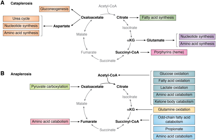
“Cataplerosis of any TCA cycle intermediate requires compensatory input to sustain TCA cycle activity, a process termed “anaplerosis” (37).”
§ Regulation and function of the mammalian tricarboxylic acid cycle > Wiring of the TCA cycle
What the sources say
- Not checked yetcites [37] Owen, 2002, The key role of anaplerosis and cataplerosis for citric acid cycle function, J. Biol. Chem. source
plaindirect- Asserted: passes — The authors put it forward as true.
- Checkable: passes — It can be checked against evidence.
- Load-bearing: passes — The paper’s conclusions depend on it.
- Based: passes — The paper gives a reason why it holds.
Probably not a substantial claim.
In the paper
“While the mechanisms dictating a cell’s preferred source of acetyl-CoA are not well understood, recent work has found that different tissues in the body have distinct fuel preferences (44), raising the possibility that the source of acetyl-CoA generation is dependent on cell type and cell state.”
§ Regulation and function of the mammalian tricarboxylic acid cycle > Wiring of the TCA cycle > Sources of acetyl-CoA
What the sources say
- Not checked yetcites [44] Jang, 2019, Metabolite exchange between mammalian organs quantified in pigs, Cell Metab. source
hedgedconditional- Asserted: passes — The authors put it forward as true.
- Checkable: passes — It can be checked against evidence.
- Load-bearing: fails — The paper’s conclusions may not depend on it.
- Based: passes — The paper gives a reason why it holds.
Probably not a substantial claim. The paper’s conclusions may not depend on it. It may state two separate claims as one.
In the paper
“Mitochondrial import and catabolism of lactate would prevent the buildup of cytosolic NADH while allowing for the mitochondrial capture of both carbon and reducing equivalents (61).”
§ Regulation and function of the mammalian tricarboxylic acid cycle > Wiring of the TCA cycle > Sources of acetyl-CoA
What the sources say
- Not checked yetcites [61] Chen, 2016, Lactate metabolism is associated with mammalian mitochondria, Nat. Chem. Biol. source
hedgeddirect- Asserted: passes — The authors put it forward as true.
- Checkable: passes — It can be checked against evidence.
- Load-bearing: passes — The paper’s conclusions depend on it.
- Based: passes — The paper gives a reason why it holds.
Probably not a substantial claim. It may state two separate claims as one.
None of the sources cited for this claim have been checked yet.
In the paper
“In cultured cells, pyruvate is largely produced from glucose via glycolysis; in vivo, circulating lactate provides an additional major source of tissue pyruvate (46, 47, 48).”
§ Regulation and function of the mammalian tricarboxylic acid cycle > Wiring of the TCA cycle > Sources of acetyl-CoA
What the sources say
- Not checked yetcites [46] Faubert, 2017, Lactate metabolism in human lung tumors, Cell source
- Not checked yetcites [47] Hui, 2017, Glucose feeds the TCA cycle via circulating lactate, Nature source
- Not checked yetcites [48] Kennedy, 2013, Catabolism of exogenous lactate reveals it as a legitimate metabolic substrate in breast cancer, PLoS One source
plaindirect- Asserted: passes — The authors put it forward as true.
- Checkable: passes — It can be checked against evidence.
- Load-bearing: unclear — It is unclear whether the paper’s conclusions depend on it.
- Based: passes — The paper gives a reason why it holds.
It may state two separate claims as one.
A conclusion the paper draws from its own results — it cites no other source for it, so there is none to check. The quote shows it is in the paper.
In the paper
“GOT2-driven glutamate metabolism thus offers several benefits to proliferating cells: GOT2 circumvents physiological limitations on glutamate catabolism, preserves the amine nitrogen of glutamate for aspartate synthesis, and reduces production of toxic ammonia.”
§ Regulation and function of the mammalian tricarboxylic acid cycle > Wiring of the TCA cycle > TCA cycle anaplerosis
plaindirect- Asserted: passes — The authors put it forward as true.
- Checkable: passes — It can be checked against evidence.
- Load-bearing: passes — The paper’s conclusions depend on it.
- Based: passes — The paper gives a reason why it holds.
It may state two separate claims as one.
None of the sources cited for this claim have been checked yet.
In the paper
“In most tissues and some tumors, the contribution of circulating lactate to TCA cycle intermediates exceeds that of glucose, supporting the idea that lactate is a fundamental TCA cycle substrate in living organisms (46, 47).”
§ Regulation and function of the mammalian tricarboxylic acid cycle > Wiring of the TCA cycle > Sources of acetyl-CoA
What the sources say
plaincorrelational- Asserted: passes — The authors put it forward as true.
- Checkable: passes — It can be checked against evidence.
- Load-bearing: passes — The paper’s conclusions depend on it.
- Based: passes — The paper gives a reason why it holds.
It may state two separate claims as one.
None of the sources cited for this claim have been checked yet.
In the paper
“Sensing of free fatty acid availability within the cell primarily occurs through peroxisome proliferator–activated receptors, which function as fatty acid–activated transcription factors that drive the expression of CPT1 (64, 65).”
§ Regulation and function of the mammalian tricarboxylic acid cycle > Wiring of the TCA cycle > Sources of acetyl-CoA
What the sources say
- Not checked yetcites [64] Chatelain, 1996, Cyclic AMP and fatty acids increase carnitine palmitoyltransferase I gene transcription in cultured fetal rat hepatocytes, Eur. J. Biochem. source
- Not checked yetcites [65] Mascaro, 1998, Control of human muscle-type carnitine palmitoyltransferase I gene transcription by peroxisome proliferator-activated receptor, J. Biol. Chem. source
plaindirect- Asserted: passes — The authors put it forward as true.
- Checkable: passes — It can be checked against evidence.
- Load-bearing: unclear — It is unclear whether the paper’s conclusions depend on it.
- Based: passes — The paper gives a reason why it holds.
It may state two separate claims as one.
None of the sources cited for this claim have been checked yet.
In the paper
“As glucose uptake and usage are under growth factor control (58, 59, 60), this observation raises the possibility that a basal level of TCA cycle oxidation in cells can persist independent of exogenous growth factor stimulation.”
§ Regulation and function of the mammalian tricarboxylic acid cycle > Wiring of the TCA cycle > Sources of acetyl-CoA
What the sources say
- Not checked yetcites [58] Christofk, 2008, Pyruvate kinase M2 is a phosphotyrosine-binding protein, Nature source
- Not checked yetcites [59] Thompson, 2011, Rethinking the regulation of cellular metabolism, Cold Spring Harb. Symp. Quant. Biol. source
- Not checked yetcites [60] Vander Heiden, 2001, Growth factors can influence cell growth and survival through effects on glucose metabolism, Mol. Cell Biol. source
hedgedconditional- Asserted: fails — The authors may not put it forward as true.
- Checkable: passes — It can be checked against evidence.
- Load-bearing: fails — The paper’s conclusions may not depend on it.
- Based: passes — The paper gives a reason why it holds.
Probably not a substantial claim. The authors may not put it forward as true. The paper’s conclusions may not depend on it.
None of the sources cited for this claim have been checked yet.
In the paper
“PC is essential for growth of succinate dehydrogenase (SDH)–mutant cells, which exhibit a truncated TCA cycle and thus require an alternative source of OAA to support TCA cycle function and sustain aspartate production for anabolic pathways (91, 92).”
§ Regulation and function of the mammalian tricarboxylic acid cycle > Wiring of the TCA cycle > TCA cycle anaplerosis
What the sources say
- Not checked yetcites [91] Cardaci, 2015, Pyruvate carboxylation enables growth of SDH-deficient cells by supporting aspartate biosynthesis, Nat. Cell Biol. source
- Not checked yetcites [92] Lussey-Lepoutre, 2015, Loss of succinate dehydrogenase activity results in dependency on pyruvate carboxylation for cellular anabolism, Nat. Commun. source
plaindirect- Asserted: passes — The authors put it forward as true.
- Checkable: passes — It can be checked against evidence.
- Load-bearing: passes — The paper’s conclusions depend on it.
- Based: passes — The paper gives a reason why it holds.
It may state two separate claims as one.
In the paper
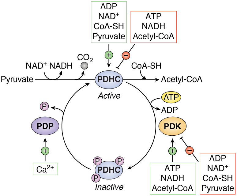
“The PDHC is allosterically inhibited by NADH, acetyl-CoA, and ATP, making it a potent sensor of TCA cycle activity and thus a critical node of TCA cycle regulation (discussed further) (52).”
§ Regulation and function of the mammalian tricarboxylic acid cycle > Wiring of the TCA cycle > Sources of acetyl-CoA
What the sources say
- Not checked yetcites [52] Behal, 1993, Regulation of the pyruvate dehydrogenase multienzyme complex, Annu. Rev. Nutr. source
plaindirect- Asserted: passes — The authors put it forward as true.
- Checkable: passes — It can be checked against evidence.
- Load-bearing: unclear — It is unclear whether the paper’s conclusions depend on it.
- Based: passes — The paper gives a reason why it holds.
It may state two separate claims as one.
A conclusion the paper draws from its own results — it cites no other source for it, so there is none to check. The quote shows it is in the paper.
In the paper
“Because the TCA cycle functions as a critical biosynthetic hub and is coupled with cellular energy production via OXPHOS, cells have evolved multiple nodes of TCA cycle regulation.”
§ Regulation and function of the mammalian tricarboxylic acid cycle > Nodes of TCA cycle control
plaindirect- Asserted: passes — The authors put it forward as true.
- Checkable: passes — It can be checked against evidence.
- Load-bearing: passes — The paper’s conclusions depend on it.
- Based: passes — The paper gives a reason why it holds.
Probably not a substantial claim. It may state two separate claims as one.
A conclusion the paper draws from its own results — it cites no other source for it, so there is none to check. The quote shows it is in the paper.
In the paper
“While not technically part of the TCA cycle, the PDHC generates the starting molecule of the cycle, acetyl-CoA, thereby functioning as a gatekeeper of TCA cycle activity.”
§ Regulation and function of the mammalian tricarboxylic acid cycle > Nodes of TCA cycle control > Allosteric regulation of TCA cycle enzyme activity
plaindirect- Asserted: passes — The authors put it forward as true.
- Checkable: passes — It can be checked against evidence.
- Load-bearing: unclear — It is unclear whether the paper’s conclusions depend on it.
- Based: passes — The paper gives a reason why it holds.
It may state two separate claims as one.
In the paper
“For example, low oxygen levels activate the hypoxia-inducible transcription factors (HIFs), which bind to hypoxia-response elements within a number of target genes including LDH-A (LDHA) and PDK1, leading to suppression of PDHC activity with a concomitant increase in aerobic glycolysis (141).”
§ Regulation and function of the mammalian tricarboxylic acid cycle > Nodes of TCA cycle control > Covalent modification of TCA cycle–related enzymes
What the sources say
- Not checked yetcites [141] Kim, 2006, HIF-1-mediated expression of pyruvate dehydrogenase kinase: A metabolic switch required for cellular adaptation to hypoxia, Cell Metab. source
plaindirect- Asserted: passes — The authors put it forward as true.
- Checkable: passes — It can be checked against evidence.
- Load-bearing: unclear — It is unclear whether the paper’s conclusions depend on it.
- Based: passes — The paper gives a reason why it holds.
It may state two separate claims as one.
In the paper
“These results indicate that cell proliferation can be impaired when the demand for mitochondrial NAD+ exceeds that of ATP and may explain why highly proliferative cells often engage in aerobic glycolysis over pyruvate oxidation and OXPHOS, even in the presence of sufficient oxygen (138).”
§ Regulation and function of the mammalian tricarboxylic acid cycle > Nodes of TCA cycle control > Allosteric regulation of TCA cycle enzyme activity
What the sources say
- Not checked yetcites [138] Luengo, 2021, Increased demand for NAD(+) relative to ATP drives aerobic glycolysis, Mol. Cell source
hedgedconditional- Asserted: passes — The authors put it forward as true.
- Checkable: passes — It can be checked against evidence.
- Load-bearing: passes — The paper’s conclusions depend on it.
- Based: passes — The paper gives a reason why it holds.
It may state two separate claims as one.
A conclusion the paper draws from its own results — it cites no other source for it, so there is none to check. The quote shows it is in the paper.
In the paper
“However, it has become increasingly appreciated that TCA cycle function is not “one size fits all,” and that components of the pathway are differentially and flexibly engaged in a context-specific fashion.”
§ Regulation and function of the mammalian tricarboxylic acid cycle > Flexibility of TCA cycle wiring
plain- Asserted: passes — The authors put it forward as true.
- Checkable: passes — It can be checked against evidence.
- Load-bearing: passes — The paper’s conclusions depend on it.
- Based: passes — The paper gives a reason why it holds.
It may state two separate claims as one.
None of the sources cited for this claim have been checked yet.
In the paper
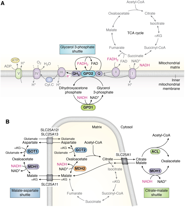
“Beyond supporting net NADH “import” into the mitochondria, these shuttles also facilitate the oxidation of cytosolic NADH, which is required to support continued glycolysis and the de novo synthesis of serine and lipids (101, 102, 103).”
§ Regulation and function of the mammalian tricarboxylic acid cycle > Wiring of the TCA cycle > Electron shuttles
What the sources say
- Not checked yetcites [101] Baksh, 2020, Extracellular serine controls epidermal stem cell fate and tumour initiation, Nat. Cell Biol. source
- Not checked yetcites [102] Diehl, 2019, Cellular redox state constrains serine synthesis and nucleotide production to impact cell proliferation, Nat. Metab. source
- Not checked yetcites [103] Li, 2022, Cancer cells depend on environmental lipids for proliferation when electron acceptors are limited, Nat. Metab. source
plaindirect- Asserted: passes — The authors put it forward as true.
- Checkable: passes — It can be checked against evidence.
- Load-bearing: unclear — It is unclear whether the paper’s conclusions depend on it.
- Based: passes — The paper gives a reason why it holds.
It may state two separate claims as one.
None of the sources cited for this claim have been checked yet.
In the paper
“The glycerol 3-phosphate shuttle is not considered the major route of NADH shuttling in most mammalian cells but has been shown to be particularly active in brown adipose tissue and may be implicated in thermoregulation (105, 106, 107).”
§ Regulation and function of the mammalian tricarboxylic acid cycle > Wiring of the TCA cycle > Electron shuttles
What the sources say
- Not checked yetcites [105] Koza, 1996, Sequence and tissue-dependent RNA expression of mouse FAD-linked glycerol-3-phosphate dehydrogenase, Arch. Biochem. Biophys. source
- Not checked yetcites [106] Ohkawa, 1969, Unusually high mitochondrial alpha glycerophosphate dehydrogenase activity in rat brown adipose tissue, J. Cell Biol. source
- Not checked yetcites [107] Safer, 1971, Control of the transport of reducing equivalents across the mitochondrial membrane in perfused rat heart, J. Mol. Cell Cardiol. source
hedgedconditional- Asserted: passes — The authors put it forward as true.
- Checkable: passes — It can be checked against evidence.
- Load-bearing: unclear — It is unclear whether the paper’s conclusions depend on it.
- Based: passes — The paper gives a reason why it holds.
It may state two separate claims as one.
In the paper
“Three enzymes in particular—CS, IDH, and oxoglutarate dehydrogenase (OGDH)—catalyze rate-controlling steps in the TCA cycle and are considered regulatory enzymes within the pathway (41).”
§ Regulation and function of the mammalian tricarboxylic acid cycle > Nodes of TCA cycle control > Allosteric regulation of TCA cycle enzyme activity
What the sources say
- Not checked yetcites [41] Chandel, 2015
plaindirect- Asserted: passes — The authors put it forward as true.
- Checkable: passes — It can be checked against evidence.
- Load-bearing: passes — The paper’s conclusions depend on it.
- Based: passes — The paper gives a reason why it holds.
It may state two separate claims as one.
In the paper
“Four pyruvate dehydrogenase kinases (PDKs; PDK1–4) catalyze phosphorylation of the serine residues, each with different site specificities (139).”
§ Regulation and function of the mammalian tricarboxylic acid cycle > Nodes of TCA cycle control > Covalent modification of TCA cycle–related enzymes
What the sources say
- Not checked yetcites [139] Korotchkina, 2001, Site specificity of four pyruvate dehydrogenase kinase isoenzymes toward the three phosphorylation sites of human pyruvate dehydrogenase, J. Biol. Chem. source
plaindirect- Asserted: passes — The authors put it forward as true.
- Checkable: passes — It can be checked against evidence.
- Load-bearing: unclear — It is unclear whether the paper’s conclusions depend on it.
- Based: passes — The paper gives a reason why it holds.
It may state two separate claims as one.
A conclusion the paper draws from its own results — it cites no other source for it, so there is none to check. The quote shows it is in the paper.
In the paper
“Thus, intramitochondrial Ca2+ regulates both carbon entry into and flux through the TCA cycle.”
§ Regulation and function of the mammalian tricarboxylic acid cycle > Nodes of TCA cycle control > Ion-mediated TCA cycle regulation
plaindirect- Asserted: passes — The authors put it forward as true.
- Checkable: passes — It can be checked against evidence.
- Load-bearing: passes — The paper’s conclusions depend on it.
- Based: passes — The paper gives a reason why it holds.
It may state two separate claims as one.
None of the sources cited for this claim have been checked yet.
In the paper
“More recently, it was demonstrated that the citrate–malate shuttle performs functions beyond that of an electron shuttle and, in fact, supports continuous citrate regeneration and TCA cycle intermediate homeostasis (119, 121).”
§ Regulation and function of the mammalian tricarboxylic acid cycle > Wiring of the TCA cycle > Electron shuttles
What the sources say
boosteddirect- Asserted: passes — The authors put it forward as true.
- Checkable: passes — It can be checked against evidence.
- Load-bearing: unclear — It is unclear whether the paper’s conclusions depend on it.
- Based: passes — The paper gives a reason why it holds.
It may state two separate claims as one.
In the paper
“Indeed, breast cancer cells can engage in reductive amination, and GDH activity supports ammonia incorporation and tumor growth in mouse models of breast cancer (128).”
§ Regulation and function of the mammalian tricarboxylic acid cycle > Flexibility of TCA cycle wiring
What the sources say
- Not checked yetcites [128] Spinelli, 2017, Metabolic recycling of ammonia via glutamate dehydrogenase supports breast cancer biomass, Science source
plaindirect- Asserted: passes — The authors put it forward as true.
- Checkable: passes — It can be checked against evidence.
- Load-bearing: unclear — It is unclear whether the paper’s conclusions depend on it.
- Based: passes — The paper gives a reason why it holds.
It may state two separate claims as one.
None of the sources cited for this claim have been checked yet.
In the paper
“OGDH undergoes substrate inhibition by succinyl-CoA, which also serves as an allosteric inhibitor of CS (134, 136).”
§ Regulation and function of the mammalian tricarboxylic acid cycle > Nodes of TCA cycle control > Allosteric regulation of TCA cycle enzyme activity
What the sources say
plaindirect- Asserted: passes — The authors put it forward as true.
- Checkable: passes — It can be checked against evidence.
- Load-bearing: unclear — It is unclear whether the paper’s conclusions depend on it.
- Based: passes — The paper gives a reason why it holds.
It may state two separate claims as one.
In the paper
“As a result of this “pseudohypoxic” state, SDH- and FH-mutant tumors exhibit enhanced glycolytic metabolism and high ROS production (186).”
§ Regulation and function of the mammalian tricarboxylic acid cycle > TCA cycle metabolites dictate cell fate and function > Metabolic control of gene expression and cell fate
What the sources say
- Not checked yetcites [186] Sullivan, 2013, The proto-oncometabolite fumarate binds glutathione to amplify ROS-dependent signaling, Mol. Cell source
plaindirect- Asserted: passes — The authors put it forward as true.
- Checkable: passes — It can be checked against evidence.
- Load-bearing: unclear — It is unclear whether the paper’s conclusions depend on it.
- Based: passes — The paper gives a reason why it holds.
It may state two separate claims as one.
In the paper
“The PI3K–AKT signaling pathway mediates both the generation and cytosolic export of glucose-derived citrate, whereas AKT-mediated phosphorylation of ACL enhances its catalytic activity (190).”
§ Regulation and function of the mammalian tricarboxylic acid cycle > TCA cycle metabolites dictate cell fate and function > Metabolic control of gene expression and cell fate
What the sources say
- Not checked yetcites [190] Berwick, 2002, The identification of ATP-citrate lyase as a protein kinase B (Akt) substrate in primary adipocytes, J. Biol. Chem. source
plaindirect- Asserted: passes — The authors put it forward as true.
- Checkable: passes — It can be checked against evidence.
- Load-bearing: passes — The paper’s conclusions depend on it.
- Based: passes — The paper gives a reason why it holds.
It may state two separate claims as one.
In the paper
“Depriving APC-mutant intestinal organoids of glutamine depletes intracellular αKG levels and promotes stem-like features and adenocarcinoma formation in vivo (162).”
§ Regulation and function of the mammalian tricarboxylic acid cycle > TCA cycle metabolites dictate cell fate and function > Metabolic control of gene expression and cell fate
What the sources say
- Not checked yetcites [162] Tran, 2020, alpha-Ketoglutarate attenuates Wnt signaling and drives differentiation in colorectal cancer, Nat. Cancer source
plaindirect- Asserted: passes — The authors put it forward as true.
- Checkable: passes — It can be checked against evidence.
- Load-bearing: passes — The paper’s conclusions depend on it.
- Based: passes — The paper gives a reason why it holds.
It may state two separate claims as one.
In the paper
“αKG supplementation rescues these effects by driving DNA and histone demethylation, which facilitates upregulation of differentiation-associated genes and blunts tumor growth in vivo (162).”
§ Regulation and function of the mammalian tricarboxylic acid cycle > TCA cycle metabolites dictate cell fate and function > Metabolic control of gene expression and cell fate
What the sources say
- Not checked yetcites [162] Tran, 2020, alpha-Ketoglutarate attenuates Wnt signaling and drives differentiation in colorectal cancer, Nat. Cancer source
plaindirect- Asserted: passes — The authors put it forward as true.
- Checkable: passes — It can be checked against evidence.
- Load-bearing: passes — The paper’s conclusions depend on it.
- Based: passes — The paper gives a reason why it holds.
It may state two separate claims as one.
In the paper
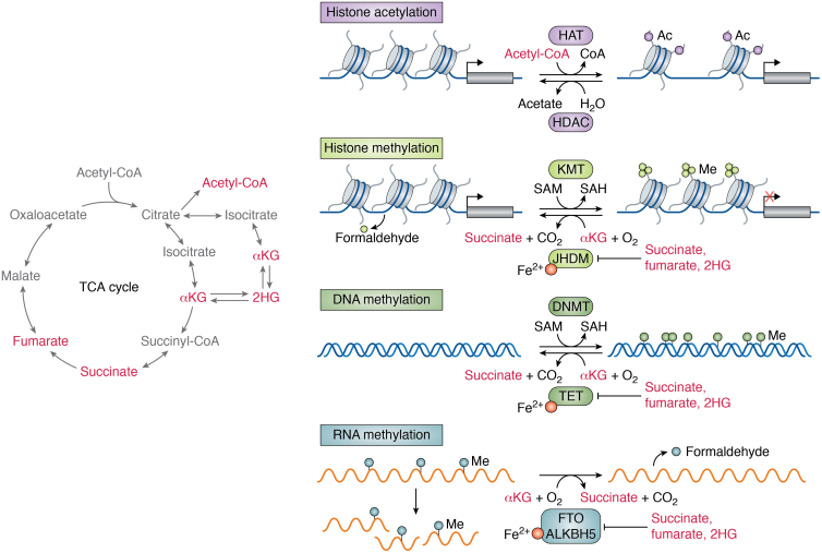
“These enzymes all require αKG, ferrous iron, and oxygen as cosubstrates and are inhibited by high levels of succinate and fumarate (161), thereby integrating multiple metabolic pathways in the control of gene expression programs and, ultimately, cell fate decisions.”
§ Regulation and function of the mammalian tricarboxylic acid cycle > TCA cycle metabolites dictate cell fate and function > Metabolic control of gene expression and cell fate
What the sources say
- Not checked yetcites [161] Baksh, 2021, Metabolic coordination of cell fate by α-ketoglutarate-dependent dioxygenases, Trends Cell Biol. source
plaindirect- Asserted: passes — The authors put it forward as true.
- Checkable: passes — It can be checked against evidence.
- Load-bearing: passes — The paper’s conclusions depend on it.
- Based: passes — The paper gives a reason why it holds.
It may state two separate claims as one.
In the paper
“Succinate, fumarate, and 2HG are all considered “oncometabolites” because they are linked to development of certain human cancers and because they affect cancer-relevant processes by virtue of their ability to act as competitive inhibitors of αKG-dependent dioxygenases (169) (Fig. 6).”
§ Regulation and function of the mammalian tricarboxylic acid cycle > TCA cycle metabolites dictate cell fate and function > Metabolic control of gene expression and cell fate
What the sources say
- Not checked yetcites [169] DeBerardinis, 2016, Fundamentals of cancer metabolism, Sci. Adv. source
plaindirect- Asserted: passes — The authors put it forward as true.
- Checkable: passes — It can be checked against evidence.
- Load-bearing: passes — The paper’s conclusions depend on it.
- Based: passes — The paper gives a reason why it holds.
It may state two separate claims as one.
None of the sources cited for this claim have been checked yet.
In the paper
“By blocking the enzymatic activity of histone and DNA demethylases, d-2HG can function to lock cells in a hypermethylated state that blocks differentiation and reinforces a malignant and stem cell–like phenotype (30, 172, 173, 174).”
§ Regulation and function of the mammalian tricarboxylic acid cycle > TCA cycle metabolites dictate cell fate and function > Metabolic control of gene expression and cell fate
What the sources say
- Not checked yetcites [30] Intlekofer, 2019, Metabolic signatures of cancer cells and stem cells, Nat. Metab. source
- Not checked yetcites [172] Figueroa, 2010, Leukemic IDH1 and IDH2 mutations result in a hypermethylation phenotype, disrupt TET2 function, and impair hematopoietic differentiation, Cancer Cell source
- Not checked yetcites [173] Lu, 2012, IDH mutation impairs histone demethylation and results in a block to cell differentiation, Nature source
plaindirect- Asserted: passes — The authors put it forward as true.
- Checkable: passes — It can be checked against evidence.
- Load-bearing: passes — The paper’s conclusions depend on it.
- Based: passes — The paper gives a reason why it holds.
It may state two separate claims as one.
In the paper
“Accordingly, oncogenic activation of the PI3K–AKT signaling axis increases acetyl-CoA levels and promotes elevated histone acetylation both in vitro and in tumors (188).”
§ Regulation and function of the mammalian tricarboxylic acid cycle > TCA cycle metabolites dictate cell fate and function > Metabolic control of gene expression and cell fate
What the sources say
- Not checked yetcites [188] Lee, 2014, Akt-dependent metabolic reprogramming regulates tumor cell histone acetylation, Cell Metab. source
plaindirect- Asserted: passes — The authors put it forward as true.
- Checkable: passes — It can be checked against evidence.
- Load-bearing: passes — The paper’s conclusions depend on it.
- Based: passes — The paper gives a reason why it holds.
It may state two separate claims as one.
A conclusion the paper draws from its own results — it cites no other source for it, so there is none to check. The quote shows it is in the paper.
In the paper
“As a central metabolic hub, the TCA cycle frequently undergoes significant remodeling during both normal and pathological changes in cell fate.”
§ Regulation and function of the mammalian tricarboxylic acid cycle > TCA cycle metabolites dictate cell fate and function
plaincorrelational- Asserted: passes — The authors put it forward as true.
- Checkable: passes — It can be checked against evidence.
- Load-bearing: fails — The paper’s conclusions may not depend on it.
- Based: fails — The paper may give no reason why it holds.
Probably not a substantial claim. The paper’s conclusions may not depend on it. The paper may give no reason why it holds.
None of the sources cited for this claim have been checked yet.
In the paper
“However, the effect of αKG on cell fate is likely context specific as interventions that increase αKG levels in mouse ESCs favor self-renewal over differentiation (165, 166).”
§ Regulation and function of the mammalian tricarboxylic acid cycle > TCA cycle metabolites dictate cell fate and function > Metabolic control of gene expression and cell fate
What the sources say
hedgedconditional- Asserted: passes — The authors put it forward as true.
- Checkable: passes — It can be checked against evidence.
- Load-bearing: unclear — It is unclear whether the paper’s conclusions depend on it.
- Based: passes — The paper gives a reason why it holds.
It may state two separate claims as one.
In the paper
“Intriguingly, the 4Fe–4S cluster present within ACO2 makes it exquisitely susceptible to inactivation by ROS, serving as another example of TCA cycle deceleration upon ETC dysfunction (150).”
§ Regulation and function of the mammalian tricarboxylic acid cycle > Nodes of TCA cycle control > Ion-mediated TCA cycle regulation
What the sources say
- Not checked yetcites [150] Castro, 2019, Aconitases: non-redox iron-sulfur proteins sensitive to reactive species, Acc. Chem. Res. source
plaindirect- Asserted: passes — The authors put it forward as true.
- Checkable: passes — It can be checked against evidence.
- Load-bearing: unclear — It is unclear whether the paper’s conclusions depend on it.
- Based: passes — The paper gives a reason why it holds.
It may state two separate claims as one.
In the paper
“While transcription factors are the ultimate controllers of cell fate, their ability to act can be shaped by chemical modifications on DNA and histones that can modulate DNA accessibility and cofactor recruitment (157).”
§ Regulation and function of the mammalian tricarboxylic acid cycle > TCA cycle metabolites dictate cell fate and function > Metabolic control of gene expression and cell fate
What the sources say
- Not checked yetcites [157] Laugesen, 2014, Chromatin repressive complexes in stem cells, development, and cancer, Cell Stem Cell source
plaindirect- Asserted: passes — The authors put it forward as true.
- Checkable: passes — It can be checked against evidence.
- Load-bearing: unclear — It is unclear whether the paper’s conclusions depend on it.
- Based: passes — The paper gives a reason why it holds.
It may state two separate claims as one.
None of the sources cited for this claim have been checked yet.
In the paper
“In addition to affecting the activity of chromatin-associated αKG-dependent dioxygenases, succinate and fumarate accumulation can also inhibit αKG-dependent prolyl hydroxylases, leading to stabilization of HIF-1 and concomitant activation of the HIF/hypoxia transcriptional program (184, 185).”
§ Regulation and function of the mammalian tricarboxylic acid cycle > TCA cycle metabolites dictate cell fate and function > Metabolic control of gene expression and cell fate
What the sources say
- Not checked yetcites [184] Isaacs, 2005, HIF overexpression correlates with biallelic loss of fumarate hydratase in renal cancer: novel role of fumarate in regulation of HIF stability, Cancer Cell source
- Not checked yetcites [185] Selak, 2005, Succinate links TCA cycle dysfunction to oncogenesis by inhibiting HIF-alpha prolyl hydroxylase, Cancer Cell source
plaindirect- Asserted: passes — The authors put it forward as true.
- Checkable: passes — It can be checked against evidence.
- Load-bearing: passes — The paper’s conclusions depend on it.
- Based: passes — The paper gives a reason why it holds.
It may state two separate claims as one.
The paper states this without a source or data behind it here, so there is nothing to check it against. The quote shows it is in the paper.
In the paper
“Succinate, fumarate, and αKG are all implicated in regulation of αKG-dependent dioxygenases, a family of enzymes that includes Jumonji C-domain–containing histone demethylases and ten–eleven translocation (TET) DNA methylcytosine dioxygenases, which remove repressive histone marks and 5-methylcytosine, respectively (Fig. 6).”
§ Regulation and function of the mammalian tricarboxylic acid cycle > TCA cycle metabolites dictate cell fate and function > Metabolic control of gene expression and cell fate
plaindirect- Asserted: passes — The authors put it forward as true.
- Checkable: passes — It can be checked against evidence.
- Load-bearing: passes — The paper’s conclusions depend on it.
- Based: passes — The paper gives a reason why it holds.
It may state two separate claims as one.
In the paper
“Acetylation of histone tails alters chromatin dynamics and generally increases accessibility to transcription factors, leading to activation of gene transcription (189) (Fig. 6).”
§ Regulation and function of the mammalian tricarboxylic acid cycle > TCA cycle metabolites dictate cell fate and function > Metabolic control of gene expression and cell fate
What the sources say
- Not checked yetcites [189] Tessarz, 2014, Histone core modifications regulating nucleosome structure and dynamics, Nat. Rev. Mol. Cell Biol. source
plaindirect- Asserted: passes — The authors put it forward as true.
- Checkable: passes — It can be checked against evidence.
- Load-bearing: passes — The paper’s conclusions depend on it.
- Based: passes — The paper gives a reason why it holds.
It may state two separate claims as one.
In the paper
“Accordingly, high BCAT1 activity leads to a hypermethylated chromatin landscape that blunts normal myeloid differentiation; reciprocally, blocking BCAT1 triggers αKG accumulation and induces myeloid differentiation (163).”
§ Regulation and function of the mammalian tricarboxylic acid cycle > TCA cycle metabolites dictate cell fate and function > Metabolic control of gene expression and cell fate
What the sources say
- Not checked yetcites [163] Raffel, 2017, BCAT1 restricts alphaKG levels in AML stem cells leading to IDHmut-like DNA hypermethylation, Nature source
plaindirect- Asserted: passes — The authors put it forward as true.
- Checkable: passes — It can be checked against evidence.
- Load-bearing: passes — The paper’s conclusions depend on it.
- Based: passes — The paper gives a reason why it holds.
It may state two separate claims as one.
In the paper
“An increased αKG/succinate ratio in this system, whether driven by p53 reactivation or suppression of OGDH, enhances activity of αKG-dependent dioxygenases like the TETs and drives reacquisition of premalignant gene expression patterns associated with tumor differentiation (164).”
§ Regulation and function of the mammalian tricarboxylic acid cycle > TCA cycle metabolites dictate cell fate and function > Metabolic control of gene expression and cell fate
What the sources say
- Not checked yetcites [164] Morris, 2019, alpha-Ketoglutarate links p53 to cell fate during tumour suppression, Nature source
plaindirect- Asserted: passes — The authors put it forward as true.
- Checkable: passes — It can be checked against evidence.
- Load-bearing: passes — The paper’s conclusions depend on it.
- Based: passes — The paper gives a reason why it holds.
It may state two separate claims as one.
In the paper
“LDHA expression and activity are high in HFSCs relative to other cell types in the epidermis, and promoting lactate production via MPC deletion boosts their activation (208).”
§ Regulation and function of the mammalian tricarboxylic acid cycle > TCA cycle heterogeneity > TCA cycle heterogeneity in stem cells
What the sources say
- Not checked yetcites [208] Flores, 2017, Lactate dehydrogenase activity drives hair follicle stem cell activation, Nat. Cell Biol. source
plaindirect- Asserted: passes — The authors put it forward as true.
- Checkable: passes — It can be checked against evidence.
- Load-bearing: passes — The paper’s conclusions depend on it.
- Based: passes — The paper gives a reason why it holds.
It may state two separate claims as one.
In the paper
“For example, we recently showed that naïve ESCs primarily oxidize citrate in the traditional TCA cycle, and, accordingly, loss of ACO2 compromises acquisition of naïve pluripotency (121).”
§ Regulation and function of the mammalian tricarboxylic acid cycle > TCA cycle heterogeneity > TCA cycle heterogeneity in stem cells
What the sources say
- Not checked yetcites [121] Arnold, 2022, A non-canonical tricarboxylic acid cycle underlies cellular identity, Nature source
plaindirect- Asserted: passes — The authors put it forward as true.
- Checkable: passes — It can be checked against evidence.
- Load-bearing: passes — The paper’s conclusions depend on it.
- Based: passes — The paper gives a reason why it holds.
It may state two separate claims as one.
In the paper
“For example, intracellular succinate accumulates rapidly upon cold temperature–mediated activation of thermogenesis in brown adipose tissue—in part because brown adipocytes begin sequestering circulating succinate more effectively (192).”
§ Regulation and function of the mammalian tricarboxylic acid cycle > TCA cycle metabolites dictate cell fate and function > TCA cycle metabolites in paracrine signaling
What the sources say
- Not checked yetcites [192] Mills, 2018, Accumulation of succinate controls activation of adipose tissue thermogenesis, Nature source
plaindirect- Asserted: passes — The authors put it forward as true.
- Checkable: passes — It can be checked against evidence.
- Load-bearing: unclear — It is unclear whether the paper’s conclusions depend on it.
- Based: passes — The paper gives a reason why it holds.
It may state two separate claims as one.
In the paper
“Succinate pools are directly oxidized by SDH, resulting in ROS production that stimulates uncoupling protein 1 activity, which in turn uncouples the mitochondrial proton gradient from respiration, thereby producing heat (192).”
§ Regulation and function of the mammalian tricarboxylic acid cycle > TCA cycle metabolites dictate cell fate and function > TCA cycle metabolites in paracrine signaling
What the sources say
- Not checked yetcites [192] Mills, 2018, Accumulation of succinate controls activation of adipose tissue thermogenesis, Nature source
plaindirect- Asserted: passes — The authors put it forward as true.
- Checkable: passes — It can be checked against evidence.
- Load-bearing: unclear — It is unclear whether the paper’s conclusions depend on it.
- Based: passes — The paper gives a reason why it holds.
It may state two separate claims as one.
None of the sources cited for this claim have been checked yet.
In the paper
“Upon lineage commitment and differentiation, however, cells increase their oxidation of pyruvate in the TCA cycle and display enhanced OXPHOS (203, 204, 205).”
§ Regulation and function of the mammalian tricarboxylic acid cycle > TCA cycle heterogeneity > TCA cycle heterogeneity in stem cells
What the sources say
- Not checked yetcites [203] Rodriguez-Colman, 2017, Interplay between metabolic identities in the intestinal crypt supports stem cell function, Nature source
- Not checked yetcites [204] Shapira, 2020, Metabolic regulation of tissue stem cells, Trends Cell Biol. source
- Not checked yetcites [205] Zheng, 2016, Metabolic reprogramming during neuronal differentiation from aerobic glycolysis to neuronal oxidative phosphorylation, Elife source
plaindirect- Asserted: passes — The authors put it forward as true.
- Checkable: passes — It can be checked against evidence.
- Load-bearing: passes — The paper’s conclusions depend on it.
- Based: passes — The paper gives a reason why it holds.
It may state two separate claims as one.
The paper states this without a source or data behind it here, so there is nothing to check it against. The quote shows it is in the paper.
In the paper
“Since this seminal finding, it has become increasingly appreciated that the TCA cycle also undergoes significant remodeling during immune cell activation and that this remodeling supports distinct immune cell functions and fates.”
§ Regulation and function of the mammalian tricarboxylic acid cycle > TCA cycle heterogeneity > TCA cycle heterogeneity in immune cells
plaindirect- Asserted: passes — The authors put it forward as true.
- Checkable: passes — It can be checked against evidence.
- Load-bearing: passes — The paper’s conclusions depend on it.
- Based: passes — The paper gives a reason why it holds.
It may state two separate claims as one.
In the paper
“While pluripotency, or the ability to give rise to all embryonic germ layers, is a state that exists only transiently during early mammalian development, it can be modeled indefinitely in vitro using ESCs derived from the inner cell mass of preimplantation blastocysts (210).”
§ Regulation and function of the mammalian tricarboxylic acid cycle > TCA cycle heterogeneity > TCA cycle heterogeneity in stem cells
What the sources say
- Not checked yetcites [210] Weinberger, 2016, Dynamic stem cell states: Naive to primed pluripotency in rodents and humans, Nat. Rev. Mol. Cell Biol. source
plain- Asserted: passes — The authors put it forward as true.
- Checkable: passes — It can be checked against evidence.
- Load-bearing: fails — The paper’s conclusions may not depend on it.
- Based: passes — The paper gives a reason why it holds.
Probably not a substantial claim. The paper’s conclusions may not depend on it. It may state two separate claims as one.
- Rests on an assumption: Pluripotency is a state that exists only transiently during early mammalian development.
None of the sources cited for this claim have been checked yet.
In the paper
“Myogenic differentiation is also coupled with increased pyruvate oxidation and OXPHOS, and this metabolic shift is likely driven, in part, by the significant energy demands of skeletal muscle (121, 204, 209).”
§ Regulation and function of the mammalian tricarboxylic acid cycle > TCA cycle heterogeneity > TCA cycle heterogeneity in stem cells
What the sources say
- Not checked yetcites [121] Arnold, 2022, A non-canonical tricarboxylic acid cycle underlies cellular identity, Nature source
- Not checked yetcites [204] Shapira, 2020, Metabolic regulation of tissue stem cells, Trends Cell Biol. source
- Not checked yetcites [209] Leary, 1998, Interactions between bioenergetics and mitochondrial biogenesis, Biochim. Biophys. Acta source
hedgedconditional- Asserted: passes — The authors put it forward as true.
- Checkable: passes — It can be checked against evidence.
- Load-bearing: unclear — It is unclear whether the paper’s conclusions depend on it.
- Based: passes — The paper gives a reason why it holds.
It may state two separate claims as one.
None of the sources cited for this claim have been checked yet.
In the paper
“TCA cycle activity also varies with PSC identity in human ESCs, which display glutamine independence and increased mitochondrial respiration upon conversion to a more naïve state (215, 216).”
§ Regulation and function of the mammalian tricarboxylic acid cycle > TCA cycle heterogeneity > TCA cycle heterogeneity in stem cells
What the sources say
- Not checked yetcites [215] Bayerl, 2021, Principles of signaling pathway modulation for enhancing human naive pluripotency induction, Cell Stem Cell source
- Not checked yetcites [216] Takashima, 2015, Resetting transcription factor control circuitry toward ground-state pluripotency in human, Cell source
plaindirect- Asserted: passes — The authors put it forward as true.
- Checkable: passes — It can be checked against evidence.
- Load-bearing: unclear — It is unclear whether the paper’s conclusions depend on it.
- Based: passes — The paper gives a reason why it holds.
It may state two separate claims as one.
In the paper
“Activated macrophages have also been shown to accumulate high levels of succinate, which induces the proinflammatory cytokine interleukin 1β (201).”
§ Regulation and function of the mammalian tricarboxylic acid cycle > TCA cycle heterogeneity > TCA cycle heterogeneity in immune cells
What the sources say
- Not checked yetcites [201] Tannahill, 2013, Succinate is an inflammatory signal that induces IL-1beta through HIF-1alpha, Nature source
plaindirect- Asserted: passes — The authors put it forward as true.
- Checkable: passes — It can be checked against evidence.
- Load-bearing: unclear — It is unclear whether the paper’s conclusions depend on it.
- Based: passes — The paper gives a reason why it holds.
It may state two separate claims as one.
In the paper
“Itaconate displays anti-inflammatory properties, in part because of its ability to activate the transcription factor Nrf2, which has antioxidant and anti-inflammatory activity (200).”
§ Regulation and function of the mammalian tricarboxylic acid cycle > TCA cycle heterogeneity > TCA cycle heterogeneity in immune cells
What the sources say
- Not checked yetcites [200] Mills, 2018, Itaconate is an anti-inflammatory metabolite that activates Nrf2 via alkylation of KEAP1, Nature source
plaindirect- Asserted: passes — The authors put it forward as true.
- Checkable: passes — It can be checked against evidence.
- Load-bearing: unclear — It is unclear whether the paper’s conclusions depend on it.
- Based: passes — The paper gives a reason why it holds.
It may state two separate claims as one.
In the paper
“In response to T-cell receptor stimulation, 2HG accumulates to millimolar levels in CD8+ T cells and enhances effector differentiation through a mechanism involving altered histone and DNA methylation (202).”
§ Regulation and function of the mammalian tricarboxylic acid cycle > TCA cycle heterogeneity > TCA cycle heterogeneity in immune cells
What the sources say
- Not checked yetcites [202] Tyrakis, 2016, S-2-hydroxyglutarate regulates CD8(+) T-lymphocyte fate, Nature source
plaindirect- Asserted: passes — The authors put it forward as true.
- Checkable: passes — It can be checked against evidence.
- Load-bearing: unclear — It is unclear whether the paper’s conclusions depend on it.
- Based: passes — The paper gives a reason why it holds.
It may state two separate claims as one.
None of the sources cited for this claim have been checked yet.
In the paper
“Some cancer types, including liver and glioblastomas, readily take up acetate, which can be converted to acetyl-CoA by ACSS and undergo oxidation in the TCA cycle (70, 195).”
§ Regulation and function of the mammalian tricarboxylic acid cycle > TCA cycle heterogeneity > TCA cycle heterogeneity in cancer
What the sources say
plaindirect- Asserted: passes — The authors put it forward as true.
- Checkable: passes — It can be checked against evidence.
- Load-bearing: unclear — It is unclear whether the paper’s conclusions depend on it.
- Based: passes — The paper gives a reason why it holds.
It may state two separate claims as one.
In the paper
“Clear cell renal carcinomas exhibit a tumor type–specific metabolic profile: these tumors do not oxidize glucose-derived carbon in the TCA cycle and instead preferentially engage in aerobic glycolysis (197).”
§ Regulation and function of the mammalian tricarboxylic acid cycle > TCA cycle heterogeneity > TCA cycle heterogeneity in cancer
What the sources say
- Not checked yetcites [197] Courtney, 2018, Isotope tracing of human clear cell renal cell carcinomas demonstrates suppressed glucose oxidation in vivo, Cell Metab. source
plain- Asserted: passes — The authors put it forward as true.
- Checkable: passes — It can be checked against evidence.
- Load-bearing: unclear — It is unclear whether the paper’s conclusions depend on it.
- Based: passes — The paper gives a reason why it holds.
It may state two separate claims as one.
None of the sources cited for this claim have been checked yet.
In the paper
“Moreover, naïve mouse ESCs accumulate pools of αKG and acetyl-CoA, which may reinforce the pluripotent state by stimulating more open chromatin (165, 211, 212, 213, 214).”
§ Regulation and function of the mammalian tricarboxylic acid cycle > TCA cycle heterogeneity > TCA cycle heterogeneity in stem cells
What the sources say
- Not checked yetcites [165] Carey, 2015, Intracellular alpha-ketoglutarate maintains the pluripotency of embryonic stem cells, Nature source
- Not checked yetcites [211] Leitch, 2013, Naive pluripotency is associated with global DNA hypomethylation, Nat. Struct. Mol. Biol. source
- Not checked yetcites [212] Moussaieff, 2015, Glycolysis-mediated changes in acetyl-CoA and histone acetylation control the early differentiation of embryonic stem cells, Cell Metab. source
hedgedconditional- Asserted: passes — The authors put it forward as true.
- Checkable: passes — It can be checked against evidence.
- Load-bearing: unclear — It is unclear whether the paper’s conclusions depend on it.
- Based: passes — The paper gives a reason why it holds.
It may state two separate claims as one.
In the paper
“Consequently, blocking the citrate–malate shuttle by disrupting ACL causes significant defects in TCA cycle metabolite homeostasis and prevents ESCs from exiting from the naïve pluripotent state (121).”
§ Regulation and function of the mammalian tricarboxylic acid cycle > TCA cycle heterogeneity > TCA cycle heterogeneity in stem cells
What the sources say
- Not checked yetcites [121] Arnold, 2022, A non-canonical tricarboxylic acid cycle underlies cellular identity, Nature source
plaindirect- Asserted: passes — The authors put it forward as true.
- Checkable: passes — It can be checked against evidence.
- Load-bearing: passes — The paper’s conclusions depend on it.
- Based: passes — The paper gives a reason why it holds.
It may state two separate claims as one.
The paper relies on this without support.
Mutations that impair FH and SDH activity are associated with pathologic accumulation of fumarate and succinate, respectively.
“Mutations that impair fumarate hydratase (FH) and SDH activity are associated with pathologic accumulation of substrates fumarate and succinate, respectively.”
The paper relies on this without support.
Acetyl-CoA itself cannot transit across the mitochondrial membrane.
“Acetyl-CoA itself cannot transit across the mitochondrial membrane.”
The paper relies on this without support.
Most mammalian cells growing in culture rely on both glucose- and glutamine-derived carbon to fuel the TCA cycle.
“Most mammalian cells growing in culture rely on both glucose- and glutamine-derived carbon to fuel the TCA cycle.”
The paper relies on this without support.
Most cultured cell lines require glutamine supplementation to support cell survival and proliferation.
“While most cultured cell lines require glutamine supplementation to support cell survival and proliferation, naïve mouse ESCs can grow in the absence of exogenous glutamine.”
The paper relies on this without support.
Adult tissue stem cells maintain organ homeostasis by balancing self-renewal and differentiation into mature cell types.
“Adult tissue stem cells maintain organ homeostasis by balancing self-renewal and differentiation into mature cell types.”
The paper relies on this without support.
Pluripotency is a state that exists only transiently during early mammalian development.
“While pluripotency, or the ability to give rise to all embryonic germ layers, is a state that exists only transiently during early mammalian development, it can be modeled indefinitely in vitro using ESCs derived from the inner cell mass of preimplantation blastocysts (210).”
The paper relies on this without support.
Mouse and human ESCs can be cultured under conditions that drive a spectrum of pluripotent states.
“Mouse and human ESCs can be cultured under conditions that drive a spectrum of pluripotent states, ranging from those that closely mimic the preimplantation state (otherwise known as the “naïve” state) to those that resemble the postimplantation epiblast.”
The paper relies on this without support.
The TCA cycle provides another critical input to chromatin regulation in the form of acetyl-CoA.
“The TCA cycle provides another critical input to chromatin regulation in the form of acetyl-CoA.”
The paper relies on this without support.
TCA cycle metabolites can function as messengers facilitating interorgan crosstalk.
“Beyond their intracellular roles, TCA cycle metabolites can also function as messengers facilitating interorgan crosstalk.”
The paper relies on this without support.
Succinate represents the most established example of a TCA cycle intermediate that serves as a paracrine signal.
“To date, succinate represents the most established example of a TCA cycle intermediate that serves as a paracrine signal.”
Claims other papers make about this one
Each is checked against this paper’s own text: does it say what the citing paper says it does?
In the citing paper
“It is a key factor in metabolism of all major biomolecules (lipids, carbohydrates, amino acids) as well as ATP synthesis by the electron transport chain [164, 165]”
From Targeting Intramyocellular Lipids to Improve Aging Muscle Function (2025) · cites this paper as [164]
Citations
Cites
Understanding the Warburg Effect: The Metabolic Requirements of Cell Proliferation
Vander Heiden, M. G., Cantley, L. C., & Thompson, C. B. (2009) · Science · doi:10.1126/science.1160809 · Cited by 16,197 · cited 1×
“However, in 1924, Otto Warburg (55, 56) found that cancer cells produce significant amounts of lactate even in the presence of oxygen.”
BackgroundIDH1 and IDH2 Mutations in Gliomas
Yan, H., Parsons, D. W., Jin, G., McLendon, R., Rasheed, B. A., Yuan, W., Kos, I., Batinic-Haberle, I., Jones, S., Riggins, G. J., et al. (2009) · New England Journal of Medicine · doi:10.1056/nejmoa0808710 · Cited by 5,847
Succinate is an inflammatory signal that induces IL-1β through HIF-1α
Tannahill, G. M., Curtis, A. M., Adamik, J., Palsson-McDermott, E. M., McGettrick, A. F., Goel, G., Frezza, C., Bernard, N. J., Kelly, B., Foley, N. H., et al. (2013) · Nature · doi:10.1038/nature11986 · Cited by 4,176 · cited 1×
“Activated macrophages have also been shown to accumulate high levels of succinate, which induces the proinflammatory cytokine interleukin 1β (201).”
BackgroundCancer-associated IDH1 mutations produce 2-hydroxyglutarate
Dang, L., White, D. W., Gross, S., Bennett, B. D., Bittinger, M. A., Driggers, E. M., Fantin, V. R., Jang, H. G., Jin, S., Keenan, M. C., et al. (2009) · Nature · doi:10.1038/nature08617 · Cited by 4,154
The Biology of Cancer: Metabolic Reprogramming Fuels Cell Growth and Proliferation
DeBerardinis, R. J., Lum, J. J., Hatzivassiliou, G., & Thompson, C. B. (2008) · Cell Metabolism · doi:10.1016/j.cmet.2007.10.002 · Cited by 4,088 · cited 1×
“While glutamine can be synthesized directly by most mammalian cells, it has been recognized for several decades that glutamine supplementation is necessary for the growth and viability of many cultured cell lines, particularly cancer cell lines (72, 75, 76).”
BackgroundKim, J.-W., Tchernyshyov, I., Semenza, G. L., & Dang, C. V. (2006) · Cell Metabolism · doi:10.1016/j.cmet.2006.02.002 · Cited by 3,905 · cited 1×
“For example, low oxygen levels activate the hypoxia-inducible transcription factors (HIFs), which bind to hypoxia-response elements within a number of target genes including LDH-A (LDHA) and PDK1, leading to suppression of PDHC activity with a concomitant increase in aerobic glycolysis (141).”
BackgroundFundamentals of Cancer Metabolism
DeBerardinis, R. J., & Chandel, N. S. (2016) · Science Advances · doi:10.1126/sciadv.1600200 · Cited by 3,013 · cited 6×
“The abbreviations used are: ACL, ATP-citrate lyase; ACO, aconitase; ACSS, acetyl-CoA synthetase; αKG, alpha-ketoglutarate; BCAT1, branched-chain amino acid transaminase 1; CCL, citryl-CoA lyase; CPT1, carnitine acetyltransferase I; CS, citrate synthase; d-2HG, d-2-hydroxyglutarate; ESC, embryonic stem cell; ETC, elect…”
BackgroundInfluentialIschaemic accumulation of succinate controls reperfusion injury through mitochondrial ROS
Chouchani, E. T., Pell, V. R., Gaude, E., Aksentijević, D., Sundier, S. Y., Robb, E. L., Logan, A., Nadtochiy, S. M., Ord, E. N. J., Smith, A. C., et al. (2014) · Nature · doi:10.1038/nature13909 · Cited by 2,907
Mitochondrial TCA cycle metabolites control physiology and disease
Martínez-Reyes, I., & Chandel, N. S. (2020) · Nature Communications · doi:10.1038/s41467-019-13668-3 · Cited by 2,813 · cited 3×
“In the ETC, complex I and complex II oxidize NADH and FADH2, respectively, to provide the oxidized electron carriers required for continuous TCA cycle activity (134).”
BackgroundFigueroa, M. E., Abdel-Wahab, O., Lu, C., Ward, P. S., Patel, J., Shih, A., Li, Y., Bhagwat, N., Vasanthakumar, A., Fernandez, H. F., et al. (2010) · Cancer Cell · doi:10.1016/j.ccr.2010.11.015 · Cited by 2,766
Research works printed but not linked: 3
- Kornberg, 1966, Anapleroic sequences and their role in metabolism, Essays Biochem., 2, 1
- Martius, 1937, Physiological breakdown of citric acid, Prelim. Z. Physiol. Chem., 246, 1
- Warburg, 1924, Ueber den stoffwechsel der tumoren, Biochem. Z., 152, 319
Cited by
Sorted by citations
Mechanisms of Oxidative Stress in Metabolic Syndrome
Masenga, S. K., Kabwe, L. S., Chakulya, M., & Kirabo, A. (2023) · International Journal of Molecular Sciences · doi:10.3390/ijms24097898 · Cited by 816
“In overnutrition, excessive glucose increases metabolism via glycolysis and the TCA cycle, resulting in increased NADH and FADH2 formation in the mitochondrial electron transport chain [50,51,56].”
BackgroundThe tricarboxylic acid (TCA) cycle: a malleable metabolic network to counter cellular stress
MacLean, A., Legendre, F., & Appanna, V. D. (2023) · Critical Reviews in Biochemistry and Molecular Biology · doi:10.1080/10409238.2023.2201945 · Cited by 181
Physiologic disruption and metabolic reprogramming in infection and sepsis
Willmann, K. L., & Moita, L. F. (2024) · Cell Metabolism · doi:10.1016/j.cmet.2024.02.013 · Cited by 155
Zhang, J., Yao, M., Xia, S., Zeng, F., & Liu, Q. (2025) · Cellular & Molecular Biology Letters · doi:10.1186/s11658-024-00682-7 · Cited by 150
Role of amino acid metabolism in mitochondrial homeostasis
Li, Q., & Hoppe, T. (2023) · Frontiers in Cell and Developmental Biology · doi:10.3389/fcell.2023.1127618 · Cited by 129
“Detailed regulation and function of TCA cycle has been recently reviewed (Arnold and Finley, 2023).”
Background
Metadata
- Authors
- Arnold, P. K., & Finley, L. W. S.
- Affiliations
- Memorial Sloan Kettering Cancer Center (US)
- Corresponding author
- Finley, Lydia W.S.
- Venue
- Journal of Biological Chemistry
- Publisher
- Elsevier BV
- Published
- 2023-02
- Volume
- 299
- Issue
- 2
- Pages
- 102838
- ISSN
- 0021-9258 (ISSN-L)
- PMC
- PMC9871338
- OpenAlex
- W4312221298
- Cited by
- 535
- References
- 215
- Citations by year
- 2026: 172 · 2025: 209 · 2024: 120 · 2023: 33
- FWCI
- 11.17
- Type
- journal article
- Language
- en
- Open access
- gold
- doi.org
- Licence
- Locations
- Journal of Biological Chemistry (publishedVersion, cc-by)
- PubMed Central (publishedVersion, cc-by)
- PubMed (publishedVersion)
- Funders
- National Cancer Institute (P30CA008748, R37CA252305)
- Eunice Kennedy Shriver National Institute of Child Health and Human Development (F31HD098824)
- Topics
- Life Sciences › Biochemistry, Genetics and Molecular Biology › Cancer Research › Cancer, Hypoxia, and Metabolism
- Life Sciences › Biochemistry, Genetics and Molecular Biology › Molecular Biology › Mitochondrial Function and Pathology
- Life Sciences › Biochemistry, Genetics and Molecular Biology › Biochemistry › Biochemical Acid Research Studies
- Keywords
- TCA cycle, cellular bioenergetics, nutrient oxidation, cancer metabolism, mammalian cells, cell state heterogeneity
- MeSH terms
- Animals, Citric Acid Cycle, Energy Metabolism, Mammals
About these checks
Registry, retraction, Cites and statistics results are computed, not judged, with one exception: a printed reference that no index lists is read once by a model before it counts as a citation error. Claims are extracted by a model and kept only when their quote appears verbatim in the text; each cited source is then checked, with a model’s help, for whether it says what the claim relies on. Those verdicts can be wrong — read the claim’s quote and the cited source before relying on one.
A language model drew the claims from the paper on 2026-10-09, under version 3 of the claim definition (model deepseek-v4.1-flash, prompt extract-v3).
A second model labelled each claim on 2026-10-09 (model jev-1.13-20260917, definition revision gold-guideline-v0.2).
Cited sources are checked with a language model’s help (gpt-6-sol).
Statistics and arithmetic are recomputed, not judged.
Confidence bands are shown only for fields calibrated on our annotated test set. None is calibrated yet, so no band is shown.
What counts as a claim ›How we measure confidence ›How we check this ›
Are you an author, or do you see a problem with this page? Request a review
