

The DOI matches its Crossref record; 248 of the 251 references it cites were found in a registry or an index; 2 of the 303 sources its claims cite were checked for support.
Macrophage defense mechanisms against intracellular bacteria
Open accessAbstract
Summary: Macrophages and neutrophils play a decisive role in host responses to intracellular bacteria including the agent of tuberculosis ( TB ), M ycobacterium tuberculosis as they represent the forefront of innate immune defense against bacterial invaders. At the same time, these phagocytes are also primary targets of intracellular bacteria to be abused as host cells. Their efficacy to contain and eliminate intracellular M . tuberculosis decides whether a patient initially becomes infected or not. However, when the infection becomes chronic or even latent (as in the case of TB ) despite development of specific immune activation, phagocytes have also important effector functions. Macrophages have evolved a myriad of defense strategies to combat infection with intracellular bacteria such as M . tuberculosis . These include induction of toxic anti‐microbial effectors such as nitric oxide and reactive oxygen intermediates, the stimulation of microbe intoxication mechanisms via acidification or metal accumulation in the phagolysosome, the restriction of the microbe's access to essential nutrients such as iron, fatty acids, or amino acids, the production of anti‐microbial peptides and cytokines, along with induction of autophagy and efferocytosis to eliminate the pathogen. On the other hand, M . tuberculosis , as a prime example of a well‐adapted facultative intracellular bacterium, has learned during evolution to counter‐balance the host's immune defense strategies to secure survival or multiplication within this otherwise hostile environment. This review provides an overview of innate immune defense of macrophages directed against intracellular bacteria with a focus on M . tuberculosis . Gaining more insights and knowledge into this complex network of host‐pathogen interaction will identify novel target sites of intervention to successfully clear infection at a time of rapidly emerging multi‐resistance of M . tuberculosis against conventional antibiotics.
Overview
AI snapshot
Written from the paper’s full text.
TL;DR
This review describes how macrophages and neutrophils defend against intracellular bacteria, especially *Mycobacterium tuberculosis*, through phagosome maturation, radicals, autophagy, cytokines, efferocytosis, and nutrient/metal restriction. It also outlines *M. tuberculosis* counter-strategies and proposes host-pathogen interfaces as potential therapeutic targets.
Key findings
- Macrophages use toxic effectors such as nitric oxide and reactive oxygen intermediates, phagolysosomal acidification, antimicrobial peptides and cytokines, autophagy, efferocytosis, and restriction of iron, fatty acids, amino acids, and other metals to contain intracellular bacteria.
- *M. tuberculosis* counters these defenses by impairing phagosome maturation, resisting acidic and radical stress, manipulating host lipid metabolism, and securing iron via siderophores and other uptake pathways.
- In human pulmonary tuberculosis, the review cites that 86% of patients were anemic and that anemic patients were three times more likely to have positive sputum smear results.
- Iron availability promotes *M. tuberculosis* growth, while host iron-restriction pathways involving IFN-γ, ferroportin, Nramp1, lipocalin-2, and hepcidin can limit bacterial iron; in one cited mouse study, transgenic NRAMP1 expression reduced mycobacterial loads by one log in lung and spleen during the first 2 weeks after aerosol infection.
Methods
- Narrative review; no primary experiments are reported.
- Synthesizes mechanistic literature on macrophage and neutrophil innate immunity against intracellular bacteria, with a focus on *M. tuberculosis*.
- Draws on in vitro, mouse, and human studies, including cited unpublished data, to describe host defense pathways and pathogen counter-strategies.
Limitations
- The paper does not present a formal limitations section.
- It notes that many mechanisms were demonstrated in mouse models or other intracellular bacteria such as *Salmonella typhimurium*, and whether they apply to *M. tuberculosis* remains to be shown.
- Some cited findings rely on unpublished data; as a narrative review, no systematic search or quality appraisal is described.
Model-written summary. Check it against the paper before relying on it.
Integrity signals
Registry
Cites
Statistics
Figures
Verification
Claims the paper makes
214 Not checked yet · 99 No basis given · 20 Own interpretation · 3 Own result · 2 Supported
Every source the paper cites for this claim was checked and supports it.
In the paper
“the enzyme heme oxygenase-1 (HO-1), which is induced in activated macrophages, exerts tissue protection by limiting the accumulation of pro-oxidant and cytotoxic heme molecules (104).”
§ Macrophage defense mechanisms against intracellular bacteria > The activated macrophage and its anti-mycobacterial defense armature > Cytokines
What the sources say
- Supportedcites [104] Silva-Gomes S Appelberg R Larsen R Soares MP Gomes MS Heme catabolism by heme oxygenase-1 confers host resistance to Mycobacterium infection Infect Immun 2013 81 2536 2545 23630967 PMC3697604 source
The introduction (p. 1) says pathogen recognition by macrophages induces HO-1 and that HO-1 catabolizes free heme. The opening summary (p. 1) reports that, without HO-1, heme accumulates and acts as a cytotoxic pro-oxidant in infected macrophages; the Discussion (p. 9) likewise describes HO-1 preventing heme-induced cell death. (LLM, conf=0.92)
plaindirect- Asserted: passes — The authors put it forward as true.
- Checkable: passes — It can be checked against evidence.
- Load-bearing: unclear — It is unclear whether the paper’s conclusions depend on it.
- Based: passes — The paper gives a reason why it holds.
- Supported
Every source the paper cites for this claim was checked and supports it.
In the paper
“In another study however, apoptosis of infected MΦs and subsequent efferocytosis by naive ones have been described as efficient way to eliminate mycobacteria (155).”
§ Macrophage defense mechanisms against intracellular bacteria > Efferocytosis: a second instance of defense
What the sources say
- Supportedcites [155] Martin CJ Efferocytosis is an innate antibacterial mechanism Cell Host Microbe 2012 12 289 300 22980326 PMC3517204 source
The Abstract reports that uninfected macrophages engulf apoptotic M. tuberculosis-infected macrophages and that the bacteria are killed after efferocytosis. The Results section on control of Mtb growth also reports reduced bacterial recovery and, in some experiments, bacterial killing. Page numbers are not available in the supplied passages. (LLM, conf=0.93)
plaindirect- Asserted: passes — The authors put it forward as true.
- Checkable: passes — It can be checked against evidence.
- Load-bearing: unclear — It is unclear whether the paper’s conclusions depend on it.
- Based: passes — The paper gives a reason why it holds.
- Supported
A result the paper reports from its own data — it cites no other source for it, so there is none to check. The quote shows it is in the paper.
In the paper
“However, our recent finding on induction of necrosis, especially of PMNs, upon M. tuberculosis infection indicates analyses of this process are warranted.”
§ Macrophage defense mechanisms against intracellular bacteria > Efferocytosis: a second instance of defense
hedgeddirect- Asserted: passes — The authors put it forward as true.
- Checkable: passes — It can be checked against evidence.
- Load-bearing: passes — The paper’s conclusions depend on it.
- Based: passes — The paper gives a reason why it holds.
A result the paper reports from its own data — it cites no other source for it, so there is none to check. The quote shows it is in the paper.
In the paper
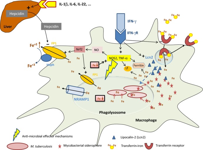
“M. tuberculosis growth in infected MΦs did not differ, whether cells were deficient for hepcidin or it was added exogenously (M. Podinovskaya, P. Masaratana, RJ. Simpson, S. Vaulont, AT. McKie, UE. Schaible, unpublished results).”
§ Macrophage defense mechanisms against intracellular bacteria > Metals in macrophage host responses to M. tuberculosis > Iron
plain- Asserted: passes — The authors put it forward as true.
- Checkable: passes — It can be checked against evidence.
- Load-bearing: unclear — It is unclear whether the paper’s conclusions depend on it.
- Based: passes — The paper gives a reason why it holds.
None of the sources cited for this claim have been checked yet.
In the paper
“Upon M. tuberculosis aerosol infection, NRAMP1-susceptible C57BL/6 mice complemented by transgenic expression of a functional NRAMP1 gene showed similar mycobacterial loads as their susceptible littermates aside from the first 2 weeks after infection, when transgenic mice carrying a functional NRAMP1 had one log less mycobacteria in lung and spleen (M. Podinovskaya, P. Masaratana, RJ. Simpson, S.”
§ Macrophage defense mechanisms against intracellular bacteria > Metals in macrophage host responses to M. tuberculosis > Iron
plain- Asserted: passes — The authors put it forward as true.
- Checkable: passes — It can be checked against evidence.
- Load-bearing: unclear — It is unclear whether the paper’s conclusions depend on it.
- Based: passes — The paper gives a reason why it holds.
The paper states this without a source or data behind it here, so there is nothing to check it against. The quote shows it is in the paper.
In the paper
“the stimulation of microbe intoxication mechanisms via acidification or metal accumulation in the phagolysosome,”
§ Macrophage defense mechanisms against intracellular bacteria > Abstract
plaindirect- Asserted: passes — The authors put it forward as true.
- Checkable: passes — It can be checked against evidence.
- Load-bearing: unclear — It is unclear whether the paper’s conclusions depend on it.
- Based: passes — The paper gives a reason why it holds.
In the paper
“Similarly, the mycobacterial cord factor TDM has been shown to delay phagosome maturation when coated onto beads (9).”
§ Macrophage defense mechanisms against intracellular bacteria > Come in and get killed: phagocytes' elimination of bacterial pathogens > Phagocytosis and phagosome maturation
What the sources say
- Not checked yetcites [9] Axelrod S Delay of phagosome maturation by a mycobacterial lipid is reversed by nitric oxide Cell Microbiol 2008 10 1530 1545 18363878 source
boosteddirect- Asserted: passes — The authors put it forward as true.
- Checkable: passes — It can be checked against evidence.
- Load-bearing: unclear — It is unclear whether the paper’s conclusions depend on it.
- Based: passes — The paper gives a reason why it holds.
The paper states this without a source or data behind it here, so there is nothing to check it against. The quote shows it is in the paper.
In the paper
“the restriction of the microbe's access to essential nutrients such as iron, fatty acids, or amino acids,”
§ Macrophage defense mechanisms against intracellular bacteria > Abstract
plaindirect- Asserted: passes — The authors put it forward as true.
- Checkable: passes — It can be checked against evidence.
- Load-bearing: unclear — It is unclear whether the paper’s conclusions depend on it.
- Based: passes — The paper gives a reason why it holds.
In the paper
“During this process, the maturing phagosome acquires and loses molecules functioning at but also characterizing individual maturation stages and diversion routes (7).”
§ Macrophage defense mechanisms against intracellular bacteria > Come in and get killed: phagocytes' elimination of bacterial pathogens > Phagocytosis and phagosome maturation
What the sources say
- Not checked yetcites [7] Flannagan RS Cosio G Grinstein S Antimicrobial mechanisms of phagocytes and bacterial evasion strategies Nat Rev Microbiol 2009 7 355 366 19369951 source
plaindirect- Asserted: passes — The authors put it forward as true.
- Checkable: passes — It can be checked against evidence.
- Load-bearing: unclear — It is unclear whether the paper’s conclusions depend on it.
- Based: passes — The paper gives a reason why it holds.
The paper states this without a source or data behind it here, so there is nothing to check it against. The quote shows it is in the paper.
In the paper
“Opsonization of invading bacteria is facilitated by soluble PRRs including surfactant proteins (SP-A, SP-D), and the mannose-binding lectin (MBL), as well as by the complement components C1q, C3, C3b, and C4.”
§ Macrophage defense mechanisms against intracellular bacteria > Introduction
plaindirect- Asserted: passes — The authors put it forward as true.
- Checkable: passes — It can be checked against evidence.
- Load-bearing: unclear — It is unclear whether the paper’s conclusions depend on it.
- Based: passes — The paper gives a reason why it holds.
- Probably not a substantial claim. 60
- It may state two separate claims as one. 123
A result the paper reports from its own data — it cites no other source for it, so there is none to check. The quote shows it is in the paper.
In the paper
“We observed decoration of M. tuberculosis-infected PMNs by cathelicidin, suggesting that this AMP can contribute to PMN necrosis (B. Corleis, UE. Schaible, unpublished results).”
§ Macrophage defense mechanisms against intracellular bacteria > Efferocytosis: a second instance of defense
hedgedconditional- Asserted: passes — The authors put it forward as true.
- Checkable: passes — It can be checked against evidence.
- Load-bearing: fails — The paper’s conclusions may not depend on it.
- Based: passes — The paper gives a reason why it holds.
Probably not a substantial claim. The paper’s conclusions may not depend on it. It may state two separate claims as one.
None of the sources cited for this claim have been checked yet.
In the paper
“Of interest, hepcidin-deficient mice were as susceptible to aerosol infection with low dose M. tuberculosis, as wildtype littermates indicated that hepcidin-triggered changes in murine iron metabolism is not affecting mycobacterial growth (M. Podinovskaya, P. Masaratana, RJ. Simpson, S. Vaulont, AT. McKie, UE. Schaible, unpublished data).”
§ Macrophage defense mechanisms against intracellular bacteria > Metals in macrophage host responses to M. tuberculosis > Iron
hedged- Asserted: passes — The authors put it forward as true.
- Checkable: passes — It can be checked against evidence.
- Load-bearing: unclear — It is unclear whether the paper’s conclusions depend on it.
- Based: passes — The paper gives a reason why it holds.
It may state two separate claims as one.
A conclusion the paper draws from its own results — it cites no other source for it, so there is none to check. The quote shows it is in the paper.
In the paper
“Their efficacy to contain and eliminate intracellular M. tuberculosis decides whether a patient initially becomes infected or not.”
§ Macrophage defense mechanisms against intracellular bacteria > Abstract
plaindirect- Asserted: passes — The authors put it forward as true.
- Checkable: passes — It can be checked against evidence.
- Load-bearing: passes — The paper’s conclusions depend on it.
- Based: fails — The paper may give no reason why it holds.
Probably not a substantial claim. The paper may give no reason why it holds.
The paper states this without a source or data behind it here, so there is nothing to check it against. The quote shows it is in the paper.
In the paper
“This process involves the GTPases, Rac1, Rac2, and Cdc42.”
§ Macrophage defense mechanisms against intracellular bacteria > Come in and get killed: phagocytes' elimination of bacterial pathogens > Phagocytosis and phagosome maturation
plaindirect- Asserted: passes — The authors put it forward as true.
- Checkable: passes — It can be checked against evidence.
- Load-bearing: fails — The paper’s conclusions may not depend on it.
- Based: passes — The paper gives a reason why it holds.
Probably not a substantial claim. The paper’s conclusions may not depend on it.
The paper states this without a source or data behind it here, so there is nothing to check it against. The quote shows it is in the paper.
In the paper
“The Toll-like receptors (TLRs) 1/2, 2/6, 4, 5, and 9, are specific for lipopeptides, lipopolysaccharides, flagellin, and low-methylated DNA sugar backbone, respectively, with TLR2/6 and 9 as the pivotal PRRs for mycobacteria.”
§ Macrophage defense mechanisms against intracellular bacteria > Introduction
plaincorrelational- Asserted: passes — The authors put it forward as true.
- Checkable: passes — It can be checked against evidence.
- Load-bearing: fails — The paper’s conclusions may not depend on it.
- Based: fails — The paper may give no reason why it holds.
Probably not a substantial claim. The paper’s conclusions may not depend on it. The paper may give no reason why it holds. It may state two separate claims as one.
The paper states this without a source or data behind it here, so there is nothing to check it against. The quote shows it is in the paper.
In the paper
“The stepwise succession of phagosomal maturation in macrophages is strikingly different from phagosome formation in PMNs.”
§ Macrophage defense mechanisms against intracellular bacteria > Come in and get killed: phagocytes' elimination of bacterial pathogens > Phagocytosis and phagosome maturation
boosted- Asserted: passes — The authors put it forward as true.
- Checkable: passes — It can be checked against evidence.
- Load-bearing: fails — The paper’s conclusions may not depend on it.
- Based: fails — The paper may give no reason why it holds.
Probably not a substantial claim. The paper’s conclusions may not depend on it. The paper may give no reason why it holds.
The paper states this without a source or data behind it here, so there is nothing to check it against. The quote shows it is in the paper.
In the paper
“These include induction of toxic anti-microbial effectors such as nitric oxide and reactive oxygen intermediates,”
§ Macrophage defense mechanisms against intracellular bacteria > Abstract
plaindirect- Asserted: passes — The authors put it forward as true.
- Checkable: passes — It can be checked against evidence.
- Load-bearing: passes — The paper’s conclusions depend on it.
- Based: passes — The paper gives a reason why it holds.
Probably not a substantial claim.
The paper states this without a source or data behind it here, so there is nothing to check it against. The quote shows it is in the paper.
In the paper
“A low pH is a prerequisite for optimal enzymatic activity of most late endosomal/lysosomal hydrolases, which are delivered to the nascent phagosome bound to the M6PR from the trans-Golgi.”
§ Macrophage defense mechanisms against intracellular bacteria > Come in and get killed: phagocytes' elimination of bacterial pathogens > Phagocytosis and phagosome maturation
plaindirect- Asserted: passes — The authors put it forward as true.
- Checkable: passes — It can be checked against evidence.
- Load-bearing: fails — The paper’s conclusions may not depend on it.
- Based: fails — The paper may give no reason why it holds.
Probably not a substantial claim. The paper’s conclusions may not depend on it. The paper may give no reason why it holds.
The paper states this without a source or data behind it here, so there is nothing to check it against. The quote shows it is in the paper.
In the paper
“Upon interaction of bacterial invaders with tissue MΦs and epithelial cells, initial inflammatory responses are triggered which comprises secretion of cytokines, chemokines, small lipid mediators (SLM) as well as anti-microbial peptides (AMPs).”
§ Macrophage defense mechanisms against intracellular bacteria > Introduction
plaindirect- Asserted: passes — The authors put it forward as true.
- Checkable: passes — It can be checked against evidence.
- Load-bearing: fails — The paper’s conclusions may not depend on it.
- Based: fails — The paper may give no reason why it holds.
Probably not a substantial claim. The paper’s conclusions may not depend on it. The paper may give no reason why it holds.
In the paper
“In addition, phenolic glycolipids promote the recruitment of M. tuberculosis permissive macrophages via stimulation of host chemokine receptor 2 expression, whereas the invasion of TLR responsive microbicidal macrophages producing reactive nitrogen and oxygen species is impaired (6).”
§ Macrophage defense mechanisms against intracellular bacteria > Come in and get killed: phagocytes' elimination of bacterial pathogens > Phagocytosis and phagosome maturation
What the sources say
- Not checked yetcites [6] Cambier CJ Mycobacteria manipulate macrophage recruitment through coordinated use of membrane lipids Nature 2014 505 218 222 24336213 PMC3961847 source
plaindirect- Asserted: passes — The authors put it forward as true.
- Checkable: passes — It can be checked against evidence.
- Load-bearing: unclear — It is unclear whether the paper’s conclusions depend on it.
- Based: passes — The paper gives a reason why it holds.
It may state two separate claims as one.
None of the sources cited for this claim have been checked yet.
In the paper
“Upon ITAM phosphorylation, the SYK tyrosine kinase is recruited to the receptor and its activation leads to downstream phosphorylation events including activation of SHP-1 and RAC (11, 12)”
§ Macrophage defense mechanisms against intracellular bacteria > Come in and get killed: phagocytes' elimination of bacterial pathogens > Phagocytosis and phagosome maturation
What the sources say
- Not checked yetcites [11] Mocsai A Ruland J Tybulewicz VL The SYK tyrosine kinase: a crucial player in diverse biological functions Nat Rev Immunol 2010 10 387 402 20467426 PMC4782221 source
- Not checked yetcites [12] Kant AM SHP-1 regulates Fcgamma receptor-mediated phagocytosis and the activation of RAC Blood 2002 100 1852 1859 12176909 source
plaindirect- Asserted: passes — The authors put it forward as true.
- Checkable: passes — It can be checked against evidence.
- Load-bearing: passes — The paper’s conclusions depend on it.
- Based: passes — The paper gives a reason why it holds.
It may state two separate claims as one.
None of the sources cited for this claim have been checked yet.
In the paper
“the fight of the host against the pathogen in M. tuberculosis infection involves several levels including containment of the microbe, generation of radicals and a hostile, acidic environment, deprivation from essential nutrients, formation of anti-mycobacterial peptides and cytokines which strengthen host responses by attracting other immune cells, down to cell damage and suicidal activities of MO”
§ Macrophage defense mechanisms against intracellular bacteria > Introduction
What the sources say
- Not checked yetcites [3] Sambarey A Prashanthi K Chandra N Mining large-scale response networks reveals ‘topmost activities’ in Mycobacterium tuberculosis infection Sci Rep 2013 3 2302 23892477 PMC3725478 source
- Not checked yetcites [4] Bruns H Stenger S New insights into the interaction of Mycobacterium tuberculosis and human macrophages Future Microbiol 2014 9 327 341 24762307 source
- Not checked yetcites [5] Schnappinger D Transcriptional adaptation of Mycobacterium tuberculosis within macrophages: insights into the phagosomal environment J Exp Med 2003 198 693 704 12953091 PMC2194186 source
boosteddirect- Asserted: passes — The authors put it forward as true.
- Checkable: passes — It can be checked against evidence.
- Load-bearing: unclear — It is unclear whether the paper’s conclusions depend on it.
- Based: passes — The paper gives a reason why it holds.
It may state two separate claims as one.
The paper states this without a source or data behind it here, so there is nothing to check it against. The quote shows it is in the paper.
In the paper
“Upon phagocytosis, epithelia-associated MΦs and other phagocytes such as dendritic cells (DCs) carry engulfed bacteria to deeper tissues and further to draining lymph nodes.”
§ Macrophage defense mechanisms against intracellular bacteria > Introduction
plaindirect- Asserted: passes — The authors put it forward as true.
- Checkable: passes — It can be checked against evidence.
- Load-bearing: fails — The paper’s conclusions may not depend on it.
- Based: fails — The paper may give no reason why it holds.
Probably not a substantial claim. The paper’s conclusions may not depend on it. The paper may give no reason why it holds.
The paper states this without a source or data behind it here, so there is nothing to check it against. The quote shows it is in the paper.
In the paper
“Subsequent enclosure of the phagocytic cup leads to formation of the phagosome and initiates its highly choreographed biogenesis driven by subsequent fusion and fission events.”
§ Macrophage defense mechanisms against intracellular bacteria > Come in and get killed: phagocytes' elimination of bacterial pathogens > Phagocytosis and phagosome maturation
plaindirect- Asserted: passes — The authors put it forward as true.
- Checkable: passes — It can be checked against evidence.
- Load-bearing: unclear — It is unclear whether the paper’s conclusions depend on it.
- Based: passes — The paper gives a reason why it holds.
It may state two separate claims as one.
The paper states this without a source or data behind it here, so there is nothing to check it against. The quote shows it is in the paper.
In the paper
“Surface PRRs include C-type lectins, mannose receptor (MR), dectin 1, dectin 2, Mincle, MCL, DC-SIGN, and scavenger receptors such as SR-A and MARCO, which not only recognize but also bind bacteria to initiate phagocytosis.”
§ Macrophage defense mechanisms against intracellular bacteria > Introduction
plaindirect- Asserted: passes — The authors put it forward as true.
- Checkable: passes — It can be checked against evidence.
- Load-bearing: fails — The paper’s conclusions may not depend on it.
- Based: passes — The paper gives a reason why it holds.
Probably not a substantial claim. The paper’s conclusions may not depend on it.
A conclusion the paper draws from its own results — it cites no other source for it, so there is none to check. The quote shows it is in the paper.
In the paper
“Macrophages and neutrophils play a decisive role in host responses to intracellular bacteria including the agent of tuberculosis (TB), Mycobacterium tuberculosis as they represent the forefront of innate immune defense against bacterial invaders.”
§ Macrophage defense mechanisms against intracellular bacteria > Abstract
plaindirect- Asserted: passes — The authors put it forward as true.
- Checkable: passes — It can be checked against evidence.
- Load-bearing: passes — The paper’s conclusions depend on it.
- Based: fails — The paper may give no reason why it holds.
Probably not a substantial claim. The paper may give no reason why it holds. It may state two separate claims as one.
The paper states this without a source or data behind it here, so there is nothing to check it against. The quote shows it is in the paper.
In the paper
“Opsonized bacteria are subsequently bound by receptors for soluble PRR, C receptors (C1qR, CR3, CR4), and, in the presence of specific antibodies, Fcγ receptors (FcγR I-III).”
§ Macrophage defense mechanisms against intracellular bacteria > Introduction
plaindirect- Asserted: passes — The authors put it forward as true.
- Checkable: passes — It can be checked against evidence.
- Load-bearing: fails — The paper’s conclusions may not depend on it.
- Based: passes — The paper gives a reason why it holds.
Probably not a substantial claim. The paper’s conclusions may not depend on it.
The paper states this without a source or data behind it here, so there is nothing to check it against. The quote shows it is in the paper.
In the paper
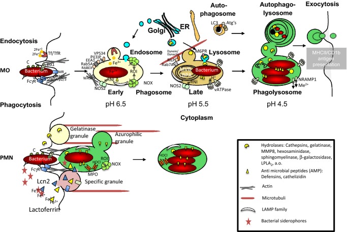
“In MΦs, upon phagosome closure, the maturing phagosome traverses an early and late phagosomal and a phagolysosomal stage paralleling endosomal maturation.”
§ Macrophage defense mechanisms against intracellular bacteria > Come in and get killed: phagocytes' elimination of bacterial pathogens > Phagocytosis and phagosome maturation
plaincorrelational- Asserted: passes — The authors put it forward as true.
- Checkable: passes — It can be checked against evidence.
- Load-bearing: fails — The paper’s conclusions may not depend on it.
- Based: passes — The paper gives a reason why it holds.
Probably not a substantial claim. The paper’s conclusions may not depend on it.
None of the sources cited for this claim have been checked yet.
In the paper
“while STING is the recently described receptor for cyclic GMP-AMP generated from microbial DNA, which is delivered into the cytoplasm by cGAMP synthase, which triggers type 1 interferon production (1, 2).”
§ Macrophage defense mechanisms against intracellular bacteria > Introduction
What the sources say
- Not checked yetcites [1] Watson RO Manzanillo PS Cox JS Extracellular M. tuberculosis DNA targets bacteria for autophagy by activating the host DNA-sensing pathway Cell 2012 150 803 815 22901810 PMC3708656 source
- Not checked yetcites [2] Sun L Wu J Du F Chen X Chen ZJ Cyclic GMP-AMP synthase is a cytosolic DNA sensor that activates the type I interferon pathway Science 2013 339 786 791 23258413 PMC3863629 source
plaindirect- Asserted: passes — The authors put it forward as true.
- Checkable: passes — It can be checked against evidence.
- Load-bearing: fails — The paper’s conclusions may not depend on it.
- Based: passes — The paper gives a reason why it holds.
Probably not a substantial claim. The paper’s conclusions may not depend on it. It may state two separate claims as one.
The paper states this without a source or data behind it here, so there is nothing to check it against. The quote shows it is in the paper.
In the paper
“A hallmark of phagosome biogenesis is acidification of the phagosomal lumen by the proton pumping vATPase.”
§ Macrophage defense mechanisms against intracellular bacteria > Come in and get killed: phagocytes' elimination of bacterial pathogens > Phagocytosis and phagosome maturation
plaincorrelational- Asserted: passes — The authors put it forward as true.
- Checkable: passes — It can be checked against evidence.
- Load-bearing: fails — The paper’s conclusions may not depend on it.
- Based: fails — The paper may give no reason why it holds.
Probably not a substantial claim. The paper’s conclusions may not depend on it. The paper may give no reason why it holds.
The paper states this without a source or data behind it here, so there is nothing to check it against. The quote shows it is in the paper.
In the paper
“Interleukin-8 (IL-8) in humans (KC in mice) as well as prostaglandins and leukotrienes attract and/or activate PMNs, which further perpetuate inflammation by secretion of additional IL-8 but also the chemokines MCP-1, MIP1α/β, and IP10 that attract monocyte-derived MΦs and other immune cells to the site of infection.”
§ Macrophage defense mechanisms against intracellular bacteria > Introduction
plaindirect- Asserted: passes — The authors put it forward as true.
- Checkable: passes — It can be checked against evidence.
- Load-bearing: fails — The paper’s conclusions may not depend on it.
- Based: fails — The paper may give no reason why it holds.
Probably not a substantial claim. The paper’s conclusions may not depend on it. The paper may give no reason why it holds. It may state two separate claims as one.
The paper states this without a source or data behind it here, so there is nothing to check it against. The quote shows it is in the paper.
In the paper
“Upon binding and recognition of bacterial invaders, intracellular signaling pathways triggered by PRR, CR3, or FcRs engaged by their respective ligands induce actin polymerization and formation of the phagocytic cup.”
§ Macrophage defense mechanisms against intracellular bacteria > Come in and get killed: phagocytes' elimination of bacterial pathogens > Phagocytosis and phagosome maturation
plaindirect- Asserted: passes — The authors put it forward as true.
- Checkable: passes — It can be checked against evidence.
- Load-bearing: fails — The paper’s conclusions may not depend on it.
- Based: passes — The paper gives a reason why it holds.
Probably not a substantial claim. The paper’s conclusions may not depend on it.
The paper states this without a source or data behind it here, so there is nothing to check it against. The quote shows it is in the paper.
In the paper
“The prime examples are alveolar MΦs, which keep the pulmonary surface under surveillance for inhaled pathogens.”
§ Macrophage defense mechanisms against intracellular bacteria > Introduction
plaindirect- Asserted: passes — The authors put it forward as true.
- Checkable: passes — It can be checked against evidence.
- Load-bearing: fails — The paper’s conclusions may not depend on it.
- Based: passes — The paper gives a reason why it holds.
Probably not a substantial claim. The paper’s conclusions may not depend on it.
None of the sources cited for this claim have been checked yet.
In the paper
“In the cytoplasm, NOD1, NOD2, and Galectin 3 sense DAP, muramyl-dipeptide, and β-galactosides, respectively,”
§ Macrophage defense mechanisms against intracellular bacteria > Introduction
plaindirect- Asserted: passes — The authors put it forward as true.
- Checkable: passes — It can be checked against evidence.
- Load-bearing: fails — The paper’s conclusions may not depend on it.
- Based: passes — The paper gives a reason why it holds.
Probably not a substantial claim. The paper’s conclusions may not depend on it. It may state two separate claims as one.
A conclusion the paper draws from its own results — it cites no other source for it, so there is none to check. The quote shows it is in the paper.
In the paper
“M. tuberculosis, as a prime example of a well-adapted facultative intracellular bacterium, has learned during evolution to counter-balance the host's immune defense strategies to secure survival or”
§ Macrophage defense mechanisms against intracellular bacteria > Abstract
plaindirect- Asserted: passes — The authors put it forward as true.
- Checkable: passes — It can be checked against evidence.
- Load-bearing: passes — The paper’s conclusions depend on it.
- Based: fails — The paper may give no reason why it holds.
Probably not a substantial claim. The paper may give no reason why it holds.
The paper states this without a source or data behind it here, so there is nothing to check it against. The quote shows it is in the paper.
In the paper
“Phagosome biogenesis is accompanied by continuous fusion and fission events, including fusion with trans-Golgi transport vesicles, endosomes, lysosomes, and autophagosomes.”
§ Macrophage defense mechanisms against intracellular bacteria > Come in and get killed: phagocytes' elimination of bacterial pathogens > Phagocytosis and phagosome maturation
plaincorrelational- Asserted: passes — The authors put it forward as true.
- Checkable: passes — It can be checked against evidence.
- Load-bearing: fails — The paper’s conclusions may not depend on it.
- Based: fails — The paper may give no reason why it holds.
Probably not a substantial claim. The paper’s conclusions may not depend on it. The paper may give no reason why it holds.
The paper states this without a source or data behind it here, so there is nothing to check it against. The quote shows it is in the paper.
In the paper
“Phagocytosis is a hallmark of anti-bacterial host defense.”
§ Macrophage defense mechanisms against intracellular bacteria > Come in and get killed: phagocytes' elimination of bacterial pathogens > Phagocytosis and phagosome maturation
plain- Asserted: passes — The authors put it forward as true.
- Checkable: passes — It can be checked against evidence.
- Load-bearing: fails — The paper’s conclusions may not depend on it.
- Based: fails — The paper may give no reason why it holds.
Probably not a substantial claim. The paper’s conclusions may not depend on it. The paper may give no reason why it holds.
A conclusion the paper draws from its own results — it cites no other source for it, so there is none to check. The quote shows it is in the paper.
In the paper
“Macrophages are thus in the center of infection with M. tuberculosis and other intracellular bacteria and use multiple strategies to eliminate this pathogens.”
§ Macrophage defense mechanisms against intracellular bacteria > Introduction
plaindirect- Asserted: passes — The authors put it forward as true.
- Checkable: passes — It can be checked against evidence.
- Load-bearing: passes — The paper’s conclusions depend on it.
- Based: passes — The paper gives a reason why it holds.
Probably not a substantial claim. It may state two separate claims as one.
The paper states this without a source or data behind it here, so there is nothing to check it against. The quote shows it is in the paper.
In the paper
“Downstream signaling cascades linked to these receptors induce receptor-mediated phagocytosis, a hallmark in host defense, in addition to a variety of pro-inflammatory responses executed by expression and/or secretion of chemokines and cytokines and release of anti-microbial effectors.”
§ Macrophage defense mechanisms against intracellular bacteria > Introduction
plaindirect- Asserted: passes — The authors put it forward as true.
- Checkable: passes — It can be checked against evidence.
- Load-bearing: fails — The paper’s conclusions may not depend on it.
- Based: fails — The paper may give no reason why it holds.
Probably not a substantial claim. The paper’s conclusions may not depend on it. The paper may give no reason why it holds. It may state two separate claims as one.
The paper states this without a source or data behind it here, so there is nothing to check it against. The quote shows it is in the paper.
In the paper
“At the same time, these phagocytes are also primary targets of intracellular bacteria to be abused as host cells.”
§ Macrophage defense mechanisms against intracellular bacteria > Abstract
plaindirect- Asserted: passes — The authors put it forward as true.
- Checkable: passes — It can be checked against evidence.
- Load-bearing: passes — The paper’s conclusions depend on it.
- Based: fails — The paper may give no reason why it holds.
Probably not a substantial claim. The paper may give no reason why it holds.
The paper states this without a source or data behind it here, so there is nothing to check it against. The quote shows it is in the paper.
In the paper
“The latter one can directly interact with WASP (Wiskott-Aldrich syndrome protein), which subsequently activates Arp2/3 as a direct initiator of actin polymerization.”
§ Macrophage defense mechanisms against intracellular bacteria > Come in and get killed: phagocytes' elimination of bacterial pathogens > Phagocytosis and phagosome maturation
plaindirect- Asserted: passes — The authors put it forward as true.
- Checkable: passes — It can be checked against evidence.
- Load-bearing: fails — The paper’s conclusions may not depend on it.
- Based: passes — The paper gives a reason why it holds.
Probably not a substantial claim. The paper’s conclusions may not depend on it. It may state two separate claims as one.
The paper states this without a source or data behind it here, so there is nothing to check it against. The quote shows it is in the paper.
In the paper
“Phagocytic cells such as macrophages (MΦs) and polymorphonuclear neutrophilic granulocytes (PMNs) represent the first line of defense against invading bacterial pathogens.”
§ Macrophage defense mechanisms against intracellular bacteria > Introduction
plaindirect- Asserted: passes — The authors put it forward as true.
- Checkable: passes — It can be checked against evidence.
- Load-bearing: fails — The paper’s conclusions may not depend on it.
- Based: fails — The paper may give no reason why it holds.
Probably not a substantial claim. The paper’s conclusions may not depend on it. The paper may give no reason why it holds.
The paper states this without a source or data behind it here, so there is nothing to check it against. The quote shows it is in the paper.
In the paper
“tissue-resident MΦs patrol epithelia of barrier organs, which represent putative entry and colonization sites for pathogens and the first location for controlling infectious invaders.”
§ Macrophage defense mechanisms against intracellular bacteria > Introduction
hedged- Asserted: passes — The authors put it forward as true.
- Checkable: passes — It can be checked against evidence.
- Load-bearing: fails — The paper’s conclusions may not depend on it.
- Based: fails — The paper may give no reason why it holds.
Probably not a substantial claim. The paper’s conclusions may not depend on it. The paper may give no reason why it holds.
The paper states this without a source or data behind it here, so there is nothing to check it against. The quote shows it is in the paper.
In the paper
“There, antigens are presented to T cells by professional antigen-presenting cells (APCs), such as DCs and MΦs (as well as B cells), to initiate specific cellular immunity and generate specific T cells.”
§ Macrophage defense mechanisms against intracellular bacteria > Introduction
plaindirect- Asserted: passes — The authors put it forward as true.
- Checkable: passes — It can be checked against evidence.
- Load-bearing: fails — The paper’s conclusions may not depend on it.
- Based: passes — The paper gives a reason why it holds.
Probably not a substantial claim. The paper’s conclusions may not depend on it.
A conclusion the paper draws from its own results — it cites no other source for it, so there is none to check. The quote shows it is in the paper.
In the paper
“Macrophages have evolved a myriad of defense strategies to combat infection with intracellular bacteria such as M. tuberculosis.”
§ Macrophage defense mechanisms against intracellular bacteria > Abstract
plaindirect- Asserted: passes — The authors put it forward as true.
- Checkable: passes — It can be checked against evidence.
- Load-bearing: passes — The paper’s conclusions depend on it.
- Based: fails — The paper may give no reason why it holds.
Probably not a substantial claim. The paper may give no reason why it holds.
The paper states this without a source or data behind it here, so there is nothing to check it against. The quote shows it is in the paper.
In the paper
“Mannose-capped lipoarabinomannan (manLAM), phosphatidyl inositol mannosides (PIMs), as well as trehalose dimycolate (TDM) are mycobacterial ligands for MR, DC-SIGN, Mincle, and MCL, respectively.”
§ Macrophage defense mechanisms against intracellular bacteria > Introduction
plaincorrelational- Asserted: passes — The authors put it forward as true.
- Checkable: passes — It can be checked against evidence.
- Load-bearing: fails — The paper’s conclusions may not depend on it.
- Based: fails — The paper may give no reason why it holds.
Probably not a substantial claim. The paper’s conclusions may not depend on it. The paper may give no reason why it holds. It may state two separate claims as one.
The paper states this without a source or data behind it here, so there is nothing to check it against. The quote shows it is in the paper.
In the paper
“specific antibodies generated during the first encounter are essential for antigen-specific recognition and opsonization of a recurrent infectious agent, and provide the basis of successful vaccines.”
§ Macrophage defense mechanisms against intracellular bacteria > Introduction
plaindirect- Asserted: passes — The authors put it forward as true.
- Checkable: passes — It can be checked against evidence.
- Load-bearing: fails — The paper’s conclusions may not depend on it.
- Based: fails — The paper may give no reason why it holds.
Probably not a substantial claim. The paper’s conclusions may not depend on it. The paper may give no reason why it holds. It may state two separate claims as one.
In the paper
“PDCD6IP also binds in a Ca2+-dependent manner to endophilins, which influence the endosomal membrane shape (bending) probably through interaction with dynamin via SH3 domains (46).”
§ Macrophage defense mechanisms against intracellular bacteria > Come in and get killed: phagocytes' elimination of bacterial pathogens > Phagocytosis and phagosome maturation
What the sources say
- Not checked yetcites [46] Bissig C Gruenberg J ALIX and the multivesicular endosome: ALIX in Wonderland Trends Cell Biol 2014 24 19 25 24287454 source
hedgedconditional- Asserted: passes — The authors put it forward as true.
- Checkable: passes — It can be checked against evidence.
- Load-bearing: unclear — It is unclear whether the paper’s conclusions depend on it.
- Based: passes — The paper gives a reason why it holds.
It may state two separate claims as one.
The paper states this without a source or data behind it here, so there is nothing to check it against. The quote shows it is in the paper.
In the paper
“Finally, the gelatinase granules spill out proteases and other enzymes to degrade extracellular matrix proteins, leading to tissue disruption to allow PMN evasion into infection site but ultimately to pathogenesis.”
§ Macrophage defense mechanisms against intracellular bacteria > Come in and get killed: phagocytes' elimination of bacterial pathogens > Phagocytosis and phagosome maturation
plaindirect- Asserted: passes — The authors put it forward as true.
- Checkable: passes — It can be checked against evidence.
- Load-bearing: fails — The paper’s conclusions may not depend on it.
- Based: fails — The paper may give no reason why it holds.
Probably not a substantial claim. The paper’s conclusions may not depend on it. The paper may give no reason why it holds. It may state two separate claims as one.
In the paper
“Some of those such as cathepsin D depend on further processing by other proteases and/or autocatalysis to generate the enzymatically active forms (40).”
§ Macrophage defense mechanisms against intracellular bacteria > Come in and get killed: phagocytes' elimination of bacterial pathogens > Phagocytosis and phagosome maturation
What the sources say
- Not checked yetcites [40] Sturgill-Koszycki S Schaible UE Russell DG Mycobacterium-containing phagosomes are accessible to early endosomes and reflect a transitional state in normal phagosome biogenesis EMBO J 1996 15 6960 6968 9003772 PMC452522 source
plaindirect- Asserted: passes — The authors put it forward as true.
- Checkable: passes — It can be checked against evidence.
- Load-bearing: fails — The paper’s conclusions may not depend on it.
- Based: passes — The paper gives a reason why it holds.
Probably not a substantial claim. The paper’s conclusions may not depend on it.
The paper states this without a source or data behind it here, so there is nothing to check it against. The quote shows it is in the paper.
In the paper
“The late phagosomal stage is characterized by the acquisition of the lysosome-associated membrane proteins (LAMP1 and 2) from endosomes as well as newly synthesized lysosomal enzymes delivered in trans-Golgi transport vesicles bound to the mannose-6-phosphate receptors (M6PR), which are transiently enriched in late phagosomes.”
§ Macrophage defense mechanisms against intracellular bacteria > Come in and get killed: phagocytes' elimination of bacterial pathogens > Phagocytosis and phagosome maturation
plaincorrelational- Asserted: passes — The authors put it forward as true.
- Checkable: passes — It can be checked against evidence.
- Load-bearing: unclear — It is unclear whether the paper’s conclusions depend on it.
- Based: passes — The paper gives a reason why it holds.
It may state two separate claims as one.
The paper states this without a source or data behind it here, so there is nothing to check it against. The quote shows it is in the paper.
In the paper
“Upon subsequent maturation, the late endosome-like phagosome is the transient stage toward an oxidative and hydrolytic compartment, where the phagosomal cargo is degraded, and associated with the loss of Rab5A and acquisition of the late endosomal Rab7A through the HOPS (VpsC-homotypic protein sorting) complex.”
§ Macrophage defense mechanisms against intracellular bacteria > Come in and get killed: phagocytes' elimination of bacterial pathogens > Phagocytosis and phagosome maturation
plaincorrelational- Asserted: passes — The authors put it forward as true.
- Checkable: passes — It can be checked against evidence.
- Load-bearing: unclear — It is unclear whether the paper’s conclusions depend on it.
- Based: passes — The paper gives a reason why it holds.
It may state two separate claims as one.
In the paper
“Inhibition of Abl tyrosine kinase by the clinically used drug imatinib resulted in increased acidification of the lysosome and improved control of M. tuberculosis (39).”
§ Macrophage defense mechanisms against intracellular bacteria > Come in and get killed: phagocytes' elimination of bacterial pathogens > Phagocytosis and phagosome maturation
What the sources say
- Not checked yetcites [39] Bruns H Abelson tyrosine kinase controls phagosomal acidification required for killing of Mycobacterium tuberculosis in human macrophages J Immunol 2012 189 4069 4078 22988030 PMC3684563 source
plaindirect- Asserted: passes — The authors put it forward as true.
- Checkable: passes — It can be checked against evidence.
- Load-bearing: unclear — It is unclear whether the paper’s conclusions depend on it.
- Based: passes — The paper gives a reason why it holds.
It may state two separate claims as one.
In the paper
“This process can be blocked by calcineurin-inhibitors cyclosporin or FK506, resulting in reduction of mycobacterial proliferation within macrophages (20).”
§ Macrophage defense mechanisms against intracellular bacteria > Come in and get killed: phagocytes' elimination of bacterial pathogens > Phagocytosis and phagosome maturation
What the sources say
- Not checked yetcites [20] Jayachandran R Survival of mycobacteria in macrophages is mediated by coronin 1-dependent activation of calcineurin Cell 2007 130 37 50 17632055 source
plaindirect- Asserted: passes — The authors put it forward as true.
- Checkable: passes — It can be checked against evidence.
- Load-bearing: unclear — It is unclear whether the paper’s conclusions depend on it.
- Based: passes — The paper gives a reason why it holds.
It may state two separate claims as one.
In the paper
“Lyso-biphosphatidic acids (LBPA) are key lipids of late endosomes/phagosomes and terminal phospholipid degradation products (44).”
§ Macrophage defense mechanisms against intracellular bacteria > Come in and get killed: phagocytes' elimination of bacterial pathogens > Phagocytosis and phagosome maturation
What the sources say
- Not checked yetcites [44] Kobayashi T Stang E Fang KS de Moerloose P Parton RG Gruenberg J A lipid associated with the antiphospholipid syndrome regulates endosome structure and function Nature 1998 392 193 197 9515966 source
plaincorrelational- Asserted: passes — The authors put it forward as true.
- Checkable: passes — It can be checked against evidence.
- Load-bearing: unclear — It is unclear whether the paper’s conclusions depend on it.
- Based: passes — The paper gives a reason why it holds.
It may state two separate claims as one.
The paper states this without a source or data behind it here, so there is nothing to check it against. The quote shows it is in the paper.
In the paper
“Together, v- and t-SNARE establish a protein complex, which facilitates membrane fusion.”
§ Macrophage defense mechanisms against intracellular bacteria > Come in and get killed: phagocytes' elimination of bacterial pathogens > Phagocytosis and phagosome maturation
plaindirect- Asserted: passes — The authors put it forward as true.
- Checkable: passes — It can be checked against evidence.
- Load-bearing: fails — The paper’s conclusions may not depend on it.
- Based: passes — The paper gives a reason why it holds.
Probably not a substantial claim. The paper’s conclusions may not depend on it.
The paper states this without a source or data behind it here, so there is nothing to check it against. The quote shows it is in the paper.
In the paper
“Phagocytosis and fusion with the lysosomal azurophilic granules is often happening simultaneously.”
§ Macrophage defense mechanisms against intracellular bacteria > Come in and get killed: phagocytes' elimination of bacterial pathogens > Phagocytosis and phagosome maturation
plaincorrelational- Asserted: passes — The authors put it forward as true.
- Checkable: passes — It can be checked against evidence.
- Load-bearing: fails — The paper’s conclusions may not depend on it.
- Based: fails — The paper may give no reason why it holds.
Probably not a substantial claim. The paper’s conclusions may not depend on it. The paper may give no reason why it holds.
The paper states this without a source or data behind it here, so there is nothing to check it against. The quote shows it is in the paper.
In the paper
“The late endosome/phagosome is the intracellular compartment for membrane degradation.”
§ Macrophage defense mechanisms against intracellular bacteria > Come in and get killed: phagocytes' elimination of bacterial pathogens > Phagocytosis and phagosome maturation
plain- Asserted: passes — The authors put it forward as true.
- Checkable: passes — It can be checked against evidence.
- Load-bearing: fails — The paper’s conclusions may not depend on it.
- Based: fails — The paper may give no reason why it holds.
Probably not a substantial claim. The paper’s conclusions may not depend on it. The paper may give no reason why it holds.
The paper states this without a source or data behind it here, so there is nothing to check it against. The quote shows it is in the paper.
In the paper
“Rab11A is involved in recycling vesicles between the early endosome and plasma membrane, and probably also early phagosomes.”
§ Macrophage defense mechanisms against intracellular bacteria > Come in and get killed: phagocytes' elimination of bacterial pathogens > Phagocytosis and phagosome maturation
hedgedcorrelational- Asserted: passes — The authors put it forward as true.
- Checkable: passes — It can be checked against evidence.
- Load-bearing: fails — The paper’s conclusions may not depend on it.
- Based: passes — The paper gives a reason why it holds.
Probably not a substantial claim. The paper’s conclusions may not depend on it. It may state two separate claims as one.
The paper states this without a source or data behind it here, so there is nothing to check it against. The quote shows it is in the paper.
In the paper
“Inherited lack of saposins or pharmacological inhibition of certain lysosomal hydrolases leads to severe lysosomal storage diseases.”
§ Macrophage defense mechanisms against intracellular bacteria > Come in and get killed: phagocytes' elimination of bacterial pathogens > Phagocytosis and phagosome maturation
plaindirect- Asserted: passes — The authors put it forward as true.
- Checkable: passes — It can be checked against evidence.
- Load-bearing: fails — The paper’s conclusions may not depend on it.
- Based: passes — The paper gives a reason why it holds.
Probably not a substantial claim. The paper’s conclusions may not depend on it.
The paper states this without a source or data behind it here, so there is nothing to check it against. The quote shows it is in the paper.
In the paper
“The fusion between vesicles requires a closely adjacent position, which is facilitated by motor proteins.”
§ Macrophage defense mechanisms against intracellular bacteria > Come in and get killed: phagocytes' elimination of bacterial pathogens > Phagocytosis and phagosome maturation
plaindirect- Asserted: passes — The authors put it forward as true.
- Checkable: passes — It can be checked against evidence.
- Load-bearing: unclear — It is unclear whether the paper’s conclusions depend on it.
- Based: passes — The paper gives a reason why it holds.
It may state two separate claims as one.
The paper states this without a source or data behind it here, so there is nothing to check it against. The quote shows it is in the paper.
In the paper
“SNAREs are essential for vesicle fusion by forming fusogenic protein complexes at the cytoplasmic side of phagosomes comprising SNARE, NSF (N-ethylmaleimide sensitive factor), and SNAP (NSF-attachment protein).”
§ Macrophage defense mechanisms against intracellular bacteria > Come in and get killed: phagocytes' elimination of bacterial pathogens > Phagocytosis and phagosome maturation
plaindirect- Asserted: passes — The authors put it forward as true.
- Checkable: passes — It can be checked against evidence.
- Load-bearing: fails — The paper’s conclusions may not depend on it.
- Based: passes — The paper gives a reason why it holds.
Probably not a substantial claim. The paper’s conclusions may not depend on it.
The paper states this without a source or data behind it here, so there is nothing to check it against. The quote shows it is in the paper.
In the paper
“Removal of this actin ‘coat’ drives mycobacteria into phagolysosomes and limits mycobacterial growth.”
§ Macrophage defense mechanisms against intracellular bacteria > Come in and get killed: phagocytes' elimination of bacterial pathogens > Phagocytosis and phagosome maturation
plaindirect- Asserted: passes — The authors put it forward as true.
- Checkable: passes — It can be checked against evidence.
- Load-bearing: passes — The paper’s conclusions depend on it.
- Based: passes — The paper gives a reason why it holds.
It may state two separate claims as one.
The paper states this without a source or data behind it here, so there is nothing to check it against. The quote shows it is in the paper.
In the paper
“Scission of the newly formed phagosome from the plasma membrane seems to involve myosins.”
§ Macrophage defense mechanisms against intracellular bacteria > Come in and get killed: phagocytes' elimination of bacterial pathogens > Phagocytosis and phagosome maturation
hedgedconditional- Asserted: passes — The authors put it forward as true.
- Checkable: passes — It can be checked against evidence.
- Load-bearing: fails — The paper’s conclusions may not depend on it.
- Based: fails — The paper may give no reason why it holds.
Probably not a substantial claim. The paper’s conclusions may not depend on it. The paper may give no reason why it holds.
The paper states this without a source or data behind it here, so there is nothing to check it against. The quote shows it is in the paper.
In the paper
“The early endosome-like phagosome represents the first stage upon closure of the phagocytic cup.”
§ Macrophage defense mechanisms against intracellular bacteria > Come in and get killed: phagocytes' elimination of bacterial pathogens > Phagocytosis and phagosome maturation
plaindirect- Asserted: passes — The authors put it forward as true.
- Checkable: passes — It can be checked against evidence.
- Load-bearing: fails — The paper’s conclusions may not depend on it.
- Based: fails — The paper may give no reason why it holds.
Probably not a substantial claim. The paper’s conclusions may not depend on it. The paper may give no reason why it holds.
None of the sources cited for this claim have been checked yet.
In the paper
“Early phagosomes recruit Tf via TfR and have therefore been suggested as an ideal niche for microbes, which require iron for growth such as M. tuberculosis (31, 32).”
§ Macrophage defense mechanisms against intracellular bacteria > Come in and get killed: phagocytes' elimination of bacterial pathogens > Phagocytosis and phagosome maturation
What the sources say
- Not checked yetcites [31] Halaas O Intracellular Mycobacterium avium intersect transferrin in the Rab11(+) recycling endocytic pathway and avoid lipocalin 2 trafficking to the lysosomal pathway J Infect Dis 2010 201 783 792 20121435 PMC2862295 source
- Not checked yetcites [32] Schaible UE Kaufmann SH Iron and microbial infection Nat Rev Microbiol 2004 2 946 953 15550940 source
hedgedcorrelational- Asserted: passes — The authors put it forward as true.
- Checkable: passes — It can be checked against evidence.
- Load-bearing: unclear — It is unclear whether the paper’s conclusions depend on it.
- Based: passes — The paper gives a reason why it holds.
It may state two separate claims as one.
The paper states this without a source or data behind it here, so there is nothing to check it against. The quote shows it is in the paper.
In the paper
“Fusion competence between two types of vesicles requires the presence of a vesicular (v) on one and a target (t) SNARE on the other vesicle such as the VAMPs and syntaxins, respectively.”
§ Macrophage defense mechanisms against intracellular bacteria > Come in and get killed: phagocytes' elimination of bacterial pathogens > Phagocytosis and phagosome maturation
plaindirect- Asserted: passes — The authors put it forward as true.
- Checkable: passes — It can be checked against evidence.
- Load-bearing: fails — The paper’s conclusions may not depend on it.
- Based: passes — The paper gives a reason why it holds.
Probably not a substantial claim. The paper’s conclusions may not depend on it.
The paper states this without a source or data behind it here, so there is nothing to check it against. The quote shows it is in the paper.
In the paper
“These granules also deliver phagocytic receptors to the PMN surface for recognition and uptake of bacteria.”
§ Macrophage defense mechanisms against intracellular bacteria > Come in and get killed: phagocytes' elimination of bacterial pathogens > Phagocytosis and phagosome maturation
plaindirect- Asserted: passes — The authors put it forward as true.
- Checkable: passes — It can be checked against evidence.
- Load-bearing: fails — The paper’s conclusions may not depend on it.
- Based: fails — The paper may give no reason why it holds.
Probably not a substantial claim. The paper’s conclusions may not depend on it. The paper may give no reason why it holds.
In the paper
“Its lack causes enhanced susceptibility to M. tuberculosis (43) and S. typhimurium infection (S. Renk, G. Graßl, P. Rausch, J. Shayman, BE. Schneider, UE. Schaible, unpublished data), although a direct microbicidal effect has not yet been found.”
§ Macrophage defense mechanisms against intracellular bacteria > Come in and get killed: phagocytes' elimination of bacterial pathogens > Phagocytosis and phagosome maturation
What the sources say
- Not checked yetcites [43] Schneider BE Behrends J Hagens K Harmel N Shayman JA Schaible UE Lysosomal phospholipase A2: a novel player in host immunity to Mycobacterium tuberculosis Eur J Immunol 2014 44 2394 2404 24825529 source
plaindirect- Asserted: passes — The authors put it forward as true.
- Checkable: passes — It can be checked against evidence.
- Load-bearing: unclear — It is unclear whether the paper’s conclusions depend on it.
- Based: passes — The paper gives a reason why it holds.
It may state two separate claims as one.
The paper states this without a source or data behind it here, so there is nothing to check it against. The quote shows it is in the paper.
In the paper
“Anti-bacterial activity has been described for a number of these hydrolases, which probably synergize with other microbicidal molecules such as defensins, cathelizidin, and ubiquitin-derived AMPs.”
§ Macrophage defense mechanisms against intracellular bacteria > Come in and get killed: phagocytes' elimination of bacterial pathogens > Phagocytosis and phagosome maturation
hedgedconditional- Asserted: passes — The authors put it forward as true.
- Checkable: passes — It can be checked against evidence.
- Load-bearing: fails — The paper’s conclusions may not depend on it.
- Based: fails — The paper may give no reason why it holds.
Probably not a substantial claim. The paper’s conclusions may not depend on it. The paper may give no reason why it holds. It may state two separate claims as one.
The paper states this without a source or data behind it here, so there is nothing to check it against. The quote shows it is in the paper.
In the paper
“At the same time, specific granules discharge iron sequestering lactoferrin and Lcn2 into both, the phagosomal lumen and the extracellular space.”
§ Macrophage defense mechanisms against intracellular bacteria > Come in and get killed: phagocytes' elimination of bacterial pathogens > Phagocytosis and phagosome maturation
plaindirect- Asserted: passes — The authors put it forward as true.
- Checkable: passes — It can be checked against evidence.
- Load-bearing: fails — The paper’s conclusions may not depend on it.
- Based: fails — The paper may give no reason why it holds.
Probably not a substantial claim. The paper’s conclusions may not depend on it. The paper may give no reason why it holds.
None of the sources cited for this claim have been checked yet.
In the paper
“Lysosomal lipid transfer proteins (LTPs), such as the four active saposins generated from pro-saposin by proteolytic cleavage, are essential for glyco-lipid degradation in lysosomes and glyco-lipid homeostasis, in addition to their immunologically relevant function in processing and presentation of lipid antigens by CD1b molecules in DC (41, 42).”
§ Macrophage defense mechanisms against intracellular bacteria > Come in and get killed: phagocytes' elimination of bacterial pathogens > Phagocytosis and phagosome maturation
What the sources say
- Not checked yetcites [41] Kolter T Winau F Schaible UE Leippe M Sandhoff K Lipid-binding proteins in membrane digestion, antigen presentation, and antimicrobial defense J Biol Chem 2005 280 41125 41128 16230343 source
- Not checked yetcites [42] Winau F Saposin C is required for lipid presentation by human CD1b Nat Immunol 2004 5 169 174 14716313 source
plaindirect- Asserted: passes — The authors put it forward as true.
- Checkable: passes — It can be checked against evidence.
- Load-bearing: unclear — It is unclear whether the paper’s conclusions depend on it.
- Based: passes — The paper gives a reason why it holds.
It may state two separate claims as one.
The paper states this without a source or data behind it here, so there is nothing to check it against. The quote shows it is in the paper.
In the paper
“This process is controlled by Abl tyrosine kinase, which negatively affects the expression of vATPase.”
§ Macrophage defense mechanisms against intracellular bacteria > Come in and get killed: phagocytes' elimination of bacterial pathogens > Phagocytosis and phagosome maturation
plaindirect- Asserted: passes — The authors put it forward as true.
- Checkable: passes — It can be checked against evidence.
- Load-bearing: passes — The paper’s conclusions depend on it.
- Based: passes — The paper gives a reason why it holds.
It may state two separate claims as one.
In the paper
“In addition to stage-specific v-, t-SNAREs and Rab proteins, phosphatidylinositide (PI) lipids synthesized by stage-specific phosphatidyl inositol kinases (PIK) at the cytoplasmic sheet of phagosomal membranes are specific for different phagosome stages and functionally contribute to maturation-associated vesicle fusion and actin-polymerization associated processes (16).”
§ Macrophage defense mechanisms against intracellular bacteria > Come in and get killed: phagocytes' elimination of bacterial pathogens > Phagocytosis and phagosome maturation
What the sources say
- Not checked yetcites [16] Yeung T Receptor activation alters inner surface potential during phagocytosis Science 2006 313 347 351 16857939 source
plaindirect- Asserted: passes — The authors put it forward as true.
- Checkable: passes — It can be checked against evidence.
- Load-bearing: unclear — It is unclear whether the paper’s conclusions depend on it.
- Based: passes — The paper gives a reason why it holds.
It may state two separate claims as one.
None of the sources cited for this claim have been checked yet.
In the paper
“A number of Rab proteins have been associated with pathogen-containing phagosomes including Rab 3, 4, 5, 7, 9, 10, 11, and 14 (14, 15) and allocated to individual maturation stages.”
§ Macrophage defense mechanisms against intracellular bacteria > Come in and get killed: phagocytes' elimination of bacterial pathogens > Phagocytosis and phagosome maturation
What the sources say
- Not checked yetcites [14] Galvez T Gilleron J Zerial M O'Sullivan GA SnapShot: Mammalian Rab proteins in endocytic trafficking Cell 2012 151 234 e2 23021225 source
- Not checked yetcites [15] Hoffmann C Functional analysis of novel Rab GTPases identified in the proteome of purified Legionella-containing vacuoles from macrophages Cell Microbiol 2014 16 1034 1052 24373249 source
plaincorrelational- Asserted: passes — The authors put it forward as true.
- Checkable: passes — It can be checked against evidence.
- Load-bearing: unclear — It is unclear whether the paper’s conclusions depend on it.
- Based: passes — The paper gives a reason why it holds.
It may state two separate claims as one.
None of the sources cited for this claim have been checked yet.
In the paper
“The small GTPase Rab5A is a marker of early phagosomes and facilitates downstream maturation by recruiting early endosomal antigen 1 (EEA1), which then binds NSF, Rabex5, and Rabaptin-5 and subsequently syntaxin13 to form a fusogenic complex with endosomes (22–24).”
§ Macrophage defense mechanisms against intracellular bacteria > Come in and get killed: phagocytes' elimination of bacterial pathogens > Phagocytosis and phagosome maturation
What the sources say
- Not checked yetcites [22] McBride HM Rybin V Murphy C Giner A Teasdale R Zerial M Oligomeric complexes link Rab5 effectors with NSF and drive membrane fusion via interactions between EEA1 and syntaxin 13 Cell 1999 98 377 386 10458612 source
- Not checked yetcites [23] Christoforidis S McBride HM Burgoyne RD Zerial M The Rab5 effector EEA1 is a core component of endosome docking Nature 1999 397 621 625 10050856 source
- Not checked yetcites [24] Lawe DC Sequential roles for phosphatidylinositol 3-phosphate and Rab5 in tethering and fusion of early endosomes via their interaction with EEA1 J Biol Chem 2002 277 8611 8617 11602609 source
plaindirect- Asserted: passes — The authors put it forward as true.
- Checkable: passes — It can be checked against evidence.
- Load-bearing: unclear — It is unclear whether the paper’s conclusions depend on it.
- Based: passes — The paper gives a reason why it holds.
It may state two separate claims as one.
The paper states this without a source or data behind it here, so there is nothing to check it against. The quote shows it is in the paper.
In the paper
“The early endosome recycles certain receptors back to the plasma membrane such as the TfR, which carries iron-loaded Tf to the early endosome. There, it is released from the TfR to get rid of the two Fe3+ ions for transfer to the cytoplasm.”
§ Macrophage defense mechanisms against intracellular bacteria > Come in and get killed: phagocytes' elimination of bacterial pathogens > Phagocytosis and phagosome maturation
plaindirect- Asserted: passes — The authors put it forward as true.
- Checkable: passes — It can be checked against evidence.
- Load-bearing: fails — The paper’s conclusions may not depend on it.
- Based: passes — The paper gives a reason why it holds.
Probably not a substantial claim. The paper’s conclusions may not depend on it. It may state two separate claims as one.
None of the sources cited for this claim have been checked yet.
In the paper
“Using arginine and oxygen, the homodimeric NOS2 generates NO and citrulline, as one subunit transfers electrons between NADPH, FAD, flavin mononucleotide to the heme iron of the second subunit (56, 57).”
§ Macrophage defense mechanisms against intracellular bacteria > The activated macrophage and its anti-mycobacterial defense armature > Radical formation
What the sources say
- Not checked yetcites [56] Nathan C Shiloh MU Reactive oxygen and nitrogen intermediates in the relationship between mammalian hosts and microbial pathogens Proc Natl Acad Sci USA 2000 97 8841 8848 10922044 PMC34021 source
- Not checked yetcites [57] Bogdan C Nitric oxide and the immune response Nat Immunol 2001 2 907 916 11577346 source
plaindirect- Asserted: passes — The authors put it forward as true.
- Checkable: passes — It can be checked against evidence.
- Load-bearing: unclear — It is unclear whether the paper’s conclusions depend on it.
- Based: passes — The paper gives a reason why it holds.
It may state two separate claims as one.
None of the sources cited for this claim have been checked yet.
In the paper
“However, M. tuberculosis can survive at pH of 4.5, which has been linked to the expression of a specific resistance gene named Rv3671c (47, 48).”
§ Macrophage defense mechanisms against intracellular bacteria > Come in and get killed: phagocytes' elimination of bacterial pathogens > Phagocytosis and phagosome maturation
What the sources say
- Not checked yetcites [47] Vandal OH Pierini LM Schnappinger D Nathan CF Ehrt S A membrane protein preserves intrabacterial pH in intraphagosomal Mycobacterium tuberculosis Nat Med 2008 14 849 854 18641659 PMC2538620 source
- Not checked yetcites [48] Baker JJ Johnson BK Abramovitch RB Slow growth of Mycobacterium tuberculosis at acidic pH is regulated by phoPR and host-associated carbon sources Mol Microbiol 2014 94 56 69 24975990 PMC4177513 source
plaincorrelational- Asserted: passes — The authors put it forward as true.
- Checkable: passes — It can be checked against evidence.
- Load-bearing: unclear — It is unclear whether the paper’s conclusions depend on it.
- Based: passes — The paper gives a reason why it holds.
It may state two separate claims as one.
The paper states this without a source or data behind it here, so there is nothing to check it against. The quote shows it is in the paper.
In the paper
“A number of bacterial infections induce lipid body formation including those by Chlamydia ssp., S. aureus, M. leprae, and M. tuberculosis, and foamy MΦs are found in infectious lesions.”
§ Macrophage defense mechanisms against intracellular bacteria > Come in and get killed: phagocytes' elimination of bacterial pathogens > Phagocytosis and phagosome maturation
plaindirect- Asserted: passes — The authors put it forward as true.
- Checkable: passes — It can be checked against evidence.
- Load-bearing: unclear — It is unclear whether the paper’s conclusions depend on it.
- Based: passes — The paper gives a reason why it holds.
It may state two separate claims as one.
None of the sources cited for this claim have been checked yet.
In the paper
“Consequently, non-steroidal drugs including COX inhibitors (aspirin, ibuprofen) ameliorate PMN-driven necrotic granulomas and M. tuberculosis growth in mice but also abolish mycobacteria-induced lipid body formation and PGE2 production (52, 53).”
§ Macrophage defense mechanisms against intracellular bacteria > Come in and get killed: phagocytes' elimination of bacterial pathogens > Phagocytosis and phagosome maturation
What the sources say
- Not checked yetcites [52] Vilaplana C Marzo E Tapia G Diaz J Garcia V Cardona PJ Ibuprofen therapy resulted in significantly decreased tissue bacillary loads and increased survival in a new murine experimental model of active tuberculosis J Infect Dis 2013 208 199 202 23564636 source
- Not checked yetcites [53] D'Avila H Melo RC Parreira GG Werneck-Barroso E Castro-Faria-Neto HC Bozza PT Mycobacterium bovis bacillus Calmette-Guerin induces TLR2-mediated formation of lipid bodies: intracellular domains for eicosanoid synthesis in vivo J Immunol 2006 176 3087 3097 16493068 source
plaindirect- Asserted: passes — The authors put it forward as true.
- Checkable: passes — It can be checked against evidence.
- Load-bearing: passes — The paper’s conclusions depend on it.
- Based: passes — The paper gives a reason why it holds.
It may state two separate claims as one.
The paper states this without a source or data behind it here, so there is nothing to check it against. The quote shows it is in the paper.
In the paper
“Non-degradable material is removed from the cell by exocytosis, i.e. outward budding of lysosome-derived vesicles.”
§ Macrophage defense mechanisms against intracellular bacteria > Come in and get killed: phagocytes' elimination of bacterial pathogens > Phagocytosis and phagosome maturation
plaindirect- Asserted: passes — The authors put it forward as true.
- Checkable: passes — It can be checked against evidence.
- Load-bearing: fails — The paper’s conclusions may not depend on it.
- Based: passes — The paper gives a reason why it holds.
Probably not a substantial claim. The paper’s conclusions may not depend on it.
The paper states this without a source or data behind it here, so there is nothing to check it against. The quote shows it is in the paper.
In the paper
“NO must diffuse through the phagosomal membranes, since NOS2 is located at the cytoplasmic side of the phagosomal membrane.”
§ Macrophage defense mechanisms against intracellular bacteria > The activated macrophage and its anti-mycobacterial defense armature > Radical formation
plaindirect- Asserted: passes — The authors put it forward as true.
- Checkable: passes — It can be checked against evidence.
- Load-bearing: unclear — It is unclear whether the paper’s conclusions depend on it.
- Based: passes — The paper gives a reason why it holds.
It may state two separate claims as one.
The paper states this without a source or data behind it here, so there is nothing to check it against. The quote shows it is in the paper.
In the paper
“Murine MΦs rely on IFN-γ plus PRR signals and/or TNF-α for full activation, whereas human MDMΦs additionally require vitamin D2 (VD) as a cofactor, which facilitates expression of AMPs such as cathelicidin (LL-37) through binding to the VD receptor (VDR).”
§ Macrophage defense mechanisms against intracellular bacteria > The activated macrophage and its anti-mycobacterial defense armature > Radical formation
plaindirect- Asserted: passes — The authors put it forward as true.
- Checkable: passes — It can be checked against evidence.
- Load-bearing: unclear — It is unclear whether the paper’s conclusions depend on it.
- Based: passes — The paper gives a reason why it holds.
It may state two separate claims as one.
The paper states this without a source or data behind it here, so there is nothing to check it against. The quote shows it is in the paper.
In the paper
“Autophagy is further accelerated by microRNA-155, resulting in maturation of phagosomes and reduced survival of intracellular mycobacteria.”
§ Macrophage defense mechanisms against intracellular bacteria > The activated macrophage and its anti-mycobacterial defense armature > Autophagy, vitamin D, and nutritional imbalances
plaindirect- Asserted: passes — The authors put it forward as true.
- Checkable: passes — It can be checked against evidence.
- Load-bearing: unclear — It is unclear whether the paper’s conclusions depend on it.
- Based: passes — The paper gives a reason why it holds.
It may state two separate claims as one.
The paper states this without a source or data behind it here, so there is nothing to check it against. The quote shows it is in the paper.
In the paper
“Formation of lipid bodies requires the structural proteins, perilipins 1–3, which are inserted in the body's rim.”
§ Macrophage defense mechanisms against intracellular bacteria > Come in and get killed: phagocytes' elimination of bacterial pathogens > Phagocytosis and phagosome maturation
plaindirect- Asserted: passes — The authors put it forward as true.
- Checkable: passes — It can be checked against evidence.
- Load-bearing: fails — The paper’s conclusions may not depend on it.
- Based: passes — The paper gives a reason why it holds.
Probably not a substantial claim. The paper’s conclusions may not depend on it.
None of the sources cited for this claim have been checked yet.
In the paper
“NOX2 consists of five catalytic subunits, which assemble after phagocytosis thereby forming an active enzymatic complex producing superoxide anions and hydrogen peroxide along with the formation of hydroxyl radicals, the latter pathway being catalyzed by iron via the Fenton reaction (56, 59–61).”
§ Macrophage defense mechanisms against intracellular bacteria > The activated macrophage and its anti-mycobacterial defense armature > Radical formation
What the sources say
- Not checked yetcites [56] Nathan C Shiloh MU Reactive oxygen and nitrogen intermediates in the relationship between mammalian hosts and microbial pathogens Proc Natl Acad Sci USA 2000 97 8841 8848 10922044 PMC34021 source
- Not checked yetcites [59] Klebanoff SJ Waltersdorph AM Michel BR Rosen H Oxygen-based free radical generation by ferrous ions and deferoxamine J Biol Chem 1989 264 19765 19771 2555330 source
- Not checked yetcites [60] Rosen GM Pou S Ramos CL Cohen MS Britigan BE Free radicals and phagocytic cells FASEB J 1995 9 200 209 7540156 source
- Not checked yetcites [61] Mastroeni P Antimicrobial actions of the NADPH phagocyte oxidase and inducible nitric oxide synthase in experimental salmonellosis. II. Effects on microbial proliferation and host survival in vivo J Exp Med 2000 192 237 248 10899910 PMC2193252 source
plaindirect- Asserted: passes — The authors put it forward as true.
- Checkable: passes — It can be checked against evidence.
- Load-bearing: unclear — It is unclear whether the paper’s conclusions depend on it.
- Based: passes — The paper gives a reason why it holds.
It may state two separate claims as one.
The paper states this without a source or data behind it here, so there is nothing to check it against. The quote shows it is in the paper.
In the paper
“The phagolysosome is the final product of late phagosome to lysosome fusion and is characterized by a pH of 4–4.5, high hydrolase activities and low amounts of M6PR, LBPA and PI(3)P.”
§ Macrophage defense mechanisms against intracellular bacteria > Come in and get killed: phagocytes' elimination of bacterial pathogens > Phagocytosis and phagosome maturation
plain- Asserted: passes — The authors put it forward as true.
- Checkable: passes — It can be checked against evidence.
- Load-bearing: fails — The paper’s conclusions may not depend on it.
- Based: fails — The paper may give no reason why it holds.
Probably not a substantial claim. The paper’s conclusions may not depend on it. The paper may give no reason why it holds. It may state two separate claims as one.
A conclusion the paper draws from its own results — it cites no other source for it, so there is none to check. The quote shows it is in the paper.
In the paper
“PGE2 and PMN influx can limit protective T-helper 1 (Th1) cell responses and will therefore be beneficial for mycobacterial survival and growth.”
§ Macrophage defense mechanisms against intracellular bacteria > Come in and get killed: phagocytes' elimination of bacterial pathogens > Phagocytosis and phagosome maturation
plaindirect- Asserted: passes — The authors put it forward as true.
- Checkable: passes — It can be checked against evidence.
- Load-bearing: passes — The paper’s conclusions depend on it.
- Based: passes — The paper gives a reason why it holds.
It may state two separate claims as one.
The paper states this without a source or data behind it here, so there is nothing to check it against. The quote shows it is in the paper.
In the paper
“The activated MO is also better equipped with the antigen-presenting and co-stimulating molecules MHC I, II, CD80, and CD86, respectively, for T-cell activation, though not exceeding the optimal APC function of DCs.”
§ Macrophage defense mechanisms against intracellular bacteria > The activated macrophage and its anti-mycobacterial defense armature > Radical formation
plaindirect- Asserted: passes — The authors put it forward as true.
- Checkable: passes — It can be checked against evidence.
- Load-bearing: fails — The paper’s conclusions may not depend on it.
- Based: passes — The paper gives a reason why it holds.
Probably not a substantial claim. The paper’s conclusions may not depend on it.
The paper states this without a source or data behind it here, so there is nothing to check it against. The quote shows it is in the paper.
In the paper
“Autophagy targets ubiquitinylated organelles/proteins to the autophagosome, which matures in similar steps as phagosomes and eventually fuses with lysosomes through recruitment of the SNARE syntaxin 17 to generate an auto-phagolysosome.”
§ Macrophage defense mechanisms against intracellular bacteria > The activated macrophage and its anti-mycobacterial defense armature > Autophagy, vitamin D, and nutritional imbalances
plaindirect- Asserted: passes — The authors put it forward as true.
- Checkable: passes — It can be checked against evidence.
- Load-bearing: unclear — It is unclear whether the paper’s conclusions depend on it.
- Based: passes — The paper gives a reason why it holds.
It may state two separate claims as one.
The paper states this without a source or data behind it here, so there is nothing to check it against. The quote shows it is in the paper.
In the paper
“The activated MO has widely strengthened anti-bacterial capacities, including generation of reactive nitrogen intermediates such as nitric oxide (NO) through nitric oxide synthase activity (NOS2) and indoleamine 2,3-dioxygenase (IDO)-mediated tryptophan depletion through generation of N-formyl-kynurenine and its subsequent degradation products.”
§ Macrophage defense mechanisms against intracellular bacteria > The activated macrophage and its anti-mycobacterial defense armature > Radical formation
plaindirect- Asserted: passes — The authors put it forward as true.
- Checkable: passes — It can be checked against evidence.
- Load-bearing: passes — The paper’s conclusions depend on it.
- Based: passes — The paper gives a reason why it holds.
It may state two separate claims as one.
The paper states this without a source or data behind it here, so there is nothing to check it against. The quote shows it is in the paper.
In the paper
“T-cell activation in the context of pro-inflammatory cytokines, such as IL-12, IL-18, and IL-23 secreted by MΦs and DCs, leads to interferon-γ (IFN-γ)-producing Th1 cells and subsequent activation of MΦs and enhanced TNF-α secretion.”
§ Macrophage defense mechanisms against intracellular bacteria > The activated macrophage and its anti-mycobacterial defense armature > Radical formation
plaindirect- Asserted: passes — The authors put it forward as true.
- Checkable: passes — It can be checked against evidence.
- Load-bearing: unclear — It is unclear whether the paper’s conclusions depend on it.
- Based: passes — The paper gives a reason why it holds.
It may state two separate claims as one.
None of the sources cited for this claim have been checked yet.
In the paper
“Activated MΦs can limit growth of intracellular bacteria including M. tuberculosis and drive phagosome maturation toward phagolysosomes and auto-phagolysosomes (54, 55).”
§ Macrophage defense mechanisms against intracellular bacteria > The activated macrophage and its anti-mycobacterial defense armature > Radical formation
What the sources say
- Not checked yetcites [54] Schneider BE A role for IL-18 in protective immunity against Mycobacterium tuberculosis Eur J Immunol 2010 40 396 405 19950174 PMC3189623 source
- Not checked yetcites [55] Deretic V Saitoh T Akira S Autophagy in infection, inflammation and immunity Nat Rev Immunol 2013 13 722 737 24064518 PMC5340150 source
plaindirect- Asserted: passes — The authors put it forward as true.
- Checkable: passes — It can be checked against evidence.
- Load-bearing: passes — The paper’s conclusions depend on it.
- Based: passes — The paper gives a reason why it holds.
It may state two separate claims as one.
In the paper
“Of note, acidification of the phagolysosome decreases mycobacterial metabolism, which becomes highly dependent on the availability of specific carbon sources such as pyruvate, acetate, or cholesterol, otherwise resulting in growth arrest (48).”
§ Macrophage defense mechanisms against intracellular bacteria > Come in and get killed: phagocytes' elimination of bacterial pathogens > Phagocytosis and phagosome maturation
What the sources say
- Not checked yetcites [48] Baker JJ Johnson BK Abramovitch RB Slow growth of Mycobacterium tuberculosis at acidic pH is regulated by phoPR and host-associated carbon sources Mol Microbiol 2014 94 56 69 24975990 PMC4177513 source
plaindirect- Asserted: passes — The authors put it forward as true.
- Checkable: passes — It can be checked against evidence.
- Load-bearing: unclear — It is unclear whether the paper’s conclusions depend on it.
- Based: passes — The paper gives a reason why it holds.
It may state two separate claims as one.
None of the sources cited for this claim have been checked yet.
In the paper
“Nonetheless, these highly reactive intermediates execute their microbicidal activity by oxidative destruction of membrane lipids, DNA, thiol and tyrosine residues, whereas NO can exert toxicity by directly targeting iron sulfur cluster of central metabolic enzymes (57, 58, 70–72).”
§ Macrophage defense mechanisms against intracellular bacteria > The activated macrophage and its anti-mycobacterial defense armature > Radical formation
What the sources say
- Not checked yetcites [57] Bogdan C Nitric oxide and the immune response Nat Immunol 2001 2 907 916 11577346 source
- Not checked yetcites [58] Fang FC Antimicrobial reactive oxygen and nitrogen species: concepts and controversies Nat Rev Microbiol 2004 2 820 832 15378046 source
- Not checked yetcites [70] Nathan CF Hibbs JB Jr Role of nitric oxide synthesis in macrophage antimicrobial activity Curr Opin Immunol 1991 3 65 70 1711326 source
- Not checked yetcites [71] Weiss G Translational regulation via iron-responsive elements by the nitric oxide/NO-synthase pathway EMBO J 1993 12 3651 3657 7504627 PMC413641 source
- Not checked yetcites [72] MacMicking JD North RJ LaCourse R Mudgett JS Shah SK Nathan CF Identification of nitric oxide synthase as a protective locus against tuberculosis Proc Natl Acad Sci USA 1997 94 5243 5248 9144222 PMC24663 source
plaindirect- Asserted: passes — The authors put it forward as true.
- Checkable: passes — It can be checked against evidence.
- Load-bearing: unclear — It is unclear whether the paper’s conclusions depend on it.
- Based: passes — The paper gives a reason why it holds.
It may state two separate claims as one.
In the paper
“However, M. tuberculosis tries to reprogram the MO to exploit lipids derived from triacyclglycerol and cholesterol, which accumulate in the bacteria-containing phagosome and lead to the formation of foamy cell macrophages (89).”
§ Macrophage defense mechanisms against intracellular bacteria > The activated macrophage and its anti-mycobacterial defense armature > Autophagy, vitamin D, and nutritional imbalances
What the sources say
- Not checked yetcites [89] Vromman F Subtil A Exploitation of host lipids by bacteria Curr Opin Microbiol 2014 17 38 45 24581691 source
plaindirect- Asserted: passes — The authors put it forward as true.
- Checkable: passes — It can be checked against evidence.
- Load-bearing: unclear — It is unclear whether the paper’s conclusions depend on it.
- Based: passes — The paper gives a reason why it holds.
It may state two separate claims as one.
In the paper
“MDSC, as these cells are termed today, of the GR1intermediate/CD11b+ phenotype expressing IL-17 and arg1 accumulate at the rim of M. tuberculosis-induced lung granulomas, especially in the absence of NOS2, where they may be protective (111).”
§ Macrophage defense mechanisms against intracellular bacteria > The activated macrophage and its anti-mycobacterial defense armature > Cytokines
What the sources say
- Not checked yetcites [111] Obregon-Henao A Henao-Tamayo M Orme IM Ordway DJ Gr1(int)CD11b+ myeloid-derived suppressor cells in Mycobacterium tuberculosis infection PLoS ONE 2013 8 e80669 24224058 PMC3815237 source
hedgedconditional- Asserted: passes — The authors put it forward as true.
- Checkable: passes — It can be checked against evidence.
- Load-bearing: unclear — It is unclear whether the paper’s conclusions depend on it.
- Based: passes — The paper gives a reason why it holds.
It may state two separate claims as one.
In the paper
“IL-1β is massively induced upon infection of macrophages with M. tuberculosis, and it appears to play important roles in anti-mycobacterial immune defenses because knockout mice are more susceptible to this infection (98)”
§ Macrophage defense mechanisms against intracellular bacteria > The activated macrophage and its anti-mycobacterial defense armature > Cytokines
What the sources say
- Not checked yetcites [98] Mayer-Barber KD Innate and adaptive interferons suppress IL-1alpha and IL-1beta production by distinct pulmonary myeloid subsets during Mycobacterium tuberculosis infection Immunity 2011 35 1023 1034 22195750 PMC3246221 source
hedgeddirect- Asserted: passes — The authors put it forward as true.
- Checkable: passes — It can be checked against evidence.
- Load-bearing: unclear — It is unclear whether the paper’s conclusions depend on it.
- Based: passes — The paper gives a reason why it holds.
It may state two separate claims as one.
In the paper
“and the inhibition of FBA impairs M. tuberculosis proliferation and increases the susceptibility to mycobacteria to host responses (92).”
§ Macrophage defense mechanisms against intracellular bacteria > The activated macrophage and its anti-mycobacterial defense armature > Autophagy, vitamin D, and nutritional imbalances
What the sources say
- Not checked yetcites [92] Puckett S Inactivation of fructose-1,6-bisphosphate aldolase prevents optimal co-catabolism of glycolytic and gluconeogenic carbon substrates in Mycobacterium tuberculosis PLoS Pathog 2014 10 e1004144 24851864 PMC4031216 source
plaindirect- Asserted: passes — The authors put it forward as true.
- Checkable: passes — It can be checked against evidence.
- Load-bearing: unclear — It is unclear whether the paper’s conclusions depend on it.
- Based: passes — The paper gives a reason why it holds.
It may state two separate claims as one.
In the paper
“Of interest, breakdown products of fatty acids, such as propionyl–coenzyme A, can exert toxic effects toward mycobacteria, which thus try to detoxify this intermediate by converting it to acetyl-CoA that can be then used by bacteria as a central metabolic compound (91).”
§ Macrophage defense mechanisms against intracellular bacteria > The activated macrophage and its anti-mycobacterial defense armature > Autophagy, vitamin D, and nutritional imbalances
What the sources say
- Not checked yetcites [91] Lee W VanderVen BC Fahey RJ Russell DG Intracellular Mycobacterium tuberculosis exploits host-derived fatty acids to limit metabolic stress J Biol Chem 2013 288 6788 6800 23306194 PMC3591590 source
plaindirect- Asserted: passes — The authors put it forward as true.
- Checkable: passes — It can be checked against evidence.
- Load-bearing: unclear — It is unclear whether the paper’s conclusions depend on it.
- Based: passes — The paper gives a reason why it holds.
It may state two separate claims as one.
None of the sources cited for this claim have been checked yet.
In the paper
“Thereby Arg1 reduces the potential of activated MΦs to produce NO and generally limits tissue nitrogen sources important for T-cell activation and proliferation (57, 107).”
§ Macrophage defense mechanisms against intracellular bacteria > The activated macrophage and its anti-mycobacterial defense armature > Cytokines
What the sources say
- Not checked yetcites [57] Bogdan C Nitric oxide and the immune response Nat Immunol 2001 2 907 916 11577346 source
- Not checked yetcites [107] Das P Lahiri A Lahiri A Chakravortty D Modulation of the arginase pathway in the context of microbial pathogenesis: a metabolic enzyme moonlighting as an immune modulator PLoS Pathog 2010 6 e1000899 20585552 PMC2887468 source
plaindirect- Asserted: passes — The authors put it forward as true.
- Checkable: passes — It can be checked against evidence.
- Load-bearing: passes — The paper’s conclusions depend on it.
- Based: passes — The paper gives a reason why it holds.
It may state two separate claims as one.
In the paper
“and thus IFN-γ-inducible activation of IDO and subsequent reduction in tryptophan availability fails to exert an anti-mycobacterial effect, whereas blockage of mycobacterial tryptophan synthesis increased the efficacy of this host defense mechanism (97).”
§ Macrophage defense mechanisms against intracellular bacteria > The activated macrophage and its anti-mycobacterial defense armature > Autophagy, vitamin D, and nutritional imbalances
What the sources say
- Not checked yetcites [97] Zhang YJ Tryptophan biosynthesis protects mycobacteria from CD4 T-cell-mediated killing Cell 2013 155 1296 1308 24315099 PMC3902092 source
plaindirect- Asserted: passes — The authors put it forward as true.
- Checkable: passes — It can be checked against evidence.
- Load-bearing: passes — The paper’s conclusions depend on it.
- Based: passes — The paper gives a reason why it holds.
It may state two separate claims as one.
In the paper
“Accordingly, macrophages from diabetic patients with low vitamin D2 levels were shown to have an impaired control of M. tuberculosis infection ex vivo, which can be significantly augmented upon vitamin D supplementation (86).”
§ Macrophage defense mechanisms against intracellular bacteria > The activated macrophage and its anti-mycobacterial defense armature > Autophagy, vitamin D, and nutritional imbalances
What the sources say
- Not checked yetcites [86] Lopez-Lopez N Vitamin D supplementation promotes macrophages' anti-mycobacterial activity in type 2 diabetes mellitus patients with low vitamin D receptor expression Microbes Infect 2014 16 755 761 25016144 source
plaindirect- Asserted: passes — The authors put it forward as true.
- Checkable: passes — It can be checked against evidence.
- Load-bearing: passes — The paper’s conclusions depend on it.
- Based: passes — The paper gives a reason why it holds.
It may state two separate claims as one.
In the paper
“Specifically, fluoxetine, a selective serotoine re-uptake inhibitor, and gefitinib, an inhibitor of epidermal growth factor receptor (EGFR) activity stimulated autophagy and reduced intra-macrophage survival of M. tuberculosis.”
§ Macrophage defense mechanisms against intracellular bacteria > The activated macrophage and its anti-mycobacterial defense armature > Autophagy, vitamin D, and nutritional imbalances
What the sources say
- Not checked yetcites [80] Stanley SA Identification of host-targeted small molecules that restrict intracellular Mycobacterium tuberculosis growth PLoS Pathog 2014 10 e1003946 24586159 PMC3930586 source
plaindirect- Asserted: passes — The authors put it forward as true.
- Checkable: passes — It can be checked against evidence.
- Load-bearing: unclear — It is unclear whether the paper’s conclusions depend on it.
- Based: passes — The paper gives a reason why it holds.
It may state two separate claims as one.
In the paper
“IDO deficient mice were able to control M. tuberculosis infection as good as wildtype animals and lack of IDO did not affect mycobacterial specific T-cell responses (96).”
§ Macrophage defense mechanisms against intracellular bacteria > The activated macrophage and its anti-mycobacterial defense armature > Autophagy, vitamin D, and nutritional imbalances
What the sources say
- Not checked yetcites [96] Blumenthal A M. tuberculosis induces potent activation of IDO-1, but this is not essential for the immunological control of infection PLoS ONE 2012 7 e37314 22649518 PMC3359358 source
plain- Asserted: passes — The authors put it forward as true.
- Checkable: passes — It can be checked against evidence.
- Load-bearing: passes — The paper’s conclusions depend on it.
- Based: passes — The paper gives a reason why it holds.
It may state two separate claims as one.
In the paper
“Recent evidence also suggests that in the absence of functional NOS2, Arg1 exerts tissue protective effects and limits exacerbated granuloma formation and mycobacterial proliferation, which could be traced back to the function of Arg1 in controlling overwhelming T-cell activation (109).”
§ Macrophage defense mechanisms against intracellular bacteria > The activated macrophage and its anti-mycobacterial defense armature > Cytokines
What the sources say
- Not checked yetcites [109] Duque-Correa MA Macrophage arginase-1 controls bacterial growth and pathology in hypoxic tuberculosis granulomas Proc Natl Acad Sci USA 2014 111 E4024 E4032 25201986 PMC4183271 source
hedgeddirect- Asserted: passes — The authors put it forward as true.
- Checkable: passes — It can be checked against evidence.
- Load-bearing: unclear — It is unclear whether the paper’s conclusions depend on it.
- Based: passes — The paper gives a reason why it holds.
It may state two separate claims as one.
In the paper
“In contrast, attenuated M. tuberculosis strains lacking the RD1 genomic virulence region not only extend the survival of PMNs before apoptotic suicide but also eventually kills those attenuated strains in a ROI (MPO, NOX2)-dependent manner.”
§ Macrophage defense mechanisms against intracellular bacteria > Granulocytes assisting macrophages in M. tuberculosis infection
What the sources say
- Not checked yetcites [120] Corleis B Korbel D Wilson R Bylund J Chee R Schaible UE Escape of Mycobacterium tuberculosis from oxidative killing by neutrophils Cell Microbiol 2012 14 1109 1121 22405091 source
plaindirect- Asserted: passes — The authors put it forward as true.
- Checkable: passes — It can be checked against evidence.
- Load-bearing: passes — The paper’s conclusions depend on it.
- Based: passes — The paper gives a reason why it holds.
It may state two separate claims as one.
In the paper
“Pyroptosis is a special case of necrosis, as it is initiated by caspase-1 and leads to release of the pro-inflammatory cytokines IL-1β and IL-18.”
§ Macrophage defense mechanisms against intracellular bacteria > Efferocytosis: a second instance of defense
What the sources say
- Not checked yetcites [138] Wang Q Imamura R Motani K Kushiyama H Nagata S Suda T Pyroptotic cells externalize eat-me and release find-me signals and are efficiently engulfed by macrophages Int Immunol 2013 25 363 372 23446850 source
plaindirect- Asserted: passes — The authors put it forward as true.
- Checkable: passes — It can be checked against evidence.
- Load-bearing: fails — The paper’s conclusions may not depend on it.
- Based: passes — The paper gives a reason why it holds.
Probably not a substantial claim. The paper’s conclusions may not depend on it. It may state two separate claims as one.
None of the sources cited for this claim have been checked yet.
In the paper
“PMNs carry three different types of granules (Fig. 1): primary azurophilic, secondary specific, and tertiary gelatinase granules. These granules contain distinct anti-microbial effector molecules.”
§ Macrophage defense mechanisms against intracellular bacteria > Granulocytes assisting macrophages in M. tuberculosis infection
What the sources say
- Not checked yetcites [114] Faurschou M Borregaard N Neutrophil granules and secretory vesicles in inflammation Microbes Infect 2003 5 1317 1327 14613775 source
- Not checked yetcites [115] McLeish KR Uriarte SM Tandon S Creed TM Le J Ward RA Exocytosis of neutrophil granule subsets and activation of prolyl isomerase 1 are required for respiratory burst priming J Innate Immun 2013 5 277 289 23363774 PMC3691856 source
- Not checked yetcites [116] Rorvig S Ostergaard O Heegaard NH Borregaard N Proteome profiling of human neutrophil granule subsets, secretory vesicles, and cell membrane: correlation with transcriptome profiling of neutrophil precursors J Leukoc Biol 2013 94 711 721 23650620 source
plain- Asserted: passes — The authors put it forward as true.
- Checkable: passes — It can be checked against evidence.
- Load-bearing: unclear — It is unclear whether the paper’s conclusions depend on it.
- Based: passes — The paper gives a reason why it holds.
It may state two separate claims as one.
The paper states this without a source or data behind it here, so there is nothing to check it against. The quote shows it is in the paper.
In the paper
“The RD1 region of M. tuberculosis encodes the secretion apparatus ESX1 as well as secreted small proteins such as ESAT-6 and CFP-10.”
§ Macrophage defense mechanisms against intracellular bacteria > Granulocytes assisting macrophages in M. tuberculosis infection
plain- Asserted: passes — The authors put it forward as true.
- Checkable: passes — It can be checked against evidence.
- Load-bearing: fails — The paper’s conclusions may not depend on it.
- Based: passes — The paper gives a reason why it holds.
Probably not a substantial claim. The paper’s conclusions may not depend on it. It may state two separate claims as one.
The paper states this without a source or data behind it here, so there is nothing to check it against. The quote shows it is in the paper.
In the paper
“Efferocytosis is the guarding mechanism to remove dying/dead cells from tissues during growth and remodeling and is executed primarily by tissue MΦs and upon onset of inflammation by monocyte-derived MΦ as well as PMN.”
§ Macrophage defense mechanisms against intracellular bacteria > Efferocytosis: a second instance of defense
plaindirect- Asserted: passes — The authors put it forward as true.
- Checkable: passes — It can be checked against evidence.
- Load-bearing: passes — The paper’s conclusions depend on it.
- Based: fails — The paper may give no reason why it holds.
Probably not a substantial claim. The paper may give no reason why it holds.
None of the sources cited for this claim have been checked yet.
In the paper
“In contrast to the successive process of phagosome maturation in MΦs, which takes several minutes, it is a matter of seconds in PMNs. Actually, phagosome maturation is almost coinciding with phagocytosis, and fusion of preformed PMN granules happens during closure of the phagocytic cup.”
§ Macrophage defense mechanisms against intracellular bacteria > Granulocytes assisting macrophages in M. tuberculosis infection
plain- Asserted: passes — The authors put it forward as true.
- Checkable: passes — It can be checked against evidence.
- Load-bearing: unclear — It is unclear whether the paper’s conclusions depend on it.
- Based: passes — The paper gives a reason why it holds.
It may state two separate claims as one.
In the paper
“Upon encountering microbes, PMNs release DNA forming NET (neutrophil extracellular traps), which bind microbes and contribute to their killing.”
§ Macrophage defense mechanisms against intracellular bacteria > Granulocytes assisting macrophages in M. tuberculosis infection
What the sources say
- Not checked yetcites [117] Brinkmann V Goosmann C Kuhn LI Zychlinsky A Automatic quantification of in vitro NET formation Front Immunol 2012 3 413 23316198 PMC3540390
plaindirect- Asserted: passes — The authors put it forward as true.
- Checkable: passes — It can be checked against evidence.
- Load-bearing: unclear — It is unclear whether the paper’s conclusions depend on it.
- Based: passes — The paper gives a reason why it holds.
It may state two separate claims as one.
In the paper
“Although MΦs killed by this cell death pathway are not efficiently phagocytosed by naive MΦs, this interaction still reduces the number of mycobacteria (154).”
§ Macrophage defense mechanisms against intracellular bacteria > Efferocytosis: a second instance of defense
What the sources say
- Not checked yetcites [154] Hartman ML Kornfeld H Interactions between naive and infected macrophages reduce Mycobacterium tuberculosis viability PLoS ONE 2011 6 e27972 22125644 PMC3220711 source
plaindirect- Asserted: passes — The authors put it forward as true.
- Checkable: passes — It can be checked against evidence.
- Load-bearing: passes — The paper’s conclusions depend on it.
- Based: passes — The paper gives a reason why it holds.
It may state two separate claims as one.
None of the sources cited for this claim have been checked yet.
In the paper
“Whereas macrophages comprise the first line of defense against M. tuberculosis by interacting with the pathogen at the primary site of infection, neutrophils are recruited following macrophage apoptosis and eventual release of mycobacteria to the extracellular space.”
§ Macrophage defense mechanisms against intracellular bacteria > Granulocytes assisting macrophages in M. tuberculosis infection
plaindirect- Asserted: passes — The authors put it forward as true.
- Checkable: passes — It can be checked against evidence.
- Load-bearing: unclear — It is unclear whether the paper’s conclusions depend on it.
- Based: passes — The paper gives a reason why it holds.
It may state two separate claims as one.
In the paper
“Interestingly, PMNs from chronic granulomatous disease patients lacking a functional NADPH-oxidase fail to succumb to necrotic cell death by wildtype M. tuberculosis but are also unable to kill attenuated strains (120).”
§ Macrophage defense mechanisms against intracellular bacteria > Granulocytes assisting macrophages in M. tuberculosis infection
What the sources say
- Not checked yetcites [120] Corleis B Korbel D Wilson R Bylund J Chee R Schaible UE Escape of Mycobacterium tuberculosis from oxidative killing by neutrophils Cell Microbiol 2012 14 1109 1121 22405091 source
plaindirect- Asserted: passes — The authors put it forward as true.
- Checkable: passes — It can be checked against evidence.
- Load-bearing: passes — The paper’s conclusions depend on it.
- Based: passes — The paper gives a reason why it holds.
It may state two separate claims as one.
In the paper
“For direct recognition of apoptotic material by efferocytes an array of receptors, which mediate tethering and/or engulfment, include CR3 and CR4, CD36, CD91 [low density lipoprotein receptor-related protein (LRP)], and other scavenging receptors (SR-A), vitronectin receptor (αvβ3) and αvβ5integrin as well as Mincle, which also recognizes the M. tuberculosis cell wall lipid TDM (134).”
§ Macrophage defense mechanisms against intracellular bacteria > Efferocytosis: a second instance of defense
What the sources say
- Not checked yetcites [134] Richardson MB Williams SJ MCL and mincle: C-type lectin receptors that sense damaged self and pathogen-associated molecular patterns Front Immunol 2014 5 288 25002863 PMC4066366 source
plaincorrelational- Asserted: passes — The authors put it forward as true.
- Checkable: passes — It can be checked against evidence.
- Load-bearing: unclear — It is unclear whether the paper’s conclusions depend on it.
- Based: passes — The paper gives a reason why it holds.
It may state two separate claims as one.
None of the sources cited for this claim have been checked yet.
In the paper
“Efferocytosis enhances cholesterol efflux via the ATP-binding cassette transporters A1 and G1 (ABCA-1, ABCG1) upon apoptosis (but not necrotic cell uptake), which protects efferocytes from cholesterol-induced ER stress and apoptosis upon massive uptake of dead cells (152, 153).”
§ Macrophage defense mechanisms against intracellular bacteria > Efferocytosis: a second instance of defense
What the sources say
- Not checked yetcites [152] Yvan-Charvet L ABCA1 and ABCG1 protect against oxidative stress-induced macrophage apoptosis during efferocytosis Circ Res 2010 106 1861 1869 20431058 PMC2995809 source
- Not checked yetcites [153] Cui D Pivotal advance: macrophages become resistant to cholesterol-induced death after phagocytosis of apoptotic cells J Leukoc Biol 2007 82 1040 1050 17576822 source
plaindirect- Asserted: passes — The authors put it forward as true.
- Checkable: passes — It can be checked against evidence.
- Load-bearing: passes — The paper’s conclusions depend on it.
- Based: passes — The paper gives a reason why it holds.
It may state two separate claims as one.
In the paper
“Under homeostatic conditions within the body, macrophages are the prime cells to clear out apoptotic bodies and remnants thereof as well as necrotic material. These cells are functionally termed efferocytes (129).”
§ Macrophage defense mechanisms against intracellular bacteria > Efferocytosis: a second instance of defense
What the sources say
- Not checked yetcites [129] Erwig LP Henson PM Clearance of apoptotic cells by phagocytes Cell Death Differ 2008 15 243 250 17571081 source
plain- Asserted: passes — The authors put it forward as true.
- Checkable: passes — It can be checked against evidence.
- Load-bearing: fails — The paper’s conclusions may not depend on it.
- Based: passes — The paper gives a reason why it holds.
Probably not a substantial claim. The paper’s conclusions may not depend on it. It may state two separate claims as one.
In the paper
“Upon co-culture with M. tuberculosis, PMNs actively migrate toward the mycobacteria in an in vitro assay and become activated upon phagocytosis as indicated by ROI production, IL-8 release and generation of active cathelicidin, MPO and NETs (120).”
§ Macrophage defense mechanisms against intracellular bacteria > Granulocytes assisting macrophages in M. tuberculosis infection
What the sources say
- Not checked yetcites [120] Corleis B Korbel D Wilson R Bylund J Chee R Schaible UE Escape of Mycobacterium tuberculosis from oxidative killing by neutrophils Cell Microbiol 2012 14 1109 1121 22405091 source
plaindirect- Asserted: passes — The authors put it forward as true.
- Checkable: passes — It can be checked against evidence.
- Load-bearing: passes — The paper’s conclusions depend on it.
- Based: passes — The paper gives a reason why it holds.
It may state two separate claims as one.
None of the sources cited for this claim have been checked yet.
In the paper
“It is well-established that increased availability of iron promotes the growth of M. tuberculosis and exacerbates TB (163–166).”
§ Macrophage defense mechanisms against intracellular bacteria > Metals in macrophage host responses to M. tuberculosis > Iron
What the sources say
- Not checked yetcites [163] Schaible UE Collins HL Priem F Kaufmann SH Correction of the iron overload defect in beta-2-microglobulin knockout mice by lactoferrin abolishes their increased susceptibility to tuberculosis J Exp Med 2002 196 1507 1513 12461085 PMC2194267 source
- Not checked yetcites [164] Lounis N Truffot-Pernot C Grosset J Gordeuk VR Boelaert JR Iron and Mycobacterium tuberculosis infection J Clin Virol 2001 20 123 126 11166659 source
- Not checked yetcites [165] McDermid JM Host iron redistribution as a risk factor for incident tuberculosis in HIV infection: an 11-year retrospective cohort study BMC Infect Dis 2013 13 48 23360117 PMC3568026 source
- Not checked yetcites [166] Gangaidzo IT Association of pulmonary tuberculosis with increased dietary iron J Infect Dis 2001 184 936 939 11528590 source
boosteddirect- Asserted: passes — The authors put it forward as true.
- Checkable: passes — It can be checked against evidence.
- Load-bearing: unclear — It is unclear whether the paper’s conclusions depend on it.
- Based: passes — The paper gives a reason why it holds.
It may state two separate claims as one.
In the paper
“The Th1-derived cytokine IFN-γ induces the transcription but also affects the translation of the major iron storage protein ferritin, the latter being due to modulation of iron regulatory protein (IRP) binding affinity by the cytokine.”
§ Macrophage defense mechanisms against intracellular bacteria > Metals in macrophage host responses to M. tuberculosis > Iron
What the sources say
- Not checked yetcites [178] Pantopoulos K Porwal SK Tartakoff A Devireddy L Mechanisms of mammalian iron homeostasis Biochemistry 2012 51 5705 5724 22703180 PMC3572738 source
plaindirect- Asserted: passes — The authors put it forward as true.
- Checkable: passes — It can be checked against evidence.
- Load-bearing: unclear — It is unclear whether the paper’s conclusions depend on it.
- Based: passes — The paper gives a reason why it holds.
It may state two separate claims as one.
The paper states this without a source or data behind it here, so there is nothing to check it against. The quote shows it is in the paper.
In the paper
“By mechanisms that remain elusive, the stimulation of hepcidin expression in the liver, which is a major mechanism for iron restriction to extracellular pathogens, is circumvented.”
§ Macrophage defense mechanisms against intracellular bacteria > Metals in macrophage host responses to M. tuberculosis > Iron
plaindirect- Asserted: passes — The authors put it forward as true.
- Checkable: passes — It can be checked against evidence.
- Load-bearing: fails — The paper’s conclusions may not depend on it.
- Based: fails — The paper may give no reason why it holds.
Probably not a substantial claim. The paper’s conclusions may not depend on it. The paper may give no reason why it holds.
In the paper
“Mice lacking NOS2 presented with MO iron loading, and intra-macrophage Salmonella were able to acquire more iron, which positively impacted on their proliferation.”
§ Macrophage defense mechanisms against intracellular bacteria > Metals in macrophage host responses to M. tuberculosis > Iron
What the sources say
- Not checked yetcites [185] Nairz M Nitric oxide-mediated regulation of ferroportin-1 controls macrophage iron homeostasis and immune function in Salmonella infection J Exp Med 2013 210 855 873 23630227 PMC3646493 source
plaindirect- Asserted: passes — The authors put it forward as true.
- Checkable: passes — It can be checked against evidence.
- Load-bearing: passes — The paper’s conclusions depend on it.
- Based: passes — The paper gives a reason why it holds.
It may state two separate claims as one.
A conclusion the paper draws from its own results — it cites no other source for it, so there is none to check. The quote shows it is in the paper.
In the paper
“All these events result in reduction in intracellular iron levels and a limited availability of iron for intra-macrophage bacteria.”
§ Macrophage defense mechanisms against intracellular bacteria > Metals in macrophage host responses to M. tuberculosis > Iron
plaindirect- Asserted: passes — The authors put it forward as true.
- Checkable: passes — It can be checked against evidence.
- Load-bearing: passes — The paper’s conclusions depend on it.
- Based: passes — The paper gives a reason why it holds.
It may state two separate claims as one.
None of the sources cited for this claim have been checked yet.
In the paper
“Stimulation of ferroportin expression results in iron limitation to intra-macrophage bacteria and an improved control of the infection, which is also due to stimulation of MO effector functions as a consequence of intracellular iron deficiency.”
§ Macrophage defense mechanisms against intracellular bacteria > Metals in macrophage host responses to M. tuberculosis > Iron
What the sources say
- Not checked yetcites [185] Nairz M Nitric oxide-mediated regulation of ferroportin-1 controls macrophage iron homeostasis and immune function in Salmonella infection J Exp Med 2013 210 855 873 23630227 PMC3646493 source
- Not checked yetcites [187] Oexle H Pathways for the regulation of interferon-gamma-inducible genes by iron in human monocytic cells J Leukoc Biol 2003 74 287 294 12885946 source
- Not checked yetcites [188] Weiss G Werner-Felmayer G Werner ER Grunewald K Wachter H Hentze MW Iron regulates nitric oxide synthase activity by controlling nuclear transcription J Exp Med 1994 180 969 976 7520477 PMC2191642 source
plaindirect- Asserted: passes — The authors put it forward as true.
- Checkable: passes — It can be checked against evidence.
- Load-bearing: passes — The paper’s conclusions depend on it.
- Based: passes — The paper gives a reason why it holds.
It may state two separate claims as one.
In the paper
“Accordingly, transcriptional analysis of macrophages and M. tuberculosis has demonstrated that Mycobacteria regulate a myriad of genes to secure a sufficient supply of iron within the cell, whereas macrophages aim to restrict the availability of this metal by different mechanisms (5).”
§ Macrophage defense mechanisms against intracellular bacteria > Metals in macrophage host responses to M. tuberculosis > Iron
What the sources say
- Not checked yetcites [5] Schnappinger D Transcriptional adaptation of Mycobacterium tuberculosis within macrophages: insights into the phagosomal environment J Exp Med 2003 198 693 704 12953091 PMC2194186 source
boosteddirect- Asserted: passes — The authors put it forward as true.
- Checkable: passes — It can be checked against evidence.
- Load-bearing: unclear — It is unclear whether the paper’s conclusions depend on it.
- Based: passes — The paper gives a reason why it holds.
It may state two separate claims as one.
The paper states this without a source or data behind it here, so there is nothing to check it against. The quote shows it is in the paper.
In the paper
“Thus, the host immune system affects the metabolism of these metals and/or their availability for microbes via the action of cytokines, cellular proteins, or hormones, for which the term ‘nutritional immunity’ has been coined.”
§ Macrophage defense mechanisms against intracellular bacteria > Metals in macrophage host responses to M. tuberculosis
plaindirect- Asserted: passes — The authors put it forward as true.
- Checkable: passes — It can be checked against evidence.
- Load-bearing: fails — The paper’s conclusions may not depend on it.
- Based: passes — The paper gives a reason why it holds.
Probably not a substantial claim. The paper’s conclusions may not depend on it.
A conclusion the paper draws from its own results — it cites no other source for it, so there is none to check. The quote shows it is in the paper.
In the paper
“The quick turnover of infected PMNs indicates that removal of dying/dead cells by efferocytes is important in TB but is either disturbed or cannot keep up with PMN influx, death, and bacterial growth.”
§ Macrophage defense mechanisms against intracellular bacteria > Efferocytosis: a second instance of defense
hedgeddirect- Asserted: passes — The authors put it forward as true.
- Checkable: passes — It can be checked against evidence.
- Load-bearing: unclear — It is unclear whether the paper’s conclusions depend on it.
- Based: passes — The paper gives a reason why it holds.
It may state two separate claims as one.
The paper states this without a source or data behind it here, so there is nothing to check it against. The quote shows it is in the paper.
In the paper
“Activated macrophages express the phagolysomal protein Nramp1, which among other effects pumps iron out of macrophages, thereby reducing the availability of the metal in the phagosome for mycobacteria.”
§ Macrophage defense mechanisms against intracellular bacteria > Metals in macrophage host responses to M. tuberculosis > Iron
plaindirect- Asserted: passes — The authors put it forward as true.
- Checkable: passes — It can be checked against evidence.
- Load-bearing: passes — The paper’s conclusions depend on it.
- Based: passes — The paper gives a reason why it holds.
It may state two separate claims as one.
In the paper
“Iron retention in MΦs via the action of hepcidin and cytokines is a good strategy to limit the availability of the metal for circulating, extracellular pathogens whereas it may be detrimental in case of an infection with a intracellular microbe, such as M. tuberculosis (161).”
§ Macrophage defense mechanisms against intracellular bacteria > Metals in macrophage host responses to M. tuberculosis > Iron
What the sources say
- Not checked yetcites [161] Nairz M Haschka D Demetz E Weiss G Iron at the interface of immunity and infection Front Pharmacol 2014 5 152 25076907 PMC4100575 source
hedgedconditional- Asserted: passes — The authors put it forward as true.
- Checkable: passes — It can be checked against evidence.
- Load-bearing: passes — The paper’s conclusions depend on it.
- Based: passes — The paper gives a reason why it holds.
It may state two separate claims as one.
In the paper
“IFN-γ stimulates NO formation, which then activates IRP-1 binding to the ferritin IRE, leading to inhibition of ferritin translation, whereas IRP-2 activity is affected depending on the type of cell and NO product (178).”
§ Macrophage defense mechanisms against intracellular bacteria > Metals in macrophage host responses to M. tuberculosis > Iron
What the sources say
- Not checked yetcites [178] Pantopoulos K Porwal SK Tartakoff A Devireddy L Mechanisms of mammalian iron homeostasis Biochemistry 2012 51 5705 5724 22703180 PMC3572738 source
plaindirect- Asserted: passes — The authors put it forward as true.
- Checkable: passes — It can be checked against evidence.
- Load-bearing: unclear — It is unclear whether the paper’s conclusions depend on it.
- Based: passes — The paper gives a reason why it holds.
It may state two separate claims as one.
None of the sources cited for this claim have been checked yet.
In the paper
“Iron restriction in monocytes and MΦs is achieved by the activity of different cytokines, such as TNF-α, IL-1, IL-6, and IL-10, on iron transport and storage proteins of macrophages and most importantly by the action of the liver-derived acute phase protein hepcidin, which binds to the only known cellular iron exporter ferroportin, resulting in its internalization and degradation, thereby blocking”
§ Macrophage defense mechanisms against intracellular bacteria > Metals in macrophage host responses to M. tuberculosis > Iron
What the sources say
plaindirect- Asserted: passes — The authors put it forward as true.
- Checkable: passes — It can be checked against evidence.
- Load-bearing: passes — The paper’s conclusions depend on it.
- Based: passes — The paper gives a reason why it holds.
It may state two separate claims as one.
None of the sources cited for this claim have been checked yet.
In the paper
“Mycobacteria acquire iron through multiple avenues including the acquisition of the metal from transferrin via the endocytic pathway, by directly binding iron-loaded transferrin via specific receptors, by uptake of cytoplasmic iron or heme iron by specific transporters, or by the production of siderophores such as mycobactin, which can bind iron and re-utilize the metal (174–177).”
§ Macrophage defense mechanisms against intracellular bacteria > Metals in macrophage host responses to M. tuberculosis > Iron
What the sources say
- Not checked yetcites [174] Olakanmi O Schlesinger LS Ahmed A Britigan BE Intraphagosomal Mycobacterium tuberculosis acquires iron from both extracellular transferrin and intracellular iron pools. Impact of interferon-gamma and hemochromatosis J Biol Chem 2002 277 49727 49734 12399453 source
- Not checked yetcites [175] Madigan CA Lipidomic discovery of deoxysiderophores reveals a revised mycobactin biosynthesis pathway in Mycobacterium tuberculosis Proc Natl Acad Sci USA 2012 109 1257 1262 22232695 PMC3268331 source
- Not checked yetcites [176] Boradia VM Mycobacterium tuberculosis acquires iron by cell-surface sequestration and internalization of human holo-transferrin Nat Commun 2014 5 4730 25163484 source
- Not checked yetcites [177] Pandey R Rodriguez GM A ferritin mutant of Mycobacterium tuberculosis is highly susceptible to killing by antibiotics and is unable to establish a chronic infection in mice Infect Immun 2012 80 3650 3659 22802345 PMC3457556 source
plaindirect- Asserted: passes — The authors put it forward as true.
- Checkable: passes — It can be checked against evidence.
- Load-bearing: unclear — It is unclear whether the paper’s conclusions depend on it.
- Based: passes — The paper gives a reason why it holds.
It may state two separate claims as one.
In the paper
“In addition, IFN-γ further stimulates MO iron egress by this pathway and reduces the iron availability for intracellular bacteria (184).”
§ Macrophage defense mechanisms against intracellular bacteria > Metals in macrophage host responses to M. tuberculosis > Iron
What the sources say
- Not checked yetcites [184] Nairz M Fritsche G Brunner P Talasz H Hantke K Weiss G Interferon-gamma limits the availability of iron for intramacrophage Salmonella typhimurium Eur J Immunol 2008 38 1923 1936 18581323 source
plaindirect- Asserted: passes — The authors put it forward as true.
- Checkable: passes — It can be checked against evidence.
- Load-bearing: unclear — It is unclear whether the paper’s conclusions depend on it.
- Based: passes — The paper gives a reason why it holds.
It may state two separate claims as one.
In the paper
“Conversely, the induction of NOS2 expression and the subsequent formation of NO resulted in the activation of the transcription factor nuclear factor erythroid 2-related factor-2 (Nrf2), which stimulated the expression of ferroportin and induced iron export from macrophages (185) (Fig. 2).”
§ Macrophage defense mechanisms against intracellular bacteria > Metals in macrophage host responses to M. tuberculosis > Iron
What the sources say
- Not checked yetcites [185] Nairz M Nitric oxide-mediated regulation of ferroportin-1 controls macrophage iron homeostasis and immune function in Salmonella infection J Exp Med 2013 210 855 873 23630227 PMC3646493 source
plaindirect- Asserted: passes — The authors put it forward as true.
- Checkable: passes — It can be checked against evidence.
- Load-bearing: passes — The paper’s conclusions depend on it.
- Based: passes — The paper gives a reason why it holds.
It may state two separate claims as one.
None of the sources cited for this claim have been checked yet.
In the paper
“The latter may be also referred to a direct inhibitory effect of IFN-γ on TfR expression in monocytes, thereby decreasing iron concentration in the mycobacteria containing phagosome (174, 181) (Fig. 2).”
§ Macrophage defense mechanisms against intracellular bacteria > Metals in macrophage host responses to M. tuberculosis > Iron
What the sources say
- Not checked yetcites [174] Olakanmi O Schlesinger LS Ahmed A Britigan BE Intraphagosomal Mycobacterium tuberculosis acquires iron from both extracellular transferrin and intracellular iron pools. Impact of interferon-gamma and hemochromatosis J Biol Chem 2002 277 49727 49734 12399453 source
- Not checked yetcites [181] Byrd TF Horwitz MA Regulation of transferrin receptor expression and ferritin content in human mononuclear phagocytes. Coordinate upregulation by iron transferrin and downregulation by interferon gamma J Clin Invest 1993 91 969 976 8450071 PMC288049 source
hedgeddirect- Asserted: passes — The authors put it forward as true.
- Checkable: passes — It can be checked against evidence.
- Load-bearing: unclear — It is unclear whether the paper’s conclusions depend on it.
- Based: passes — The paper gives a reason why it holds.
It may state two separate claims as one.
None of the sources cited for this claim have been checked yet.
In the paper
“These alterations of iron traffic are thought to result from defense strategy of the body to limit the availability of iron for invading pathogens (167–169), which lead to retention of iron in MΦs and an impaired iron absorption from the diet (170).”
§ Macrophage defense mechanisms against intracellular bacteria > Metals in macrophage host responses to M. tuberculosis > Iron
What the sources say
- Not checked yetcites [167] Nairz M Schroll A Sonnweber T Weiss G The struggle for iron – a metal at the host-pathogen interface Cell Microbiol 2010 12 1691 1702 20964797 source
- Not checked yetcites [168] Cassat JE Skaar EP Iron in infection and immunity Cell Host Microbe 2013 13 509 519 23684303 PMC3676888 source
- Not checked yetcites [169] Drakesmith H Prentice AM Hepcidin and the iron-infection axis Science 2012 338 768 772 23139325 source
- Not checked yetcites [170] Weiss G Schett G Anaemia in inflammatory rheumatic diseases Nat Rev Rheumatol 2013 9 205 215 23147894 source
hedgeddirect- Asserted: passes — The authors put it forward as true.
- Checkable: passes — It can be checked against evidence.
- Load-bearing: passes — The paper’s conclusions depend on it.
- Based: passes — The paper gives a reason why it holds.
It may state two separate claims as one.
In the paper
“However, M. tuberculosis can also stimulate the formation of hepcidin in MΦs and subvert the stimulation of ferroportin-mediated iron excess and ensure iron retention within the MO (197).”
§ Macrophage defense mechanisms against intracellular bacteria > Metals in macrophage host responses to M. tuberculosis > Iron
What the sources say
- Not checked yetcites [197] Sow FB Florence WC Satoskar AR Schlesinger LS Zwilling BS Lafuse WP Expression and localization of hepcidin in macrophages: a role in host defense against tuberculosis J Leukoc Biol 2007 82 934 945 17609338 source
plaindirect- Asserted: passes — The authors put it forward as true.
- Checkable: passes — It can be checked against evidence.
- Load-bearing: unclear — It is unclear whether the paper’s conclusions depend on it.
- Based: passes — The paper gives a reason why it holds.
It may state two separate claims as one.
In the paper
“Of interest, while Lcn2 formation is increased in macrophages infected with M. avium, these bacteria avoided population of Lcn2-expressing phagosomes but resided within Rab11+ recycling endosomes, where they have excess to transferrin (31).”
§ Macrophage defense mechanisms against intracellular bacteria > Metals in macrophage host responses to M. tuberculosis > Iron
What the sources say
- Not checked yetcites [31] Halaas O Intracellular Mycobacterium avium intersect transferrin in the Rab11(+) recycling endocytic pathway and avoid lipocalin 2 trafficking to the lysosomal pathway J Infect Dis 2010 201 783 792 20121435 PMC2862295 source
plaindirect- Asserted: passes — The authors put it forward as true.
- Checkable: passes — It can be checked against evidence.
- Load-bearing: unclear — It is unclear whether the paper’s conclusions depend on it.
- Based: passes — The paper gives a reason why it holds.
It may state two separate claims as one.
In the paper
“Ferroportin was found in the membrane of the M. tuberculosis-containing phagosome, which can be referred to a direct stimulatory effect of M. tuberculosis on ferroportin transcription.”
§ Macrophage defense mechanisms against intracellular bacteria > Metals in macrophage host responses to M. tuberculosis > Iron
What the sources say
- Not checked yetcites [172] Van Zandt KE The iron export protein ferroportin 1 is differentially expressed in mouse macrophage populations and is present in the mycobacterial-containing phagosome J Leukoc Biol 2008 84 689 700 18586980 PMC2516892 source
plainconditional- Asserted: passes — The authors put it forward as true.
- Checkable: passes — It can be checked against evidence.
- Load-bearing: unclear — It is unclear whether the paper’s conclusions depend on it.
- Based: passes — The paper gives a reason why it holds.
It may state two separate claims as one.
In the paper
“These mice were able to control invasive infection with S. typhimurium significantly better than wildtype littermates, which translated into reduced bacterial burden in spleen and liver and improved survival of Hfe−/− mice (204).”
§ Macrophage defense mechanisms against intracellular bacteria > Metals in macrophage host responses to M. tuberculosis > Iron
What the sources say
- Not checked yetcites [204] Nairz M Absence of functional Hfe protects mice from invasive Salmonella enterica serovar Typhimurium infection via induction of lipocalin-2 Blood 2009 114 3642 3651 19700664 PMC2766679 source
plaindirect- Asserted: passes — The authors put it forward as true.
- Checkable: passes — It can be checked against evidence.
- Load-bearing: passes — The paper’s conclusions depend on it.
- Based: passes — The paper gives a reason why it holds.
It may state two separate claims as one.
In the paper
“This beneficial effect was linked to increased formation of lipocalin-2 (Lcn2) by Hfe−/− mice, whereas the improved control of infection was abolished in Hfe−/− and Lcn2−/− double knockout mice (204).”
§ Macrophage defense mechanisms against intracellular bacteria > Metals in macrophage host responses to M. tuberculosis > Iron
What the sources say
- Not checked yetcites [204] Nairz M Absence of functional Hfe protects mice from invasive Salmonella enterica serovar Typhimurium infection via induction of lipocalin-2 Blood 2009 114 3642 3651 19700664 PMC2766679 source
plaincorrelational- Asserted: passes — The authors put it forward as true.
- Checkable: passes — It can be checked against evidence.
- Load-bearing: passes — The paper’s conclusions depend on it.
- Based: passes — The paper gives a reason why it holds.
It may state two separate claims as one.
In the paper
“This goes along with the observation in systemic M. avium infection demonstrating that alterations of iron traffic were paralleled by increased expression of ferroportin whereas hepcidin formation in the liver remained unchanged (200).”
§ Macrophage defense mechanisms against intracellular bacteria > Metals in macrophage host responses to M. tuberculosis > Iron
What the sources say
- Not checked yetcites [200] Rodrigues PN Mycobacteria-induced anaemia revisited: a molecular approach reveals the involvement of NRAMP1 and lipocalin-2, but not of hepcidin Immunobiology 2011 216 1127 1134 21601942 source
boostedcorrelational- Asserted: passes — The authors put it forward as true.
- Checkable: passes — It can be checked against evidence.
- Load-bearing: unclear — It is unclear whether the paper’s conclusions depend on it.
- Based: passes — The paper gives a reason why it holds.
It may state two separate claims as one.
None of the sources cited for this claim have been checked yet.
In the paper
“Genetic mutations affecting siderophore utilization by the bacteria resulted in significant reduction in bacterial virulence or bacterial numbers in the lung and absent mortality in mice with tuberculosis (207, 208).”
§ Macrophage defense mechanisms against intracellular bacteria > Metals in macrophage host responses to M. tuberculosis > Iron
What the sources say
- Not checked yetcites [207] Wells RM Discovery of a siderophore export system essential for virulence of Mycobacterium tuberculosis PLoS Pathog 2013 9 e1003120 23431276 PMC3561183 source
- Not checked yetcites [208] Reddy PV Disruption of mycobactin biosynthesis leads to attenuation of Mycobacterium tuberculosis for growth and virulence J Infect Dis 2013 208 1255 1265 23788726 source
plaindirect- Asserted: passes — The authors put it forward as true.
- Checkable: passes — It can be checked against evidence.
- Load-bearing: passes — The paper’s conclusions depend on it.
- Based: passes — The paper gives a reason why it holds.
It may state two separate claims as one.
In the paper
“The HFE mutation leads to an impaired formation of hepcidin and thus to an iron deficiency phenotype of MΦs, whereas excessively absorbed iron is stored in parenchymal organs such as the liver, heart, or pancreas.”
§ Macrophage defense mechanisms against intracellular bacteria > Metals in macrophage host responses to M. tuberculosis > Iron
What the sources say
- Not checked yetcites [201] Pietrangelo A Hereditary hemochromatosis – a new look at an old disease N Engl J Med 2004 350 2383 2397 15175440 source
plaindirect- Asserted: passes — The authors put it forward as true.
- Checkable: passes — It can be checked against evidence.
- Load-bearing: passes — The paper’s conclusions depend on it.
- Based: passes — The paper gives a reason why it holds.
It may state two separate claims as one.
None of the sources cited for this claim have been checked yet.
In the paper
“While subjects with HFE-related HH are more susceptible to infections with pathogens such as Yersinia spp. or Vibrio spp., they appear to be protected from infection with the intracellular bacteria M. tuberculosis and S. typhimurium (161, 202).”
§ Macrophage defense mechanisms against intracellular bacteria > Metals in macrophage host responses to M. tuberculosis > Iron
What the sources say
- Not checked yetcites [161] Nairz M Haschka D Demetz E Weiss G Iron at the interface of immunity and infection Front Pharmacol 2014 5 152 25076907 PMC4100575 source
- Not checked yetcites [202] Silva-Gomes S Vale-Costa S Appelberg R Gomes MS Iron in intracellular infection: to provide or to deprive? Front Cell Infect Microbiol 2013 3 96 24367768 PMC3856365 source
hedgedcorrelational- Asserted: passes — The authors put it forward as true.
- Checkable: passes — It can be checked against evidence.
- Load-bearing: passes — The paper’s conclusions depend on it.
- Based: passes — The paper gives a reason why it holds.
It may state two separate claims as one.
In the paper
“because mycobacterial ferritin expression is associated with resistance to oxidative stress and reduced susceptibility to antibiotics (177).”
§ Macrophage defense mechanisms against intracellular bacteria > Metals in macrophage host responses to M. tuberculosis > Iron
What the sources say
- Not checked yetcites [177] Pandey R Rodriguez GM A ferritin mutant of Mycobacterium tuberculosis is highly susceptible to killing by antibiotics and is unable to establish a chronic infection in mice Infect Immun 2012 80 3650 3659 22802345 PMC3457556 source
plaincorrelational- Asserted: passes — The authors put it forward as true.
- Checkable: passes — It can be checked against evidence.
- Load-bearing: passes — The paper’s conclusions depend on it.
- Based: passes — The paper gives a reason why it holds.
It may state two separate claims as one.
In the paper
“Lcn2 has been shown to bind mycobacterial siderophores and to limit the growth of M. tuberculosis (210).”
§ Macrophage defense mechanisms against intracellular bacteria > Metals in macrophage host responses to M. tuberculosis > Iron
What the sources say
- Not checked yetcites [210] Holmes MA Paulsene W Jide X Ratledge C Strong RK Siderocalin (Lcn 2) also binds carboxymycobactins, potentially defending against mycobacterial infections through iron sequestration Structure 2005 13 29 41 15642259 source
plaindirect- Asserted: passes — The authors put it forward as true.
- Checkable: passes — It can be checked against evidence.
- Load-bearing: passes — The paper’s conclusions depend on it.
- Based: passes — The paper gives a reason why it holds.
It may state two separate claims as one.
None of the sources cited for this claim have been checked yet.
In the paper
“This outcome may be linked to impaired lymphocyte proliferation in association with iron deficiency and impaired macrophage effector function in association with iron loading (161, 169).”
§ Macrophage defense mechanisms against intracellular bacteria > Metals in macrophage host responses to M. tuberculosis > Iron
What the sources say
hedgedcorrelational- Asserted: passes — The authors put it forward as true.
- Checkable: passes — It can be checked against evidence.
- Load-bearing: passes — The paper’s conclusions depend on it.
- Based: passes — The paper gives a reason why it holds.
It may state two separate claims as one.
A conclusion the paper draws from its own results — it cites no other source for it, so there is none to check. The quote shows it is in the paper.
In the paper
“is part of a regulatory feedback loop, by which NO modulates the IRE-binding function of IRPs, thereby affecting ferritin translation and intracellular iron availability and linking maintenance of iron homeostasis to NO formation for host defense.”
§ Macrophage defense mechanisms against intracellular bacteria > Metals in macrophage host responses to M. tuberculosis > Iron
plaindirect- Asserted: passes — The authors put it forward as true.
- Checkable: passes — It can be checked against evidence.
- Load-bearing: passes — The paper’s conclusions depend on it.
- Based: passes — The paper gives a reason why it holds.
It may state two separate claims as one.
None of the sources cited for this claim have been checked yet.
In the paper
“In addition, Lcn2 may contribute to this effect by shuttling iron out of macrophages, which is supposed to result from binding of a mammalian siderophore (211, 212).”
§ Macrophage defense mechanisms against intracellular bacteria > Metals in macrophage host responses to M. tuberculosis > Iron
What the sources say
- Not checked yetcites [211] Devireddy LR Hart DO Goetz DH Green MR A mammalian siderophore synthesized by an enzyme with a bacterial homolog involved in enterobactin production Cell 2010 141 1006 1017 20550936 PMC2910436 source
- Not checked yetcites [212] Bao G Iron traffics in circulation bound to a siderocalin (Ngal)-catechol complex Nat Chem Biol 2010 6 602 609 20581821 PMC2907470 source
hedgedconditional- Asserted: passes — The authors put it forward as true.
- Checkable: passes — It can be checked against evidence.
- Load-bearing: unclear — It is unclear whether the paper’s conclusions depend on it.
- Based: passes — The paper gives a reason why it holds.
It may state two separate claims as one.
In the paper
“Recent evidence suggests that macrophages reduce the synthesis of the siderophore 2,5-dihydroxybenzoic acid while increasing Lcn2 formation upon bacterial infection to limit microbial iron access (213), the relevance of this finding in M. tuberculosis infection remains to be shown.”
§ Macrophage defense mechanisms against intracellular bacteria > Metals in macrophage host responses to M. tuberculosis > Iron
What the sources say
- Not checked yetcites [213] Liu Z Regulation of mammalian siderophore 2,5-DHBA in the innate immune response to infection J Exp Med 2014 211 1197 1213 24863067 PMC4042634 source
hedgeddirect- Asserted: passes — The authors put it forward as true.
- Checkable: passes — It can be checked against evidence.
- Load-bearing: fails — The paper’s conclusions may not depend on it.
- Based: passes — The paper gives a reason why it holds.
Probably not a substantial claim. The paper’s conclusions may not depend on it. It may state two separate claims as one.
In the paper
“Recent evidence suggest that M. tuberculosis recycle siderophores to enable efficient iron use, whereas disruption of this process results in iron-mediated self-poisoning of the bacteria (209).”
§ Macrophage defense mechanisms against intracellular bacteria > Metals in macrophage host responses to M. tuberculosis > Iron
What the sources say
- Not checked yetcites [209] Jones CM Self-poisoning of Mycobacterium tuberculosis by interrupting siderophore recycling Proc Natl Acad Sci USA 2014 111 1945 1950 24497493 PMC3918798 source
hedgeddirect- Asserted: passes — The authors put it forward as true.
- Checkable: passes — It can be checked against evidence.
- Load-bearing: unclear — It is unclear whether the paper’s conclusions depend on it.
- Based: passes — The paper gives a reason why it holds.
It may state two separate claims as one.
In the paper
“Siderophores are synthesized and secreted by bacteria into their microenvironment to acquire soluble iron or to steal the metal from host iron proteins (205).”
§ Macrophage defense mechanisms against intracellular bacteria > Metals in macrophage host responses to M. tuberculosis > Iron
What the sources say
- Not checked yetcites [205] Chakraborty S Kaur S Guha S Batra SK The multifaceted roles of neutrophil gelatinase associated lipocalin (NGAL) in inflammation and cancer Biochim Biophys Acta 2012 1826 129 169 22513004 PMC3362670 source
plaindirect- Asserted: passes — The authors put it forward as true.
- Checkable: passes — It can be checked against evidence.
- Load-bearing: passes — The paper’s conclusions depend on it.
- Based: passes — The paper gives a reason why it holds.
It may state two separate claims as one.
None of the sources cited for this claim have been checked yet.
In the paper
“In addition, Nramp1-mediated alterations of iron homeostasis stimulate anti-microbial immune effector function in murine macrophages as reflected by increased formation of NO or TNF-α whereas the expression of the anti-inflammatory cytokine IL-10 is significantly reduced (167, 190).”
§ Macrophage defense mechanisms against intracellular bacteria > Metals in macrophage host responses to M. tuberculosis > Iron
What the sources say
- Not checked yetcites [167] Nairz M Schroll A Sonnweber T Weiss G The struggle for iron – a metal at the host-pathogen interface Cell Microbiol 2010 12 1691 1702 20964797 source
- Not checked yetcites [190] Fritsche G Nairz M Werner ER Barton HC Weiss G Nramp1-functionality increases iNOS expression via repression of IL-10 formation Eur J Immunol 2008 38 3060 3067 18991287 source
plaindirect- Asserted: passes — The authors put it forward as true.
- Checkable: passes — It can be checked against evidence.
- Load-bearing: passes — The paper’s conclusions depend on it.
- Based: passes — The paper gives a reason why it holds.
It may state two separate claims as one.
A conclusion the paper draws from its own results — it cites no other source for it, so there is none to check. The quote shows it is in the paper.
In the paper
“Thus, copper deficiency leads to iron overload and subsequent iron-mediated tissue damage.”
§ Macrophage defense mechanisms against intracellular bacteria > Metals in macrophage host responses to M. tuberculosis > Alterations of copper, zinc, and manganese homeostasis
plaindirect- Asserted: passes — The authors put it forward as true.
- Checkable: passes — It can be checked against evidence.
- Load-bearing: unclear — It is unclear whether the paper’s conclusions depend on it.
- Based: passes — The paper gives a reason why it holds.
It may state two separate claims as one.
None of the sources cited for this claim have been checked yet.
In the paper
“In addition, S100 proteins can be induced by IL-17 and IL-22, and S100 proteins exert pro-inflammatory activity and may promote chemotaxis of PMN (233, 244).”
§ Macrophage defense mechanisms against intracellular bacteria > Metals in macrophage host responses to M. tuberculosis > Alterations of copper, zinc, and manganese homeostasis
What the sources say
- Not checked yetcites [233] Hood MI Skaar EP Nutritional immunity: transition metals at the pathogen-host interface Nat Rev Microbiol 2012 10 525 537 22796883 PMC3875331 source
- Not checked yetcites [244] Diaz-Ochoa VE Jellbauer S Klaus S Raffatellu M Transition metal ions at the crossroads of mucosal immunity and microbial pathogenesis Front Cell Infect Microbiol 2014 4 2 24478990 PMC3900919 source
hedgedconditional- Asserted: passes — The authors put it forward as true.
- Checkable: passes — It can be checked against evidence.
- Load-bearing: unclear — It is unclear whether the paper’s conclusions depend on it.
- Based: passes — The paper gives a reason why it holds.
It may state two separate claims as one.
None of the sources cited for this claim have been checked yet.
In the paper
“NRAMP1 is expressed in the late phagolysosome and has been characterized as a transporter for divalent metals and protons (221–225).”
§ Macrophage defense mechanisms against intracellular bacteria > Metals in macrophage host responses to M. tuberculosis > Iron
What the sources say
- Not checked yetcites [221] Jabado N Jankowski A Dougaparsad S Picard V Grinstein S Gros P Natural resistance to intracellular infections: natural resistance-associated macrophage protein 1 (Nramp1) functions as a pH-dependent manganese transporter at the phagosomal membrane J Exp Med 2000 192 1237 1248 11067873 PMC2193348 source
- Not checked yetcites [225] Cellier MF Nramp: from sequence to structure and mechanism of divalent metal import Curr Top Membr 2012 69 249 293 23046654 source
plain- Asserted: passes — The authors put it forward as true.
- Checkable: passes — It can be checked against evidence.
- Load-bearing: unclear — It is unclear whether the paper’s conclusions depend on it.
- Based: passes — The paper gives a reason why it holds.
It may state two separate claims as one.
In the paper
“DMT1 expression and iron transport capacity are increased in inflammatory MΦs (245); however, it has not been investigated whether this is also paralleled by increased accumulation of copper, zinc, or manganese in macrophages and whether or not this strengthens anti-microbial activities against M. tuberculosis.”
§ Macrophage defense mechanisms against intracellular bacteria > Metals in macrophage host responses to M. tuberculosis > Alterations of copper, zinc, and manganese homeostasis
What the sources say
- Not checked yetcites [245] Ludwiczek S Aigner E Theurl I Weiss G Cytokine-mediated regulation of iron transport in human monocytic cells Blood 2003 101 4148 4154 12522003 source
plaincorrelational- Asserted: passes — The authors put it forward as true.
- Checkable: passes — It can be checked against evidence.
- Load-bearing: unclear — It is unclear whether the paper’s conclusions depend on it.
- Based: passes — The paper gives a reason why it holds.
It may state two separate claims as one.
In the paper
“Pro-inflammatory cytokines such as IFN-γ induce the expression of the copper permease Ctr1 in macrophages, which results in increased uptake of copper into macrophages and translocation of the P-type ATPase ATP7A to phagolysosomes, thereby mediating copper influx into these vesicles and subsequent metal poisoning of bacteria (236).”
§ Macrophage defense mechanisms against intracellular bacteria > Metals in macrophage host responses to M. tuberculosis > Alterations of copper, zinc, and manganese homeostasis
What the sources say
- Not checked yetcites [236] White C Lee J Kambe T Fritsche K Petris MJ A role for the ATP7A copper-transporting ATPase in macrophage bactericidal activity J Biol Chem 2009 284 33949 33956 19808669 PMC2797165 source
plaindirect- Asserted: passes — The authors put it forward as true.
- Checkable: passes — It can be checked against evidence.
- Load-bearing: passes — The paper’s conclusions depend on it.
- Based: passes — The paper gives a reason why it holds.
It may state two separate claims as one.
None of the sources cited for this claim have been checked yet.
In the paper
“Two of these proteins, S100A8 and S100A9, form a heterodimeric complex named calprotectin and are mainly expressed by PMN.”
§ Macrophage defense mechanisms against intracellular bacteria > Metals in macrophage host responses to M. tuberculosis > Alterations of copper, zinc, and manganese homeostasis
What the sources say
- Not checked yetcites [233] Hood MI Skaar EP Nutritional immunity: transition metals at the pathogen-host interface Nat Rev Microbiol 2012 10 525 537 22796883 PMC3875331 source
- Not checked yetcites [244] Diaz-Ochoa VE Jellbauer S Klaus S Raffatellu M Transition metal ions at the crossroads of mucosal immunity and microbial pathogenesis Front Cell Infect Microbiol 2014 4 2 24478990 PMC3900919 source
plain- Asserted: passes — The authors put it forward as true.
- Checkable: passes — It can be checked against evidence.
- Load-bearing: unclear — It is unclear whether the paper’s conclusions depend on it.
- Based: passes — The paper gives a reason why it holds.
It may state two separate claims as one.
In the paper
“Bacterial periplasmic superoxide-dismutases (SOD) contain zinc or manganese to elaborate their defense against host-mediated oxidative stress, and bacteria thus have a certain need for these metals (233).”
§ Macrophage defense mechanisms against intracellular bacteria > Metals in macrophage host responses to M. tuberculosis > Alterations of copper, zinc, and manganese homeostasis
What the sources say
- Not checked yetcites [233] Hood MI Skaar EP Nutritional immunity: transition metals at the pathogen-host interface Nat Rev Microbiol 2012 10 525 537 22796883 PMC3875331 source
plaindirect- Asserted: passes — The authors put it forward as true.
- Checkable: passes — It can be checked against evidence.
- Load-bearing: unclear — It is unclear whether the paper’s conclusions depend on it.
- Based: passes — The paper gives a reason why it holds.
It may state two separate claims as one.
In the paper
“In addition, Nramp1 expression increases Lcn2 formation, which contributes to the control of infection at least with intra-macrophage Salmonella (228).”
§ Macrophage defense mechanisms against intracellular bacteria > Metals in macrophage host responses to M. tuberculosis > Iron
What the sources say
- Not checked yetcites [228] Fritsche G Nairz M Libby SJ Fang FC Weiss G Slc11a1 (Nramp1) impairs growth of Salmonella enterica serovar typhimurium in macrophages via stimulation of lipocalin-2 expression J Leukoc Biol 2012 92 353 359 22706314 PMC3395419 source
hedgeddirect- Asserted: passes — The authors put it forward as true.
- Checkable: passes — It can be checked against evidence.
- Load-bearing: unclear — It is unclear whether the paper’s conclusions depend on it.
- Based: passes — The paper gives a reason why it holds.
It may state two separate claims as one.
In the paper
“Investigations of RAW264.7 MO cell line stably transfected with functional or non-functional Nramp1 demonstrated that MΦs expressing functional Nramp1 exhibited a lower iron uptake via TfR and an increased iron release mediated via increased ferroportin expression, resulting in reduced cellular iron content (228).”
§ Macrophage defense mechanisms against intracellular bacteria > Metals in macrophage host responses to M. tuberculosis > Iron
What the sources say
- Not checked yetcites [228] Fritsche G Nairz M Libby SJ Fang FC Weiss G Slc11a1 (Nramp1) impairs growth of Salmonella enterica serovar typhimurium in macrophages via stimulation of lipocalin-2 expression J Leukoc Biol 2012 92 353 359 22706314 PMC3395419 source
boosteddirect- Asserted: passes — The authors put it forward as true.
- Checkable: passes — It can be checked against evidence.
- Load-bearing: passes — The paper’s conclusions depend on it.
- Based: passes — The paper gives a reason why it holds.
It may state two separate claims as one.
None of the sources cited for this claim have been checked yet.
In the paper
“Iron homeostasis and Lnc2 formation are also linked to the activity of natural resistance associated macrophage protein 1 (NRAMP1 or SLC11A1), conferring resistance to infections with intracellular pathogens, such as Leishmania, Salmonella, or Mycobacteria (219, 220).”
§ Macrophage defense mechanisms against intracellular bacteria > Metals in macrophage host responses to M. tuberculosis > Iron
What the sources say
- Not checked yetcites [219] Forbes JR Gros P Divalent-metal transport by NRAMP proteins at the interface of host-pathogen interactions Trends Microbiol 2001 9 397 403 11514223 source
- Not checked yetcites [220] Blackwell JM Searle S Goswami T Miller EN Understanding the multiple functions of Nramp1 Microbes Infect 2000 2 317 321 10758409 source
plaincorrelational- Asserted: passes — The authors put it forward as true.
- Checkable: passes — It can be checked against evidence.
- Load-bearing: unclear — It is unclear whether the paper’s conclusions depend on it.
- Based: passes — The paper gives a reason why it holds.
It may state two separate claims as one.
In the paper
“The transition metal zinc plays central roles for the function of structural proteins and is essential for immune cell proliferation and differentiation (240).”
§ Macrophage defense mechanisms against intracellular bacteria > Metals in macrophage host responses to M. tuberculosis > Alterations of copper, zinc, and manganese homeostasis
What the sources say
- Not checked yetcites [240] Liu JZ Zinc sequestration by the neutrophil protein calprotectin enhances Salmonella growth in the inflamed gut Cell Host Microbe 2012 11 227 239 22423963 PMC3308348 source
plaindirect- Asserted: passes — The authors put it forward as true.
- Checkable: passes — It can be checked against evidence.
- Load-bearing: fails — The paper’s conclusions may not depend on it.
- Based: passes — The paper gives a reason why it holds.
Probably not a substantial claim. The paper’s conclusions may not depend on it. It may state two separate claims as one.
In the paper
“However metal depletion strategies of the host not only affect pathogenic bacteria but also the commensal/protective flora, and thus bacteria that have evolved strategies to outcompete these metal restrictions benefit from a developmental advantage and may become more pathogenic (240).”
§ Macrophage defense mechanisms against intracellular bacteria > Metals in macrophage host responses to M. tuberculosis > Alterations of copper, zinc, and manganese homeostasis
What the sources say
- Not checked yetcites [240] Liu JZ Zinc sequestration by the neutrophil protein calprotectin enhances Salmonella growth in the inflamed gut Cell Host Microbe 2012 11 227 239 22423963 PMC3308348 source
hedgedconditional- Asserted: passes — The authors put it forward as true.
- Checkable: passes — It can be checked against evidence.
- Load-bearing: unclear — It is unclear whether the paper’s conclusions depend on it.
- Based: passes — The paper gives a reason why it holds.
It may state two separate claims as one.
In the paper
“As with iron, copper is a redox-active metal able to catalyze the formation of toxic hydroxyl radicals, and copper accumulation is associated with increased anti-microbial toxicity also termed as metal poisoning (234).”
§ Macrophage defense mechanisms against intracellular bacteria > Metals in macrophage host responses to M. tuberculosis > Alterations of copper, zinc, and manganese homeostasis
What the sources say
- Not checked yetcites [234] Samanovic MI Ding C Thiele DJ Darwin KH Copper in microbial pathogenesis: meddling with the metal Cell Host Microbe 2012 11 106 115 22341460 PMC3285254 source
plaincorrelational- Asserted: passes — The authors put it forward as true.
- Checkable: passes — It can be checked against evidence.
- Load-bearing: passes — The paper’s conclusions depend on it.
- Based: passes — The paper gives a reason why it holds.
It may state two separate claims as one.
In the paper
“In addition, copper plays important roles as a prostethic group for many enzymes such as cytochromes, proteins involved in oxidative phosphorylation or copper/zinc-superoxide dismutase (233).”
§ Macrophage defense mechanisms against intracellular bacteria > Metals in macrophage host responses to M. tuberculosis > Alterations of copper, zinc, and manganese homeostasis
What the sources say
- Not checked yetcites [233] Hood MI Skaar EP Nutritional immunity: transition metals at the pathogen-host interface Nat Rev Microbiol 2012 10 525 537 22796883 PMC3875331 source
plain- Asserted: passes — The authors put it forward as true.
- Checkable: passes — It can be checked against evidence.
- Load-bearing: fails — The paper’s conclusions may not depend on it.
- Based: passes — The paper gives a reason why it holds.
Probably not a substantial claim. The paper’s conclusions may not depend on it.
In the paper
“Specifically, GM-CSF induced the expression of two zinc transport proteins leading to accumulation of the metal in the Golgi apparatus, which triggered the formation of toxic radicals by NADPH-oxidase and thereby exerting anti-fungal activity against Histoplasma capsulatum (241).”
§ Macrophage defense mechanisms against intracellular bacteria > Metals in macrophage host responses to M. tuberculosis > Alterations of copper, zinc, and manganese homeostasis
What the sources say
- Not checked yetcites [241] Subramanian Vignesh K Landero Figueroa JA Porollo A Caruso JA Deepe GS Jr Granulocyte macrophage-colony stimulating factor induced zn sequestration enhances macrophage superoxide and limits intracellular pathogen survival Immunity 2013 39 697 710 24138881 PMC3841917 source
plaindirect- Asserted: passes — The authors put it forward as true.
- Checkable: passes — It can be checked against evidence.
- Load-bearing: unclear — It is unclear whether the paper’s conclusions depend on it.
- Based: passes — The paper gives a reason why it holds.
It may state two separate claims as one.
Claims other papers make about this one
Each is checked against this paper’s own text: does it say what the citing paper says it does?
The source backs part of the claim, not all of it — or backs it for a different population, condition or comparison.
In the citing paper
“T H 1 cells produce IFN-γ, TNF, IL-2, GM-CSF and related cytokines which stimulate macrophages and neutrophils to kill intracellular but also extracellular bacteria (Seder and Paul, 1994;Weiss and Schaible, 2015).”
From Iron in infection and immunity (2020) · cites this paper as Weiss and Schaible, 2015
Experimental — accuracy not yet measuredThe cited source says what the claim attributes to it.
In the citing paper
“In contrast, distinct amounts of iron are a prerequisite for sufficient translation of TNF and other cytokine messenger RNAs (Wang et al., 2008) and promotion of antimicrobial oxidative stress responses (Vazquez-Torres et al., 2000;Weiss and Schaible, 2015).”
From Iron in infection and immunity (2020) · cites this paper as Weiss and Schaible, 2015
In the citing paper
“Alongside O2•− production, macrophages employ several direct antimicrobial mechanisms in the phagosome, such as reactive nitrogen species RNS, as well as delivery of cathepsins and other hydrolases [201,202,203,204].”
From Superoxide Anion Chemistry—Its Role at the Core of the Innate Immunity (2023) · cites this paper as [203]
In the citing paper
“Beyond the production of ROS, macrophages also employ an array of directly antimicrobial mechanisms, e.g., the generation of reactive nitrogen species (RNS) in the phagosome and the delivery of cathepsins and other hydrolases into maturing phagosomes [59,225,226,227,228,229,230].”
From Functions of ROS in Macrophages and Antimicrobial Immunity (2021) · cites this paper as [226]
In the citing paper
“Macrophage iron metabolism and immune functions are interconnected [57, 79, 165, 247].”
From “Pumping iron”—how macrophages handle iron at the systemic, microenvironmental, and cellular levels (2017) · cites this paper as [247]
In the citing paper
“In addition of the well-known ferroptosis inhibitor ferrostatin, also iron chelation led to significant lower bacterial load which may also referred to iron withdrawal from bacteria or strengthening of certain macrophage effector functions directed against mycobacteria”
From Iron in immune cell function and host defense (2021) · cites this paper as [107]
Citations
Cites
Cyclic GMP-AMP Synthase Is a Cytosolic DNA Sensor That Activates the Type I Interferon Pathway
Sun, L., Wu, J., Du, F., Chen, X., & Chen, Z. J. (2013) · Science · doi:10.1126/science.1232458 · Cited by 4,996
Toll-Like Receptor Triggering of a Vitamin D-Mediated Human Antimicrobial Response
Liu, P. T., Stenger, S., Li, H., Wenzel, L., Tan, B. H., Krutzik, S. R., Ochoa, M. T., Schauber, J., Wu, K., Meinken, C., et al. (2006) · Science · doi:10.1126/science.1123933 · Cited by 3,924
Weiss, G., & Goodnough, L. T. (2005) · New England Journal of Medicine · doi:10.1056/nejmra041809 · Cited by 3,800
Nitric oxide and the immune response
Bogdan, C. (2001) · Nature Immunology · doi:10.1038/ni1001-907 · Cited by 3,447
Cho, Y., Challa, S., Moquin, D., Genga, R., Ray, T. D., Guildford, M., & Chan, F. K.-M. (2009) · Cell · doi:10.1016/j.cell.2009.05.037 · Cited by 2,435
Receptor Interacting Protein Kinase-3 Determines Cellular Necrotic Response to TNF-α
He, S., Wang, L., Miao, L., Wang, T., Du, F., Zhao, L., & Wang, X. (2009) · Cell · doi:10.1016/j.cell.2009.05.021 · Cited by 2,214
RIP3, an Energy Metabolism Regulator That Switches TNF-Induced Cell Death from Apoptosis to Necrosis
Zhang, D.-W., Shao, J., Lin, J., Zhang, N., Lu, B.-J., Lin, S.-C., Dong, M.-Q., & Han, J. (2009) · Science · doi:10.1126/science.1172308 · Cited by 1,931
Autophagy in infection, inflammation and immunity
Deretic, V., Saitoh, T., & Akira, S. (2013) · Nature Reviews Immunology · doi:10.1038/nri3532 · Cited by 1,919
An interferon-inducible neutrophil-driven blood transcriptional signature in human tuberculosis
Berry, M. P. R., Graham, C. M., McNab, F. W., Xu, Z., Bloch, S. A. A., Oni, T., Wilkinson, K. A., Banchereau, R., Skinner, J., Wilkinson, R. J., et al. (2010) · Nature · doi:10.1038/nature09247 · Cited by 1,898
Antimicrobial reactive oxygen and nitrogen species: concepts and controversies
Fang, F. C. (2004) · Nature Reviews Microbiology · doi:10.1038/nrmicro1004 · Cited by 1,676
Cited by
Sorted by citations
Items 401–500 of 990
Chen, Y., Xiao, J.-N., Li, Y., et al. (2019) · Frontiers in Immunology · doi:10.3389/fimmu.2018.03190 · Cited by 17
Infant Alveolar Macrophages Are Unable to Effectively Contain Mycobacterium tuberculosis
Goenka, A., Prise, I. E., Connolly, E., Fernández-Soto, P., Morgan, D. J., Cavet, J. S., Grainger, J. R., Nichani, J., Arkwright, P. D., & Hussell, T. (2020) · Frontiers in Immunology · doi:10.3389/fimmu.2020.00486 · Cited by 17
Silva, C. A. M., Rojony, R., Bermudez, L. E., & Danelishvili, L. (2020) · Pathogens · doi:10.3390/pathogens9090700 · Cited by 17
Immunometabolism in human brucellosis: An emerging field of investigation
Mirzaei, R., Sholeh, M., Jalalifar, S., Zafari, E., Kazemi, S., Rasouli‐Saravani, A., Karampoor, S., & Yousefimashouf, R. (2021) · Microbial Pathogenesis · doi:10.1016/j.micpath.2021.105115 · Cited by 17
Rasaei, R., Tyagi, A., Rasaei, S., Lee, S., Yang, S., Kim, K.-S., Ramakrishna, S., & Hong, S. (2022) · Stem Cell Research & Therapy · doi:10.1186/s13287-022-03136-z · Cited by 18
Nikitina, I., Panteleev, A. V., Sosunova, E., et al. (2016) · Journal of Immunology Research · doi:10.1155/2016/7249369 · Cited by 17
Helm, E. T., Burrough, E. R., Leite, F., et al. (2021) · Veterinary Research · doi:10.1186/s13567-021-00958-2 · Cited by 16
Xiao, G., Zhang, S., Zhang, L., Liu, S., Li, G., Ou, M., Zeng, X., Wang, Z., Zhang, G., & Lu, S. (2022) · BMC Microbiology · doi:10.1186/s12866-022-02659-y · Cited by 16
Specificity of the innate immune responses to different classes of non-tuberculous mycobacteria
Hu, W., Koch, B. E. V., Lamers, G. E. M., Forn‐Cuní, G., & Spaink, H. P. (2023) · Frontiers in Immunology · doi:10.3389/fimmu.2022.1075473 · Cited by 15
The Role of Vitamins in the Pathogenesis of Asthma
Zając, D., & Wojciechowski, P. (2023) · International Journal of Molecular Sciences · doi:10.3390/ijms24108574 · Cited by 18
Mycobacterium tuberculosis and its clever approaches to escape the deadly macrophage
Krishnan, V., Nath, S., Nair, P., & Das, B. (2023) · World Journal of Microbiology and Biotechnology · doi:10.1007/s11274-023-03735-9 · Cited by 17
Antonello, A. M., Sartori, T., Silva, M. B., et al. (2019) · Experimental Parasitology · doi:10.1016/j.exppara.2019.107724 · Cited by 16
Guillonneau, R., Murphy, A. R. J., Teng, Z., Wang, P., Zhang, Y., Scanlan, D. J., & Chen, Y. (2022) · Proceedings of the National Academy of Sciences · doi:10.1073/pnas.2203057119 · Cited by 15
Luo, H., Zeng, J., Huang, Q., Liu, M., Abdalla, A. E., Xie, L., Wang, H., & Xie, J. (2015) · Journal of Biomolecular Structure and Dynamics · doi:10.1080/07391102.2015.1046935 · Cited by 15
Cell scale host-pathogen modeling: another branch in the evolution of constraint-based methods
Jamshidi, N., & Raghunathan, A. (2015) · Frontiers in Microbiology · doi:10.3389/fmicb.2015.01032 · Cited by 16
Role of MicroRNA in the Lung's Innate Immune Response
Cohen, T. S. (2016) · Journal of Innate Immunity · doi:10.1159/000452669 · Cited by 16
Protection Against Lethal Multidrug-Resistant Bacterial Infections Using Macrophage Cell Therapy
Tacke, R., Sun, J., Uchiyama, S., Polovina, A., Nguyen, D. G., & Nizet, V. (2019) · Infectious Microbes & Diseases · doi:10.1097/im9.0000000000000012 · Cited by 16
The Innate Immune System: A Trigger for Many Chronic Inflammatory Intestinal Diseases
Kamada, N., & Rogler, G. (2016) · Inflammatory Intestinal Diseases · doi:10.1159/000445261 · Cited by 16
Pertactin contributes to shedding and transmission of Bordetella bronchiseptica
Ma, L., Dewan, K. K., Taylor, D. L., Wagner, S. M., Linz, B., Rivera, I., Su, Y., Caulfield, A. D., Blas‐Machado, U., & Harvill, E. T. (2021) · PLoS Pathogens · doi:10.1371/journal.ppat.1009735 · Cited by 14
Carbohydrate Metabolism Affects Macrophage-Mediated Killing of Enterococcus faecalis
Wei, L., Xia, F., Wang, J., Ran, S., Liang, Y., Zhou, W., Huang, Z., & Liang, J. (2021) · mSystems · doi:10.1128/msystems.00434-21 · Cited by 15
Legionella and mitochondria, an intriguing relationship
García-Rodríguez, F. J., Buchrieser, C., & Escoll, P. (2022) · International review of cytology · doi:10.1016/bs.ircmb.2022.10.001 · Cited by 12
Maina, J. N. (2023) · Biological reviews/Biological reviews of the Cambridge Philosophical Society · doi:10.1111/brv.13000 · Cited by 14
The role of transcriptional regulators in metal ion homeostasis of Mycobacterium tuberculosis
Wang, S., Ren, F., Wang, H., Li, X., Xing, J., Li, Z., & Song, N. (2024) · Frontiers in Cellular and Infection Microbiology · doi:10.3389/fcimb.2024.1360880 · Cited by 16
The function of CD36 in Mycobacterium tuberculosis infection
Wang, J., Cao, H., Yang, H.-W., Wang, N., Weng, Y., & Luo, H. L. (2024) · Frontiers in Immunology · doi:10.3389/fimmu.2024.1413947 · Cited by 15
Mukhtar, F., Guarnieri, A., Brancazio, N., Falcone, M., Naro, M. D., Azeem, M., Zubair, M., Nicolosi, D., Marco, R. D., & Petronio, G. P. (2024) · Frontiers in Microbiology · doi:10.3389/fmicb.2024.1441781 · Cited by 16
Dhusia, K., Bajpai, A., & Ramteke, P. W. (2017) · Journal of Controlled Release · doi:10.1016/j.jconrel.2017.11.001 · Cited by 15
RetractedMolecular Basis of Oxidative Stress and Inflammation
Mesa-Garcia, M. D., Plaza‐Díaz, J., & Gómez‐Llorente, C. (2018) · Elsevier eBooks · doi:10.1016/b978-0-12-812504-5.00003-9 · Cited by 18
Yao, C.-X., Lin, T., Su, Y., Zou, H., Yan, Z., & Wu, S. (2019) · Enzyme and Microbial Technology · doi:10.1016/j.enzmictec.2019.04.011 · Cited by 15
Zhu, Y., Lu, Y., Yuan, L., Ling, W., Jiang, X., Chen, S., & Hu, B. (2021) · International Journal of Medical Microbiology · doi:10.1016/j.ijmm.2021.151515 · Cited by 17
Siregar, T. A. P., Prombutara, P., Kanjanasirirat, P., Kunkaew, N., Tubsuwan, A., Boonmee, A., Palaga, T., Khumpanied, T., Borwornpinyo, S., Chaiprasert, A., et al. (2022) · Pathogens and Disease · doi:10.1093/femspd/ftac004 · Cited by 15
The Missing Link in Correlates of Protective Tuberculosis Immunity: Recognizing the Infected Cell
Lewinsohn, D. M., & Lewinsohn, D. A. (2022) · Frontiers in Immunology · doi:10.3389/fimmu.2022.869057 · Cited by 13
Yugami, M., Odagiri, H., Endo, M., Tsutsuki, H., Fujii, S., Kadomatsu, T., Masuda, T., Miyata, K., Terada, K., Tanoue, H., et al. (2016) · Journal of Biological Chemistry · doi:10.1074/jbc.m116.720870 · Cited by 17
Striking the right immunological balance prevents progression of tuberculosis
Vyas, S., & Goswami, R. (2017) · Inflammation Research · doi:10.1007/s00011-017-1081-z · Cited by 17
Mori, M., Mode, R., & Pieters, J. (2018) · Frontiers in Cellular and Infection Microbiology · doi:10.3389/fcimb.2018.00077 · Cited by 15
Macrophage Bactericidal Assays
Aribi, M. (2018) · Methods in molecular biology · doi:10.1007/978-1-4939-7837-3_14 · Cited by 15
Zhao, X., Shen, H., Liang, S., et al. (2021) · Veterinary Research · doi:10.1186/s13567-021-00910-4 · Cited by 15
Intracellular growth of Brucella is mediated by Dps‐dependent activation of ferritinophagy
Hop, H. T., Huy, T. X. N., Lee, H.-J., & Kim, S. (2023) · EMBO Reports · doi:10.15252/embr.202255376 · Cited by 15
Antigen recognition reinforces regulatory T cell mediated Leishmania major persistence
Zayats, R., Mou, Z., Yazdanpanah, A., Gupta, G., Lopez, P. G., Nayar, D., Koh, W. H., Uzonna, J. E., & Murooka, T. T. (2023) · Nature Communications · doi:10.1038/s41467-023-44297-6 · Cited by 15
Sun, J., Huang, Y., Li, X., Xu, X., Cui, X., Hao, F., Ji, Q., Chen, C., Bao, G., & Liu, Y. (2024) · Applied Microbiology and Biotechnology · doi:10.1007/s00253-024-13060-2 · Cited by 15
Yahyazadeh, R., Rahimi, V. B., Yahyazadeh, A., & Askari, V. R. (2024) · Current Neuropharmacology · doi:10.2174/1570159x22666240212142655 · Cited by 14
Engineering macrophages and their derivatives: A new hope for antitumor therapy
Fang, W., Liu, H., Wang, Y., Li, Y., & Han, S. (2024) · Biomedicine & Pharmacotherapy · doi:10.1016/j.biopha.2024.116925 · Cited by 17
Santos, J. H. A., Bührer-Sékula, S., de Melo, G. C., Cordeiro‐Santos, M., Pimentel, J. P. D., Gomes-Silva, A., Costa, A. G., Saraceni, V., Da‐Cruz, A. M., & Lacerda, M. (2019) · Revista da Sociedade Brasileira de Medicina Tropical · doi:10.1590/0037-8682-0315-2019 · Cited by 14
Saha, S., Das, P., & Dasgupta, S. B. (2020) · Frontiers in Cellular and Infection Microbiology · doi:10.3389/fcimb.2020.582563 · Cited by 14
Increased Th1 Cells with Disease Resolution of Active Pulmonary Tuberculosis in Non-Atopic Patients
Lo, C., Huang, Y., Huang, H.-Y., Chung, F., Lin, C., Chung, K. F., & Wang, C. (2021) · Biomedicines · doi:10.3390/biomedicines9070724 · Cited by 14
An Overview on the Manufacturing of Functional and Mature Cellular Skin Substitutes
Ríos-Galacho, M., Martínez-Moreno, D., López‐Ruiz, E., Gálvez-Martín, P., & Marchal, J. A. (2021) · Tissue Engineering Part B Reviews · doi:10.1089/ten.teb.2021.0131 · Cited by 17
Liu, T., Yang, R., Zhou, J., Lu, X., Yuan, Z., Wei, X., & Guo, L. (2022) · Frontiers in Cellular and Infection Microbiology · doi:10.3389/fcimb.2021.783323 · Cited by 16
Shen, Y., Xu, J., Zhi, S., et al. (2022) · Frontiers in Cellular and Infection Microbiology · doi:10.3389/fcimb.2022.810865 · Cited by 14
Ahmed, S., Manning, A. J., Flint, L., Awasthi, D., Ovechkina, Y., & Parish, T. (2022) · Frontiers in Pharmacology · doi:10.3389/fphar.2021.790583 · Cited by 12
Cargoes of exosomes function as potential biomarkers for Mycobacterium tuberculosis infection
Wang, N., Yao, Y., Qian, Y., Qiu, D., Cao, H., Xiang, H., & Wang, J. (2023) · Frontiers in Immunology · doi:10.3389/fimmu.2023.1254347 · Cited by 14
MafB enhances efferocytosis in RAW264.7 macrophages by regulating Axl expression
Sato, M., Shibata, Y., Inoue, S., Igarashi, A., Tokairin, Y., Yamauchi, K., Kimura, T., Nemoto, T., Sato, K., Nakano, H., et al. (2017) · Immunobiology · doi:10.1016/j.imbio.2017.10.007 · Cited by 13
Aparna, V., Shiva, M., Biswas, R., & Jayakumar, R. (2018) · International Journal of Biological Macromolecules · doi:10.1016/j.ijbiomac.2018.01.030 · Cited by 14
Remy, K. E., Cortés‐Puch, I., Sun, J., Feng, J., Lertora, J. J., Risoleo, T., Katz, J., Basu, S., Liu, X., Perlegas, A., et al. (2019) · Transfusion · doi:10.1111/trf.15567 · Cited by 14
Veiras, L., Cao, D.-Y., Saito, S., Peng, Z.-Z., Bernstein, E., Shen, J. Z. Y., Koronyo-Hamaoui, M., Okwan-Duodu, D., Giani, J., Khan, Z., et al. (2020) · Current Hypertension Reports · doi:10.1007/s11906-019-1008-x · Cited by 11
Namdeo, M., Kandel, R., Thakur, P. K., Mohan, A., Dey, A. B., & Mitra, D. K. (2020) · European Journal of Immunology · doi:10.1002/eji.201948261 · Cited by 14
Chen, K., Yoshimura, T., Gong, W., Tian, C., Huang, J., Trinchieri, G., & Wang, J. (2021) · Journal of Cell Science · doi:10.1242/jcs.252148 · Cited by 13
Zhang, Y., Lin, Y., Yu, H., Tian, R., & Li, F. (2019) · International Journal of Molecular Medicine · doi:10.3892/ijmm.2019.4293 · Cited by 14
Wall, D. A., Tarrant, S., Wang, C., Mills, K. V., & Lennon, C. W. (2021) · Frontiers in Molecular Biosciences · doi:10.3389/fmolb.2021.752824 · Cited by 14
Role of UPRmt and mitochondrial dynamics in host immunity: it takes two to tango
Kumar, M., Sharma, S., & Mazumder, S. (2023) · Frontiers in Cellular and Infection Microbiology · doi:10.3389/fcimb.2023.1135203 · Cited by 13
Non-traditional approaches for control of antibiotic resistance
Umarje, S. C., & Banerjee, S. K. (2023) · Expert Opinion on Biological Therapy · doi:10.1080/14712598.2023.2279644 · Cited by 13
Maso, A. M. D., & Ruiz, C. (2024) · MicrobiologyOpen · doi:10.1002/mbo3.70006 · Cited by 14
Methods and Models for Studying Mycobacterium tuberculosis in Respiratory Infections
Franco, C., & Rezzani, R. (2024) · International Journal of Molecular Sciences · doi:10.3390/ijms26010018 · Cited by 13
Biomimetic Silk Nanoparticle Manufacture: Calcium Ion-Mediated Assembly
Roamcharern, N., Matthew, S. A. L., Brady, D. J., et al. (2025) · ACS Biomaterials Science & Engineering · doi:10.1021/acsbiomaterials.4c02175 · Cited by 13
Evaluating lipopolysaccharide-induced oxidative stress in bovine granulosa cells
Bromfield, J. J., & Iacovides, S. (2017) · Journal of Assisted Reproduction and Genetics · doi:10.1007/s10815-017-1031-2 · Cited by 13
Wu, Y., Li, C., Peng, H., Swaidan, A., Riehle, A., Pollmeier, B., Zhang, Y., Gulbins, E., & Grassmé, H. (2020) · Cells · doi:10.3390/cells9112406 · Cited by 12
Salmonella Enteritidis GalE Protein Inhibits LPS-Induced NLRP3 Inflammasome Activation
Huang, T., Gu, D., Guo, Y., Li, A., Kang, X., Jiao, X., & Pan, Z. (2022) · Microorganisms · doi:10.3390/microorganisms10050911 · Cited by 13
Biswas, V. K., Sen, K., Ahad, A., Ghosh, A., Verma, S., Pati, R., Prusty, S., Nayak, S. P., Podder, S., Kumar, D., et al. (2023) · PLoS Biology · doi:10.1371/journal.pbio.3002231 · Cited by 14
Cord factor as an invisibility cloak? A hypothesis for asymptomatic TB persistence
Hunter, R. L., Hwang, S., Jagannath, C., & Actor, J. K. (2016) · Tuberculosis · doi:10.1016/j.tube.2016.09.023 · Cited by 13
Tuning the Anti(myco)bacterial Activity of 3-Hydroxy-4-pyridinone Chelators through Fluorophores
Rangel, M., Moniz, T., Silva, A. M. N., & Leite, A. (2018) · Pharmaceuticals · doi:10.3390/ph11040110 · Cited by 15
Xie, X., Yang, C., Duan, C., Chen, H., Zeng, T., Huang, S., Li, H., Ren, M., Lin, W., & Yan, L. (2020) · European Journal of Immunology · doi:10.1002/eji.201948477 · Cited by 12
Hao, L., Wu, Y., Zhang, Y., Zhou, Z., Lei, Q., Ullah, N., Ndzouboukou, J. B., Lin, X., & Fan, X. (2020) · Frontiers in Immunology · doi:10.3389/fimmu.2020.575504 · Cited by 14
Phagocytosis of apoptotic endothelial cells reprograms macrophages in skin wounds
Xu, M., Chen, Z., Chen, K., Ma, D., Chen, L., & DiPietro, L. (2021) · Journal of Immunology and Regenerative Medicine · doi:10.1016/j.regen.2021.100038 · Cited by 14
Lyadova, I. V., & Nikitina, I. (2019) · Frontiers in Immunology · doi:10.3389/fimmu.2019.00972 · Cited by 13
Fish requires FasL to facilitate CD8+ T-cell function and antimicrobial immunity
Li, K., Zhu, Y., Fang, Z., Geng, M., Zhang, J., Zheng, Y., Cao, Y., Wei, X., & Yang, J. (2025) · The Journal of Immunology · doi:10.1093/jimmun/vkaf022 · Cited by 12
Host Immune Response to Mycobacterium tuberculosis Infection: Implications for Vaccine Development
Liu, Q., Que, S., Qiu, Y., Tang, M., Liu, S., Yang, G., Wang, Y., Deng, A., Hu, X., Lian, X. L., et al. (2025) · Journal of Inflammation Research · doi:10.2147/jir.s517034 · Cited by 17
Survival and evasion of Neisseria meningitidis from macrophages
Joshi, R., & Saroj, S. D. (2023) · Medicine in Microecology · doi:10.1016/j.medmic.2023.100087 · Cited by 14
Lee, M., Kim, H. L., Seo, H., et al. (2023) · Journal of Biomedical Science · doi:10.1186/s12929-023-00988-2 · Cited by 12
Grubwieser, P., Brigo, N., Seifert, M., Grander, M., Theurl, I., Nairz, M., Weiß, G., & Pfeifhofer‐Obermair, C. (2024) · BIO-PROTOCOL · doi:10.21769/bioprotoc.4929 · Cited by 14
Self-citationTantibhadrasapa, A., Li, S., Buddhasiri, S., Sukjoi, C., Mongkolkarvin, P., Boonpan, P., Wongpalee, S. P., Paenkaew, P., Sutheeworapong, S., Nakphaichit, M., et al. (2024) · Frontiers in Cellular and Infection Microbiology · doi:10.3389/fcimb.2024.1401462 · Cited by 12
Chen, C., Xing, Z., Zhang, W., Zheng, L., Zhao, H., Zhang, X., Ding, Y., Tong, Q. Z., Li, Y., Meyron‐Holtz, E. G., et al. (2024) · Proceedings of the National Academy of Sciences · doi:10.1073/pnas.2321929121 · Cited by 13
Kumar, R., & Bhardwaj, V. K. (2024) · Journal of Fluorescence · doi:10.1007/s10895-024-03874-8 · Cited by 12
Ferroptosis: A new way to intervene in the game between Mycobacterium tuberculosis and macrophages
Jumabayi, W., Reyimu, A., Zheng, R., Paerhati, P., Rahman, M., Zou, X., & Xu, A. (2024) · Microbial Pathogenesis · doi:10.1016/j.micpath.2024.107014 · Cited by 12
Sheng, J., You, X., Nie, D., Fu, Y., Ling, Q., Yang, X., Chen, Y., Ma, L., & Hu, S. (2025) · Science Advances · doi:10.1126/sciadv.adq7706 · Cited by 13
Insight into the environmental cues modulating the expression of bacterial toxin–antitoxin systems
Ostyn, E., Augagneur, Y., & Pinel‐Marie, M. (2025) · FEMS Microbiology Reviews · doi:10.1093/femsre/fuaf007 · Cited by 16
Pupylation-dependent and -independent proteasomal degradation in mycobacteria
Imkamp, F., Ziemski, M., & Weber-Ban, E. U. (2015) · BioMolecular Concepts · doi:10.1515/bmc-2015-0017 · Cited by 12
Zhao, Y., Fu, H., Liang, X., Zhang, B., Wei, L., Zhu, J., Chen, M., & Zhao, Y. (2019) · Cell Biology International · doi:10.1002/cbin.11204 · Cited by 12
Bustamante-Rengifo, J. A., Astudillo-Hernández, M., & Crespo-Ortiz, M. D. P. (2021) · Current Microbiology · doi:10.1007/s00284-021-02604-8 · Cited by 14
Kiselinova, M., Naesens, L., Veld, D. H. I. ‘., Boelens, J., Braeckel, E. V., Weygaerde, Y. V., & Callens, S. F. J. (2023) · Pathogens · doi:10.3390/pathogens13010012 · Cited by 13
Host Delipidation Mediated by Bacterial Effectors
Mei, L., Qiu, X., Jiang, C., et al. (2020) · Trends in Microbiology · doi:10.1016/j.tim.2020.09.012 · Cited by 12
Lu, X., Yu, C., Zhang, C., Zhang, H., Li, Y., Cheng, X., & Jia, Y. (2021) · Cytokine · doi:10.1016/j.cyto.2021.155424 · Cited by 11
Marinho, F. V., Fahel, J. S., Scanga, C. A., Gomes, M. T. R., Guimarães, G. R., Carvalho, G. R. M., Morales, S., Báfica, A. L. B., & Oliveira, S. C. (2016) · The Journal of Immunology · doi:10.4049/jimmunol.1502157 · Cited by 12
Maciag, K., Raychowdhury, R., Smith, K., Schneider, A. M., Coers, J., Mumbach, M. R., Schwartz, S., & Hacohen, N. (2021) · Journal of Leukocyte Biology · doi:10.1002/jlb.3a0218-069rr · Cited by 10
Postantibiotic leukocyte enhancement-mediated reduction of intracellular bacteria by macrophages
Wu, Y., Gong, X., Shen, J., et al. (2023) · Journal of Advanced Research · doi:10.1016/j.jare.2023.05.010 · Cited by 11
Pichardo, A. H., Wilm, B., Liptrott, N. J., & Murray, P. A. (2023) · Stem Cells International · doi:10.1155/2023/7397819 · Cited by 11
Impact of Mycobacterium tuberculosis Glycolipids on the CD4+ T Cell–Macrophage Immunological Synapse
Mwebaza, I., Shaw, R., Li, Q., Fletcher, S., Achkar, J. M., Harding, C. V., Carpenter, S. M., & Boom, W. H. (2023) · The Journal of Immunology · doi:10.4049/jimmunol.2300107 · Cited by 9
Song, Y., Peng, Y., Qin, C., Jiang, S., Lin, J., Lai, S., Wu, J., Ding, M., Du, Y., Yu, L., et al. (2024) · International Journal of Surgery · doi:10.1097/js9.0000000000001901 · Cited by 12
Beadell, B., Yamauchi, J., & Wong‐Beringer, A. (2024) · Journal of Antimicrobial Chemotherapy · doi:10.1093/jac/dkae241 · Cited by 13
Hu, M., Jin, F., Zhang, C., et al. (2024) · Biomedicine & Pharmacotherapy · doi:10.1016/j.biopha.2024.117358 · Cited by 11
Banerjee, R. (2025) · Non-Coding RNA · doi:10.3390/ncrna11030036 · Cited by 14
Liu, H., Xiong, X., Zhu, T., Zhu, Y., Peng, Y., Zhu, X., Wang, J., Chen, H., Chen, Y., & Guo, A. (2020) · Microbial Pathogenesis · doi:10.1016/j.micpath.2020.104303 · Cited by 11
Gonçalves, L. G., Santos, S. P. A. D., Gomes, L. P., Armengaud, J., Miragaia, M., & Coelho, A. V. (2022) · Frontiers in Microbiology · doi:10.3389/fmicb.2022.1000737 · Cited by 13
Metadata
- Authors
- Weiss, G., & Schaible, U. E.
- Affiliations
- Innsbruck Medical University (AT)
- London School of Hygiene & Tropical Medicine (GB)
- Corresponding author
- Weiss, Günter
- Venue
- Immunological Reviews
- Publisher
- Wiley
- Published
- 2015-02-20
- Volume
- 264
- Issue
- 1
- Pages
- 182-203
- ISSN
- 0105-2896 (ISSN-L)
- 1600-065X
- PMC
- PMC4368383
- OpenAlex
- W2055224823
- Cited by
- 989
- References
- 251
- Citations by year
- 2026: 67 · 2025: 80 · 2024: 107 · 2023: 112 · 2022: 112 · 2021: 106 · 2020: 91 · 2019: 103 · 2018: 72 · 2017: 72 · 2016: 48 · 2015: 18
- FWCI
- 10.54
- Type
- journal article
- Language
- en
- Open access
- hybrid
- doi.org
- Licence
- Locations
- Immunological Reviews (publishedVersion, cc-by-nc-nd)
- Europe PMC (PubMed Central) (publishedVersion, cc-by-nc-nd)
- PubMed Central (publishedVersion, cc-by-nc-nd)
- PubMed (publishedVersion)
- Funders
- Deutsches Zentrum für Infektionsforschung
- Austrian Science Fund
- Topics
- Health Sciences › Medicine › Infectious Diseases › Tuberculosis Research and Epidemiology
- Life Sciences › Immunology and Microbiology › Immunology › Immune responses and vaccinations
- Life Sciences › Immunology and Microbiology › Immunology › Immune Response and Inflammation
- Keywords
- macrophages, Mycobacterium tuberculosis, tuberculosis, innate immunity, intracellular bacteria, reactive oxygen species, nitric oxide, iron limitation, antimicrobial peptides, autophagy, efferocytosis, host-pathogen interaction, antibiotic resistance
- MeSH terms
- Animals, Cell Communication, Humans, Immunity, Innate, Lysosomes, Macrophage Activation, Macrophages, Metals, Mycobacterium tuberculosis, Phagocytes, Phagocytosis, Phagosomes, Tuberculosis, Host-Pathogen Interactions
About these checks
Registry, retraction, Cites and statistics results are computed, not judged, with one exception: a printed reference that no index lists is read once by a model before it counts as a citation error. Claims are extracted by a model and kept only when their quote appears verbatim in the text; each cited source is then checked, with a model’s help, for whether it says what the claim relies on. Those verdicts can be wrong — read the claim’s quote and the cited source before relying on one.
A language model drew the claims from the paper on 2026-10-07, under version 3 of the claim definition (model deepseek-v4.1-flash, prompt extract-v3).
A second model labelled each claim on 2026-10-07 (model jev-1.13-20260917, definition revision gold-guideline-v0.2).
Cited sources are checked with a language model’s help (gpt-6-sol).
Statistics and arithmetic are recomputed, not judged.
Confidence bands are shown only for fields calibrated on our annotated test set. None is calibrated yet, so no band is shown.
What counts as a claim ›How we measure confidence ›How we check this ›
Are you an author, or do you see a problem with this page? Request a review
