

The DOI matches its Crossref record; 108 of the 116 references it cites were found in a registry or an index; 5 of the 89 sources its claims cite were checked for support.
The Pathogenesis of Adenomyosis vis-à-vis Endometriosis
Open accessAbstract
Adenomyosis is used to be called endometriosis interna, and deep endometriosis is now called adenomyosis externa. Thus, there is a question as to whether adenomyosis is simply endometriosis of the uterus, either from the perspective of pathogenesis or pathophysiology. In this manuscript, a comprehensive review was performed with a literature search using PubMed for all publications in English, related to adenomyosis and endometriosis, from inception to June 20, 2019. In addition, two prevailing theories, i.e., invagination—based on tissue injury and repair (TIAR) hypothesis—and metaplasia, on adenomyosis pathogenesis, are briefly overviewed and then critically scrutinized. Both theories have apparent limitations, i.e., difficulty in falsification, explaining existing data, and making useful predictions. Based on the current understanding of wound healing, a new hypothesis, called endometrial-myometrial interface disruption (EMID), is proposed to account for adenomyosis resulting from iatrogenic trauma to EMI. The EMID hypothesis not only highlights the more salient feature, i.e., hypoxia, at the wounding site, but also incorporates epithelial mesenchymal transition, recruitment of bone-marrow-derived stem cells, and enhanced survival and dissemination of endometrial cells dispersed and displaced due to iatrogenic procedures. More importantly, the EMID hypothesis predicts that the risk of adenomyosis can be reduced if certain perioperative interventions are performed. Consequently, from a pathogenic standpoint, adenomyosis is not simply endometriosis of the uterus, and, as such, may call for interventional procedures that are somewhat different from those for endometriosis to achieve the best results.
Overview
AI snapshot
Written from the paper’s full text.
TL;DR
This review argues that adenomyosis is not simply endometriosis of the uterus, because the two conditions have different risk profiles, age-specific incidence, and likely pathogenesis. It critiques the invagination/TIAR and metaplasia theories and proposes the endometrial–myometrial interface disruption (EMID) hypothesis, centered on iatrogenic trauma, hypoxia, EMT, and bone-marrow-derived stem cell recruitment.
Key findings
- Reported adenomyosis prevalence varies widely, from 10% to 57%; in a population-based study in Northeast Italy, estimated prevalence was 0.18% for adenomyosis versus 1.14% for endometriosis, with adenomyosis incidence peaking at ages 46–50 years.
- Parity is consistently a risk factor for adenomyosis, smoking is inconsistently protective, and a history of dilatation and curettage (D&C) is suggested as a risk factor; this risk profile differs from endometriosis, where greater parity is protective and earlier menarche/shorter menstrual cycles are risk factors.
- The review criticizes both prevailing adenomyosis theories—invagination/TIAR and metaplasia—as difficult to falsify, unable to fully explain existing data, and limited in making useful predictions; it notes the TIAR feed-forward loop is based almost exclusively on endometriotic stromal cells, whereas normal endometrial stromal cells have hypermethylated/silenced aromatase, SF-1, and ERβ.
- The proposed EMID hypothesis holds that iatrogenic trauma to the endometrial–myometrial interface causes hypoxia, platelet aggregation, increased estrogen production, TGF-β1/VEGF/PDGF/COX-2/SDF-1 signaling, elevated PGE2, EMT, bone-marrow-derived stem cell recruitment, and impaired cell-mediated immunity, promoting survival and myometrial implantation of displaced endometrial cells; it predicts perioperative interventions may reduce adenomyosis risk.
Methods
- Comprehensive review with a PubMed literature search for all English publications related to adenomyosis and endometriosis from inception to June 20, 2019.
- Briefly overviews and critically scrutinizes the two prevailing adenomyosis pathogenesis theories—invagination/TIAR and metaplasia—then proposes the EMID hypothesis based on current understanding of wound healing.
- No primary experimental data are reported in this review; it mentions that EMID can robustly induce adenomyosis in mice (Hao et al., submitted for publication) as cited/unpublished evidence.
Limitations
- The paper states the EMID hypothesis may account for adenomyosis arising from iatrogenic trauma and perhaps persistent hyperperistalsis-induced EMID, but probably not focal adenomyosis of the outer myometrium (FAOM) or extrinsic/exterior adenomyosis.
- The paper states the model is by no means final or complete and is subject to change or modification; other aiders and abettors may yet be identified, and upstream cues or risk factors for hyperperistalsis-induced EMID need elucidation.
- Evident from the text: as a narrative review, it does not report a systematic review protocol, risk-of-bias assessment, or meta-analysis, and the mouse induction evidence is cited as submitted/unpublished rather than detailed in the paper.
Model-written summary. Check it against the paper before relying on it.
Integrity signals
Registry
Cites
Statistics
Figures
Verification
Claims the paper makes
49 Not checked yet · 42 Own interpretation · 3 Partly supported · 3 No basis given · accuracy not yet measured
The cited sources support only part of this claim — read each source’s verdict below.
In the paper
“Yet normal endometrial stromal cells are very different in behavior from endometriotic or even adenomyotic stromal cells.”
§ The Pathogenesis of Adenomyosis vis-à-vis Endometriosis > 3. Pathogenesis > 3.3. The TIAR Theory: The Need for Patching up
What the sources say
- Partly supportedcites [65] Herndon, 2016, Global Transcriptome Abnormalities of the Eutopic Endometrium from Women with Adenomyosis, Reprod. Sci. sourceExperimental — accuracy not yet measured
The Discussion (Apoptosis and Proliferation; page not provided) reports that stromal cells from women with adenomyosis proliferate more rapidly than controls, supporting a behavioral difference. The Introduction describes abnormalities in endometrium from patients with endometriosis, but the passages do not establish the claimed difference in behavior of endometriotic stromal cells. The paper’s own comparison is of whole-endometrium gene expression, not stromal-cell behavior. (LLM, conf=0.84)
- Partly supportedcites [66] Wu, 2006, Transcriptional characterizations of differences between eutopic and ectopic endometrium, Endocrinology sourceExperimental — accuracy not yet measured
The introduction says ectopic endometrium behaves differently from eutopic endometrium, supporting the broad comparison. But the methods discussion says this study examined epithelial cells only and excluded stromal cells; the cited passages do not establish a behavioral difference between stromal cells or address adenomyotic cells. No page numbers are available in our copy. (LLM, conf=0.86)
plain- Asserted: passes — The authors put it forward as true.
- Checkable: passes — It can be checked against evidence.
- Load-bearing: passes — The paper’s conclusions depend on it.
- Based: passes — The paper gives a reason why it holds.
- Partly supported
The cited sources support only part of this claim — read each source’s verdict below.
In the paper
“The activation of HIF-1α and the formation of the HIF complex induce several angiogenic growth factor genes, including VEGF and stromal cell-derived factor-1 (SDF-1)/C-X-C motif chemokine 12 (CXCL12) [82,83,84].”
§ The Pathogenesis of Adenomyosis vis-à-vis Endometriosis > 3. Pathogenesis > 3.4. Revamping the TIAR Theory: The Endometrial-Myometrial Interface Disruption (EMID)
What the sources say
- Not checked yetcites [82] Forsythe, 1996, Activation of vascular endothelial growth factor gene transcription by hypoxia-inducible factor 1, Mol. Cell. Biol. source
- Not checked yetcites [83] Ceradini, 2004, Progenitor cell trafficking is regulated by hypoxic gradients through HIF-1 induction of SDF-1, Nat. Med. source
- Partly supportedcites [84] Yamakawa, 2003, Hypoxia-inducible factor-1 mediates activation of cultured vascular endothelial cells by inducing multiple angiogenic factors, Circ. Res. sourceExperimental — accuracy not yet measured
The Abstract and the section ‘Role of HIF-1 in Hypoxic Induction of Multiple Angiogenic Factors’ (page not available) report that HIF-1 induces VEGF and angiopoietins. The paper describes HIF-1 as a complex containing HIF-1α, but our copy does not identify SDF-1/CXCL12 among the genes it induces. (LLM, conf=0.88)
plaindirect- Asserted: passes — The authors put it forward as true.
- Checkable: passes — It can be checked against evidence.
- Load-bearing: passes — The paper’s conclusions depend on it.
- Based: passes — The paper gives a reason why it holds.
In the paper
“In fact, EMID can robustly induce adenomyosis in mice (Hao et al., submitted for publication).”
§ The Pathogenesis of Adenomyosis vis-à-vis Endometriosis > 3. Pathogenesis > 3.4. Revamping the TIAR Theory: The Endometrial-Myometrial Interface Disruption (EMID)
What the sources say
- Not checked yetcites Hao et al., submitted for publication
boosteddirect- Asserted: passes — The authors put it forward as true.
- Checkable: passes — It can be checked against evidence.
- Load-bearing: passes — The paper’s conclusions depend on it.
- Based: passes — The paper gives a reason why it holds.
In the paper
“This wide variability stems from variations in the histological criteria adopted for diagnosis and the techniques used to procure myometrial samples [15].”
§ The Pathogenesis of Adenomyosis vis-à-vis Endometriosis > 2. Epidemiology
What the sources say
- Not checked yetcites [15] Vercellini, 2006, Adenomyosis, epidemiological factors, Best Pr. Res. Clin. Obs. Gynaecol. source
plaindirect- Asserted: passes — The authors put it forward as true.
- Checkable: passes — It can be checked against evidence.
- Load-bearing: unclear — It is unclear whether the paper’s conclusions depend on it.
- Based: passes — The paper gives a reason why it holds.
None of the sources cited for this claim have been checked yet.
In the paper
“A preponderance of evidence suggests that a history of receiving dilatation and curettage (D&C) procedures is a risk factor for adenomyosis [19,20,21,22].”
§ The Pathogenesis of Adenomyosis vis-à-vis Endometriosis > 2. Epidemiology
What the sources say
- Not checked yetcites [19] Parazzini, 1997, Risk factors for adenomyosis, Hum. Reprod source
- Not checked yetcites [20] Levgur, 2000, Adenomyosis, symptoms, histology, and pregnancy terminations, Obs. Gynecol.
- Not checked yetcites [21] Curtis, 2002, Disruption of the endometrial-myometrial border during pregnancy as a risk factor for adenomyosis, Am. J. Obs. Gynecol. source
- Not checked yetcites [22] Panganamamula, 2004, Is prior uterine surgery a risk factor for adenomyosis?, Obs. Gynecol. source
hedgedcorrelational- Asserted: passes — The authors put it forward as true.
- Checkable: passes — It can be checked against evidence.
- Load-bearing: unclear — It is unclear whether the paper’s conclusions depend on it.
- Based: passes — The paper gives a reason why it holds.
A conclusion the paper draws from its own results — it cites no other source for it, so there is none to check. The quote shows it is in the paper.
In the paper
“The different risk profiles and the different age-specific incidence between the two diseases indicate that they are likely to have different pathogenesis.”
§ The Pathogenesis of Adenomyosis vis-à-vis Endometriosis > 2. Epidemiology
hedgedconditional- Asserted: passes — The authors put it forward as true.
- Checkable: passes — It can be checked against evidence.
- Load-bearing: passes — The paper’s conclusions depend on it.
- Based: passes — The paper gives a reason why it holds.
In the paper
“Even in women with leiomyomas, women with both leiomyomas and adenomyosis are reported to be older than those with leiomyomas alone [24].”
§ The Pathogenesis of Adenomyosis vis-à-vis Endometriosis > 2. Epidemiology
What the sources say
- Not checked yetcites [24] Brucker, 2014, Clinical characteristics indicating adenomyosis coexisting with leiomyomas, a retrospective, questionnaire-based study, Fertil. Steril. source
hedgedcorrelational- Asserted: passes — The authors put it forward as true.
- Checkable: passes — It can be checked against evidence.
- Load-bearing: passes — The paper’s conclusions depend on it.
- Based: passes — The paper gives a reason why it holds.
In the paper
“The age-specific incidence of adenomyosis is very different from that of endometriosis, reaching its peak at the age interval of 46–50 years [17].”
§ The Pathogenesis of Adenomyosis vis-à-vis Endometriosis > 2. Epidemiology
What the sources say
- Not checked yetcites [17] Morassutto, C., Monasta, L., Ricci, G., Barbone, F., and Ronfani, L. (2016). Incidence and Estimated Prevalence of Endometriosis and Adenomyosis in Northeast Italy, A Data Linkage Study. PLoS ONE, 11. source
plaincorrelational- Asserted: passes — The authors put it forward as true.
- Checkable: passes — It can be checked against evidence.
- Load-bearing: unclear — It is unclear whether the paper’s conclusions depend on it.
- Based: passes — The paper gives a reason why it holds.
In the paper
“In particular, neonatal exposure to diethylstilbestrol followed by continuous hormonal treatment—even progesterone alone—also results in adenomyosis [58].”
§ The Pathogenesis of Adenomyosis vis-à-vis Endometriosis > 3. Pathogenesis > 3.3. The TIAR Theory: The Need for Patching up
What the sources say
- Not checked yetcites [58] Ostrander, 1985, Long-term responses of the mouse uterus to neonatal diethylstilbestrol treatment and to later sex hormone exposure, J. Natl. Cancer Inst.
plaindirect- Asserted: passes — The authors put it forward as true.
- Checkable: passes — It can be checked against evidence.
- Load-bearing: unclear — It is unclear whether the paper’s conclusions depend on it.
- Based: passes — The paper gives a reason why it holds.
None of the sources cited for this claim have been checked yet.
In the paper
“Consequent to vascular damage, activated platelets can also release an array of cellular growth factors and angiogenic factors such as PDGF and VEGF, as well as inflammatory mediators such as IL-8 [79,80,81].”
§ The Pathogenesis of Adenomyosis vis-à-vis Endometriosis > 3. Pathogenesis > 3.4. Revamping the TIAR Theory: The Endometrial-Myometrial Interface Disruption (EMID)
What the sources say
- Not checked yetcites [79] Holt, 1985, Biochemistry of alpha granule proteins, Semin. Hematol.
- Not checked yetcites [80] Gear, 2003, Platelet chemokines and chemokine receptors, linking hemostasis, inflammation, and host defense, Microcirculation source
- Not checked yetcites [81] Wagner, 2003, Platelets in inflammation and thrombosis, Arter. Thromb. Vasc. Biol. source
plaindirect- Asserted: passes — The authors put it forward as true.
- Checkable: passes — It can be checked against evidence.
- Load-bearing: unclear — It is unclear whether the paper’s conclusions depend on it.
- Based: passes — The paper gives a reason why it holds.
- It may state two separate claims as one. 31
- Probably not a substantial claim. 15
The cited sources support only part of this claim — read each source’s verdict below.
In the paper
“In fact, over 1000 genes are documented to be differentially expressed between adenomyotic lesions and eutopic endometrium [65] and close to 1000 genes are differentially expressed between ectopic and eutopic endometrium [66].”
§ The Pathogenesis of Adenomyosis vis-à-vis Endometriosis > 3. Pathogenesis > 3.3. The TIAR Theory: The Need for Patching up
What the sources say
- Partly supportedcites [65] Herndon, 2016, Global Transcriptome Abnormalities of the Eutopic Endometrium from Women with Adenomyosis, Reprod. Sci. sourceExperimental — accuracy not yet measured
The Results report 140 upregulated and 884 downregulated genes—1,024 in total—but compare eutopic endometrium from women with adenomyosis against eutopic endometrium from controls. They do not compare adenomyotic lesions with eutopic endometrium, as the clause citing this paper claims. The separate ectopic-versus-eutopic clause cites [66] and is not assessed here. (LLM, conf=0.90)
- Supportedcites [66] Wu, 2006, Transcriptional characterizations of differences between eutopic and ectopic endometrium, Endocrinology source
Citation [66] is attached to the ectopic-versus-eutopic endometrium clause, not the adenomyotic-lesion clause. The Conclusions report 904 differentially expressed genes/ESTs between ectopic and eutopic endometrium, which supports “close to 1000.” (LLM, conf=0.94)
plain- Asserted: passes — The authors put it forward as true.
- Checkable: passes — It can be checked against evidence.
- Load-bearing: passes — The paper’s conclusions depend on it.
- Based: passes — The paper gives a reason why it holds.
It may state two separate claims as one.
- Partly supported
In the paper
“Sampson’s retrograde menstruation theory is the most widely accepted and has the most robust evidence [27].”
§ The Pathogenesis of Adenomyosis vis-à-vis Endometriosis > 3. Pathogenesis
What the sources say
- Not checked yetcites [27] Vercellini, 2014, Endometriosis, pathogenesis and treatment, Nat. Rev. Endocrinol. source
boosted- Asserted: passes — The authors put it forward as true.
- Checkable: passes — It can be checked against evidence.
- Load-bearing: unclear — It is unclear whether the paper’s conclusions depend on it.
- Based: passes — The paper gives a reason why it holds.
It may state two separate claims as one.
A conclusion the paper draws from its own results — it cites no other source for it, so there is none to check. The quote shows it is in the paper.
In the paper
“The D&C procedure, and perhaps other uterine surgeries as well, may disrupt the endometrial–myometrial interface (EMI) and facilitate the invasion, implantation, embedding, and establishment of endometrial colonies within the myometrial wall, increasing the risk of adenomyosis.”
§ The Pathogenesis of Adenomyosis vis-à-vis Endometriosis > 2. Epidemiology
hedgedconditional- Asserted: passes — The authors put it forward as true.
- Checkable: passes — It can be checked against evidence.
- Load-bearing: passes — The paper’s conclusions depend on it.
- Based: passes — The paper gives a reason why it holds.
It may state two separate claims as one.
In the paper
“In a rare population-based study, the estimated prevalence of adenomyosis in Northeast Italy is reported to be 0.18%, much lower than that of endometriosis (1.14%) [17].”
§ The Pathogenesis of Adenomyosis vis-à-vis Endometriosis > 2. Epidemiology
What the sources say
- Not checked yetcites [17] Morassutto, C., Monasta, L., Ricci, G., Barbone, F., and Ronfani, L. (2016). Incidence and Estimated Prevalence of Endometriosis and Adenomyosis in Northeast Italy, A Data Linkage Study. PLoS ONE, 11. source
hedged- Asserted: passes — The authors put it forward as true.
- Checkable: passes — It can be checked against evidence.
- Load-bearing: fails — The paper’s conclusions may not depend on it.
- Based: passes — The paper gives a reason why it holds.
Probably not a substantial claim. The paper’s conclusions may not depend on it. It may state two separate claims as one.
None of the sources cited for this claim have been checked yet.
In the paper
“Parity is consistently shown to be a risk factor, while smoking is inconsistently shown to be a protective factor [15,18].”
§ The Pathogenesis of Adenomyosis vis-à-vis Endometriosis > 2. Epidemiology
What the sources say
- Not checked yetcites [15] Vercellini, 2006, Adenomyosis, epidemiological factors, Best Pr. Res. Clin. Obs. Gynaecol. source
- Not checked yetcites [18] Taran, 2013, Adenomyosis, Epidemiology, Risk Factors, Clinical Phenotype and Surgical and Interventional Alternatives to Hysterectomy, Geburtshilfe Frauenheilkd source
boostedcorrelational- Asserted: passes — The authors put it forward as true.
- Checkable: passes — It can be checked against evidence.
- Load-bearing: unclear — It is unclear whether the paper’s conclusions depend on it.
- Based: passes — The paper gives a reason why it holds.
It may state two separate claims as one.
In the paper
“Aside from their similar symptomologies, both are estrogen-dependent [10] and share many molecular aberrations.”
§ The Pathogenesis of Adenomyosis vis-à-vis Endometriosis > 1. Introduction
What the sources say
- Not checked yetcites [10] Kitawaki, 2006, Adenomyosis, the pathophysiology of an oestrogen-dependent disease, Best Pr. Res. Clin. Obs. Gynaecol. source
plaincorrelational- Asserted: passes — The authors put it forward as true.
- Checkable: passes — It can be checked against evidence.
- Load-bearing: passes — The paper’s conclusions depend on it.
- Based: passes — The paper gives a reason why it holds.
It may state two separate claims as one.
None of the sources cited for this claim have been checked yet.
In the paper
“Adenomyosis, defined as the presence of endometrial glands and stroma infiltrated deep and haphazardly into the myometrium [1], is a common gynecologic disorder with poorly understood pathogenesis and pathophysiology [2,3,4].”
§ The Pathogenesis of Adenomyosis vis-à-vis Endometriosis > 1. Introduction
What the sources say
- Not checked yetcites [2] Bergeron, 2006, Pathology and physiopathology of adenomyosis, Best Pr. Res. Clin. Obs. Gynaecol. source
- Not checked yetcites [3] Vannuccini, 2017, Pathogenesis of adenomyosis, an update on molecular mechanisms, Reprod. Biomed. Online source
- Not checked yetcites [4] Donnez, 2018, Pathogenesis of uterine adenomyosis, invagination or metaplasia?, Fertil. Steril. source
plain- Asserted: passes — The authors put it forward as true.
- Checkable: passes — It can be checked against evidence.
- Load-bearing: fails — The paper’s conclusions may not depend on it.
- Based: passes — The paper gives a reason why it holds.
Probably not a substantial claim. The paper’s conclusions may not depend on it. It may state two separate claims as one.
The paper states this without a source or data behind it here, so there is nothing to check it against. The quote shows it is in the paper.
In the paper
“The definition of adenomyosis is essentially the presence of ectopic endometrium within the myometrium, very similar to that of endometriosis, which is the presence of ectopic endometrium outside of the uterine cavity.”
§ The Pathogenesis of Adenomyosis vis-à-vis Endometriosis > 1. Introduction
plaincorrelational- Asserted: passes — The authors put it forward as true.
- Checkable: passes — It can be checked against evidence.
- Load-bearing: passes — The paper’s conclusions depend on it.
- Based: passes — The paper gives a reason why it holds.
Probably not a substantial claim. It may state two separate claims as one.
A conclusion the paper draws from its own results — it cites no other source for it, so there is none to check. The quote shows it is in the paper.
In the paper
“Hence, the short answer to the question would be: adenomyosis is not simply endometriosis of the uterus; rather, it shares with endometriosis similar developmental features but probably not the causes.”
§ The Pathogenesis of Adenomyosis vis-à-vis Endometriosis > 1. Introduction
hedged- Asserted: passes — The authors put it forward as true.
- Checkable: passes — It can be checked against evidence.
- Load-bearing: passes — The paper’s conclusions depend on it.
- Based: passes — The paper gives a reason why it holds.
It may state two separate claims as one.
None of the sources cited for this claim have been checked yet.
In the paper
“The reported prevalence of adenomyosis varies greatly among different studies, from as low as 10% to as high as 57% [15,16].”
§ The Pathogenesis of Adenomyosis vis-à-vis Endometriosis > 2. Epidemiology
What the sources say
plain- Asserted: passes — The authors put it forward as true.
- Checkable: passes — It can be checked against evidence.
- Load-bearing: fails — The paper’s conclusions may not depend on it.
- Based: passes — The paper gives a reason why it holds.
Probably not a substantial claim. The paper’s conclusions may not depend on it.
In the paper
“The clinical treatment strategy for the two diseases also seems to be similar, and the drugs to treat the two diseases can be used interchangeably [11].”
§ The Pathogenesis of Adenomyosis vis-à-vis Endometriosis > 1. Introduction
What the sources say
- Not checked yetcites [11] Vannuccini, 2018, Role of medical therapy in the management of uterine adenomyosis, Fertil. Steril. source
hedgedcorrelational- Asserted: passes — The authors put it forward as true.
- Checkable: passes — It can be checked against evidence.
- Load-bearing: unclear — It is unclear whether the paper’s conclusions depend on it.
- Based: passes — The paper gives a reason why it holds.
It may state two separate claims as one.
In the paper
“The risk profiles for adenomyosis are thus quite different from that of endometriosis, in which greater parity is a protective factor, while earlier age at menarche and shorter menstrual cycle length—factors that are consistent with Sampson’s retrograde menstruation theory for the etiology of endometriosis—are risk factors [23].”
§ The Pathogenesis of Adenomyosis vis-à-vis Endometriosis > 2. Epidemiology
What the sources say
- Not checked yetcites [23] Shafrir, 2018, Risk for and consequences of endometriosis, A critical epidemiologic review, Best Pract. Res. Clin. Obstet. Gynaecol. source
plaincorrelational- Asserted: passes — The authors put it forward as true.
- Checkable: passes — It can be checked against evidence.
- Load-bearing: passes — The paper’s conclusions depend on it.
- Based: passes — The paper gives a reason why it holds.
It may state two separate claims as one.
None of the sources cited for this claim have been checked yet.
In the paper
“In a nutshell, they showed that uterine peristalsis is critical for sperm transport, and women with endometriosis exhibit hyperperistalsis and dysperistalsis [38,39,40,41].”
§ The Pathogenesis of Adenomyosis vis-à-vis Endometriosis > 3. Pathogenesis > 3.1. Invagination
What the sources say
- Not checked yetcites [38] Leyendecker, 1996, Uterine hyperperistalsis and dysperistalsis as dysfunctions of the mechanism of rapid sperm transport in patients with endometriosis and infertility, Hum. Reprod. source
- Not checked yetcites [39] Kunz, 1996, The dynamics of rapid sperm transport through the female genital tract, evidence from vaginal sonography of uterine peristalsis and hysterosalpingoscintigraphy, Hum. Reprod. source
- Not checked yetcites [40] Kunz, 1998, Sonographic evidence for the involvement of the utero-ovarian counter-current system in the ovarian control of directed uterine sperm transport, Hum. Reprod. Update source
- Not checked yetcites [41] Wildt, 1998, Sperm transport in the human female genital tract and its modulation by oxytocin as assessed by hysterosalpingoscintigraphy, hysterotonography, electrohysterography and Doppler sonography, Hum. Reprod. Update source
boosteddirect- Asserted: passes — The authors put it forward as true.
- Checkable: passes — It can be checked against evidence.
- Load-bearing: passes — The paper’s conclusions depend on it.
- Based: passes — The paper gives a reason why it holds.
It may state two separate claims as one.
The paper states this without a source or data behind it here, so there is nothing to check it against. The quote shows it is in the paper.
In the paper
“In sharp contrast to endometriosis, the exact pathogenesis of adenomyosis is largely a conundrum.”
§ The Pathogenesis of Adenomyosis vis-à-vis Endometriosis > 3. Pathogenesis
plain- Asserted: passes — The authors put it forward as true.
- Checkable: fails — It may be too vague to check against evidence.
- Load-bearing: fails — The paper’s conclusions may not depend on it.
- Based: passes — The paper gives a reason why it holds.
Probably not a substantial claim. It may be too vague to check against evidence. The paper’s conclusions may not depend on it.
A conclusion the paper draws from its own results — it cites no other source for it, so there is none to check. The quote shows it is in the paper.
In the paper
“Both theories have apparent limitations, i.e., difficulty in falsification, explaining existing data, and making useful predictions.”
§ The Pathogenesis of Adenomyosis vis-à-vis Endometriosis > Abstract
hedged- Asserted: passes — The authors put it forward as true.
- Checkable: unclear — It is unclear whether it could be checked against evidence.
- Load-bearing: passes — The paper’s conclusions depend on it.
- Based: passes — The paper gives a reason why it holds.
It may state two separate claims as one.
A conclusion the paper draws from its own results — it cites no other source for it, so there is none to check. The quote shows it is in the paper.
In the paper
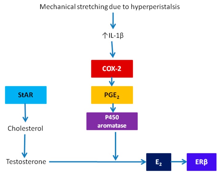
“Thus, hyperperistalsis may induce COX-2 expression and higher PGE2 production in endometrial stromal cells, but it is unlikely to further induce increased estrogen production.”
§ The Pathogenesis of Adenomyosis vis-à-vis Endometriosis > 3. Pathogenesis > 3.3. The TIAR Theory: The Need for Patching up
hedgedconditional- Asserted: passes — The authors put it forward as true.
- Checkable: passes — It can be checked against evidence.
- Load-bearing: passes — The paper’s conclusions depend on it.
- Based: passes — The paper gives a reason why it holds.
It may state two separate claims as one.
None of the sources cited for this claim have been checked yet.
In the paper
“Indeed, the newly reported association between focal adenomyosis of the outer myometrium (FAOM) and deep endometriosis (DE) [61,62] suggests that not all subtypes of adenomyosis are the same and may have a different pathogenesis.”
§ The Pathogenesis of Adenomyosis vis-à-vis Endometriosis > 3. Pathogenesis > 3.3. The TIAR Theory: The Need for Patching up
What the sources say
- Not checked yetcites [61] Chapron, 2017, Relationship between the magnetic resonance imaging appearance of adenomyosis and endometriosis phenotypes, Hum. Reprod. source
- Not checked yetcites [62] Marcellin, 2018, Anterior Focal Adenomyosis and Bladder Deep Infiltrating Endometriosis, Is There a Link?, J. Minim. Invasive Gynecol. source
hedgedcorrelational- Asserted: passes — The authors put it forward as true.
- Checkable: passes — It can be checked against evidence.
- Load-bearing: passes — The paper’s conclusions depend on it.
- Based: passes — The paper gives a reason why it holds.
It may state two separate claims as one.
In the paper
“but inhibition of pituitary prolactin secretion during youth in mice suppressed the induction of adenomyosis [60], suggesting the potential role of prolactin in the pathogenesis of adenomyosis.”
§ The Pathogenesis of Adenomyosis vis-à-vis Endometriosis > 3. Pathogenesis > 3.3. The TIAR Theory: The Need for Patching up
What the sources say
- Not checked yetcites [60] Nagasawa, 1982, Stimulation of mammary tumorigenesis and suppression of uterine adenomyosis by temporary inhibition of pituitary prolactin secretion during youth in mice (41492), Proc. Soc. Exp. Biol. Med. source
hedgedconditional- Asserted: passes — The authors put it forward as true.
- Checkable: passes — It can be checked against evidence.
- Load-bearing: unclear — It is unclear whether the paper’s conclusions depend on it.
- Based: passes — The paper gives a reason why it holds.
It may state two separate claims as one.
A conclusion the paper draws from its own results — it cites no other source for it, so there is none to check. The quote shows it is in the paper.
In the paper
“More importantly, it is difficult, if not impossible, to devise an experiment to prove or disprove either theory.”
§ The Pathogenesis of Adenomyosis vis-à-vis Endometriosis > 3. Pathogenesis > 3.3. The TIAR Theory: The Need for Patching up
plain- Asserted: passes — The authors put it forward as true.
- Checkable: fails — It may be too vague to check against evidence.
- Load-bearing: passes — The paper’s conclusions depend on it.
- Based: fails — The paper may give no reason why it holds.
Probably not a substantial claim. It may be too vague to check against evidence. The paper may give no reason why it holds.
None of the sources cited for this claim have been checked yet.
In the paper
“Thus, hypoxia may well be viewed as the single most important event following tissue injury, and, indeed, adaptation to hypoxic stress via HIF signaling plays a critical role in promoting wound healing processes [74,75].”
§ The Pathogenesis of Adenomyosis vis-à-vis Endometriosis > 3. Pathogenesis > 3.4. Revamping the TIAR Theory: The Endometrial-Myometrial Interface Disruption (EMID)
What the sources say
hedgedconditional- Asserted: passes — The authors put it forward as true.
- Checkable: passes — It can be checked against evidence.
- Load-bearing: passes — The paper’s conclusions depend on it.
- Based: passes — The paper gives a reason why it holds.
It may state two separate claims as one.
A conclusion the paper draws from its own results — it cites no other source for it, so there is none to check. The quote shows it is in the paper.
In the paper
“Hence, the injury to the EMI constitutes a trauma, leading to subsequent tissue hypoxia in myocytes and fibroblasts.”
§ The Pathogenesis of Adenomyosis vis-à-vis Endometriosis > 3. Pathogenesis > 3.4. Revamping the TIAR Theory: The Endometrial-Myometrial Interface Disruption (EMID)
plaindirect- Asserted: passes — The authors put it forward as true.
- Checkable: passes — It can be checked against evidence.
- Load-bearing: passes — The paper’s conclusions depend on it.
- Based: unclear — It is unclear why the paper says it holds.
It may state two separate claims as one.
A conclusion the paper draws from its own results — it cites no other source for it, so there is none to check. The quote shows it is in the paper.
In the paper
“Even though adenomyotic lesions (used to be called endometriosis interna) may resemble DE lesions (now called adenomyosis externa [107]) in their look and feel, their developmental paths may be similar but not entirely the same, due to their different microenvironment.”
§ The Pathogenesis of Adenomyosis vis-à-vis Endometriosis > 4. Conclusions
hedgedconditional- Asserted: passes — The authors put it forward as true.
- Checkable: passes — It can be checked against evidence.
- Load-bearing: passes — The paper’s conclusions depend on it.
- Based: passes — The paper gives a reason why it holds.
It may state two separate claims as one.
In the paper
“it raises a tantalizing possibility that perioperative intervention could reduce the risk of adenomyosis, as in the case of a reduction in recurrence risk in endometriosis [100].”
§ The Pathogenesis of Adenomyosis vis-à-vis Endometriosis > 3. Pathogenesis > 3.4. Revamping the TIAR Theory: The Endometrial-Myometrial Interface Disruption (EMID)
What the sources say
- Not checked yetcites [100] Long, 2019, Perioperative Intervention by beta-Blockade and NF-kappaB Suppression Reduces the Recurrence Risk of Endometriosis in Mice Due to Incomplete Excision, Reprod. Sci. source
hedgedconditional- Asserted: passes — The authors put it forward as true.
- Checkable: passes — It can be checked against evidence.
- Load-bearing: passes — The paper’s conclusions depend on it.
- Based: passes — The paper gives a reason why it holds.
Probably not a substantial claim.
A conclusion the paper draws from its own results — it cites no other source for it, so there is none to check. The quote shows it is in the paper.
In the paper
“Conceivably, these surgical procedures would result in the direct implantation of endometrial cells ectopically within the abdominal wall.”
§ The Pathogenesis of Adenomyosis vis-à-vis Endometriosis > 3. Pathogenesis > 3.5. Similarities and Difference in Pathogenesis between Adenomyosis and Endometriosis
hedgedconditional- Asserted: passes — The authors put it forward as true.
- Checkable: passes — It can be checked against evidence.
- Load-bearing: fails — The paper’s conclusions may not depend on it.
- Based: passes — The paper gives a reason why it holds.
Probably not a substantial claim. The paper’s conclusions may not depend on it.
In the paper
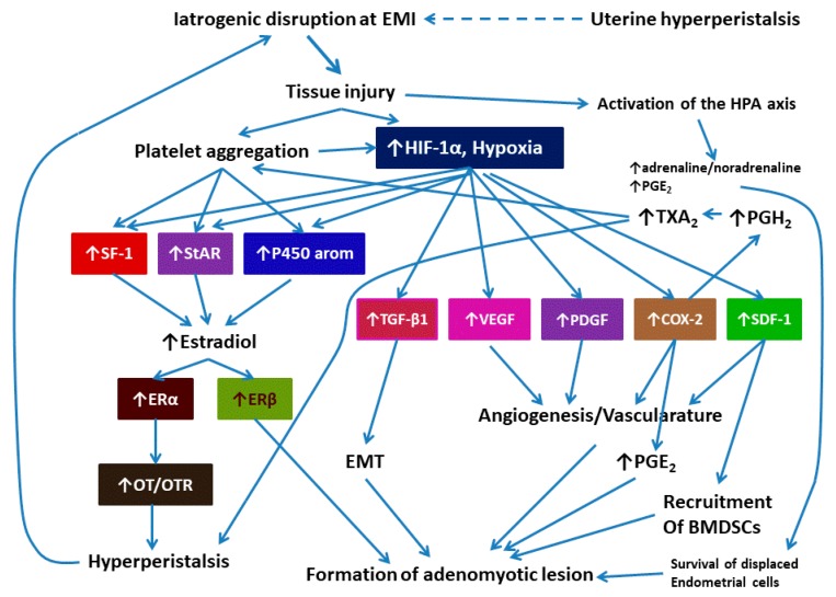
“Note that hypoxia-induced COX-2 overexpression can increase the production of not only PGE2 but also PGH2, which leads to increased production of thromboxane A2 (TXA2), a vasoconstrictor, a potent platelet activator, and a potent inducer of uterine contraction [99].”
§ The Pathogenesis of Adenomyosis vis-à-vis Endometriosis > 3. Pathogenesis > 3.4. Revamping the TIAR Theory: The Endometrial-Myometrial Interface Disruption (EMID)
What the sources say
- Not checked yetcites [99] Wilhelmsson, 1981, PGH2, TxA2 and PGI2 have potent and differentiated actions on human uterine contractility, Prostaglandins source
plaindirect- Asserted: passes — The authors put it forward as true.
- Checkable: passes — It can be checked against evidence.
- Load-bearing: passes — The paper’s conclusions depend on it.
- Based: passes — The paper gives a reason why it holds.
It may state two separate claims as one.
A conclusion the paper draws from its own results — it cites no other source for it, so there is none to check. The quote shows it is in the paper.
In the paper
“The release of adrenaline/noradrenaline and PGE2 can reduce cell-mediated immunity, promoting the survival of spread and displaced endometrial cells.”
§ The Pathogenesis of Adenomyosis vis-à-vis Endometriosis > 3. Pathogenesis > 3.4. Revamping the TIAR Theory: The Endometrial-Myometrial Interface Disruption (EMID)
plaindirect- Asserted: passes — The authors put it forward as true.
- Checkable: passes — It can be checked against evidence.
- Load-bearing: passes — The paper’s conclusions depend on it.
- Based: fails — The paper may give no reason why it holds.
Probably not a substantial claim. The paper may give no reason why it holds.
A conclusion the paper draws from its own results — it cites no other source for it, so there is none to check. The quote shows it is in the paper.
In the paper
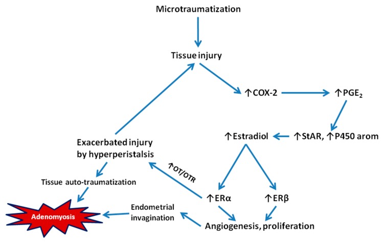
“Thus, the increased estrogen levels may induce an ERα-mediated OT/OTR system, enhancing uterine peristalsis.”
§ The Pathogenesis of Adenomyosis vis-à-vis Endometriosis > 3. Pathogenesis > 3.4. Revamping the TIAR Theory: The Endometrial-Myometrial Interface Disruption (EMID)
hedgedconditional- Asserted: passes — The authors put it forward as true.
- Checkable: passes — It can be checked against evidence.
- Load-bearing: passes — The paper’s conclusions depend on it.
- Based: unclear — It is unclear why the paper says it holds.
It may state two separate claims as one.
None of the sources cited for this claim have been checked yet.
In the paper
“HIF-1α-induced COX-2 expression leads to elevated PGE2 levels, which can induce PGE2-mediated vascularization [87,88].”
§ The Pathogenesis of Adenomyosis vis-à-vis Endometriosis > 3. Pathogenesis > 3.4. Revamping the TIAR Theory: The Endometrial-Myometrial Interface Disruption (EMID)
What the sources say
plaindirect- Asserted: passes — The authors put it forward as true.
- Checkable: passes — It can be checked against evidence.
- Load-bearing: passes — The paper’s conclusions depend on it.
- Based: passes — The paper gives a reason why it holds.
It may state two separate claims as one.
A conclusion the paper draws from its own results — it cites no other source for it, so there is none to check. The quote shows it is in the paper.
In the paper
“While it can explain the genesis of adenomyosis induced by iatrogenic trauma, it may also explain the pathogenesis of adenomyosis due to persistent hyperperistalsis that causes EMID, but probably not FAOM or extrinsic/exterior adenomyosis.”
§ The Pathogenesis of Adenomyosis vis-à-vis Endometriosis > 3. Pathogenesis > 3.4. Revamping the TIAR Theory: The Endometrial-Myometrial Interface Disruption (EMID)
hedgedconditional- Asserted: passes — The authors put it forward as true.
- Checkable: passes — It can be checked against evidence.
- Load-bearing: passes — The paper’s conclusions depend on it.
- Based: passes — The paper gives a reason why it holds.
It may state two separate claims as one.
A conclusion the paper draws from its own results — it cites no other source for it, so there is none to check. The quote shows it is in the paper.
In the paper
“For example, perioperative intervention when iatrogenic procedures such as D&C are performed may reduce the risk of developing adenomyosis in the first place.”
§ The Pathogenesis of Adenomyosis vis-à-vis Endometriosis > 4. Conclusions
hedgedconditional- Asserted: passes — The authors put it forward as true.
- Checkable: passes — It can be checked against evidence.
- Load-bearing: passes — The paper’s conclusions depend on it.
- Based: fails — The paper may give no reason why it holds.
Probably not a substantial claim. The paper may give no reason why it holds.
In the paper
“As adenomyosis shares with endometriosis in cyclic bleeding and thus ReTIAR”
§ The Pathogenesis of Adenomyosis vis-à-vis Endometriosis > 4. Conclusions
What the sources say
- Not checked yetcites [104] Guo, 2018, Fibrogenesis resulting from cyclic bleeding, the Holy Grail of the natural history of ectopic endometrium, Hum. Reprod. source
plaindirect- Asserted: passes — The authors put it forward as true.
- Checkable: passes — It can be checked against evidence.
- Load-bearing: passes — The paper’s conclusions depend on it.
- Based: passes — The paper gives a reason why it holds.
It may state two separate claims as one.
A conclusion the paper draws from its own results — it cites no other source for it, so there is none to check. The quote shows it is in the paper.
In the paper
“In addition, increased estradiol levels would induce ERα, which further activate OT/OTR signaling pathway, causing increased peristalsis.”
§ The Pathogenesis of Adenomyosis vis-à-vis Endometriosis > 3. Pathogenesis > 3.4. Revamping the TIAR Theory: The Endometrial-Myometrial Interface Disruption (EMID)
hedgeddirect- Asserted: passes — The authors put it forward as true.
- Checkable: passes — It can be checked against evidence.
- Load-bearing: passes — The paper’s conclusions depend on it.
- Based: fails — The paper may give no reason why it holds.
Probably not a substantial claim. The paper may give no reason why it holds. It may state two separate claims as one.
A conclusion the paper draws from its own results — it cites no other source for it, so there is none to check. The quote shows it is in the paper.
In the paper
“That adenomyosis is not simply endometriosis of the uterus implies that, when it comes to management and prevention, interventions that are carefully tailored to adenomyosis should be more effective.”
§ The Pathogenesis of Adenomyosis vis-à-vis Endometriosis > 4. Conclusions
hedgeddirect- Asserted: passes — The authors put it forward as true.
- Checkable: fails — It may be too vague to check against evidence.
- Load-bearing: passes — The paper’s conclusions depend on it.
- Based: passes — The paper gives a reason why it holds.
Probably not a substantial claim. It may be too vague to check against evidence.
A conclusion the paper draws from its own results — it cites no other source for it, so there is none to check. The quote shows it is in the paper.
In the paper
“In fact, even for adenomyosis, there might be two or more different pathogenic origins, for example, the FAOM, or extrinsic or external adenomyosis, might originate from the invasion of neighboring DE lesions.”
§ The Pathogenesis of Adenomyosis vis-à-vis Endometriosis > 3. Pathogenesis > 3.5. Similarities and Difference in Pathogenesis between Adenomyosis and Endometriosis
hedgedconditional- Asserted: passes — The authors put it forward as true.
- Checkable: passes — It can be checked against evidence.
- Load-bearing: passes — The paper’s conclusions depend on it.
- Based: passes — The paper gives a reason why it holds.
It may state two separate claims as one.
A conclusion the paper draws from its own results — it cites no other source for it, so there is none to check. The quote shows it is in the paper.
In the paper
“Together, these catecholamines and PGE2 would impair the cell-mediated immunity, promoting the survival of displaced and dispersed cells that spread to the EMI and possibly beyond due to the procedure.”
§ The Pathogenesis of Adenomyosis vis-à-vis Endometriosis > 3. Pathogenesis > 3.4. Revamping the TIAR Theory: The Endometrial-Myometrial Interface Disruption (EMID)
hedgeddirect- Asserted: passes — The authors put it forward as true.
- Checkable: passes — It can be checked against evidence.
- Load-bearing: passes — The paper’s conclusions depend on it.
- Based: unclear — It is unclear why the paper says it holds.
It may state two separate claims as one.
A conclusion the paper draws from its own results — it cites no other source for it, so there is none to check. The quote shows it is in the paper.
In the paper
“In addition, since uterine procedures using different energy sources result in different severities of injury and bleeding patterns, the resultant EMID would lead to differential = risk of developing adenomyosis.”
§ The Pathogenesis of Adenomyosis vis-à-vis Endometriosis > 3. Pathogenesis > 3.4. Revamping the TIAR Theory: The Endometrial-Myometrial Interface Disruption (EMID)
hedgeddirect- Asserted: passes — The authors put it forward as true.
- Checkable: passes — It can be checked against evidence.
- Load-bearing: unclear — It is unclear whether the paper’s conclusions depend on it.
- Based: unclear — It is unclear why the paper says it holds.
It may state two separate claims as one.
A conclusion the paper draws from its own results — it cites no other source for it, so there is none to check. The quote shows it is in the paper.
In the paper
“The model depicted in Figure 3 is completely falsifiable.”
§ The Pathogenesis of Adenomyosis vis-à-vis Endometriosis > 3. Pathogenesis > 3.4. Revamping the TIAR Theory: The Endometrial-Myometrial Interface Disruption (EMID)
boosted- Asserted: passes — The authors put it forward as true.
- Checkable: fails — It may be too vague to check against evidence.
- Load-bearing: fails — The paper’s conclusions may not depend on it.
- Based: fails — The paper may give no reason why it holds.
Probably not a substantial claim. It may be too vague to check against evidence. The paper’s conclusions may not depend on it. The paper may give no reason why it holds.
A conclusion the paper draws from its own results — it cites no other source for it, so there is none to check. The quote shows it is in the paper.
In the paper
“Unlike the TIAR theory, the EMID hypothesis is easily falsifiable, at least in mouse.”
§ The Pathogenesis of Adenomyosis vis-à-vis Endometriosis > 4. Conclusions
plain- Asserted: passes — The authors put it forward as true.
- Checkable: fails — It may be too vague to check against evidence.
- Load-bearing: passes — The paper’s conclusions depend on it.
- Based: fails — The paper may give no reason why it holds.
Probably not a substantial claim. It may be too vague to check against evidence. The paper may give no reason why it holds.
A conclusion the paper draws from its own results — it cites no other source for it, so there is none to check. The quote shows it is in the paper.
In the paper
“Thus, when EMID occurs, such as in the D&C procedure, the resultant tissue hypoxia would lead to platelet aggregation, increased estrogen production, induction of the TGF-β1, VEGF, PDGF, COX-2, and SDF-1 signaling and the resultant elevated PGE2 production, induction of EMT, and increased recruitment of BMDSCs in the wound site, all of which can promote the mobility and implantation of endometrial”
§ The Pathogenesis of Adenomyosis vis-à-vis Endometriosis > 3. Pathogenesis > 3.4. Revamping the TIAR Theory: The Endometrial-Myometrial Interface Disruption (EMID)
hedgeddirect- Asserted: passes — The authors put it forward as true.
- Checkable: passes — It can be checked against evidence.
- Load-bearing: passes — The paper’s conclusions depend on it.
- Based: passes — The paper gives a reason why it holds.
It may state two separate claims as one.
A conclusion the paper draws from its own results — it cites no other source for it, so there is none to check. The quote shows it is in the paper.
In the paper
“Thus, EMID may induce EMT in adjacent endometrial epithelial cells and enhance their mobility and invasiveness, resulting in the crossing of EMI to the myometrium by endometrial epithelial cells.”
§ The Pathogenesis of Adenomyosis vis-à-vis Endometriosis > 3. Pathogenesis > 3.4. Revamping the TIAR Theory: The Endometrial-Myometrial Interface Disruption (EMID)
hedgedconditional- Asserted: passes — The authors put it forward as true.
- Checkable: passes — It can be checked against evidence.
- Load-bearing: passes — The paper’s conclusions depend on it.
- Based: passes — The paper gives a reason why it holds.
It may state two separate claims as one.
A conclusion the paper draws from its own results — it cites no other source for it, so there is none to check. The quote shows it is in the paper.
In the paper
“For example, the use of coagulation might lead to a lower risk of adenomyosis than the D&C procedure.”
§ The Pathogenesis of Adenomyosis vis-à-vis Endometriosis > 3. Pathogenesis > 3.4. Revamping the TIAR Theory: The Endometrial-Myometrial Interface Disruption (EMID)
hedgedconditional- Asserted: passes — The authors put it forward as true.
- Checkable: passes — It can be checked against evidence.
- Load-bearing: fails — The paper’s conclusions may not depend on it.
- Based: passes — The paper gives a reason why it holds.
Probably not a substantial claim. The paper’s conclusions may not depend on it.
A conclusion the paper draws from its own results — it cites no other source for it, so there is none to check. The quote shows it is in the paper.
In the paper
“This model can at least account for the genesis of adenomyosis arising from iatrogenic trauma such as uterine procedures, but perhaps not for adenomyosis induced by other causes.”
§ The Pathogenesis of Adenomyosis vis-à-vis Endometriosis > 3. Pathogenesis > 3.4. Revamping the TIAR Theory: The Endometrial-Myometrial Interface Disruption (EMID)
hedgedconditional- Asserted: passes — The authors put it forward as true.
- Checkable: passes — It can be checked against evidence.
- Load-bearing: passes — The paper’s conclusions depend on it.
- Based: passes — The paper gives a reason why it holds.
It may state two separate claims as one.
The paper relies on this without support.
The molecular mechanisms of wound healing in non-endometrial tissues apply to the endometrial–myometrial interface and adenomyosis pathogenesis.
“Based on the current understanding of wound healing, a new hypothesis, called endometrial-myometrial interface disruption (EMID), is proposed to account for adenomyosis resulting from iatrogenic trauma to EMI.”
The paper relies on this without support.
The mouse EMID model is a valid model for human adenomyosis.
“In fact, EMID can robustly induce adenomyosis in mice (Hao et al., submitted for publication).”
The paper relies on this without support.
Normal endometrial stromal cells studied in vitro reflect normal endometrial stromal cells in vivo.
“Yet normal endometrial stromal cells are very different in behavior from endometriotic or even adenomyotic stromal cells.”
The paper relies on this without support.
Iatrogenic procedures are common in gynecological practice nowadays.
“iatrogenic procedures aplenty nowadays in gynecological practice”
The paper relies on this without support.
Many medications used primarily for treating endometriosis can and have been used also for treating adenomyosis.
“many medications used primarily for treating endometriosis can and have been used also for treating adenomyosis”
Claims other papers make about this one
Each is checked against this paper’s own text: does it say what the citing paper says it does?
In the citing paper
“The majority of research includes instances of gynecologic surgery for benign conditions such as adenomyosis, uterine leiomyomas, and fibroids [17,18]”
From Challenges in Diagnosis and Prevention of Iatrogenic Endometriosis as a Long-Term Surgical Complication after C-Section (2022) · cites this paper as [18]
Citations
Cites
Activation of vascular endothelial growth factor gene transcription by hypoxia-inducible factor 1
Forsythe, J. A., Jiang, B.-H., Iyer, N. V., Agani, F., Leung, S. W., Koos, R. D., & Semenza, G. L. (1996) · Molecular and Cellular Biology · doi:10.1128/mcb.16.9.4604 · Cited by 3,981
Hypoxia-inducible factors in physiology and medicine
Semenza, G. L. (2012) · Cell · doi:10.1016/j.cell.2012.01.021 · Cited by 3,645 · cited 1×
“Once hypoxic, evolutionarily conserved adaptive processes, such as hypoxia-inducible factors (HIFs), are activated [73].”
BackgroundGiudice, L. C., & Kao, L. C. (2004) · The Lancet · doi:10.1016/s0140-6736(04)17403-5 · Cited by 3,166
Progenitor cell trafficking is regulated by hypoxic gradients through HIF-1 induction of SDF-1
Ceradini, D. J., Kulkarni, A. R., Callaghan, M. J., Tepper, O. M., Bastidas, N., Kleinman, M. E., Capla, J. M., Galiano, R. D., Levine, J. P., & Gurtner, G. C. (2004) · Nature Medicine · doi:10.1038/nm1075 · Cited by 2,600
Cyclooxygenase regulates angiogenesis induced by colon cancer cells
Tsujii, M., Kawano, S., Tsuji, S., Sawaoka, H., Hori, M., & DuBois, R. N. (1998) · Cell · doi:10.1016/s0092-8674(00)81433-6 · Cited by 2,272
Endometriosis: pathogenesis and treatment
Vercellini, P., Viganò, P., Somigliana, E., & Fedele, L. (2014) · Nature Reviews Endocrinology · doi:10.1038/nrendo.2013.255 · Cited by 1,940 · cited 1×
“Sampson’s retrograde menstruation theory is the most widely accepted and has the most robust evidence [27].”
BackgroundPathogenesis and pathophysiology of endometriosis
Burney, R. O., & Giudice, L. C. (2012) · Fertility and Sterility · doi:10.1016/j.fertnstert.2012.06.029 · Cited by 1,590 · cited 1×
“For endometriosis, several theories regarding its pathogenesis have been proposed, and these hypotheses can be divided into three themes: in situ development (such as coelomic metaplasia or embryonic cell rests), implantation, or a combination of in situ development and implantation [25,26].”
BackgroundHIF‐1α is required for solid tumor formation and embryonic vascularization
Ryan, H. E., Lo, J., & Johnson, R. S. (1998) · The EMBO Journal · doi:10.1093/emboj/17.11.3005 · Cited by 1,579 · cited 2×
“Hypoxia can also enhance cellular viability and proliferation through upregulation of insulin growth factor 2 (IGF-2) and IGF binding protein 3 (IGF-BP3) [85].”
BackgroundGreenhough, A., Smartt, H. J. M., Moore, A. E., Roberts, H. R., Williams, A. C., Paraskeva, C., & Kaidi, A. (2009) · Carcinogenesis · doi:10.1093/carcin/bgp014 · Cited by 1,220 · cited 1×
“HIF-1α-induced COX-2 expression leads to elevated PGE2 levels, which can induce PGE2-mediated vascularization [87,88].”
BackgroundRisk for and consequences of endometriosis: A critical epidemiologic review
Shafrir, A. L., Farland, L. V., Shah, D. K., Harris, H. R., Kvaskoff, M., Zondervan, K., & Missmer, S. A. (2018) · Best Practice & Research Clinical Obstetrics & Gynaecology · doi:10.1016/j.bpobgyn.2018.06.001 · Cited by 823 · cited 1×
“The risk profiles for adenomyosis are thus quite different from that of endometriosis, in which greater parity is a protective factor, while earlier age at menarche and shorter menstrual cycle length—factors that are consistent with Sampson’s retrograde menstruation theory for the etiology of endometriosis—are risk fa…”
BackgroundDifferential Transcriptional Regulation of the Two Vascular Endothelial Growth Factor Receptor Genes
Gerber, H.-P., Condorelli, F., Park, J., & Ferrara, N. (1997) · Journal of Biological Chemistry · doi:10.1074/jbc.272.38.23659 · Cited by 794
World Endometriosis Society consensus on the classification of endometriosis
Johnson, N. P., Hummelshoj, L., Adamson, G. D., Keckstein, J., Taylor, H. S., Abrao, M. S., Bush, D., Kiesel, L., Tamimi, R., Sharpe-Timms, K. L., et al. (2017) · Human Reproduction · doi:10.1093/humrep/dew293 · Cited by 664 · cited 1×
“Both are badly in need of a better classification system [8,13,14].”
BackgroundPlatelets in inflammation and thrombosis
Wagner, D. D., & Burger, P. C. (2003) · Arteriosclerosis Thrombosis and Vascular Biology · doi:10.1161/01.atv.0000095974.95122.ec · Cited by 570
Endometrial stem/progenitor cells: the first 10 years
Gargett, C. E., Schwab, K. E., & Deane, J. A. (2015) · Human Reproduction Update · doi:10.1093/humupd/dmv051 · Cited by 561 · cited 1×
“Is there any trigger, if any, that leads to the metaplasia, or the microtraumatization, that eventually results in invagination? If microtraumatization is ubiquitous, why do only a small portion of women eventually develop adenomyosis? Can we prevent the microtrauma, or at least tame it, so that the risk of developing…”
BackgroundThe elusive adenomyosis of the uterus—revisited
Bird, C. C., McElin, T. W., & Manalo-Estrella, P. (1972) · American Journal of Obstetrics and Gynecology · doi:10.1016/0002-9378(72)90781-8 · Cited by 531
Bulun, S. E., Lin, Z., Imir, G., Amin, S., Demura, M., Yilmaz, B., Martin, R., Utsunomiya, H., Thung, S., Gurates, B., et al. (2005) · Pharmacological Reviews · doi:10.1124/pr.57.3.6 · Cited by 531 · cited 1×
“In fact, the feed-forward loop depicted in Figure 1 is embedded within the positive feedback model depicted in Figure 11 in [64].”
Pathophysiology of adenomyosis
Ferenczy, A. S. (1998) · Human Reproduction Update · doi:10.1093/humupd/4.4.312 · Cited by 512 · cited 1×
“The metaplasia theory proposes that adenomyotic lesions may originate from the metaplasia of displaced embryonic pluripotent Müllerian remnants [50].”
BackgroundHypoxia Regulates Macrophage Functions in Inflammation
Murdoch, C., Muthana, M., & Lewis, C. E. (2005) · The Journal of Immunology · doi:10.4049/jimmunol.175.10.6257 · Cited by 455
The Role of Hypoxia-Inducible Factor in Wound Healing
Hong, W. X., Hu, M. S., Esquivel, M., Liang, G. Y., Rennert, R. C., McArdle, A., Paik, K. J., Duscher, D., Gurtner, G. C., Lorenz, H. P., et al. (2014) · Advances in Wound Care · doi:10.1089/wound.2013.0520 · Cited by 448 · cited 1×
“Thus, hypoxia may well be viewed as the single most important event following tissue injury, and, indeed, adaptation to hypoxic stress via HIF signaling plays a critical role in promoting wound healing processes [74,75].”
BackgroundThe pathophysiology of endometriosis and adenomyosis, tissue injury and repair
Leyendecker, G., Wildt, L., & Mall, G. (2009) · Archives of Gynecology and Obstetrics · doi:10.1007/s00404-009-1191-0 · Cited by 418 · cited 3×
“Moreover, there is a remarkable similarity between hyperestrogenism in adenomyosis and the role of estrogen in wound repair [29,30].”
BackgroundInfluentialUterine adenomyosis and in vitro fertilization outcome: a systematic review and meta-analysis
Vercellini, P., Consonni, D., Dridi, D., Bracco, B., Frattaruolo, M. P., & Somigliana, E. (2014) · Human Reproduction · doi:10.1093/humrep/deu041 · Cited by 411
Kunz, G., Beil, D., Deininger, H., Wildt, L., & Leyendecker, G. (1996) · Human Reproduction · doi:10.1093/humrep/11.3.627 · Cited by 408
Uterine stem cells: What is the evidence?
Gargett, C. E. (2007) · Human Reproduction Update · doi:10.1093/humupd/dml045 · Cited by 373 · cited 1×
“Alternatively, endometrial stem/progenitor cells or their niche cells may behave aberrantly for some unknown reason, and their differentiating progeny cells move towards the myometrium, rather than functionalis, resulting in adenomyosis [51].”
BackgroundPathology and physiopathology of adenomyosis
Bergeron, C., Amant, F., & Ferenczy, A. (2006) · Best Practice & Research Clinical Obstetrics & Gynaecology · doi:10.1016/j.bpobgyn.2006.01.016 · Cited by 359
Promoter Methylation Regulates Estrogen Receptor 2 in Human Endometrium and Endometriosis1
Xue, Q., Lin, Z., Cheng, Y.-H., Huang, C.-C., Marsh, E., Yin, P., Milad, M. P., Confino, E., Reierstad, S., Innes, J., et al. (2007) · Biology of Reproduction · doi:10.1095/biolreprod.107.061804 · Cited by 346 · cited 1×
“In addition, ERβ is also hypermethylated and silenced in normal endometrial stromal cells [71].”
BackgroundPathogenesis of uterine adenomyosis, invagination or metaplasia?
García-Solares, J., Donnez, J., Donnez, O., & Dolmans, M.-M. (2018) · Fertility and Sterility · doi:10.1016/j.fertnstert.2017.12.030 · Cited by 339 · cited 3×
“The metaplasia theory could account for some cases of adenomyosis in the rudimentary muscular uterine wall of patients with Mayer–Rokitansky–Kuster–Hauser syndrome [4].”
BackgroundMethodInfluentialYamakawa, M., Liu, L. X., Date, T., Belanger, A. J., Vincent, K. A., Akita, G. Y., Kuriyama, T., Cheng, S. H., Gregory, R. J., & Jiang, C. (2003) · Circulation Research · doi:10.1161/01.res.0000093984.48643.d7 · Cited by 327
Pathogenesis of adenomyosis, an update on molecular mechanisms
Vannuccini, S., Tosti, C., Carmona, F., Huang, S. J., Chapron, C., Guo, S.-W., & Petraglia, F. (2017) · Reproductive BioMedicine Online · doi:10.1016/j.rbmo.2017.06.016 · Cited by 326 · cited 1×
“With the shift in diagnosis from hysterectomy to imaging due to advancing imaging technologies, it is certain that the incidence peak may change to women younger than 46–50 years [3].”
MethodSelf-citationHoming to Hypoxia: HIF-1 as a Mediator of Progenitor Cell Recruitment to Injured Tissue
Ceradini, D. J., & Gurtner, G. C. (2005) · Trends in Cardiovascular Medicine · doi:10.1016/j.tcm.2005.02.002 · Cited by 324 · cited 1×
“In addition, hypoxia-induced SDF-1/CXCL12 can attract hematopoietic and endothelial progenitor cells (EPCs) expressing its receptor CXCR4, facilitating vasculogenesis [89].”
BackgroundAdenomyosis, epidemiological factors
Vercellini, P., Viganò, P., Somigliana, E., Daguati, R., Abbiati, A., & Fedele, L. (2006) · Best Practice & Research Clinical Obstetrics & Gynaecology · doi:10.1016/j.bpobgyn.2006.01.017 · Cited by 319 · cited 3×
“This wide variability stems from variations in the histological criteria adopted for diagnosis and the techniques used to procure myometrial samples [15].”
BackgroundPlatelet chemokines and chemokine receptors, linking hemostasis, inflammation, and host defense
GEAR, A. R. L., & CAMERINI, D. (2003) · Microcirculation · doi:10.1038/sj.mn.7800198 · Cited by 317
Leyendecker, G., Kunz, G., Wildt, L., Beil, D., & Deininger, H. (1996) · Human Reproduction · doi:10.1093/oxfordjournals.humrep.a019435 · Cited by 307 · cited 1×
“Third, uterine hyperperistalsis/dysperistalsis may be a result of archimetral hyperestrogenism [38,47].”
BackgroundKitawaki, J., Noguchi, T., Amatsu, T., Maeda, K., Tsukamoto, K., Yamamoto, T., Fushiki, S., Osawa, Y., & Honjo, H. (1997) · Biology of Reproduction · doi:10.1095/biolreprod57.3.514 · Cited by 305 · cited 1×
“In addition, genes involved in estrogen biosynthesis, such as steroidogenic acute regulatory protein (StAR) and P450 aromatase, are upregulated in adenomyosis [48,49].”
BackgroundKaidi, A., Qualtrough, D., Williams, A. C., & Paraskeva, C. (2006) · Cancer Research · doi:10.1158/0008-5472.can-06-0425 · Cited by 300 · cited 11×
“Lastly, many genes, such as ERβ and cyclooxygenase-2 (COX-2), that are involved in tissue repair and inflammation, are upregulated in adenomyosis.”
InfluentialChapron, C., Tosti, C., Marcellin, L., Bourdon, M., Lafay-Pillet, M.-C., Millischer, A.-E., Streuli, I., Borghese, B., Petraglia, F., & Santulli, P. (2017) · Human Reproduction · doi:10.1093/humrep/dex088 · Cited by 294
Yang, G., Im, H.-J., & Wang, J. H.-C. (2005) · Gene · doi:10.1016/j.gene.2005.08.006 · Cited by 282 · cited 13×
“HIF-1α-induced COX-2 expression leads to elevated PGE2 levels, which can induce PGE2-mediated vascularization [87,88].”
BackgroundInfluentialLyons, E. A., Taylor, P. J., Zheng, X. H., Ballard, G., Levi, C. S., & Kredentser, J. V. (1991) · Fertility and Sterility · doi:10.1016/s0015-0282(16)54246-0 · Cited by 272 · cited 2×
“In particular, the JZ is involved in the modulation of uterine contractions throughout the menstrual cycle, ostensibly serving the purpose of sperm transport during ovulation and antegrade menstruation during the follicular and menstrual phases [35,36].”
BackgroundLeyendecker, G., Bilgicyildirim, A., Inacker, M., Stalf, T., Huppert, P., Mall, G., Böttcher, B., & Wildt, L. (2015) · Archives of Gynecology and Obstetrics · doi:10.1007/s00404-014-3437-8 · Cited by 261
The Impact of Adenomyosis on Women's Fertility
Harada, T., Khine, Y. M., Kaponis, A. I., Nikellis, T., Decavalas, G., & Taniguchi, F. (2016) · Obstetrical & Gynecological Survey · doi:10.1097/ogx.0000000000000346 · Cited by 258
Magnetic resonance imaging of the female pelvis: initial experience
Hricak, H., Alpers, C. E., Crooks, L. E., & Sheldon, P. (1983) · American Journal of Roentgenology · doi:10.2214/ajr.141.6.1119 · Cited by 257 · cited 1×
“The former consists of endometrium, the subendometrial unit called the junctional zone (JZ) [32], and the underlying stratum subvasculare of the myometrium.”
BackgroundSymptoms and classification of uterine adenomyosis, including the place of hysteroscopy in diagnosis
Gordts, S., Grimbizis, G., & Campo, R. (2018) · Fertility and Sterility · doi:10.1016/j.fertnstert.2018.01.006 · Cited by 246 · cited 1×
“Both are badly in need of a better classification system [8,13,14].”
BackgroundContractions of the inner third of the myometrium
de Vries, K., Lyons, E. A., Ballard, G., Levi, C. S., & Lindsay, D. J. (1990) · American Journal of Obstetrics and Gynecology · doi:10.1016/0002-9378(90)90983-e · Cited by 243 · cited 2×
“In particular, the JZ is involved in the modulation of uterine contractions throughout the menstrual cycle, ostensibly serving the purpose of sperm transport during ovulation and antegrade menstruation during the follicular and menstrual phases [35,36].”
BackgroundPathogenesis of deep endometriosis
Gordts, S., Koninckx, P., & Brosens, I. (2017) · Fertility and Sterility · doi:10.1016/j.fertnstert.2017.08.036 · Cited by 241 · cited 1×
“Even though adenomyotic lesions (used to be called endometriosis interna) may resemble DE lesions (now called adenomyosis externa [107]) in their look and feel, their developmental paths may be similar but not entirely the same, due to their different microenvironment.”
BackgroundRole of medical therapy in the management of uterine adenomyosis
Vannuccini, S., Luisi, S., Tosti, C., Sorbi, F., & Petraglia, F. (2018) · Fertility and Sterility · doi:10.1016/j.fertnstert.2018.01.013 · Cited by 231 · cited 1×
“be similar, and the drugs to treat the two diseases can be used interchangeably [11].”
BackgroundPrevalence and risk factors of adenomyosis at hysterectomy
Bergholt, T., Eriksen, L., Berendt, N., Jacobsen, M., & Hertz, J. B. (2001) · Human Reproduction · doi:10.1093/humrep/16.11.2418 · Cited by 227 · cited 1×
“The reported prevalence of adenomyosis varies greatly among different studies, from as low as 10% to as high as 57% [15,16].”
BackgroundKappel, A., Rönicke, V., Damert, A., Flamme, I., Risau, W., & Breier, G. (1999) · Blood · doi:10.1182/blood.v93.12.4284 · Cited by 223
Xue, Q., Lin, Z., Yin, P., Milad, M. P., Cheng, Y.-H., Confino, E., Reierstad, S., & Bulun, S. E. (2007) · The Journal of Clinical Endocrinology & Metabolism · doi:10.1210/jc.2007-0494 · Cited by 219 · cited 1×
“Similarly, the gene encoding for steroidogenic factor-1 (SF-1), the protein critically involved in estrogen biosynthesis, is also hypermethylated and silenced in normal endometrial stromal cells [69].”
BackgroundUterine junctional zone, function and disease
Brosens, J. J., de Souza, N. M., & Barker, F. G. (1995) · The Lancet · doi:10.1016/s0140-6736(95)91387-4 · Cited by 215 · cited 2×
“However, the JZ thickening, in and of itself, does not provide proof of mucosal invasion of the myometrium [37].”
BackgroundTaran, F., Stewart, E., & Brucker, S. (2013) · Geburtshilfe und Frauenheilkunde · doi:10.1055/s-0033-1350840 · Cited by 213
Parazzini, F., Vercellini, P., Panazza, S., Chatenoud, L., Oldani, S., & Crosignani, P. G. (1997) · Human Reproduction · doi:10.1093/humrep/12.6.1275 · Cited by 213
Adenomyosis: symptoms, histology, and pregnancy terminations
Levgur, M. (2000) · Obstetrics and Gynecology · doi:10.1016/s0029-7844(99)00659-6 · Cited by 209
Transcriptional characterizations of differences between eutopic and ectopic endometrium
Wu, Y., Kajdacsy-Balla, A., Strawn, E., Basir, Z., Halverson, G., Jailwala, P., Wang, Y., Wang, X., Ghosh, S., & Guo, S.-W. (2006) · Endocrinology · doi:10.1210/en.2005-0426 · Cited by 208 · cited 1×
“In fact, over 1000 genes are documented to be differentially expressed between adenomyotic lesions and eutopic endometrium [65] and close to 1000 genes are differentially expressed between ectopic and eutopic endometrium [66].”
BackgroundAttar, E., Tokunaga, H., Imir, G., Yilmaz, M. B., Redwine, D., Putman, M., Gurates, B., Attar, R., Yaegashi, N., Hales, D. B., et al. (2009) · The Journal of Clinical Endocrinology & Metabolism · doi:10.1210/jc.2008-1180 · Cited by 207 · cited 1×
“Even when stimulated with PGE2, the expression of most genes involved in estrogen biosynthesis, including StAR, SF-1, and aromatase, are still negligibly low, concomitant with the very low production of estradiol in normal endometrial stromal cells [70].”
BackgroundEndometriosis: A dysfunction and disease of the archimetra
Leyendecker, G. (1998) · Human Reproduction Update · doi:10.1093/humupd/4.5.752 · Cited by 199 · cited 3×
“Of note, the archimetra and the neometra function differently during the process of reproduction [31].”
BackgroundInfluentialLiu, X., Shen, M., Qi, Q., Zhang, H., & Guo, S.-W. (2016) · Human Reproduction · doi:10.1093/humrep/dew018 · Cited by 190 · cited 1×
“, EMT, fibroblast-to-myofibroblast transdifferentation, smooth muscle metaplasia, and fibrogenesis [105,106].”
BackgroundSelf-citationKappel, A., Rönicke, V., Damert, A., et al. (1999) · Blood · doi:10.1182/blood.v93.12.4284.412k25_4284_4292 · Cited by 183
Medical and surgical management of adenomyosis
Farquhar, C. M., & Brosens, I. A. (2006) · Best Practice & Research Clinical Obstetrics & Gynaecology · doi:10.1016/j.bpobgyn.2006.01.012 · Cited by 171
Uterine peristaltic activity and the development of endometriosis
LEYENDECKER, G., KUNZ, G., HERBERTZ, M., BEIL, D., HUPPERT, P., MALL, G., KISSLER, S., NOE, M., & WILDT, L. (2004) · Annals of the New York Academy of Sciences · doi:10.1196/annals.1335.036 · Cited by 162 · cited 1×
“Third, uterine hyperperistalsis/dysperistalsis may be a result of archimetral hyperestrogenism [38,47].”
BackgroundMorassutto, C., Monasta, L., Ricci, G., Barbone, F., & Ronfani, L. (2016) · PLOS ONE · doi:10.1371/journal.pone.0154227 · Cited by 157 · cited 1×
“The age-specific incidence of adenomyosis is very different from that of endometriosis, reaching its peak at the age interval of 46– 50 years [17].”
BackgroundWildt, L. (1998) · Human Reproduction Update · doi:10.1093/humupd/4.5.655 · Cited by 149
A new concept of endometriosis and adenomyosis, tissue injury and repair (TIAR)
Leyendecker, G., & Wildt, L. (2011) · hmbci · doi:10.1515/hmbci.2011.002 · Cited by 147
Platelet Chemokines and Chemokine Receptors: Linking Hemostasis, Inflammation, and Host Defense
Gear, A. R. L., & Camerini, D. (2003) · Microcirculation · doi:10.1080/mic.10.3-4.335.350 · Cited by 144
Adenomyosis: the pathophysiology of an oestrogen-dependent disease
Kitawaki, J. (2006) · Best Practice & Research Clinical Obstetrics & Gynaecology · doi:10.1016/j.bpobgyn.2006.01.010 · Cited by 143 · cited 1×
“Aside from their similar symptomologies, both are estrogen-dependent [10] and share many molecular aberrations.”
BackgroundKunz, G. (1998) · Human Reproduction Update · doi:10.1093/humupd/4.5.647 · Cited by 136 · cited 1×
“Uterine peristalsis can be induced by oxytocin (OT) [43], which is mediated by its receptor OTR.”
BackgroundTaylor, C. T., & McElwain, J. C. (2010) · Physiology · doi:10.1152/physiol.00029.2010 · Cited by 135 · cited 1×
“The tissue hypoxia can be further exacerbated by an influx of inflammatory and stromal cells—all with high metabolic demands for oxygen, which is essential for all aerobic organisms to produce energy via mitochondrial oxidative respiration and to perform other vital biological functions [72].”
BackgroundFeng, W., Madajka, M., Kerr, B. A., Mahabeleshwar, G. H., Whiteheart, S. W., & Byzova, T. V. (2011) · Blood · doi:10.1182/blood-2010-08-304808 · Cited by 133 · cited 1×
“Similarly, platelets stimulate the homing of BMDSCs, facilitating angiogenesis in injured and hypoxic tissues [92].”
BackgroundGuo, S.-W., Mao, X., Ma, Q., & Liu, X. (2013) · Fertility and Sterility · doi:10.1016/j.fertnstert.2012.08.038 · Cited by 131 · cited 1×
“In adenomyosis, OTR overexpression in myometrium, concomitant with uterine hyperactivity, has been reported and is found to correlate to the severity of dysmenorrhea [46].”
BackgroundShen, M., Liu, X., Zhang, H., & Guo, S.-W. (2015) · Human Reproduction · doi:10.1093/humrep/dev314 · Cited by 124
Self-citationUterine MR imaging: effects of hormonal stimulation.
Demas, B. E., Hricak, H., & Jaffe, R. B. (1986) · Radiology · doi:10.1148/radiology.159.1.3952297 · Cited by 116 · cited 1×
“Remarkably, the change in uterine zonal anatomy depends on ovarian sex steroid hormones [33,34].”
BackgroundGuo, S.-W. (2018) · Human Reproduction · doi:10.1093/humrep/dey015 · Cited by 116 · cited 1×
“For adenomyosis pathophysiology, the hypothesis of repeated tissue injury and repair (ReTIAR) has been proposed [104].”
BackgroundSelf-citationEvidence for estrogen synthesis in adenomyotic tissues
Yamamoto, T., Noguchi, T., Tamura, T., Kitawaki, J., & Okada, H. (1993) · American Journal of Obstetrics and Gynecology · doi:10.1016/0002-9378(93)90654-2 · Cited by 111 · cited 1×
“In addition, genes involved in estrogen biosynthesis, such as steroidogenic acute regulatory protein (StAR) and P450 aromatase, are upregulated in adenomyosis [48,49].”
BackgroundBiochemistry of alpha granule proteins.
Holt, J., & Niewiarowski, S. (1985) · PubMed · Cited by 111
Global Transcriptome Abnormalities of the Eutopic Endometrium from Women with Adenomyosis
Herndon, C. N., Aghajanova, L., Balayan, S., Erikson, D., Barragan, F., Goldfien, G., Vo, K. C., Hawkins, S., & Giudice, L. C. (2016) · Reproductive Sciences · doi:10.1177/1933719116650758 · Cited by 106 · cited 1×
“In fact, over 1000 genes are documented to be differentially expressed between adenomyotic lesions and eutopic endometrium [65] and close to 1000 genes are differentially expressed between ectopic and eutopic endometrium [66].”
BackgroundXiong, Y., Liu, Y., Xiong, W., Zhang, L., Liu, H., Du, Y., & Li, N. (2016) · Human Reproduction · doi:10.1093/humrep/dew081 · Cited by 105
Risk Factors, Clinical Presentation, and Outcomes for Abdominal Wall Endometriosis
Khan, Z., Zanfagnin, V., El-Nashar, S. A., Famuyide, A. O., Daftary, G. S., & Hopkins, M. R. (2017) · Journal of Minimally Invasive Gynecology · doi:10.1016/j.jmig.2017.01.005 · Cited by 97 · cited 1×
“One probable exception might be the abdominal wall endometriosis (AWE), which is associated with cesarean delivery and abdominal hysterectomy [102,103].”
BackgroundWang, X., Mamillapalli, R., Mutlu, L., Du, H., & Taylor, H. S. (2015) · Stem Cell Research · doi:10.1016/j.scr.2015.04.004 · Cited by 93 · cited 1×
“Incidentally or not, SDF-1 plays a critical role in recruiting bone marrow derived stem cells (BMDSCs) to endometrium and ectopic endometrium [90,91].”
BackgroundIs it time for a paradigm shift in drug research and development in endometriosis/adenomyosis?
Guo, S., & Groothuis, P. G. (2018) · Human Reproduction Update · doi:10.1093/humupd/dmy020 · Cited by 93 · cited 2×
“In fact, few drugs are being developed exclusively for adenomyosis [12].”
BackgroundSelf-citationDisruption of the endometrial-myometrial border during pregnancy as a risk factor for adenomyosis
Curtis, K. M., Hillis, S. D., Marchbanks, P. A., & Peterson, H. B. (2002) · American Journal of Obstetrics and Gynecology · doi:10.1067/mob.2002.124285 · Cited by 91
Is prior uterine surgery a risk factor for adenomyosis?
Panganamamula, U. R., Harmanli, O. H., Isik-Akbay, E. F., Grotegut, C. A., Dandolu, V., & Gaughan, J. P. (2004) · Obstetrics & Gynecology · doi:10.1097/01.aog.0000143264.59822.73 · Cited by 90
Izawa, M., Harada, T., Taniguchi, F., Ohama, Y., Takenaka, Y., & Terakawa, N. (2008) · Fertility and Sterility · doi:10.1016/j.fertnstert.2007.03.078 · Cited by 83 · cited 1×
“In stark contrast to endometriotic stromal cells, in fact, the promoter of the gene coding for P450 aromatase is hypermethylated in normal endometrial stromal cells, and thus silenced [67,68].”
BackgroundCampo, S., Campo, V., & Benagiano, G. (2011) · Obstetrics and Gynecology International · doi:10.1155/2012/786132 · Cited by 83 · cited 1×
“It is well accepted that uterine peristalsis due to myometrial contraction originates from the JZ, while the outer myometrium remains quiescent [42].”
BackgroundPGH2, TxA2 and PGI2 have potent and differentiated actions on human uterine contractility
Wilhelmsson, L., Wikland, M., & Wiqvist, N. (1981) · Prostaglandins · doi:10.1016/0090-6980(81)90145-3 · Cited by 77
Current Insights into the role of HIF-1 in cutaneous wound healing
Andrikopoulou, E., Zhang, X., Sebastian, R., Marti, G., Liu, L., M. Milner, S., & W. Harmon, J. (2011) · Current Molecular Medicine · doi:10.2174/156652411795243414 · Cited by 77 · cited 1×
“Thus, hypoxia may well be viewed as the single most important event following tissue injury, and, indeed, adaptation to hypoxic stress via HIF signaling plays a critical role in promoting wound healing processes [74,75].”
BackgroundOxytocin and Oxytocin Receptor Gene Expression in the Uterus
Zingg, H. H., Rozen, F., Chu, K., Larcher, A., ARSLAN, A., Richard, S., & Lefebvre, D. L. (1995) · Elsevier eBooks · doi:10.1016/b978-0-12-571150-0.50015-9 · Cited by 75 · cited 1×
“In myometrium, OT production and OTR expression seem to be regulated by estrogen receptor α (ERα) [44,45].”
BackgroundOstrander, P. L., Mills, K. T., & Bern, H. A. (1985) · JNCI Journal of the National Cancer Institute · doi:10.1093/jnci/74.1.121 · Cited by 75 · cited 1×
“In particular, neonatal exposure to diethylstilbestrol followed by continuous hormonal treatment—even progesterone alone—also results in adenomyosis [58].”
BackgroundIzawa, M., Taniguchi, F., Uegaki, T., Takai, E., Iwabe, T., Terakawa, N., & Harada, T. (2011) · Fertility and Sterility · doi:10.1016/j.fertnstert.2010.06.024 · Cited by 73 · cited 1×
“In stark contrast to endometriotic stromal cells, in fact, the promoter of the gene coding for P450 aromatase is hypermethylated in normal endometrial stromal cells, and thus silenced [67,68].”
BackgroundKunz, G. (1998) · Human Reproduction Update · doi:10.1093/humupd/4.5.667 · Cited by 69
Nelson, W. O. (1937) · The Anatomical Record · doi:10.1002/ar.1090680107 · Cited by 67
The induction of adenomyosis in mice by intrauterine pituitary isografts
Mori, T., Nagasawa, H., & Takahashi, S. (1981) · Life Sciences · doi:10.1016/0024-3205(81)90234-4 · Cited by 65 · cited 1×
“In addition, the grafting of a single anterior pituitary into the uterine lumen induced a high incidence of adenomyosis in some strains of mice [59], but inhibition of pituitary”
BackgroundPathological functions of hypoxia in endometriosis
Hsiao, K.-Y. (2015) · Frontiers in Bioscience · doi:10.2741/e736 · Cited by 61 · cited 1×
“Remarkably, hypoxic conditions alone can dramatically change the behavior of endometrial cells that are otherwise “normal”, manifesting in increased estrogen production and other “abnormal” phenotypes [96].”
BackgroundBone Marrow Stem Cell Chemotactic Activity Is Induced by Elevated CXCl12 in Endometriosis
Moridi, I., Mamillapalli, R., Cosar, E., Ersoy, G. S., & Taylor, H. S. (2017) · Reproductive Sciences · doi:10.1177/1933719116672587 · Cited by 59 · cited 1×
“Incidentally or not, SDF-1 plays a critical role in recruiting bone marrow derived stem cells (BMDSCs) to endometrium and ectopic endometrium [90,91].”
BackgroundCancer driver mutations in endometriosis: Variations on the major theme of fibrogenesis
Guo, S. (2018) · Reproductive Medicine and Biology · doi:10.1002/rmb2.12221 · Cited by 58 · cited 1×
“Future phylogenic analysis based on the next-generation sequencing should be able to resolve this issue [101].”
BackgroundSelf-citationBurla, L., Scheiner, D., Samartzis, E. P., Seidel, S., Eberhard, M., Fink, D. A., Boss, A., & Imesch, P. (2019) · Archives of Gynecology and Obstetrics · doi:10.1007/s00404-019-05157-1 · Cited by 56
Liu, H., Du, Y., Zhang, Z., Lv, L., Xiong, W., Zhang, L., Li, N., He, H., Li, Q., & Liu, Y. (2018) · Biology of Reproduction · doi:10.1093/biolre/ioy128 · Cited by 53
Anterior Focal Adenomyosis and Bladder Deep Infiltrating Endometriosis, Is There a Link?
Marcellin, L., Santulli, P., Bortolato, S., Morin, C., Millischer, A. E., Borghese, B., & Chapron, C. (2018) · Journal of Minimally Invasive Gynecology · doi:10.1016/j.jmig.2018.02.002 · Cited by 51 · cited 1×
“Indeed, the newly reported association between focal adenomyosis of the outer myometrium (FAOM) and deep endometriosis (DE) [61,62] suggests that not all subtypes of adenomyosis are the same and may have a different pathogenesis.”
BackgroundBrucker, S. Y., Huebner, M., Wallwiener, M., Stewart, E. A., Ebersoll, S., Schoenfisch, B., & Taran, F. A. (2014) · Fertility and Sterility · doi:10.1016/j.fertnstert.2013.09.038 · Cited by 50 · cited 1×
“Even in women with leiomyomas, women with both leiomyomas and adenomyosis are reported to be older than those with leiomyomas alone [24].”
BackgroundLipschutz, A., Iglesias, R., Panasevich, V. I., & Salinas, S. (1967) · British Journal of Cancer · doi:10.1038/bjc.1967.15 · Cited by 44
Qi, Q., Liu, X., Zhang, Q., & Guo, S.-W. (2020) · Scientific Reports · doi:10.1038/s41598-020-57997-6 · Cited by 44 · cited 1×
“In fact, activated platelets can also upregulate the gene expression of StAR, SF-1 and aromatase, increasing the estrogen production significantly in endometriotic stromal cells [98].”
BackgroundSelf-citationFrancica, G. (2012) · World Journal of Radiology · doi:10.4329/wjr.v4.i4.135 · Cited by 41 · cited 1×
“One probable exception might be the abdominal wall endometriosis (AWE), which is associated with cesarean delivery and abdominal hysterectomy [102,103].”
BackgroundMagnetic resonance imaging changes in uterine zonal anatomy during a conception cycle
Turnbull, L. W., Manton, D. J., Horsman, A., & Killick, S. R. (1995) · BJOG An International Journal of Obstetrics & Gynaecology · doi:10.1111/j.1471-0528.1995.tb09141.x · Cited by 38 · cited 1×
“Remarkably, the change in uterine zonal anatomy depends on ovarian sex steroid hormones [33,34].”
BackgroundLiu, K., Sun, B., Zhao, X., Wang, X., Li, Y., Qiu, Z., Gu, Q., Dong, X., Zhang, Y., Wang, Y., et al. (2015) · Experimental and Molecular Pathology · doi:10.1016/j.yexmp.2015.08.009 · Cited by 37
Nagasawa, H., & Mori, T. (1982) · Experimental Biology and Medicine · doi:10.3181/00379727-171-41492 · Cited by 20
Long, Q., Zheng, H., Liu, X., & Guo, S.-W. (2019) · Reproductive Sciences · doi:10.1177/1933719119828066 · Cited by 16 · cited 1×
“cell-mediated immunity that promotes the survival of endometrial cells dispersed and displaced by the procedure, it raises a tantalizing possibility that perioperative intervention could reduce the risk of adenomyosis, as in the case of a reduction in recurrence risk in endometriosis [100].”
BackgroundSelf-citationGene expression of oxytocin receptor in human ectopic endometrial tissues.
Mizumoto, Y., Furuya, K., Makimura, N., et al. (1995) · PubMed · Cited by 15
Qi, Q.-M., Guo, S.-W., & Liu, X.-S. (2019) · Reproductive and Developmental Medicine · doi:10.4103/2096-2924.262390 · Cited by 15 · cited 1×
“More remarkably, activated platelets can activate HIF-1α in endometrial and endometriotic stromal cells, effectively generating a state of hypoxia [97].”
BackgroundSelf-citationEndometrial polyps and hyperplasia produced in an aged monkey with estrogen plus progesterone.
Crossen, R. J., & Suntzeff, V. (1950) · PubMed · Cited by 13
PROBLEMS IN THE DIAGNOSIS OF ADENOMYOSIS UTERI. WITH SPECIAL REFERENCE TO DYSFUNCTIONAL BLEEDING
Emge, L. A. (1957) · Obstetrical & Gynecological Survey · doi:10.1097/00006254-195702000-00043 · Cited by 8 · cited 1×
“As such, adenomyosis is used to be called “endometriosis interna” [9], presumably due to such a similarity.”
Meissner (1957) · doi:10.1002/1097-0142(195705/06)10:3
Research works printed but not linked: 1
- Lacassagne, 1935, Modifications progressive de I’uterus de la souris sous l’action prolongee de I’oestrone, C R Seances Soc. Bioi., 35, 320
Cited by
Sorted by citations
Pathogenesis of Endometriosis: The Origin of Pain and Subfertility
Gruber, T. M., & Mechsner, S. (2021) · Cells · doi:10.3390/cells10061381 · Cited by 237
“The actual AM prevalence has therefore been underestimated for many years [69].”
BackgroundImmunological changes associated with adenomyosis: a systematic review
Bourdon, M., Santulli, P., Jeljeli, M. M., Vannuccini, S., Marcellin, L., Doridot, L., Petraglia, F., Batteux, F., & Chapron, C. (2020) · Human Reproduction Update · doi:10.1093/humupd/dmaa038 · Cited by 195
Uterine bleeding: how understanding endometrial physiology underpins menstrual health
Jain, V., Chodankar, R. R., Maybin, J. A., & Critchley, H. (2022) · Nature Reviews Endocrinology · doi:10.1038/s41574-021-00629-4 · Cited by 206
Molecular Basis of Endometriosis and Endometrial Cancer: Current Knowledge and Future Perspectives
Terzić, M., Aimagambetova, G. N., Kunz, J., Bapayeva, G., Aitbayeva, B., Terzic, S., & Laganà, A. S. (2021) · International Journal of Molecular Sciences · doi:10.3390/ijms22179274 · Cited by 115
“Adenomyosis can be classified in several different subtypes: (a) intrinsic adenomyosis, (b) extrinsic adenomyosis, (c) adenomyosis externa, and (d) focal adenomyosis located in the outer myometrium [35,37,38].”
BackgroundCracking the enigma of adenomyosis: an update on its pathogenesis and pathophysiology
Guo, S. (2022) · Reproduction · doi:10.1530/rep-22-0224 · Cited by 93
Self-citation
Metadata
- Authors
- Guo, S.-W.
- Affiliations
- Fudan University (CN)
- Corresponding author
- Guo, Sun-Wei
- ORCID
- Guo, Sun-Wei
- Venue
- Journal of Clinical Medicine
- Publisher
- MDPI AG
- Published
- 2020-02-10
- Volume
- 9
- Issue
- 2
- Pages
- 485
- ISSN
- 2077-0383 (ISSN-L)
- PMC
- PMC7073526
- OpenAlex
- W3005308188
- Semantic Scholar
- 5a6ac6de5d69707a16c5a1e3292560d153248759
- Cited by
- 165
- References
- 116
- Citations by year
- 2026: 26 · 2025: 18 · 2024: 30 · 2023: 22 · 2022: 32 · 2021: 28 · 2020: 8
- FWCI
- 3.33
- Type
- journal article
- Language
- en
- Open access
- gold
- dx.doi.org
- Licence
- Locations
- Journal of Clinical Medicine (publishedVersion, cc-by)
- PubMed Central (publishedVersion, cc-by)
- PubMed (publishedVersion)
- DOAJ (DOAJ: Directory of Open Access Journals) (submittedVersion)
- Funders
- National Natural Science Foundation of China (81530040, 81771553)
- Topics
- Health Sciences › Medicine › Reproductive Medicine › Endometriosis Research and Treatment
- Health Sciences › Medicine › Obstetrics and Gynecology › Gynecological conditions and treatments
- Health Sciences › Medicine › Obstetrics and Gynecology › Uterine Myomas and Treatments
- Keywords
- adenomyosis, endometriosis, wound healing, hypoxia, epithelial-mesenchymal transition, bone marrow-derived stem cells, iatrogenic injury, perioperative interventions, deep infiltrating endometriosis
About these checks
Registry, retraction, Cites and statistics results are computed, not judged, with one exception: a printed reference that no index lists is read once by a model before it counts as a citation error. Claims are extracted by a model and kept only when their quote appears verbatim in the text; each cited source is then checked, with a model’s help, for whether it says what the claim relies on. Those verdicts can be wrong — read the claim’s quote and the cited source before relying on one.
A language model drew the claims from the paper on 2026-10-10, under version 3 of the claim definition (model deepseek-v4.1-flash, prompt extract-v3).
A second model labelled each claim on 2026-10-10 (model jev-1.13-20260917, definition revision gold-guideline-v0.2).
Cited sources are checked with a language model’s help (gpt-6-sol).
Statistics and arithmetic are recomputed, not judged.
Confidence bands are shown only for fields calibrated on our annotated test set. None is calibrated yet, so no band is shown.
What counts as a claim ›How we measure confidence ›How we check this ›
Are you an author, or do you see a problem with this page? Request a review
