

The DOI matches its Crossref record; 1,858 of the 1,862 references it cites were found in a registry or an index.
Recent advances in 2D and 3D in vitro systems using primary hepatocytes, alternative hepatocyte sources and non-parenchymal liver cells and their use in investigating mechanisms of hepatotoxicity, cell signaling and ADME
Open accessAbstract
This review encompasses the most important advances in liver functions and hepatotoxicity and analyzes which mechanisms can be studied in vitro. In a complex architecture of nested, zonated lobules, the liver consists of approximately 80 % hepatocytes and 20 % non-parenchymal cells, the latter being involved in a secondary phase that may dramatically aggravate the initial damage. Hepatotoxicity, as well as hepatic metabolism, is controlled by a set of nuclear receptors (including PXR, CAR, HNF-4α, FXR, LXR, SHP, VDR and PPAR) and signaling pathways. When isolating liver cells, some pathways are activated, e.g., the RAS/MEK/ERK pathway, whereas others are silenced (e.g. HNF-4α), resulting in up- and downregulation of hundreds of genes. An understanding of these changes is crucial for a correct interpretation of in vitro data. The possibilities and limitations of the most useful liver in vitro systems are summarized, including three-dimensional culture techniques, co-cultures with non-parenchymal cells, hepatospheres, precision cut liver slices and the isolated perfused liver. Also discussed is how closely hepatoma, stem cell and iPS cell-derived hepatocyte-like-cells resemble real hepatocytes. Finally, a summary is given of the state of the art of liver in vitro and mathematical modeling systems that are currently used in the pharmaceutical industry with an emphasis on drug metabolism, prediction of clearance, drug interaction, transporter studies and hepatotoxicity. One key message is that despite our enthusiasm for in vitro systems, we must never lose sight of the in vivo situation. Although hepatocytes have been isolated for decades, the hunt for relevant alternative systems has only just begun.
Overview
The claims and statistics of this paper haven’t been checked yet.
Run the check — free, no account needed. It reads the open-access text and checks the sources it cites.
Integrity signals
Registry
Cites
Statistics
Figures
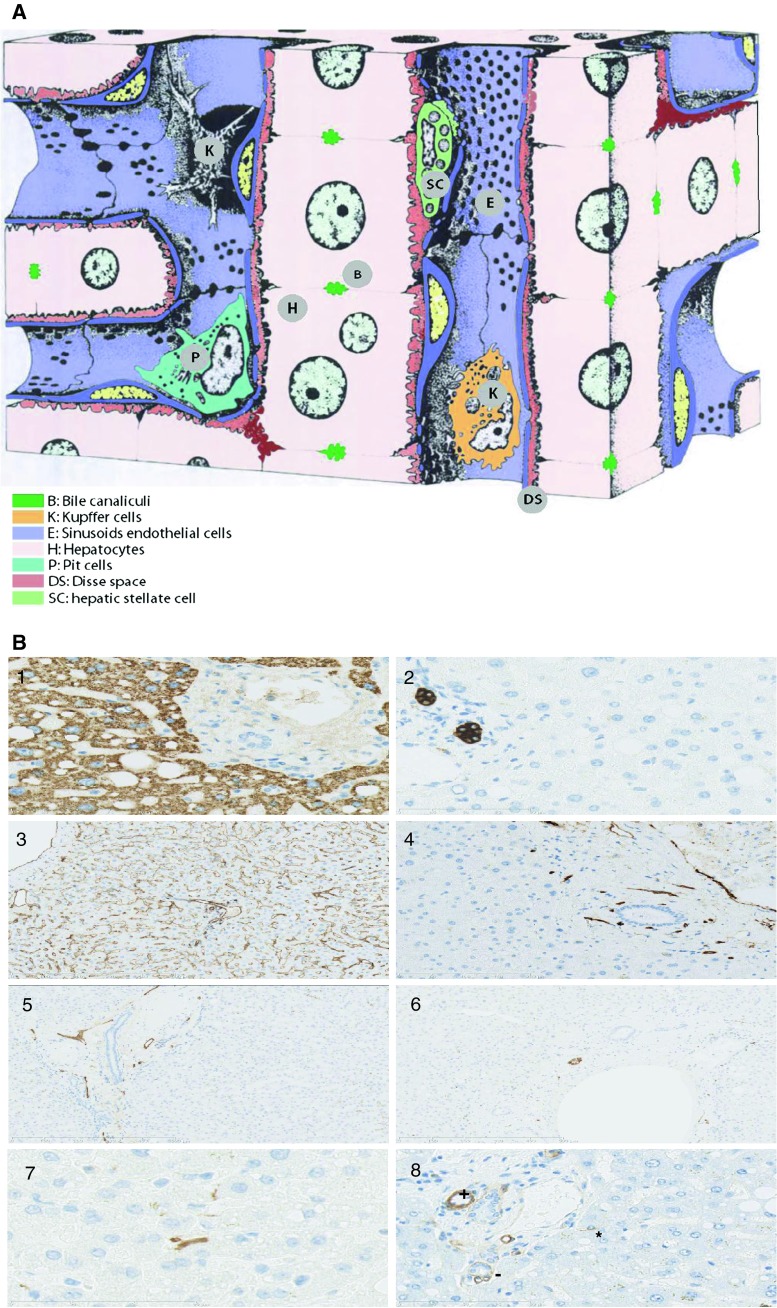
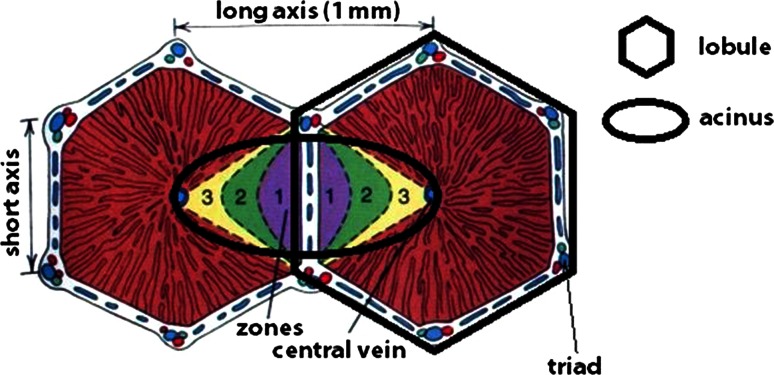
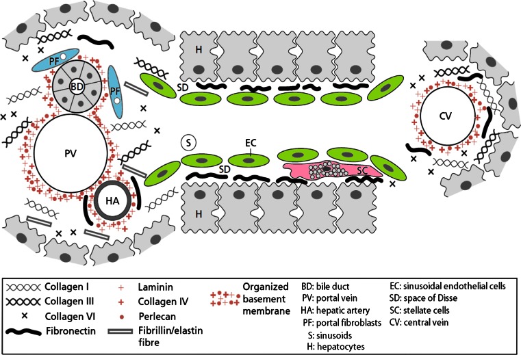
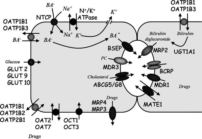
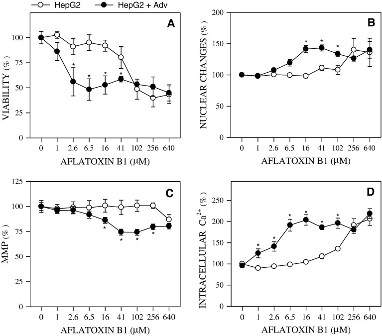
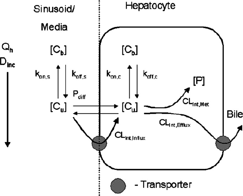
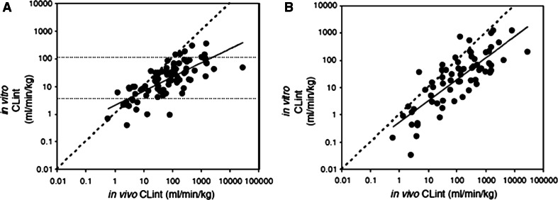
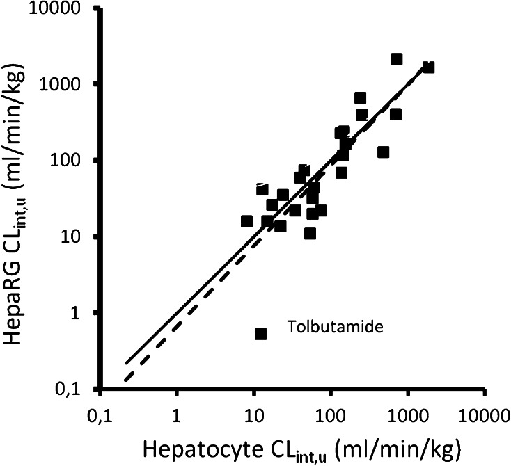
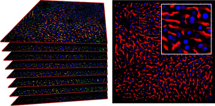
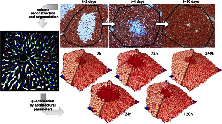
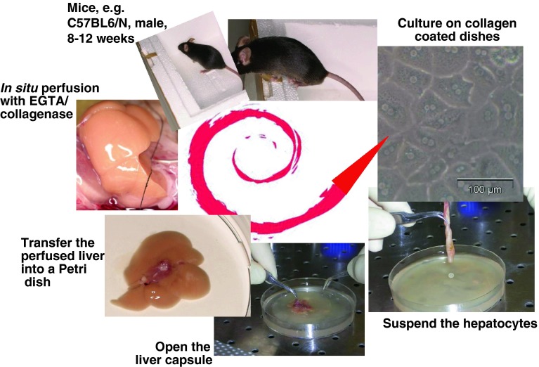
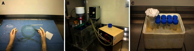
Verification
Claims the paper makes
The claims and statistics of this paper haven’t been checked yet.
Run the check — free, no account needed. It reads the open-access text and checks the sources it cites.
Claims other papers make about this one
Each is checked against this paper’s own text: does it say what the citing paper says it does?
The source backs part of the claim, not all of it — or backs it for a different population, condition or comparison.
In the citing paper
“Already in the first few days of cultivation, a reduction of hepatic functions can be observed, e.g. production of plasma proteins, drug metabolizing enzyme activities, glucose metabolism and urea synthesis (Godoy et al., 2013[15]).”
From Optimization of extracellular matrix for primary human hepatocyte cultures using mixed collagen-matrigel matrices (2023) · cites this paper as Godoy et al., 2013[15]
Experimental — accuracy not yet measuredThe source backs part of the claim, not all of it — or backs it for a different population, condition or comparison.
In the citing paper
“Primary human hepatocytes (PHHs) are an important tool for studying human liver disease and drug-induced liver injury (DILI), with other models, such as rat hepatocytes, hepatic cell lines and stem cell-derived hepatocyte-like cells failing to offer metabolically competent and species-relevant alternatives (Godoy et al. 2013).”
From Mechanistic evaluation of primary human hepatocyte culture using global proteomic analysis reveals a selective dedifferentiation profile (2017) · cites this paper as Godoy et al. 2013
Experimental — accuracy not yet measuredThe source backs part of the claim, not all of it — or backs it for a different population, condition or comparison.
In the citing paper
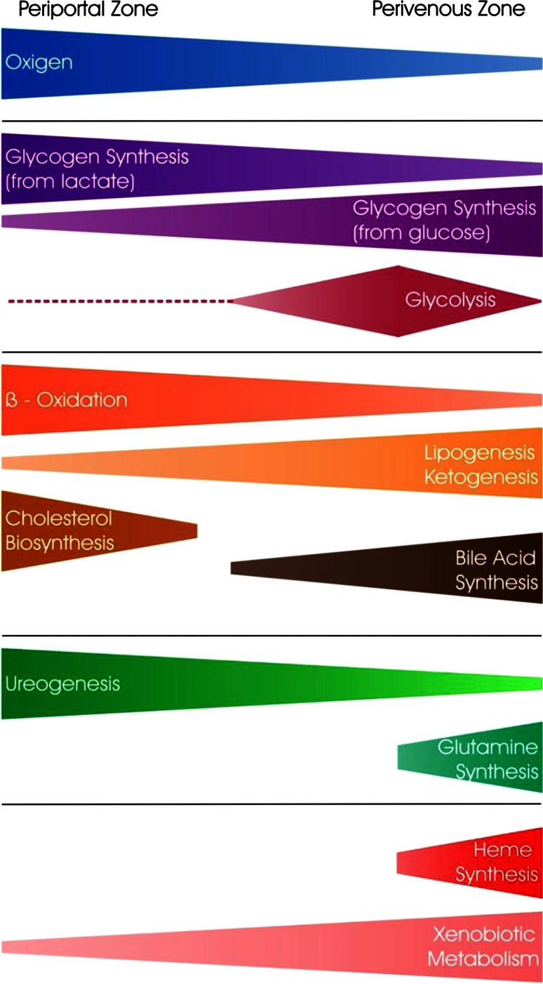
“In fact, zonation appears to be a large influencing factor on variation during culture as our pathway analysis of the most variable proteins revealed significant association with ketogenesis, cholesterol biosynthesis, fatty acid β oxidation and glutamine synthesis (table S4), all of these are related to the portocentral axis gradient of the lobule (Godoy et al. 2013).”
From Mechanistic evaluation of primary human hepatocyte culture using global proteomic analysis reveals a selective dedifferentiation profile (2017) · cites this paper as Godoy et al. 2013
Experimental — accuracy not yet measuredThe cited source says what the claim attributes to it.
In the citing paper
“although cytotoxicity measures are fundamental in the assessment of a drug’s potential for hepatotoxicity in vitro33,34, gene expression and various phenotypic changes can occur at much lower concentrations35,36.”
From Performance assessment and economic analysis of a human Liver-Chip for predictive toxicology (2022) · cites this paper as [34]
The cited source says what the claim attributes to it.
In the citing paper
“The apical membrane is equipped with transport proteins that eliminate bile acids and xenobiotics from the cell, like multidrug resistance-associated protein 2 (MRP2) (Godoy et al., 2013[15]; Ito et al., 2005[20]).”
From Optimization of extracellular matrix for primary human hepatocyte cultures using mixed collagen-matrigel matrices (2023) · cites this paper as Godoy et al., 2013[15]
The cited source says what the claim attributes to it.
In the citing paper
“The hepatocyte's basal membrane faces the sinusoid. It expresses several transmembrane transporters that manage the exchange of substances with the sinusoidal blood stream (Godoy et al., 2013[15]).”
From Optimization of extracellular matrix for primary human hepatocyte cultures using mixed collagen-matrigel matrices (2023) · cites this paper as Godoy et al., 2013[15]
The cited source says what the claim attributes to it.
In the citing paper
“In vivo, the distribution of ECM components varies between the different parts of the liver acinus, the smallest organizational unit of the liver (Godoy et al., 2013[15]; Martinez-Hernandez and Amenta, 1993[26]).”
From Optimization of extracellular matrix for primary human hepatocyte cultures using mixed collagen-matrigel matrices (2023) · cites this paper as Godoy et al., 2013[15]
The cited source says what the claim attributes to it.
In the citing paper
“The application of primary human hepatocytes following isolation from human tissue is well accepted to be compromised by the process of dedifferentiation.”
From Mechanistic evaluation of primary human hepatocyte culture using global proteomic analysis reveals a selective dedifferentiation profile (2017) · cites this paper as Godoy et al. 2013
The check wasn’t confident either way, so it gives no verdict — never disputed.
In the citing paper
“In practice, strategies for optimizing HepG2 can be divided into several approaches. The first approach is based on genetic modification of cells to express key biotransformation enzymes, in particular cytochrome P450 isoforms [109]. The second approach involves epigenetic and cultural maturation of cells, which allows for increased expression of nuclear receptors and, consequently, of xenobiotic”
From The HepG2 Cell Line as a Model for Studying Metabolic Dysfunction-Associated Steatotic Liver Disease (2026) · cites this paper as [163]
Experimental — accuracy not yet measured
Self-check
Percentages, sums and reported differences re-computed from the paper’s own numbers.
The self-check needs the paper’s full text.
Citations
Cites
1 retracted
RETRACTED: Wheat proteins improve cryopreservation of rat hepatocytes
Grondin, M., Hamel, F., Averill‐Bates, D. A., & Sarhan, F. (2009) · Biotechnology and Bioengineering · doi:10.1002/bit.22270 · Cited by 18
RetractedBartel (2004) · doi:10.1016/s0092-8674(04)00045-5 · Cited by 34,714
Computer Simulation of Liquids
Allen, M. P., & Tildesley, D. J. (2017) · doi:10.1093/oso/9780198803195.001.0001 · Cited by 20,945
Induction of Pluripotent Stem Cells from Adult Human Fibroblasts by Defined Factors
Takahashi, K., Tanabe, K., Ohnuki, M., Narita, M., Ichisaka, T., Tomoda, K., & Yamanaka, S. (2007) · Cell · doi:10.1016/j.cell.2007.11.019 · Cited by 20,221
Computer Simulation of Liquids
P, S., & J, K. (1988) · Journal of Molecular Liquids · doi:10.1016/0167-7322(88)80022-9 · Cited by 16,088
Apoptosis: A Basic Biological Phenomenon with Wideranging Implications in Tissue Kinetics
Kerr, J. F. R., Wyllie, A. H., & Currie, A. R. (1972) · British Journal of Cancer · doi:10.1038/bjc.1972.33 · Cited by 15,673
Adverse Renal Effects of Immune Checkpoint Inhibitors: A Narrative Review
Wanchoo, R., Karam, S., Uppal, N. N., Barta, V. S., Deray, G., Devoe, C., Launay‐Vacher, V., Jhaveri, K. D., & on behalf of Cancer and Kidney International Network Workgroup on Immune Checkpoint Inhibitors (2017) · American Journal of Nephrology · doi:10.1159/000455014 · Cited by 15,320
Lewis, B. P., Burge, C. B., & Bartel, D. P. (2005) · Cell · doi:10.1016/j.cell.2004.12.035 · Cited by 11,855
Induced Pluripotent Stem Cell Lines Derived from Human Somatic Cells
Yu, J., Vodyanik, M. A., Smuga-Otto, K., Antosiewicz-Bourget, J., Frane, J. L., Tian, S., Nie, J., Jonsdottir, G. A., Ruotti, V., Stewart, R., et al. (2007) · Science · doi:10.1126/science.1151526 · Cited by 10,310
Two-photon laser scanning fluorescence microscopy
Denk, W., Strickler, J. H., & Webb, W. W. (1990) · Science · doi:10.1126/science.2321027 · Cited by 9,488
Research works printed but not linked: 9
- Bode JG (2011) Inflammation and liver regeneration. In: Häussinger D (ed) Liver regneration. DeGruyter Verlag, Berlin
- Bücher T, Sies H (1976) Mitochondrial and cytosolic redox states in perfused rat liver: methods and problems in metabolic compartmentation. In: Tager jm WJ, Söling D (eds) Use of isolated liver cells and kidney tubules in metabolic studies. North Holland Publishing Company/American Elsevier, New York/Amsterdam
- EPA (2012) EPA’s reanalysis of key issues related to dioxin toxicity and response to NAS Comments. Environmental Protection Agency (EPA), Washington, DC
- Hallifax D, Foster JA, Houston JB (2010) Prediction of human metabolic clearance from in vitro systems: retrospective analysis and prospective view. Pharm Res 27:2150–2161 · DOI
- ITC (International Transporter Consortium) (2010) Membrane transporters in drug development (Report from the FDA Critical Path Workshop). Nat Rev Drug Discov 9:215–236 · DOI · Paper page
- Lamers W, Moorman A, Charles R (1989) The metabolic lobulus, a key to the architecture of the liver. In: Revisiones Sobre Biologia Celular, Springer, New York
- Sahin S (2003) Perfused liver preparation and its applications. FABAD J Pharm Sci 28:39–49
- Waddington CH (1942) The epigenotype. Endeavour 1:18–20
- Warburg O (1923) Versuche an überlebendem Karzinomgewebe. Biochem Z 142:317
Cited by
Sorted by citations
Current advances and future perspectives in extrusion-based bioprinting
Ozbolat, I. T., & Hospodiuk, M. (2015) · Biomaterials · doi:10.1016/j.biomaterials.2015.10.076 · Cited by 1,642
EASL Clinical Practice Guidelines: Drug-induced liver injury
Andrade, R. J., Aithal, G. P., Björnsson, E. S., Kaplowitz, N., Kullak-Ublick, G. A., Larrey, D., & Karlsen, T. H. (2019) · Journal of Hepatology · doi:10.1016/j.jhep.2019.02.014 · Cited by 1,276
Self-citationBell, C. C., Hendriks, D. F. G., Moro, S. M. L., Ellis, E., Walsh, J., Renblom, A., Fredriksson Puigvert, L., Dankers, A. C. A., Jacobs, F., Snoeys, J., et al. (2016) · Scientific Reports · doi:10.1038/srep25187 · Cited by 739
Self-citationOrgans-on-a-Chip: A Fast Track for Engineered Human Tissues in Drug Development
Ronaldson-Bouchard, K., & Vunjak-Novakovic, G. (2018) · Cell Stem Cell · doi:10.1016/j.stem.2018.02.011 · Cited by 657
Du, K., Ramachandran, A., & Jaeschke, H. (2016) · Redox Biology · doi:10.1016/j.redox.2016.10.001 · Cited by 573
Self-citationDrug-induced liver injury: recent advances in diagnosis and risk assessment
Kullak-Ublick, G. A., Andrade, R. J., Merz, M., End, P., Benesic, A., Gerbes, A. L., & Aithal, G. P. (2017) · Gut · doi:10.1136/gutjnl-2016-313369 · Cited by 438
Self-citationSpatial heterogeneity in the mammalian liver
Ben-Moshe, S., & Itzkovitz, S. (2019) · Nature Reviews Gastroenterology & Hepatology · doi:10.1038/s41575-019-0134-x · Cited by 485
3D Bioprinting: from Benches to Translational Applications
Heinrich, M. A., Liu, W. H., Jimenez, A., Yang, J., Akpek, A., Liu, X., Pi, Q., Mu, X., Hu, N., Schiffelers, R. M., et al. (2019) · Small · doi:10.1002/smll.201805510 · Cited by 455
Decellularized human liver as a natural 3D-scaffold for liver bioengineering and transplantation
Mazza, G., Rombouts, K., Rennie Hall, A., Urbani, L., Vinh Luong, T., Al-Akkad, W., Longato, L., Brown, D., Maghsoudlou, P., Dhillon, A. P., et al. (2015) · Scientific Reports · doi:10.1038/srep13079 · Cited by 415
The Curious Case of the HepG2 Cell Line: 40 Years of Expertise
Arzumanian, V. A., Kiseleva, O. I., & Poverennaya, E. V. (2021) · International Journal of Molecular Sciences · doi:10.3390/ijms222313135 · Cited by 387
Nguyen, D. G., Funk, J., Robbins, J. B., Crogan‐Grundy, C., Presnell, S. C., Singer, T., & Roth, A. B. (2016) · PLoS ONE · doi:10.1371/journal.pone.0158674 · Cited by 389
Paired-cell sequencing enables spatial gene expression mapping of liver endothelial cells
Halpern, K. B., Shenhav, R., Massalha, H., Toth, B., Egozi, A., Massasa, E. E., Medgalia, C., David, E., Giladi, A., Moor, A. E., et al. (2018) · Nature Biotechnology · doi:10.1038/nbt.4231 · Cited by 368
Ramaiahgari, S., den Braver, M. W., Herpers, B., Terpstra, V., Commandeur, J. N. M., van de Water, B., & Price, L. (2014) · Archives of Toxicology · doi:10.1007/s00204-014-1215-9 · Cited by 373
Adverse outcome pathways: opportunities, limitations and open questions
Leist, M., Ghallab, A., Graepel, R., Marchan, R., Hassan, R., Bennekou, S. H., Limonciel, A., Vinken, M., Schildknecht, S., Waldmann, T., et al. (2017) · Archives of Toxicology · doi:10.1007/s00204-017-2045-3 · Cited by 394
Self-citationPerformance assessment and economic analysis of a human Liver-Chip for predictive toxicology
Ewart, L., Apostolou, A., Briggs, S. A., Carman, C. V., Chaff, J. T., Heng, A. R., Jadalannagari, S., Janardhanan, J., Jang, K.-J., Joshipura, S. R., et al. (2022) · Communications Medicine · doi:10.1038/s43856-022-00209-1 · Cited by 337
Utility of spherical human liver microtissues for prediction of clinical drug-induced liver injury
Proctor, W. R., Foster, A. J., Vogt, J., Summers, C., Middleton, B., Pilling, M. A., Shienson, D., Kijanska, M., Ströbel, S., Kelm, J. M., et al. (2017) · Archives of Toxicology · doi:10.1007/s00204-017-2002-1 · Cited by 328
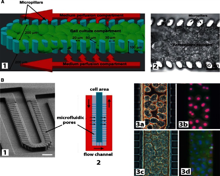
“In most cases, these systems displayed enhanced metabolic activity, hepatocellular phenotype, and stability in culture, previously not attainable with traditional hepatic cell line or hepatocyte models [refer to review (Godoy et al. 2013)].”
BackgroundSelf-citationThe evolving role of investigative toxicology in the pharmaceutical industry
Pognan, F., Beilmann, M., Boonen, H. C. M., Czich, A., Dear, G. J., Hewitt, P. G., Mow, T. J., Oinonen, T., Roth, A. B., Steger‐Hartmann, T., et al. (2023) · Nature Reviews Drug Discovery · doi:10.1038/s41573-022-00633-x · Cited by 280
Self-citationA microfluidically perfused three dimensional human liver model
Rennert, K., Steinborn, S., Gröger, M., Ungerböck, B., Jank, A., Ehgartner, J., Nietzsche, S., Dinger, J., Kiehntopf, M., Funke, H., et al. (2015) · Biomaterials · doi:10.1016/j.biomaterials.2015.08.043 · Cited by 265
Simulating tissue mechanics with agent-based models: concepts, perspectives and some novel results
Liedekerke, P. V., Palm, M. M., Jagiella, N., & Drasdo, D. (2015) · Computational Particle Mechanics · doi:10.1007/s40571-015-0082-3 · Cited by 267
Self-citationWeaver, R. J., Blomme, E. A. G., Chadwick, A. E., Copple, I. M., Gerets, H. H. J., Goldring, C. E., Guillouzo, A., Hewitt, P. G., Ingelman‐Sundberg, M., Jensen, K. G., et al. (2019) · Nature Reviews Drug Discovery · doi:10.1038/s41573-019-0048-x · Cited by 261
Self-citationLauschke, V. M., Hendriks, D. F. G., Bell, C. C., Andersson, T. B., & Ingelman-Sundberg, M. (2016) · Chemical Research in Toxicology · doi:10.1021/acs.chemrestox.6b00150 · Cited by 241
Deng, J., Wei, W., Chen, Z., Lin, B., Zhao, W., Luo, Y., & Zhang, X. (2019) · Micromachines · doi:10.3390/mi10100676 · Cited by 238
Cell sources for in vitro human liver cell culture models
Zeilinger, K., Freyer, N., Damm, G., Seehofer, D., & Knöspel, F. (2016) · Experimental Biology and Medicine · doi:10.1177/1535370216657448 · Cited by 230
Self-citationA human liver microphysiology platform for investigating physiology, drug safety, and disease models
Vernetti, L. A., Senutovitch, N., Boltz, R. C., DeBiasio, R., Shun, T. Y., Gough, A., & Taylor, D. L. (2015) · Experimental Biology and Medicine · doi:10.1177/1535370215592121 · Cited by 233
Optimization of collagen type I-hyaluronan hybrid bioink for 3D bioprinted liver microenvironments
Mazzocchi, A. R., Devarasetty, M., Huntwork, R. C., Söker, S., & Skardal, A. (2018) · Biofabrication · doi:10.1088/1758-5090/aae543 · Cited by 235
Acetaminophen-induced Liver Injury: from Animal Models to Humans
Jaeschke, H., Xie, Y., & McGill, M. R. (2014) · Journal of Clinical and Translational Hepatology · doi:10.14218/jcth.2014.00014 · Cited by 227
Self-citationLife in 3D is never flat: 3D models to optimise drug delivery
Fitzgerald, K. A., Malhotra, M., Curtin, C. M., O’Brien, F. J., & O’Driscoll, C. M. (2015) · Journal of Controlled Release · doi:10.1016/j.jconrel.2015.07.020 · Cited by 240
State-of-the-art of 3D cultures (organs-on-a-chip) in safety testing and pathophysiology
Alépée, N. (2014) · ALTEX · doi:10.14573/altex1406111 · Cited by 228
Long‐term maintenance of a microfluidic 3D human liver sinusoid
Prodanov, L., Jindal, R., Bale, S. S., Hegde, M., McCarty, W. J., Golberg, I., Bhushan, A., Yarmush, M. L., & Usta, O. B. (2016) · Biotechnology and Bioengineering · doi:10.1002/bit.25700 · Cited by 214
Beckwitt, C. H., Clark, A. M., Wheeler, S., Taylor, D. L., Stolz, D. B., Griffith, L. G., & Wells, A. H. (2017) · Experimental Cell Research · doi:10.1016/j.yexcr.2017.12.023 · Cited by 216
Microfluidic organ-on-a-chip models of human liver tissue
Moradi, E., Jalili‐Firoozinezhad, S., & Solati‐Hashjin, M. (2020) · Acta Biomaterialia · doi:10.1016/j.actbio.2020.08.041 · Cited by 213
Spatial sorting enables comprehensive characterization of liver zonation
Ben-Moshe, S., Shapira, Y., Moor, A. E., Halpern, K. B., & Itzkovitz, S. (2019) · doi:10.1101/529784 · Cited by 205
Spatial sorting enables comprehensive characterization of liver zonation
Ben-Moshe, S., Shapira, Y., Moor, A. E., Manco, R., Veg, T., Bahar Halpern, K., & Itzkovitz, S. (2019) · Nature Metabolism · doi:10.1038/s42255-019-0109-9 · Cited by 205
Experimental models of hepatotoxicity related to acute liver failure
Maes, M., Vinken, M., & Jaeschke, H. (2015) · Toxicology and Applied Pharmacology · doi:10.1016/j.taap.2015.11.016 · Cited by 209
Self-citationDrug‐induced liver injury: Advances in mechanistic understanding that will inform risk management
Mosedale, M., & Watkins, P. B. (2016) · Clinical Pharmacology & Therapeutics · doi:10.1002/cpt.564 · Cited by 211
Competency of different cell models to predict human hepatotoxic drugs
Gómez-Lechón, M. J., Tolosa, L., Conde, I., & Donato, M. T. (2014) · Expert Opinion on Drug Metabolism & Toxicology · doi:10.1517/17425255.2014.967680 · Cited by 199
Self-citationA 3D microfluidic liver model for high throughput compound toxicity screening in the OrganoPlate®
Bircsak, K. M., DeBiasio, R., Miedel, M. T., Alsebahi, A., Reddinger, R. M., Saleh, A. D., Shun, T., Vernetti, L. A., & Gough, A. (2021) · Toxicology · doi:10.1016/j.tox.2020.152667 · Cited by 198
Ware, B. R., Berger, D. R., & Khetani, S. R. (2015) · Toxicological Sciences · doi:10.1093/toxsci/kfv048 · Cited by 198
Toxicology Strategies for Drug Discovery: Present and Future
Blomme, E. A. G., & Will, Y. (2015) · Chemical Research in Toxicology · doi:10.1021/acs.chemrestox.5b00407 · Cited by 201
A lung/liver-on-a-chip platform for acute and chronic toxicity studies
Bovard, D., Sandoz, A., Luettich, K., Frentzel, S., Iskandar, A., Marescotti, D., Trivedi, K., Guedj, E., Dutertre, Q., Peitsch, M. C., et al. (2018) · Lab on a Chip · doi:10.1039/c8lc01029c · Cited by 195
Berger, D. R., Ware, B. R., Davidson, M. D., Allsup, S. R., & Khetani, S. R. (2014) · Hepatology · doi:10.1002/hep.27621 · Cited by 196
Malinen, M. M., Kanninen, L. K., Corlu, A., Isoniemi, H., Lou, Y., Yliperttula, M. L., & Urtti, A. O. (2014) · Biomaterials · doi:10.1016/j.biomaterials.2014.03.020 · Cited by 192
Organic Anion-Transporting Polypeptides
Stieger, B., & Hagenbuch, B. (2014) · Current topics in membranes · doi:10.1016/b978-0-12-800223-0.00005-0 · Cited by 192
Self-citationWiśniewski, J. R., Vildhede, A., Norén, A., & Artursson, P. (2016) · Journal of Proteomics · doi:10.1016/j.jprot.2016.01.016 · Cited by 185
Engineering a perfusable 3D human liver platform from iPS cells
Schepers, A. G., Li, C. Y., Chhabra, A., Tschudy-Seney, B., & Bhatia, S. N. (2016) · Lab on a Chip · doi:10.1039/c6lc00598e · Cited by 179
Strategies, models and biomarkers in experimental non-alcoholic fatty liver disease research
Willebrords, J., Pereira, I. V. A., Maes, M., Yanguas, S. C., Colle, I. O., Bossche, B. V. D., da Silva, T. C., de Oliveira, C. P. M. S., Andraus, W., Alves, V. A. F., et al. (2015) · Progress in Lipid Research · doi:10.1016/j.plipres.2015.05.002 · Cited by 177
Self-citationGodoy, P., Schmidt‐Heck, W., Natarajan, K., Lucendo‐Villarin, B., Szkolnicka, D., Asplund, A., Björquist, P., Widera, A., Stöber, R. M., Campos, G., et al. (2015) · Journal of Hepatology · doi:10.1016/j.jhep.2015.05.013 · Cited by 170
Self-citationBioengineered Liver Models for Drug Testing and Cell Differentiation Studies
Underhill, G. H., & Khetani, S. R. (2017) · Cellular and Molecular Gastroenterology and Hepatology · doi:10.1016/j.jcmgh.2017.11.012 · Cited by 169
Naturally-Derived Biomaterials for Tissue Engineering Applications
Brovold, M., Almeida, J. I., Plá-Palacín, I., Sainz‐Arnal, P., Sánchez‐Romero, N., Rivas, J. J., Almeida, H., Dachary, P. R., Serrano, T., Söker, S., et al. (2018) · Advances in experimental medicine and biology · doi:10.1007/978-981-13-0947-2_23 · Cited by 168
Yang, S., & Kar, S. (2023) · Artificial Intelligence Chemistry · doi:10.1016/j.aichem.2023.100011 · Cited by 162
Large-scale perfused tissues via synthetic 3D soft microfluidics
Grebenyuk, S. E., Fattah, A. R. A., Kumar, M., Toprakhisar, B., Rustandi, G., Vananroye, A., Salmon, I., Verfaillie, C. M., Grillo, M. P., & Ranga, A. (2023) · Nature Communications · doi:10.1038/s41467-022-35619-1 · Cited by 160
Jaeschke, H., Adelusi, O. B., Akakpo, J. Y., Nguyen, N. T., Sánchez-Guerrero, G., Umbaugh, D. S., Ding, W., & Ramachandran, A. (2021) · Acta Pharmaceutica Sinica B · doi:10.1016/j.apsb.2021.09.023 · Cited by 155
Self-citationMarch, S., Ramanan, V., Trehan, K., Ng, S., Galstian, A., Gural, N., Scull, M. A., Shlomai, A., Mota, M. M., Fleming, H. E., et al. (2015) · Nature Protocols · doi:10.1038/nprot.2015.128 · Cited by 152
Heslop, J. A., Rowe, C., Walsh, J., Sison-Young, R., Jenkins, R., Kamalian, L., Kia, R., Hay, D., Jones, R. P., Malik, H. Z., et al. (2017) · Archives of Toxicology · doi:10.1007/s00204-016-1694-y · Cited by 141
Self-citationReactive Metabolites: Current and Emerging Risk and Hazard Assessments
Thompson, R. A., Isin, E. M., Ogese, M. O., Mettetal, J. T., & Williams, D. P. (2016) · Chemical Research in Toxicology · doi:10.1021/acs.chemrestox.5b00410 · Cited by 144
Lee-Montiel, F. T., George, S. M., Gough, A., Sharma, A. D., Wu, J., DeBiasio, R., Vernetti, L. A., & Taylor, D. L. (2017) · Experimental Biology and Medicine · doi:10.1177/1535370217703978 · Cited by 141
Toxicogenomics directory of chemically exposed human hepatocytes
Grinberg, M., Stöber, R. M., Edlund, K., Rempel, E., Godoy, P., Reif, R., Widera, A., Madjar, K., Schmidt‐Heck, W., Marchan, R., et al. (2014) · Archives of Toxicology · doi:10.1007/s00204-014-1400-x · Cited by 139
Self-citationLi, X., George, S. M., Vernetti, L. A., Gough, A., & Taylor, D. L. (2018) · Lab on a Chip · doi:10.1039/c8lc00418h · Cited by 141
A critical perspective on 3D liver models for drug metabolism and toxicology studies
Serras, A. S., Rodrigues, J. S., Cipriano, M., Rodrigues, A. V., Oliveira, N. G., & Miranda, J. P. (2021) · Frontiers in Cell and Developmental Biology · doi:10.3389/fcell.2021.626805 · Cited by 138
Hammad, S., Hoehme, S., Friebel, A., von Recklinghausen, I., Othman, A., Begher‐Tibbe, B., Reif, R., Godoy, P., Johann, T., Damle‐Vartak, A., et al. (2014) · Archives of Toxicology · doi:10.1007/s00204-014-1243-5 · Cited by 135
Self-citationBioreactor technologies to support liver function in vitro
Ebrahimkhani, M. R., Neiman, J. A. S., Raredon, M. S. B., Hughes, D. J., & Griffith, L. G. (2014) · Advanced Drug Delivery Reviews · doi:10.1016/j.addr.2014.02.011 · Cited by 141
Self‐assembled liver organoids recapitulate hepatobiliary organogenesis in vitro
Vyas, D., Baptista, P. M., Brovold, M., Moran, E. C., Gaston, B., Booth, C., Samuel, M., Atala, A., & Söker, S. (2017) · Hepatology · doi:10.1002/hep.29483 · Cited by 134
Comparison of minipig, dog, monkey and human drug metabolism and disposition
Dalgaard, L. (2015) · Journal of Pharmacological and Toxicological Methods · doi:10.1016/j.vascn.2014.12.005 · Cited by 132
Next generation organoids for biomedical research and applications
Lou, Y., & Leung, A. W. (2017) · Biotechnology Advances · doi:10.1016/j.biotechadv.2017.10.005 · Cited by 138
Characterization of a functional C3A liver spheroid model
Gaskell, H., Sharma, P., Colley, H. E., Murdoch, C., Williams, D. P., & Webb, S. D. (2016) · Toxicology Research · doi:10.1039/c6tx00101g · Cited by 131
Application of Microphysiological Systems to Enhance Safety Assessment in Drug Discovery
Ewart, L. C., Dehne, E., Fabre, K. M., Gibbs, S., Hickman, J. J., Hornberg, E., Ingelman‐Sundberg, M., Jang, K., Jones, D. R., Lauschke, V. M., et al. (2017) · The Annual Review of Pharmacology and Toxicology · doi:10.1146/annurev-pharmtox-010617-052722 · Cited by 136
Experimental in vitro and in vivo models for the study of human hepatitis B virus infection
Allweiss, L., & Dandri, M. (2016) · Journal of Hepatology · doi:10.1016/j.jhep.2016.02.012 · Cited by 134
Subramanya, S. B., Venkataraman, B., Meeran, M. F. N., Goyal, S. N., Patil, C. R., & Ojha, S. K. (2018) · International Journal of Molecular Sciences · doi:10.3390/ijms19123776 · Cited by 128
Mihajlovic, M., & Vinken, M. (2022) · International Journal of Molecular Sciences · doi:10.3390/ijms23063315 · Cited by 132
Self-citationIn Vitro Models for Non-Alcoholic Fatty Liver Disease: Emerging Platforms and Their Applications
Ramos, M. J., Bandiera, L., Menolascina, F., & Fallowfield, J. A. (2022) · iScience · doi:10.1016/j.isci.2021.103549 · Cited by 127
King, S. M., Higgins, J. W., Nino, C., Smith, T. R., Paffenroth, E. H., Fairbairn, C. E., Docuyanan, A., Shah, V., Chen, A. E., Presnell, S. C., et al. (2017) · Frontiers in Physiology · doi:10.3389/fphys.2017.00123 · Cited by 127
Schimek, K. M. S., Frentzel, S., Luettich, K., Bovard, D., Rütschle, I., Boden, L., Rambo, F., Erfurth, H., Dehne, E., Winter, A., et al. (2020) · Scientific Reports · doi:10.1038/s41598-020-64219-6 · Cited by 130
3D spherical microtissues and microfluidic technology for multi-tissue experiments and analysis
Kim, J.-Y., Fluri, D. A., Marchan, R., Boonen, K., Mohanty, S., Singh, P., Hammad, S., Landuyt, B., Hengstler, J. G., Kelm, J. M., et al. (2015) · Journal of Biotechnology · doi:10.1016/j.jbiotec.2015.01.003 · Cited by 126
Self-citationKenna, J. G., Taskar, K. S., Battista, C., Bourdet, D. L., Brouwer, K. L. R., Brouwer, K. R., Dai, D., Funk, C., Hafey, M. J., Lai, Y., et al. (2018) · Clinical Pharmacology & Therapeutics · doi:10.1002/cpt.1222 · Cited by 126
Current progress in hepatic tissue regeneration by tissue engineering
Hosseini, V., Maroufi, N. F., Saghati, S., Asadi, N., Darabi, M., Ahmad, S. N. S., Hosseinkhani, H., & Rahbarghazi, R. (2019) · Journal of Translational Medicine · doi:10.1186/s12967-019-02137-6 · Cited by 122
Zebrafish: A Marvel of High-Throughput Biology for 21st Century Toxicology
Bugel, S. M., Tanguay, R. L., & Planchart, A. (2014) · Current Environmental Health Reports · doi:10.1007/s40572-014-0029-5 · Cited by 128
High-resolution mass spectrometry in toxicology: current status and future perspectives
Maurer, H. H., & Meyer, M. R. (2016) · Archives of Toxicology · doi:10.1007/s00204-016-1764-1 · Cited by 120
Drug Discovery Approaches Utilizing Three-Dimensional Cell Culture
Ryan, S.-L., Baird, A., Vaz, G. M. F., Urquhart, A. J., Senge, M. O., Richard, D. J., O'Byrne, K. J., & Davies, A. M. (2016) · Assay and Drug Development Technologies · doi:10.1089/adt.2015.670 · Cited by 126
McGill, M. R., & Jaeschke, H. (2014) · Expert Opinion on Drug Metabolism & Toxicology · doi:10.1517/17425255.2014.920823 · Cited by 119
Self-citationNatural killer cells and liver fibrosis
Fasbender, F., Widera, A., Hengstler, J. G., & Watzl, C. (2016) · Frontiers in Immunology · doi:10.3389/fimmu.2016.00019 · Cited by 118
Self-citationGheytanchi, E., Naseri, M., Karimi‐Busheri, F., Atyabi, F., Mirsharif, E. S., Bozorgmehr, M., Ghods, R., & Madjd, Z. (2021) · Cancer Cell International · doi:10.1186/s12935-021-01898-9 · Cited by 118
Human in vitro models of nonalcoholic fatty liver disease
Müller, F. A., & Sturla, S. J. (2019) · Current Opinion in Toxicology · doi:10.1016/j.cotox.2019.03.001 · Cited by 118
Thiel, C., Schneckener, S., Krauß, M., Ghallab, A., Hofmann, U., Kanacher, T., Zellmer, S., Gebhardt, R., Hengstler, J. G., & Kuepfer, L. (2014) · Journal of Pharmaceutical Sciences · doi:10.1002/jps.24214 · Cited by 119
Self-citationComparative analysis of 3D culture methods on human HepG2 cells
Luckert, C., Schulz, C., Lehmann, N., Thomas, M., Hofmann, U., Hammad, S., Hengstler, J. G., Braeuning, A., Lampen, A., & Hessel‐Pras, S. (2016) · Archives of Toxicology · doi:10.1007/s00204-016-1677-z · Cited by 117
Self-citationMicroengineered Liver Tissues for Drug Testing
Khetani, S. R., Berger, D. R., Ballinger, K. R., Davidson, M. D., Lin, C., & Ware, B. R. (2015) · SLAS Technology · doi:10.1177/2211068214566939 · Cited by 116
Bale, S. S., Geerts, S., Jindal, R., & Yarmush, M. L. (2016) · Scientific Reports · doi:10.1038/srep25329 · Cited by 119
Brown, J. H., Das, P., DiVito, M. D., Ivancic, D., Tan, L. P., & Wertheim, J. A. (2018) · Acta Biomaterialia · doi:10.1016/j.actbio.2018.02.009 · Cited by 114
Generation of Multilayered 3D Structures of HepG2 Cells Using a Bio-printing Technique
Jeon, H., Kang, K., Park, S. A., Kim, W. D., Paik, S. S., Lee, S., Jeong, J., & Choi, D. (2016) · Gut and Liver · doi:10.5009/gnl16010 · Cited by 113
Tissue Engineering in Liver Regenerative Medicine: Insights into Novel Translational Technologies
Heydari, Z., Najimi, M., Mirzaei, H., Shpichka, A. I., Ruoß, M., Farzaneh, Z., Montazeri, L., Piryaei, A., Timashev, P. S., Gramignoli, R., et al. (2020) · Cells · doi:10.3390/cells9020304 · Cited by 113
Self-citationMessina, A., Luce, E., Hussein, M., & Dubart‐Kupperschmitt, A. (2020) · Cells · doi:10.3390/cells9020420 · Cited by 111
Model-guided identification of a therapeutic strategy to reduce hyperammonemia in liver diseases
Ghallab, A., Cellière, G., Henkel, S., Driesch, D., Hoehme, S., Hofmann, U., Zellmer, S., Godoy, P., Sachinidis, A., Blaszkewicz, M., et al. (2015) · Journal of Hepatology · doi:10.1016/j.jhep.2015.11.018 · Cited by 110
Self-citationAlbrecht, W., Kappenberg, F., Brecklinghaus, T., Stöber, R. M., Marchan, R., Zhang, M., Ebbert, K. E., Kirschner, H., Grinberg, M., Leist, M., et al. (2019) · Archives of Toxicology · doi:10.1007/s00204-019-02492-9 · Cited by 110
Self-citationLiver organ-on-chip models for toxicity studies and risk assessment
Messelmani, T., Morisseau, L., Sakai, Y., Legallais, C., Goff, A. L., Leclerc, É., & Jellali, R. (2022) · Lab on a Chip · doi:10.1039/d2lc00307d · Cited by 113
Modeling human nonalcoholic fatty liver disease (NAFLD) with an organoids-on-a-chip system
Wang, Y., Wang, H., Deng, P., Tao, T., Liu, H., Wu, S., Chen, W., & Qin, J. (2020) · ACS Biomaterials Science & Engineering · doi:10.1021/acsbiomaterials.0c00682 · Cited by 109
Graffmann, N., Ring, S., Kawala, M.-A., Wruck, W., Ncube, A., Trompeter, H., & Adjaye, J. (2016) · Stem Cells and Development · doi:10.1089/scd.2015.0383 · Cited by 109
Organ-on-a-chip platforms for accelerating the evaluation of nanomedicine
Chen, X., Zhang, Y. S., Zhang, X., & Liu, C. (2020) · Bioactive Materials · doi:10.1016/j.bioactmat.2020.09.022 · Cited by 112
A Bioreactor Technology for Modeling Fibrosis in Human and Rodent Precision‐Cut Liver Slices
Paish, H. L., Reed, L. H., Brown, H., Bryan, M. C., Govaere, O., Leslie, J., Barksby, B. S., Garcia Macia, M., Watson, A., Xu, X., et al. (2019) · Hepatology · doi:10.1002/hep.30651 · Cited by 106
Fowler, S., Chen, W. L. K., Duignan, D. B., Gupta, A., Hariparsad, N., Kenny, J. R., Lai, W. G., Liras, J., Phillips, J. A., & Gan, J. (2020) · Lab on a Chip · doi:10.1039/c9lc00857h · Cited by 109
Rodrigues, A. D., Lai, Y., Cvijic, M. E., Elkin, L. L., Zvyaga, T. A., & Soars, M. G. (2013) · Drug Metabolism and Disposition · doi:10.1124/dmd.113.054205 · Cited by 107
Kegel, V., Deharde, D., Pfeiffer, E., Zeilinger, K., Seehofer, D., & Damm, G. (2016) · Journal of Visualized Experiments · doi:10.3791/53069 · Cited by 104
Self-citation
Metadata
- Authors
- Godoy, P., Hewitt, N. J., Albrecht, U., Andersen, M. E., Ansari, N., Bhattacharya, S., Bode, J. G., Bolleyn, J., Borner, C., Böttger, J., Braeuning, A., Budinsky, R. A., Burkhardt, B., Cameron, N. R., Camussi, G., Cho, C.-S., Choi, Y.-J., Craig Rowlands, J., Dahmen, U., Damm, G., Dirsch, O., Donato, M. T., Dong, J., Dooley, S., Drasdo, D., Eakins, R., Ferreira, K. S., Fonsato, V., Fraczek, J., Gebhardt, R., Gibson, A., Glanemann, M., Goldring, C. E. P., Gómez-Lechón, M. J., Groothuis, G. M. M., Gustavsson, L., Guyot, C., Hallifax, D., Hammad, S., Hayward, A., Häussinger, D., Hellerbrand, C., Hewitt, P., Hoehme, S., Holzhütter, H.-G., Houston, J. B., Hrach, J., Ito, K., Jaeschke, H., Keitel, V., Kelm, J. M., Kevin Park, B., Kordes, C., Kullak-Ublick, G. A., LeCluyse, E. L., Lu, P., Luebke-Wheeler, J., Lutz, A., Maltman, D. J., Matz-Soja, M., McMullen, P., Merfort, I., Messner, S., Meyer, C., Mwinyi, J., Naisbitt, D. J., Nussler, A. K., Olinga, P., Pampaloni, F., Pi, J., Pluta, L., Przyborski, S. A., Ramachandran, A., Rogiers, V., Rowe, C., Schelcher, C., Schmich, K., Schwarz, M., Singh, B., Stelzer, E. H. K., Stieger, B., Stöber, R., Sugiyama, Y., Tetta, C., Thasler, W. E., Vanhaecke, T., Vinken, M., Weiss, T. S., Widera, A., Woods, C. G., Xu, J. J., Yarborough, K. M., & Hengstler, J. G.
- Affiliations
- University of Zurich (CH)
- Leibniz Research Centre for Working Environment and Human Factors (DE)
- The Hamner Institutes for Health Sciences (US)
- Goethe University Frankfurt (DE)
- Vrije Universiteit Brussel (BE)
- University of Freiburg (DE)
- Leipzig University (DE)
- Dow Chemical (United States) (US)
- Durham University (GB)
- Seoul National University (KR)
- Instituto de Investigación Sanitaria La Fe (ES)
- Heidelberg University (DE)
- University of Liverpool (GB)
- University of Groningen (NL)
- Lund University (SE)
- University of Manchester (GB)
- South Valley University (EG)
- Heinrich Heine University Düsseldorf (DE)
- University Hospital Regensburg (DE)
- Merck KGaA, Darmstadt (Germany) (DE)
- Charité – Universitätsmedizin Berlin (DE)
- Musashino University (JP)
- University Hospital Zurich (CH)
- Merck & Co., Inc., Rahway, NJ, USA (United States) (US)
- Corresponding author
- Hewitt, Nicola J.
- ORCID
- Hewitt, Nicola J.
- Andersen, Melvin E.
- Bhattacharya, Sudin
- Bode, Johannes Georg
- Borner, Christoph
- Böttger, Jan
- Braeuning, Albert
- Budinsky, Robert A.
- Cameron, Neil R.
- Camussi, Giovanni
- Cho, Chong-Su
- Choi, Yun-Jaie
- Dahmen, Uta
- Damm, Georg
- Dirsch, Olaf
- Donato, María Teresa
- Dong, Jian
- Dooley, Steven
- Drasdo, Dirk
- Gibson, Andrew
- Glanemann, Matthias
- Groothuis, Geny M. M.
- Hallifax, David
- Hammad, Seddik
- Hayward, Adam
- Häussinger, Dieter
- Hellerbrand, Claus
- Hewitt, Philip
- Hoehme, Stefan
- Holzhütter, Hermann-Georg
- Houston, J. Brian
- Ito, Kiyomi
- Jaeschke, Hartmut
- Keitel, Verena
- Kordes, Claus
- Kullak-Ublick, Gerd A.
- LeCluyse, Edward L.
- Maltman, Daniel J.
- McMullen, Patrick
- Merfort, Irmgard
- Meyer, Christoph
- Mwinyi, Jessica
- Naisbitt, Dean J.
- Nussler, Andreas K.
- Olinga, Peter
- Pampaloni, Francesco
- Pi, Jingbo
- Pluta, Linda
- Przyborski, Stefan A.
- Ramachandran, Anup
- Rogiers, Vera
- Schwarz, Michael
- Singh, Bijay
- Stelzer, Ernst H. K.
- Stieger, Bruno
- Sugiyama, Yuichi
- Vanhaecke, Tamara
- Vinken, Mathieu
- Weiss, Thomas S.
- Woods, Courtney G.
- Hengstler, Jan G.
- Venue
- Archives of Toxicology
- Published
- 2013-08
- Volume
- 87
- Issue
- 8
- Pages
- 1315-1530
- ISSN
- 0340-5761 (ISSN-L)
- 1432-0738
- PMC
- PMC3753504
- OpenAlex
- W2134520937
- Cited by
- 1,280
- References
- 1,862
- Citations by year
- 2026: 54 · 2025: 63 · 2024: 59 · 2023: 80 · 2022: 98 · 2021: 96 · 2020: 130 · 2019: 132 · 2018: 129 · 2017: 102 · 2016: 118 · 2015: 110 · 2014: 91 · 2013: 12
- FWCI
- 18.06
- Type
- journal article
- Language
- en
- Open access
- hybrid
- doi.org
- Licence
- Locations
- Archives of Toxicology (publishedVersion, other-oa)
- Europe PMC (PubMed Central) (publishedVersion)
- PubMed Central (publishedVersion, cc-by-nc)
- PubMed (publishedVersion)
- Research Explorer (The University of Manchester) (publishedVersion)
- University of Regensburg Publication Server (University of Regensburg) (acceptedVersion)
- Zurich Open Repository and Archive (University of Zurich) (acceptedVersion, other-oa)
- Durham Research Online (Durham University) (acceptedVersion, other-oa)
- Durham Research Online (Durham University) (submittedVersion, cc-by)
- HAL (submittedVersion)
- Institutional Research Information System University of Turin (University of Turin) (submittedVersion)
- Lund University Publications (Lund University) (submittedVersion, other-oa)
- University of Groningen research database (University of Groningen / Centre for Information Technology) (submittedVersion, cc-by)
- Repository of Digital Objects for Teaching Research and Culture (University of Valencia) (submittedVersion, cc-by-nc)
- VUBIR (Vrije Universiteit Brussel) (submittedVersion)
- Zurich Open Repository and Archive (University of Zurich) (submittedVersion, other-oa)
- Zurich Open Repository and Archive (University of Zurich)
- University of Regensburg Publication Server (University of Regensburg)
- Funders
- Leibniz-Institut für Arbeitsforschung an der TU Dortmund
- Deutsche Forschungsgemeinschaft (GRK1104, (EXC 294))
- Agence Nationale de la Recherche
- Bundesministerium für Bildung und Forschung
- Belgian Federal Science Policy Office
- Fonds Wetenschappelijk Onderzoek
- National Research Foundation of Korea
- Innoviris
- Universität Regensburg
- Vlaamse regering
- National Institutes of Health (AA12916)
- Biotechnology and Biological Sciences Research Council (1233823)
- Leibniz-Gemeinschaft
- Japan Society for the Promotion of Science (23590204)
- National Institute on Alcohol Abuse and Alcoholism (R56AA012916, R01AA012916)
- National Institute of Diabetes and Digestive and Kidney Diseases (R01DK070195)
- Topics
- Life Sciences › Pharmacology, Toxicology and Pharmaceutics › Pharmacology › Drug-Induced Hepatotoxicity and Protection
- Health Sciences › Medicine › Hepatology › Liver physiology and pathology
- Health Sciences › Veterinary › Small Animals › Animal testing and alternatives
- Keywords
- hepatotoxicity, primary hepatocytes, non-parenchymal liver cells, hepatocyte co-culture, precision-cut liver slices, isolated perfused liver, stem cell-derived hepatocytes, induced pluripotent stem cell-derived hepatocytes, nuclear receptors, Ras/MEK/ERK pathway, HNF4α, drug metabolism, ADME, drug interactions
- MeSH terms
- Animals, Gene Expression Regulation, Humans, Liver, Inactivation, Metabolic, Organ Culture Techniques, Signal Transduction, Receptors, Cytoplasmic and Nuclear, Toxicity Tests, Coculture Techniques, Hepatocytes, Toxicogenetics, Culture Techniques, High-Throughput Screening Assays
About these checks
Registry, retraction, Cites and statistics results are computed, not judged, with one exception: a printed reference that no index lists is read once by a model before it counts as a citation error. Claims are extracted by a model and kept only when their quote appears verbatim in the text; each cited source is then checked, with a model’s help, for whether it says what the claim relies on. Those verdicts can be wrong — read the claim’s quote and the cited source before relying on one.
Statistics and arithmetic are recomputed, not judged.
Confidence bands are shown only for fields calibrated on our annotated test set. None is calibrated yet, so no band is shown.
What counts as a claim ›How we measure confidence ›How we check this ›
Are you an author, or do you see a problem with this page? Request a review
