

The DOI matches its Crossref record; 200 of the 200 references it cites were found in a registry or an index; 26 of the 189 sources its claims cite were checked for support.
The ASD Living Biology: from cell proliferation to clinical phenotype
Open accessAbstract
Autism spectrum disorder (ASD) has captured the attention of scientists, clinicians and the lay public because of its uncertain origins and striking and unexplained clinical heterogeneity. Here we review genetic, genomic, cellular, postmortem, animal model, and cell model evidence that shows ASD begins in the womb. This evidence leads to a new theory that ASD is a multistage, progressive disorder of brain development, spanning nearly all of prenatal life. ASD can begin as early as the 1st and 2nd trimester with disruption of cell proliferation and differentiation. It continues with disruption of neural migration, laminar disorganization, altered neuron maturation and neurite outgrowth, disruption of synaptogenesis and reduced neural network functioning. Among the most commonly reported high-confidence ASD ( hcASD ) genes, 94% express during prenatal life and affect these fetal processes in neocortex, amygdala, hippocampus, striatum and cerebellum. A majority of hcASD genes are pleiotropic, and affect proliferation/differentiation and/or synapse development. Proliferation and subsequent fetal stages can also be disrupted by maternal immune activation in the 1st trimester. Commonly implicated pathways, PI3K/AKT and RAS/ERK, are also pleiotropic and affect multiple fetal processes from proliferation through synapse and neural functional development. In different ASD individuals, variation in how and when these pleiotropic pathways are dysregulated, will lead to different, even opposing effects, producing prenatal as well as later neural and clinical heterogeneity. Thus, the pathogenesis of ASD is not set at one point in time and does not reside in one process, but rather is a cascade of prenatal pathogenic processes in the vast majority of ASD toddlers. Despite this new knowledge and theory that ASD biology begins in the womb, current research methods have not provided individualized information: What are the fetal processes and early-age molecular and cellular differences that underlie ASD in each individual child? Without such individualized knowledge, rapid advances in biological-based diagnostic, prognostic, and precision medicine treatments cannot occur. Missing, therefore, is what we call ASD Living Biology. This is a conceptual and paradigm shift towards a focus on the abnormal prenatal processes underlying ASD within each living individual. The concept emphasizes the specific need for foundational knowledge of a living child’s development from abnormal prenatal beginnings to early clinical stages. The ASD Living Biology paradigm seeks this knowledge by linking genetic and in vitro prenatal molecular, cellular and neural measurements with in vivo post-natal molecular, neural and clinical presentation and progression in each ASD child. We review the first such study, which confirms the multistage fetal nature of ASD and provides the first in vitro fetal-stage explanation for in vivo early brain overgrowth. Within-child ASD Living Biology is a novel research concept we coin here that advocates the integration of in vitro prenatal and in vivo early post-natal information to generate individualized and group-level explanations, clinically useful prognoses, and precision medicine approaches that are truly beneficial for the individual infant and toddler with ASD.
Overview
AI snapshot
Written from the paper’s full text.
TL;DR
This review argues that ASD is a multistage, progressive prenatal disorder beginning with disrupted cell proliferation/differentiation in the 1st and 2nd trimesters and continuing through migration, synaptogenesis, and neural network dysfunction, with 94% of high-confidence ASD genes expressed prenatally. It coins “ASD Living Biology” to call for within-child integration of in vitro prenatal iPSC measures with in vivo postnatal neural and clinical data.
Key findings
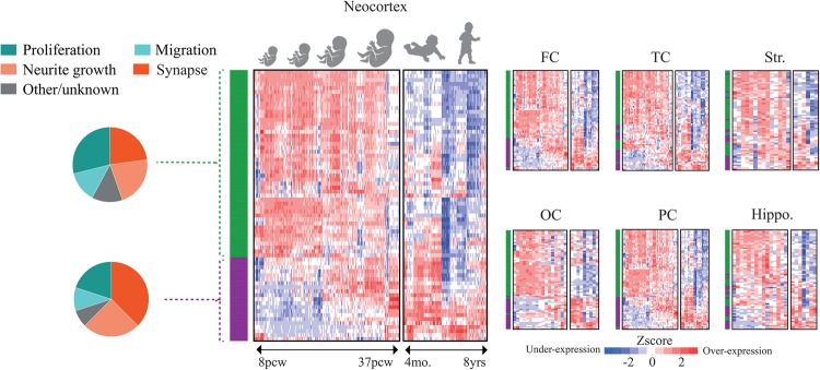
- ASD begins in the womb: 94% of 69 reliably expressed high-confidence ASD (hcASD) genes are expressed prenatally, 68% peak prenatally then decline after birth, and 62% of 58 functionally annotated hcASD genes are pleiotropic across multiple developmental processes.
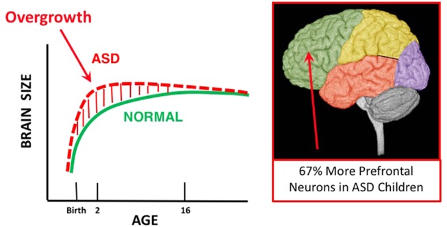
- Postmortem evidence: young ASD male children have on average 67% more prefrontal neurons than controls (79% more in dorsolateral and 29% more in medial prefrontal cortex; range 12–106%), 36% more cortical neurons in another study, and ~53% more von Economo neurons in frontoinsular cortex; neuron size reduction varies from −4% in occipital to −18% in prefrontal cortex.
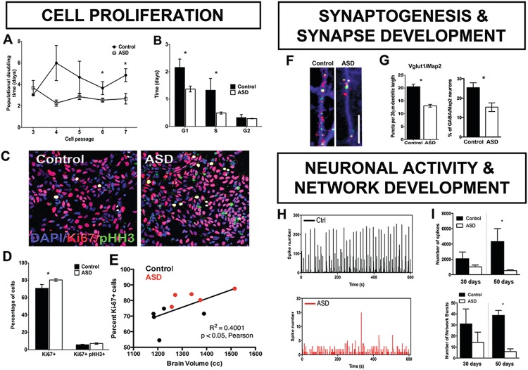
- iPSC evidence: ASD neural progenitors doubled in number twice as fast as controls due to a shorter G1/S phase; ASD-derived neurons showed fewer excitatory synapses, and by 50 days synchronized neural bursts were 6 times lower than controls; one ASD child-based cellular model found a 10-fold decrease in spontaneous neural activity.
- Maternal immune activation (MIA) models show upregulated cell cycle/proliferation, overproduction of neurons, a 24% increase in neuron numbers in cortical layers II/III, and when combined with Pten haploinsufficiency increased brain enlargement from 8% to 44%; non-genetic etiologies may account for 30–41% of ASD risk.
Methods
- Narrative review integrating postmortem, genetic, genomic, transcriptomic, iPSC/cell model, animal model, and MIA evidence; no new primary human cohort is reported.
- Reanalysis of 72 hcASD genes (38 recurrent/penetrant genes overlapping with 65 SFARI Level 1/2 genes), focusing on 69 with reliable BrainSpan RNA-Seq expression, plus manual literature search annotating 58 genes across proliferation/neurogenesis, cell fate/migration, neurite outgrowth, and spine/synaptogenesis/synapse function.
- Reviews the first ASD Living Biology study (Marchetto et al.) linking iPSC-derived prenatal molecular/cellular measures to MRI-based early brain overgrowth in living ASD toddlers.
Limitations
- Stated: current approaches cannot provide individualized fetal-stage information; postnatal phenotype data alone cannot reveal fetal-stage biology, and fetal-stage cellular models alone cannot explain postnatal development and clinical heterogeneity.
- Stated: many proposed ASD gene associations may be “noise,” and the hcASD gene set is limited to 72 genes, with 3 not reliably detected and only 58 having functional annotation from the literature.
- Evident: this is a narrative review rather than a systematic review or new primary study; causal and multistage claims rely on heterogeneous existing postmortem, animal, and cell-model evidence, with some key iPSC–MRI links being correlational.
Model-written summary. Check it against the paper before relying on it.
Integrity signals
Registry
Cites
Statistics
Figures
Verification
Claims the paper makes
88 Not checked yet · 26 Own interpretation · 8 Own result · 7 Partly supported · 6 No basis given · 3 Supported · accuracy not yet measured
The cited sources support only part of this claim — read each source’s verdict below.
In the paper
“Studies on CHD8 and iPS-derived ASD neurons indicate the dysregulation of proliferation and macrocephaly in ASD could be linked to the disruption of these signaling pathways and shortened G1/S transition[54, 55, 57, 94, 183, 192, 193].”
§ The ASD Living Biology: from cell proliferation to clinical phenotype > Pleiotropic common pathways implicated in ASD > Dysregulation of proliferation and neurogenesis
What the sources say
- Supportedcites [54] Marchetto MC, Belinson H, Tian Y, Freitas BC, Fu C, Vadodaria KC, et al. Altered proliferation and networks in neural cells derived from idiopathic autistic individuals. Mol Psychiatry. 2016;22:820–35. source
The Results section on rapid proliferation reports that iPSC-derived neural progenitor cells from ASD individuals with early brain overgrowth proliferated faster, principally because their G1 and S phases were shorter. The Results section on β-catenin/BRN2 links that accelerated proliferation to reduced β-catenin transcriptional activity. This supports the hedged claim as it relates to this iPSC study; the paper studied a selected group with brain overgrowth, not all individuals with ASD. (LLM, conf=0.88)
- Partly supportedcites [55] Mariani J, Coppola G, Zhang P, Abyzov A, Provini L, Tomasini L, et al. FOXG1-dependent dysregulation of GABA/Glutamate neuron differentiation in autism spectrum disorders. Cell. 2015;162:375–90. sourceExperimental — accuracy not yet measured
The Results section reports a shorter overall cell cycle in ASD-derived iPSCs, and the Discussion links increased proliferative potential with macrocephaly in the studied patients. The paper also identifies altered cell-cycle-related gene programs. It does not establish a shortened G1/S transition specifically, and this is not a CHD8 study. (LLM, conf=0.86)
- Partly supportedcites [57] Krumm N, O’Roak BJ, Shendure J, Eichler EE. A de novo convergence of autism genetics and molecular neuroscience. Trends Neurosci. 2014;37:95–105. sourceExperimental — accuracy not yet measured
The section “Protein interaction networks converge on common pathways” links CHD8–beta-catenin/WNT signaling to neural growth and reports macrocephaly in ASD cases with truncating CHD8 mutations. The paper does not link that finding to shortened G1/S transition or report proliferation findings from iPS-derived ASD neurons; its discussion of induced pluripotent stem cells concerns Timothy syndrome. (LLM, conf=0.88)
- Partly supportedcites [94] Subtil-Rodriguez A, Vazquez-Chavez E, Ceballos-Chavez M, Rodriguez-Paredes M, Martin-Subero JI, Esteller M, et al. The chromatin remodeller CHD8 is required for E2F-dependent transcription activation of S-phase genes. Nucleic Acids Res. 2014;42:2185–96. sourceExperimental — accuracy not yet measured
The Results sections on E2F targets and cell-cycle re-entry show that CHD8 regulates G1/S gene expression and cell proliferation in RPE1 cells. They do not establish a link to ASD, iPS-derived neurons, or macrocephaly. Rather than a shortened G1/S transition, CHD8 depletion caused a strong delay in cell-cycle progression. (LLM, conf=0.88)
- Partly supportedcites [183] Gompers AL, Su-Feher L, Ellegood J, Copping NA, Riyadh MA, Stradleigh TW, et al. Germline Chd8 haploinsufficiency alters brain development in mouse. Nature Neuroscience. 2017;20:1062–73. sourceExperimental — accuracy not yet measured
The Results section, “Increased prenatal proliferation of neural progenitors in Chd8+/del5 mice” (page unavailable), reports increased proliferation in mutant mice and suggests that altered neuron production may contribute to megalencephaly. The Discussion also describes perturbed cell-cycle-related gene networks. This paper does not establish a shortened G1/S transition or report findings from iPS-derived ASD neurons, so it supports only part of the multi-citation claim. (LLM, conf=0.86)
- Partly supportedcites [192] Sugathan A, Biagioli M, Golzio C, Erdin S, Blumenthal I, Manavalan P, et al. CHD8 regulates neurodevelopmental pathways associated with autism spectrum disorder in neural progenitors. Proceedings of the National Academy of Sciences. 2014;111:E4468–E4477. sourceExperimental — accuracy not yet measured
The Results (p. 7) report increased proliferating cells after chd8 suppression in zebrafish and suggest that disturbed neuronal proliferation likely causes the observed macrocephaly. The paper also reports altered neurodevelopmental pathways (Discussion, p. 9), but it does not establish a shortened G1/S transition. Its iPSC-derived cells were neural progenitors from a control individual, not ASD neurons (Results, p. 3). (LLM, conf=0.86)
- Partly supportedcites [193] Bernier R, Golzio C, Xiong B, Stessman HA, Coe BP, et al. Disruptive CHD8 mutations define a subtype of autism early in development. Cell. 2014;158:263–276. sourceExperimental — accuracy not yet measured
The Discussion reports macrocephaly in people with CHD8 mutations and in chd8-disrupted zebrafish, where forebrain/midbrain expansion is described as likely due to over-proliferation of neuronal progenitors. The Introduction identifies CHD8 as a potential regulator of Wnt signaling, but this paper does not establish disruption of that pathway as the mechanism, study iPS-derived ASD neurons, or report a shortened G1/S transition. No page numbers are available in our copy. (LLM, conf=0.85)
hedgedcorrelational- Asserted: passes — The authors put it forward as true.
- Checkable: passes — It can be checked against evidence.
- Load-bearing: passes — The paper’s conclusions depend on it.
- Based: passes — The paper gives a reason why it holds.
- Supported
The cited sources support only part of this claim — read each source’s verdict below.
In the paper
“MIA models have ASD-like social, vocalization, ritualistic, exploration and other behavioral deficits [123–125].”
§ The ASD Living Biology: from cell proliferation to clinical phenotype > The prenatal maternal immune activation model of ASD
What the sources say
- Partly supportedcites [123] Shi L, Fatemi SH, Sidwell RW, Patterson PH. Maternal influenza infection causes marked behavioral and pharmacological changes in the offspring. J Neurosci. 2003;23:297–302. sourceExperimental — accuracy not yet measured
The Social behavior and Exploratory behavior sections report reduced social contact and exploration in offspring of influenza-infected mice. The Discussion says their behavior is possibly consistent with a rodent version of autism, but notes that the tests are not disorder-specific. Our copy does not report vocalization or ritualistic-behavior deficits. (LLM, conf=0.89)
- Supportedcites [124] Malkova NV, Yu CZ, Hsiao EY, Moore MJ, Patterson PH. Maternal immune activation yields offspring displaying mouse versions of the three core symptoms of autism. Brain Behav Immun. 2012;26:607–16. source
The Results (sections 3.2, 3.4 and 3.5) report reduced social preference and vocalizations, alongside increased repetitive behavior, in MIA offspring. The Discussion (section 4) also reports a previously observed deficit in interaction with a novel object, supporting the exploration component. (LLM, conf=0.86)
- Partly supportedcites [125] Smith SE, Li J, Garbett K, Mirnics K, Patterson PH. Maternal immune activation alters fetal brain development through interleukin-6. J Neurosci. 2007;27:10695–702. sourceExperimental — accuracy not yet measured
The Results section, “Effects of anti-IL-6 on abnormal behaviors evoked by MIA” (page unavailable), reports social-interaction and open-field exploration deficits in offspring of poly(I:C)-treated mice; it also reports latent-inhibition and prepulse-inhibition deficits. The passages found do not report vocalization or ritualistic-behavior deficits, so this source supports only part of the cited list. (LLM, conf=0.88)
plaincorrelational- Asserted: passes — The authors put it forward as true.
- Checkable: passes — It can be checked against evidence.
- Load-bearing: passes — The paper’s conclusions depend on it.
- Based: passes — The paper gives a reason why it holds.
- Partly supported
The cited sources support only part of this claim — read each source’s verdict below.
In the paper
“Importantly, increased levels of IL-6 alone recapitulate aspects of ASD pathophysiology [138, 142, 143].”
§ The ASD Living Biology: from cell proliferation to clinical phenotype > The prenatal maternal immune activation model of ASD
What the sources say
- Not checked yetcites [138] Deverman BE, Patterson PH. Cytokines and CNS development. Neuron. 2009;64:61–78. source
- Partly supportedcites [142] Gumusoglu SB, Fine RS, Murray SJ, Bittle JL, Stevens HE. The role of IL-6 in neurodevelopment after prenatal stress. Brain Behav Immun. 2017;65:274–83. sourceExperimental — accuracy not yet measured
The Abstract reports that prenatal IL-6 alone reproduced stress-induced microglial changes and delayed GABAergic progenitor migration in mice. But the Discussion says IL-6 administration did not produce the decreased sociability, anxiety-like changes, or reduced locomotion that the paper links to autism symptoms. The evidence supports some neurodevelopmental effects, not a general claim that IL-6 alone recapitulates ASD pathophysiology. (LLM, conf=0.86)
- Supportedcites [143] Wu WL, Hsiao EY, Yan Z, Mazmanian SK, Patterson PH. The placental interleukin-6 signaling controls fetal brain development and behavior. Brain Behav Immun. 2017;62:11–23. source
The Introduction states that injection of recombinant IL-6 alone into mice produces behavioral phenotypes similar to those in the maternal immune activation model, which the paper describes as having ASD-related features. The support is for aspects of ASD-related behavior in a mouse model, not for reproducing ASD in humans. (LLM, conf=0.90)
plaindirect- Asserted: passes — The authors put it forward as true.
- Checkable: passes — It can be checked against evidence.
- Load-bearing: passes — The paper’s conclusions depend on it.
- Based: passes — The paper gives a reason why it holds.
The cited sources support only part of this claim — read each source’s verdict below.
In the paper
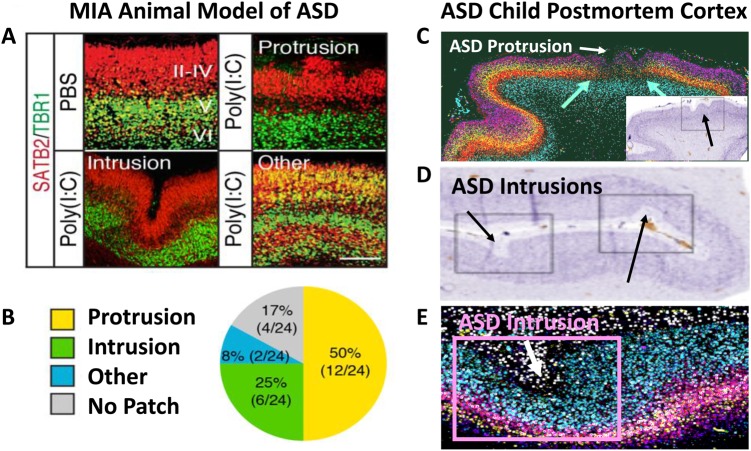
“The Choi and colleagues [132, 81] MIA ASD mouse model study showed that the same fetal MIA perturbation of littermates causes patches of focal cortical dysplasia that have marked heterogeneity, analogous to heterogeneity found in cortex in unrelated individual ASD cases postmortem [34] (Fig. 7).”
§ The ASD Living Biology: from cell proliferation to clinical phenotype > Conclusions
What the sources say
- Partly supportedcites [132] Choi GB, Yim YS, Wong H, Kim S, Kim H, Kim SV, et al. The maternal interleukin-17a pathway in mice promotes autism-like phenotypes in offspring. Science. 2016;351:933–9. sourceExperimental — accuracy not yet measured
In “Maternal IL-17a promotes abnormal cortical development in offspring” (page unavailable), the paper reports a patch of disorganized cortex after maternal immune activation and notes that focal cortical abnormalities have been found in people with ASD. It does not report marked heterogeneity among littermates; instead, it says the singular patch appeared at a similar position in a majority of affected fetal brains. The comparison to heterogeneity across postmortem ASD cases is not established by this paper. (LLM, conf=0.87)
- Partly supportedcites [81] Yim YS, Park A, Berrios J, Lafourcade M, Pascual LM, Soares N, et al. Reversing behavioural abnormalities in mice exposed to maternal inflammation. Nature. 2017;549:482–7. sourceExperimental — accuracy not yet measured
The Characterization of cortical patches section reports that MIA offspring developed patches whose locations and sizes varied among individual mice; the Cortical patches are predictive of MIA behaviors section reports patches after maternal poly(I:C) exposure at E12.5. The copy does not establish that the compared mice were littermates or characterize the patches as focal cortical dysplasia. The Discussion notes similar patches in cortical tissue from children with autism, but does not compare the degree of heterogeneity with postmortem ASD cases. (LLM, conf=0.83)
- Supportedcites [34] Stoner R, Chow ML, Boyle MP, Sunkin SM, Mouton PR, Roy S, et al. Patches of disorganization in the neocortex of children with autism. N Engl J Med. 2014;370:1209–19. source
The citation to this paper attaches to the comparison with postmortem autism cases, not to the preceding claim about the MIA mouse model. In Results (Aberrant Laminar Expression Patterns), the paper reports that patches varied between case samples in the cortical layers and cell types most affected; the Discussion likewise says their presentation varied across cases. (LLM, conf=0.93)
plaindirect- Asserted: passes — The authors put it forward as true.
- Checkable: passes — It can be checked against evidence.
- Load-bearing: passes — The paper’s conclusions depend on it.
- Based: passes — The paper gives a reason why it holds.
- Partly supported
The paper states this without a source or data behind it here, so there is nothing to check it against. The quote shows it is in the paper.
In the paper
“Among the most commonly reported high-confidence ASD (hcASD) genes, 94% express during prenatal life and affect these fetal processes in neocortex, amygdala, hippocampus, striatum and cerebellum.”
§ The ASD Living Biology: from cell proliferation to clinical phenotype > Abstract
plaindirect- Asserted: passes — The authors put it forward as true.
- Checkable: passes — It can be checked against evidence.
- Load-bearing: passes — The paper’s conclusions depend on it.
- Based: unclear — It is unclear why the paper says it holds.
A result the paper reports from its own data — it cites no other source for it, so there is none to check. The quote shows it is in the paper.
In the paper
“Of the 72 genes, 3 are not detected (RPKM > 0.5) in >10% of BrainSpan samples between prenatal and up to 8 years old post-natal.”
§ The ASD Living Biology: from cell proliferation to clinical phenotype > ASD begins in the womb > High-confidence ASD genes and prenatal development
plain- Asserted: passes — The authors put it forward as true.
- Checkable: passes — It can be checked against evidence.
- Load-bearing: passes — The paper’s conclusions depend on it.
- Based: passes — The paper gives a reason why it holds.
- Rests on an assumption: BrainSpan RNA-Seq expression patterns accurately represent developmental expression timing and roles of hcASD genes in living human brain development.
A result the paper reports from its own data — it cites no other source for it, so there is none to check. The quote shows it is in the paper.
In the paper
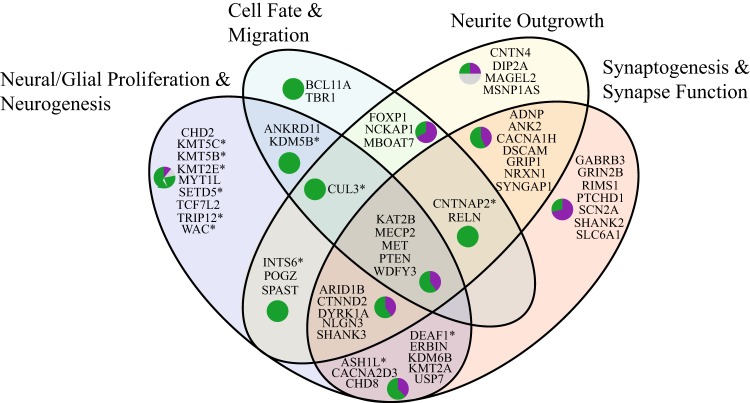
“Importantly, 47 genes (68%; green cluster in Fig. 4) have peak expression during much of prenatal development, and then are downregulated following birth.”
§ The ASD Living Biology: from cell proliferation to clinical phenotype > ASD begins in the womb > High-confidence ASD genes and prenatal development
plaincorrelational- Asserted: passes — The authors put it forward as true.
- Checkable: passes — It can be checked against evidence.
- Load-bearing: passes — The paper’s conclusions depend on it.
- Based: passes — The paper gives a reason why it holds.
- Rests on an assumption: BrainSpan RNA-Seq expression patterns accurately represent developmental expression timing and roles of hcASD genes in living human brain development.
A result the paper reports from its own data — it cites no other source for it, so there is none to check. The quote shows it is in the paper.
In the paper
“Of these 69 genes, 65 (94%) are expressed (RPKM > 1) in prenatal brain development.”
§ The ASD Living Biology: from cell proliferation to clinical phenotype > ASD begins in the womb > High-confidence ASD genes and prenatal development
plain- Asserted: passes — The authors put it forward as true.
- Checkable: passes — It can be checked against evidence.
- Load-bearing: passes — The paper’s conclusions depend on it.
- Based: passes — The paper gives a reason why it holds.
- Rests on an assumption: BrainSpan RNA-Seq expression patterns accurately represent developmental expression timing and roles of hcASD genes in living human brain development.
A result the paper reports from its own data — it cites no other source for it, so there is none to check. The quote shows it is in the paper.
In the paper
“The remaining 22 (32%) of the hcASD genes start to increase in expression levels during the 3rd trimester and the first several years of post-natal life (Fig. 4; purple cluster).”
§ The ASD Living Biology: from cell proliferation to clinical phenotype > ASD begins in the womb > High-confidence ASD genes and prenatal development
plaindirect- Asserted: passes — The authors put it forward as true.
- Checkable: passes — It can be checked against evidence.
- Load-bearing: passes — The paper’s conclusions depend on it.
- Based: passes — The paper gives a reason why it holds.
- Rests on an assumption: BrainSpan RNA-Seq expression patterns accurately represent developmental expression timing and roles of hcASD genes in living human brain development.
A result the paper reports from its own data — it cites no other source for it, so there is none to check. The quote shows it is in the paper.
In the paper
“Furthermore, they display peak expression in brain regions that are abnormal in ASD, including major cerebral cortical areas, hippocampus, striatum (Fig. 4), and cerebellum.”
§ The ASD Living Biology: from cell proliferation to clinical phenotype > ASD begins in the womb > High-confidence ASD genes and prenatal development
plaincorrelational- Asserted: passes — The authors put it forward as true.
- Checkable: passes — It can be checked against evidence.
- Load-bearing: passes — The paper’s conclusions depend on it.
- Based: passes — The paper gives a reason why it holds.
- Rests on an assumption: BrainSpan RNA-Seq expression patterns accurately represent developmental expression timing and roles of hcASD genes in living human brain development.
In the paper
“Second, we addressed “what” developmental role all 72 hcASD genes play, and found studies that support involvement of 58 out of 72 hcASD genes in at least one of the four main brain development processes including proliferation/neurogenesis, cell fate/migration, neurite outgrowth, and spine development/synaptogenesis/synapse function (Table S2).”
§ The ASD Living Biology: from cell proliferation to clinical phenotype > ASD begins in the womb > High-confidence ASD genes and prenatal development
What the sources say
- Not checked yetcites Table S2
plaincorrelational- Asserted: passes — The authors put it forward as true.
- Checkable: passes — It can be checked against evidence.
- Load-bearing: passes — The paper’s conclusions depend on it.
- Based: passes — The paper gives a reason why it holds.
- Rests on an assumption: Functional annotation from animal/cell models and literature can be generalized to human hcASD gene roles in brain development.
A result the paper reports from its own data — it cites no other source for it, so there is none to check. The quote shows it is in the paper.
In the paper
“Indeed, 36 out of 58 (62%) hcASD genes are involved in multiple major developmental processes (Fig. 5).”
§ The ASD Living Biology: from cell proliferation to clinical phenotype > ASD begins in the womb > High-confidence ASD genes and prenatal development
plaincorrelational- Asserted: passes — The authors put it forward as true.
- Checkable: passes — It can be checked against evidence.
- Load-bearing: passes — The paper’s conclusions depend on it.
- Based: passes — The paper gives a reason why it holds.
- Rests on an assumption: Functional annotation from animal/cell models and literature can be generalized to human hcASD gene roles in brain development.
The paper states this without a source or data behind it here, so there is nothing to check it against. The quote shows it is in the paper.
In the paper
“Among the hcASD genes, at least 48 (67%) genes are involved in gene regulation including chromatin structure, transcription, translation, and post-translation stages.”
§ The ASD Living Biology: from cell proliferation to clinical phenotype > Pleiotropic common pathways implicated in ASD
plain- Asserted: passes — The authors put it forward as true.
- Checkable: passes — It can be checked against evidence.
- Load-bearing: passes — The paper’s conclusions depend on it.
- Based: unclear — It is unclear why the paper says it holds.
The paper states this without a source or data behind it here, so there is nothing to check it against. The quote shows it is in the paper.
In the paper
“The majority are also pleiotropic, affecting multiple developmental stages, not just one.”
§ The ASD Living Biology: from cell proliferation to clinical phenotype > Conclusions
plaincorrelational- Asserted: passes — The authors put it forward as true.
- Checkable: passes — It can be checked against evidence.
- Load-bearing: passes — The paper’s conclusions depend on it.
- Based: passes — The paper gives a reason why it holds.
A conclusion the paper draws from its own results — it cites no other source for it, so there is none to check. The quote shows it is in the paper.
In the paper
“Here we review genetic, genomic, cellular, postmortem, animal model, and cell model evidence that shows ASD begins in the womb.”
- Also stated in: § Abstract · plain
“Despite this new knowledge and theory that ASD biology begins in the womb, current research methods have not provided individualized information: What are the fetal processes and early-age molecular and cellular differences that underlie ASD in each individual child?”
§ The ASD Living Biology: from cell proliferation to clinical phenotype > Abstract
boosteddirect- Asserted: passes — The authors put it forward as true.
- Checkable: passes — It can be checked against evidence.
- Load-bearing: passes — The paper’s conclusions depend on it.
- Based: passes — The paper gives a reason why it holds.
- Also stated in: § Abstract · plain
A conclusion the paper draws from its own results — it cites no other source for it, so there is none to check. The quote shows it is in the paper.
In the paper
“This evidence leads to a new theory that ASD is a multistage, progressive disorder of brain development, spanning nearly all of prenatal life.”
§ The ASD Living Biology: from cell proliferation to clinical phenotype > Abstract
plain- Asserted: passes — The authors put it forward as true.
- Checkable: passes — It can be checked against evidence.
- Load-bearing: passes — The paper’s conclusions depend on it.
- Based: passes — The paper gives a reason why it holds.
A conclusion the paper draws from its own results — it cites no other source for it, so there is none to check. The quote shows it is in the paper.
In the paper
“ASD can begin as early as the 1st and 2nd trimester with disruption of cell proliferation and differentiation.”
§ The ASD Living Biology: from cell proliferation to clinical phenotype > Abstract
plaindirect- Asserted: passes — The authors put it forward as true.
- Checkable: passes — It can be checked against evidence.
- Load-bearing: passes — The paper’s conclusions depend on it.
- Based: passes — The paper gives a reason why it holds.
A conclusion the paper draws from its own results — it cites no other source for it, so there is none to check. The quote shows it is in the paper.
In the paper
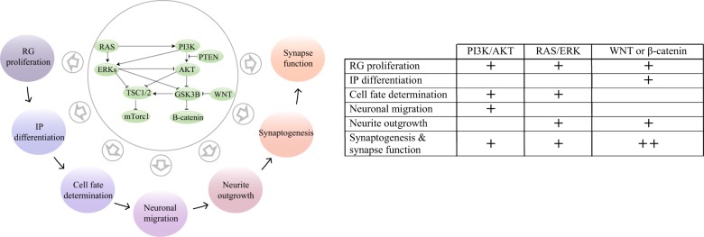
“Commonly implicated pathways, PI3K/AKT and RAS/ERK, are also pleiotropic and affect multiple fetal processes from proliferation through synapse and neural functional development.”
§ The ASD Living Biology: from cell proliferation to clinical phenotype > Abstract
plaindirect- Asserted: passes — The authors put it forward as true.
- Checkable: passes — It can be checked against evidence.
- Load-bearing: passes — The paper’s conclusions depend on it.
- Based: unclear — It is unclear why the paper says it holds.
A conclusion the paper draws from its own results — it cites no other source for it, so there is none to check. The quote shows it is in the paper.
In the paper
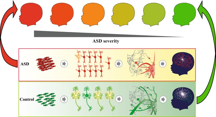
“In different ASD individuals, variation in how and when these pleiotropic pathways are dysregulated, will lead to different, even opposing effects, producing prenatal as well as later neural and clinical heterogeneity.”
- Also stated in: § Conclusions · plain
“In different ASD individuals, variation in how and when these pleiotropic pathways are dysregulated will lead to different, even opposing effects, producing prenatal as well as later neural and clinical heterogeneity.”
§ The ASD Living Biology: from cell proliferation to clinical phenotype > Abstract
plaindirect- Asserted: passes — The authors put it forward as true.
- Checkable: passes — It can be checked against evidence.
- Load-bearing: passes — The paper’s conclusions depend on it.
- Based: passes — The paper gives a reason why it holds.
- Also stated in: § Conclusions · plain
In the paper
“Strong evidence that ASD begins in the womb comes from observations that young ASD male children have 67% more prefrontal neurons than controls [24] (Fig. 1).”
§ The ASD Living Biology: from cell proliferation to clinical phenotype > ASD begins in the womb > ASD postmortem evidence
What the sources say
- Not checked yetcites [24] Courchesne E, Mouton PR, Calhoun ME, Semendeferi K, Ahrens-Barbeau C, Hallet MJ, et al. Neuron number and size in prefrontal cortex of children with autism. JAMA. 2011;306:2001–10. source
boostedcorrelational- Asserted: passes — The authors put it forward as true.
- Checkable: passes — It can be checked against evidence.
- Load-bearing: passes — The paper’s conclusions depend on it.
- Based: passes — The paper gives a reason why it holds.
In the paper
“In Marchetto et al. [54], ASD neural progenitors doubled in number twice as fast compared to controls due to a shorter cell cycle G1/S phase.”
§ The ASD Living Biology: from cell proliferation to clinical phenotype > ASD begins in the womb > ASD patient-derived iPS cells
What the sources say
- Not checked yetcites [54] Marchetto MC, Belinson H, Tian Y, Freitas BC, Fu C, Vadodaria KC, et al. Altered proliferation and networks in neural cells derived from idiopathic autistic individuals. Mol Psychiatry. 2016;22:820–35. source
plaindirect- Asserted: passes — The authors put it forward as true.
- Checkable: passes — It can be checked against evidence.
- Load-bearing: passes — The paper’s conclusions depend on it.
- Based: passes — The paper gives a reason why it holds.
- Rests on an assumption: iPSC-derived neural progenitor cells and neurons from ASD toddlers recapitulate the prenatal brain developmental processes relevant to ASD in vivo.
A conclusion the paper draws from its own results — it cites no other source for it, so there is none to check. The quote shows it is in the paper.
In the paper
“This suggests dysregulated proliferation contributes to early brain overgrowth in ASD toddlers.”
§ The ASD Living Biology: from cell proliferation to clinical phenotype > ASD begins in the womb > ASD patient-derived iPS cells
hedgedconditional- Asserted: passes — The authors put it forward as true.
- Checkable: passes — It can be checked against evidence.
- Load-bearing: passes — The paper’s conclusions depend on it.
- Based: passes — The paper gives a reason why it holds.
- Rests on an assumption: iPSC-derived neural progenitor cells and neurons from ASD toddlers recapitulate the prenatal brain developmental processes relevant to ASD in vivo.
In the paper
“Young ASD males have ~53% more von Economo neurons in frontoinsular cortex than controls [41].”
§ The ASD Living Biology: from cell proliferation to clinical phenotype > ASD begins in the womb > ASD postmortem evidence
What the sources say
- Not checked yetcites [41] Santos M, Uppal N, Butti C, Wicinski B, Schmeidler J, Giannakopoulos P, et al. Von Economo neurons in autism: a stereologic study of the frontoinsular cortex in children. Brain Res. 2011;1380:206–17. source
plaincorrelational- Asserted: passes — The authors put it forward as true.
- Checkable: passes — It can be checked against evidence.
- Load-bearing: passes — The paper’s conclusions depend on it.
- Based: passes — The paper gives a reason why it holds.
A conclusion the paper draws from its own results — it cites no other source for it, so there is none to check. The quote shows it is in the paper.
In the paper
“Thus, in living ASD infants and toddlers, patient-derived iPS cell experiments and leukocyte transcriptomics point to dysregulation of cell cycle G1/S.”
§ The ASD Living Biology: from cell proliferation to clinical phenotype > ASD begins in the womb > Transcriptomic evidence in living ASD toddlers points to prenatal periods
plaincorrelational- Asserted: passes — The authors put it forward as true.
- Checkable: passes — It can be checked against evidence.
- Load-bearing: passes — The paper’s conclusions depend on it.
- Based: passes — The paper gives a reason why it holds.
- Rests on an assumption: iPSC-derived neural progenitor cells and neurons from ASD toddlers recapitulate the prenatal brain developmental processes relevant to ASD in vivo.
- Rests on an assumption: Leukocyte transcriptomics in ASD toddlers reflects molecular processes in the developing brain.
In the paper
“Functional genomic analyses identified 23 candidate genes for brain maldevelopment linked to 32 upstream high-confidence ASD genes [56].”
§ The ASD Living Biology: from cell proliferation to clinical phenotype > ASD begins in the womb > Transcriptomic evidence in living ASD toddlers points to prenatal periods
What the sources say
- Not checked yetcites [56] Pramparo T, Lombardo MV, Campbell K, Barnes CC, Marinero S, Solso S, et al. Cell cycle networks link gene expression dysregulation, mutation, and brain maldevelopment in autistic toddlers. Mol Syst Biol. 2015;11:841. source
plaincorrelational- Asserted: passes — The authors put it forward as true.
- Checkable: passes — It can be checked against evidence.
- Load-bearing: passes — The paper’s conclusions depend on it.
- Based: passes — The paper gives a reason why it holds.
- Rests on an assumption: Leukocyte transcriptomics in ASD toddlers reflects molecular processes in the developing brain.
In the paper
“Patches of focal laminar disorganization and nearby clusters of mis-migrated neurons were found in prefrontal and temporal cortices in 10 of 11 young ASD cases [34].”
§ The ASD Living Biology: from cell proliferation to clinical phenotype > ASD begins in the womb > ASD postmortem evidence
What the sources say
- Not checked yetcites [34] Stoner R, Chow ML, Boyle MP, Sunkin SM, Mouton PR, Roy S, et al. Patches of disorganization in the neocortex of children with autism. N Engl J Med. 2014;370:1209–19. source
plaincorrelationaln = 11- Asserted: passes — The authors put it forward as true.
- Checkable: passes — It can be checked against evidence.
- Load-bearing: passes — The paper’s conclusions depend on it.
- Based: passes — The paper gives a reason why it holds.
In the paper
“In another study, cortex in young ASD cases had 36% more neurons than controls [40] (Table S1).”
§ The ASD Living Biology: from cell proliferation to clinical phenotype > ASD begins in the womb > ASD postmortem evidence
What the sources say
- Not checked yetcites [40] van Kooten IA, Palmen SJ, von Cappeln P, Steinbusch HW, Korr H, Heinsen H, et al. Neurons in the fusiform gyrus are fewer and smaller in autism. Brain. 2008;131(Pt 4):987–99. source
plaincorrelational- Asserted: passes — The authors put it forward as true.
- Checkable: passes — It can be checked against evidence.
- Load-bearing: passes — The paper’s conclusions depend on it.
- Based: passes — The paper gives a reason why it holds.
None of the sources cited for this claim have been checked yet.
In the paper
“Consistently, studies on hcASD genes and iPSC-derived ASD neurons highlight signaling pathways that are commonly disrupted, such as PI3K/AKT [79, 80], RAS/ERK [79, 80, 82, 83], WNT and β-catenin [57, 83, 84].”
§ The ASD Living Biology: from cell proliferation to clinical phenotype > Pleiotropic common pathways implicated in ASD
What the sources say
- Not checked yetcites [79] Pinto D, Delaby E, Merico D, Barbosa M, Merikangas A, Klei L, et al. Convergence of genes and cellular pathways dysregulated in autism spectrum disorders. Am J Hum Genet. 2014;94:677–94. source
- Not checked yetcites [80] Sahin M, Sur M Genes, circuits, and precision therapies for autism and related neurodevelopmental disorders. Science. 2015; 350. source
- Not checked yetcites [82] Wen Y, Alshikho MJ, Herbert MR. Pathway Network Analyses for Autism Reveal Multisystem Involvement, Major Overlaps with Other Diseases and Convergence upon MAPK and Calcium Signaling. PLoS ONE. 2016;11:e0153329. source
- Not checked yetcites [83] Luo W, Zhang C, Jiang YH, Brouwer CR. Systematic reconstruction of autism biology from massive genetic mutation profiles. Sci Adv 2018; 4(4): e1701799. source
- Not checked yetcites [57] Krumm N, O’Roak BJ, Shendure J, Eichler EE. A de novo convergence of autism genetics and molecular neuroscience. Trends Neurosci. 2014;37:95–105. source
- Not checked yetcites [84] Kalkman HO. A review of the evidence for the canonical Wnt pathway in autism spectrum disorders. Mol Autism. 2012;3:10. source
plaincorrelational- Asserted: passes — The authors put it forward as true.
- Checkable: passes — It can be checked against evidence.
- Load-bearing: passes — The paper’s conclusions depend on it.
- Based: passes — The paper gives a reason why it holds.
In the paper
“Moreover, the degree of neuron excess across ASD cases was heterogeneous, ranging from 12 to 106%.”
§ The ASD Living Biology: from cell proliferation to clinical phenotype > ASD begins in the womb > ASD postmortem evidence
What the sources say
- Not checked yetcites [24] Courchesne E, Mouton PR, Calhoun ME, Semendeferi K, Ahrens-Barbeau C, Hallet MJ, et al. Neuron number and size in prefrontal cortex of children with autism. JAMA. 2011;306:2001–10. source
plainn = 4- Asserted: passes — The authors put it forward as true.
- Checkable: passes — It can be checked against evidence.
- Load-bearing: unclear — It is unclear whether the paper’s conclusions depend on it.
- Based: passes — The paper gives a reason why it holds.
In the paper
“By 50 days, synchronized neural bursts of activity were 6 times lower in ASD than controls (Fig. 3h, i).”
§ The ASD Living Biology: from cell proliferation to clinical phenotype > ASD begins in the womb > ASD patient-derived iPS cells
What the sources say
- Not checked yetcites [54] Marchetto MC, Belinson H, Tian Y, Freitas BC, Fu C, Vadodaria KC, et al. Altered proliferation and networks in neural cells derived from idiopathic autistic individuals. Mol Psychiatry. 2016;22:820–35. source
plaincorrelational- Asserted: passes — The authors put it forward as true.
- Checkable: passes — It can be checked against evidence.
- Load-bearing: passes — The paper’s conclusions depend on it.
- Based: passes — The paper gives a reason why it holds.
- Rests on an assumption: iPSC-derived neural progenitor cells and neurons from ASD toddlers recapitulate the prenatal brain developmental processes relevant to ASD in vivo.
In the paper
“Further, reduced β-catenin and BRN2 transcriptional activity was related to this proliferation abnormality.”
§ The ASD Living Biology: from cell proliferation to clinical phenotype > ASD begins in the womb > ASD patient-derived iPS cells
What the sources say
- Not checked yetcites [54] Marchetto MC, Belinson H, Tian Y, Freitas BC, Fu C, Vadodaria KC, et al. Altered proliferation and networks in neural cells derived from idiopathic autistic individuals. Mol Psychiatry. 2016;22:820–35. source
plaincorrelational- Asserted: passes — The authors put it forward as true.
- Checkable: passes — It can be checked against evidence.
- Load-bearing: passes — The paper’s conclusions depend on it.
- Based: passes — The paper gives a reason why it holds.
- Rests on an assumption: iPSC-derived neural progenitor cells and neurons from ASD toddlers recapitulate the prenatal brain developmental processes relevant to ASD in vivo.
None of the sources cited for this claim have been checked yet.
In the paper
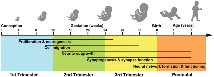
“In ASD, neural migration and cortical laminar organization, stages that occur in second and third trimesters, are also abnormal (Fig. 2) [34, 45–48].”
§ The ASD Living Biology: from cell proliferation to clinical phenotype > ASD begins in the womb > ASD postmortem evidence
What the sources say
- Not checked yetcites [34] Stoner R, Chow ML, Boyle MP, Sunkin SM, Mouton PR, Roy S, et al. Patches of disorganization in the neocortex of children with autism. N Engl J Med. 2014;370:1209–19. source
- Not checked yetcites [45] Wegiel J, Kuchna I, Nowicki K, Imaki H, Marchi E, Ma SY, et al. The neuropathology of autism: defects of neurogenesis and neuronal migration, and dysplastic changes. Acta Neuropathol. 2010;119:755–70. source
- Not checked yetcites [46] Varghese M, Keshav N, Jacot-Descombes S, Warda T, Wicinski B, Dickstein DL, et al. Autism spectrum disorder: neuropathology and animal models. Acta Neuropathol. 2017;134:537–66. source
- Not checked yetcites [47] Hutsler JJ, Casanova MF. Review: Cortical construction in autism spectrum disorder: columns, connectivity and the subplate. Neuropathol Appl Neurobiol. 2016;42:115–34. source
- Not checked yetcites [48] Bailey A, Luthert P, Dean A, Harding B, Janota I, Montgomery M, et al. A clinicopathological study of autism. Brain. 1998;121(Pt 5):889–905. source
plainn = 11- Asserted: passes — The authors put it forward as true.
- Checkable: passes — It can be checked against evidence.
- Load-bearing: passes — The paper’s conclusions depend on it.
- Based: passes — The paper gives a reason why it holds.
None of the sources cited for this claim have been checked yet.
In the paper
“Thus, neurons fail to migrate to the cortical plate and accumulate within the intermediate zone [88, 104].”
§ The ASD Living Biology: from cell proliferation to clinical phenotype > Pleiotropic common pathways implicated in ASD > Disruption of cell fate and migration
What the sources say
- Not checked yetcites [88] Munji RN, Choe Y, Li G, Siegenthaler JA, Pleasure SJ. Wnt signaling regulates neuronal differentiation of cortical intermediate progenitors. J Neurosci. 2011;31:1676–87. source
- Not checked yetcites [104] Morgan-Smith M, Wu Y, Zhu X, Pringle J, Snider WD. GSK-3 signaling in developing cortical neurons is essential for radial migration and dendritic orientation. eLife. 2014;3:e02663. source
plaindirect- Asserted: passes — The authors put it forward as true.
- Checkable: passes — It can be checked against evidence.
- Load-bearing: passes — The paper’s conclusions depend on it.
- Based: passes — The paper gives a reason why it holds.
In the paper
“PI3K/AKT and RAS/ERK signaling pathways also control processes associated with neural excitotoxicity and cell survival [191].”
§ The ASD Living Biology: from cell proliferation to clinical phenotype > Pleiotropic common pathways implicated in ASD > Dysregulation of neurite outgrowth and neuronal function
What the sources say
- Not checked yetcites [191] Hardingham GE, Bading H. Synaptic versus extrasynaptic NMDA receptor signalling: implications for neurodegenerative disorders. Nat Rev Neurosci. 2010;11:682–96. source
plaindirect- Asserted: passes — The authors put it forward as true.
- Checkable: passes — It can be checked against evidence.
- Load-bearing: unclear — It is unclear whether the paper’s conclusions depend on it.
- Based: passes — The paper gives a reason why it holds.
None of the sources cited for this claim have been checked yet.
In the paper
“Interestingly, G1/S transition rates might be associated with these cell decisions as symmetric divisions can have shorter G1/S transition times than asymmetric divisions [93, 101].”
§ The ASD Living Biology: from cell proliferation to clinical phenotype > Pleiotropic common pathways implicated in ASD > Dysregulation of proliferation and neurogenesis
What the sources say
- Not checked yetcites [93] Dehay C, Kennedy H. Cell-cycle control and cortical development. Nat Rev Neurosci. 2007;8:438–50. source
- Not checked yetcites [101] Calegari F, Haubensak W, Haffner C, Huttner WB. Selective lengthening of the cell cycle in the neurogenic subpopulation of neural progenitor cells during mouse brain development. J Neurosci. 2005;25:6533–8. source
hedgedcorrelational- Asserted: passes — The authors put it forward as true.
- Checkable: passes — It can be checked against evidence.
- Load-bearing: unclear — It is unclear whether the paper’s conclusions depend on it.
- Based: passes — The paper gives a reason why it holds.
A conclusion the paper draws from its own results — it cites no other source for it, so there is none to check. The quote shows it is in the paper.
In the paper
“Thus, in addition to modulating neuron development, the perturbed pathways in ASD also alter cell fate determination and neural migration.”
§ The ASD Living Biology: from cell proliferation to clinical phenotype > Pleiotropic common pathways implicated in ASD > Disruption of cell fate and migration
plaindirect- Asserted: passes — The authors put it forward as true.
- Checkable: passes — It can be checked against evidence.
- Load-bearing: passes — The paper’s conclusions depend on it.
- Based: passes — The paper gives a reason why it holds.
In the paper
“While downregulation of RAS/ERK can lead to a thicker cortex from increased proliferation and an increased neuronal count [86]”
§ The ASD Living Biology: from cell proliferation to clinical phenotype > Pleiotropic common pathways implicated in ASD > Dysregulation of proliferation and neurogenesis
What the sources say
- Not checked yetcites [86] Rhim JH, Luo X, Gao D, Xu X, Zhou T, Li F, et al. Cell type-dependent Erk-Akt pathway crosstalk regulates the proliferation of fetal neural progenitor cells. Sci Rep. 2016;6:26547. source
plaindirect- Asserted: passes — The authors put it forward as true.
- Checkable: passes — It can be checked against evidence.
- Load-bearing: unclear — It is unclear whether the paper’s conclusions depend on it.
- Based: passes — The paper gives a reason why it holds.
In the paper
“RGCs in a mouse model of Gsk3 knockout, a major convergence point of the signaling pathways (Fig. 6), show more highly active β-catenin and Notch pathways [102].”
§ The ASD Living Biology: from cell proliferation to clinical phenotype > Pleiotropic common pathways implicated in ASD > Dysregulation of proliferation and neurogenesis
What the sources say
- Not checked yetcites [102] Kim WY, Wang X, Wu Y, Doble BW, Patel S, Woodgett JR, et al. GSK-3 is a master regulator of neural progenitor homeostasis. Nat Neurosci. 2009;12:1390–7. source
plaincorrelational- Asserted: passes — The authors put it forward as true.
- Checkable: passes — It can be checked against evidence.
- Load-bearing: passes — The paper’s conclusions depend on it.
- Based: passes — The paper gives a reason why it holds.
None of the sources cited for this claim have been checked yet.
In the paper
“These pathways control soma size, dendritic arborization, axon generation, spine development and synapse function [184–188].”
§ The ASD Living Biology: from cell proliferation to clinical phenotype > Pleiotropic common pathways implicated in ASD > Dysregulation of neurite outgrowth and neuronal function
What the sources say
- Not checked yetcites [184] Kumar V, Zhang MX, Swank MW, Kunz J, Wu GY. Regulation of dendritic morphogenesis by Ras-PI3K-Akt-mTOR and Ras-MAPK signaling pathways. J Neurosci. 2005;25:11288–99. source
- Not checked yetcites [185] Yoshii A, Constantine-Paton M. Postsynaptic BDNF-TrkB signaling in synapse maturation, plasticity, and disease. Dev Neurobiol. 2010;70:304–22. source
- Not checked yetcites [186] Rosso SB, Inestrosa NC. WNT signaling in neuronal maturation and synaptogenesis. Front Cell Neurosci. 2013;7:103 source
- Not checked yetcites [187] Zhang X, Zhu J, Yang G-Y, Wang Q-J, Qian L, Chen Y-M, et al. Dishevelled promotes axon differentiation by regulating atypical protein kinase C. Nature Cell Biology. 2007;9:743–54. source
- Not checked yetcites [188] Thomas GM, Huganir RL. MAPK cascade signalling and synaptic plasticity. Nat Rev Neurosci. 2004;5:173–83. source
plaindirect- Asserted: passes — The authors put it forward as true.
- Checkable: passes — It can be checked against evidence.
- Load-bearing: passes — The paper’s conclusions depend on it.
- Based: passes — The paper gives a reason why it holds.
In the paper
“During human brain development, precursors differentiate into neurons first and glial cells later [91].”
§ The ASD Living Biology: from cell proliferation to clinical phenotype > Pleiotropic common pathways implicated in ASD > Dysregulation of proliferation and neurogenesis
What the sources say
- Not checked yetcites [91] Miller FD, Gauthier AS. Timing is everything: making neurons versus glia in the developing cortex. Neuron. 2007;54:357–69. source
plaindirect- Asserted: passes — The authors put it forward as true.
- Checkable: passes — It can be checked against evidence.
- Load-bearing: passes — The paper’s conclusions depend on it.
- Based: passes — The paper gives a reason why it holds.
In the paper
“Upregulation of WNT and β-catenin signaling, for example, prevents the multipolar to bipolar transition of pyramidal precursors [103].”
§ The ASD Living Biology: from cell proliferation to clinical phenotype > Pleiotropic common pathways implicated in ASD > Disruption of cell fate and migration
What the sources say
- Not checked yetcites [103] Boitard M, Bocchi R, Egervari K, Petrenko V, Viale B, Gremaud S, et al. Wnt signaling regulates multipolar-to-bipolar transition of migrating neurons in the cerebral cortex. Cell Rep. 2015;10:1349–61. source
plaindirect- Asserted: passes — The authors put it forward as true.
- Checkable: passes — It can be checked against evidence.
- Load-bearing: passes — The paper’s conclusions depend on it.
- Based: passes — The paper gives a reason why it holds.
In the paper
“The hcASD genes SHANK3, FMR1, CTNND2, CHD8, SYNGAP1, MECP2, PTEN, ARID1B and NF1 are associated with dysregulation of neurite outgrowth and neuronal function through the PI3K/AKT, RAS/ERK, WNT and β-catenin signaling pathways (Table 1).”
§ The ASD Living Biology: from cell proliferation to clinical phenotype > Pleiotropic common pathways implicated in ASD > Dysregulation of neurite outgrowth and neuronal function
What the sources say
- Not checked yetcites Table 1
plaincorrelational- Asserted: passes — The authors put it forward as true.
- Checkable: passes — It can be checked against evidence.
- Load-bearing: passes — The paper’s conclusions depend on it.
- Based: passes — The paper gives a reason why it holds.
None of the sources cited for this claim have been checked yet.
In the paper
“IPCs can undergo 1–3 proliferation cycles before differentiation to neurons [97, 98].”
§ The ASD Living Biology: from cell proliferation to clinical phenotype > Pleiotropic common pathways implicated in ASD > Dysregulation of proliferation and neurogenesis
What the sources say
- Not checked yetcites [97] Noctor SC, Martinez-Cerdeno V, Ivic L, Kriegstein AR. Cortical neurons arise in symmetric and asymmetric division zones and migrate through specific phases. Nat Neurosci. 2004;7:136–44. source
- Not checked yetcites [98] Wu SX, Goebbels S, Nakamura K, Nakamura K, Kometani K, Minato N, et al. Pyramidal neurons of upper cortical layers generated by NEX-positive progenitor cells in the subventricular zone. Proc Natl Acad Sci USA. 2005;102:17172–7. source
plaindirect- Asserted: passes — The authors put it forward as true.
- Checkable: passes — It can be checked against evidence.
- Load-bearing: unclear — It is unclear whether the paper’s conclusions depend on it.
- Based: passes — The paper gives a reason why it holds.
None of the sources cited for this claim have been checked yet.
In the paper
“Cortical surface area and thickness are controlled by the mechanisms involved in RGC and IPC proliferation and differentiation decisions [99, 100].”
§ The ASD Living Biology: from cell proliferation to clinical phenotype > Pleiotropic common pathways implicated in ASD > Dysregulation of proliferation and neurogenesis
What the sources say
- Not checked yetcites [99] Pontious A, Kowalczyk T, Englund C, Hevner RF. Role of intermediate progenitor cells in cerebral cortex development. Dev Neurosci. 2008;30:24–32. source
- Not checked yetcites [100] Lui JH, Hansen DV, Kriegstein AR. Development and evolution of the human neocortex. Cell. 2011;146:18–36. source
plaindirect- Asserted: passes — The authors put it forward as true.
- Checkable: passes — It can be checked against evidence.
- Load-bearing: passes — The paper’s conclusions depend on it.
- Based: passes — The paper gives a reason why it holds.
In the paper
“Studies on MECP2, KAT2B, REELIN and FOXG1 hcASD genes suggest that they are associated with changes to the cell fate determination and neural migration processes in ASD through PI3K/AKT, RAS/ERK, WNT and β-catenin pathways (Table 1).”
§ The ASD Living Biology: from cell proliferation to clinical phenotype > Pleiotropic common pathways implicated in ASD > Disruption of cell fate and migration
What the sources say
- Not checked yetcites Table 1
hedgedcorrelational- Asserted: passes — The authors put it forward as true.
- Checkable: passes — It can be checked against evidence.
- Load-bearing: passes — The paper’s conclusions depend on it.
- Based: passes — The paper gives a reason why it holds.
The paper states this without a source or data behind it here, so there is nothing to check it against. The quote shows it is in the paper.
In the paper
“Evidence suggests that PI3K/AKT, RAS/ERK, WNT and β-catenin pathways are major players of these cell decisions as well.”
§ The ASD Living Biology: from cell proliferation to clinical phenotype > Pleiotropic common pathways implicated in ASD > Dysregulation of proliferation and neurogenesis
hedgedcorrelational- Asserted: passes — The authors put it forward as true.
- Checkable: passes — It can be checked against evidence.
- Load-bearing: passes — The paper’s conclusions depend on it.
- Based: passes — The paper gives a reason why it holds.
None of the sources cited for this claim have been checked yet.
In the paper
“the induced proliferation by the signaling pathways can result in either a thicker or thinner cortex in mice models depending on the strength of the dysregulation.”
§ The ASD Living Biology: from cell proliferation to clinical phenotype > Pleiotropic common pathways implicated in ASD > Dysregulation of proliferation and neurogenesis
What the sources say
- Not checked yetcites [86] Rhim JH, Luo X, Gao D, Xu X, Zhou T, Li F, et al. Cell type-dependent Erk-Akt pathway crosstalk regulates the proliferation of fetal neural progenitor cells. Sci Rep. 2016;6:26547. source
- Not checked yetcites [95] Samuels IS, Karlo JC, Faruzzi AN, Pickering K, Herrup K, Sweatt JD, et al. Deletion of ERK2 mitogen-activated protein kinase identifies its key roles in cortical neurogenesis and cognitive function. J Neurosci. 2008;28:6983–95. source
plaindirect- Asserted: passes — The authors put it forward as true.
- Checkable: passes — It can be checked against evidence.
- Load-bearing: passes — The paper’s conclusions depend on it.
- Based: passes — The paper gives a reason why it holds.
In the paper
“Multiple ASD risk factors, including PTEN, MECP2, CHD8, ARID1B, ERBIN, and the 16p11.2 locus, link ASD to the disruption of neuron production through the PI3K/AKT, RAS/ERK, WNT and β-catenin pathways (Table 1).”
§ The ASD Living Biology: from cell proliferation to clinical phenotype > Pleiotropic common pathways implicated in ASD > Dysregulation of proliferation and neurogenesis
What the sources say
- Not checked yetcites Table 1
plaincorrelational- Asserted: passes — The authors put it forward as true.
- Checkable: passes — It can be checked against evidence.
- Load-bearing: passes — The paper’s conclusions depend on it.
- Based: passes — The paper gives a reason why it holds.
None of the sources cited for this claim have been checked yet.
In the paper
“Somatic activating mutations in the PI3K/AKT pathway are associated with hemimegalencephaly and focal cortical dysplasia syndromes that are marked by cortical dyslamination, dysmorphic neurons and loss of radial neuronal orientation [50, 105, 106].”
§ The ASD Living Biology: from cell proliferation to clinical phenotype > Pleiotropic common pathways implicated in ASD > Disruption of cell fate and migration
What the sources say
- Not checked yetcites [50] McConnell MJ, Moran JV, Abyzov A, Akbarian S, Bae T, Cortes-Ciriano I et al. Intersection of diverse neuronal genomes and neuropsychiatric disease: The Brain Somatic Mosaicism Network. Science. 2017;356. source
- Not checked yetcites [105] Jansen LA, Mirzaa GM, Ishak GE, O’Roak BJ, Hiatt JB, Roden WH, et al. PI3K/AKT pathway mutations cause a spectrum of brain malformations from megalencephaly to focal cortical dysplasia. Brain. 2015;138(Pt 6):1613–28. source
- Not checked yetcites [106] Casanova MF, El-Baz AS, Kamat SS, Dombroski BA, Khalifa F, Elnakib A, et al. Focal cortical dysplasias in autism spectrum disorders. Acta Neuropathol Commun. 2013;1:67. source
plaincorrelational- Asserted: passes — The authors put it forward as true.
- Checkable: passes — It can be checked against evidence.
- Load-bearing: passes — The paper’s conclusions depend on it.
- Based: passes — The paper gives a reason why it holds.
In the paper
“MIA disrupts fetal development by involving transcriptional programs that are abnormal in adult ASD brain tissue and genes with high-confidence ASD mutations or their downstream targets [127].”
§ The ASD Living Biology: from cell proliferation to clinical phenotype > The prenatal maternal immune activation model of ASD
What the sources say
- Not checked yetcites [127] Lombardo MV, Moon HM, Su J, Palmer TD, Courchesne E, Pramparo T. Maternal immune activation dysregulation of the fetal brain transcriptome and relevance to the pathophysiology of autism spectrum disorder. Mol Psychiatry. 2017;23:1001–13. source
plaindirect- Asserted: passes — The authors put it forward as true.
- Checkable: passes — It can be checked against evidence.
- Load-bearing: passes — The paper’s conclusions depend on it.
- Based: passes — The paper gives a reason why it holds.
None of the sources cited for this claim have been checked yet.
In the paper
“while downregulation of the pathway in Fmr1 null mice ameliorated aberrations in protein synthesis, dendritic spine density and cortical network connectivity [108, 109].”
§ The ASD Living Biology: from cell proliferation to clinical phenotype > Pleiotropic common pathways implicated in ASD > Dysregulation of neurite outgrowth and neuronal function
What the sources say
- Not checked yetcites [108] Gross C, Chang CW, Kelly SM, Bhattacharya A, McBride SM, Danielson SW, et al. Increased expression of the PI3K enhancer PIKE mediates deficits in synaptic plasticity and behavior in fragile X syndrome. Cell Rep. 2015;11:727–36. source
- Not checked yetcites [109] Gross C, Raj N, Molinaro G, Allen AG, Whyte AJ, Gibson JR et al. Selective role of the catalytic PI3K subunit p110beta in impaired higher order cognition in fragile X syndrome. Cell Rep 2015;11:681–88. source
plaindirect- Asserted: passes — The authors put it forward as true.
- Checkable: passes — It can be checked against evidence.
- Load-bearing: passes — The paper’s conclusions depend on it.
- Based: passes — The paper gives a reason why it holds.
A conclusion the paper draws from its own results — it cites no other source for it, so there is none to check. The quote shows it is in the paper.
In the paper
“Interestingly, it seems that hcASD genes do not impact these pathways in the same way.”
§ The ASD Living Biology: from cell proliferation to clinical phenotype > Pleiotropic common pathways implicated in ASD > Dysregulation of neurite outgrowth and neuronal function
hedged- Asserted: passes — The authors put it forward as true.
- Checkable: passes — It can be checked against evidence.
- Load-bearing: passes — The paper’s conclusions depend on it.
- Based: passes — The paper gives a reason why it holds.
None of the sources cited for this claim have been checked yet.
In the paper
“in cases of Mecp2 and Shank3, neurons could be partially rescued by activating PI3K/AKT pathway through Pten inhibition or IGF1 treatment”
§ The ASD Living Biology: from cell proliferation to clinical phenotype > Pleiotropic common pathways implicated in ASD > Dysregulation of neurite outgrowth and neuronal function
What the sources say
- Not checked yetcites [110] Tropea D, Giacometti E, Wilson NR, Beard C, McCurry C, Fu DD, et al. Partial reversal of Rett Syndrome-like symptoms in MeCP2 mutant mice. Proc Natl Acad Sci USA. 2009;106:2029–34. source
- Not checked yetcites [111] Bozdagi O, Sakurai T, Papapetrou D, Wang X, Dickstein DL, Takahashi N, et al. Haploinsufficiency of the autism-associated Shank3 gene leads to deficits in synaptic function, social interaction, and social communication. Mol Autism. 2010;1:15. source
hedgedconditional- Asserted: passes — The authors put it forward as true.
- Checkable: passes — It can be checked against evidence.
- Load-bearing: unclear — It is unclear whether the paper’s conclusions depend on it.
- Based: passes — The paper gives a reason why it holds.
In the paper
“This was accompanied by ASD-like abnormal social, vocalization, and ritualistic behaviors.”
§ The ASD Living Biology: from cell proliferation to clinical phenotype > Pleiotropic common pathways implicated in ASD > Dysregulation of neurite outgrowth and neuronal function
What the sources say
- Not checked yetcites [112] Fang WQ, Chen WW, Jiang L, Liu K, Yung WH, Fu AK, et al. Overproduction of upper-layer neurons in the neocortex leads to autism-like features in mice. Cell Rep. 2014;9:1635–43. source
plaincorrelational- Asserted: passes — The authors put it forward as true.
- Checkable: passes — It can be checked against evidence.
- Load-bearing: passes — The paper’s conclusions depend on it.
- Based: passes — The paper gives a reason why it holds.
In the paper
“Current MIA ASD models expose developing rodent pups to bacterial or viral mimetics [126] to elicit strong immune responses.”
§ The ASD Living Biology: from cell proliferation to clinical phenotype > The prenatal maternal immune activation model of ASD
What the sources say
- Not checked yetcites [126] Estes ML, McAllister AK. Maternal immune activation: Implications for neuropsychiatric disorders. Science. 2016;353:772–7. source
plaindirect- Asserted: passes — The authors put it forward as true.
- Checkable: passes — It can be checked against evidence.
- Load-bearing: unclear — It is unclear whether the paper’s conclusions depend on it.
- Based: passes — The paper gives a reason why it holds.
None of the sources cited for this claim have been checked yet.
In the paper
“Such augmented disruption occurs via the activation of the cellular redox signaling in response to infection [130,133].”
§ The ASD Living Biology: from cell proliferation to clinical phenotype > The prenatal maternal immune activation model of ASD
What the sources say
- Not checked yetcites [130] Le Belle JE, Sperry J, Ngo A, Ghochani Y, Laks DR, Lopez-Aranda M, et al. Maternal inflammation contributes to brain overgrowth and autism-associated behaviors through altered redox signaling in stem and progenitor cells. Stem Cell Rep. 2014;3:725–34. source
- Not checked yetcites [133] Lanté F, Meunier J, Guiramand J, Maurice T, Cavalier M, de Jesus Ferreira MC, et al. Neurodevelopmental damage after prenatal infection: role of oxidative stress in the fetal brain. Free Radic Biol Med. 2006;42:1231–45. source
plaindirect- Asserted: passes — The authors put it forward as true.
- Checkable: passes — It can be checked against evidence.
- Load-bearing: passes — The paper’s conclusions depend on it.
- Based: passes — The paper gives a reason why it holds.
In the paper
“MIA also causes long-lasting deleterious effects on the post-natal brain similar to those expected from germline mutations in patients with neurodevelopmental disorders [127].”
§ The ASD Living Biology: from cell proliferation to clinical phenotype > The prenatal maternal immune activation model of ASD
What the sources say
- Not checked yetcites [127] Lombardo MV, Moon HM, Su J, Palmer TD, Courchesne E, Pramparo T. Maternal immune activation dysregulation of the fetal brain transcriptome and relevance to the pathophysiology of autism spectrum disorder. Mol Psychiatry. 2017;23:1001–13. source
plaindirect- Asserted: passes — The authors put it forward as true.
- Checkable: passes — It can be checked against evidence.
- Load-bearing: passes — The paper’s conclusions depend on it.
- Based: passes — The paper gives a reason why it holds.
None of the sources cited for this claim have been checked yet.
In the paper
“MIA can also induce more complex cortical phenotypes such as focal cortical dysplasia [81, 132], similar to those found in ASD [34] (Fig. 7).”
§ The ASD Living Biology: from cell proliferation to clinical phenotype > The prenatal maternal immune activation model of ASD
What the sources say
- Not checked yetcites [81] Yim YS, Park A, Berrios J, Lafourcade M, Pascual LM, Soares N, et al. Reversing behavioural abnormalities in mice exposed to maternal inflammation. Nature. 2017;549:482–7. source
- Not checked yetcites [132] Choi GB, Yim YS, Wong H, Kim S, Kim H, Kim SV, et al. The maternal interleukin-17a pathway in mice promotes autism-like phenotypes in offspring. Science. 2016;351:933–9. source
- Not checked yetcites [34] Stoner R, Chow ML, Boyle MP, Sunkin SM, Mouton PR, Roy S, et al. Patches of disorganization in the neocortex of children with autism. N Engl J Med. 2014;370:1209–19. source
plaindirect- Asserted: passes — The authors put it forward as true.
- Checkable: passes — It can be checked against evidence.
- Load-bearing: passes — The paper’s conclusions depend on it.
- Based: passes — The paper gives a reason why it holds.
None of the sources cited for this claim have been checked yet.
In the paper
“A well-established non-genetic model system for studying ASD pathophysiology is the prenatal maternal immune activation (MIA) model [114–116].”
§ The ASD Living Biology: from cell proliferation to clinical phenotype > The prenatal maternal immune activation model of ASD
What the sources say
- Not checked yetcites [114] Patterson PH. Maternal infection and immune involvement in autism. Trends Mol Med. 2011;17:389–94. source
- Not checked yetcites [115] Patterson PH. Immune involvement in schizophrenia and autism: etiology, pathology and animal models. Behav Brain Res. 2009;204:313–21. source
- Not checked yetcites [116] Meltzer A, Van de Water J. The role of the immune system in autism spectrum disorder. Neuropsychopharmacology. 2017;42:284–98. source
boosted- Asserted: passes — The authors put it forward as true.
- Checkable: passes — It can be checked against evidence.
- Load-bearing: passes — The paper’s conclusions depend on it.
- Based: passes — The paper gives a reason why it holds.
None of the sources cited for this claim have been checked yet.
In the paper
“The link between MIA and enhanced risk for ASD is bolstered by large-scale population-based studies showing small but significantly increased risk due to maternal prenatal infections [117–122].”
§ The ASD Living Biology: from cell proliferation to clinical phenotype > The prenatal maternal immune activation model of ASD
What the sources say
- Not checked yetcites [117] Atladottir HO, Thorsen P, Ostergaard L, Schendel DE, Lemcke S, Abdallah M, et al. Maternal infection requiring hospitalization during pregnancy and autism spectrum disorders. J Autism Dev Disord. 2010;40:1423–30. source
- Not checked yetcites [118] Atladottir HO, Thorsen P, Schendel DE, Ostergaard L, Lemcke S, Parner ET. Association of hospitalization for infection in childhood with diagnosis of autism spectrum disorders: a Danish cohort study. Arch Pediatr Adolesc Med. 2010;164:470–7. source
- Not checked yetcites [119] Lee BK, Magnusson C, Gardner RM, Blomstrom A, Newschaffer CJ, Burstyn I, et al. Maternal hospitalization with infection during pregnancy and risk of autism spectrum disorders. Brain Behav Immun. 2015;44:100–5. source
- Not checked yetcites [120] Jiang HY, Xu LL, Shao L, Xia RM, Yu ZH, Ling ZX, et al. Maternal infection during pregnancy and risk of autism spectrum disorders: a systematic review and meta-analysis. Brain Behav Immun. 2016;58:165–72. source
- Not checked yetcites [121] Hornig M, Bresnahan MA, Che X, Schultz AF, Ukaigwe JE, Eddy ML et al. Prenatal fever and autism risk. Mol Psychiatry. 2018;23:759–66. source
- Not checked yetcites [122] Brown AS, Sourander A, Hinkka-Yli-Salomaki S, McKeague IW, Sundvall J, Surcel HM. Elevated maternal C-reactive protein and autism in a national birth cohort. Mol Psychiatry. 2014;19:259–64. source
boostedcorrelational- Asserted: passes — The authors put it forward as true.
- Checkable: passes — It can be checked against evidence.
- Load-bearing: passes — The paper’s conclusions depend on it.
- Based: passes — The paper gives a reason why it holds.
In the paper
“MIA animal models also lead to many other neuroanatomical and molecular abnormalities found in ASD (Table 2).”
§ The ASD Living Biology: from cell proliferation to clinical phenotype > The prenatal maternal immune activation model of ASD
What the sources say
- Not checked yetcites Table 2
plaindirect- Asserted: passes — The authors put it forward as true.
- Checkable: passes — It can be checked against evidence.
- Load-bearing: unclear — It is unclear whether the paper’s conclusions depend on it.
- Based: passes — The paper gives a reason why it holds.
None of the sources cited for this claim have been checked yet.
In the paper
“Such effects likely involve the epigenetic dysregulation of transcriptional programs that are sensitive to environmental changes or insults [135–137].”
§ The ASD Living Biology: from cell proliferation to clinical phenotype > The prenatal maternal immune activation model of ASD
What the sources say
- Not checked yetcites [135] Coiro P, Padmashri R, Suresh A, Spartz E, Pendyala G, Chou S, et al. Impaired synaptic development in a maternal immune activation mouse model of neurodevelopmental disorders. Brain Behav Immun. 2015;50:249–58. source
- Not checked yetcites [136] Richetto J, Massart R, Weber-Stadlbauer U, Szyf M, Riva MA, Meyer U. Genome-wide DNA Methylation Changes in a Mouse Model of Infection-Mediated Neurodevelopmental Disorders. Biol Psychiatry. 2017;81:265–76. source
- Not checked yetcites [137] Weber-Stadlbauer U, Richetto J, Labouesse MA, Bohacek J, Mansuy IM, Meyer U. Transgenerational transmission and modification of pathological traits induced by prenatal immune activation. Mol Psychiatry. 2017;22:102–12. source
hedgedconditional- Asserted: passes — The authors put it forward as true.
- Checkable: passes — It can be checked against evidence.
- Load-bearing: passes — The paper’s conclusions depend on it.
- Based: passes — The paper gives a reason why it holds.
None of the sources cited for this claim have been checked yet.
In the paper
“This is timed to roughly mimic infection between 8 to 12 weeks during the 1st trimester in humans, when neural progenitor cells are proliferating to expand cortical surface area and promote cortical layering [37, 39].”
§ The ASD Living Biology: from cell proliferation to clinical phenotype > The prenatal maternal immune activation model of ASD
What the sources say
- Not checked yetcites [37] Hansen DV, Lui JH, Parker PR, Kriegstein AR. Neurogenic radial glia in the outer subventricular zone of human neocortex. Nature. 2010;464:554–61. source
- Not checked yetcites [39] Kriegstein A, Noctor S, Martinez-Cerdeno V. Patterns of neural stem and progenitor cell division may underlie evolutionary cortical expansion. Nat Rev Neurosci. 2006;7:883–90. source
plain- Asserted: passes — The authors put it forward as true.
- Checkable: passes — It can be checked against evidence.
- Load-bearing: unclear — It is unclear whether the paper’s conclusions depend on it.
- Based: passes — The paper gives a reason why it holds.
In the paper
“This dysfunctional network endpoint is not the “cause” of ASD but rather appears to stand at the end of a long chain of prenatal and early post-natal pathogenic changes (Fig. 8).”
§ The ASD Living Biology: from cell proliferation to clinical phenotype > Conclusions
What the sources say
- Not checked yetcites Fig. 8
hedgedSingle case- Asserted: passes — The authors put it forward as true.
- Checkable: passes — It can be checked against evidence.
- Load-bearing: passes — The paper’s conclusions depend on it.
- Based: passes — The paper gives a reason why it holds.
None of the sources cited for this claim have been checked yet.
In the paper
“Studies demonstrate that ASD risk can be measured as young as 12 to 24 months using parent report screening tools such as the CSBS IT-Checklist and M-CHAT at well-baby check-ups with follow-up confirmation at ASD specialty clinics [179, 180].”
§ The ASD Living Biology: from cell proliferation to clinical phenotype > Conclusions
What the sources say
- Not checked yetcites [179] Pierce K, Carter C, Weinfeld M, Desmond J, Hazin R, Bjork R, et al. Detecting, studying, and treating autism early: the one-year well-baby check-up approach. J Pediatr. 2011;159:458–65. e451-456. source
- Not checked yetcites [180] Robins DL, Casagrande K, Barton M, Chen CM, Dumont-Mathieu T, Fein D. Validation of the modified checklist for Autism in toddlers, revised with follow-up (M-CHAT-R/F). Pediatrics. 2014;133:37–45. source
boostedcorrelational- Asserted: passes — The authors put it forward as true.
- Checkable: passes — It can be checked against evidence.
- Load-bearing: passes — The paper’s conclusions depend on it.
- Based: passes — The paper gives a reason why it holds.
In the paper
“Fang et al. show robust synaptic, neural and behavioral downstream effects of excess neuron proliferation [112]”
§ The ASD Living Biology: from cell proliferation to clinical phenotype > Conclusions
What the sources say
- Not checked yetcites [112] Fang WQ, Chen WW, Jiang L, Liu K, Yung WH, Fu AK, et al. Overproduction of upper-layer neurons in the neocortex leads to autism-like features in mice. Cell Rep. 2014;9:1635–43. source
boosteddirect- Asserted: passes — The authors put it forward as true.
- Checkable: passes — It can be checked against evidence.
- Load-bearing: passes — The paper’s conclusions depend on it.
- Based: passes — The paper gives a reason why it holds.
A conclusion the paper draws from its own results — it cites no other source for it, so there is none to check. The quote shows it is in the paper.
In the paper
“This cascade can begin as early as the first trimester, with alterations in cell proliferation and differentiation.”
§ The ASD Living Biology: from cell proliferation to clinical phenotype > Conclusions
plaindirect- Asserted: passes — The authors put it forward as true.
- Checkable: passes — It can be checked against evidence.
- Load-bearing: passes — The paper’s conclusions depend on it.
- Based: passes — The paper gives a reason why it holds.
None of the sources cited for this claim have been checked yet.
In the paper
“Cell and animal models of high-confidence ASD genes, such as CHD8, PTEN, WDFY3, definitively show disruption of cell proliferation (Fig. 5; Table S2);”
§ The ASD Living Biology: from cell proliferation to clinical phenotype > Conclusions
What the sources say
- Not checked yetcites Fig. 5
- Not checked yetcites Table S2
boosteddirect- Asserted: passes — The authors put it forward as true.
- Checkable: passes — It can be checked against evidence.
- Load-bearing: passes — The paper’s conclusions depend on it.
- Based: passes — The paper gives a reason why it holds.
None of the sources cited for this claim have been checked yet.
In the paper
“In our theory, a single perturbation can trigger a cascade of pathogenic changes, such as an MIA event, a single fetal injection of XAV939 [112] or PTEN mutation in a child [54].”
§ The ASD Living Biology: from cell proliferation to clinical phenotype > Conclusions
What the sources say
- Not checked yetcites [112] Fang WQ, Chen WW, Jiang L, Liu K, Yung WH, Fu AK, et al. Overproduction of upper-layer neurons in the neocortex leads to autism-like features in mice. Cell Rep. 2014;9:1635–43. source
- Not checked yetcites [54] Marchetto MC, Belinson H, Tian Y, Freitas BC, Fu C, Vadodaria KC, et al. Altered proliferation and networks in neural cells derived from idiopathic autistic individuals. Mol Psychiatry. 2016;22:820–35. source
plaindirect- Asserted: passes — The authors put it forward as true.
- Checkable: passes — It can be checked against evidence.
- Load-bearing: passes — The paper’s conclusions depend on it.
- Based: passes — The paper gives a reason why it holds.
In the paper
“Nonetheless, in each individual ASD child, multiple fetal-stage processes are disrupted; one pathogenesis pattern is illustrated in Fig. 8 and is based on Marchetto et al. [54].”
§ The ASD Living Biology: from cell proliferation to clinical phenotype > Conclusions
What the sources say
- Not checked yetcites [54] Marchetto MC, Belinson H, Tian Y, Freitas BC, Fu C, Vadodaria KC, et al. Altered proliferation and networks in neural cells derived from idiopathic autistic individuals. Mol Psychiatry. 2016;22:820–35. source
plaindirect- Asserted: passes — The authors put it forward as true.
- Checkable: passes — It can be checked against evidence.
- Load-bearing: passes — The paper’s conclusions depend on it.
- Based: passes — The paper gives a reason why it holds.
In the paper
“Affected regions include frontal, temporal, parietal, occipital, cerebellar, striatal, amygdala and hippocampal regions (Fig. 4).”
§ The ASD Living Biology: from cell proliferation to clinical phenotype > Conclusions
What the sources say
- Not checked yetcites Fig. 4
plain- Asserted: passes — The authors put it forward as true.
- Checkable: passes — It can be checked against evidence.
- Load-bearing: passes — The paper’s conclusions depend on it.
- Based: passes — The paper gives a reason why it holds.
In the paper
“Such regulation may also be altered by activation of microglia cells under stress conditions including MIA and/or in response to increase levels of IL-6 [142].”
§ The ASD Living Biology: from cell proliferation to clinical phenotype > The prenatal maternal immune activation model of ASD
What the sources say
- Not checked yetcites [142] Gumusoglu SB, Fine RS, Murray SJ, Bittle JL, Stevens HE. The role of IL-6 in neurodevelopment after prenatal stress. Brain Behav Immun. 2017;65:274–83. source
hedgedconditional- Asserted: passes — The authors put it forward as true.
- Checkable: passes — It can be checked against evidence.
- Load-bearing: unclear — It is unclear whether the paper’s conclusions depend on it.
- Based: passes — The paper gives a reason why it holds.
A conclusion the paper draws from its own results — it cites no other source for it, so there is none to check. The quote shows it is in the paper.
In the paper
“The process then continues throughout prenatal life and into the early post-natal period of life.”
§ The ASD Living Biology: from cell proliferation to clinical phenotype > Conclusions
plaindirect- Asserted: passes — The authors put it forward as true.
- Checkable: passes — It can be checked against evidence.
- Load-bearing: passes — The paper’s conclusions depend on it.
- Based: passes — The paper gives a reason why it holds.
None of the sources cited for this claim have been checked yet.
In the paper
“This provides a strong link between pro-inflammatory immune factors, body growth through activation of the mTOR pathway and uncontrolled proliferation typical of cancer-related disease [150, 151–153].”
§ The ASD Living Biology: from cell proliferation to clinical phenotype > The prenatal maternal immune activation model of ASD
What the sources say
- Not checked yetcites [150] Ma XM, Blenis J. Molecular mechanisms of mTOR-mediated translational control. Nat Rev Mol Cell Biol. 2009;10:307–18. source
- Not checked yetcites [151] Lee DF, Hung MC. All roads lead to mTOR: integrating inflammation and tumor angiogenesis. Cell Cycle. 2007;6:3011–14. source
- Not checked yetcites [152] Fleming JD, Giresi PG, Lindahl-Allen M, Krall EB, Lieb JD, Struhl K. STAT3 acts through pre-existing nucleosome-depleted regions bound by FOS during an epigenetic switch linking inflammation to cancer. Epigenetics Chromatin. 2015;8:7. source
- Not checked yetcites [153] Banerjee K, Resat H. Constitutive activation of STAT3 in breast cancer cells: A review. Int J Cancer. 2016;138:2570–8. source
boostedcorrelational- Asserted: passes — The authors put it forward as true.
- Checkable: passes — It can be checked against evidence.
- Load-bearing: passes — The paper’s conclusions depend on it.
- Based: passes — The paper gives a reason why it holds.
None of the sources cited for this claim have been checked yet.
In the paper
“ASD patient-derived iPSC models show that excess proliferation is a robust early fetal stage defect in every case examined [54, 55].”
§ The ASD Living Biology: from cell proliferation to clinical phenotype > Conclusions
What the sources say
- Not checked yetcites [54] Marchetto MC, Belinson H, Tian Y, Freitas BC, Fu C, Vadodaria KC, et al. Altered proliferation and networks in neural cells derived from idiopathic autistic individuals. Mol Psychiatry. 2016;22:820–35. source
- Not checked yetcites [55] Mariani J, Coppola G, Zhang P, Abyzov A, Provini L, Tomasini L, et al. FOXG1-dependent dysregulation of GABA/Glutamate neuron differentiation in autism spectrum disorders. Cell. 2015;162:375–90. source
boosteddirect- Asserted: passes — The authors put it forward as true.
- Checkable: passes — It can be checked against evidence.
- Load-bearing: passes — The paper’s conclusions depend on it.
- Based: passes — The paper gives a reason why it holds.
A conclusion the paper draws from its own results — it cites no other source for it, so there is none to check. The quote shows it is in the paper.
In the paper
“Culprit prenatal perturbations do not necessarily produce identical neural and clinical outcomes even when the perturbations are themselves identical in siblings.”
§ The ASD Living Biology: from cell proliferation to clinical phenotype > Conclusions
hedgedn = 2- Asserted: passes — The authors put it forward as true.
- Checkable: passes — It can be checked against evidence.
- Load-bearing: passes — The paper’s conclusions depend on it.
- Based: passes — The paper gives a reason why it holds.
In the paper
“Remarkably, one ASD child-based cellular model finds a 10-fold decrease in spontaneous neural activity and a 6-fold decrease in synchronized bursts of neural network activity, a type of defect theorized long ago [6].”
§ The ASD Living Biology: from cell proliferation to clinical phenotype > Conclusions
What the sources say
- Not checked yetcites [6] Courchesne E, Pierce K. Why the frontal cortex in autism might be talking only to itself: local over-connectivity but long-distance disconnection. Curr Opin Neurobiol. 2005;15:225–30. source
plain- Asserted: passes — The authors put it forward as true.
- Checkable: passes — It can be checked against evidence.
- Load-bearing: passes — The paper’s conclusions depend on it.
- Based: passes — The paper gives a reason why it holds.
A conclusion the paper draws from its own results — it cites no other source for it, so there is none to check. The quote shows it is in the paper.
In the paper
“A commonly implicated pathway, RAS/ERK-PI3K/AKT, is also pleiotropic and impacts multiple prenatal stages from proliferation to synapse formation and function.”
§ The ASD Living Biology: from cell proliferation to clinical phenotype > Conclusions
plaindirect- Asserted: passes — The authors put it forward as true.
- Checkable: passes — It can be checked against evidence.
- Load-bearing: passes — The paper’s conclusions depend on it.
- Based: passes — The paper gives a reason why it holds.
None of the sources cited for this claim have been checked yet.
In the paper
“Dysregulated proliferation/differentiation can be a potent perturbation because it can lead to abnormal changes in subsequent stages of cell fate, migration, organization, maturation, and synapse and neural development [54, 112].”
§ The ASD Living Biology: from cell proliferation to clinical phenotype > Conclusions
What the sources say
- Not checked yetcites [54] Marchetto MC, Belinson H, Tian Y, Freitas BC, Fu C, Vadodaria KC, et al. Altered proliferation and networks in neural cells derived from idiopathic autistic individuals. Mol Psychiatry. 2016;22:820–35. source
- Not checked yetcites [112] Fang WQ, Chen WW, Jiang L, Liu K, Yung WH, Fu AK, et al. Overproduction of upper-layer neurons in the neocortex leads to autism-like features in mice. Cell Rep. 2014;9:1635–43. source
plaindirect- Asserted: passes — The authors put it forward as true.
- Checkable: passes — It can be checked against evidence.
- Load-bearing: passes — The paper’s conclusions depend on it.
- Based: passes — The paper gives a reason why it holds.
None of the sources cited for this claim have been checked yet.
In the paper
“Genetic or epigenetic dysregulation of such pathways is associated with several neurodevelopmental disorders, in particular psychiatric, characterized by abnormal brain size (enlarged and reduced) [154, 155].”
§ The ASD Living Biology: from cell proliferation to clinical phenotype > The prenatal maternal immune activation model of ASD
What the sources say
- Not checked yetcites [154] Wang L, Zhou K, Fu Z, Yu D, Huang H, Zang X, et al. Brain Development and Akt Signaling: the Crossroads of Signaling Pathway and Neurodevelopmental Diseases. J Mol Neurosci. 2017;61:379–84. source
- Not checked yetcites [155] Keppler-Noreuil KM, Parker VE, Darling TN, Martinez-Agosto JA. Somatic overgrowth disorders of the PI3K/AKT/mTOR pathway & therapeutic strategies. Am J Med Genet C Semin Med Genet. 2016;172:402–21. source
plaincorrelational- Asserted: passes — The authors put it forward as true.
- Checkable: passes — It can be checked against evidence.
- Load-bearing: unclear — It is unclear whether the paper’s conclusions depend on it.
- Based: passes — The paper gives a reason why it holds.
In the paper
“Multiple perturbations may substantially amplify both the severity of disorder and outcome heterogeneity, such as, for example, a combination of PTEN mutation and MIA [130].”
§ The ASD Living Biology: from cell proliferation to clinical phenotype > Conclusions
What the sources say
- Not checked yetcites [130] Le Belle JE, Sperry J, Ngo A, Ghochani Y, Laks DR, Lopez-Aranda M, et al. Maternal inflammation contributes to brain overgrowth and autism-associated behaviors through altered redox signaling in stem and progenitor cells. Stem Cell Rep. 2014;3:725–34. source
hedgedconditional- Asserted: passes — The authors put it forward as true.
- Checkable: passes — It can be checked against evidence.
- Load-bearing: passes — The paper’s conclusions depend on it.
- Based: passes — The paper gives a reason why it holds.
None of the sources cited for this claim have been checked yet.
In the paper
“Studies show that when pediatricians and ASD specialty clinics work together, risk detection and diagnostic evaluation can occur as early as 12 to 20 months, and interventions soon thereafter [179, 181].”
§ The ASD Living Biology: from cell proliferation to clinical phenotype > Conclusions
What the sources say
- Not checked yetcites [179] Pierce K, Carter C, Weinfeld M, Desmond J, Hazin R, Bjork R, et al. Detecting, studying, and treating autism early: the one-year well-baby check-up approach. J Pediatr. 2011;159:458–65. e451-456. source
- Not checked yetcites [181] Pierce K, Courchesne E, Bacon E. To screen or not to screen universally for autism is not the question: why the task force got it wrong. J Pediatr. 2016;176:182–94. source
plaindirect- Asserted: passes — The authors put it forward as true.
- Checkable: passes — It can be checked against evidence.
- Load-bearing: passes — The paper’s conclusions depend on it.
- Based: passes — The paper gives a reason why it holds.
A conclusion the paper draws from its own results — it cites no other source for it, so there is none to check. The quote shows it is in the paper.
In the paper
“If an infant or toddler with ASD is identified and behavioral treatment begun before or while early brain connections are being actively established, then brain function for that toddler stands the best chance of being improved.”
§ The ASD Living Biology: from cell proliferation to clinical phenotype > Conclusions
plainconditional- Asserted: passes — The authors put it forward as true.
- Checkable: passes — It can be checked against evidence.
- Load-bearing: passes — The paper’s conclusions depend on it.
- Based: passes — The paper gives a reason why it holds.
- Rests on an assumption: Neural circuits are sculpted by experience.
In the paper
“Patient-derived brain organoids allow investigation of fetal processes that are remarkably similar to in vivo development.”
§ The ASD Living Biology: from cell proliferation to clinical phenotype > Conclusions
What the sources say
- Not checked yetcites [182] Lancaster MA, Knoblich JA. Organogenesis in a dish: modeling development and disease using organoid technologies. Science. 2014;345:1247125. source
boosteddirect- Asserted: passes — The authors put it forward as true.
- Checkable: passes — It can be checked against evidence.
- Load-bearing: passes — The paper’s conclusions depend on it.
- Based: passes — The paper gives a reason why it holds.
- It may state two separate claims as one. 54
- Probably not a substantial claim. 1
Every source the paper cites for this claim was checked and supports it.
In the paper
“In analyses of gene expression and MRI measures of brain size at young ages, ASD toddlers with the most abnormal expression of cell cycle hub genes had the most brain overgrowth, while those with milder cell cycle disruption had smaller than normal brain size [56].”
§ The ASD Living Biology: from cell proliferation to clinical phenotype > ASD begins in the womb > Transcriptomic evidence in living ASD toddlers points to prenatal periods
What the sources say
- Supportedcites [56] Pramparo T, Lombardo MV, Campbell K, Barnes CC, Marinero S, Solso S, et al. Cell cycle networks link gene expression dysregulation, mutation, and brain maldevelopment in autistic toddlers. Mol Syst Biol. 2015;11:841. source
Split into 2 atoms — 2 confirmed, 0 partially supported, 0 disputed, 0 misattributed, 0 unverifiable. Expand the claim to see per-atom verdicts.
plaincorrelational- Asserted: passes — The authors put it forward as true.
- Checkable: passes — It can be checked against evidence.
- Load-bearing: passes — The paper’s conclusions depend on it.
- Based: passes — The paper gives a reason why it holds.
It may state two separate claims as one.
- Rests on an assumption: Leukocyte transcriptomics in ASD toddlers reflects molecular processes in the developing brain.
- Supported
The cited sources support only part of this claim — read each source’s verdict below.
In the paper
“Thus, ASD patient-derived iPSC studies find evidence of multiple fetal stage defects with commonality in excess proliferation and cell cycle acceleration defects, but heterogeneity in synaptogenesis effects. This points to fetal stage subtypes in ASD.”
§ The ASD Living Biology: from cell proliferation to clinical phenotype > ASD begins in the womb > ASD patient-derived iPS cells
What the sources say
- Partly supportedcites [54] Marchetto MC, Belinson H, Tian Y, Freitas BC, Fu C, Vadodaria KC, et al. Altered proliferation and networks in neural cells derived from idiopathic autistic individuals. Mol Psychiatry. 2016;22:820–35. sourceExperimental — accuracy not yet measured
Split into 2 atoms — 0 confirmed, 2 partially supported, 0 disputed, 0 misattributed, 0 unverifiable. Expand the claim to see per-atom verdicts.
- Partly supportedcites [55] Mariani J, Coppola G, Zhang P, Abyzov A, Provini L, Tomasini L, et al. FOXG1-dependent dysregulation of GABA/Glutamate neuron differentiation in autism spectrum disorders. Cell. 2015;162:375–90. sourceExperimental — accuracy not yet measured
Split into 2 atoms — 0 confirmed, 2 partially supported, 0 disputed, 0 misattributed, 0 unverifiable. Expand the claim to see per-atom verdicts.
plaincorrelational- Asserted: passes — The authors put it forward as true.
- Checkable: passes — It can be checked against evidence.
- Load-bearing: passes — The paper’s conclusions depend on it.
- Based: passes — The paper gives a reason why it holds.
It may state two separate claims as one.
- Rests on an assumption: iPSC-derived neural progenitor cells and neurons from ASD toddlers recapitulate the prenatal brain developmental processes relevant to ASD in vivo.
- Partly supported
The cited sources support only part of this claim — read each source’s verdict below.
In the paper
“In precursor and radial glial cells (RGCs), upregulation of PI3K/AKT, WNT and β-catenin or downregulation of RAS/ERK leads to elevated proliferation [86, 88, 89].”
§ The ASD Living Biology: from cell proliferation to clinical phenotype > Pleiotropic common pathways implicated in ASD > Dysregulation of proliferation and neurogenesis
What the sources say
- Partly supportedcites [86] Rhim JH, Luo X, Gao D, Xu X, Zhou T, Li F, et al. Cell type-dependent Erk-Akt pathway crosstalk regulates the proliferation of fetal neural progenitor cells. Sci Rep. 2016;6:26547. sourceExperimental — accuracy not yet measured
The Results sections on Erk–Akt crosstalk and ERK/GSK-3 inhibition report increased proliferation in fetal rat neural progenitor cells, associated with Akt activation or Wnt signaling and β-catenin accumulation. The section on cell-type selectivity says the ERK-inhibition effect was not found in several other cell types. The paper does not establish the claim for radial glial cells or precursor cells generally. Page numbers were not available in our copy. (LLM, conf=0.88)
- Partly supportedcites [88] Munji RN, Choe Y, Li G, Siegenthaler JA, Pleasure SJ. Wnt signaling regulates neuronal differentiation of cortical intermediate progenitors. J Neurosci. 2011;31:1676–87. sourceExperimental — accuracy not yet measured
The section “Wnt3a promotes expansion of RG and differentiation of IPs” reports more radial glial cells after Wnt3a overexpression and interprets this as increased self-renewal. The paper does not support the sentence’s claims about PI3K/AKT upregulation or RAS/ERK downregulation; it also distinguishes radial glial self-renewal from intermediate progenitor differentiation. No page number is provided in our copy. (LLM, conf=0.88)
- Partly supportedcites [89] Zhang J, Shemezis JR, McQuinn ER, Wang J, Sverdlov M, Chenn A. AKT activation by N-cadherin regulates beta-catenin signaling and neuronal differentiation during cortical development. Neural Dev. 2013;8:7. sourceExperimental — accuracy not yet measured
The Results report active AKT targets in dividing radial glia and cite evidence that AKT1 overexpression increases cortical neural stem-cell self-renewal (page unavailable). The Discussion links PI3K/AKT and β-catenin signaling to cortical growth, but does not establish that upregulating WNT increases proliferation or that downregulating RAS/ERK does so. (LLM, conf=0.85)
plaindirect- Asserted: passes — The authors put it forward as true.
- Checkable: passes — It can be checked against evidence.
- Load-bearing: passes — The paper’s conclusions depend on it.
- Based: passes — The paper gives a reason why it holds.
It may state two separate claims as one.
- Partly supported
Every source the paper cites for this claim was checked and supports it.
In the paper
“Similar to some ASD cases, MIA induces overproduction of neurons [129], and increases cortical thickness [129, 130] and brain size [130] as seen in many toddlers with ASD [5, 8, 10, 13, 14, 18].”
§ The ASD Living Biology: from cell proliferation to clinical phenotype > The prenatal maternal immune activation model of ASD
What the sources say
- Supportedcites [129] Smith SE, Elliott RM, Anderson MP. Maternal immune activation increases neonatal mouse cortex thickness and cell density. J Neuroimmune Pharmacol. 2012;7:529–32. source
The Results report significantly increased cortical thickness and cell density in neonatal mice exposed to maternal immune activation; the Discussion describes this as increased numbers of neurons. The cited paper supports the two claims attached to [129]. Brain size and findings in toddlers with ASD are attached to other citations, not this one. (LLM, conf=0.91)
- Supportedcites [130] Le Belle JE, Sperry J, Ngo A, Ghochani Y, Laks DR, Lopez-Aranda M, et al. Maternal inflammation contributes to brain overgrowth and autism-associated behaviors through altered redox signaling in stem and progenitor cells. Stem Cell Rep. 2014;3:725–34. source
The cited paper supports the claims attached to [130]: its Results report increased cortical thickness in LPS-exposed pups and increased brain size after maternal inflammatory exposure. The sentence attaches the separate neuron-overproduction claim only to [129], so that claim is not being attributed to this paper. (LLM, conf=0.92)
plaindirect- Asserted: passes — The authors put it forward as true.
- Checkable: passes — It can be checked against evidence.
- Load-bearing: passes — The paper’s conclusions depend on it.
- Based: passes — The paper gives a reason why it holds.
It may state two separate claims as one.
- Supported
Every source the paper cites for this claim was checked and supports it.
In the paper
“Also, cytoarchitecture heterogeneity is seen that is characteristic of ASD cases [34, 132].”
§ The ASD Living Biology: from cell proliferation to clinical phenotype > The prenatal maternal immune activation model of ASD
What the sources say
- Supportedcites [34] Stoner R, Chow ML, Boyle MP, Sunkin SM, Mouton PR, Roy S, et al. Patches of disorganization in the neocortex of children with autism. N Engl J Med. 2014;370:1209–19. source
The Results report focal patches of abnormal laminar cytoarchitecture in 10 of 11 children with autism and heterogeneity between cases in the affected cell types and layers. These findings support the cited sentence’s characterization of cytoarchitectural heterogeneity in ASD cases. (LLM, conf=0.90)
- Supportedcites [132] Choi GB, Yim YS, Wong H, Kim S, Kim H, Kim SV, et al. The maternal interleukin-17a pathway in mice promotes autism-like phenotypes in offspring. Science. 2016;351:933–9. source
In “Maternal IL-17a promotes abnormal cortical development in offspring,” the paper reports a patch of disorganized cortical cytoarchitecture in MIA mouse offspring. The same section notes focal patches of abnormal laminar cytoarchitecture in the brains of ASD patients. Page numbers were not provided. (LLM, conf=0.91)
plaincorrelational- Asserted: passes — The authors put it forward as true.
- Checkable: passes — It can be checked against evidence.
- Load-bearing: passes — The paper’s conclusions depend on it.
- Based: passes — The paper gives a reason why it holds.
It may state two separate claims as one.
- Supported
The cited sources support only part of this claim — read each source’s verdict below.
In the paper
“In the Marchetto et al. [54] study, even though neural progenitors from every ASD toddler doubled cell numbers faster than every control, the proliferation rates among ASD toddlers varied and that variation correlated with variation in MRI volume.”
§ The ASD Living Biology: from cell proliferation to clinical phenotype > Conclusions
What the sources say
- Partly supportedcites [54] Marchetto MC, Belinson H, Tian Y, Freitas BC, Fu C, Vadodaria KC, et al. Altered proliferation and networks in neural cells derived from idiopathic autistic individuals. Mol Psychiatry. 2016;22:820–35. sourceExperimental — accuracy not yet measured
The Results section (page unavailable) says all ASD-derived neural progenitor clones proliferated faster than all control clones. It also reports a correlation between proliferation and brain volume, but that analysis included ASD and control samples together; it does not establish the claimed correlation among ASD toddlers alone. (LLM, conf=0.88)
plaincorrelational- Asserted: passes — The authors put it forward as true.
- Checkable: passes — It can be checked against evidence.
- Load-bearing: passes — The paper’s conclusions depend on it.
- Based: passes — The paper gives a reason why it holds.
It may state two separate claims as one.
- Partly supported
The paper states this without a source or data behind it here, so there is nothing to check it against. The quote shows it is in the paper.
In the paper
“A majority of hcASD genes are pleiotropic, and affect proliferation/differentiation and/or synapse development.”
§ The ASD Living Biology: from cell proliferation to clinical phenotype > Abstract
plaincorrelational- Asserted: passes — The authors put it forward as true.
- Checkable: passes — It can be checked against evidence.
- Load-bearing: passes — The paper’s conclusions depend on it.
- Based: passes — The paper gives a reason why it holds.
It may state two separate claims as one.
A result the paper reports from its own data — it cites no other source for it, so there is none to check. The quote shows it is in the paper.
In the paper
“From the perspective of biological roles, these hcASD genes are largely associated with proliferation and differentiation. Many are also involved in neural migration, neurite outgrowth, and early synaptogenesis (Fig. 4).”
§ The ASD Living Biology: from cell proliferation to clinical phenotype > ASD begins in the womb > High-confidence ASD genes and prenatal development
plaincorrelational- Asserted: passes — The authors put it forward as true.
- Checkable: passes — It can be checked against evidence.
- Load-bearing: passes — The paper’s conclusions depend on it.
- Based: passes — The paper gives a reason why it holds.
It may state two separate claims as one.
- Rests on an assumption: BrainSpan RNA-Seq expression patterns accurately represent developmental expression timing and roles of hcASD genes in living human brain development.
A result the paper reports from its own data — it cites no other source for it, so there is none to check. The quote shows it is in the paper.
In the paper
“Of these 58 hcASD genes, 33 (57%) hcASD genes are involved in cell proliferation and/or neurogenesis, while 34 (59%) are involved in spine development, synaptogenesis, and/or synapse function (Fig. 5).”
§ The ASD Living Biology: from cell proliferation to clinical phenotype > ASD begins in the womb > High-confidence ASD genes and prenatal development
plaincorrelational- Asserted: passes — The authors put it forward as true.
- Checkable: passes — It can be checked against evidence.
- Load-bearing: passes — The paper’s conclusions depend on it.
- Based: passes — The paper gives a reason why it holds.
It may state two separate claims as one.
- Rests on an assumption: Functional annotation from animal/cell models and literature can be generalized to human hcASD gene roles in brain development.
None of the sources cited for this claim have been checked yet.
In the paper
“Consistently, we found that MIA alters key components of the TSC/mTOR signaling pathway with ultimate activation of the translation initiation factor EIF4E which is predicted to alter the regulation of neural progenitor cell divisions during midgestation [127, 156–158].”
§ The ASD Living Biology: from cell proliferation to clinical phenotype > The prenatal maternal immune activation model of ASD
What the sources say
- Not checked yetcites [127] Lombardo MV, Moon HM, Su J, Palmer TD, Courchesne E, Pramparo T. Maternal immune activation dysregulation of the fetal brain transcriptome and relevance to the pathophysiology of autism spectrum disorder. Mol Psychiatry. 2017;23:1001–13. source
- Not checked yetcites [156] Chen J, Alberts I, Li X. Dysregulation of the IGF-I/PI3K/AKT/mTOR signaling pathway in autism spectrum disorders. Int J Dev Neurosci. 2014;35:35–41. source
- Not checked yetcites [157] Huber KM, Klann E, Costa-Mattioli M, Zukin RS. Dysregulation of mammalian target of rapamycin signaling in mouse models of autism. J Neurosci. 2015;35:13836–42. source
- Not checked yetcites [158] Gkogkas CG, Khoutorsky A, Ran I, Rampakakis E, Nevarko T, Weatherill DB, et al. Autism-related deficits via dysregulated eIF4E-dependent translational control. Nature. 2013;493:371–7. source
hedgedconditional- Asserted: passes — The authors put it forward as true.
- Checkable: passes — It can be checked against evidence.
- Load-bearing: passes — The paper’s conclusions depend on it.
- Based: passes — The paper gives a reason why it holds.
It may state two separate claims as one.
The paper states this without a source or data behind it here, so there is nothing to check it against. The quote shows it is in the paper.
In the paper
“Ninety-four percent of hcASD genes have peak expression prenatally, with the vast majority affecting proliferation/differentiation, cell fate, migration, neurite outgrowth and synaptogenesis.”
§ The ASD Living Biology: from cell proliferation to clinical phenotype > Conclusions
plaincorrelational- Asserted: passes — The authors put it forward as true.
- Checkable: passes — It can be checked against evidence.
- Load-bearing: passes — The paper’s conclusions depend on it.
- Based: unclear — It is unclear why the paper says it holds.
It may state two separate claims as one.
A conclusion the paper draws from its own results — it cites no other source for it, so there is none to check. The quote shows it is in the paper.
In the paper
“It continues with disruption of neural migration, laminar disorganization, altered neuron maturation and neurite outgrowth, disruption of synaptogenesis and reduced neural network functioning.”
§ The ASD Living Biology: from cell proliferation to clinical phenotype > Abstract
plaindirect- Asserted: passes — The authors put it forward as true.
- Checkable: passes — It can be checked against evidence.
- Load-bearing: passes — The paper’s conclusions depend on it.
- Based: passes — The paper gives a reason why it holds.
It may state two separate claims as one.
A conclusion the paper draws from its own results — it cites no other source for it, so there is none to check. The quote shows it is in the paper.
In the paper
“Thus, the pathogenesis of ASD is not set at one point in time and does not reside in one process, but rather is a cascade of prenatal pathogenic processes in the vast majority of ASD toddlers.”
§ The ASD Living Biology: from cell proliferation to clinical phenotype > Abstract
plaindirect- Asserted: passes — The authors put it forward as true.
- Checkable: passes — It can be checked against evidence.
- Load-bearing: passes — The paper’s conclusions depend on it.
- Based: unclear — It is unclear why the paper says it holds.
It may state two separate claims as one.
In the paper
“ASD patient-derived neurons additionally displayed fewer excitatory synapses, and downregulation of multiple neurotransmitter and synapse markers including GABA receptor markers (Fig. 3f, g).”
§ The ASD Living Biology: from cell proliferation to clinical phenotype > ASD begins in the womb > ASD patient-derived iPS cells
What the sources say
- Not checked yetcites [54] Marchetto MC, Belinson H, Tian Y, Freitas BC, Fu C, Vadodaria KC, et al. Altered proliferation and networks in neural cells derived from idiopathic autistic individuals. Mol Psychiatry. 2016;22:820–35. source
plaincorrelational- Asserted: passes — The authors put it forward as true.
- Checkable: passes — It can be checked against evidence.
- Load-bearing: passes — The paper’s conclusions depend on it.
- Based: passes — The paper gives a reason why it holds.
It may state two separate claims as one.
- Rests on an assumption: iPSC-derived neural progenitor cells and neurons from ASD toddlers recapitulate the prenatal brain developmental processes relevant to ASD in vivo.
In the paper
“ASD neural progenitor cells display excess proliferation compared to normal controls (Fig. 3a–d), and that excess proliferation was correlated with the degree of each child’s MRI-based brain overgrowth (Fig. 3e).”
§ The ASD Living Biology: from cell proliferation to clinical phenotype > ASD begins in the womb > ASD patient-derived iPS cells
What the sources say
- Not checked yetcites [54] Marchetto MC, Belinson H, Tian Y, Freitas BC, Fu C, Vadodaria KC, et al. Altered proliferation and networks in neural cells derived from idiopathic autistic individuals. Mol Psychiatry. 2016;22:820–35. source
plaincorrelational- Asserted: passes — The authors put it forward as true.
- Checkable: passes — It can be checked against evidence.
- Load-bearing: passes — The paper’s conclusions depend on it.
- Based: passes — The paper gives a reason why it holds.
It may state two separate claims as one.
- Rests on an assumption: iPSC-derived neural progenitor cells and neurons from ASD toddlers recapitulate the prenatal brain developmental processes relevant to ASD in vivo.
None of the sources cited for this claim have been checked yet.
In the paper
“Because proliferation of cortical neurons is exponential between 10 and 20 weeks of gestation [43, 44] and does not occur postnatally, pathological neuron excess cannot be caused by post-natal events and instead indicates neural disorder in ASD has a prenatal origin, likely by the first and/or second trimester.”
§ The ASD Living Biology: from cell proliferation to clinical phenotype > ASD begins in the womb > ASD postmortem evidence
What the sources say
- Not checked yetcites [43] Rabinowicz T, de Courten-Myers GM, Petetot JM, Xi G, de los Reyes E. Human cortex development: estimates of neuronal numbers indicate major loss late during gestation. J Neuropathol Exp Neurol. 1996;55:320–8. source
- Not checked yetcites [44] Gohlke JM, Griffith WC, Faustman EM. Computational models of neocortical neuronogenesis and programmed cell death in the developing mouse, monkey, and human. Cereb Cortex. 2007;17:2433–42. source
hedgeddirectn = 4- Asserted: passes — The authors put it forward as true.
- Checkable: passes — It can be checked against evidence.
- Load-bearing: passes — The paper’s conclusions depend on it.
- Based: passes — The paper gives a reason why it holds.
It may state two separate claims as one.
In the paper
“Neuropathology examination of 4-year-old to 60-year-old ASD brains shows evidence of multiregional dysregulation of neurogenesis and neuronal migration in several cases, including subcortical, periventricular, hippocampal and cerebellar heterotopias reflecting abnormal neuronal migration and multifocal cerebral dysplasias [45].”
§ The ASD Living Biology: from cell proliferation to clinical phenotype > ASD begins in the womb > ASD postmortem evidence
What the sources say
- Not checked yetcites [45] Wegiel J, Kuchna I, Nowicki K, Imaki H, Marchi E, Ma SY, et al. The neuropathology of autism: defects of neurogenesis and neuronal migration, and dysplastic changes. Acta Neuropathol. 2010;119:755–70. source
plaincorrelational- Asserted: passes — The authors put it forward as true.
- Checkable: passes — It can be checked against evidence.
- Load-bearing: passes — The paper’s conclusions depend on it.
- Based: passes — The paper gives a reason why it holds.
It may state two separate claims as one.
A conclusion the paper draws from its own results — it cites no other source for it, so there is none to check. The quote shows it is in the paper.
In the paper
“These ASD Living Biology experiments suggest that neural developmental disorder in ASD spans prenatal and early post-natal life, beginning with dysregulation of cell cycle G1/S and excess cell proliferation and ending with disrupted synaptogenesis and dysfunctional nascent neural circuit activity.”
§ The ASD Living Biology: from cell proliferation to clinical phenotype > ASD begins in the womb > ASD patient-derived iPS cells
hedgeddirect- Asserted: passes — The authors put it forward as true.
- Checkable: passes — It can be checked against evidence.
- Load-bearing: passes — The paper’s conclusions depend on it.
- Based: passes — The paper gives a reason why it holds.
It may state two separate claims as one.
- Rests on an assumption: iPSC-derived neural progenitor cells and neurons from ASD toddlers recapitulate the prenatal brain developmental processes relevant to ASD in vivo.
In the paper
“Neuron overabundance is not uniform in prefrontal cortex: Neuron excess was 79% in dorsolateral and 29% in medial prefrontal cortex [24].”
§ The ASD Living Biology: from cell proliferation to clinical phenotype > ASD begins in the womb > ASD postmortem evidence
What the sources say
- Not checked yetcites [24] Courchesne E, Mouton PR, Calhoun ME, Semendeferi K, Ahrens-Barbeau C, Hallet MJ, et al. Neuron number and size in prefrontal cortex of children with autism. JAMA. 2011;306:2001–10. source
plain- Asserted: passes — The authors put it forward as true.
- Checkable: passes — It can be checked against evidence.
- Load-bearing: unclear — It is unclear whether the paper’s conclusions depend on it.
- Based: passes — The paper gives a reason why it holds.
It may state two separate claims as one.
None of the sources cited for this claim have been checked yet.
In the paper
“Reduced growth of neuronal cell size and dendritic arbors in ASD postmortem cases have long been reported [25, 52] and are now recognized as an important neuropathological attribute of ASD cortex [46]; neuron size reduction varies from −4% in occipital to −18% in prefrontal cortex [24, 40, 53].”
§ The ASD Living Biology: from cell proliferation to clinical phenotype > ASD begins in the womb > ASD postmortem evidence
What the sources say
- Not checked yetcites [25] Bauman ML, Kemper TL. Neuroanatomic observations of the brain in autism: a review and future directions. Int J Dev Neurosci. 2005;23:183–7. source
- Not checked yetcites [52] Raymond GV, Bauman ML, Kemper TL. Hippocampus in autism: a Golgi analysis. Acta Neuropathol. 1996;91:117–9. source
- Not checked yetcites [46] Varghese M, Keshav N, Jacot-Descombes S, Warda T, Wicinski B, Dickstein DL, et al. Autism spectrum disorder: neuropathology and animal models. Acta Neuropathol. 2017;134:537–66. source
- Not checked yetcites [24] Courchesne E, Mouton PR, Calhoun ME, Semendeferi K, Ahrens-Barbeau C, Hallet MJ, et al. Neuron number and size in prefrontal cortex of children with autism. JAMA. 2011;306:2001–10. source
- Not checked yetcites [40] van Kooten IA, Palmen SJ, von Cappeln P, Steinbusch HW, Korr H, Heinsen H, et al. Neurons in the fusiform gyrus are fewer and smaller in autism. Brain. 2008;131(Pt 4):987–99. source
- Not checked yetcites [53] Jacot-Descombes S, Uppal N, Wicinski B, Santos M, Schmeidler J, Giannakopoulos P, et al. Decreased pyramidal neuron size in Brodmann areas 44 and 45 in patients with autism. Acta Neuropathol. 2012;124:67–79. source
boostedcorrelational- Asserted: passes — The authors put it forward as true.
- Checkable: passes — It can be checked against evidence.
- Load-bearing: unclear — It is unclear whether the paper’s conclusions depend on it.
- Based: passes — The paper gives a reason why it holds.
It may state two separate claims as one.
In the paper
“Cell cycle, differentiation, and DNA damage response genes and pathways are dysregulated in prefrontal cortex at young ages in ASD, and immune gene expression is upregulated [42].”
§ The ASD Living Biology: from cell proliferation to clinical phenotype > ASD begins in the womb > ASD postmortem evidence
What the sources say
- Not checked yetcites [42] Chow ML, Pramparo T, Winn ME, Barnes CC, Li HR, Weiss L, et al. Age-dependent brain gene expression and copy number anomalies in autism suggest distinct pathological processes at young versus mature ages. PLoS Genet. 2012;8:e1002592. source
plaincorrelational- Asserted: passes — The authors put it forward as true.
- Checkable: passes — It can be checked against evidence.
- Load-bearing: passes — The paper’s conclusions depend on it.
- Based: passes — The paper gives a reason why it holds.
It may state two separate claims as one.
A conclusion the paper draws from its own results — it cites no other source for it, so there is none to check. The quote shows it is in the paper.
In the paper
“Therefore, dysregulation of proliferating precursor cells will perturb the number of generated neurons as well as the balance between neurogenesis and gliogenesis in the developing brain.”
§ The ASD Living Biology: from cell proliferation to clinical phenotype > Pleiotropic common pathways implicated in ASD > Dysregulation of proliferation and neurogenesis
plaindirect- Asserted: passes — The authors put it forward as true.
- Checkable: passes — It can be checked against evidence.
- Load-bearing: passes — The paper’s conclusions depend on it.
- Based: passes — The paper gives a reason why it holds.
It may state two separate claims as one.
In the paper
“The newly generated cell can differentiate into a neuron, or become an IPC [97].”
§ The ASD Living Biology: from cell proliferation to clinical phenotype > Pleiotropic common pathways implicated in ASD > Dysregulation of proliferation and neurogenesis
What the sources say
- Not checked yetcites [97] Noctor SC, Martinez-Cerdeno V, Ivic L, Kriegstein AR. Cortical neurons arise in symmetric and asymmetric division zones and migrate through specific phases. Nat Neurosci. 2004;7:136–44. source
plaindirect- Asserted: passes — The authors put it forward as true.
- Checkable: passes — It can be checked against evidence.
- Load-bearing: unclear — It is unclear whether the paper’s conclusions depend on it.
- Based: passes — The paper gives a reason why it holds.
It may state two separate claims as one.
None of the sources cited for this claim have been checked yet.
In the paper
“In neural development, these pathways are crucial in different fetal and early post-natal stages (Fig. 6; Table 1), suggesting that the pleiotropic concept can be extended from genes to molecular pathways.”
§ The ASD Living Biology: from cell proliferation to clinical phenotype > Pleiotropic common pathways implicated in ASD
What the sources say
- Not checked yetcites Fig. 6
- Not checked yetcites Table 1
hedgedcorrelational- Asserted: passes — The authors put it forward as true.
- Checkable: passes — It can be checked against evidence.
- Load-bearing: passes — The paper’s conclusions depend on it.
- Based: passes — The paper gives a reason why it holds.
It may state two separate claims as one.
None of the sources cited for this claim have been checked yet.
In the paper
“Activity of β-catenin (downstream of PI3K/AKT and WNT, Fig. 6) enhances premature differentiation of neurons [88], while activity of RAS/ERK reduces neurogenesis of IPCs and increases astrogenesis [90].”
§ The ASD Living Biology: from cell proliferation to clinical phenotype > Pleiotropic common pathways implicated in ASD > Dysregulation of proliferation and neurogenesis
What the sources say
- Not checked yetcites [88] Munji RN, Choe Y, Li G, Siegenthaler JA, Pleasure SJ. Wnt signaling regulates neuronal differentiation of cortical intermediate progenitors. J Neurosci. 2011;31:1676–87. source
- Not checked yetcites [90] Wang Y, Kim E, Wang X, Novitch BG, Yoshikawa K, Chang LS, et al. ERK inhibition rescues defects in fate specification of Nf1-deficient neural progenitors and brain abnormalities. Cell. 2012;150:816–30. source
plaindirect- Asserted: passes — The authors put it forward as true.
- Checkable: passes — It can be checked against evidence.
- Load-bearing: passes — The paper’s conclusions depend on it.
- Based: passes — The paper gives a reason why it holds.
It may state two separate claims as one.
None of the sources cited for this claim have been checked yet.
In the paper
“Activity of receptor tyrosine kinases, metabotropic glutamic receptors and NMDA receptors lead to the activation of PI3K/AKT and RAS/ERK signaling pathways [185, 189], and thereby influence synaptic plasticity by mediating expression and trafficking of AMPA receptors in excitatory synapses and controlling long-term depression (LTD) and long-term potentiation (LTP) responses [188–190].”
§ The ASD Living Biology: from cell proliferation to clinical phenotype > Pleiotropic common pathways implicated in ASD > Dysregulation of neurite outgrowth and neuronal function
What the sources say
- Not checked yetcites [185] Yoshii A, Constantine-Paton M. Postsynaptic BDNF-TrkB signaling in synapse maturation, plasticity, and disease. Dev Neurobiol. 2010;70:304–22. source
- Not checked yetcites [189] Ebert DH, Greenberg ME. Activity-dependent neuronal signalling and autism spectrum disorder. Nature. 2013;493:327–37. source
- Not checked yetcites [188] Thomas GM, Huganir RL. MAPK cascade signalling and synaptic plasticity. Nat Rev Neurosci. 2004;5:173–83. source
- Not checked yetcites [190] Derkach VA, Oh MC, Guire ES, Soderling TR. Regulatory mechanisms of AMPA receptors in synaptic plasticity. Nat Rev Neurosci. 2007;8:101–13. source
plaindirect- Asserted: passes — The authors put it forward as true.
- Checkable: passes — It can be checked against evidence.
- Load-bearing: passes — The paper’s conclusions depend on it.
- Based: passes — The paper gives a reason why it holds.
It may state two separate claims as one.
In the paper
“During brain development, RGCs can divide symmetrically or asymmetrically. While symmetric divisions result in two daughter RGCs, asymmetric divisions produce a RGC and one another cell [96].”
§ The ASD Living Biology: from cell proliferation to clinical phenotype > Pleiotropic common pathways implicated in ASD > Dysregulation of proliferation and neurogenesis
What the sources say
- Not checked yetcites [96] Chenn A, McConnell SK. Cleavage orientation and the asymmetric inheritance of Notch1 immunoreactivity in mammalian neurogenesis. Cell. 1995;82:631–41. source
plaindirect- Asserted: passes — The authors put it forward as true.
- Checkable: passes — It can be checked against evidence.
- Load-bearing: unclear — It is unclear whether the paper’s conclusions depend on it.
- Based: passes — The paper gives a reason why it holds.
It may state two separate claims as one.
In the paper
“This resulted in expanded RGC pools, fewer IPCs, reduced cortical thickness and increased ventricular apical surface length [102].”
§ The ASD Living Biology: from cell proliferation to clinical phenotype > Pleiotropic common pathways implicated in ASD > Dysregulation of proliferation and neurogenesis
What the sources say
- Not checked yetcites [102] Kim WY, Wang X, Wu Y, Doble BW, Patel S, Woodgett JR, et al. GSK-3 is a master regulator of neural progenitor homeostasis. Nat Neurosci. 2009;12:1390–7. source
plaindirect- Asserted: passes — The authors put it forward as true.
- Checkable: passes — It can be checked against evidence.
- Load-bearing: passes — The paper’s conclusions depend on it.
- Based: passes — The paper gives a reason why it holds.
It may state two separate claims as one.
In the paper
“loss of Erk2 can induce such strong G1/S dysregulation that mice cells miss the time window of neurogenesis (E14.5–E16.5), resulting in fewer neurons, precocious astrogenesis, and potentially microcephaly [95].”
§ The ASD Living Biology: from cell proliferation to clinical phenotype > Pleiotropic common pathways implicated in ASD > Dysregulation of proliferation and neurogenesis
What the sources say
- Not checked yetcites [95] Samuels IS, Karlo JC, Faruzzi AN, Pickering K, Herrup K, Sweatt JD, et al. Deletion of ERK2 mitogen-activated protein kinase identifies its key roles in cortical neurogenesis and cognitive function. J Neurosci. 2008;28:6983–95. source
plaindirect- Asserted: passes — The authors put it forward as true.
- Checkable: passes — It can be checked against evidence.
- Load-bearing: passes — The paper’s conclusions depend on it.
- Based: passes — The paper gives a reason why it holds.
It may state two separate claims as one.
In the paper
“As a result, RGCs were locked in proliferative state with attenuated neurogenesis [102].”
§ The ASD Living Biology: from cell proliferation to clinical phenotype > Pleiotropic common pathways implicated in ASD > Dysregulation of proliferation and neurogenesis
What the sources say
- Not checked yetcites [102] Kim WY, Wang X, Wu Y, Doble BW, Patel S, Woodgett JR, et al. GSK-3 is a master regulator of neural progenitor homeostasis. Nat Neurosci. 2009;12:1390–7. source
plaindirect- Asserted: passes — The authors put it forward as true.
- Checkable: passes — It can be checked against evidence.
- Load-bearing: passes — The paper’s conclusions depend on it.
- Based: passes — The paper gives a reason why it holds.
It may state two separate claims as one.
None of the sources cited for this claim have been checked yet.
In the paper
“MIA strongly upregulates cell cycle gene expression and cell proliferation, and may cause brain overgrowth [127–129].”
§ The ASD Living Biology: from cell proliferation to clinical phenotype > The prenatal maternal immune activation model of ASD
What the sources say
- Not checked yetcites [127] Lombardo MV, Moon HM, Su J, Palmer TD, Courchesne E, Pramparo T. Maternal immune activation dysregulation of the fetal brain transcriptome and relevance to the pathophysiology of autism spectrum disorder. Mol Psychiatry. 2017;23:1001–13. source
- Not checked yetcites [128] Oskvig DB, Elkahloun AG, Johnson KR, Phillips TM, Herkenham M. Maternal immune activation by LPS selectively alters specific gene expression profiles of interneuron migration and oxidative stress in the fetus without triggering a fetal immune response. Brain Behav Immun. 2012;26:623–34. source
- Not checked yetcites [129] Smith SE, Elliott RM, Anderson MP. Maternal immune activation increases neonatal mouse cortex thickness and cell density. J Neuroimmune Pharmacol. 2012;7:529–32. source
boostedconditional- Asserted: passes — The authors put it forward as true.
- Checkable: passes — It can be checked against evidence.
- Load-bearing: passes — The paper’s conclusions depend on it.
- Based: passes — The paper gives a reason why it holds.
It may state two separate claims as one.
In the paper
“Non-genetic etiologies may account for 30–41% of the risk for ASD [113], and such factors may act independently or in combination with genetic factors [113].”
§ The ASD Living Biology: from cell proliferation to clinical phenotype > The prenatal maternal immune activation model of ASD
What the sources say
- Not checked yetcites [113] Gaugler T, Klei L, Sanders SJ, Bodea CA, Goldberg AP, Lee AB, et al. Most genetic risk for autism resides with common variation. Nat Genet. 2014;46:881–5. source
hedgedconditional- Asserted: passes — The authors put it forward as true.
- Checkable: passes — It can be checked against evidence.
- Load-bearing: unclear — It is unclear whether the paper’s conclusions depend on it.
- Based: passes — The paper gives a reason why it holds.
It may state two separate claims as one.
None of the sources cited for this claim have been checked yet.
In the paper
“mutations in Mecp2 and Shank3 are reported to suppress the pathway, leading to delayed and sparse spine development and reduced synaptic amplitude [110, 111].”
§ The ASD Living Biology: from cell proliferation to clinical phenotype > Pleiotropic common pathways implicated in ASD > Dysregulation of neurite outgrowth and neuronal function
What the sources say
- Not checked yetcites [110] Tropea D, Giacometti E, Wilson NR, Beard C, McCurry C, Fu DD, et al. Partial reversal of Rett Syndrome-like symptoms in MeCP2 mutant mice. Proc Natl Acad Sci USA. 2009;106:2029–34. source
- Not checked yetcites [111] Bozdagi O, Sakurai T, Papapetrou D, Wang X, Dickstein DL, Takahashi N, et al. Haploinsufficiency of the autism-associated Shank3 gene leads to deficits in synaptic function, social interaction, and social communication. Mol Autism. 2010;1:15. source
hedgeddirect- Asserted: passes — The authors put it forward as true.
- Checkable: passes — It can be checked against evidence.
- Load-bearing: passes — The paper’s conclusions depend on it.
- Based: passes — The paper gives a reason why it holds.
It may state two separate claims as one.
In the paper
“Thus, RAS/ERK, PI3K/AKT, WNT and β-catenin are highly integrated and pleiotropic, exhibiting different and successive roles throughout prenatal and early post-natal life (Fig. 6).”
§ The ASD Living Biology: from cell proliferation to clinical phenotype > Pleiotropic common pathways implicated in ASD > Dysregulation of neurite outgrowth and neuronal function
What the sources say
- Not checked yetcites Fig. 6
plaincorrelational- Asserted: passes — The authors put it forward as true.
- Checkable: passes — It can be checked against evidence.
- Load-bearing: passes — The paper’s conclusions depend on it.
- Based: passes — The paper gives a reason why it holds.
It may state two separate claims as one.
In the paper
“Reactive oxygen species (ROS) can increase stem cell self-renewal and neurogenesis through the reversible inactivation of PTEN and hyper-activation of the NADPH oxidase (NOX)-PI3K pathway [134], which ultimately, during early post-natal development, will result in enlarged brains and autistic behaviors.”
§ The ASD Living Biology: from cell proliferation to clinical phenotype > The prenatal maternal immune activation model of ASD
What the sources say
- Not checked yetcites [134] Le Belle JE, Orozco NM, Paucar AA, Saxe JP, Mottahedeh J, Pyle AD, et al. Proliferative neural stem cells have high endogenous ROS levels that regulate self-renewal and neurogenesis in a PI3K/Akt-dependant manner. Cell Stem Cell. 2011;8:59–71. source
plaindirect- Asserted: passes — The authors put it forward as true.
- Checkable: passes — It can be checked against evidence.
- Load-bearing: passes — The paper’s conclusions depend on it.
- Based: passes — The paper gives a reason why it holds.
It may state two separate claims as one.
None of the sources cited for this claim have been checked yet.
In the paper
“Gene expression alterations also involve neuronal migration processes [128] that could underlie observed cortical layering defects, characterized by a 24% increase in neuron numbers in cortical layers II/III [131].”
§ The ASD Living Biology: from cell proliferation to clinical phenotype > The prenatal maternal immune activation model of ASD
What the sources say
- Not checked yetcites [128] Oskvig DB, Elkahloun AG, Johnson KR, Phillips TM, Herkenham M. Maternal immune activation by LPS selectively alters specific gene expression profiles of interneuron migration and oxidative stress in the fetus without triggering a fetal immune response. Brain Behav Immun. 2012;26:623–34. source
- Not checked yetcites [131] Soumiya H, Fukumitsu H, Furukawa S. Prenatal immune challenge compromises the normal course of neurogenesis during development of the mouse cerebral cortex. J Neurosci Res. 2011;89:1575–85. source
hedgedconditional- Asserted: passes — The authors put it forward as true.
- Checkable: passes — It can be checked against evidence.
- Load-bearing: unclear — It is unclear whether the paper’s conclusions depend on it.
- Based: passes — The paper gives a reason why it holds.
It may state two separate claims as one.
None of the sources cited for this claim have been checked yet.
In the paper
“while mutations in Fmr1 and Pten lead to the over-activity of PI3K/AKT pathway with increased synaptogenesis and hyperconnectivity of the neurons [107–109]”
§ The ASD Living Biology: from cell proliferation to clinical phenotype > Pleiotropic common pathways implicated in ASD > Dysregulation of neurite outgrowth and neuronal function
What the sources say
- Not checked yetcites [107] Huang WC, Chen Y, Page DT. Hyperconnectivity of prefrontal cortex to amygdala projections in a mouse model of macrocephaly/autism syndrome. Nat Commun. 2016;7:13421. source
- Not checked yetcites [108] Gross C, Chang CW, Kelly SM, Bhattacharya A, McBride SM, Danielson SW, et al. Increased expression of the PI3K enhancer PIKE mediates deficits in synaptic plasticity and behavior in fragile X syndrome. Cell Rep. 2015;11:727–36. source
- Not checked yetcites [109] Gross C, Raj N, Molinaro G, Allen AG, Whyte AJ, Gibson JR et al. Selective role of the catalytic PI3K subunit p110beta in impaired higher order cognition in fragile X syndrome. Cell Rep 2015;11:681–88. source
plaindirect- Asserted: passes — The authors put it forward as true.
- Checkable: passes — It can be checked against evidence.
- Load-bearing: passes — The paper’s conclusions depend on it.
- Based: passes — The paper gives a reason why it holds.
It may state two separate claims as one.
In the paper
“When focal cortical dysplasia is present, ASD-like behaviors occur; but when its development is prevented, ASD-like behaviors do not occur [81].”
§ The ASD Living Biology: from cell proliferation to clinical phenotype > The prenatal maternal immune activation model of ASD
What the sources say
- Not checked yetcites [81] Yim YS, Park A, Berrios J, Lafourcade M, Pascual LM, Soares N, et al. Reversing behavioural abnormalities in mice exposed to maternal inflammation. Nature. 2017;549:482–7. source
plaindirect- Asserted: passes — The authors put it forward as true.
- Checkable: passes — It can be checked against evidence.
- Load-bearing: passes — The paper’s conclusions depend on it.
- Based: passes — The paper gives a reason why it holds.
It may state two separate claims as one.
In the paper
“Work by Le Belle et al. [130] identified that brain enlargement due to Pten haploinsufficiency in mouse increased from 8 to 44% when combined with MIA, thus demonstrating the interactive effects of MIA and ASD gene mutations in the disruption of cortical development.”
§ The ASD Living Biology: from cell proliferation to clinical phenotype > The prenatal maternal immune activation model of ASD
What the sources say
- Not checked yetcites [130] Le Belle JE, Sperry J, Ngo A, Ghochani Y, Laks DR, Lopez-Aranda M, et al. Maternal inflammation contributes to brain overgrowth and autism-associated behaviors through altered redox signaling in stem and progenitor cells. Stem Cell Rep. 2014;3:725–34. source
boosteddirect- Asserted: passes — The authors put it forward as true.
- Checkable: passes — It can be checked against evidence.
- Load-bearing: passes — The paper’s conclusions depend on it.
- Based: passes — The paper gives a reason why it holds.
It may state two separate claims as one.
A conclusion the paper draws from its own results — it cites no other source for it, so there is none to check. The quote shows it is in the paper.
In the paper
“Across ASD individuals, variation in how and when these pleiotropic pathways are dysregulated will cause early heterogeneity of pathophysiology and potentially later neural and clinical heterogeneous outcomes.”
§ The ASD Living Biology: from cell proliferation to clinical phenotype > Pleiotropic common pathways implicated in ASD > Dysregulation of neurite outgrowth and neuronal function
hedgedconditional- Asserted: passes — The authors put it forward as true.
- Checkable: passes — It can be checked against evidence.
- Load-bearing: passes — The paper’s conclusions depend on it.
- Based: passes — The paper gives a reason why it holds.
It may state two separate claims as one.
In the paper
“by prenatal injection of a small molecule that stabilizes axin, a component of the WNT and β-catenin signaling pathway, cell cycle proliferation is prolonged and causes an excess of layer 2 and 3 neurons and early brain overgrowth, abnormal synaptogenesis and increased excitatory neural activity [112].”
§ The ASD Living Biology: from cell proliferation to clinical phenotype > Pleiotropic common pathways implicated in ASD > Dysregulation of neurite outgrowth and neuronal function
What the sources say
- Not checked yetcites [112] Fang WQ, Chen WW, Jiang L, Liu K, Yung WH, Fu AK, et al. Overproduction of upper-layer neurons in the neocortex leads to autism-like features in mice. Cell Rep. 2014;9:1635–43. source
plaindirect- Asserted: passes — The authors put it forward as true.
- Checkable: passes — It can be checked against evidence.
- Load-bearing: passes — The paper’s conclusions depend on it.
- Based: passes — The paper gives a reason why it holds.
It may state two separate claims as one.
A conclusion the paper draws from its own results — it cites no other source for it, so there is none to check. The quote shows it is in the paper.
In the paper
“This evidence suggests shared disrupted pathways between genetic and non-genetic etiologies and the possibility for a much more relevant interplay between a genetic background at-risk for ASD and environmental triggers.”
§ The ASD Living Biology: from cell proliferation to clinical phenotype > The prenatal maternal immune activation model of ASD
hedgedcorrelational- Asserted: passes — The authors put it forward as true.
- Checkable: passes — It can be checked against evidence.
- Load-bearing: passes — The paper’s conclusions depend on it.
- Based: passes — The paper gives a reason why it holds.
It may state two separate claims as one.
A conclusion the paper draws from its own results — it cites no other source for it, so there is none to check. The quote shows it is in the paper.
In the paper
“These results strengthen the causal link between cell cycle disruption and ASD and also suggest that non-genetic etiologies may underlie deviant neurodevelopmental trajectories.”
§ The ASD Living Biology: from cell proliferation to clinical phenotype > Pleiotropic common pathways implicated in ASD > Dysregulation of neurite outgrowth and neuronal function
hedgedconditional- Asserted: passes — The authors put it forward as true.
- Checkable: passes — It can be checked against evidence.
- Load-bearing: passes — The paper’s conclusions depend on it.
- Based: passes — The paper gives a reason why it holds.
It may state two separate claims as one.
In the paper
“Moreover, many known and proposed ASD etiologies (e.g., MECP2, PTEN, MIA, XAV939) may perturb the pleiotropic RAS/ERK-PI3K/AKT pathway (Fig. 6), which in turn modulates each of the ASD-critical fetal and early post-natal stages discussed here.”
§ The ASD Living Biology: from cell proliferation to clinical phenotype > Conclusions
What the sources say
- Not checked yetcites Fig. 6
hedgedconditional- Asserted: passes — The authors put it forward as true.
- Checkable: passes — It can be checked against evidence.
- Load-bearing: passes — The paper’s conclusions depend on it.
- Based: passes — The paper gives a reason why it holds.
It may state two separate claims as one.
None of the sources cited for this claim have been checked yet.
In the paper
“Inflammatory mediators (e.g., cytokines) induce such alterations during neurodevelopment [138] by histone modifications [139], methylation changes at the promoter of ASD-critical genes (e.g., MECP2) [140] or genes involved in synaptic functions [141] and global methylation changes [136].”
§ The ASD Living Biology: from cell proliferation to clinical phenotype > The prenatal maternal immune activation model of ASD
What the sources say
- Not checked yetcites [138] Deverman BE, Patterson PH. Cytokines and CNS development. Neuron. 2009;64:61–78. source
- Not checked yetcites [139] Tang B, Jia H, Kast RJ, Thomas EA. Epigenetic changes at gene promoters in response to immune activation in utero. Brain Behav Immun. 2013;30:168–75. source
- Not checked yetcites [140] Basil P, Li Q, Dempster EL, Mill J, Sham PC, Wong CC, et al. Prenatal maternal immune activation causes epigenetic differences in adolescent mouse brain. Transl Psychiatry. 2014;4:e434. source
plaindirect- Asserted: passes — The authors put it forward as true.
- Checkable: passes — It can be checked against evidence.
- Load-bearing: passes — The paper’s conclusions depend on it.
- Based: passes — The paper gives a reason why it holds.
It may state two separate claims as one.
A conclusion the paper draws from its own results — it cites no other source for it, so there is none to check. The quote shows it is in the paper.
In the paper
“Thus, the pathogenesis of ASD is not set at one point in time and does not reside in one process, but rather is a cascade of pathogenic processes.”
§ The ASD Living Biology: from cell proliferation to clinical phenotype > Conclusions
plaindirect- Asserted: passes — The authors put it forward as true.
- Checkable: passes — It can be checked against evidence.
- Load-bearing: passes — The paper’s conclusions depend on it.
- Based: passes — The paper gives a reason why it holds.
It may state two separate claims as one.
In the paper
“The Wdfy3 mutation mouse model of ASD likewise displays heterogeneity of migration and focal patch defects across individual animals, as well as disrupted proliferation/differentiation and cortical overgrowth [177].”
§ The ASD Living Biology: from cell proliferation to clinical phenotype > Conclusions
What the sources say
- Not checked yetcites [177] Orosco LA, Ross AP, Cates SL, Scott SE, Wu D, Sohn J, et al. Loss of Wdfy3 in mice alters cerebral cortical neurogenesis reflecting aspects of the autism pathology. Nat Commun. 2014;5:4692. source
plaindirect- Asserted: passes — The authors put it forward as true.
- Checkable: passes — It can be checked against evidence.
- Load-bearing: passes — The paper’s conclusions depend on it.
- Based: passes — The paper gives a reason why it holds.
It may state two separate claims as one.
A conclusion the paper draws from its own results — it cites no other source for it, so there is none to check. The quote shows it is in the paper.
In the paper
“Thus, an excess or reduction of synapse formation may occur in different ASD cases; excess brain growth occurs in most while reduced size occurs in a small subset.”
§ The ASD Living Biology: from cell proliferation to clinical phenotype > Conclusions
hedgedconditional- Asserted: passes — The authors put it forward as true.
- Checkable: passes — It can be checked against evidence.
- Load-bearing: passes — The paper’s conclusions depend on it.
- Based: passes — The paper gives a reason why it holds.
It may state two separate claims as one.
None of the sources cited for this claim have been checked yet.
In the paper
“Impairment in the regulation of microglia functions is consistently seen in ASD studies [42, 159, 160] and is likely involved in abnormal regulation of neuron numbers both at pre- and post-natal stages.”
§ The ASD Living Biology: from cell proliferation to clinical phenotype > The prenatal maternal immune activation model of ASD
What the sources say
- Not checked yetcites [42] Chow ML, Pramparo T, Winn ME, Barnes CC, Li HR, Weiss L, et al. Age-dependent brain gene expression and copy number anomalies in autism suggest distinct pathological processes at young versus mature ages. PLoS Genet. 2012;8:e1002592. source
- Not checked yetcites [159] Morgan JT, Chana G, Abramson I, Semendeferi K, Courchesne E, Everall IP. Abnormal microglial-neuronal spatial organization in the dorsolateral prefrontal cortex in autism. Brain Res. 2012;1456:72–81. source
- Not checked yetcites [160] Voineagu I, Wang X, Johnston P, Lowe JK, Tian Y, Horvath S, et al. Transcriptomic analysis of autistic brain reveals convergent molecular pathology. Nature. 2011;474:380–4. source
hedgedconditional- Asserted: passes — The authors put it forward as true.
- Checkable: passes — It can be checked against evidence.
- Load-bearing: passes — The paper’s conclusions depend on it.
- Based: passes — The paper gives a reason why it holds.
It may state two separate claims as one.
In the paper
“In Courchesne et al. [24], two ASD individuals had >100% the normal mean prefrontal neuron number, but one had small neurons and a modestly enlarged brain weight while the other had typical neuron size and one of the largest brain weights ever recorded for ASD.”
§ The ASD Living Biology: from cell proliferation to clinical phenotype > Conclusions
What the sources say
- Not checked yetcites [24] Courchesne E, Mouton PR, Calhoun ME, Semendeferi K, Ahrens-Barbeau C, Hallet MJ, et al. Neuron number and size in prefrontal cortex of children with autism. JAMA. 2011;306:2001–10. source
plainn = 2- Asserted: passes — The authors put it forward as true.
- Checkable: passes — It can be checked against evidence.
- Load-bearing: passes — The paper’s conclusions depend on it.
- Based: passes — The paper gives a reason why it holds.
It may state two separate claims as one.
A conclusion the paper draws from its own results — it cites no other source for it, so there is none to check. The quote shows it is in the paper.
In the paper
“ASD is a multi-etiology, multi-stage, progressive disorder that spans most of fetal life.”
§ The ASD Living Biology: from cell proliferation to clinical phenotype > Conclusions
plain- Asserted: passes — The authors put it forward as true.
- Checkable: passes — It can be checked against evidence.
- Load-bearing: passes — The paper’s conclusions depend on it.
- Based: passes — The paper gives a reason why it holds.
It may state two separate claims as one.
A conclusion the paper draws from its own results — it cites no other source for it, so there is none to check. The quote shows it is in the paper.
In the paper
“Evidence shows that ASD begins during prenatal life, which is the least studied and understood of all developmental periods of ASD.”
§ The ASD Living Biology: from cell proliferation to clinical phenotype > Future directions: ASD Living Biology
plaindirect- Asserted: passes — The authors put it forward as true.
- Checkable: passes — It can be checked against evidence.
- Load-bearing: passes — The paper’s conclusions depend on it.
- Based: fails — The paper may give no reason why it holds.
Probably not a substantial claim. The paper may give no reason why it holds. It may state two separate claims as one.
None of the sources cited for this claim have been checked yet.
In the paper
“At the molecular level, IL-6 increases the activity of DNA methyltransferases 1 (DNMT1) with activating effects on the JAK/STAT3, NF-κB and PI3K/AKT/mTOR signaling pathways [144–148] affecting the regulation of autophagy-related functions, that are required to maintain cellular homeostasis under stress [149], such as a prenatal insult.”
§ The ASD Living Biology: from cell proliferation to clinical phenotype > The prenatal maternal immune activation model of ASD
What the sources say
- Not checked yetcites [144] Liu CC, Lin JH, Hsu TW, Su K, Li AF, Hsu HS, et al. IL-6 enriched lung cancer stem-like cell population by inhibition of cell cycle regulators via DNMT1 upregulation. Int J Cancer. 2015;136:547–59. source
- Not checked yetcites [145] Huang L, Hu B, Ni J, Wu J, Jiang W, Chen C, et al. Transcriptional repression of SOCS3 mediated by IL-6/STAT3 signaling via DNMT1 promotes pancreatic cancer growth and metastasis. J Exp Clin Cancer Res. 2016;35:27. source
- Not checked yetcites [146] Gerard C, Gonze D, Lemaigre F, Novak B. A model for the epigenetic switch linking inflammation to cell transformation: deterministic and stochastic approaches. PLoS Comput Biol. 2014;10:e1003455. source
- Not checked yetcites [147] Hodge DR, Cho E, Copeland TD, Guszczynski T, Yang E, Seth AK, et al. IL-6 enhances the nuclear translocation of DNA cytosine-5-methyltransferase 1 (DNMT1) via phosphorylation of the nuclear localization sequence by the AKT kinase. Cancer Genom Proteom. 2007;4:387–98.
- Not checked yetcites [148] Mundi PS, Sachdev J, McCourt C, Kalinsky K. AKT in cancer: new molecular insights and advances in drug development. Br J Clin Pharmacol. 2016;82:943–56. source
- Not checked yetcites [149] You L, Wang Z, Li H, Shou J, Jing Z, Xie J, et al. The role of STAT3 in autophagy. Autophagy. 2015;11:729–39. source
plaindirect- Asserted: passes — The authors put it forward as true.
- Checkable: passes — It can be checked against evidence.
- Load-bearing: passes — The paper’s conclusions depend on it.
- Based: passes — The paper gives a reason why it holds.
It may state two separate claims as one.
A conclusion the paper draws from its own results — it cites no other source for it, so there is none to check. The quote shows it is in the paper.
In the paper
“Early interventions and services may improve an ASD child’s developmental outcome and help parents at a crucial time in human brain development.”
§ The ASD Living Biology: from cell proliferation to clinical phenotype > Conclusions
hedgedconditional- Asserted: passes — The authors put it forward as true.
- Checkable: passes — It can be checked against evidence.
- Load-bearing: passes — The paper’s conclusions depend on it.
- Based: passes — The paper gives a reason why it holds.
It may state two separate claims as one.
- Rests on an assumption: Neural circuits are sculpted by experience.
The paper relies on this without support.
iPSC-derived neural progenitor cells and neurons from ASD toddlers recapitulate the prenatal brain developmental processes relevant to ASD in vivo.
“To test whether disruptions in proliferation and prenatal neural developmental stages are related to early brain overgrowth in vitro in ASD, Marchetto et al [54] derived iPS cells from fibroblasts from living ASD toddlers with early brain overgrowth and from control toddlers.”
“These ASD Living Biology experiments suggest that neural developmental disorder in ASD spans prenatal and early post-natal life, beginning with dysregulation of cell cycle G1/S and excess cell proliferation and ending with disrupted synaptogenesis and dysfunctional nascent neural circuit activity.”
The paper relies on this without support.
Leukocyte transcriptomics in ASD toddlers reflects molecular processes in the developing brain.
“Transcriptomics of leukocytes from ASD toddlers reveal further insights into ASD Living Biology.”
The paper relies on this without support.
BrainSpan RNA-Seq expression patterns accurately represent developmental expression timing and roles of hcASD genes in living human brain development.
“First, we addressed the “when and where” roles of these hcASD genes by leveraging the most recent BrainSpan RNA-Seq dataset (http://www.brainspan.org/).”
The paper relies on this without support.
Functional annotation from animal/cell models and literature can be generalized to human hcASD gene roles in brain development.
“Functional annotation could be found for 58 hcASD genes based on a manual literature search.”
The paper relies on this without support.
Maternal immune activation in the 1st trimester can disrupt proliferation and subsequent fetal stages in ASD.
“Proliferation and subsequent fetal stages can also be disrupted by maternal immune activation in the 1st trimester.”
The paper relies on this without support.
The balance between cell cycle re-entry (proliferative divisions) and exit (neurogenesis) controls neuron numbers.
“The other key factor that controls neuron numbers is the balance between cell cycle re-entry (proliferative divisions) and exit (neurogenesis).”
The paper relies on this without support.
Somatic mutations can lead to dysregulation of these signaling pathways during brain development.
“Somatic mutations can also lead to dysregulation of these signaling pathways during brain development.”
The paper relies on this without support.
Nearly all current approaches—postmortem, molecular, genomic, genetic, computational and animal models—cannot get answers to foundational questions about fetal and early post-natal ASD processes.
“nearly all current approaches—postmortem, molecular, genomic, genetic, computational and animal models—cannot get answers to foundational questions”
The paper relies on this without support.
Post-natal phenotype data alone cannot reveal fetal-stage biology, and fetal-stage cellular models alone cannot explain post-natal development and clinical heterogeneity.
“Post-natal phenotype data alone cannot reveal fetal-stage biology, and fetal-stage cellular models alone cannot explain post-natal development and clinical heterogeneity.”
The paper relies on this without support.
Neural circuits are sculpted by experience.
“Because neural circuits are sculpted by experience”
Claims other papers make about this one
Each is checked against this paper’s own text: does it say what the citing paper says it does?
The cited source says what the claim attributes to it.
In the citing paper
“Aberrant cell proliferation during brain development has been suggested to contribute to ASD pathology33.”
From Single-cell brain organoid screening identifies developmental defects in autism (2023) · cites this paper as [33]
In the citing paper
“Alterations to neural migration, laminar disorganization, neuron maturation and neurite outgrowth, synaptogenesis and reduced neural network functioning likely play a crucial role in autism development [258]”
From The contribution of environmental exposure to the etiology of autism spectrum disorder (2019) · cites this paper as [258]
In the citing paper
“Autism spectrum disorder is a common disorder that begins in prenatal life.2”
From Evaluation of the Diagnostic Stability of the Early Autism Spectrum Disorder Phenotype in the General Population Starting at 12 Months (2019) · cites this paper as [2]
Self-check
Percentages, sums and reported differences re-computed from the paper’s own numbers.
Each contradiction between two passages listed here was confirmed by an independent check.
Rated major: it would touch a result — a value a result rests on, reported and recomputed p-values on opposite sides of the significance level, two passages that conflict with no sign which is right, or a cited work retracted before this paper was published.
In the paper
§ The ASD Living Biology: from cell proliferation to clinical phenotype > Conclusions
“Ninety-four percent of hcASD genes have peak expression prenatally, with the vast majority affecting proliferation/differentiation, cell fate, migration, neurite outgrowth and synaptogenesis.”
§ The ASD Living Biology: from cell proliferation to clinical phenotype > ASD begins in the womb > High-confidence ASD genes and prenatal development
“Of these 69 genes, 65 (94%) are expressed (RPKM > 1) in prenatal brain development. Importantly, 47 genes (68%; green cluster in Fig. 4) have peak expression during much of prenatal development, and then are downregulated following birth.”
Rated minor: it would not touch a result — a value no result rests on, a p-value that stays on the same side of the significance level, or a conflict that is clerical, resolved elsewhere in the paper, or outside its own findings.
In the paper
§ The ASD Living Biology: from cell proliferation to clinical phenotype > ASD begins in the womb > ASD postmortem evidence
“In another study, cortex in young ASD cases had 36% more neurons than controls [40] (Table S1).”
§ The ASD Living Biology: from cell proliferation to clinical phenotype > References
“40. van Kooten IA Palmen SJ von Cappeln P Steinbusch HW Korr H Heinsen H Neurons in the fusiform gyrus are fewer and smaller in autism Brain 2008 131 Pt 4 987 99 18332073 10.1093/brain/awn033”
Citations
Cites
1 retracted
Huang, L., Hu, B., Ni, J., Wu, J., Jiang, W., Chen, C., Yang, L., Zeng, Y., Wan, R., Hu, G., et al. (2016) · Journal of Experimental & Clinical Cancer Research · doi:10.1186/s13046-016-0301-7 · Cited by 86
RetractedShrestha, S., & Offer, S. M. (2016) · Medical Epigenetics · doi:10.1159/000444713 · Cited by 8,951
(1943) · The Nervous child · Cited by 8,870
Hsiao, E. Y., McBride, S. W., Hsien, S., Sharon, G., Hyde, E. R., McCue, T., Codelli, J. A., Chow, J., Reisman, S. E., Petrosino, J. F., et al. (2013) · Cell · doi:10.1016/j.cell.2013.11.024 · Cited by 3,283
CANALIZATION OF DEVELOPMENT AND THE INHERITANCE OF ACQUIRED CHARACTERS
WADDINGTON, C. H. (1942) · Nature · doi:10.1038/150563a0 · Cited by 3,255 · cited 1×
“…biology studies could be designed to look at the interaction between atypical prenatal biology and postnatal experience-based adaptation and how this could lead to a spiral in early development that canalizes 172 over time towards multiple, heterogeneous end points (e.g., multifinality) 173 .”
Organogenesis in a dish: Modeling development and disease using organoid technologies
Lancaster, M. A., & Knoblich, J. A. (2014) · Science · doi:10.1126/science.1247125 · Cited by 2,931
Synaptic, transcriptional and chromatin genes disrupted in autism
Study, T. D., De Rubeis, S., Autism, H. M. C. F., UK10K Consortium, The Autism Sequencing Consortium, He, X., Goldberg, A. P., Poultney, C. S., Samocha, K., Ercument Cicek, A., et al. (2014) · Nature · doi:10.1038/nature13772 · Cited by 2,746
The contribution of de novo coding mutations to autism spectrum disorder
Iossifov, I., O’Roak, B. J., Sanders, S. J., Ronemus, M., Krumm, N., Levy, D., Stessman, H. A., Witherspoon, K. T., Vives, L., Patterson, K. E., et al. (2014) · Nature · doi:10.1038/nature13908 · Cited by 2,585
Molecular mechanisms of mTOR-mediated translational control
Ma, X. M., & Blenis, J. (2009) · Nature Reviews Molecular Cell Biology · doi:10.1038/nrm2672 · Cited by 2,558
Autistic disturbances of affective contact.
Kanner, L. (1968) · PubMed · Cited by 2,282
Cited by
Sorted by citations
The contribution of environmental exposure to the etiology of autism spectrum disorder
Bölte, S., Girdler, S., & Marschik, P. B. (2019) · Cellular and Molecular Life Sciences · doi:10.1007/s00018-018-2988-4 · Cited by 509
Pierce, K., Gazestani, V. H., Bacon, E., Barnes, C. C., Cha, D., Nalabolu, S., Lopez, L., Moore, A., Pence-Stophaeros, S., & Courchesne, E. (2019) · JAMA Pediatrics · doi:10.1001/jamapediatrics.2019.0624 · Cited by 333
Self-citationSingle-cell brain organoid screening identifies developmental defects in autism
Li, C., Fleck, J. S., Martins-Costa, C., Burkard, T. R., Themann, J., Stuempflen, M., Peer, A. M., Vertesy, Á., Littleboy, J. B., Esk, C., et al. (2023) · Nature · doi:10.1038/s41586-023-06473-y · Cited by 283
Aylward, B. S., Gal‐Szabo, D. E., & Taraman, S. (2021) · Journal of Developmental & Behavioral Pediatrics · doi:10.1097/dbp.0000000000000996 · Cited by 241
Prenatal Origins of ASD: The When, What, and How of ASD Development
Courchesne, E., Gazestani, V., & Lewis, N. E. (2020) · Trends in Neurosciences · doi:10.1016/j.tins.2020.03.005 · Cited by 200
Self-citation
Metadata
- Authors
- Courchesne, E., Pramparo, T., Gazestani, V. H., Lombardo, M. V., Pierce, K., & Lewis, N. E.
- Affiliations
- University of California San Diego (US)
- University of Cambridge (GB)
- Corresponding author
- Courchesne, Eric
- ORCID
- Venue
- Molecular Psychiatry
- Published
- 2018-06-22
- Volume
- 24
- Issue
- 1
- Pages
- 88-107
- ISSN
- 1359-4184 (ISSN-L)
- 1476-5578
- PMC
- PMC6309606
- OpenAlex
- W2809176836
- Semantic Scholar
- 7a329c7a1b967c4dec4df0d83af4d604a469e01c
- Cited by
- 333
- References
- 200
- Citations by year
- 2026: 33 · 2025: 29 · 2024: 41 · 2023: 42 · 2022: 44 · 2021: 44 · 2020: 74 · 2019: 17 · 2018: 8
- FWCI
- 4.79
- Type
- journal article
- Language
- en
- Open access
- hybrid
- doi.org
- Locations
- Molecular Psychiatry (publishedVersion, cc-by)
- Europe PMC (PubMed Central) (publishedVersion, other-oa)
- PubMed Central (publishedVersion, cc-by)
- PubMed (publishedVersion)
- eScholarship (California Digital Library) (submittedVersion)
- eScholarship (California Digital Library) (submittedVersion, cc-by)
- Funders
- National Alliance for Research on Schizophrenia and Depression
- Novo Nordisk Fonden (NNF10CC1016517)
- National Institutes of Health (5r01mh110558-05)
- National Institute of Mental Health (MH036840, R01MH104446, R01-MH110558, R01MH036840, U01MH108898, R01MH080134)
- National Institute on Deafness and Other Communication Disorders (R01DC016385)
- National Institute of General Medical Sciences (GM119850, R35GM119850)
- National Institute of Neurological Disorders and Stroke (P30NS047101)
- Congressionally Directed Medical Research Programs (AR130409)
- H2020 European Research Council (755816)
- Topics
- Life Sciences › Neuroscience › Cognitive Neuroscience › Autism Spectrum Disorder Research
- Life Sciences › Biochemistry, Genetics and Molecular Biology › Genetics › Genetics and Neurodevelopmental Disorders
- Health Sciences › Medicine › Psychiatry and Mental health › Child Nutrition and Feeding Issues
- Keywords
- autism spectrum disorder, fetal brain development, cell proliferation, neuronal differentiation, neuronal migration, synaptogenesis, neuronal maturation, neurite outgrowth, maternal immune activation, PI3K/Akt signaling, Ras/ERK pathway, pleiotropic genes, clinical heterogeneity, precision medicine, prenatal biomarkers, cell culture model
- MeSH terms
- Autism Spectrum Disorder, Amygdala, Animals, Brain, Cell Differentiation, Female, Humans, Male, Phenotype, Pregnancy, Prenatal Exposure Delayed Effects, Cell Proliferation
About these checks
Registry, retraction, Cites and statistics results are computed, not judged, with one exception: a printed reference that no index lists is read once by a model before it counts as a citation error. Claims are extracted by a model and kept only when their quote appears verbatim in the text; each cited source is then checked, with a model’s help, for whether it says what the claim relies on. Those verdicts can be wrong — read the claim’s quote and the cited source before relying on one.
A language model drew the claims from the paper on 2026-10-06, under version 3 of the claim definition (model deepseek-v4.1-flash, prompt extract-v3).
A second model labelled each claim on 2026-10-06 (model jev-1.13-20260917, definition revision gold-guideline-v0.2).
Cited sources are checked with a language model’s help (gpt-6-sol).
Statistics and arithmetic are recomputed, not judged.
Confidence bands are shown only for fields calibrated on our annotated test set. None is calibrated yet, so no band is shown.
What counts as a claim ›How we measure confidence ›How we check this ›
Are you an author, or do you see a problem with this page? Request a review
