

The DOI matches its Crossref record; 256 of the 256 references it cites were found in a registry or an index.
Metabolic reprogramming: a bridge between aging and tumorigenesis
Open accessAbstract
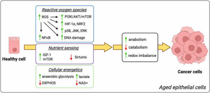
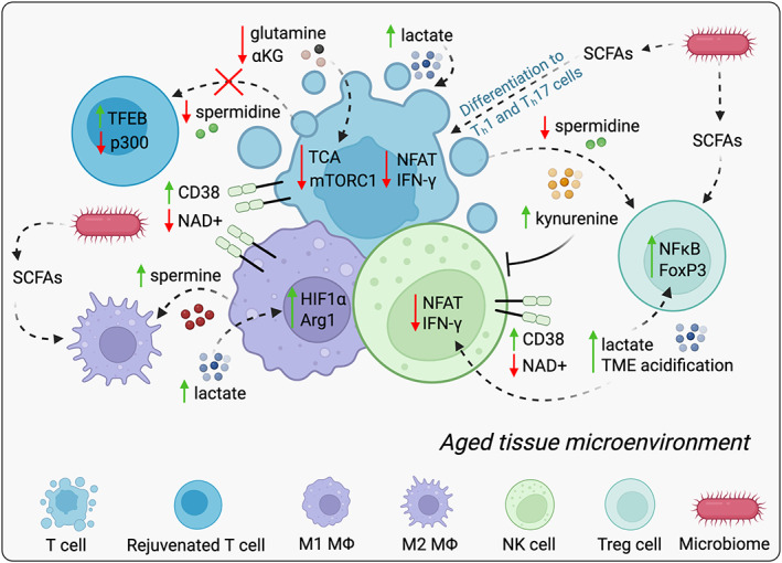
Aging is the most robust risk factor for cancer development, with more than 60% of cancers occurring in those aged 60 and above. However, how aging and tumorigenesis are intertwined is poorly understood and a matter of significant debate. Metabolic changes are hallmarks of both aging and tumorigenesis. The deleterious consequences of aging include dysfunctional cellular processes, the build-up of metabolic byproducts and waste molecules in circulation and within tissues, and stiffer connective tissues that impede blood flow and oxygenation. Collectively, these age-driven changes lead to metabolic reprogramming in different cell types of a given tissue that significantly affects their cellular functions. Here, we put forward the idea that metabolic changes that happen during aging help create a favorable environment for tumorigenesis. We review parallels in metabolic changes that happen during aging and how these changes function both as adaptive mechanisms that enable the development of malignant phenotypes in a cell-autonomous manner and as mechanisms that suppress immune surveillance, collectively creating the perfect environment for cancers to thrive. Hence, antiaging therapeutic strategies that target the metabolic reprogramming that occurs as we age might provide new opportunities to prevent cancer initiation and/or improve responses to standard-of-care anticancer therapies.
Overview
The claims and statistics of this paper haven’t been checked yet.
Run the check — free, no account needed. It reads the open-access text and checks the sources it cites.
Integrity signals
Registry
Cites
Statistics
Figures
Verification
Claims the paper makes
The claims and statistics of this paper haven’t been checked yet.
Run the check — free, no account needed. It reads the open-access text and checks the sources it cites.
Claims other papers make about this one
Each is checked against this paper’s own text: does it say what the citing paper says it does?
In the citing paper
“Both hallmarks exhibit clear commonalities, but there are also some differences or even antagonisms between aging-associated deregulated nutrient sensing and cancer-associated deregulation of cellular energy metabolism.[204][205][206]”
From Meta-hallmarks of aging and cancer (2023) · cites this paper as [206]
Self-check
Percentages, sums and reported differences re-computed from the paper’s own numbers.
The self-check needs the paper’s full text.
Citations
Cites
1 retracted
Concomitant suppression of three target genes can explain the impact of a microRNA on metastasisPreprint
Valastyan, S., Benaich, N., Chang, A., Reinhardt, F., & Weinberg, R. A. (2009) · Genes & Development · doi:10.1101/gad.1832709 · Cited by 104
RetractedHallmarks of Cancer: The Next Generation
Hanahan, D., & Weinberg, R. A. (2011) · Cell · doi:10.1016/j.cell.2011.02.013 · Cited by 66,590
Understanding the Warburg Effect: The Metabolic Requirements of Cell Proliferation
Vander Heiden, M. G., Cantley, L. C., & Thompson, C. B. (2009) · Science · doi:10.1126/science.1160809 · Cited by 16,197
López-Otín, C., Blasco, M. A., Partridge, L., Serrano, M., & Kroemer, G. (2013) · Cell · doi:10.1016/j.cell.2013.05.039 · Cited by 14,982
Mutation and cancer: statistical study of retinoblastoma
Knudson, A. G. (1971) · Proceedings of the National Academy of Sciences · doi:10.1073/pnas.68.4.820 · Cited by 7,612
ROS function in redox signaling and oxidative stress
Schieber, M., & Chandel, N. S. (2014) · Current Biology · doi:10.1016/j.cub.2014.03.034 · Cited by 6,931
Nowell, P. C. (1976) · Science · doi:10.1126/science.959840 · Cited by 6,471
The emerging hallmarks of cancer metabolism
Pavlova, N. N., & Thompson, C. B. (2016) · Cell Metabolism · doi:10.1016/j.cmet.2015.12.006 · Cited by 5,926
Gut microbiome modulates response to anti-PD-1 immunotherapy in melanoma patients
Gopalakrishnan, V., Spencer, C. N., Nezi, L., Reuben, A., Andrews, M. C., Karpinets, T. V., Prieto, P. A., Vicente, D., Hoffman, K., Wei, S. C., et al. (2018) · Science · doi:10.1126/science.aan4236 · Cited by 4,812
Metabolic regulation of gene expression by histone lactylation
Zhang, D., Tang, Z., Huang, H., Zhou, G., Cui, C., Weng, Y., Liu, W., Kim, S., Lee, S., Perez-Neut, M., et al. (2019) · Nature · doi:10.1038/s41586-019-1678-1 · Cited by 4,371
Rapamycin fed late in life extends lifespan in genetically heterogeneous mice
Harrison, D. E., Strong, R., Sharp, Z. D., Nelson, J. F., Astle, C. M., Flurkey, K., Nadon, N. L., Wilkinson, J. E., Frenkel, K., Carter, C. S., et al. (2009) · Nature · doi:10.1038/nature08221 · Cited by 3,766
The impact of the gut microbiota on human health: an integrative view
Clemente, J. C., Ursell, L. K., Parfrey, L. W., & Knight, R. (2012) · Cell · doi:10.1016/j.cell.2012.01.035 · Cited by 3,710
Reactive oxygen species in cancer
Liou, G., & Störz, P. (2010) · Free Radical Research · doi:10.3109/10715761003667554 · Cited by 3,528
Crosstalk of reactive oxygen species and NF-κB signaling
Morgan, M. J., & Liu, Z.-G. (2011) · Cell Research · doi:10.1038/cr.2010.178 · Cited by 3,505
Wallace, D. C. (2005) · Annual Review of Genetics · doi:10.1146/annurev.genet.39.110304.095751 · Cited by 3,185
Functional Polarization of Tumour-Associated Macrophages by Tumour-Derived Lactic Acid
Colegio, O. R., Chu, N.-Q., Szabo, A. L., Chu, T., Rhebergen, A. M., Jairam, V., Cyrus, N., Brokowski, C. E., Eisenbarth, S. C., Phillips, G. M., et al. (2014) · Nature · doi:10.1038/nature13490 · Cited by 3,046
MAP kinase signalling pathways in cancer
Dhillon, A. S., Hagan, S., Rath, O., & Kolch, W. (2007) · Oncogene · doi:10.1038/sj.onc.1210421 · Cited by 2,995
The gut microbiota, bacterial metabolites and colorectal cancer
Louis, P., Hold, G. L., & Flint, H. J. (2014) · Nature Reviews Microbiology · doi:10.1038/nrmicro3344 · Cited by 2,925
Defining the role of mTOR in cancer
Guertin, D. A., & Sabatini, D. M. (2007) · Cancer Cell · doi:10.1016/j.ccr.2007.05.008 · Cited by 2,833
mTOR at the nexus of nutrition, growth, ageing and disease
Liu, G. Y., & Sabatini, D. M. (2020) · Nature Reviews Molecular Cell Biology · doi:10.1038/s41580-019-0199-y · Cited by 2,737
Premature ageing in mice expressing defective mitochondrial DNA polymerase
Trifunovic, A., Wredenberg, A., Falkenberg, M., Spelbrink, J. N., Rovio, A. T., Bruder, C. E., Bohlooly-Y, M., Gidlöf, S., Oldfors, A., Wibom, R., et al. (2004) · Nature · doi:10.1038/nature02517 · Cited by 2,734
Kostic, A. D., Chun, E., Robertson, L., Glickman, J. N., Gallini, C. A., Michaud, M., Clancy, T. E., Chung, D. C., Lochhead, P., Hold, G. L., et al. (2013) · Cell Host & Microbe · doi:10.1016/j.chom.2013.07.007 · Cited by 2,718
The human tumor microbiome is composed of tumor type–specific intracellular bacteria
Nejman, D., Livyatan, I., Fuks, G., Gavert, N., Zwang, Y., Geller, L. T., Rotter-Maskowitz, A., Weiser, R., Mallel, G., Gigi, E., et al. (2020) · Science · doi:10.1126/science.aay9189 · Cited by 2,464
Understanding the Intersections between Metabolism and Cancer Biology
Heiden, M. G. V., & DeBerardinis, R. J. (2017) · Cell · doi:10.1016/j.cell.2016.12.039 · Cited by 2,369
Rejuvenation of aged progenitor cells by exposure to a young systemic environment
Conboy, I. M., Conboy, M. J., Wagers, A. J., Girma, E. R., Weissman, I. L., & Rando, T. A. (2005) · Nature · doi:10.1038/nature03260 · Cited by 2,287
Caloric Restriction Delays Disease Onset and Mortality in Rhesus Monkeys
Colman, R. J., Anderson, R. M., Johnson, S. C., Kastman, E. K., Kosmatka, K. J., Beasley, T. M., Allison, D. B., Cruzen, C., Simmons, H. A., Kemnitz, J. W., et al. (2009) · Science · doi:10.1126/science.1173635 · Cited by 2,277
Bjelakovic, G., Nikolova, D., Gluud, L. L., Simonetti, R. G., & Gluud, C. (2007) · JAMA · doi:10.1001/jama.297.8.842 · Cited by 2,275
LDHA-Associated Lactic Acid Production Blunts Tumor Immunosurveillance by T and NK Cells
Brand, A., Singer, K., Koehl, G. E., Kolitzus, M., Schoenhammer, G., Thiel, A., Matos, C., Bruss, C., Klobuch, S., Peter, K., et al. (2016) · Cell Metabolism · doi:10.1016/j.cmet.2016.08.011 · Cited by 2,039
Inhibitory effect of tumor cell-derived lactic acid on human T cells
Fischer, K., Hoffmann, P., Voelkl, S., Meidenbauer, N., Ammer, J., Edinger, M., Gottfried, E., Schwarz, S., Rothe, G., Hoves, S., et al. (2007) · Blood · doi:10.1182/blood-2006-07-035972 · Cited by 1,946
Shaw, R. J., Kosmatka, M., Bardeesy, N., Hurley, R. L., Witters, L. A., DePinho, R. A., & Cantley, L. C. (2004) · Proceedings of the National Academy of Sciences · doi:10.1073/pnas.0308061100 · Cited by 1,797
Lin, S.-J., Defossez, P.-A., & Guarente, L. (2000) · Science · doi:10.1126/science.289.5487.2126 · Cited by 1,775
L-Arginine Modulates T Cell Metabolism and Enhances Survival and Anti-tumor Activity
Geiger, R., Rieckmann, J. C., Wolf, T., Basso, C., Feng, Y., Fuhrer, T., Kogadeeva, M., Picotti, P., Meissner, F., Mann, M., et al. (2016) · Cell · doi:10.1016/j.cell.2016.09.031 · Cited by 1,775
Glutamine addiction: a new therapeutic target in cancer
Wise, D. R., & Thompson, C. B. (2010) · Trends in Biochemical Sciences · doi:10.1016/j.tibs.2010.05.003 · Cited by 1,770
Cytotoxic CD8+ T cells in cancer and cancer immunotherapy
Raskov, H., Orhan, A., Christensen, J. P., & Gögenur, I. (2021) · British Journal of Cancer · doi:10.1038/s41416-020-01048-4 · Cited by 1,687
Induction of autophagy by spermidine promotes longevity
Eisenberg, T., Knauer, H., Schauer, A., Büttner, S., Ruckenstuhl, C., Carmona-Gutierrez, D., Ring, J., Schroeder, S., Magnes, C., Antonacci, L., et al. (2009) · Nature Cell Biology · doi:10.1038/ncb1975 · Cited by 1,680
An Interaction between Kynurenine and the Aryl Hydrocarbon Receptor Can Generate Regulatory T Cells
Mezrich, J. D., Fechner, J. H., Zhang, X., Johnson, B. P., Burlingham, W. J., & Bradfield, C. A. (2010) · The Journal of Immunology · doi:10.4049/jimmunol.0903670 · Cited by 1,674
The role of ROS in tumour development and progression
Cheung, E. C., & Vousden, K. H. (2022) · Nature reviews. Cancer · doi:10.1038/s41568-021-00435-0 · Cited by 1,663
Odamaki, T., Kato, K., Sugahara, H., Hashikura, N., Takahashi, S., Xiao, J.-Z., Abe, F., & Osawa, R. (2016) · BMC Microbiology · doi:10.1186/s12866-016-0708-5 · Cited by 1,521
Gomes, A. P., Price, N. L., Ling, A. J. Y., Moslehi, J. J., Montgomery, M. K., Rajman, L., White, J. P., Teodoro, J. S., Wrann, C. D., Hubbard, B. P., et al. (2013) · Cell · doi:10.1016/j.cell.2013.11.037 · Cited by 1,482
Self-citationMetformin improves healthspan and lifespan in mice
Martin-Montalvo, A., Mercken, E. M., Mitchell, S. J., Palacios, H. H., Mote, P. L., Scheibye-Knudsen, M., Gomes, A. P., Ward, T. M., Minor, R. K., Blouin, M.-J., et al. (2013) · Nature Communications · doi:10.1038/ncomms3192 · Cited by 1,445
Self-citationThe hallmarks of cancer metabolism: still emerging
Pavlova, N. N., Zhu, J., & Thompson, C. B. (2022) · Cell Metabolism · doi:10.1016/j.cmet.2022.01.007 · Cited by 1,418
The Keap1-Nrf2 pathway: Mechanisms of activation and dysregulation in cancer
Kansanen, E., Kuosmanen, S. M., Leinonen, H., & Levonen, A.-L. (2013) · Redox Biology · doi:10.1016/j.redox.2012.10.001 · Cited by 1,313
Regulation of lifespan in Drosophila by modulation of genes in the TOR signaling pathway
Kapahi, P., Zid, B. M., Harper, T., Koslover, D., Sapin, V., & Benzer, S. (2004) · Current Biology · doi:10.1016/j.cub.2004.03.059 · Cited by 1,293
Regulation of Yeast Replicative Life Span by TOR and Sch9 in Response to Nutrients
Kaeberlein, M., Powers, R. W., Steffen, K. K., Westman, E. A., Hu, D., Dang, N., Kerr, E. O., Kirkland, K. T., Fields, S., & Kennedy, B. K. (2005) · Science · doi:10.1126/science.1115535 · Cited by 1,293
Rapamycin: one drug, many effects
Li, J., Kim, S. G., & Blenis, J. (2014) · Cell Metabolism · doi:10.1016/j.cmet.2014.01.001 · Cited by 1,287
Rac1b and reactive oxygen species mediate MMP-3-induced EMT and genomic instability
Radisky, D. C., Levy, D. D., Littlepage, L. E., Liu, H., Nelson, C. M., Fata, J. E., Leake, D., Godden, E. L., Albertson, D. G., Angela Nieto, M., et al. (2005) · Nature · doi:10.1038/nature03688 · Cited by 1,284
Oxidative stress inhibits distant metastasis by human melanoma cells
Piskounova, E., Agathocleous, M., Murphy, M. M., Hu, Z., Huddlestun, S. E., Zhao, Z., Leitch, A. M., Johnson, T. M., DeBerardinis, R. J., & Morrison, S. J. (2015) · Nature · doi:10.1038/nature15726 · Cited by 1,269
α-ketoglutarate orchestrates macrophage activation through metabolic and epigenetic reprogramming
Liu, P.-S., Wang, H., Li, X., Chao, T., Teav, T., Christen, S., Di Conza, G., Cheng, W.-C., Chou, C.-H., Vavakova, M., et al. (2017) · Nature Immunology · doi:10.1038/ni.3796 · Cited by 1,264
Metabolic Regulation of T Lymphocytes
MacIver, N. J., Michalek, R. D., & Rathmell, J. C. (2013) · Annual Review of Immunology · doi:10.1146/annurev-immunol-032712-095956 · Cited by 1,244
Mitogen-Activated Protein Kinases and Reactive Oxygen Species: How Can ROS Activate MAPK Pathways?
Son, Y., Cheong, Y.-K., Kim, N.-H., Chung, H.-T., Kang, D. G., & Pae, H.-O. (2011) · Journal of Signal Transduction · doi:10.1155/2011/792639 · Cited by 1,219
Rapamycin-Induced Insulin Resistance Is Mediated by mTORC2 Loss and Uncoupled from Longevity
Lamming, D. W., Ye, L., Katajisto, P., Goncalves, M. D., Saitoh, M., Stevens, D. M., Davis, J. G., Salmon, A. B., Richardson, A., Ahima, R. S., et al. (2012) · Science · doi:10.1126/science.1215135 · Cited by 1,199
Cardioprotection and lifespan extension by the natural polyamine spermidine
Eisenberg, T., Abdellatif, M., Schroeder, S., Primessnig, U., Stekovic, S., Pendl, T., Harger, A., Schipke, J., Zimmermann, A., Schmidt, A., et al. (2016) · Nature Medicine · doi:10.1038/nm.4222 · Cited by 1,168
Someya, S., Yu, W., Hallows, W. C., Xu, J., Vann, J. M., Leeuwenburgh, C., Tanokura, M., Denu, J. M., & Prolla, T. A. (2010) · Cell · doi:10.1016/j.cell.2010.10.002 · Cited by 1,150
DNA methylation-based measures of biological age: meta-analysis predicting time to death
Chen, B. H., Marioni, R. E., Colicino, E., Peters, M. J., Ward-Caviness, C. K., Tsai, P.-C., Roetker, N. S., Just, A. C., Demerath, E. W., Guan, W., et al. (2016) · Aging · doi:10.18632/aging.101020 · Cited by 1,126
Reversible Inactivation of the Tumor Suppressor PTEN by H2O2
Lee, S.-R., Yang, K., Kwon, J., Lee, C., Jeong, W., & Rhee, S. G. (2002) · Journal of Biological Chemistry · doi:10.1074/jbc.m111899200 · Cited by 1,061
The sirtuin SIRT6 regulates lifespan in male mice
Kanfi, Y., Naiman, S., Amir, G., Peshti, V., Zinman, G., Nahum, L., Bar-Joseph, Z., & Cohen, H. Y. (2012) · Nature · doi:10.1038/nature10815 · Cited by 1,059
Influence of TOR kinase on lifespan in C. elegans
Vellai, T., Takacs-Vellai, K., Zhang, Y., Kovacs, A. L., Orosz, L., & Müller, F. (2003) · Nature · doi:10.1038/426620a · Cited by 1,057
Mechanisms of Life Span Extension by Rapamycin in the Fruit Fly Drosophila Melanogaster
Bjedov, I., Toivonen, J. M., Kerr, F., Slack, C., Jacobson, J., Foley, A., & Partridge, L. (2010) · Cell Metabolism · doi:10.1016/j.cmet.2009.11.010 · Cited by 1,057
In Vivo Amelioration of Age-Associated Hallmarks by Partial Reprogramming.
Ocampo, A., Reddy, P., Martinez-Redondo, P., Platero-Luengo, A., Hatanaka, F., Hishida, T., Li, M., Lam, D., Kurita, M., Beyret, E., et al. (2016) · Cell · doi:10.1016/j.cell.2016.11.052 · Cited by 988
Metformin retards aging in C. elegans by altering microbial folate and methionine metabolism
Cabreiro, F., Au, C., Leung, K.-Y., Vergara-Irigaray, N., Cochemé, H. M., Noori, T., Weinkove, D., Schuster, E., Greene, N. D. E., & Gems, D. (2013) · Cell · doi:10.1016/j.cell.2013.02.035 · Cited by 967
Redox regulation of protein tyrosine phosphatase 1B involves a sulphenyl-amide intermediate
Salmeen, A., Andersen, J. N., Myers, M. P., Meng, T.-C., Hinks, J. A., Tonks, N. K., & Barford, D. (2003) · Nature · doi:10.1038/nature01680 · Cited by 951
How cancer metabolism is tuned for proliferation and vulnerable to disruption
Schulze, A., & Harris, A. L. (2012) · Nature · doi:10.1038/nature11706 · Cited by 945
Lactate in the Regulation of Tumor Microenvironment and Therapeutic Approaches
de la Cruz-López, K. G., Castro-Muñoz, L. J., Reyes-Hernández, D. O., García-Carrancá, A., & Manzo-Merino, J. (2019) · Frontiers in Oncology · doi:10.3389/fonc.2019.01143 · Cited by 940
Bjelakovic, G., Nikolova, D., Gluud, L. L., Simonetti, R. G., & Gluud, C. (2012) · Cochrane Database of Systematic Reviews · doi:10.1002/14651858.cd007176.pub2 · Cited by 924
Nakaya, M., Xiao, Y., Zhou, X., Chang, J., Chang, M., Cheng, X., Blonska, M., Lin, X., & Sun, S. (2014) · Immunity · doi:10.1016/j.immuni.2014.04.007 · Cited by 902
Long-Term Calorie Restriction Is Highly Effective in Reducing the Risk for Atherosclerosis in Humans
Fontana, L., Meyer, T. E., Klein, S., & Holloszy, J. O. (2004) · Proceedings of the National Academy of Sciences · doi:10.1073/pnas.0308291101 · Cited by 902
Levine, M. E. (2012) · The Journals of Gerontology Series A · doi:10.1093/gerona/gls233 · Cited by 897
SIRT1 redistribution on chromatin promotes genomic stability but alters gene expression during aging
Oberdoerffer, P., Michan, S., McVay, M., Mostoslavsky, R., Vann, J., Park, S.-K., Hartlerode, A., Stegmuller, J., Hafner, A., Loerch, P., et al. (2008) · Cell · doi:10.1016/j.cell.2008.10.025 · Cited by 852
mtDNA mutations increase tumorigenicity in prostate cancer
Petros, J. A., Baumann, A. K., Ruiz‐Pesini, E., Amin, M. B., Sun, C. Q., Hall, J., Lim, S.-D., Issa, M. M., Flanders, W. D., Hosseini, S. H., et al. (2005) · Proceedings of the National Academy of Sciences · doi:10.1073/pnas.0408894102 · Cited by 850
Carr, E. L., Kelman, A., Wu, G. S., Gopaul, R., Senkevitch, E., Aghvanyan, A., Turay, A. M., & Frauwirth, K. A. (2010) · The Journal of Immunology · doi:10.4049/jimmunol.0903586 · Cited by 830
Reprogramming to recover youthful epigenetic information and restore vision
Lu, Y., Brommer, B., Tian, X., Krishnan, A., Meer, M., Wang, C., Vera, D. L., Zeng, Q., Yu, D., Bonkowski, M. S., et al. (2020) · Nature · doi:10.1038/s41586-020-2975-4 · Cited by 820
Causes, consequences, and reversal of immune system aging
Montecino-Rodriguez, E., Berent-Maoz, B., & Dorshkind, K. (2013) · Journal of Clinical Investigation · doi:10.1172/jci64096 · Cited by 793
Metabolic coordination of T cell quiescence and activation
Chapman, N. M., Boothby, M. R., & Chi, H. (2019) · Nature reviews. Immunology · doi:10.1038/s41577-019-0203-y · Cited by 793
Detection of a specific mitochondrial DNA deletion in tissues of older humans
Cortopassi, G. A., & Arnheim, N. (1990) · Nucleic Acids Research · doi:10.1093/nar/18.23.6927 · Cited by 788
SIRT3 Opposes Reprogramming of Cancer Cell Metabolism through HIF1α Destabilization
Finley, L. W. S., Carracedo, A., Lee, J., Souza, A., Egia, A., Zhang, J., Teruya-Feldstein, J., Moreira, P. I., Cardoso, S. M., Clish, C. B., et al. (2011) · Cancer Cell · doi:10.1016/j.ccr.2011.02.014 · Cited by 782
Metabolic Control of Longevity
López-Otín, C., Galluzzi, L., Freije, J. M. P., Madeo, F., & Kroemer, G. (2016) · Cell · doi:10.1016/j.cell.2016.07.031 · Cited by 753
ROS signaling under metabolic stress: cross-talk between AMPK and AKT pathway
Zhao, Y., Hu, X., Liu, Y., Dong, S., Wen, Z., He, W., Zhang, S., Huang, Q., & Shi, M. (2017) · Molecular Cancer · doi:10.1186/s12943-017-0648-1 · Cited by 735
Decline in skeletal muscle mitochondrial respiratory chain function: possible factor in ageing
Trounce, I., Byrne, E., & Marzuki, S. (1989) · The Lancet · doi:10.1016/s0140-6736(89)92143-0 · Cited by 730
Distinct Regulation of Th17 and Th1 Cell Differentiation by Glutaminase-Dependent Metabolism
Johnson, M. O., Wolf, M. M., Madden, M. Z., Andrejeva, G., Sugiura, A., Contreras, D. C., Maseda, D., Liberti, M. V., Paz, K. G., Kishton, R. J., et al. (2018) · Cell · doi:10.1016/j.cell.2018.10.001 · Cited by 729
The histone deacetylase SIRT6 is a tumor suppressor that controls cancer metabolism
Sebastián, C., Zwaans, B. M. M., Silberman, D. M., Gymrek, M., Goren, A., Zhong, L., Ram, O., Truelove, J., Guimaraes, A. R., Toiber, D., et al. (2012) · Cell · doi:10.1016/j.cell.2012.10.047 · Cited by 660
The double-edged roles of ROS in cancer prevention and therapy
Wang, Y., Qi, H., Liu, Y., Duan, C., Liu, X., Xia, T., Chen, D., Piao, H., & Liu, H. (2021) · Theranostics · doi:10.7150/thno.56747 · Cited by 657
The Two Faces of Reactive Oxygen Species in Cancer
Reczek, C. R., & Chandel, N. S. (2017) · Annual Review of Cancer Biology · doi:10.1146/annurev-cancerbio-041916-065808 · Cited by 602
Disturbed mitochondrial dynamics in CD8+ TILs reinforce T cell exhaustion
Yu, Y.-R., Imrichova, H., Wang, H., Chao, T., Xiao, Z., Gao, M., Rincon-Restrepo, M., Franco, F., Genolet, R., Cheng, W.-C., et al. (2020) · Nature Immunology · doi:10.1038/s41590-020-0793-3 · Cited by 593
Martens, C. R., Denman, B. A., Mazzo, M. R., Armstrong, M. L., Reisdorph, N., McQueen, M. B., Chonchol, M., & Seals, D. R. (2018) · Nature Communications · doi:10.1038/s41467-018-03421-7 · Cited by 588
Aging of the Immune System. Mechanisms and Therapeutic Targets
Weyand, C. M., & Goronzy, J. J. (2016) · Annals of the American Thoracic Society · doi:10.1513/annalsats.201602-095aw · Cited by 586
Hopkins, M. J., Sharp, R., & Macfarlane, G. T. (2001) · Gut · doi:10.1136/gut.48.2.198 · Cited by 577
Fang, E. F., Kassahun, H., Croteau, D. L., Scheibye-Knudsen, M., Marosi, K., Lu, H., Shamanna, R. A., Kalyanasundaram, S., Bollineni, R. C., Wilson, M. A., et al. (2016) · Cell Metabolism · doi:10.1016/j.cmet.2016.09.004 · Cited by 575
Mitochondrial dysfunction and oxidative stress in aging and cancer
Kudryavtseva, A. V., Krasnov, G. S., Dmitriev, A. A., Alekseev, B. Y., Kardymon, O. L., Садритдинова, А. Ф., Fedorova, M., Pokrovsky, A. V., Melnikova, N. V., Каприн, А. Д., et al. (2016) · Oncotarget · doi:10.18632/oncotarget.9821 · Cited by 564
Lactate: A Metabolic Driver in the Tumour Landscape
Ippolito, L., Morandi, A., Giannoni, E., & Chiarugi, P. (2018) · Trends in Biochemical Sciences · doi:10.1016/j.tibs.2018.10.011 · Cited by 541
ROS in Cancer: The Burning Question
Chio, I. I. C., & Tuveson, D. A. (2017) · Trends in Molecular Medicine · doi:10.1016/j.molmed.2017.03.004 · Cited by 532
Klysz, D., Tai, X., Robert, P. A., Craveiro, M., Cretenet, G., Oburoglu, L., Mongellaz, C., Floess, S., Fritz, V., Matias, M. I., et al. (2015) · Science Signaling · doi:10.1126/scisignal.aab2610 · Cited by 522
NAD(+) in aging: molecular mechanisms and translational implications
Fang, E. F., Lautrup, S., Hou, Y., Demarest, T. G., Croteau, D. L., Mattson, M. P., & Bohr, V. A. (2017) · Trends in Molecular Medicine · doi:10.1016/j.molmed.2017.08.001 · Cited by 501
Comprehensive molecular characterization of mitochondrial genomes in human cancers
Yuan, Y., Ju, Y. S., Kim, Y.-W., Li, J., Wang, Y., Yoon, S.-S., Yang, J. Y. H., Martincorena, I., Creighton, C. J., Weinstein, J. N., et al. (2020) · Nature Genetics · doi:10.1038/s41588-019-0557-x · Cited by 485
The Maintenance of Mitochondrial DNA Integrity--Critical Analysis and UpdatePreprint
ALEXEYEV, M. F., Shokolenko, I. N., Wilson, G. L., & LeDoux, S. P. (2013) · Cold Spring Harbor Perspectives in Biology · doi:10.1101/cshperspect.a012641 · Cited by 478
Hou, Y., Lautrup, S., Cordonnier, S., Wang, Y., Croteau, D. L., Zavala, E., Zhang, Y., Moritoh, K., O’Connell, J. F., Baptiste, B. A., et al. (2018) · Proceedings of the National Academy of Sciences · doi:10.1073/pnas.1718819115 · Cited by 472
Zhu, X.-H., Lu, M., Lee, B.-Y., Ugurbil, K., & Chen, W. (2015) · Proceedings of the National Academy of Sciences · doi:10.1073/pnas.1417921112 · Cited by 468
Elhassan, Y. S., Kluckova, K., Fletcher, R. S., Schmidt, M. S., Garten, A., Doig, C. L., Cartwright, D. M., Oakey, L., Burley, C. V., Jenkinson, N., et al. (2019) · Cell Reports · doi:10.1016/j.celrep.2019.07.043 · Cited by 448
Jeong, S. M., Xiao, C., Finley, L. W. S., Lahusen, T., Souza, A. L., Pierce, K., Li, Y.-H., Wang, X., Laurent, G., German, N. J., et al. (2013) · Cancer Cell · doi:10.1016/j.ccr.2013.02.024 · Cited by 445
Bell, E. L., Emerling, B. M., Ricoult, S. J. H., & Guarente, L. (2011) · Oncogene · doi:10.1038/onc.2011.37 · Cited by 440
Kaur, A., Ecker, B. L., Douglass, S. M., Kugel, C. H., Webster, M. R., Almeida, F. V., Somasundaram, R., Hayden, J. E., Ban, E., Ahmadzadeh, H., et al. (2018) · Cancer Discovery · doi:10.1158/2159-8290.cd-18-0193 · Cited by 437
SIRT3 Reverses Aging-Associated Degeneration
Brown, K., Xie, S., Qiu, X., Mohrin, M., Shin, J., Liu, Y., Zhang, D., Scadden, D. T., & Chen, D. (2013) · Cell Reports · doi:10.1016/j.celrep.2013.01.005 · Cited by 436
Hagen, T. M., Yowe, D. L., Bartholomew, J. C., Wehr, C. M., Katherine, L. B. B., Park, J.-Y., & Ames, B. N. (1997) · Proceedings of the National Academy of Sciences · doi:10.1073/pnas.94.7.3064 · Cited by 434
Phosphoinositide 3-Kinase/Akt Signaling and Redox Metabolism in Cancer
Koundouros, N., & Poulogiannis, G. (2018) · Frontiers in Oncology · doi:10.3389/fonc.2018.00160 · Cited by 425
Senescent cells promote tissue NAD+ decline during ageing via the activation of CD38+ macrophages
Covarrubias, A. J., Kale, A., Perrone, R., Lopez-Dominguez, J. A., Pisco, A. O., Kasler, H. G., Schmidt, M. S., Heckenbach, I., Kwok, R., Wiley, C. D., et al. (2020) · Nature Metabolism · doi:10.1038/s42255-020-00305-3 · Cited by 420
Chiesa, M. D., Carlomagno, S., Frumento, G., Balsamo, M., Cantoni, C., Conte, R., Moretta, L., Moretta, A., & Vitale, M. (2006) · Blood · doi:10.1182/blood-2006-03-006700 · Cited by 417
Mitochondrial point mutations do not limit the natural lifespan of mice
Vermulst, M., Bielas, J. H., Kujoth, G. C., Ladiges, W. C., Rabinovitch, P. S., Prolla, T. A., & Loeb, L. A. (2007) · Nature Genetics · doi:10.1038/ng1988 · Cited by 404
Using DNA methylation profiling to evaluate biological age and longevity interventions
Petkovich, D. A., Podolskiy, D. I., Lobanov, A. V., Lee, S.-G., Miller, R. A., & Gladyshev, V. N. (2017) · Cell Metabolism · doi:10.1016/j.cmet.2017.03.016 · Cited by 398
Mitochondrial DNA mutations and mitochondrial DNA depletion in gastric cancer
Wu, C., Yin, P., Hung, W., Li, A. F., Li, S., Chi, C., Wei, Y., & Lee, H. (2005) · Genes Chromosomes and Cancer · doi:10.1002/gcc.20213 · Cited by 381
Mitochondrial and nuclear DNA matching shapes metabolism and healthy ageing
Latorre-Pellicer, A., Moreno-Loshuertos, R., Lechuga-Vieco, A. V., Sánchez-Cabo, F., Torroja, C., Acín-Pérez, R., Calvo, E., Aix, E., González-Guerra, A., Logan, A., et al. (2016) · Nature · doi:10.1038/nature18618 · Cited by 377
Lactate Limits T Cell Proliferation via the NAD(H) Redox State
Quinn, W. J., Jiao, J., TeSlaa, T. A., Stadanlick, J. E., Wang, Z., Wang, L., Akimova, T., Angelin, A., Schäfer, P. M., Cully, M. D., et al. (2020) · Cell Reports · doi:10.1016/j.celrep.2020.108500 · Cited by 376
Immune regulation by microbiome metabolites
Kim, C. H. (2018) · Immunology · doi:10.1111/imm.12930 · Cited by 375
Metabolic positron emission tomography imaging in cancer detection and therapy response
Zhu, A., Lee, D., & Shim, H. (2011) · Seminars in Oncology · doi:10.1053/j.seminoncol.2010.11.012 · Cited by 368
Thymic involution and rising disease incidence with age
Palmer, S., Albergante, L., Blackburn, C. C., & Newman, T. J. (2018) · Proceedings of the National Academy of Sciences · doi:10.1073/pnas.1714478115 · Cited by 364
Kennedy, S. R., Salk, J. J., Schmitt, M. W., & Loeb, L. A. (2013) · PLoS Genetics · doi:10.1371/journal.pgen.1003794 · Cited by 363
Spermidine induces autophagy by inhibiting the acetyltransferase EP300
Pietrocola, F., Lachkar, S., Enot, D. P., Niso-Santano, M., Bravo-San Pedro, J. M., Sica, V, Izzo, V, Maiuri, M. C., Madeo, F., Mariño, G., et al. (2015) · Cell Death & Differentiation · doi:10.1038/cdd.2014.215 · Cited by 357
The overexpression of major antioxidant enzymes does not extend the lifespan of mice
Pérez, V. I., Van Remmen, H., Bokov, A., Epstein, C. J., Vijg, J., & Richardson, A. (2009) · Aging Cell · doi:10.1111/j.1474-9726.2008.00449.x · Cited by 353
Rapamycin increases lifespan and inhibits spontaneous tumorigenesis in inbred female mice
Anisimov, V. N., Zabezhinski, M. A., Popovich, I. G., Piskunova, T. S., Semenchenko, A. V., Тындык, М. Л., Yurova, M. N., Rosenfeld, S. V., & Blagosklonny, M. V. (2011) · Cell Cycle · doi:10.4161/cc.10.24.18486 · Cited by 353
A high-fat diet and NAD(+) activate Sirt1 to rescue premature aging in Cockayne syndrome
Scheibye-Knudsen, M., Mitchell, S. J., Fang, E. F., Iyama, T., Ward, T., Wang, J., Dunn, C. A., Singh, N., Veith, S., Hasan-Olive, M. M., et al. (2014) · Cell Metabolism · doi:10.1016/j.cmet.2014.10.005 · Cited by 352
CD38: An Immunomodulatory Molecule in Inflammation and Autoimmunity
Piedra-Quintero, Z. L., Wilson, Z., Nava, P., & Guerau‐de‐Arellano, M. (2020) · Frontiers in Immunology · doi:10.3389/fimmu.2020.597959 · Cited by 351
Caloric Restriction Reprograms the Single-Cell Transcriptional Landscape of Rattus Norvegicus Aging
Ma, S., Sun, S., Geng, L., Song, M., Wang, W., Ye, Y., Ji, Q., Zou, Z., Wang, S., He, X., et al. (2020) · Cell · doi:10.1016/j.cell.2020.02.008 · Cited by 349
CD38 ecto-enzyme in immune cells is induced during aging and regulates NAD+ and NMN levels
Chini, C. C. S., Peclat, T. R., Warner, G. M., Kashyap, S., Espindola-Netto, J. M., de Oliveira, G. C., Gomez, L. S., Hogan, K. A., Tarragó, M. G., Puranik, A. S., et al. (2020) · Nature Metabolism · doi:10.1038/s42255-020-00298-z · Cited by 349
Polyamines control eIF5A hypusination, TFEB translation, and autophagy to reverse B cell senescence
Zhang, H., Alsaleh, G., Feltham, J., Sun, Y., Napolitano, G., Riffelmacher, T., Charles, P., Frau, L., Hublitz, P., Yu, Z., et al. (2019) · Molecular Cell · doi:10.1016/j.molcel.2019.08.005 · Cited by 341
Sekhar, R. V., Patel, S. G., Guthikonda, A. P., Reid, M., Balasubramanyam, A., Taffet, G. E., & Jahoor, F. (2011) · The American Journal of Clinical Nutrition · doi:10.3945/ajcn.110.003483 · Cited by 336
Elyahu, Y., Hekselman, I., Eizenberg-Magar, I., Berner, O., Strominger, I., Schiller, M., Mittal, K., Nemirovsky, A., Eremenko, E., Vital, A., et al. (2019) · Science Advances · doi:10.1126/sciadv.aaw8330 · Cited by 324
Triplett, T. A., Garrison, K. C., Marshall, N., Donkor, M., Blazeck, J., Lamb, C., Qerqez, A., Dekker, J. D., Tanno, Y., Lu, W.-C., et al. (2018) · Nature Biotechnology · doi:10.1038/nbt.4180 · Cited by 321
Caloric restriction in humans reveals immunometabolic regulators of health span
Spadaro, O., Youm, Y., Shchukina, I., Ryu, S., Sidorov, S., Ravussin, A., Nguyen, K., Aladyeva, E., Predeus, A. N., Smith, S. R., et al. (2022) · Science · doi:10.1126/science.abg7292 · Cited by 317
Wu, J. J., Liu, J., Chen, E. B., Wang, J. J., Cao, L., Narayan, N., Fergusson, M. M., Rovira, I. I., Allen, M., Springer, D. A., et al. (2013) · Cell Reports · doi:10.1016/j.celrep.2013.07.030 · Cited by 307
Dietary deprivation extends lifespan in Caenorhabditis elegans
Lee, G. D., Wilson, M. A., Zhu, M., Wolkow, C. A., De Cabo, R., Ingram, D. K., & Zou, S. (2006) · Aging Cell · doi:10.1111/j.1474-9726.2006.00241.x · Cited by 307
Polyamine metabolism is a central determinant of helper T cell lineage fidelity
Puleston, D. J., Baixauli, F., Sanin, D. E., Edwards-Hicks, J., Villa, M., Kabat, A. M., Kamiński, M. M., Stanckzak, M., Weiss, H. J., Grzes, K. M., et al. (2021) · Cell · doi:10.1016/j.cell.2021.06.007 · Cited by 305
Ross, J. M., Öberg, J., Brené, S., Coppotelli, G., Terzioglu, M., Pernold, K., Goiny, M., Sitnikov, R., Kehr, J., Trifunovic, A., et al. (2010) · Proceedings of the National Academy of Sciences · doi:10.1073/pnas.1008189107 · Cited by 299
The Free Radical Theory of Aging Is Dead. Long Live the Damage Theory!
Gladyshev, V. N. (2013) · Antioxidants and Redox Signaling · doi:10.1089/ars.2013.5228 · Cited by 298
Gao, Y., Zhang, W., Zeng, L.-Q., Bai, H., Li, J., Zhou, J., Zhou, G., Fang, C., Wang, F., & Qin, X.-J. (2020) · Redox Biology · doi:10.1016/j.redox.2020.101635 · Cited by 298
Wang, T., Tsui, B., Kreisberg, J. F., Robertson, N., Gross, A. M., Yu, M., Carter, H. K., Brown‐Borg, H. M., Adams, P. D., & Ideker, T. (2017) · Genome biology · doi:10.1186/s13059-017-1186-2 · Cited by 296
Serrano, L., Martínez‐Redondo, P., Marazuela-Duque, A., Vázquez, B. N., Dooley, S., Voigt, P., Beck, D. B., Kane‐Goldsmith, N., Tong, Q., Rabanal, R. M., et al. (2013) · Genes & Development · doi:10.1101/gad.211342.112 · Cited by 290
Rapamycin Extends Life and Health in C57BL/6 Mice
Zhang, Y., Bokov, A. F., Gelfond, J., Soto, V., Ikeno, Y., Hubbard, G. B., Diaz, V., Sloane, L. B., Maslin, K., Treaster, S. B., et al. (2013) · The Journals of Gerontology Series A · doi:10.1093/gerona/glt056 · Cited by 283
Kibe, R., Kurihara, S., Sakai, Y., Suzuki, H., Ooga, T., Sawaki, E., Muramatsu, K., Nakamura, A., Yamashita, A., Kitada, Y., et al. (2014) · Scientific Reports · doi:10.1038/srep04548 · Cited by 273
CD38-NAD+Axis Regulates Immunotherapeutic Anti-Tumor T Cell Response
Chatterjee, S., Daenthanasanmak, A., Chakraborty, P., Wyatt, M. W., Dhar, P., Selvam, S. P., Fu, J., Zhang, J., Nguyen, H., Kang, I., et al. (2018) · Cell Metabolism · doi:10.1016/j.cmet.2017.10.006 · Cited by 270
Glutathione contents of tissues in the aging mouse
Hazelton, G. A., & Lang, C. A. (1980) · Biochemical Journal · doi:10.1042/bj1880025 · Cited by 268
Fontana, L., Villareal, D. T., Weiss, E. P., Racette, S. B., Steger-May, K., Klein, S. B., & Holloszy, J. O. (2007) · American Journal of Physiology-Endocrinology and Metabolism · doi:10.1152/ajpendo.00102.2007 · Cited by 267
Role of Nicotinamide in DNA Damage, Mutagenesis, and DNA Repair
Surjana, D., Halliday, G. M., & Damian, D. L. (2010) · Journal of Nucleic Acids · doi:10.4061/2010/157591 · Cited by 264
Age‐Dependent Impairment of Mitochondrial Function in Primate Brain
Bowling, A. C., Mutisya, E. M., Walker, L. C., Price, D. L., Cork, L. C., & Beal, M. H. (1993) · Journal of Neurochemistry · doi:10.1111/j.1471-4159.1993.tb13430.x · Cited by 263
Reactive Oxygen Species and Regulation of Gene Expression
Turpaev, K. T. (2002) · Biochemistry (Moscow) · doi:10.1023/a:1014819832003 · Cited by 258
Comito, G., Iscaro, A., Bacci, M., Morandi, A., Ippolito, L., Parri, M., Montagnani, I., Raspollini, M. R., Serni, S., Simeoni, L., et al. (2019) · Oncogene · doi:10.1038/s41388-019-0688-7 · Cited by 257
DNA Damage as the Primary Cause of Aging
Gensler, H. L., & Bernstein, H. (1981) · The Quarterly Review of Biology · doi:10.1086/412317 · Cited by 252
Age-induced accumulation of methylmalonic acid promotes tumour progression
Gomes, A. P., Ilter, D., Low, V., Endress, J. E., Fernández-García, J., Rosenzweig, A., Schild, T., Broekaert, D., Ahmed, A., Planque, M., et al. (2020) · Nature · doi:10.1038/s41586-020-2630-0 · Cited by 244
Self-citationHigh incidence of somatic mitochondrial DNA mutations in human ovarian carcinomas.
Liu, V. W. S., Shi, H., Cheung, A., Chiu, P. M., Leung, T., NAGLEY, P., Wong, L. C., & Ngan, H. (2001) · PubMed · Cited by 243
Spectrum and Risk of Neoplasia in Werner Syndrome: A Systematic Review
Lauper, J. M., Krause, A. J., Vaughan, T. L., & Monnat, R. J. (2013) · PLoS ONE · doi:10.1371/journal.pone.0059709 · Cited by 234
Age related human T cell subset evolution and senescence
Li, M., Yao, D., Zeng, X., Kasakovski, D., Zhang, Y., Chen, S., Zha, X., Li, Y., & Xu, L. (2019) · Immunity & Ageing · doi:10.1186/s12979-019-0165-8 · Cited by 230
Dostal, A., Chassard, C., Hilty, F. M., Zimmermann, M. B., Jaeggi, T., Rossi, S., & Lacroix, C. (2011) · Journal of Nutrition · doi:10.3945/jn.111.148643 · Cited by 229
Skokowa, J., Lan, D., Thakur, B. K., Wang, F., Gupta, K., Cario, G., Brechlin, A. M., Schambach, A., Hinrichsen, L., Meyer, G., et al. (2009) · Nature Medicine · doi:10.1038/nm.1913 · Cited by 221
The human NAD metabolome: Functions, metabolism and compartmentalization
Nikiforov, A. A., Kulikova, V., & Ziegler, M. (2015) · Critical Reviews in Biochemistry and Molecular Biology · doi:10.3109/10409238.2015.1028612 · Cited by 221
Deleterious mitochondrial DNA mutations accumulate in aging human tissues
Arnheim, N., & Cortopassi, G. A. (1992) · Mutation Research/DNAging · doi:10.1016/0921-8734(92)90020-p · Cited by 217
van Karnebeek, C., & Stöckler, S. (2011) · Molecular Genetics and Metabolism · doi:10.1016/j.ymgme.2011.11.191 · Cited by 214
Low blood glutathione levels in healthy aging adults.
Lang, C. A., Naryshkin, S., Schneider, D. L., Mills, B. J., & Lindeman, R. D. (1992) · PubMed · Cited by 213
Gut dysbiosis: a potential link between increased cancer risk in ageing and inflammaging
Biragyn, A., & Ferrucci, L. (2018) · The Lancet Oncology · doi:10.1016/s1470-2045(18)30095-0 · Cited by 209
Sesso, H. D., Christen, W. G., Bubes, V., Smith, J. P., MacFadyen, J., Schvartz, M., Manson, J. E., Glynn, R. J., Buring, J. E., & Gaziano, J. M. (2012) · JAMA · doi:10.1001/jama.2012.14805 · Cited by 208
Regulating T-cell differentiation through the polyamine spermidine
Carriche, G. M., Almeida, L., Stüve, P., Velásquez, L. N., Dhillon-LaBrooy, A., Roy, U., Lindenberg, M., Strowig, T., Plaza‐Sirvent, C., Schmitz, I., et al. (2020) · Journal of Allergy and Clinical Immunology · doi:10.1016/j.jaci.2020.04.037 · Cited by 205
van der Goot, A. T., Zhu, W., Vázquez‐Manrique, R. P., Seinstra, R. I., Dettmer, K., Michels, H., Farina, F., Krijnen, J., Melki, R., Buijsman, R. C., et al. (2012) · Proceedings of the National Academy of Sciences · doi:10.1073/pnas.1203083109 · Cited by 199
Clinical Phenotype of Families with Longevity
Atzmon, G., Schechter, C. B., Greiner, W., Davidson, D. E., Rennert, G., & Barzilai, N. (2004) · Journal of the American Geriatrics Society · doi:10.1111/j.1532-5415.2004.52068.x · Cited by 197
Age as a prognostic factor in ovarian carcinoma: The gynecologic oncology group experience
Thigpen, T., Brady, M. F., OMURA, G. A., Creasman, W. T., McGuire, W. P., Hoskins, W. J., & Williams, S. D. (2010) · Cancer · doi:10.1002/cncr.2820710218 · Cited by 197
Reactive Oxygen Species Regulate Hypoxia-Inducible Factor 1α Differentially in Cancer and Ischemia
Qutub, A. A., & Popel, A. S. (2008) · Molecular and Cellular Biology · doi:10.1128/mcb.00060-08 · Cited by 196
Methylation-Based Biological Age and Breast Cancer Risk
Kresovich, J. K., Xu, Z., O’Brien, K. M., Weinberg, C. R., Sandler, D. P., & Taylor, J. A. (2019) · JNCI Journal of the National Cancer Institute · doi:10.1093/jnci/djz020 · Cited by 195
Mitochondrial DNA copy number and mitochondrial DNA deletion in adult and senescent rats
Gadaleta, M. N., Rainaldi, G., Lezza, A. M. S., Milella, F., Fracasso, F., & Cantatore, P. (1992) · Mutation Research/DNAging · doi:10.1016/0921-8734(92)90022-h · Cited by 194
Lifestyle Factors in Cancer Survivorship
Ligibel, J. A. (2012) · Journal of Clinical Oncology · doi:10.1200/jco.2012.42.0638 · Cited by 189
A conserved NAD + binding pocket that regulates protein-protein interactions during aging
Li, J., Bonkowski, M. S., Moniot, S., Zhang, D., Hubbard, B. P., Ling, A. J. Y., Rajman, L. A., Qin, B., Lou, Z., Gorbunova, V. A., et al. (2017) · Science · doi:10.1126/science.aad8242 · Cited by 188
The role of polyamines in the regulation of macrophage polarization and function
Latour, Y. L., Gobert, A. P., & Wilson, K. T. (2019) · Amino Acids · doi:10.1007/s00726-019-02719-0 · Cited by 182
Mechanisms linking mtDNA damage and aging
Pinto, M., & Moraes, C. T. (2015) · Free Radical Biology and Medicine · doi:10.1016/j.freeradbiomed.2015.05.005 · Cited by 179
McReynolds, M. R., Chellappa, K., & Baur, J. A. (2020) · Experimental Gerontology · doi:10.1016/j.exger.2020.110888 · Cited by 179
Yue, F., Li, W., Zou, J., Jiang, X., Xu, G., Huang, H. W., & Liu, L. (2017) · Cancer Research · doi:10.1158/0008-5472.can-16-3462 · Cited by 178
NAD+ biosynthesis, aging, and disease
Johnson, S., & Imai, S. (2018) · F1000Research · doi:10.12688/f1000research.12120.1 · Cited by 178
The evolutionary relationships among known life forms
Cedergren, R., Gray, M. W., Abel, Y., & Sankoff, D. (1988) · Journal of Molecular Evolution · doi:10.1007/bf02143501 · Cited by 177
Swindell, W. R. (2009) · BMC Genomics · doi:10.1186/1471-2164-10-585 · Cited by 171
Petrosillo, G., Matera, M. G., Casanova, G., Ruggiero, F. M., & Paradies, G. (2008) · Neurochemistry International · doi:10.1016/j.neuint.2008.07.001 · Cited by 171
Bruzzone, S., Fruscione, F., Morando, S., Ferrando, T., Poggi, A. D., Garuti, A., D’Urso, A., Selmo, M., Benvenuto, F., Cea, M., et al. (2009) · PLoS ONE · doi:10.1371/journal.pone.0007897 · Cited by 171
Lactic acid promotes macrophage polarization through MCT-HIF1α signaling in gastric cancer
Zhang, L., & Li, S. (2020) · Experimental Cell Research · doi:10.1016/j.yexcr.2020.111846 · Cited by 168
Kiss, T., Nyúl-Tóth, Á., Balasubramanian, P., Tarantini, S., Ahire, C., Yabluchanskiy, A., Csipo, T., Farkas, E., Wren, J. D., Garman, L., et al. (2020) · GeroScience · doi:10.1007/s11357-020-00165-5 · Cited by 163
Mitochondrial ROS-derived PTEN oxidation activates PI3K pathway for mTOR-induced myogenic autophagy
Kim, J. H., Choi, T. G., Park, S., Yun, H. R., Nguyen, N. N. Y., Jo, Y. H., Jang, M., Kim, J., Kim, J., Kang, I., et al. (2018) · Cell Death and Differentiation · doi:10.1038/s41418-018-0165-9 · Cited by 158
Ecker, B. L., Kaur, A., Douglass, S. M., Webster, M. R., Almeida, F. V., Marino, G. E., Sinnamon, A. J., Neuwirth, M. G., Alicea, G. M., Ndoye, A., et al. (2019) · Cancer Discovery · doi:10.1158/2159-8290.cd-18-0168 · Cited by 153
Yalcin, S., Marinković, D., Mungamuri, S. K., Zhang, X., Tong, W., Sellers, R. S., & Ghaffari, S. (2010) · The EMBO Journal · doi:10.1038/emboj.2010.292 · Cited by 150
Schöpf, B., Weißensteiner, H., Schäfer, G., Fazzini, F., Charoentong, P., Naschberger, A., Rupp, B., Fendt, L., Bukur, V., Giese, I., et al. (2020) · Nature Communications · doi:10.1038/s41467-020-15237-5 · Cited by 148
Ancient mtDNA Genetic Variants Modulate mtDNA Transcription and Replication
Suissa, S., Wang, Z., Poole, J. C., Wittkopp, S., Feder, J., Shutt, T. E., Wallace, D. C., Shadel, G. S., & Mishmar, D. (2009) · PLoS Genetics · doi:10.1371/journal.pgen.1000474 · Cited by 147
Polyamines and Kynurenines at the Intersection of Immune Modulation
Proietti, E., Rossini, S., Grohmann, U., & Mondanelli, G. (2020) · Trends in Immunology · doi:10.1016/j.it.2020.09.007 · Cited by 146
Spermidine and spermine are enriched in whole blood of nona/centenarians
Pucciarelli, S., Moreschini, B., Micozzi, D., De Fronzo, G. S., Carpi, F. M., Polzonetti, V., Vincenzetti, S., Mignini, F., & Napolioni, V. (2012) · Rejuvenation Research · doi:10.1089/rej.2012.1349 · Cited by 144
The microbiome: An emerging key player in aging and longevity
Kim, M., & Benayoun, B. A. (2020) · Translational Medicine of Aging · doi:10.1016/j.tma.2020.07.004 · Cited by 144
Regulation of macrophage activation and inflammation by spermine: A new chapter in an old story
Zhang, M., Wang, H., & Tracey, K. J. (2000) · Critical Care Medicine · doi:10.1097/00003246-200004001-00007 · Cited by 142
Regulation of NK Cell Function by Human Granulocyte Arginase
Oberlies, J., Watzl, C., Giese, T., Luckner, C., Kropf, P., Müller, I., Ho, A. D., & Munder, M. (2009) · The Journal of Immunology · doi:10.4049/jimmunol.0803523 · Cited by 142
Bjelaković, G., Nikolova, D., Gluud, L. L., Simonetti, R. G., & Gluud, C. N. (2015) · Sao Paulo Medical Journal · doi:10.1590/1516-3180.20151332t1 · Cited by 140
Smith, A. L. M., Whitehall, J. C., Bradshaw, C., Gay, D. M., Robertson, F. M., Blain, A. P., Hudson, G., Pyle, A., Houghton, D., Hunt, M., et al. (2020) · Nature Cancer · doi:10.1038/s43018-020-00112-5 · Cited by 140
Tarumi, T., Gonzales, M. M., Fallow, B. A., Nualnim, N., Pyron, M. I., Tanaka, H., & Haley, A. P. (2013) · Journal of Hypertension · doi:10.1097/hjh.0b013e328364decc · Cited by 134
Why NAD + Declines during Aging: It’s Destroyed
Schultz, M. B., & Sinclair, D. A. (2016) · Cell Metabolism · doi:10.1016/j.cmet.2016.05.022 · Cited by 126
High carbohydrate–low protein consumption maximizes Drosophila lifespan
Bruce, K. D., Hoxha, S., Carvalho, G. B., Yamada, R., Wang, H., Karayan, P., He, S., Brummel, T., Kapahi, P., & Ja, W. W. (2013) · Experimental Gerontology · doi:10.1016/j.exger.2013.02.003 · Cited by 125
Wang, Y., Wang, F., Wang, L., Qiu, S., Yao, Y., Yan, C., Xiong, X., Chen, X., Ji, Q., Cao, J., et al. (2021) · Cell Reports · doi:10.1016/j.celrep.2021.109516 · Cited by 125
Alsaleh, G., Panse, I., Swadling, L., Zhang, H., Richter, F. C., Meyer, A., Lord, J., Barnes, E., Klenerman, P., Green, C., et al. (2020) · eLife · doi:10.7554/elife.57950 · Cited by 120
The Changing Landscape of Naive T Cell Receptor Repertoire With Human Aging
Egorov, E. S., Kasatskaya, S. A., Zubov, V. N., Izraelson, M., Nakonechnaya, T. O., Staroverov, D. B., Angius, A., Cucca, F., Mamedov, I. Z., Rosati, E., et al. (2018) · Frontiers in Immunology · doi:10.3389/fimmu.2018.01618 · Cited by 117
Respiratory complex and tissue lineage drive recurrent mutations in tumour mtDNA
Gorelick, A. N., Kim, M., Chatila, W. K., La, K., Hakimi, A. A., Berger, M. F., Taylor, B. S., Gammage, P. A., & Reznik, E. (2021) · Nature Metabolism · doi:10.1038/s42255-021-00378-8 · Cited by 116
Cancer prevalence and mortality in centenarians: a systematic review
Pavlidis, N., Stanta, G., & Audisio, R. A. (2012) · Critical Reviews in Oncology/Hematology · doi:10.1016/j.critrevonc.2011.09.007 · Cited by 115
A nexus for cellular homeostasis: the interplay between metabolic and signal transduction pathways
Gomes, A. P., & Blenis, J. (2015) · Current Opinion in Biotechnology · doi:10.1016/j.copbio.2014.12.007 · Cited by 112
Self-citationKiss, T., Giles, C. B., Tarantini, S., Yabluchanskiy, A., Balasubramanian, P., Gautam, T., Csípő, T., Nyúl‐Tóth, Á., Lipécz, Á., Szabó, C., et al. (2019) · GeroScience · doi:10.1007/s11357-019-00095-x · Cited by 112
Macpherson, H., Pipingas, A., & Pase, M. P. (2013) · The American Journal of Clinical Nutrition · doi:10.3945/ajcn.112.049304 · Cited by 107
Caloric restriction reduces growth of mammary tumors and metastases
Lorenzo, M. S. D., Baljinnyam, E., Vatner, D. E., Abarzúa, P., Vatner, S. F., & Rabson, A. B. (2011) · Carcinogenesis · doi:10.1093/carcin/bgr107 · Cited by 107
Cited by
Sorted by citations
Meta-hallmarks of aging and cancer
López-Otín, C., Pietrocola, F., Roiz-Valle, D., Galluzzi, L., & Kroemer, G. (2023) · Cell Metabolism · doi:10.1016/j.cmet.2022.11.001 · Cited by 529
Metabolic interplays between the tumour and the host shape the tumour macroenvironment
Altea‐Manzano, P., Decker-Farrell, A. R., Janowitz, T., & Erez, A. (2025) · Nature reviews. Cancer · doi:10.1038/s41568-024-00786-4 · Cited by 138
Mitochondrial dysfunction and aging: multidimensional mechanisms and therapeutic strategies
Wei, P., Zhang, X., Yan, C., Sun, S., Chen, Z., & Lin, F. (2025) · Biogerontology · doi:10.1007/s10522-025-10273-4 · Cited by 68
Host-directed therapy for tuberculosis
Tian, N., Chu, H., Li, Q., Sun, H., Zhang, J., Chu, N., & Sun, Z. (2025) · European journal of medical research · doi:10.1186/s40001-025-02443-4 · Cited by 43
Stangis, M. M., Chen, Z., Min, J., Glass, S. E., Jackson, J. O., Radyk, M. D., Hoi, X. P., Brennen, W. N., Yu, M., Dinh, H. Q., et al. (2024) · Cancer Discovery · doi:10.1158/2159-8290.cd-23-1550 · Cited by 37
Metadata
- Authors
- Drapela, S., Ilter, D., & Gomes, A. P.
- Affiliations
- Department of Molecular Oncology H. Lee Moffit Cancer Center & Research Institute Tampa FL USA
- Moffitt Cancer Center (US)
- Corresponding author
- Gomes, Ana P.
- Venue
- Molecular Oncology
- Publisher
- Wiley
- Published
- 2022-06-19
- Volume
- 16
- Issue
- 18
- Pages
- 3295-3318
- ISSN
- 1574-7891 (ISSN-L)
- 1878-0261
- PMC
- PMC9490145
- OpenAlex
- W4281620894
- Cited by
- 41
- References
- 256
- Citations by year
- 2026: 11 · 2025: 18 · 2024: 5 · 2023: 4 · 2022: 3
- FWCI
- 0.91
- Type
- journal article
- Language
- en
- Open access
- gold
- doi.org
- Licence
- Locations
- Molecular Oncology (publishedVersion, cc-by)
- PubMed Central (publishedVersion, cc-by)
- PubMed (publishedVersion)
- DOAJ (DOAJ: Directory of Open Access Journals) (submittedVersion, cc-by-sa)
- Funders
- American Lung Association
- Florida Breast Cancer Foundation
- National Cancer Institute (R00CA218686)
- NIH Office of the Director (DP2 AG0776980)
- Topics
- Life Sciences › Biochemistry, Genetics and Molecular Biology › Cancer Research › Cancer, Hypoxia, and Metabolism
- Life Sciences › Biochemistry, Genetics and Molecular Biology › Molecular Biology › Metabolism, Diabetes, and Cancer
- Life Sciences › Biochemistry, Genetics and Molecular Biology › Aging › Genetics, Aging, and Longevity in Model Organisms
- Keywords
- metabolic reprogramming, aging, carcinogenesis, cellular senescence, immune surveillance, cancer prevention, tumor microenvironment, metabolic waste accumulation
- MeSH terms
- Cell Transformation, Neoplastic, Humans, Neoplasms
About these checks
Registry, retraction, Cites and statistics results are computed, not judged, with one exception: a printed reference that no index lists is read once by a model before it counts as a citation error. Claims are extracted by a model and kept only when their quote appears verbatim in the text; each cited source is then checked, with a model’s help, for whether it says what the claim relies on. Those verdicts can be wrong — read the claim’s quote and the cited source before relying on one.
Statistics and arithmetic are recomputed, not judged.
Confidence bands are shown only for fields calibrated on our annotated test set. None is calibrated yet, so no band is shown.
What counts as a claim ›How we measure confidence ›How we check this ›
Are you an author, or do you see a problem with this page? Request a review
