

The DOI matches its Crossref record; 172 of the 178 references it cites were found in a registry or an index, and 3 are not checked yet; 1 of the 125 sources its claims cite were checked for support.
Gastric pouches and the mucociliary sole: setting the stage for nervous system evolution
Open accessAbstract
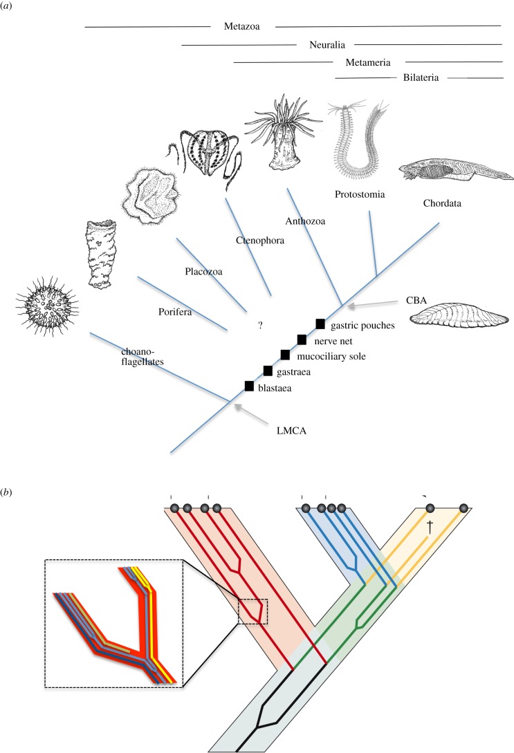
Prerequisite for tracing nervous system evolution is understanding of the body plan, feeding behaviour and locomotion of the first animals in which neurons evolved. Here, a comprehensive scenario is presented for the diversification of cell types in early metazoans, which enhanced feeding efficiency and led to the emergence of larger animals that were able to move. Starting from cup-shaped, gastraea-like animals with outer and inner choanoflagellate-like cells, two major innovations are discussed that set the stage for nervous system evolution. First, the invention of a mucociliary sole entailed a switch from intra- to extracellular digestion and increased the concentration of nutrients flowing into the gastric cavity. In these animals, an initial nerve net may have evolved via division of labour from mechanosensory-contractile cells in the lateral body wall, enabling coordinated movement of the growing body that involved both mucociliary creeping and changes of body shape. Second, the inner surface of the animals folded into metameric series of gastric pouches, which optimized nutrient resorption and allowed larger body sizes. The concomitant acquisition of bilateral symmetry may have allowed more directed locomotion and, with more demanding coordinative tasks, triggered the evolution of specialized nervous subsystems. Animals of this organizational state would have resembled Ediacarian fossils such as Dickinsonia and may have been close to the cnidarian–bilaterian ancestor. In the bilaterian lineage, the mucociliary sole was used mostly for creeping, or frequently lost. One possible remnant is the enigmatic Reissner's fibre in the ventral neural tube of cephalochordates and vertebrates.
Verified 2026-10-06 · 3 references not checked yet
Overview
AI snapshot
Written from the paper’s full text.
TL;DR
This review proposes a cell-type–based scenario for early metazoan evolution in which a mucociliary sole for external digestion and metameric gastric pouches set the stage for nervous system evolution. It argues that the first nerve net arose by division of labour from mechanosensory-contractile ectochoanocytes, and that animals of this grade resembled Ediacaran *Dickinsonia* and were close to the cnidarian–bilaterian ancestor.
Key findings
- Two major innovations are proposed as prerequisites for nervous system evolution: a mucociliary sole, which switched feeding from intra- to extracellular digestion and increased nutrient concentration in the gastric cavity, and metameric gastric pouches, which expanded resorptive surface and allowed larger body sizes.
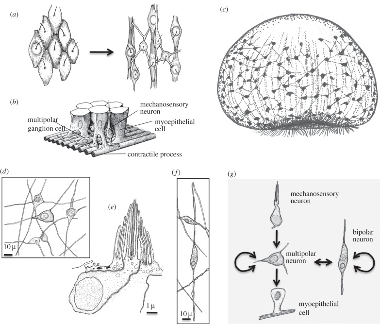
- The first nerve net is hypothesized to have evolved by division of labour from mechanosensory-contractile cells in the lateral body wall, enabling coordinated movement involving mucociliary creeping and body-shape changes; glutamate is proposed as the first excitatory transmitter and GABA as inhibitory.
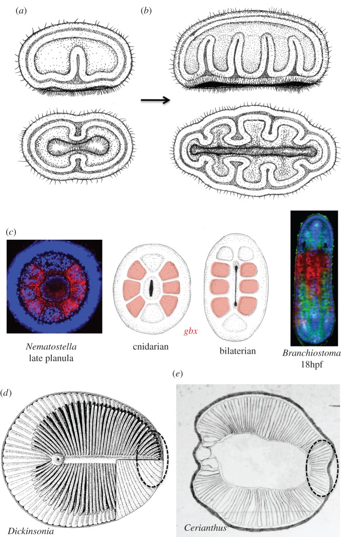
- Gastric pouches are proposed as homologous to bilaterian coelomic cavities, supported by *gbx* expression in all but the first pair of pouches in sea anemone and amphioxus, staggered/collinear *hox* expression, and expression of somitic musculature genes such as *mox* and *twist*.
- The organizational grade is compared to Ediacaran *Dickinsonia*, with its metameric digestive diverticula, and placed close to the cnidarian–bilaterian ancestor; Reissner’s fibre in cephalochordates and vertebrates is suggested as a possible remnant of the mucociliary sole.
Methods
- This is a review and synthetic evolutionary scenario, not a primary data paper; it integrates comparative structural and ultrastructural data, molecular studies on basal metazoans and choanoflagellates, developmental biology, and palaeontology.
- The main analytical framework is cell-type diversification via the sister cell-type concept and division of labour, used to infer ancestral cell types such as ectochoanocytes, endochoanocytes, kopeocytes, mucocytes, zymocytes, enterocytes, and nerve net neurons.
- Homology arguments draw on gene expression data, including *gbx*, *hox*, *mox*, *twist*, *foxj*, *foxA*, *atonal*, and *achaete-scute* family genes, alongside fossil comparisons such as *Dickinsonia*.
Limitations
- The paper states that in the absence of comparative molecular data for most cell types discussed, the postulated sequence of cell-type diversification remains tentative.
- The paper notes that the early metazoan tree remains unsolved, the placement of ctenophores is uncertain, and the existence of a “Neuralia” grouping excluding sponges and placozoans remains unsettled.
- The paper states it cannot explain why Reissner’s fibre is so persistent in extant vertebrates; more broadly, the text makes evident that many proposed cell types and transitions are hypothetical and not yet directly tested by comparative single-cell profiling.
Model-written summary. Check it against the paper before relying on it.
Integrity signals
Registry
Cites
Statistics
Figures
Verification
Claims the paper makes
62 Not checked yet · 52 Own interpretation · 2 No basis given · 1 Supported · 1 Own method
Every source the paper cites for this claim was checked and supports it.
In the paper
“In line with the postulated origin of nerve net neurons from ectochoanocytes, nerve net synaptic transmission appears to be largely glutamatergic, as recently postulated for ctenophores [156] and as indicated by many lines of circumstantial evidence in cnidarians.”
§ Gastric pouches and the mucociliary sole: setting the stage for nervous system evolution > The benthic gastraea: evolution of the nerve net > Glutamate and GABA
What the sources say
- Supportedcites [156] Moroz LL et al. 2014 The ctenophore genome and the evolutionary origins of neural systems. Nature 510, 109– 114. (10.1038/nature13400) 24847885 PMC4337882 source
In “Parallel evolution of neural organization” (page not available), the paper identifies neural nets in the ctenophore Pleurobrachia and proposes glutamate as a signalling molecule, citing glutamate-receptor expression and transmitter tests. That supports the citing sentence’s attribution of a postulate about glutamatergic signalling in ctenophores; this citation is not offered for the separate statement about cnidarians. (LLM, conf=0.87)
hedgedcorrelational- Asserted: passes — The authors put it forward as true.
- Checkable: passes — It can be checked against evidence.
- Load-bearing: unclear — It is unclear whether the paper’s conclusions depend on it.
- Based: passes — The paper gives a reason why it holds.
- Supported
A conclusion the paper draws from its own results — it cites no other source for it, so there is none to check. The quote shows it is in the paper.
In the paper
“In these animals, an initial nerve net may have evolved via division of labour from mechanosensory-contractile cells in the lateral body wall, enabling coordinated movement of the growing body that involved both mucociliary creeping and changes of body shape.”
§ Gastric pouches and the mucociliary sole: setting the stage for nervous system evolution > Abstract
hedgedconditional- Asserted: passes — The authors put it forward as true.
- Checkable: passes — It can be checked against evidence.
- Load-bearing: passes — The paper’s conclusions depend on it.
- Based: unclear — It is unclear why the paper says it holds.
None of the sources cited for this claim have been checked yet.
In the paper
“In addition, we can infer that early metazoan cells formed lamellipodia and filopodia, using proteins that reorganize cortical actin filaments such as the Arp2/3 complex, the actin cross-linking protein fascin, and myosin X”
§ Gastric pouches and the mucociliary sole: setting the stage for nervous system evolution > The choanoblastaea, a sphere of choanoflagellate-like cells > The first metazoan cells: choanoflagellate-like and multifunctional
What the sources say
- Not checked yetcites [39] Sebe-Pedros A, Burkhardt P, Sanchez-Pons N, Fairclough SR, Lang BF, King N, Ruiz-Trillo I 2013 Insights into the origin of metazoan filopodia and microvilli. Mol. Biol. Evol. 30, 2013– 2023. (10.1093/molbev/mst110) 23770652 PMC3748353 source
- Not checked yetcites [40] Jekely G 2014 Origin and evolution of the self-organizing cytoskeleton in the network of eukaryotic organelles. Cold Spring Harb. Perspect. Biol. 6, a016030 (10.1101/cshperspect.a016030) 25183829 PMC4142967 source
hedgeddirect- Asserted: passes — The authors put it forward as true.
- Checkable: passes — It can be checked against evidence.
- Load-bearing: unclear — It is unclear whether the paper’s conclusions depend on it.
- Based: passes — The paper gives a reason why it holds.
In the paper
“Such equipped animals may have been close to some simple Ediacaran fossils that have been interpreted as grazing on algal mats with a mucociliary sole for external digestion [3].”
§ Gastric pouches and the mucociliary sole: setting the stage for nervous system evolution > Introduction
What the sources say
- Not checked yetcites [3] Sperling EA, Vinther J 2010 A placozoan affinity for Dickinsonia and the evolution of Late Proterozoic metazoan feeding modes. Evol. Dev. 12, 201– 209. (10.1111/j.1525-142X.2010.00404.x) 20433459 source
hedgedcorrelational- Asserted: passes — The authors put it forward as true.
- Checkable: passes — It can be checked against evidence.
- Load-bearing: unclear — It is unclear whether the paper’s conclusions depend on it.
- Based: passes — The paper gives a reason why it holds.
A conclusion the paper draws from its own results — it cites no other source for it, so there is none to check. The quote shows it is in the paper.
In the paper
“Many of these molecules and associated functions were key to the evolution of the first neural cell types, as will be detailed below.”
§ Gastric pouches and the mucociliary sole: setting the stage for nervous system evolution > The choanoblastaea, a sphere of choanoflagellate-like cells > The first metazoan cells: choanoflagellate-like and multifunctional
plaindirect- Asserted: passes — The authors put it forward as true.
- Checkable: passes — It can be checked against evidence.
- Load-bearing: passes — The paper’s conclusions depend on it.
- Based: passes — The paper gives a reason why it holds.
None of the sources cited for this claim have been checked yet.
In the paper
“Stem metazoans were likely composed of one main characteristic cell type, resembling, structurally and functionally, the choanoflagellates [8–10; see however 11].”
§ Gastric pouches and the mucociliary sole: setting the stage for nervous system evolution > The choanoblastaea, a sphere of choanoflagellate-like cells
What the sources say
- Not checked yetcites [8] Nielsen C 2008 Six major steps in animal evolution: are we derived sponge larvae? Evol. Dev. 10, 241– 257. (10.1111/j.1525-142X.2008.00231.x) 18315817 source
- Not checked yetcites [9] Haeckel E 1874 Die Gastraea-Theorie, die phylogenetische Classification des Thierreiches und die Homologie der Keimblätter. Jena Z. Naturwiss. 8, 1– 55.
- Not checked yetcites [10] Maldonado M 2004 Choanoflagellates, choanocytes, and animal multicellularity. Invertebr. Biol. 123, 1– 22. (10.1111/j.1744-7410.2004.tb00138.x) source
hedged- Asserted: passes — The authors put it forward as true.
- Checkable: passes — It can be checked against evidence.
- Load-bearing: passes — The paper’s conclusions depend on it.
- Based: passes — The paper gives a reason why it holds.
A conclusion the paper draws from its own results — it cites no other source for it, so there is none to check. The quote shows it is in the paper.
In the paper
“This supports homology of sponge inner layer with the endomesoderm (i.e. the gastroderm) of other animals.”
§ Gastric pouches and the mucociliary sole: setting the stage for nervous system evolution > The microphageous gastraea
plaincorrelational- Asserted: passes — The authors put it forward as true.
- Checkable: passes — It can be checked against evidence.
- Load-bearing: passes — The paper’s conclusions depend on it.
- Based: passes — The paper gives a reason why it holds.
A conclusion the paper draws from its own results — it cites no other source for it, so there is none to check. The quote shows it is in the paper.
In the paper
“further diversification of the cell types making up the nerve net and the mucociliary sole gave rise to the plethora of receptor cells, neuron types and muscular and ciliary effector cells present in today's cnidarians and bilaterians.”
§ Gastric pouches and the mucociliary sole: setting the stage for nervous system evolution > Introduction
plaindirect- Asserted: passes — The authors put it forward as true.
- Checkable: passes — It can be checked against evidence.
- Load-bearing: passes — The paper’s conclusions depend on it.
- Based: unclear — It is unclear why the paper says it holds.
A conclusion the paper draws from its own results — it cites no other source for it, so there is none to check. The quote shows it is in the paper.
In the paper
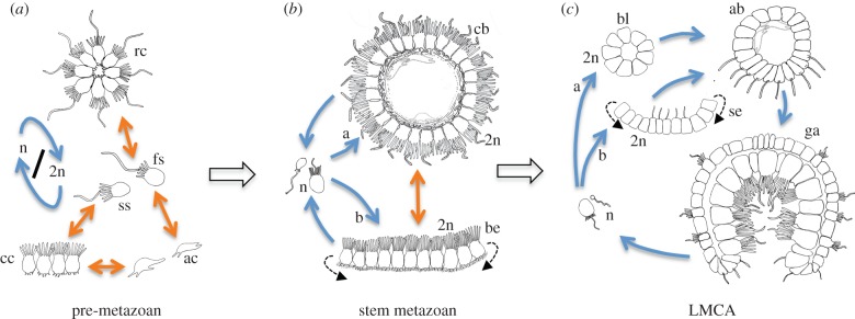
“Filopodia are highly dynamic structures that probably played a role in anchoring and stabilizing cells in the blastaea (figure 2a,b).”
§ Gastric pouches and the mucociliary sole: setting the stage for nervous system evolution > The choanoblastaea, a sphere of choanoflagellate-like cells > The first metazoan cells: choanoflagellate-like and multifunctional
hedgedconditional- Asserted: passes — The authors put it forward as true.
- Checkable: passes — It can be checked against evidence.
- Load-bearing: unclear — It is unclear whether the paper’s conclusions depend on it.
- Based: passes — The paper gives a reason why it holds.
In the paper
“a recent study comparing cellular transcriptomes indicates that a hierarchical tree structure indeed dominates cell-type interrelationships [6]”
§ Gastric pouches and the mucociliary sole: setting the stage for nervous system evolution > Introduction
What the sources say
- Not checked yetcites [6] Liang C, Consortium F, Forrest AR, Wagner GP 2015 The statistical geometry of transcriptome divergence in cell-type evolution and cancer. Nat. Commun. 6, 6066 (10.1038/ncomms7066) 25585899 source
hedgedcorrelational- Asserted: passes — The authors put it forward as true.
- Checkable: passes — It can be checked against evidence.
- Load-bearing: passes — The paper’s conclusions depend on it.
- Based: passes — The paper gives a reason why it holds.
- It may state two separate claims as one. 41
- Probably not a substantial claim. 25
A conclusion the paper draws from its own results — it cites no other source for it, so there is none to check. The quote shows it is in the paper.
In the paper
“Animals of this organizational state would have resembled Ediacarian fossils such as Dickinsonia and may have been close to the cnidarian–bilaterian”
§ Gastric pouches and the mucociliary sole: setting the stage for nervous system evolution > Abstract
hedgedcorrelational- Asserted: passes — The authors put it forward as true.
- Checkable: passes — It can be checked against evidence.
- Load-bearing: unclear — It is unclear whether the paper’s conclusions depend on it.
- Based: unclear — It is unclear why the paper says it holds.
It may state two separate claims as one.
A conclusion the paper draws from its own results — it cites no other source for it, so there is none to check. The quote shows it is in the paper.
In the paper
“The concomitant acquisition of bilateral symmetry may have allowed more directed locomotion and, with more demanding coordinative tasks, triggered the evolution of specialized nervous subsystems.”
§ Gastric pouches and the mucociliary sole: setting the stage for nervous system evolution > Abstract
hedgedconditional- Asserted: passes — The authors put it forward as true.
- Checkable: unclear — It is unclear whether it could be checked against evidence.
- Load-bearing: passes — The paper’s conclusions depend on it.
- Based: unclear — It is unclear why the paper says it holds.
It may state two separate claims as one.
A conclusion the paper draws from its own results — it cites no other source for it, so there is none to check. The quote shows it is in the paper.
In the paper
“Second, the inner surface of the animals folded into metameric series of gastric pouches, which optimized nutrient resorption and allowed larger body sizes.”
§ Gastric pouches and the mucociliary sole: setting the stage for nervous system evolution > Abstract
plaindirect- Asserted: passes — The authors put it forward as true.
- Checkable: passes — It can be checked against evidence.
- Load-bearing: passes — The paper’s conclusions depend on it.
- Based: unclear — It is unclear why the paper says it holds.
It may state two separate claims as one.
A conclusion the paper draws from its own results — it cites no other source for it, so there is none to check. The quote shows it is in the paper.
In the paper
“In the bilaterian lineage, the mucociliary sole was used mostly for creeping, or frequently lost.”
§ Gastric pouches and the mucociliary sole: setting the stage for nervous system evolution > Abstract
plain- Asserted: passes — The authors put it forward as true.
- Checkable: passes — It can be checked against evidence.
- Load-bearing: fails — The paper’s conclusions may not depend on it.
- Based: fails — The paper may give no reason why it holds.
Probably not a substantial claim. The paper’s conclusions may not depend on it. The paper may give no reason why it holds. It may state two separate claims as one.
The paper states this without a source or data behind it here, so there is nothing to check it against. The quote shows it is in the paper.
In the paper
“Prerequisite for tracing nervous system evolution is understanding of the body plan, feeding behaviour and locomotion of the first animals in which neurons evolved.”
§ Gastric pouches and the mucociliary sole: setting the stage for nervous system evolution > Abstract
plaindirect- Asserted: passes — The authors put it forward as true.
- Checkable: fails — It may be too vague to check against evidence.
- Load-bearing: passes — The paper’s conclusions depend on it.
- Based: fails — The paper may give no reason why it holds.
Probably not a substantial claim. It may be too vague to check against evidence. The paper may give no reason why it holds.
A conclusion the paper draws from its own results — it cites no other source for it, so there is none to check. The quote shows it is in the paper.
In the paper
“First, the invention of a mucociliary sole entailed a switch from intra- to extracellular digestion and increased the concentration of nutrients flowing into the gastric cavity.”
§ Gastric pouches and the mucociliary sole: setting the stage for nervous system evolution > Abstract
plaindirect- Asserted: passes — The authors put it forward as true.
- Checkable: passes — It can be checked against evidence.
- Load-bearing: passes — The paper’s conclusions depend on it.
- Based: fails — The paper may give no reason why it holds.
Probably not a substantial claim. The paper may give no reason why it holds. It may state two separate claims as one.
A conclusion the paper draws from its own results — it cites no other source for it, so there is none to check. The quote shows it is in the paper.
In the paper
“One possible remnant is the enigmatic Reissner's fibre in the ventral neural tube of cephalochordates and vertebrates.”
§ Gastric pouches and the mucociliary sole: setting the stage for nervous system evolution > Abstract
hedgedcorrelational- Asserted: passes — The authors put it forward as true.
- Checkable: passes — It can be checked against evidence.
- Load-bearing: passes — The paper’s conclusions depend on it.
- Based: fails — The paper may give no reason why it holds.
Probably not a substantial claim. The paper may give no reason why it holds.
A conclusion the paper draws from its own results — it cites no other source for it, so there is none to check. The quote shows it is in the paper.
In the paper
“Starting from cup-shaped, gastraea-like animals with outer and inner choanoflagellate-like cells, two major innovations are discussed that set the stage for nervous system evolution.”
§ Gastric pouches and the mucociliary sole: setting the stage for nervous system evolution > Abstract
plaindirect- Asserted: passes — The authors put it forward as true.
- Checkable: passes — It can be checked against evidence.
- Load-bearing: passes — The paper’s conclusions depend on it.
- Based: fails — The paper may give no reason why it holds.
Probably not a substantial claim. The paper may give no reason why it holds.
In the paper
“The slow myosin is also involved in amoeboid movement [38], which probably belonged to the behavioural repertoire of early metazoan cells.”
§ Gastric pouches and the mucociliary sole: setting the stage for nervous system evolution > The choanoblastaea, a sphere of choanoflagellate-like cells > The first metazoan cells: choanoflagellate-like and multifunctional
What the sources say
- Not checked yetcites [38] Liu YJ, Le Berre M, Lautenschlaeger F, Maiuri P, Callan-Jones A, Heuze M, Takaki T, Voituriez R, Piel M 2015 Confinement and low adhesion induce fast amoeboid migration of slow mesenchymal cells. Cell 160, 659– 672. (10.1016/j.cell.2015.01.007) 25679760
hedgedcorrelational- Asserted: passes — The authors put it forward as true.
- Checkable: passes — It can be checked against evidence.
- Load-bearing: unclear — It is unclear whether the paper’s conclusions depend on it.
- Based: passes — The paper gives a reason why it holds.
It may state two separate claims as one.
In the paper
“The flagellar cilia were both motile and sensory (as reported for unicellular eukaryotes and human airway cilia), and thus contained a very short and direct sensory-effector arc [22].”
§ Gastric pouches and the mucociliary sole: setting the stage for nervous system evolution > The choanoblastaea, a sphere of choanoflagellate-like cells > The first metazoan cells: choanoflagellate-like and multifunctional
What the sources say
- Not checked yetcites [22] Salathe M 2007 Regulation of mammalian ciliary beating. Annu. Rev. Physiol. 69, 401– 422. (10.1146/annurev.physiol.69.040705.141253) 16945069
plaindirect- Asserted: passes — The authors put it forward as true.
- Checkable: passes — It can be checked against evidence.
- Load-bearing: passes — The paper’s conclusions depend on it.
- Based: passes — The paper gives a reason why it holds.
It may state two separate claims as one.
None of the sources cited for this claim have been checked yet.
In the paper
“These ancestors represented an epithelium [14] that took the shape of a sphere, called choanoblastaea [8], which later gave rise to the cup-shaped gastraea”
§ Gastric pouches and the mucociliary sole: setting the stage for nervous system evolution > The choanoblastaea, a sphere of choanoflagellate-like cells
What the sources say
- Not checked yetcites [8] Nielsen C 2008 Six major steps in animal evolution: are we derived sponge larvae? Evol. Dev. 10, 241– 257. (10.1111/j.1525-142X.2008.00231.x) 18315817 source
- Not checked yetcites [14] Leys SP, Riesgo A 2012 Epithelia, an evolutionary novelty of metazoans. J. Exp. Zool. B Mol. Dev. Evol. 318, 438– 447. (10.1002/jez.b.21442) 22057924
plaindirect- Asserted: passes — The authors put it forward as true.
- Checkable: passes — It can be checked against evidence.
- Load-bearing: passes — The paper’s conclusions depend on it.
- Based: passes — The paper gives a reason why it holds.
It may state two separate claims as one.
A conclusion the paper draws from its own results — it cites no other source for it, so there is none to check. The quote shows it is in the paper.
In the paper
“the extensive folding of the inner surface into metameric pouches, which optimized nutrient distribution and resorption”
§ Gastric pouches and the mucociliary sole: setting the stage for nervous system evolution > Introduction
plaindirect- Asserted: passes — The authors put it forward as true.
- Checkable: passes — It can be checked against evidence.
- Load-bearing: passes — The paper’s conclusions depend on it.
- Based: fails — The paper may give no reason why it holds.
Probably not a substantial claim. The paper may give no reason why it holds.
A conclusion the paper draws from its own results — it cites no other source for it, so there is none to check. The quote shows it is in the paper.
In the paper
“the endochoanocytes into cell types specializing in internal nutrient distribution and resorption”
§ Gastric pouches and the mucociliary sole: setting the stage for nervous system evolution > Introduction
plaindirect- Asserted: passes — The authors put it forward as true.
- Checkable: passes — It can be checked against evidence.
- Load-bearing: passes — The paper’s conclusions depend on it.
- Based: fails — The paper may give no reason why it holds.
Probably not a substantial claim. The paper may give no reason why it holds.
A conclusion the paper draws from its own results — it cites no other source for it, so there is none to check. The quote shows it is in the paper.
In the paper
“and the ectochoanocytes into cell types specializing in mechanosensation, information integration and body contractions in response to environmental stimuli”
§ Gastric pouches and the mucociliary sole: setting the stage for nervous system evolution > Introduction
plaindirect- Asserted: passes — The authors put it forward as true.
- Checkable: passes — It can be checked against evidence.
- Load-bearing: passes — The paper’s conclusions depend on it.
- Based: fails — The paper may give no reason why it holds.
Probably not a substantial claim. The paper may give no reason why it holds.
A conclusion the paper draws from its own results — it cites no other source for it, so there is none to check. The quote shows it is in the paper.
In the paper
“One prominent example is the division of labour that accompanied the birth of the first neurons and muscle cells (see §2).”
§ Gastric pouches and the mucociliary sole: setting the stage for nervous system evolution > Introduction
plaincorrelational- Asserted: passes — The authors put it forward as true.
- Checkable: passes — It can be checked against evidence.
- Load-bearing: fails — The paper’s conclusions may not depend on it.
- Based: fails — The paper may give no reason why it holds.
Probably not a substantial claim. The paper’s conclusions may not depend on it. The paper may give no reason why it holds.
In the paper
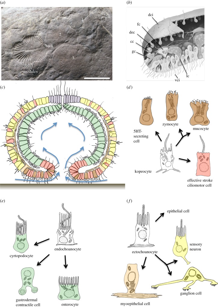
“Various lines of evidence indicate that a new feeding mode evolved in the Precambrian ocean, referred to as mucoid-ciliary particle feeding [3] (figure 4a,b).”
- Also stated in: § Grazing algal mats with mucus and digestive gland cells · hedged
“A rich body of fossil and comparative evidence indicates that mucoid-ciliary particle feeding evolved in early metazoans.”
§ Gastric pouches and the mucociliary sole: setting the stage for nervous system evolution > The benthic gastraea: external digestion with a mucociliary sole
What the sources say
- Not checked yetcites [3] Sperling EA, Vinther J 2010 A placozoan affinity for Dickinsonia and the evolution of Late Proterozoic metazoan feeding modes. Evol. Dev. 12, 201– 209. (10.1111/j.1525-142X.2010.00404.x) 20433459 source
hedgeddirect- Asserted: passes — The authors put it forward as true.
- Checkable: passes — It can be checked against evidence.
- Load-bearing: passes — The paper’s conclusions depend on it.
- Based: passes — The paper gives a reason why it holds.
It may state two separate claims as one.
- Also stated in: § Grazing algal mats with mucus and digestive gland cells · hedged
A conclusion the paper draws from its own results — it cites no other source for it, so there is none to check. The quote shows it is in the paper.
In the paper
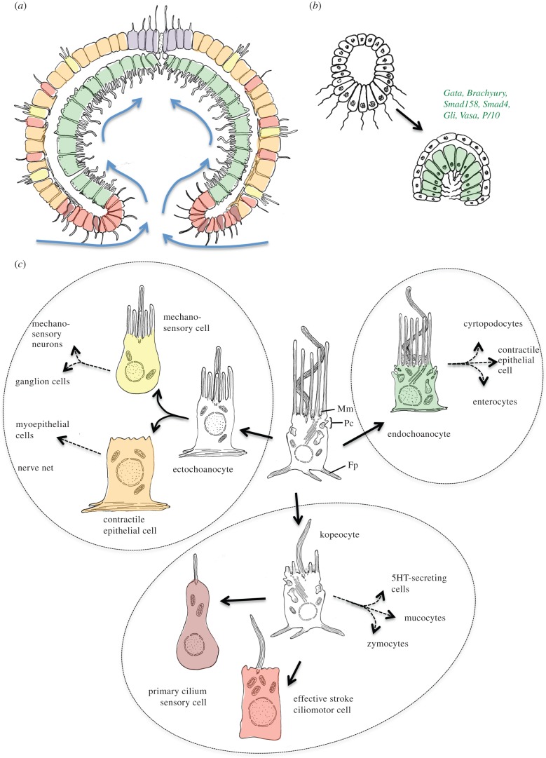
“We will hypothesize that the simple, bilayered, cup-shaped anatomy of this ancestor was reflected by a first diversification at the cellular level, including: (i) the external ‘ectochoanocytes’, mediating interactions with the environment; (ii) the internal ‘endochoanocytes’ that specialized towards particle capture and nutrient resorption; and (iii) the ‘kopeocytes’, regulating water currents”
§ Gastric pouches and the mucociliary sole: setting the stage for nervous system evolution > Introduction
hedgeddirect- Asserted: fails — The authors may not put it forward as true.
- Checkable: passes — It can be checked against evidence.
- Load-bearing: passes — The paper’s conclusions depend on it.
- Based: fails — The paper may give no reason why it holds.
Probably not a substantial claim. The authors may not put it forward as true. The paper may give no reason why it holds.
None of the sources cited for this claim have been checked yet.
In the paper
“choanoflagellates and metazoans share synaptic adhesion/signalling molecules, a primordial neurosecretory apparatus active on the apical cell surface (but no presynaptic active zone proteins) [32], postsynaptic density proteins such as Homer or Shank [33,34] and a variety of receptors including the metabotropic glutamate receptor [35] (but not ionotropic glutamate receptors, which only appeared in”
§ Gastric pouches and the mucociliary sole: setting the stage for nervous system evolution > The choanoblastaea, a sphere of choanoflagellate-like cells > The first metazoan cells: choanoflagellate-like and multifunctional
What the sources say
- Not checked yetcites [32] Burkhardt P 2015 The origin and evolution of synaptic proteins—choanoflagellates lead the way. J. Exp. Biol. 218, 506– 514. (10.1242/jeb.110247) 25696814 source
- Not checked yetcites [33] Alie A, Manuel M 2010 The backbone of the post-synaptic density originated in a unicellular ancestor of choanoflagellates and metazoans. BMC Evol. Biol. 10, 34 (10.1186/1471-2148-10-34) 20128896 PMC2824662
- Not checked yetcites [34] Ryan TJ, Grant SG 2009 The origin and evolution of synapses. Nat. Rev. Neurosci. 10, 701– 712. (10.1038/nrn2717) 19738623
- Not checked yetcites [35] Krishnan A, Schioth HB 2015 The role of G protein-coupled receptors in the early evolution of neurotransmission and the nervous system. J. Exp. Biol. 218, 562– 571. (10.1242/jeb.110312) 25696819 source
plaincorrelational- Asserted: passes — The authors put it forward as true.
- Checkable: passes — It can be checked against evidence.
- Load-bearing: passes — The paper’s conclusions depend on it.
- Based: passes — The paper gives a reason why it holds.
It may state two separate claims as one.
A conclusion the paper draws from its own results — it cites no other source for it, so there is none to check. The quote shows it is in the paper.
In the paper
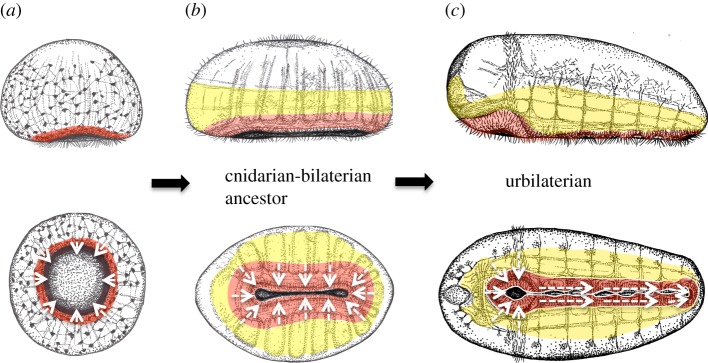
“We propose that, with these changes, basal metazoans had reached the organizational grade of the Ediacaran fossil Dickinsonia, which may have been close to the last common ancestor of cnidarians and bilaterians (that we collectively refer to as ‘Metameria’, figure 1a).”
§ Gastric pouches and the mucociliary sole: setting the stage for nervous system evolution > Introduction
hedgeddirect- Asserted: passes — The authors put it forward as true.
- Checkable: passes — It can be checked against evidence.
- Load-bearing: passes — The paper’s conclusions depend on it.
- Based: unclear — It is unclear why the paper says it holds.
It may state two separate claims as one.
In the paper
“Cell-type diversification involves the segregation and divergence of existing cellular modules or functions [5].”
§ Gastric pouches and the mucociliary sole: setting the stage for nervous system evolution > Introduction
What the sources say
- Not checked yetcites [5] Arendt D 2008 The evolution of cell types in animals: emerging principles from molecular studies. Nature reviews. Genetics 9, 868– 882. (10.1038/nrg2416) 18927580
plaindirect- Asserted: passes — The authors put it forward as true.
- Checkable: passes — It can be checked against evidence.
- Load-bearing: passes — The paper’s conclusions depend on it.
- Based: passes — The paper gives a reason why it holds.
Probably not a substantial claim.
A conclusion the paper draws from its own results — it cites no other source for it, so there is none to check. The quote shows it is in the paper.
In the paper
“Sponges might be regarded as representatives of a gastraea-type organization (which then would have undergone surface extension independent of other metazoan lineages”
§ Gastric pouches and the mucociliary sole: setting the stage for nervous system evolution > The microphageous gastraea
hedgedconditional- Asserted: passes — The authors put it forward as true.
- Checkable: passes — It can be checked against evidence.
- Load-bearing: passes — The paper’s conclusions depend on it.
- Based: fails — The paper may give no reason why it holds.
Probably not a substantial claim. The paper may give no reason why it holds. It may state two separate claims as one.
A conclusion the paper draws from its own results — it cites no other source for it, so there is none to check. The quote shows it is in the paper.
In the paper
“Food was predigested externally in a layer of mucus and the predigested nutrients transported into the body cavity with water currents.”
§ Gastric pouches and the mucociliary sole: setting the stage for nervous system evolution > The benthic gastraea: external digestion with a mucociliary sole
plaindirect- Asserted: passes — The authors put it forward as true.
- Checkable: passes — It can be checked against evidence.
- Load-bearing: passes — The paper’s conclusions depend on it.
- Based: passes — The paper gives a reason why it holds.
It may state two separate claims as one.
In the paper
“Its most characteristic feature is the infolding of the lower body surface, resulting in an outer ectoderm, an inner gastroderm and a gastric opening (figure 3a).”
§ Gastric pouches and the mucociliary sole: setting the stage for nervous system evolution > The microphageous gastraea
What the sources say
- Not checked yetcites [9] Haeckel E 1874 Die Gastraea-Theorie, die phylogenetische Classification des Thierreiches und die Homologie der Keimblätter. Jena Z. Naturwiss. 8, 1– 55.
plaindirect- Asserted: passes — The authors put it forward as true.
- Checkable: passes — It can be checked against evidence.
- Load-bearing: passes — The paper’s conclusions depend on it.
- Based: passes — The paper gives a reason why it holds.
Probably not a substantial claim.
None of the sources cited for this claim have been checked yet.
In the paper
“classical authors agreed that the first metazoan cells had an apical collar with an undulating flagellum [16,17]”
§ Gastric pouches and the mucociliary sole: setting the stage for nervous system evolution > The choanoblastaea, a sphere of choanoflagellate-like cells
What the sources say
- Not checked yetcites [16] Hibberd DJ 1975 Observations on the ultrastructure of the choanoflagellate Codosiga botrytis (Ehr.) Saville-Kent with special reference to the flagellar apparatus. J. Cell Sci. 17, 191– 219. 1089676
- Not checked yetcites [17] Leadbeater BSC 1983 Distribution and chemistry of microfilaments in choanoflagellates, with special reference to the collar and other tentacle systems. Protistologia 10, 157– 166.
plain- Asserted: passes — The authors put it forward as true.
- Checkable: passes — It can be checked against evidence.
- Load-bearing: fails — The paper’s conclusions may not depend on it.
- Based: passes — The paper gives a reason why it holds.
Probably not a substantial claim. The paper’s conclusions may not depend on it.
A conclusion the paper draws from its own results — it cites no other source for it, so there is none to check. The quote shows it is in the paper.
In the paper
“Digestive enzymes were secreted into this mucus layer for external digestion. The coordinated and directional beating of effective stroke cilia distributed and transported the mucus over the digestive epithelium and washed the nutrients into the gastric cavity.”
§ Gastric pouches and the mucociliary sole: setting the stage for nervous system evolution > The benthic gastraea: external digestion with a mucociliary sole
plaindirect- Asserted: passes — The authors put it forward as true.
- Checkable: passes — It can be checked against evidence.
- Load-bearing: passes — The paper’s conclusions depend on it.
- Based: passes — The paper gives a reason why it holds.
It may state two separate claims as one.
None of the sources cited for this claim have been checked yet.
In the paper
“It is a long-standing notion that the second step in the evolution of the metazoan body was the transformation of the spherical blastaea into the gastraea [8,9], which may have been close to the last metazoan common ancestor (figure 1b).”
§ Gastric pouches and the mucociliary sole: setting the stage for nervous system evolution > The microphageous gastraea
What the sources say
- Not checked yetcites [8] Nielsen C 2008 Six major steps in animal evolution: are we derived sponge larvae? Evol. Dev. 10, 241– 257. (10.1111/j.1525-142X.2008.00231.x) 18315817 source
- Not checked yetcites [9] Haeckel E 1874 Die Gastraea-Theorie, die phylogenetische Classification des Thierreiches und die Homologie der Keimblätter. Jena Z. Naturwiss. 8, 1– 55.
hedged- Asserted: passes — The authors put it forward as true.
- Checkable: passes — It can be checked against evidence.
- Load-bearing: passes — The paper’s conclusions depend on it.
- Based: passes — The paper gives a reason why it holds.
Probably not a substantial claim. It may state two separate claims as one.
In the paper
“Even in sponges, a transitory cup-like stage forms during metamorphosis, as recently confirmed [41]; and in cnidarians and ctenophores, the overall body form resembles that of a gastrula during the entire life cycle—at larval, polyp and medusa stages.”
§ Gastric pouches and the mucociliary sole: setting the stage for nervous system evolution > The microphageous gastraea
What the sources say
- Not checked yetcites [41] Leys SP, Eerkes-Medrano D 2005 Gastrulation in calcareous sponges: in search of Haeckel's gastraea. Integr. Comp. Biol. 45, 342– 351. (10.1093/icb/45.2.342) 21676779
boosted- Asserted: passes — The authors put it forward as true.
- Checkable: passes — It can be checked against evidence.
- Load-bearing: passes — The paper’s conclusions depend on it.
- Based: passes — The paper gives a reason why it holds.
It may state two separate claims as one.
A conclusion the paper draws from its own results — it cites no other source for it, so there is none to check. The quote shows it is in the paper.
In the paper
“the transition to bilateral symmetry, which enabled directed movement, facilitated food search and avoided grazing the same area twice”
§ Gastric pouches and the mucociliary sole: setting the stage for nervous system evolution > Introduction
plaindirect- Asserted: fails — The authors may not put it forward as true.
- Checkable: passes — It can be checked against evidence.
- Load-bearing: passes — The paper’s conclusions depend on it.
- Based: fails — The paper may give no reason why it holds.
Probably not a substantial claim. The authors may not put it forward as true. The paper may give no reason why it holds. It may state two separate claims as one.
A conclusion the paper draws from its own results — it cites no other source for it, so there is none to check. The quote shows it is in the paper.
In the paper
“These latter cells would have constituted the first nerve net, which possibly controlled some form of amoeboid locomotion.”
§ Gastric pouches and the mucociliary sole: setting the stage for nervous system evolution > Introduction
hedgedconditional- Asserted: passes — The authors put it forward as true.
- Checkable: passes — It can be checked against evidence.
- Load-bearing: passes — The paper’s conclusions depend on it.
- Based: unclear — It is unclear why the paper says it holds.
It may state two separate claims as one.
None of the sources cited for this claim have been checked yet.
In the paper
“They were swimming or lived partially benthic in shallow waters, filtering and concentrating dissolved organic carbon and bacteria as microphageous suspension feeders [3,15].”
§ Gastric pouches and the mucociliary sole: setting the stage for nervous system evolution > The choanoblastaea, a sphere of choanoflagellate-like cells
What the sources say
- Not checked yetcites [3] Sperling EA, Vinther J 2010 A placozoan affinity for Dickinsonia and the evolution of Late Proterozoic metazoan feeding modes. Evol. Dev. 12, 201– 209. (10.1111/j.1525-142X.2010.00404.x) 20433459 source
- Not checked yetcites [15] Leys SP 2015 Elements of a ‘nervous system’ in sponges. J. Exp. Biol. 218, 581– 591. (10.1242/jeb.110817) 25696821 source
plain- Asserted: passes — The authors put it forward as true.
- Checkable: passes — It can be checked against evidence.
- Load-bearing: unclear — It is unclear whether the paper’s conclusions depend on it.
- Based: passes — The paper gives a reason why it holds.
It may state two separate claims as one.
A conclusion the paper draws from its own results — it cites no other source for it, so there is none to check. The quote shows it is in the paper.
In the paper
“Subsequently, the kopeocytes may have further diversified into cell types specializing in ciliary movement and control, mucus production and secretion of digestive enzymes, thus enabling external digestion and forming a ‘mucociliary sole’ around the gastric opening”
§ Gastric pouches and the mucociliary sole: setting the stage for nervous system evolution > Introduction
hedgedconditional- Asserted: passes — The authors put it forward as true.
- Checkable: fails — It may be too vague to check against evidence.
- Load-bearing: passes — The paper’s conclusions depend on it.
- Based: fails — The paper may give no reason why it holds.
Probably not a substantial claim. It may be too vague to check against evidence. The paper may give no reason why it holds. It may state two separate claims as one.
A conclusion the paper draws from its own results — it cites no other source for it, so there is none to check. The quote shows it is in the paper.
In the paper
“However, digestion was intracellular at first and took place in the same multifunctional cells that also trapped the food—just as in unicellular eukaryotes.”
§ Gastric pouches and the mucociliary sole: setting the stage for nervous system evolution > The choanoblastaea, a sphere of choanoflagellate-like cells > The first metazoan cells: choanoflagellate-like and multifunctional
plain- Asserted: passes — The authors put it forward as true.
- Checkable: passes — It can be checked against evidence.
- Load-bearing: passes — The paper’s conclusions depend on it.
- Based: passes — The paper gives a reason why it holds.
It may state two separate claims as one.
In the paper
“The sister cell-type concept [5] posits that cell-type evolution can be resolved into a scheme of stepwise diversifications, giving rise to sister cell types [5]”
§ Gastric pouches and the mucociliary sole: setting the stage for nervous system evolution > Introduction
What the sources say
- Not checked yetcites [5] Arendt D 2008 The evolution of cell types in animals: emerging principles from molecular studies. Nature reviews. Genetics 9, 868– 882. (10.1038/nrg2416) 18927580
plaindirect- Asserted: passes — The authors put it forward as true.
- Checkable: passes — It can be checked against evidence.
- Load-bearing: passes — The paper’s conclusions depend on it.
- Based: passes — The paper gives a reason why it holds.
Probably not a substantial claim.
None of the sources cited for this claim have been checked yet.
In the paper
“Five subfamilies of transient receptor potential (TRP) channels were present [24], among which TRPV (vanilloid TRP) channels likely played an ancient role in mechanosensation”
§ Gastric pouches and the mucociliary sole: setting the stage for nervous system evolution > The choanoblastaea, a sphere of choanoflagellate-like cells > The first metazoan cells: choanoflagellate-like and multifunctional
What the sources say
- Not checked yetcites [24] Cai X, Clapham DE 2012 Ancestral Ca2+ signaling machinery in early animal and fungal evolution. Mol. Biol. Evol. 29, 91– 100. (10.1093/molbev/msr149) 21680871 PMC4037924
- Not checked yetcites [25] Fujiu K, Nakayama Y, Iida H, Sokabe M, Yoshimura K 2011 Mechanoreception in motile flagella of Chlamydomonas. Nat. Cell Biol. 13, 630– 632. (10.1038/ncb2214) 21478860
hedgedconditional- Asserted: passes — The authors put it forward as true.
- Checkable: passes — It can be checked against evidence.
- Load-bearing: unclear — It is unclear whether the paper’s conclusions depend on it.
- Based: passes — The paper gives a reason why it holds.
It may state two separate claims as one.
The paper states this without a source or data behind it here, so there is nothing to check it against. The quote shows it is in the paper.
In the paper
“From Haeckel's times until today, strong support for the gastraea hypothesis comes from the prevalence of gastrula-like stages during animal development that are interpreted as recapitulation of the gastraea-like ancestral body plan.”
§ Gastric pouches and the mucociliary sole: setting the stage for nervous system evolution > The microphageous gastraea
boostedcorrelational- Asserted: passes — The authors put it forward as true.
- Checkable: passes — It can be checked against evidence.
- Load-bearing: fails — The paper’s conclusions may not depend on it.
- Based: fails — The paper may give no reason why it holds.
Probably not a substantial claim. The paper’s conclusions may not depend on it. The paper may give no reason why it holds.
A conclusion the paper draws from its own results — it cites no other source for it, so there is none to check. The quote shows it is in the paper.
In the paper
“The initial function of this nerve net might have been the control of amoeboid locomotion of the gastraea, grazing on algal mats (resembling the movement of an amoeba, but at multicellular scale).”
§ Gastric pouches and the mucociliary sole: setting the stage for nervous system evolution > The benthic gastraea: evolution of the nerve net
hedgedconditional- Asserted: passes — The authors put it forward as true.
- Checkable: passes — It can be checked against evidence.
- Load-bearing: fails — The paper’s conclusions may not depend on it.
- Based: fails — The paper may give no reason why it holds.
Probably not a substantial claim. The paper’s conclusions may not depend on it. The paper may give no reason why it holds.
In the paper
“the thyroid-related transcription factors TTF-2/foxE4, foxq1 and foxA are jointly expressed in the amphioxus and ascidian endostyle, a presumed homolog of the thyroidea that harbours mucus-secreting and motile ciliated cells; and this function of the Fox genes is considered ancestral for the chordates [119].”
§ Gastric pouches and the mucociliary sole: setting the stage for nervous system evolution > The benthic gastraea: external digestion with a mucociliary sole > A link between the mucociliary cell types and Fox family evolution
What the sources say
- Not checked yetcites [119] Hiruta J, Mazet F, Yasui K, Zhang P, Ogasawara M 2005 Comparative expression analysis of transcription factor genes in the endostyle of invertebrate chordates. Dev. Dyn. 233, 1031– 1037. (10.1002/dvdy.20401) 15861404
hedgedcorrelational- Asserted: passes — The authors put it forward as true.
- Checkable: passes — It can be checked against evidence.
- Load-bearing: unclear — It is unclear whether the paper’s conclusions depend on it.
- Based: passes — The paper gives a reason why it holds.
It may state two separate claims as one.
In the paper
“Feeding traces indicate that one of the most characteristic fossils of the Ediacaran fauna, Dickinsonia, was grazing on the sea floor, which was covered by ubiquitous microbial mats [3] (figure 4a).”
§ Gastric pouches and the mucociliary sole: setting the stage for nervous system evolution > The benthic gastraea: external digestion with a mucociliary sole > Grazing algal mats with mucus and digestive gland cells
What the sources say
- Not checked yetcites [3] Sperling EA, Vinther J 2010 A placozoan affinity for Dickinsonia and the evolution of Late Proterozoic metazoan feeding modes. Evol. Dev. 12, 201– 209. (10.1111/j.1525-142X.2010.00404.x) 20433459 source
hedged- Asserted: passes — The authors put it forward as true.
- Checkable: passes — It can be checked against evidence.
- Load-bearing: passes — The paper’s conclusions depend on it.
- Based: passes — The paper gives a reason why it holds.
It may state two separate claims as one.
- The authors state this as an assumption: Dickinsonia's entire ventral body surface was a digestive sole covered with mucus and cilia.
A conclusion the paper draws from its own results — it cites no other source for it, so there is none to check. The quote shows it is in the paper.
In the paper
“Importantly, neuromuscular orthogon and mucociliary sole appear to have overlapped, because longitudinal muscles reach the gastric opening (at least in cnidarians).”
§ Gastric pouches and the mucociliary sole: setting the stage for nervous system evolution > The benthic gastraea: evolution of the nerve net > The neuromuscular orthogon
hedgedcorrelational- Asserted: passes — The authors put it forward as true.
- Checkable: passes — It can be checked against evidence.
- Load-bearing: unclear — It is unclear whether the paper’s conclusions depend on it.
- Based: passes — The paper gives a reason why it holds.
It may state two separate claims as one.
None of the sources cited for this claim have been checked yet.
In the paper
“As a rule, the ectodermal muscle fibres associated with the nerve are oriented longitudinally, that is in parallel to the primary body axis (while the inner layer forms circular fibres [61,62]).”
§ Gastric pouches and the mucociliary sole: setting the stage for nervous system evolution > The benthic gastraea: evolution of the nerve net > The neuromuscular orthogon
What the sources say
- Not checked yetcites [61] Hertwig O, Hertwig R 1879 Die Actinien. In Studien zur Blättertheorie. Jena, Germany: Gustav Fischer.
- Not checked yetcites [62] Hyman LH 1940 The invertebrates: protozoa through Ctenophora. New York, NY: McGraw-Hill Book Company.
plaincorrelational- Asserted: passes — The authors put it forward as true.
- Checkable: passes — It can be checked against evidence.
- Load-bearing: passes — The paper’s conclusions depend on it.
- Based: passes — The paper gives a reason why it holds.
It may state two separate claims as one.
None of the sources cited for this claim have been checked yet.
In the paper
“Most prominently, foxj has been identified as an ancient key regulator of motile cilia biogenesis [72,112,113], conserved in cnidarians [72] and even sponges [114].”
§ Gastric pouches and the mucociliary sole: setting the stage for nervous system evolution > The benthic gastraea: external digestion with a mucociliary sole > A link between the mucociliary cell types and Fox family evolution
What the sources say
- Not checked yetcites [72] Vij S et al. 2012 Evolutionarily ancient association of the FoxJ1 transcription factor with the motile ciliogenic program. PLoS Genet. 8, e1003019 (10.1371/journal.pgen.1003019) 23144623 PMC3493443
- Not checked yetcites [112] Stubbs JL, Oishi I, Izpisua Belmonte JC, Kintner C 2008 The forkhead protein Foxj1 specifies node-like cilia in Xenopus and zebrafish embryos. Nat. Genet. 40, 1454– 1460. (10.1038/ng.267) 19011629 PMC4648715
- Not checked yetcites [113] Yu X, Ng CP, Habacher H, Roy S 2008 Foxj1 transcription factors are master regulators of the motile ciliogenic program. Nat. Genet. 40, 1445– 1453. (10.1038/ng.263) 19011630
plaindirect- Asserted: passes — The authors put it forward as true.
- Checkable: passes — It can be checked against evidence.
- Load-bearing: passes — The paper’s conclusions depend on it.
- Based: passes — The paper gives a reason why it holds.
It may state two separate claims as one.
None of the sources cited for this claim have been checked yet.
In the paper
“A choanocyte-origin has been previously postulated for enterocytes [45] and for flame cells [46], based on morphological, functional and molecular resemblance.”
§ Gastric pouches and the mucociliary sole: setting the stage for nervous system evolution > The benthic gastraea: external digestion with a mucociliary sole > Diversification of endochoanocytes: enterocytes and excretory cells
What the sources say
- Not checked yetcites [45] Takashima S, Gold D, Hartenstein V 2012 Stem cells and lineages of the intestine: a developmental and evolutionary perspective. Dev. Genes Evol. 223, 85– 102. (10.1007/s00427-012-0422-8) 23179635 PMC3873164
- Not checked yetcites [46] Jacobs DK, Nakanishi N, Yuan D, Camara A, Nichols SA, Hartenstein V 2007 Evolution of sensory structures in basal metazoa. Integr. Comp. Biol. 47, 712– 723. (10.1093/icb/icm094) 21669752
plaincorrelational- Asserted: passes — The authors put it forward as true.
- Checkable: passes — It can be checked against evidence.
- Load-bearing: fails — The paper’s conclusions may not depend on it.
- Based: passes — The paper gives a reason why it holds.
Probably not a substantial claim. The paper’s conclusions may not depend on it.
None of the sources cited for this claim have been checked yet.
In the paper
“Interestingly, the metazoan foxA, B, C, D and E subfamilies are evolutionarily related [120–122] and resulted from duplications of an ancient precursor gene;”
§ Gastric pouches and the mucociliary sole: setting the stage for nervous system evolution > The benthic gastraea: external digestion with a mucociliary sole > A link between the mucociliary cell types and Fox family evolution
What the sources say
- Not checked yetcites [120] Adell T, Muller WE 2004 Isolation and characterization of five Fox (Forkhead) genes from the sponge Suberites domuncula. Gene 334, 35– 46. (10.1016/j.gene.2004.02.036) 15256253
- Not checked yetcites [121] Mazet F, Yu JK, Liberles DA, Holland LZ, Shimeld SM 2003 Phylogenetic relationships of the Fox (Forkhead) gene family in the Bilateria. Gene 316, 79– 89. (10.1016/S0378-1119(03)00741-8) 14563554
- Not checked yetcites [122] Magie CR, Pang K, Martindale MQ 2005 Genomic inventory and expression of Sox and Fox genes in the cnidarian Nematostella vectensis. Dev. Genes Evol. 215, 618– 630. (10.1007/s00427-005-0022-y) 16193320
plaindirect- Asserted: passes — The authors put it forward as true.
- Checkable: passes — It can be checked against evidence.
- Load-bearing: unclear — It is unclear whether the paper’s conclusions depend on it.
- Based: passes — The paper gives a reason why it holds.
It may state two separate claims as one.
In the paper
“Placozoans move by ciliary gliding with the thick mucociliary sole facing the sea floor, and by concomitant contraction of the syncytial fibre cells (figure 4b).”
§ Gastric pouches and the mucociliary sole: setting the stage for nervous system evolution > The benthic gastraea: external digestion with a mucociliary sole > Grazing algal mats with mucus and digestive gland cells
What the sources say
- Not checked yetcites [104] Ruthmann A, Behrendt G, Wahl R 1986 The ventral epithelium of Trichoplax adhaerens (Placozoa): cytoskeletal structures, cell contacts and endocytosis. Zoomorphology 106, 115– 122. (10.1007/BF00312113)
plaindirect- Asserted: passes — The authors put it forward as true.
- Checkable: passes — It can be checked against evidence.
- Load-bearing: passes — The paper’s conclusions depend on it.
- Based: passes — The paper gives a reason why it holds.
It may state two separate claims as one.
In the paper
“Such an ectodermal nerve net essentially controlled body wall contractions, possibly mediating local to more widespread ‘avoidance’ responses to mechanical stimuli [141].”
§ Gastric pouches and the mucociliary sole: setting the stage for nervous system evolution > The benthic gastraea: evolution of the nerve net > The neuromuscular orthogon
What the sources say
- Not checked yetcites [141] Pantin CFA 1956 The origin of the nervous system. Pubbl. Staz. Zool. Napoli 28, 171– 181.
hedgedconditional- Asserted: passes — The authors put it forward as true.
- Checkable: passes — It can be checked against evidence.
- Load-bearing: passes — The paper’s conclusions depend on it.
- Based: passes — The paper gives a reason why it holds.
It may state two separate claims as one.
What the paper did — it cites no source for this, so there is none to check. The quote shows it is in the paper.
In the paper
“Necessarily, in the absence of comparative molecular data for most of the cell types discussed here, the postulated sequence of cell-type diversification events underlying these transitions remains tentative.”
§ Gastric pouches and the mucociliary sole: setting the stage for nervous system evolution > Conclusion
hedgedconditional- Asserted: passes — The authors put it forward as true.
- Checkable: passes — It can be checked against evidence.
- Load-bearing: passes — The paper’s conclusions depend on it.
- Based: passes — The paper gives a reason why it holds.
Probably not a substantial claim.
A conclusion the paper draws from its own results — it cites no other source for it, so there is none to check. The quote shows it is in the paper.
In the paper
“In essence, we hypothesize that such locomotion required more sensory integration and coordination with increasing body size (and that the increase in body size in turn resulted from the invention of the mucociliary sole with external digestion).”
§ Gastric pouches and the mucociliary sole: setting the stage for nervous system evolution > The benthic gastraea: evolution of the nerve net > The neuromuscular orthogon
hedgeddirect- Asserted: fails — The authors may not put it forward as true.
- Checkable: passes — It can be checked against evidence.
- Load-bearing: passes — The paper’s conclusions depend on it.
- Based: fails — The paper may give no reason why it holds.
Probably not a substantial claim. The authors may not put it forward as true. The paper may give no reason why it holds. It may state two separate claims as one.
None of the sources cited for this claim have been checked yet.
In the paper
“These diverticulae have been compared with cnidarian pouches repeatedly [169,170] (figure 6e), which would imply that modern cnidarians resemble ‘a grade of Late Precambrian organization from which bilaterians evolved’ [171].”
§ Gastric pouches and the mucociliary sole: setting the stage for nervous system evolution > Evolution of the Metamera: the invention of gastric pouches > The Dickinsoniids
What the sources say
- Not checked yetcites [169] Valentine JW 1992 Dickinsonia as a polypoid organism. Palaeobiology 18, 178– 182. source
- Not checked yetcites [170] Malakhov VV 2004 New ideas on the origin of bilateral animals. Russ. J. Mar. Biol. 30, S22– S33. (10.1007/s11179-005-0019-4)
- Not checked yetcites [171] Budd GE, Jensen S 2000 A critical reappraisal of the fossil record of the bilaterian phyla. Biol. Rev. 75, 253– 295. (10.1017/S000632310000548X) 10881389 source
hedgedcorrelational- Asserted: passes — The authors put it forward as true.
- Checkable: passes — It can be checked against evidence.
- Load-bearing: passes — The paper’s conclusions depend on it.
- Based: passes — The paper gives a reason why it holds.
Probably not a substantial claim. It may state two separate claims as one.
A conclusion the paper draws from its own results — it cites no other source for it, so there is none to check. The quote shows it is in the paper.
In the paper
“Our scenario explains the evolutionary origin of this fibre, yet cannot explain why it is so persistent in extant vertebrates.”
§ Gastric pouches and the mucociliary sole: setting the stage for nervous system evolution > Towards the Bilateria > From digestive sole to creeping sole and Reissner's fibre
plain- Asserted: passes — The authors put it forward as true.
- Checkable: passes — It can be checked against evidence.
- Load-bearing: unclear — It is unclear whether the paper’s conclusions depend on it.
- Based: passes — The paper gives a reason why it holds.
It may state two separate claims as one.
In the paper
“The obvious advantage of this innovation was the extension of the internal surface, i.e. the surface of the resorptive epithelium, and would have intensified body physiology and probably allowed the maintenance and movement of larger body forms [163]”
§ Gastric pouches and the mucociliary sole: setting the stage for nervous system evolution > Evolution of the Metamera: the invention of gastric pouches
What the sources say
- Not checked yetcites [163] Laflamme M, Xiao S, Kowalewski M 2009 From the cover: osmotrophy in modular Ediacara organisms. Proc. Natl Acad. Sci. USA 106, 14 438– 14 443. (10.1073/pnas.0904836106) PMC2732876 19706530 source
hedgedconditional- Asserted: passes — The authors put it forward as true.
- Checkable: passes — It can be checked against evidence.
- Load-bearing: passes — The paper’s conclusions depend on it.
- Based: passes — The paper gives a reason why it holds.
It may state two separate claims as one.
None of the sources cited for this claim have been checked yet.
In the paper
“Similar ideas have been put forward repeatedly [5,66,67] and imply that the first synapse arose from the aggregation of vesicle-related intercellular signalling modules, including synaptotagmin, SNARE-complex components and postsynaptic receptor complexes, as extensively reviewed elsewhere [15,32,34,46,60].”
§ Gastric pouches and the mucociliary sole: setting the stage for nervous system evolution > The benthic gastraea: evolution of the nerve net > The birth of first neurons by division of labour
What the sources say
- Not checked yetcites [15] Leys SP 2015 Elements of a ‘nervous system’ in sponges. J. Exp. Biol. 218, 581– 591. (10.1242/jeb.110817) 25696821 source
- Not checked yetcites [32] Burkhardt P 2015 The origin and evolution of synaptic proteins—choanoflagellates lead the way. J. Exp. Biol. 218, 506– 514. (10.1242/jeb.110247) 25696814 source
- Not checked yetcites [34] Ryan TJ, Grant SG 2009 The origin and evolution of synapses. Nat. Rev. Neurosci. 10, 701– 712. (10.1038/nrn2717) 19738623
- Not checked yetcites [46] Jacobs DK, Nakanishi N, Yuan D, Camara A, Nichols SA, Hartenstein V 2007 Evolution of sensory structures in basal metazoa. Integr. Comp. Biol. 47, 712– 723. (10.1093/icb/icm094) 21669752
- Not checked yetcites [60] Nickel M 2010 Evolutionary emergence of synaptic nervous systems: what can we learn from the non-synaptic, nerveless Porifera? Invertebrate Biol. 129, 1– 16. (10.1111/j.1744-7410.2010.00193.x) source
plaindirect- Asserted: passes — The authors put it forward as true.
- Checkable: passes — It can be checked against evidence.
- Load-bearing: fails — The paper’s conclusions may not depend on it.
- Based: passes — The paper gives a reason why it holds.
Probably not a substantial claim. The paper’s conclusions may not depend on it.
None of the sources cited for this claim have been checked yet.
In the paper
“In cerianthids, these are the only neurons to directly receive sensory innervation, to connect to each other, and to directly innervate, via multiple collaterals, the underlying myofibres [128]; they can thus be regarded as the core of the ancestral nerve net [130].”
§ Gastric pouches and the mucociliary sole: setting the stage for nervous system evolution > The benthic gastraea: evolution of the nerve net > Cell types of the nerve net
What the sources say
- Not checked yetcites [128] Peteya DJ 1973 A light and electron microscope study of the nervous system of Ceriantheopsis americanus (Cnidaria, Ceriantharia). Z. Zellforsch. 141, 301– 317. (10.1007/BF00307408) 4147590
- Not checked yetcites [130] McFarlane ID 1984 Nerve nets and conducting systems in sea anemones: two pathways excite tentacle contractions in Calliactis parasitica. J. Exp. Biol. 108, 137– 149.
plaincorrelational- Asserted: passes — The authors put it forward as true.
- Checkable: passes — It can be checked against evidence.
- Load-bearing: passes — The paper’s conclusions depend on it.
- Based: passes — The paper gives a reason why it holds.
It may state two separate claims as one.
None of the sources cited for this claim have been checked yet.
In the paper
“These data suggest that glutamate was the first excitatory transmitter in nerve net neurons, and that GABA acted as an inhibitory signal, possibly released in a refractory manner by the innervated muscle cells themselves.”
§ Gastric pouches and the mucociliary sole: setting the stage for nervous system evolution > The benthic gastraea: evolution of the nerve net > Glutamate and GABA
What the sources say
- Not checked yetcites [15] Leys SP 2015 Elements of a ‘nervous system’ in sponges. J. Exp. Biol. 218, 581– 591. (10.1242/jeb.110817) 25696821 source
- Not checked yetcites [156] Moroz LL et al. 2014 The ctenophore genome and the evolutionary origins of neural systems. Nature 510, 109– 114. (10.1038/nature13400) 24847885 PMC4337882 source
- Not checked yetcites [158] Delgado LM, Couve E, Schmachtenberg O 2010 GABA and glutamate immunoreactivity in tentacles of the sea anemone Phymactis papillosa (LESSON 1830). J. Morphol. 271, 845– 852. 20309875
hedgeddirect- Asserted: passes — The authors put it forward as true.
- Checkable: passes — It can be checked against evidence.
- Load-bearing: passes — The paper’s conclusions depend on it.
- Based: passes — The paper gives a reason why it holds.
It may state two separate claims as one.
A conclusion the paper draws from its own results — it cites no other source for it, so there is none to check. The quote shows it is in the paper.
In the paper
“First, external digestion with the mucociliary sole would have enabled external digestion and thus enhanced the concentration of nutrients in the gastric cavity.”
§ Gastric pouches and the mucociliary sole: setting the stage for nervous system evolution > Conclusion
hedgeddirect- Asserted: passes — The authors put it forward as true.
- Checkable: passes — It can be checked against evidence.
- Load-bearing: passes — The paper’s conclusions depend on it.
- Based: unclear — It is unclear why the paper says it holds.
It may state two separate claims as one.
In the paper
“The medial section corresponds to the tissue of the former mucociliary sole; this tissue most probably evolved into the medial motor column of the bilaterian nerve cord [44], and the more lateral section into the lateral sensory-integrative columns.”
§ Gastric pouches and the mucociliary sole: setting the stage for nervous system evolution > Towards the Bilateria
What the sources say
- Not checked yetcites [44] Arendt D, Tosches MA, Marlow H In press From nerve net to nerve ring, nerve cord and brain—evolution of animal nervous systems. Nat. Rev. Neurosci. 26675821
hedgedconditional- Asserted: passes — The authors put it forward as true.
- Checkable: passes — It can be checked against evidence.
- Load-bearing: passes — The paper’s conclusions depend on it.
- Based: passes — The paper gives a reason why it holds.
It may state two separate claims as one.
None of the sources cited for this claim have been checked yet.
In the paper
“It has long been suggested that the invention of gastric pouches predated the CBA [164]; this notion can now be revived based on molecular evidence [44].”
§ Gastric pouches and the mucociliary sole: setting the stage for nervous system evolution > Evolution of the Metamera: the invention of gastric pouches
What the sources say
- Not checked yetcites [164] Sedgwick A 1884 On the origin of metameric segmentation and some other morphological questions. Q. J. Microsc. Sci. 24, 43– 82.
- Not checked yetcites [44] Arendt D, Tosches MA, Marlow H In press From nerve net to nerve ring, nerve cord and brain—evolution of animal nervous systems. Nat. Rev. Neurosci. 26675821
hedged- Asserted: passes — The authors put it forward as true.
- Checkable: passes — It can be checked against evidence.
- Load-bearing: unclear — It is unclear whether the paper’s conclusions depend on it.
- Based: passes — The paper gives a reason why it holds.
It may state two separate claims as one.
A conclusion the paper draws from its own results — it cites no other source for it, so there is none to check. The quote shows it is in the paper.
In the paper
“Second, the substantial increase of the internal nutrient-resorbing surface via folding into gastric pouches might have boosted physiology and, consequently, energy supply for locomotion, thus triggering the regionalization and specialization of parts of the nerve net needed for the control of more sophisticated and targeted movements.”
§ Gastric pouches and the mucociliary sole: setting the stage for nervous system evolution > Conclusion
hedgedconditional- Asserted: passes — The authors put it forward as true.
- Checkable: passes — It can be checked against evidence.
- Load-bearing: passes — The paper’s conclusions depend on it.
- Based: unclear — It is unclear why the paper says it holds.
It may state two separate claims as one.
A conclusion the paper draws from its own results — it cites no other source for it, so there is none to check. The quote shows it is in the paper.
In the paper
“Plausibly, body movement changed from multidirectional to unidirectional towards the new anterior end. This would have allowed more targeted food search and avoided passing through areas that had already been grazed (cf. figure 4a).”
§ Gastric pouches and the mucociliary sole: setting the stage for nervous system evolution > Evolution of the Metamera: the invention of gastric pouches > Optimizing water flow
hedgedconditional- Asserted: passes — The authors put it forward as true.
- Checkable: passes — It can be checked against evidence.
- Load-bearing: unclear — It is unclear whether the paper’s conclusions depend on it.
- Based: passes — The paper gives a reason why it holds.
It may state two separate claims as one.
The paper relies on this without support.
A nervous system was present in the last common ancestor of cnidarians and bilaterians (the cnidarian-bilaterian ancestor, CBA).
“Almost certainly, a nervous system was present in the last common ancestor of cnidarians and bilaterians (cnidarian-bilaterian ancestor, CBA)”
The authors state this as an assumption.
The ancient gastraea crept on the sea floor with the gastric opening facing downwards.
“The ancient gastraea is originally assumed as creeping on the sea floor, with the gastric opening facing downwards”
The authors state this as an assumption.
Dickinsonia's entire ventral body surface was a digestive sole covered with mucus and cilia.
“It is assumed that their entire ventral body surface was a digestive ‘sole’ covered with mucus and cilia [3].”
The authors state this as an assumption.
A nerve net existed in the last common ancestor of cnidarians, ctenophores and bilaterians (Neuralia), at the exclusion of sponges and placozoans.
“As sponges and placozoans do not form a nerve net, the most parsimonious assumption appears to be that a nerve net existed in the last common ancestor of cnidarians, ctenophores and bilaterians (a grouping referred to as Neuralia by Nielsen [8]); yet, in light of the current uncertainties about the placement of the ctenophores [137], the existence of the ‘Neuralia’ at the exclusion of sponges and”
The authors state this as an assumption.
The overall establishment of internal currents passing through the surface-extended pouches closely resembled that of some extant cnidarians.
“Apart from that, we can assume that the overall establishment of internal currents passing through the surface-extended pouches might have closely resembled that of some extant cnidarians.”
Claims other papers make about this one
Each is checked against this paper’s own text: does it say what the citing paper says it does?
In the citing paper
“The ciliated mechanosensory cells in cnidarians are also similar to the hair cells of vertebrates, and may represent descendants of an ancient metazoan sensory cell type [64].”
From Back to the Basics: Cnidarians Start to Fire (2017) · cites this paper as [64]
Citations
Cites
Non-muscle myosin II takes centre stage in cell adhesion and migration
Vicente‐Manzanares, M., Ma, X., Adelstein, R. S., & Horwitz, A. R. (2009) · Nature Reviews Molecular Cell Biology · doi:10.1038/nrm2786 · Cited by 2,137
The Primary Cilium as the Cell's Antenna: Signaling at a Sensory Organelle
Singla, V., & Reiter, J. F. (2006) · Science · doi:10.1126/science.1124534 · Cited by 1,218
Confinement and Low Adhesion Induce Fast Amoeboid Migration of Slow Mesenchymal Cells
Liu, Y., Berre, M. L., Lautenschläger, F., Maiuri, P., Callan-Jones, A., Heuzé, M. L., Takaki, T., Voituriez, R., & Piel, M. (2015) · Cell · doi:10.1016/j.cell.2015.01.007 · Cited by 866
The ctenophore genome and the evolutionary origins of neural systems
Moroz, L. L., Kocot, K. M., Citarella, M. R., Dosung, S., Norekian, T. P., Povolotskaya, I. S., Grigorenko, A. P., Dailey, C., Berezikov, E., Buckley, K. M., et al. (2014) · Nature · doi:10.1038/nature13400 · Cited by 716
(1940) · Andalas University Repository (Andalas University) · Cited by 591
The origin and evolution of synapses
Ryan, T. J., & Grant, S. G. N. (2009) · Nature reviews. Neuroscience · doi:10.1038/nrn2717 · Cited by 535
The evolution of cell types in animals: emerging principles from molecular studies
Arendt, D. (2008) · Nature Reviews Genetics · doi:10.1038/nrg2416 · Cited by 525
Self-citationatonal is the proneural gene for Drosophila photoreceptors
Jarman, A. P., Grell, E. H., Ackerman, L. D., Jan, L. Y., & Jan, Y. N. (1994) · Nature · doi:10.1038/369398a0 · Cited by 512
Foxj1 transcription factors are master regulators of the motile ciliogenic program
Yu, X., Ng, C. P., Habacher, H., & Roy, S. (2008) · Nature Genetics · doi:10.1038/ng.263 · Cited by 495
The VGL-Chanome: A Protein Superfamily Specialized for Electrical Signaling and Ionic Homeostasis
Yu, F. H., & Catterall, W. A. (2004) · Science s STKE · doi:10.1126/stke.2532004re15 · Cited by 488
Regulation of Mammalian Ciliary Beating
Salathe, M. A. (2006) · Annual Review of Physiology · doi:10.1146/annurev.physiol.69.040705.141253 · Cited by 404
Martindale, M. Q., Pang, K., & Finnerty, J. R. (2004) · Development · doi:10.1242/dev.01119 · Cited by 396
High-throughput spatial mapping of single-cell RNA-seq data to tissue of origin
Achim, K., Pettit, J.-B., Saraiva, L. R., Gavriouchkina, D., Larsson, T., Arendt, D., & Marioni, J. C. (2015) · Nature Biotechnology · doi:10.1038/nbt.3209 · Cited by 395
Self-citationSwitching on cilia: transcriptional networks regulating ciliogenesis
Choksi, S. P., Lauter, G., Swoboda, P., & Roy, S. (2014) · Development · doi:10.1242/dev.074666 · Cited by 376
Coelenterate Ecology and Behavior
Mackie, G. O. (1976) · Springer eBooks · doi:10.1007/978-1-4757-9724-4 · Cited by 369
Liedtke, W., Tobin, D. M., Bargmann, C. I., & Friedman, J. M. (2003) · Proceedings of the National Academy of Sciences · doi:10.1073/pnas.2235619100 · Cited by 352
Six major steps in animal evolution: Are we derived sponge larvae?
Nielsen, C. (2008) · Evolution & Development · doi:10.1111/j.1525-142x.2008.00231.x · Cited by 343
Merkel Cells Are Essential for Light-Touch Responses
Maricich, S. M., Wellnitz, S. A., Nelson, A. M., Lesniak, D. R., Gerling, G. J., Lumpkin, E. A., & Zoghbi, H. Y. (2009) · Science · doi:10.1126/science.1172890 · Cited by 327
The forkhead protein Foxj1 specifies node-like cilia in Xenopus and zebrafish embryos
Stubbs, J. L., Oishi, I., Izpisúa Belmonte, J. C., & Kintner, C. (2008) · Nature Genetics · doi:10.1038/ng.267 · Cited by 327
Gel-forming mucins appeared early in metazoan evolution
Lang, T., Hansson, G. C., & Samuelsson, T. (2007) · Proceedings of the National Academy of Sciences · doi:10.1073/pnas.0705984104 · Cited by 310
Simionato, E., Ledent, V., Richards, G. S., Thomas‐Chollier, M., Kerner, P., Coornaert, D., Degnan, B. M., & Vervoort, M. (2007) · BMC Evolutionary Biology · doi:10.1186/1471-2148-7-33 · Cited by 292
From nerve net to nerve ring, nerve cord and brain — evolution of the nervous system
Arendt, D., Tosches, M. A., & Marlow, H. (2016) · Nature Reviews Neuroscience · doi:10.1038/nrn.2015.15 · Cited by 279
Self-citationCell differentiation and morphogenesis in the colony-forming choanoflagellate Salpingoeca rosetta
Dayel, M. J., Alegado, R. A., Fairclough, S. R., Levin, T. C., Nichols, S. A., McDonald, K., & King, N. (2011) · Developmental Biology · doi:10.1016/j.ydbio.2011.06.003 · Cited by 277
Functional conservation of atonal and Math1 in the CNS and PNS
Ben‐Arie, N., Hassan, B. A., Bermingham, N. A., Malicki, D. M., Armstrong, D. L., Matzuk, M. M., Bellen, H. J., & Zoghbi, H. Y. (2000) · Development · doi:10.1242/dev.127.5.1039 · Cited by 260
The Invertebrates. Protozoa Through Ctenophora
Walford, L. A., & Hyman, L. H. (1941) · Copeia · doi:10.2307/1437456 · Cited by 259
Smith, C. L., Varoqueaux, F., Kittelmann, M., Azzam, R. N., Cooper, B., Winters, C. A., Eitel, M., Fasshauer, D., & Reese, T. S. (2014) · Current Biology · doi:10.1016/j.cub.2014.05.046 · Cited by 254
Independent evolution of striated muscles in cnidarians and bilaterians
Steinmetz, P. R. H., Kraus, J. E. M., Larroux, C., Hammel, J. U., Amon-Hassenzahl, A., Houliston, E., Wörheide, G., Nickel, M., Degnan, B. M., & Technau, U. (2012) · Nature · doi:10.1038/nature11180 · Cited by 252
Anatomy and development of the nervous system of Nematostella vectensis , an anthozoan cnidarian
Marlow, H. Q., Srivastava, M., Matus, D. Q., Rokhsar, D., & Martindale, M. Q. (2009) · Developmental Neurobiology · doi:10.1002/dneu.20698 · Cited by 247
Self-citationStephenson, T. A. (1928) · Medical Entomology and Zoology · Cited by 243
Neuroid Conduction and the Evolution of Conducting Tissues
Mackie, G. O. (1970) · The Quarterly Review of Biology · doi:10.1086/406645 · Cited by 230
The Elementary Nervous System.
Parkhurst, H. H., & Parker, G. H. (1919) · The Journal of Philosophy Psychology and Scientific Methods · doi:10.2307/2940224 · Cited by 229
The Nervous Systems of Invertebrates: An Evolutionary and Comparative Approach
(1995) · Proceedings of the Fourth International Symposium on Polarization Phenomena in Nuclear Reactions · doi:10.1007/978-3-0348-9219-3 · Cited by 227
The Diversity of Cadherins and Implications for a Synaptic Adhesive Code in the CNS
Shapiro, L. S., & Colman, D. (1999) · Neuron · doi:10.1016/s0896-6273(00)80796-5 · Cited by 219
TRPV4 channel is involved in the coupling of fluid viscosity changes to epithelial ciliary activity
Andrade, Y. N., Fernandes, J., Vázquez, E., Fernández‐Fernández, J. M., Arniges, M., Sanchez, T. M., Villalón, M., & Valverde, M. A. (2005) · The Journal of Cell Biology · doi:10.1083/jcb.200409070 · Cited by 215
Cytological Basis of Photoresponsive Behavior in a Sponge Larva
Leys, S. P., & Degnan, B. M. (2001) · The Biological Bulletin · doi:10.2307/1543611 · Cited by 212
Choanoflagellates, choanocytes, and animal multicellularity
Maldonado, M. (2004) · Invertebrate Biology · doi:10.1111/j.1744-7410.2004.tb00138.x · Cited by 207
Qin, H., Burnette, D. T., Bae, Y.-K., Forscher, P., Barr, M. M., & Rosenbaum, J. L. (2005) · Current Biology · doi:10.1016/j.cub.2005.08.047 · Cited by 206
Cnidarians and the evolutionary origin of the nervous system
Watanabe, H., Fujisawa, T., & Holstein, T. W. (2009) · Development, Growth & Differentiation · doi:10.1111/j.1440-169x.2009.01103.x · Cited by 206
The elementary nervous system.
Parker, G. H. (1919) · doi:10.1037/13832-000 · Cited by 200
The Elementary Nervous System Revisited
Mackie, G. O. (1990) · American Zoologist · doi:10.1093/icb/30.4.907 · Cited by 197
Sebe-Pedros, A., de Mendoza, A., Lang, B. F., Degnan, B. M., & Ruiz-Trillo, I. (2011) · Molecular Biology and Evolution · doi:10.1093/molbev/msq309 · Cited by 195
The system of cerebrospinal fluid-contacting neurons.
VIGH-TEICHMANN, I., & VIGH, B. (1983) · Archivum histologicum japonicum · doi:10.1679/aohc.46.427 · Cited by 195
A placozoan affinity for Dickinsonia and the evolution of late Proterozoic metazoan feeding modes
Sperling, E. A., & Vinther, J. (2010) · Evolution & Development · doi:10.1111/j.1525-142x.2010.00404.x · Cited by 194 · cited 8×
“Various lines of evidence indicate that a new feeding mode evolved in the Precambrian ocean, referred to as mucoid-ciliary particle feeding [3] (figure 4a,b).”
BackgroundInfluentialPhotomechanical Responses in Drosophila Photoreceptors
Hardie, R., & Franze, K. (2012) · Science · doi:10.1126/science.1222376 · Cited by 194
Mammalian Merkel cells are descended from the epidermal lineage
Morrison, K. M., Miesegaes, G. R., Lumpkin, E. A., & Maricich, S. M. (2009) · Developmental Biology · doi:10.1016/j.ydbio.2009.09.032 · Cited by 194
Arendt, D., & Nübler‐Jung, K. (1997) · Mechanisms of Development · doi:10.1016/s0925-4773(96)00620-x · Cited by 181
Self-citationOsmotrophy in modular Ediacara organisms
Laflamme, M., Xiao, S., & Kowalewski, M. (2009) · Proceedings of the National Academy of Sciences · doi:10.1073/pnas.0904836106 · Cited by 180
Regulated aggregative multicellularity in a close unicellular relative of metazoa
Sebé-Pedrós, A., Irimia, M., del Campo, J., Parra-Acero, H., Russ, C., Nusbaum, C., Blencowe, B. J., & Ruiz‐Trillo, I. (2013) · eLife · doi:10.7554/elife.01287 · Cited by 178
Larroux, C., Fahey, B., Liubicich, D., Hinman, V. F., Gauthier, M., Gongora, M., Green, K., Wörheide, G., Leys, S. P., & Degnan, B. M. (2006) · Evolution & Development · doi:10.1111/j.1525-142x.2006.00086.x · Cited by 173
Sponge Genes Provide New Insight into the Evolutionary Origin of the Neurogenic Circuit
Richards, G. S., Simionato, E., Perron, M., Adamska, M., Vervoort, M., & Degnan, B. M. (2008) · Current Biology · doi:10.1016/j.cub.2008.06.074 · Cited by 165
Fritzenwanker, J. H., Saina, M., & Technau, U. (2004) · Developmental Biology · doi:10.1016/j.ydbio.2004.08.014 · Cited by 165
Drosophila Regulatory factor X is necessary for ciliated sensory neuron differentiation
Dubruille, R., Laurençon, A., Vandaele, C., Shishido, E., Coulon-Bublex, M., Swoboda, P., Couble, P., Kernan, M. J., & Durand, B. (2002) · Development · doi:10.1242/dev.00148 · Cited by 164
Epithelial Conduction in Hydromedusae
Mackie, G. O., & Passano, L. M. (1968) · The Journal of General Physiology · doi:10.1085/jgp.52.4.600 · Cited by 159
Genomic inventory and expression of Sox and Fox genes in the cnidarian Nematostella vectensis
Magie, C. R., Pang, K., & Martindale, M. Q. (2005) · Development Genes and Evolution · doi:10.1007/s00427-005-0022-y · Cited by 158
Adaptive evolution of voltage-gated sodium channels: The first 800 million years
Zakon, H. H. (2012) · Proceedings of the National Academy of Sciences · doi:10.1073/pnas.1201884109 · Cited by 156
Trochophora larvae: Cell‐lineages, ciliary bands, and body regions. 1. Annelida and Mollusca
Nielsen, C. (2004) · Journal of Experimental Zoology Part B: Molecular and Developmental Evolution · doi:10.1002/jez.b.20001 · Cited by 150
Cnidarian chemical neurotransmission, an updated overview
Kass-Simon, G., & Pierobon, P. (2007) · Comparative Biochemistry and Physiology Part A: Molecular & Integrative Physiology · doi:10.1016/j.cbpa.2006.09.008 · Cited by 150
Martinez, D. E., Dirksen, M.-L., Bode, P. M., Jamrich, M., Steele, R. E., & Bode, H. R. (1997) · Developmental Biology · doi:10.1006/dbio.1997.8715 · Cited by 150
Evolution of sodium channels predates the origin of nervous systems in animals
Liebeskind, B. J., Hillis, D. M., & Zakon, H. H. (2011) · Proceedings of the National Academy of Sciences · doi:10.1073/pnas.1106363108 · Cited by 149
FMRFamide immunoreactivity is generally occurring in the nervous systems of coelenterates
Grimmelikhuijzen, C. J. P. (1983) · Histochemistry and Cell Biology · doi:10.1007/bf00496623 · Cited by 149
A critical reappraisal of the fossil record of the bilaterian phyla
BUDD, G. E., & JENSEN, S. (2000) · Biological Reviews · doi:10.1111/j.1469-185x.1999.tb00046.x · Cited by 145
A second sensory?motor?interneuron with neurosecretory granules inHydra
Westfall, J. A., & Kinnamon, J. C. (1978) · Journal of Neurocytology · doi:10.1007/bf01176999 · Cited by 140
Chemical transmission in the sea anemone Nematostella vectensis: a genomic perspective
Anctil, M. (2009) · Comparative Biochemistry and Physiology Part D Genomics and Proteomics · doi:10.1016/j.cbd.2009.07.001 · Cited by 139
Elements of a ‘nervous system’ in sponges
Leys, S. P. (2015) · Journal of Experimental Biology · doi:10.1242/jeb.110817 · Cited by 138
Leininger, S., Adamski, M., Bergum, B., Guder, C., Liu, J., Laplante, M., Bråte, J., Hoffmann, F., Fortunato, S., Jordal, S., et al. (2014) · Nature Communications · doi:10.1038/ncomms4905 · Cited by 136
Convergent evolution of neural systems in ctenophores
Moroz, L. L. (2015) · Journal of Experimental Biology · doi:10.1242/jeb.110692 · Cited by 134
Ludeman, D. A., Farrar, N. R., Riesgo, A., Paps, J., & Leys, S. P. (2014) · BMC Evolutionary Biology · doi:10.1186/1471-2148-14-3 · Cited by 133
On the Origin of Metameric Segmentation and some other Morphological Questions
Sedgwick, A. C. (1884) · Journal of Cell Science · doi:10.1242/jcs.s2-24.93.43 · Cited by 132
The Physiology and Molecular Biology of Sponge Tissues
Leys, S. P., & Hill, A. (2012) · Advances in Marine Biology · doi:10.1016/b978-0-12-394283-8.00001-1 · Cited by 128
The nervous systems of Cnidarians
Grimmelikhuijzen, C. J. P., & Westfall, J. A. (1995) · Experientia Supplementum · doi:10.1007/978-3-0348-9219-3_2 · Cited by 128
Anatomical information content in the Ediacaran fossils and their possible zoological affinities
Dzik, J. (2003) · Integrative and Comparative Biology · doi:10.1093/icb/43.1.114 · Cited by 127
Neuronal control of swimming in jellyfish: a comparative story
Satterlie, R. A. (2002) · Canadian Journal of Zoology · doi:10.1139/z02-132 · Cited by 125
Hibberd, D. J. (1975) · Journal of Cell Science · doi:10.1242/jcs.17.1.191 · Cited by 125
Phylogenetic relationships of the Fox (Forkhead) gene family in the Bilateria
Mazet, F., Yu, J., Liberles, D. A., Holland, L. Z., & Shimeld, S. M. (2003) · Gene · doi:10.1016/s0378-1119(03)00741-8 · Cited by 125
Development of chromaffin cells depends on MASH1 function
Huber, K., Brühl, B., Guillemot, F., Olson, E. N., Ernsberger, U., & Unsicker, K. (2002) · Development · doi:10.1242/dev.129.20.4729 · Cited by 117
On the Occurrence and Structure of Choanocyte-like Cells in Some Echinoderms
Nerrevang, A., & Wingstrand, K. G. (1970) · Acta Zoologica · doi:10.1111/j.1463-6395.1970.tb00436.x · Cited by 114
Do jellyfish have central nervous systems?
Satterlie, R. A. (2011) · Journal of Experimental Biology · doi:10.1242/jeb.043687 · Cited by 113
Feeding in a Calcareous Sponge: Particle Uptake by Pseudopodia
Leys, S. P., & Eerkes‐Medrano, D. (2006) · Biological Bulletin · doi:10.2307/4134590 · Cited by 113
Molecular evolution of the vertebrate mechanosensory cell and ear
Fritzsch, B., Beisel, K. W., Pauley, S., & Soukup, G. A. (2007) · The International Journal of Developmental Biology · doi:10.1387/ijdb.072367bf · Cited by 113
Martindale, M. Q., & Henry, J. Q. (1999) · Developmental Biology · doi:10.1006/dbio.1999.9427 · Cited by 112
Regulatory Factor X (RFX)-mediated transcriptional rewiring of ciliary genes in animals
Piasecki, B. P., Burghoorn, J., & Swoboda, P. (2010) · Proceedings of the National Academy of Sciences · doi:10.1073/pnas.0914241107 · Cited by 110
Elliott, G. R. D., & Leys, S. P. (2010) · Journal of Experimental Biology · doi:10.1242/jeb.039859 · Cited by 108
Jager, M., Chiori, R., Alié, A., Dayraud, C., Quéinnec, E., & Manuel, M. (2011) · Journal of Experimental Zoology Part B: Molecular and Developmental Evolution · doi:10.1002/jez.b.21386 · Cited by 107
Epithelia, an Evolutionary Novelty of Metazoans
Leys, S. P., & Riesgo, A. (2011) · Journal of Experimental Zoology Part B Molecular and Developmental Evolution · doi:10.1002/jez.b.21442 · Cited by 107
Linck, R. W. (1973) · Journal of Cell Science · doi:10.1242/jcs.12.3.951 · Cited by 105
Mechanoreception in motile flagella of Chlamydomonas
Fujiu, K., Nakayama, Y., Iida, H., Sokabe, M., & Yoshimura, K. (2011) · Nature Cell Biology · doi:10.1038/ncb2214 · Cited by 104
Calcisponges have a ParaHox gene and dynamic expression of dispersed NK homeobox genes
Fortunato, S., Adamski, M., Ramos, O. M., Leininger, S., Liu, J. L., Ferrier, D. E. K., & Adamska, M. (2014) · Nature · doi:10.1038/nature13881 · Cited by 104
Ancestral Ca2+ Signaling Machinery in Early Animal and Fungal Evolution
Cai, X., & Clapham, D. E. (2011) · Molecular Biology and Evolution · doi:10.1093/molbev/msr149 · Cited by 103
Dubaissi, E., & Papalopulu, N. (2011) · Disease Models & Mechanisms · doi:10.1242/dmm.006494 · Cited by 102
Chia, F., & Koss, R. (1979) · Journal of Morphology · doi:10.1002/jmor.1051600303 · Cited by 102
Drosophila atonal Fully Rescues the Phenotype of Math1 Null Mice
Wang, V. Y., Hassan, B. A., Bellen, H. J., & Zoghbi, H. Y. (2002) · Current Biology · doi:10.1016/s0960-9822(02)01144-2 · Cited by 101
Layden, M. J., Boekhout, M., & Martindale, M. Q. (2012) · Development · doi:10.1242/dev.073221 · Cited by 98
Middle Cambrian ctenophores from the Stephen Formation, British Columbia, Canada
Morris, S. C., & Collins, D. H. (1996) · Philosophical Transactions of the Royal Society of London. Series B: Biological Sciences · doi:10.1098/rstb.1996.0024 · Cited by 96
Choanoflagellate and choanocyte collar-flagellar systems and the assumption of homology
Mah, J. L., Christensen‐Dalsgaard, K. K., & Leys, S. P. (2014) · Evolution & Development · doi:10.1111/ede.12060 · Cited by 96
Deuterostomic Development in the Protostome Priapulus caudatus
Martín-Durán, J. M., Janßen, R., Wennberg, S. A., Budd, G. E., & Hejnol, A. (2012) · Current Biology · doi:10.1016/j.cub.2012.09.037 · Cited by 95
Nickel, M. (2010) · Invertebrate Biology · doi:10.1111/j.1744-7410.2010.00193.x · Cited by 93
The ‘division of labour’ model of eye evolution
Arendt, D., Hausen, H., & Purschke, G. (2009) · Philosophical Transactions of the Royal Society B: Biological Sciences · doi:10.1098/rstb.2009.0104 · Cited by 90
Self-citationRNAs everywhere: genome‐wide annotation of structured RNAs
Backofen, R., Bernhart, S. H., Flamm, C., Fried, C., Guido Fritzsch, A., Hackermüller, J., Jana Hertel, A., Hofacker, I. L., Kristin Missal, A., Mosig, A., et al. (2006) · Journal of Experimental Zoology Part B Molecular and Developmental Evolution · doi:10.1002/jez.b.21130 · Cited by 90
Evolution of sensory structures in basal metazoa
Jacobs, D. K., Nakanishi, N., Yuan, D., Camara, A., Nichols, S., & Hartenstein, V. (2007) · Integrative and Comparative Biology · doi:10.1093/icb/icm094 · Cited by 89
Ultrastructure of mechanoreceptors of the polyp Coryne pintneri (hydrozoa, athecata)
Tardent, P., & Schmid, V. J. (1972) · Experimental Cell Research · doi:10.1016/0014-4827(72)90589-7 · Cited by 88
Downstream collecting in ciliary suspension feeders: the catch-up principle
Riisgård, H. U., Nielsen, C., & Larsen, P. S. (2000) · Marine Ecology Progress Series · doi:10.3354/meps207033 · Cited by 86
Insights into the Origin of Metazoan Filopodia and Microvilli
Sebé-Pedrós, A., Burkhardt, P., Sánchez-Pons, N., Fairclough, S. R., Lang, B. F., King, N., & Ruiz-Trillo, I. (2013) · Molecular Biology and Evolution · doi:10.1093/molbev/mst110 · Cited by 84
Westfall, J. A., Elliott, C. F., & Carlin, R. W. (2002) · Journal of Morphology · doi:10.1002/jmor.1075 · Cited by 83
Krishnan, A., & Schiöth, H. B. (2015) · Journal of Experimental Biology · doi:10.1242/jeb.110312 · Cited by 81
Le Syst�me Nerveux des Ct�naires: I. Structure et Ultrastructure des R�seaux Epith�liaux
Hernandez-Nicaise, M.-L. (1973) · Zeitschrift f�r Zellforschung und Mikroskopische Anatomie · doi:10.1007/bf00307432 · Cited by 81
GABA and glutamate specifically induce contractions in the sponge Tethya wilhelma
Ellwanger, K., Eich, A., & Nickel, M. (2006) · Journal of Comparative Physiology A · doi:10.1007/s00359-006-0165-y · Cited by 81
Dubaissi, E., Rousseau, K., Lea, R., Soto, X., Nardeosingh, S., Schweickert, A., Amaya, E., Thornton, D. J., & Papalopulu, N. (2014) · Development · doi:10.1242/dev.102426 · Cited by 81
Alié, A., & Manuel, M. (2010) · BMC Evolutionary Biology · doi:10.1186/1471-2148-10-34 · Cited by 79
Ultrastructure of synapses in the first-evolved nervous systems
Westfall, J. A. (1996) · Journal of Neurocytology · doi:10.1007/bf02284838 · Cited by 78
A two‐step process in the emergence of neurogenesis
Galliot, B., & Quiquand, M. (2011) · European Journal of Neuroscience · doi:10.1111/j.1460-9568.2011.07829.x · Cited by 77
Functional selectivity of GPCR signaling in animals
Zhou, L., & Bohn, L. (2013) · Current Opinion in Cell Biology · doi:10.1016/j.ceb.2013.11.010 · Cited by 76
cAMP and cGMP signaling: sensory systems with prokaryotic roots adopted by eukaryotic cilia
Johnson, J.-L., & Leroux, M. R. (2010) · Trends in Cell Biology · doi:10.1016/j.tcb.2010.05.005 · Cited by 74
On the Origin of The Nervous System
Miller, G. L. (2009) · Science · doi:10.1126/science.325_24 · Cited by 74
Kumar, M. S., Hendrix, J. A., Johnson, A. D., & Owens, G. K. (2003) · Circulation Research · doi:10.1161/01.res.0000069031.55281.7c · Cited by 73
Ruthmann, A., Behrendt, G., & Wahl, R. (1986) · Zoomorphology · doi:10.1007/bf00312113 · Cited by 72
Evolution: Ctenophore Genomes and the Origin of Neurons
Marlow, H., & Arendt, D. (2014) · Current Biology · doi:10.1016/j.cub.2014.06.057 · Cited by 71
Self-citationSpontaneous and electrically induced movements of ampullary kinocilia and stereovilli
Rüsch, A., & Thurm, U. (1990) · Hearing Research · doi:10.1016/0378-5955(90)90065-w · Cited by 71
The fine structure of ciliated sensory cells in the epidermis of the earthworm Lumbricus terrestris
Knapp, M. F., & Mill, P. J. (1971) · Tissue and Cell · doi:10.1016/s0040-8166(71)80009-5 · Cited by 70
Tzetlin, A., & Purschke, G. (2005) · Developments in Hydrobiology · doi:10.1007/1-4020-3240-4_12 · Cited by 69
Stem cells and lineages of the intestine: a developmental and evolutionary perspective
Takashima, S., Gold, D., & Hartenstein, V. (2013) · Development Genes and Evolution · doi:10.1007/s00427-012-0422-8 · Cited by 67
The ultrastructure of ciliary sensory cells in two turbellaria acoela
Bedini, C., Ferrero, E. A., & Lanfranchi, A. (1973) · Tissue and Cell · doi:10.1016/s0040-8166(73)80030-8 · Cited by 67
Newton, F. G., Lage, P. Z., Karak, S., Toddie-Moore, D., Göpfert, M. C., & Jarman, A. P. (2012) · Developmental Cell · doi:10.1016/j.devcel.2012.05.010 · Cited by 66
Vij, S., Rink, J. C., Ho, H. K., Babu, D., Eitel, M., Narasimhan, V., Tiku, V., Westbrook, J., Schierwater, B., & Roy, S. (2012) · PLoS Genetics · doi:10.1371/journal.pgen.1003019 · Cited by 65
Lyons, K. M. (1973) · Zeitschrift f�r Zellforschung und Mikroskopische Anatomie · doi:10.1007/bf00307189 · Cited by 64
The filtration apparatus for food collection in freshwater sponges (Porifera, Spongillidae)
Weissenfels, N. (1992) · Zoomorphology · doi:10.1007/bf01632994 · Cited by 64
Convergent evolution of RFX transcription factors and ciliary genes predated the origin of metazoans
Chu, J. S., Baillie, D. L., & Chen, N. (2010) · BMC Evolutionary Biology · doi:10.1186/1471-2148-10-130 · Cited by 63
Seipel, K., Yanze, N., & Schmid, V. J. (2004) · Developmental Biology · doi:10.1016/j.ydbio.2004.01.035 · Cited by 62
Hiruta, J., Mazet, F., Yasui, K., Zhang, P., & Ogasawara, M. (2005) · Developmental Dynamics · doi:10.1002/dvdy.20401 · Cited by 62
Walentek, P., Bogusch, S., Thumberger, T., Vick, P., Dubaissi, E., Beyer, T., Blum, M., & Schweickert, A. (2014) · Development · doi:10.1242/dev.102343 · Cited by 61
Development of the enteropneust Ptychodera flava: Ciliary bands and nervous system
Nielsen, C., & Hay‐Schmidt, A. (2007) · Journal of Morphology · doi:10.1002/jmor.10533 · Cited by 61
Finnerty, J. R. (2005) · BioEssays · doi:10.1002/bies.20299 · Cited by 59
The origin and evolution of synaptic proteins – choanoflagellates lead the way
Burkhardt, P. (2015) · Journal of Experimental Biology · doi:10.1242/jeb.110247 · Cited by 59
The statistical geometry of transcriptome divergence in cell-type evolution and cancer
Liang, C., the FANTOM Consortium, Forrest, A. R. R., & Wagner, G. P. (2015) · Nature Communications · doi:10.1038/ncomms7066 · Cited by 59
Cachero, S., Simpson, T. I., Lage, P. Z., Ma, L., Newton, F. G., Holohan, E. E., Armstrong, J. D., & Jarman, A. P. (2011) · PLoS Biology · doi:10.1371/journal.pbio.1000568 · Cited by 59
The ciliary-cone sensory cell of anemones and cerianthids
Peteya, D. J. (1975) · Tissue and Cell · doi:10.1016/0040-8166(75)90003-8 · Cited by 58
Gastrulation in Calcareous Sponges: In Search of Haeckel's Gastraea
Leys, S. P. (2005) · Integrative and Comparative Biology · doi:10.1093/icb/45.2.342 · Cited by 57
Structural evolution of cell types by step-wise assembly of cellular modules
Achim, K., & Arendt, D. (2014) · Current Opinion in Genetics & Development · doi:10.1016/j.gde.2014.05.001 · Cited by 56
Self-citationHydra : eine Anatomisch-entwicklungsgeschichtliche Untersuchung / von Nicloaus Kleinenberg ...
Kleinenberg, N. (1872) · doi:10.5962/bhl.title.4670 · Cited by 55
The role of Atonal transcription factors in the development of mechanosensitive cells
Jarman, A. P., & Groves, A. K. (2013) · Seminars in Cell and Developmental Biology · doi:10.1016/j.semcdb.2013.03.010 · Cited by 55
Somatic motoneurones in amphioxus larvae: cell types, cell position and innervation patterns
Lacalli, T. C., & Kelly, S. J. (1999) · Acta Zoologica · doi:10.1046/j.1463-6395.1999.80220004.x · Cited by 55
Isolation and characterization of five Fox (Forkhead) genes from the sponge Suberites domuncula
Adell, T., & Müller, W. E. G. (2004) · Gene · doi:10.1016/j.gene.2004.02.036 · Cited by 54
Hayakawa, E., Fujisawa, C., & Fujisawa, T. (2004) · Development Genes and Evolution · doi:10.1007/s00427-004-0430-4 · Cited by 53
Insight into planar cell polarity
Sebbagh, M., & Borg, J. (2014) · Experimental Cell Research · doi:10.1016/j.yexcr.2014.09.005 · Cited by 53
Ghost Loci Imply Hox and ParaHox Existence in the Last Common Ancestor of Animals
Ramos, O. M., Barker, D., & Ferrier, D. E. K. (2012) · Current Biology · doi:10.1016/j.cub.2012.08.023 · Cited by 53
Dayraud, C., Alié, A., Jager, M., Chang, P., Le Guyader, H., Manuel, M., & Quéinnec, E. (2012) · BMC Evolutionary Biology · doi:10.1186/1471-2148-12-107 · Cited by 49
Origin of the neuro‐sensory system: new and expected insights from sponges
Renard, E., Vacelet, J., Gazave, E., Lapébie, P., Borchiellini, C., & Ereskovsky, A. V. (2009) · Integrative Zoology · doi:10.1111/j.1749-4877.2009.00167.x · Cited by 49
Lateral line, otic and epibranchial placodes: developmental and evolutionary links?
Baker, C. V. H., O'Neill, P. A., & McCole, R. B. (2007) · Journal of Experimental Zoology Part B Molecular and Developmental Evolution · doi:10.1002/jez.b.21188 · Cited by 49
Fgf signaling controls pharyngeal taste bud formation through miR-200 and Delta-Notch activity
Kapsimali, M., Kaushik, A.-L., Gibon, G., Dirian, L., Ernest, S., & Rosa, F. (2011) · Development · doi:10.1242/dev.058669 · Cited by 48
Beyond generalized hair cells: Molecular cues for hair cell types
Jahan, I., Pan, N., Kersigo, J., & Fritzsch, B. (2012) · Hearing Research · doi:10.1016/j.heares.2012.11.008 · Cited by 46
A possible proprioceptor in Ceriantheopsis americanus (cnidaria, ceriantharia)
Peteya, D. J. (1973) · Cell and Tissue Research · doi:10.1007/bf00306682 · Cited by 44
Dickinsonia as a polypoid organism
Valentine, J. W. (1992) · Paleobiology · doi:10.1017/s0094837300010952 · Cited by 42
NK homeobox genes with choanocyte-specific expression in homoscleromorph sponges
Gazave, E., Lapébie, P., Renard, E., Bézac, C., Boury-Esnault, N., Vacelet, J., Pérez, T., Manuel, M., & Borchiellini, C. (2008) · Development Genes and Evolution · doi:10.1007/s00427-008-0242-z · Cited by 42
Ultrastructural Studies of the Development of Nerves in Hydra
DAVIS, L. E. (1974) · American Zoologist · doi:10.1093/icb/14.2.551 · Cited by 39
Seta, Y., Stoick-Cooper, C. L., Toyono, T., Kataoka, S., Toyoshima, K., & Barlow, L. A. (2006) · Archivum histologicum japonicum · doi:10.1679/aohc.69.189 · Cited by 39
Origin and Evolution of the Self-Organizing Cytoskeleton in the Network of Eukaryotic Organelles
Jekely, G. (2014) · Cold Spring Harbor Perspectives in Biology · doi:10.1101/cshperspect.a016030 · Cited by 38
A Qualitative and Quantitative Inventory of Nervous Cells in Hydra Attenuata Pall
Tardent, P., & Weber, C. (1976) · Springer eBooks · doi:10.1007/978-1-4757-9724-4_52 · Cited by 34
Die Reusengeißelzellen (Cyrtocyten)
Kümmel, G., & Brandenburg, J. (1961) · Zeitschrift für Naturforschung B · doi:10.1515/znb-1961-1011 · Cited by 33
Mariscal, R. N., & Bigger, C. H. (1976) · Springer eBooks · doi:10.1007/978-1-4757-9724-4_58 · Cited by 30
Peteya, D. J. (1973) · Zeitschrift f�r Zellforschung und Mikroskopische Anatomie · doi:10.1007/bf00307408 · Cited by 29
Developmental expression of FoxJ1.2, FoxJ2, and FoxQ1 in Xenopus tropicalis
Choi, V. M., Harland, R. M., & Khokha, M. K. (2006) · Gene Expression Patterns · doi:10.1016/j.modgep.2005.11.007 · Cited by 27
Rf, C. (1974) · The Journal of Physiology · doi:10.1113/jphysiol.1974.sp010457 · Cited by 22
Ontogeny of O2 and CO2//H+ chemosensitivity in adrenal chromaffin cells: role of innervation
Salman, S., Buttigieg, J. A., & Nurse, C. A. (2014) · Journal of Experimental Biology · doi:10.1242/jeb.086165 · Cited by 20
Mcfarlane, I. D. (1984) · Journal of Experimental Biology · doi:10.1242/jeb.108.1.137 · Cited by 20
Expression of GAD67 and Dlx5 in the Taste Buds of Mice Genetically Lacking Mash1
Kito-Shingaki, A., Seta, Y., Toyono, T., Kataoka, S., Kakinoki, Y., Yanagawa, Y., & Toyoshima, K. (2014) · Chemical Senses · doi:10.1093/chemse/bju010 · Cited by 18
Role of proneural genes in the formation of the larval olfactory organ of Drosophila
Grillenzoni, N., de Vaux, V., Meuwly, J., Vuichard, S., Jarman, A. P., Holohan, E. E., Gendre, N., & Stocker, R. F. (2007) · Development Genes and Evolution · doi:10.1007/s00427-007-0135-6 · Cited by 17
Metameric features in the Vendian metazoans
Fedonkin, M. A. (1998) · The European Zoological Journal · doi:10.1080/11250009809386723 · Cited by 16
New ideas on the origin of bilateral animals
Malakhov, V. V. (2004) · Russian Journal of Marine Biology · doi:10.1007/s11179-005-0019-4 · Cited by 15
Delgado, L. M., Couve, E., & Schmachtenberg, O. (2010) · Journal of Morphology · doi:10.1002/jmor.10838 · Cited by 14
Reversal of the Direction of Mucus-Flow on the Ciliated Pharynx of a Sea Anemone
Holley, M. C., & Shelton, G. A. B. (1984) · Journal of Experimental Biology · doi:10.1242/jeb.108.1.151 · Cited by 13
Ultrastructure of the tentacle nerve plexus and putative neural pathways in sea anemones
Westfall, J. A., & Elliott, C. F. (2002) · Invertebrate Biology · doi:10.1111/j.1744-7410.2002.tb00060.x · Cited by 10
Ray, K., & Rodrigues, V. (1994) · Development Genes and Evolution · doi:10.1007/bf00457805 · Cited by 9
Cerianthus membranaceus Haime. Ein Beitrag zur Anatomie der Actinien
Heider, V., & Ritter, A. (1879) · Sitzungsberichte der Kaiserlichen Akademie der Wissenschaften. Mathematisch-Naturwissenschaftliche Classe. · Cited by 8
Springer, J. T., & Holley, D. (2013) · Jones & Bartlett Learning eBooks · Cited by 4
Cerianthus membranaceus Haime. Ein Beitrag zur Anatomie der Actinien
Heider, A. R. V. (. R. V. (1879) · Zenodo (CERN European Organization for Nuclear Research) · doi:10.5281/zenodo.15932677 · Cited by 1
Research works printed but not linked: 7
- Haeckel E, 1874, Die Gastraea-Theorie, die phylogenetische Classification des Thierreiches und die Homologie der Keimblätter, Jena Z. Naturwiss, 8, 1
- Leadbeater BSC, 1983, Distribution and chemistry of microfilaments in choanoflagellates, with special reference to the collar and other tentacle systems, Protistologia, 10, 157
- Aiello E, 1974, Cilia and flagella, 353
- Arendt D, 2004, Gastrulation
- Hertwig O, 1879, Die Actinien. In Studien zur Blättertheorie
- Bullock TH, 1965, Structure and function in the nervous system of invertebrates
- Hernandez-Nicaise ML, 1991, Microscopic anatomy of the invertebrates, volume Placozoa, Porifera, Cnidaria, and Ctenophora, 359
Cited by
Sorted by citations
The origin and evolution of cell types
Arendt, D., Musser, J. M., Baker, C. V. H., Bergman, A., Cepko, C., Erwin, D. H., Pavlicev, M., Schlosser, G., Widder, S., Laubichler, M. D., et al. (2016) · Nature Reviews Genetics · doi:10.1038/nrg.2016.127 · Cited by 764
Self-citationThe Origin of Animal Multicellularity and Cell Differentiation
(2017) · doi:10.1016/j.devcel.2017.09.016 · Cited by 419
Self-citationThe origin of Metazoa: a unicellular perspective
Sebé-Pedrós, A., Degnan, B. M., & Ruiz-Trillo, I. (2017) · Nature Reviews Genetics · doi:10.1038/nrg.2017.21 · Cited by 332
From nerve net to nerve ring, nerve cord and brain — evolution of the nervous system
Arendt, D., Tosches, M. A., & Marlow, H. (2016) · Nature Reviews Neuroscience · doi:10.1038/nrn.2015.15 · Cited by 279
Self-citationProfiling cellular diversity in sponges informs animal cell type and nervous system evolution
Musser, J. M., Schippers, K. J., Nickel, M., Mizzon, G., Kohn, A. B., Pape, C., Ronchi, P., Papadopoulos, N., Tarashansky, A. J., Hammel, J. U., et al. (2021) · Science · doi:10.1126/science.abj2949 · Cited by 242
Self-citation
Metadata
- Authors
- Arendt, D., Benito-Gutierrez, E., Brunet, T., & Marlow, H.
- Affiliations
- Heidelberg University (DE)
- European Molecular Biology Laboratory, Meyerhofstrasse 1, 69012 Heidelberg, Germany
- European Molecular Biology Laboratory (DE)
- Corresponding author
- Arendt, Detlev
- Venue
- Philosophical Transactions of the Royal Society B: Biological Sciences
- Publisher
- The Royal Society
- Published
- 2015-12-19
- Volume
- 370
- Issue
- 1684
- Pages
- 20150286
- ISSN
- 0962-8436 (ISSN-L)
- 1471-2970
- PMC
- PMC4650134
- OpenAlex
- W2178149281
- Cited by
- 112
- References
- 178
- Citations by year
- 2026: 6 · 2025: 11 · 2024: 8 · 2023: 19 · 2022: 7 · 2021: 12 · 2020: 5 · 2019: 16 · 2018: 6 · 2017: 13 · 2016: 7 · 2015: 2
- FWCI
- 1.18
- Type
- journal article
- Language
- en
- Open access
- hybrid
- doi.org
- Locations
- Philosophical Transactions of the Royal Society B Biological Sciences (publishedVersion, cc-by)
- PubMed Central (publishedVersion, cc-by)
- PubMed (publishedVersion)
- Apollo (University of Cambridge) (submittedVersion, cc-by)
- Apollo (cc-by)
- Topics
- Life Sciences › Agricultural and Biological Sciences › Animal Science and Zoology › Marine Invertebrate Physiology and Ecology
- Life Sciences › Biochemistry, Genetics and Molecular Biology › Molecular Biology › Developmental Biology and Gene Regulation
- Life Sciences › Biochemistry, Genetics and Molecular Biology › Molecular Biology › Planarian Biology and Electrostimulation
- Keywords
- nervous system evolution, gastric pouch, feeding behaviour, locomotion, nerve net, bilateral symmetry, Ediacaran fossils, Dickinsonia, cephalochordates, nutrient resorption
- MeSH terms
- Animals, Biological Evolution, Fossils, Nerve Net, Nervous System, Gastrointestinal Tract
About these checks
Registry, retraction, Cites and statistics results are computed, not judged, with one exception: a printed reference that no index lists is read once by a model before it counts as a citation error. Claims are extracted by a model and kept only when their quote appears verbatim in the text; each cited source is then checked, with a model’s help, for whether it says what the claim relies on. Those verdicts can be wrong — read the claim’s quote and the cited source before relying on one.
A language model drew the claims from the paper on 2026-10-06, under version 3 of the claim definition (model deepseek-v4.1-flash, prompt extract-v3).
A second model labelled each claim on 2026-10-06 (model jev-1.13-20260917, definition revision gold-guideline-v0.2).
Cited sources are checked with a language model’s help (gpt-6-sol).
Statistics and arithmetic are recomputed, not judged.
Confidence bands are shown only for fields calibrated on our annotated test set. None is calibrated yet, so no band is shown.
What counts as a claim ›How we measure confidence ›How we check this ›
Are you an author, or do you see a problem with this page? Request a review
