

The DOI matches its Crossref record; 556 of the 556 references it cites were found in a registry or an index; 25 of the 482 sources its claims cite were checked for support.
Microglia in neurodegenerative diseases: mechanism and potential therapeutic targets
Open accessAbstract
Microglia activation is observed in various neurodegenerative diseases. Recent advances in single-cell technologies have revealed that these reactive microglia were with high spatial and temporal heterogeneity. Some identified microglia in specific states correlate with pathological hallmarks and are associated with specific functions. Microglia both exert protective function by phagocytosing and clearing pathological protein aggregates and play detrimental roles due to excessive uptake of protein aggregates, which would lead to microglial phagocytic ability impairment, neuroinflammation, and eventually neurodegeneration. In addition, peripheral immune cells infiltration shapes microglia into a pro-inflammatory phenotype and accelerates disease progression. Microglia also act as a mobile vehicle to propagate protein aggregates. Extracellular vesicles released from microglia and autophagy impairment in microglia all contribute to pathological progression and neurodegeneration. Thus, enhancing microglial phagocytosis, reducing microglial-mediated neuroinflammation, inhibiting microglial exosome synthesis and secretion, and promoting microglial conversion into a protective phenotype are considered to be promising strategies for the therapy of neurodegenerative diseases. Here we comprehensively review the biology of microglia and the roles of microglia in neurodegenerative diseases, including Alzheimer’s disease, Parkinson’s disease, multiple system atrophy, amyotrophic lateral sclerosis, frontotemporal dementia, progressive supranuclear palsy, corticobasal degeneration, dementia with Lewy bodies and Huntington’s disease. We also summarize the possible microglia-targeted interventions and treatments against neurodegenerative diseases with preclinical and clinical evidence in cell experiments, animal studies, and clinical trials.
Overview
AI snapshot
Written from the paper’s full text.
TL;DR
This review synthesizes the biology of microglia and their roles across nine neurodegenerative diseases, arguing that reactive microglia are highly heterogeneous and act as a "double-edged sword" that both clears and propagates pathological protein aggregates. It catalogs microglia-targeted therapeutic strategies with preclinical and clinical evidence, concluding that modulating neuroinflammation is the most widely pursued approach.
Key findings
- Microglia are not a simple M1/M2 binary; single-cell technologies (scRNA-seq, snRNA-seq, CyTOF) have identified disease-specific states such as disease-associated microglia (DAMs) localized near Aβ plaques in AD, and distinct Aβ- and tau-associated signatures in patients.
- Microglia both protect and harm: they phagocytose Aβ, tau, α-syn, and TDP-43, but excessive uptake impairs phagocytosis and triggers neuroinflammation; microglia also propagate pathology as mobile vehicles and via exosomes (e.g., MGnD microglia hypersecrete pTau-containing EVs).
- TREM2 is a central node: the R47H variant confers a 2- to 4-fold increased AD risk, and TREM2 p.R47H is a risk factor for FTD (OR = 5.06; p = 0.001); TREM2 effects on tau differ by disease stage.
- Clinical translation is mixed: minocycline reduced microglial activation on PET in MSA but showed no clinical benefit (NCT00146809) and had harmful effects in ALS (NCT00047723); verdiperstat improved clinical scores and reduced neuroinflammation in a 12-week phase 2 MSA trial (NCT02388295); IONIS-HTTRx produced a 40% reduction in CSF mHTT at the two highest doses.
Methods
Narrative review; no primary data collection or meta-analysis. The authors synthesize mechanistic, preclinical (cell and mouse model), and clinical trial evidence across AD, PD, MSA, ALS, FTD, PSP, CBD, DLB, and HD, and compile a table of interventions with mouse-model findings and trial design, sample size, duration, and outcomes.
Limitations
The paper states that microglial heterogeneity and dynamics during disease progression still need to be better clarified, and that it is beyond its scope to thoroughly review all progress in microglial research. Evident from the text: many mechanistic conclusions rest on mouse models and postmortem tissue, and the authors repeatedly note unresolved questions (e.g., whether early microglial activation in DLB is protective or deleterious, and whether microglial activation in MSA is secondary to α-syn aggregation or neuronal loss). Several cited trials are listed as ongoing, recruiting, or completed without posted results, so their outcomes are not yet evaluable.
Model-written summary. Check it against the paper before relying on it.
Integrity signals
Registry
Cites
Statistics
Figures
Verification
Claims the paper makes
332 Not checked yet · 72 Own interpretation · 9 No basis given · 8 Supported · 7 Partly supported · 3 Supported so far · accuracy not yet measured
The sources with a verdict support this claim; the rest weren’t checked yet, have no full text we could read, or their verdicts aren’t rated yet.
In the paper
“Microglial activation has also been detected by PET in patients with mild cognitive impairment (MCI).”
§ Microglia in neurodegenerative diseases: mechanism and potential therapeutic targets > Microglial dysfunction in neurodegenerative diseases > Alzheimer’s disease
What the sources say
- Not checked yetcites [138] Fan, Z. et al. Influence of microglial activation on neuronal function in Alzheimer’s and Parkinson’s disease dementia. Alzheimers Dement. 11, 608–621.e607 (2015). source
- Not checked yetcites [139] Yasuno, F. et al. Increased binding of peripheral benzodiazepine receptor in mild cognitive impairment-dementia converters measured by positron emission tomography with [(1)(1)C]DAA1106. Psychiatry Res. 203, 67–74 (2012). source
- Supportedcites [140] Okello, A. et al. Microglial activation and amyloid deposition in mild cognitive impairment: a PET study. Neurology 72, 56–62 (2009). source
The 11 C-(R)-PK11195 binding results (indexed under GLOSSARY; page not supplied) report significantly increased PET-tracer binding in a subgroup of patients with amnestic MCI compared with controls. The claim says activation was detected in patients with MCI, not in all patients, which matches the finding. (LLM, conf=0.93)
plaincorrelational- Asserted: passes — The authors put it forward as true.
- Checkable: passes — It can be checked against evidence.
- Load-bearing: unclear — It is unclear whether the paper’s conclusions depend on it.
- Based: passes — The paper gives a reason why it holds.
- Rests on an assumption: Increases in TSPO-PET ligand binding in AD and PD brains reflect microglial activation.
Every source the paper cites for this claim was checked and supports it.
In the paper
“Mechanismly, various factors, including genetics, amyloid protein, tau, ApoE, and neuroimmune activation, are involved in the pathogenesis of AD.”
§ Microglia in neurodegenerative diseases: mechanism and potential therapeutic targets > Microglial dysfunction in neurodegenerative diseases > Alzheimer’s disease
What the sources say
- Supportedcites [123] Long, J. M. & Holtzman, D. M. Alzheimer disease: an update on pathobiology and treatment strategies. Cell 179, 312–339 (2019). source
The Amyloid section links genetic forms of AD and Aβ to pathogenesis and identifies tau accumulation as a downstream factor. The ApoE and Neuroimmune activation sections describe their roles in AD pathophysiology. Page numbers were not provided in the supplied passages. (LLM, conf=0.94)
plaincorrelational- Asserted: passes — The authors put it forward as true.
- Checkable: passes — It can be checked against evidence.
- Load-bearing: unclear — It is unclear whether the paper’s conclusions depend on it.
- Based: passes — The paper gives a reason why it holds.
- Supported
Every source the paper cites for this claim was checked and supports it.
In the paper
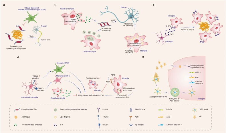
“On the other hand, microglia could also drive tau spreading and toxicity by promoting neuroinflammation, such as activating NACHT-, LRR- and pyrin (PYD)-domain-containing protein 3 (NLRP3) inflammasome155 or inducing NF-kB signaling.”
§ Microglia in neurodegenerative diseases: mechanism and potential therapeutic targets > Microglial dysfunction in neurodegenerative diseases > Alzheimer’s disease
What the sources say
- Supportedcites [155] Ising, C. et al. NLRP3 inflammasome activation drives tau pathology. Nature 575, 669–673 (2019). source
The Abstract identifies microglial NLRP3 activation as a driver of tau pathology. In “NLRP3 mediates Aβ-induced tau pathology,” the paper reports NLRP3-dependent tau seeding and hyperphosphorylation in mice; “NLRP3 regulates kinases and phosphatases” links microglial inflammasome activation to worsened tau pathology through IL-1β. The citation is attached to the NLRP3 example, not to the separate NF-κB example. (LLM, conf=0.89)
- Supportedcites [156] Wang, C. et al. Microglial NF-kappaB drives tau spreading and toxicity in a mouse model of tauopathy. Nat. Commun. 13, 1969 (2022). source
The citation is offered for the NF-κB part of the sentence; the NLRP3 example has a separate citation. In the Discussion, the paper reports that microglial NF-κB activation accelerates tau spreading in PS19 mice and that its inhibition protects against tau-related toxicity and cognitive deficits. (LLM, conf=0.93)
hedgeddirect- Asserted: passes — The authors put it forward as true.
- Checkable: passes — It can be checked against evidence.
- Load-bearing: passes — The paper’s conclusions depend on it.
- Based: passes — The paper gives a reason why it holds.
- Supported
Every source the paper cites for this claim was checked and supports it.
In the paper
“However, its aggregation results in a gain of toxic function.”
§ Microglia in neurodegenerative diseases: mechanism and potential therapeutic targets > Microglial dysfunction in neurodegenerative diseases > Parkinson’s disease
What the sources say
- Supportedcites [205] Bras, I. C. & Outeiro, T. F. Alpha-synuclein: mechanisms of release and pathology progression in synucleinopathies. Cells 10, 375 (2021). source
Section 2 describes α-synuclein monomers aggregating into pathological oligomers and fibrils. Section 5 reports that oligomeric species are implicated in neuronal degeneration and that injected fibrils cause dopamine-neuron loss in mice. Together, these passages support the paraphrase that aggregation gives rise to toxic activity. (LLM, conf=0.88)
plaindirect- Asserted: passes — The authors put it forward as true.
- Checkable: passes — It can be checked against evidence.
- Load-bearing: unclear — It is unclear whether the paper’s conclusions depend on it.
- Based: passes — The paper gives a reason why it holds.
- Supported
The cited sources support only part of this claim — read each source’s verdict below.
In the paper
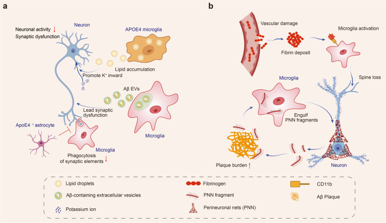
“The extracellular lipid droplets directly decreased neuronal activity by increasing inward K+ currents.”
§ Microglia in neurodegenerative diseases: mechanism and potential therapeutic targets > Microglial dysfunction in neurodegenerative diseases > Alzheimer’s disease
What the sources say
- Partly supportedcites [197] Young, J. E. & Jayadev, S. Neighborhood matters: altered lipid metabolism in APOE4 microglia causes problems for neurons. Cell Stem Cell 29, 1159–1160 (2022). sourceExperimental — accuracy not yet measured
The paper’s main text (indexed under Abstract; no page supplied) says that APOE4 microglial conditioned medium is associated with increased neuronal inward K+ currents and reduced activity. It describes lipid droplets accumulating inside microglia, while increased cholesterol—not extracellular lipid droplets—is found in the medium; cholesterol exposure is proposed as a putative mechanism. The claim therefore identifies the wrong extracellular agent and overstates its direct effect. (LLM, conf=0.89)
plaindirect- Asserted: passes — The authors put it forward as true.
- Checkable: passes — It can be checked against evidence.
- Load-bearing: unclear — It is unclear whether the paper’s conclusions depend on it.
- Based: passes — The paper gives a reason why it holds.
- Partly supported
The cited sources support only part of this claim — read each source’s verdict below.
In the paper
“The byproduct of synthetic heroin, MPTP, could be taken up by dopaminergic neurons and induces parkinsonism in humans, non-human primates, and mice.”
§ Microglia in neurodegenerative diseases: mechanism and potential therapeutic targets > Microglial dysfunction in neurodegenerative diseases > Parkinson’s disease
What the sources say
- Partly supportedcites [232] Tieu, K. A guide to neurotoxic animal models of Parkinson’s disease. Cold Spring Harb. Perspect. Med. 1, a009316 (2011). sourceExperimental — accuracy not yet measured
The MPTP section reports PD-like effects in humans and nonhuman primates and parkinsonian motor deficits in mice. But its mechanism passage says MPTP is converted in astrocytes to MPP+, which is then taken up by dopaminergic neurons; it does not say MPTP itself is taken up. The paper describes synthetic meperidine contaminated with MPTP, rather than calling MPTP a byproduct of synthetic heroin. (LLM, conf=0.88)
hedgeddirect- Asserted: passes — The authors put it forward as true.
- Checkable: passes — It can be checked against evidence.
- Load-bearing: unclear — It is unclear whether the paper’s conclusions depend on it.
- Based: passes — The paper gives a reason why it holds.
- Partly supported
Every source the paper cites for this claim was checked and supports it.
In the paper
“LRRK2 (PARK8) gene mutations are the most common genetic cause of both familial and sporadic PD.257”
§ Microglia in neurodegenerative diseases: mechanism and potential therapeutic targets > Microglial dysfunction in neurodegenerative diseases > Parkinson’s disease
What the sources say
- Supportedcites [257] Li, J. Q., Tan, L. & Yu, J. T. The role of the LRRK2 gene in Parkinsonism. Mol. Neurodegener. 9, 47 (2014). source
The Introduction explicitly identifies LRRK2 as PARK8 and states that its mutations are the most common genetic cause of both familial and sporadic PD. This directly matches the cited claim. (LLM, conf=0.95)
plaindirect- Asserted: passes — The authors put it forward as true.
- Checkable: passes — It can be checked against evidence.
- Load-bearing: unclear — It is unclear whether the paper’s conclusions depend on it.
- Based: passes — The paper gives a reason why it holds.
- Supported
Every source the paper cites for this claim was checked and supports it.
In the paper
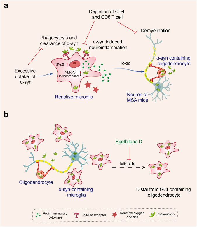
“TLR4 gene ablation in a transgenic mouse model of MSA with oligodendroglial α-syn overexpression impaired the phagocytic ability of microglia to α-syn, leading to enhanced motor impairment and augmented loss of nigrostriatal dopaminergic neurons.295”
§ Microglia in neurodegenerative diseases: mechanism and potential therapeutic targets > Microglial dysfunction in neurodegenerative diseases > Multiple system atrophy
What the sources say
- Supportedcites [295] Stefanova, N. et al. Toll-like receptor 4 promotes alpha-synuclein clearance and survival of nigral dopaminergic neurons. Am. J. Pathol. 179, 954–963 (2011). source
Split into 2 atoms — 2 confirmed, 0 partially supported, 0 disputed, 0 misattributed, 0 unverifiable. Expand the claim to see per-atom verdicts.
plaindirect- Asserted: passes — The authors put it forward as true.
- Checkable: passes — It can be checked against evidence.
- Load-bearing: passes — The paper’s conclusions depend on it.
- Based: passes — The paper gives a reason why it holds.
- Supported
The cited sources support only part of this claim — read each source’s verdict below.
In the paper
“Microglial activation in middle frontal and superior or middle temporal gyrus regions was also highly found in vivo PET imaging studies in humans”
§ Microglia in neurodegenerative diseases: mechanism and potential therapeutic targets > Microglial dysfunction in neurodegenerative diseases > Amyotrophic lateral sclerosis and frontotemporal dementia
What the sources say
- Not checked yetcites [311] Turner, M. R. et al. Evidence of widespread cerebral microglial activation in amyotrophic lateral sclerosis: an [11C](R)-PK11195 positron emission tomography study. Neurobiol. Dis. 15, 601–609 (2004). source
- Partly supportedcites [312] Zurcher, N. R. et al. Increased in vivo glial activation in patients with amyotrophic lateral sclerosis: assessed with [(11)C]-PBR28. Neuroimage Clin. 7, 409–414 (2015). sourceExperimental — accuracy not yet measured
The Results (Whole brain between-group analysis; page unavailable) report increased PET tracer binding in the motor cortices and corticospinal tract of patients with ALS. The Introduction also reports an earlier PET finding in the temporal cortex. Neither passage establishes the claimed high activation specifically in the middle frontal and superior or middle temporal gyri. (LLM, conf=0.83)
- Partly supportedcites [313] Tondo, G. et al. (11) C-PK11195 PET-based molecular study of microglia activation in SOD1 amyotrophic lateral sclerosis. Ann. Clin. Transl. Neurol. 7, 1513–1523 (2020). sourceExperimental — accuracy not yet measured
The Results (Group level analysis; page unavailable) report significant PET-detected microglial activation in temporal cortex and premotor frontal cortex in people with SOD1-ALS. They do not identify the middle frontal or superior or middle temporal gyri specifically. The Results (Single-subject analysis; page unavailable) also say frontal-lobe clusters were less frequent in asymptomatic carriers, so the claim that activation was highly found in all the named regions overstates the findings. (LLM, conf=0.84)
plaincorrelational- Asserted: passes — The authors put it forward as true.
- Checkable: passes — It can be checked against evidence.
- Load-bearing: unclear — It is unclear whether the paper’s conclusions depend on it.
- Based: passes — The paper gives a reason why it holds.
Every source the paper cites for this claim was checked and supports it.
In the paper
“Moreover, MSA mice with Tcrb (CD4 and CD8 T cell knockout) or Cd4 (CD4 T cell knockout) genetic deletion showed reduced inflammation and demyelination after exposure to syn301”
§ Microglia in neurodegenerative diseases: mechanism and potential therapeutic targets > Microglial dysfunction in neurodegenerative diseases > Multiple system atrophy
What the sources say
- Supportedcites [301] Williams, G. P. et al. T cell infiltration in both human multiple system atrophy and a novel mouse model of the disease. Acta Neuropathol. 139, 855–874 (2020). source
The Results section (‘Knockout of αβ T cells or CD4 T cells alone…’) identifies Tcrb−/− and Cd4−/− mice as the respective T-cell knockouts. After Olig001-SYN treatment, both showed reduced CNS myeloid activation and less demyelination than wild-type mice (Fig. 7). (LLM, conf=0.94)
plaindirect- Asserted: passes — The authors put it forward as true.
- Checkable: passes — It can be checked against evidence.
- Load-bearing: passes — The paper’s conclusions depend on it.
- Based: passes — The paper gives a reason why it holds.
- Supported
- It may state two separate claims as one. 186
- Probably not a substantial claim. 23
The cited sources support only part of this claim — read each source’s verdict below.
In the paper
“Recent advances in single-cell technologies have revealed that these reactive microglia were with high spatial and temporal heterogeneity.”
- Also stated in: § Introduction · plain
“Recent advances in scRNA-seq and snRNA-seq technologies have identified high spatial and temporal heterogeneity levels and unique disease-related signatures of these reactive microglia without correspondence to the canonical M1/M2 classification in neurodegenerative diseases.”
§ Microglia in neurodegenerative diseases: mechanism and potential therapeutic targets > Abstract
What the sources say
- Partly supportedcites [13] Keren-Shaul, H. et al. A unique microglia type associated with restricting development of Alzheimer’s disease. Cell 169, 1276–1290.e1217 (2017). sourceExperimental — accuracy not yet measured
The Results describe distinct disease-associated microglial states and a transition from homeostatic microglia as Alzheimer’s disease progresses; the Introduction locates these cells near amyloid plaques. This supports spatial and temporal differences in the studied disease models, but the cited passages do not establish high heterogeneity across neurodegenerative diseases or address correspondence with the M1/M2 classification. (LLM, conf=0.82)
- Partly supportedcites [14] Friedman, B. A. et al. Diverse brain myeloid expression profiles reveal distinct microglial activation states and aspects of Alzheimer’s disease not evident in mouse models. Cell Rep. 22, 832–847 (2018). sourceExperimental — accuracy not yet measured
The Results identify distinct microglial states in a single-cell dataset from an Alzheimer’s mouse model, and the Discussion reports expression differences between cerebellar and cortical microglia and between aged and young mice. These passages support heterogeneity across cells, regions, and ages, but do not establish that single-cell technologies revealed high spatial and temporal heterogeneity of reactive microglia across neurodegenerative diseases. The copy also does not directly compare those states with the M1/M2 classification. (LLM, conf=0.84)
- Not checked yetcites [15] Gerrits, E. et al. Distinct amyloid-beta and tau-associated microglia profiles in Alzheimer’s disease. Acta Neuropathol. 141, 681–696 (2021). source
plain- Asserted: passes — The authors put it forward as true.
- Checkable: passes — It can be checked against evidence.
- Load-bearing: passes — The paper’s conclusions depend on it.
- Based: passes — The paper gives a reason why it holds.
It may state two separate claims as one.
- Also stated in: § Introduction · plain
Every source the paper cites for this claim was checked and supports it.
In the paper
“Notably, DAMs were localized near Aβ plaques and participated in the clearance of β-amyloid.”
§ Microglia in neurodegenerative diseases: mechanism and potential therapeutic targets > Introduction
What the sources say
- Supportedcites [13] Keren-Shaul, H. et al. A unique microglia type associated with restricting development of Alzheimer’s disease. Cell 169, 1276–1290.e1217 (2017). source
The Results (page unavailable) place DAM-marker-positive microglia near Aβ plaques and report intracellular Aβ particles in those cells. The authors conclude that DAM actively participate in dismantling and digesting amyloid plaques, supporting the paraphrase about clearance. (LLM, conf=0.90)
plaindirect- Asserted: passes — The authors put it forward as true.
- Checkable: passes — It can be checked against evidence.
- Load-bearing: unclear — It is unclear whether the paper’s conclusions depend on it.
- Based: passes — The paper gives a reason why it holds.
It may state two separate claims as one.
- Supported
The sources with a verdict support this claim; the rest weren’t checked yet, have no full text we could read, or their verdicts aren’t rated yet.
In the paper
“Firstly, microglial activation colocalized and correlated with tau pathology in PSP patients374,378 and transgenic mice expressing mutant tau P301L.380”
§ Microglia in neurodegenerative diseases: mechanism and potential therapeutic targets > Microglial dysfunction in neurodegenerative diseases > Progressive supranuclear palsy
What the sources say
- Not checked yetcites [374] Ishizawa, K. & Dickson, D. W. Microglial activation parallels system degeneration in progressive supranuclear palsy and corticobasal degeneration. J. Neuropathol. Exp. Neurol. 60, 647–657 (2001). source
- Supportedcites [378] Malpetti, M. et al. Neuroinflammation and tau colocalize in vivo in progressive supranuclear palsy. Ann. Neurol. 88, 1194–1204 (2020). source
The Discussion reports a positive correlation between PET measures of microglial activation and tau pathology across brain regions in PSP patients and concludes that the two are macroscopically co-localized. The transgenic-mouse clause has a separate citation (380) and is not attributed to this source. (LLM, conf=0.93)
- Not checked yetcites [380] Sasaki, A. et al. Microglial activation in brain lesions with tau deposits: comparison of human tauopathies and tau transgenic mice TgTauP301L. Brain Res. 1214, 159–168 (2008). source
plaincorrelational- Asserted: passes — The authors put it forward as true.
- Checkable: passes — It can be checked against evidence.
- Load-bearing: passes — The paper’s conclusions depend on it.
- Based: passes — The paper gives a reason why it holds.
It may state two separate claims as one.
Every source the paper cites for this claim was checked and supports it.
In the paper
“Microglial elimination in R6/2 mice prevented the loss of PNNs and dramatically increased PNNs in the brains of naïve littermates.”
§ Microglia in neurodegenerative diseases: mechanism and potential therapeutic targets > Microglial dysfunction in neurodegenerative diseases > Huntington’s disease
What the sources say
- Supportedcites [456] Crapser, J. D. et al. Microglial depletion prevents extracellular matrix changes and striatal volume reduction in a model of Huntington’s disease. Brain 143, 266–288 (2020). source
The PNN results section (p. 17) reports that microglial elimination prevented disease-associated PNN loss in the somatosensory and motor cortices of R6/2 mice. It also reports a dramatic increase in PNN density across every examined brain region of naïve non-transgenic mice; the methods (p. 6) identify the non-transgenic mice as littermates. (LLM, conf=0.94)
plaindirect- Asserted: passes — The authors put it forward as true.
- Checkable: passes — It can be checked against evidence.
- Load-bearing: passes — The paper’s conclusions depend on it.
- Based: passes — The paper gives a reason why it holds.
It may state two separate claims as one.
- Supported
In the paper
“Microglia, the resident macrophages of the central nervous system (CNS), utilize their specific receptor repertoire to monitor the microenvironment dynamically in the brain.”
§ Microglia in neurodegenerative diseases: mechanism and potential therapeutic targets > Introduction
What the sources say
- Not checked yetcites [1] Arcuri, C., Mecca, C., Bianchi, R., Giambanco, I. & Donato, R. The pathophysiological role of microglia in dynamic surveillance, phagocytosis and structural remodeling of the developing CNS. Front. Mol. Neurosci. 10, 191 (2017). source
plain- Asserted: passes — The authors put it forward as true.
- Checkable: passes — It can be checked against evidence.
- Load-bearing: fails — The paper’s conclusions may not depend on it.
- Based: passes — The paper gives a reason why it holds.
Probably not a substantial claim. The paper’s conclusions may not depend on it.
A conclusion the paper draws from its own results — it cites no other source for it, so there is none to check. The quote shows it is in the paper.
In the paper
“Extracellular vesicles released from microglia and autophagy impairment in microglia all contribute to pathological progression and neurodegeneration.”
§ Microglia in neurodegenerative diseases: mechanism and potential therapeutic targets > Abstract
plaindirect- Asserted: passes — The authors put it forward as true.
- Checkable: passes — It can be checked against evidence.
- Load-bearing: passes — The paper’s conclusions depend on it.
- Based: passes — The paper gives a reason why it holds.
It may state two separate claims as one.
A conclusion the paper draws from its own results — it cites no other source for it, so there is none to check. The quote shows it is in the paper.
In the paper
“peripheral immune cells infiltration shapes microglia into a pro-inflammatory phenotype and accelerates disease progression.”
- Also stated in: § Conclusion · plain
“Peripheral immune cells infiltration shapes microglia into a pro-inflammatory phenotype and accelerates disease progression.”
§ Microglia in neurodegenerative diseases: mechanism and potential therapeutic targets > Abstract
plaindirect- Asserted: passes — The authors put it forward as true.
- Checkable: passes — It can be checked against evidence.
- Load-bearing: passes — The paper’s conclusions depend on it.
- Based: fails — The paper may give no reason why it holds.
Probably not a substantial claim. The paper may give no reason why it holds. It may state two separate claims as one.
- Also stated in: § Conclusion · plain
None of the sources cited for this claim have been checked yet.
In the paper
“Some identified microglia in specific states correlate with pathological hallmarks and are associated with specific functions.”
§ Microglia in neurodegenerative diseases: mechanism and potential therapeutic targets > Abstract
What the sources say
- Not checked yetcites [11] Savage, J. C., Carrier, M. & Tremblay, M. E. Morphology of microglia across contexts of health and disease. Methods Mol. Biol. 2034, 13–26 (2019). source
- Not checked yetcites [12] Bouvier, D. S. et al. High resolution dissection of reactive glial nets in Alzheimer’s disease. Sci. Rep. 6, 24544 (2016). source
plaincorrelational- Asserted: passes — The authors put it forward as true.
- Checkable: passes — It can be checked against evidence.
- Load-bearing: passes — The paper’s conclusions depend on it.
- Based: passes — The paper gives a reason why it holds.
It may state two separate claims as one.
A conclusion the paper draws from its own results — it cites no other source for it, so there is none to check. The quote shows it is in the paper.
In the paper
“play detrimental roles due to excessive uptake of protein aggregates, which would lead to microglial phagocytic ability impairment, neuroinflammation, and eventually neurodegeneration.”
§ Microglia in neurodegenerative diseases: mechanism and potential therapeutic targets > Abstract
hedgeddirect- Asserted: passes — The authors put it forward as true.
- Checkable: passes — It can be checked against evidence.
- Load-bearing: passes — The paper’s conclusions depend on it.
- Based: passes — The paper gives a reason why it holds.
It may state two separate claims as one.
A conclusion the paper draws from its own results — it cites no other source for it, so there is none to check. The quote shows it is in the paper.
In the paper
“However, microglia activation is more varied and dynamic than ever anticipated, both in -omics features and functional consequences, indicating that microglia respond differently in different diseases.”
§ Microglia in neurodegenerative diseases: mechanism and potential therapeutic targets > Microglial dysfunction in neurodegenerative diseases
hedgedcorrelational- Asserted: passes — The authors put it forward as true.
- Checkable: passes — It can be checked against evidence.
- Load-bearing: passes — The paper’s conclusions depend on it.
- Based: fails — The paper may give no reason why it holds.
Probably not a substantial claim. The paper may give no reason why it holds. It may state two separate claims as one.
In the paper
“Both intrinsic factors (species, sex, genetic background, etc.) and extrinsic factors (pathogens, nutrition, microbiota, etc.) influence microglial states.”
§ Microglia in neurodegenerative diseases: mechanism and potential therapeutic targets > Introduction
What the sources say
- Not checked yetcites [8] Paolicelli, R. C. et al. Microglia states and nomenclature: a field at its crossroads. Neuron 110, 3458–3483 (2022). source
plaindirect- Asserted: passes — The authors put it forward as true.
- Checkable: passes — It can be checked against evidence.
- Load-bearing: unclear — It is unclear whether the paper’s conclusions depend on it.
- Based: passes — The paper gives a reason why it holds.
It may state two separate claims as one.
The paper states this without a source or data behind it here, so there is nothing to check it against. The quote shows it is in the paper.
In the paper
“Microglia activation was observed in various neurodegenerative diseases.”
§ Microglia in neurodegenerative diseases: mechanism and potential therapeutic targets > Microglial dysfunction in neurodegenerative diseases
plaincorrelational- Asserted: passes — The authors put it forward as true.
- Checkable: passes — It can be checked against evidence.
- Load-bearing: passes — The paper’s conclusions depend on it.
- Based: fails — The paper may give no reason why it holds.
Probably not a substantial claim. The paper may give no reason why it holds.
None of the sources cited for this claim have been checked yet.
In the paper
“Microglial cell colonization of the CNS is evolutionarily conserved across vertebrate species and occurs before the formation of the neuroectoderm-derived glial cell types such as oligodendrocytes and astrocytes.”
§ Microglia in neurodegenerative diseases: mechanism and potential therapeutic targets > Origin and development of brain microglia
What the sources say
- Not checked yetcites [48] Verney, C., Monier, A., Fallet-Bianco, C. & Gressens, P. Early microglial colonization of the human forebrain and possible involvement in periventricular white-matter injury of preterm infants. J. Anat. 217, 436–448 (2010). source
- Not checked yetcites [49] Schlegelmilch, T., Henke, K. & Peri, F. Microglia in the developing brain: from immunity to behaviour. Curr. Opin. Neurobiol. 21, 5–10 (2011). source
plain- Asserted: passes — The authors put it forward as true.
- Checkable: passes — It can be checked against evidence.
- Load-bearing: unclear — It is unclear whether the paper’s conclusions depend on it.
- Based: passes — The paper gives a reason why it holds.
It may state two separate claims as one.
In the paper
“Thus, primary EMPs, secondary EMP-derived fetal liver monocytes, and HSC-derived fetal liver monocytes contribute to all tissue macrophages, with microglia arising solely from primary EMP.”
§ Microglia in neurodegenerative diseases: mechanism and potential therapeutic targets > Ontogeny of microglia
What the sources say
- Not checked yetcites [41] Li, Q. & Barres, B. A. Microglia and macrophages in brain homeostasis and disease. Nat. Rev. Immunol. 18, 225–242 (2018). source
plaindirect- Asserted: passes — The authors put it forward as true.
- Checkable: passes — It can be checked against evidence.
- Load-bearing: unclear — It is unclear whether the paper’s conclusions depend on it.
- Based: passes — The paper gives a reason why it holds.
It may state two separate claims as one.
In the paper
“Once A1 astrocytes are induced, they lose their essential functions such as supporting neuronal survival, and they also promote neuroinflammation, which contributes to the progression of neurodegenerative diseases.”
§ Microglia in neurodegenerative diseases: mechanism and potential therapeutic targets > Microglia in the homeostatic brain > Microglia-astrocyte crosstalk
What the sources say
- Not checked yetcites [113] Yun, S. P. et al. Block of A1 astrocyte conversion by microglia is neuroprotective in models of Parkinson’s disease. Nat. Med. 24, 931–938 (2018). source
plaindirect- Asserted: passes — The authors put it forward as true.
- Checkable: passes — It can be checked against evidence.
- Load-bearing: unclear — It is unclear whether the paper’s conclusions depend on it.
- Based: passes — The paper gives a reason why it holds.
It may state two separate claims as one.
- The authors state this as an assumption: The M1/M2 and A1/A2 classifications, although inaccurate, are helpful for elucidating microglia-astrocyte interaction.
None of the sources cited for this claim have been checked yet.
In the paper
“Microglia communicate with nearly all cell types in the brain to facilitate developmental process, maintain homeostasis, assist in tissue repair, and contribute to the pathogenesis of diseases.”
§ Microglia in neurodegenerative diseases: mechanism and potential therapeutic targets > Microglia in the homeostatic brain > Microglia-neuron crosstalk
What the sources say
- Not checked yetcites [53] Prinz, M., Masuda, T., Wheeler, M. A. & Quintana, F. J. Microglia and central nervous system-associated macrophages-from origin to disease modulation. Annu. Rev. Immunol. 39, 251–277 (2021). source
- Not checked yetcites [97] Frost, J. L. & Schafer, D. P. Microglia: architects of the developing nervous system. Trends Cell Biol. 26, 587–597 (2016). source
plaindirect- Asserted: passes — The authors put it forward as true.
- Checkable: passes — It can be checked against evidence.
- Load-bearing: unclear — It is unclear whether the paper’s conclusions depend on it.
- Based: passes — The paper gives a reason why it holds.
It may state two separate claims as one.
In the paper
“Microglia phagocytose misfolded proteins, cellular debris, and dying cells to maintain homeostasis.”
§ Microglia in neurodegenerative diseases: mechanism and potential therapeutic targets > Introduction
What the sources say
- Not checked yetcites [2] Sarlus, H. & Heneka, M. T. Microglia in Alzheimer’s disease. J. Clin. Invest. 127, 3240–3249 (2017). source
plaindirect- Asserted: passes — The authors put it forward as true.
- Checkable: passes — It can be checked against evidence.
- Load-bearing: fails — The paper’s conclusions may not depend on it.
- Based: passes — The paper gives a reason why it holds.
Probably not a substantial claim. The paper’s conclusions may not depend on it.
In the paper
“The primary pathological features of AD are extraneuronal amyloid plaques formed by the deposition of Aβ peptide in the brain and intracellular neurofibrillary tangles (NFTs) caused by abnormal aggregation of tau protein.”
§ Microglia in neurodegenerative diseases: mechanism and potential therapeutic targets > Microglial dysfunction in neurodegenerative diseases > Alzheimer’s disease
What the sources say
- Not checked yetcites [122] Duyckaerts, C., Delatour, B. & Potier, M. C. Classification and basic pathology of Alzheimer disease. Acta Neuropathol. 118, 5–36 (2009). source
plaindirect- Asserted: passes — The authors put it forward as true.
- Checkable: passes — It can be checked against evidence.
- Load-bearing: unclear — It is unclear whether the paper’s conclusions depend on it.
- Based: passes — The paper gives a reason why it holds.
It may state two separate claims as one.
In the paper
“TREM2 is expressed highly and exclusively in microglia in the brain.”
§ Microglia in neurodegenerative diseases: mechanism and potential therapeutic targets > Microglial dysfunction in neurodegenerative diseases > Alzheimer’s disease
What the sources say
- Not checked yetcites [162] Jay, T. R., von Saucken, V. E. & Landreth, G. E. TREM2 in neurodegenerative diseases. Mol. Neurodegener. 12, 56 (2017). source
plain- Asserted: passes — The authors put it forward as true.
- Checkable: passes — It can be checked against evidence.
- Load-bearing: unclear — It is unclear whether the paper’s conclusions depend on it.
- Based: passes — The paper gives a reason why it holds.
It may state two separate claims as one.
In the paper
“In a humanized APP mouse model, MGnD microglia, a class of disease-reactive microglia common in neurodegeneration, hypersecrete EVs containing phosphorylated tau (pTau), accelerating tau propagation.”
§ Microglia in neurodegenerative diseases: mechanism and potential therapeutic targets > Microglial dysfunction in neurodegenerative diseases > Alzheimer’s disease
What the sources say
- Not checked yetcites [160] Clayton, K. et al. Plaque associated microglia hyper-secrete extracellular vesicles and accelerate tau propagation in a humanized APP mouse model. Mol. Neurodegener. 16, 18 (2021). source
plaindirect- Asserted: passes — The authors put it forward as true.
- Checkable: passes — It can be checked against evidence.
- Load-bearing: unclear — It is unclear whether the paper’s conclusions depend on it.
- Based: passes — The paper gives a reason why it holds.
It may state two separate claims as one.
In the paper
“Keren-Shaul et al.13 first identified a subgroup of microglia in AD, termed disease-associated microglia (DAM), which participated in the clearance of Aβ. DAMs were localized near Aβ plaques”
§ Microglia in neurodegenerative diseases: mechanism and potential therapeutic targets > Microglial dysfunction in neurodegenerative diseases > Alzheimer’s disease
What the sources say
- Not checked yetcites [13] Keren-Shaul, H. et al. A unique microglia type associated with restricting development of Alzheimer’s disease. Cell 169, 1276–1290.e1217 (2017). source
plaindirect- Asserted: passes — The authors put it forward as true.
- Checkable: passes — It can be checked against evidence.
- Load-bearing: passes — The paper’s conclusions depend on it.
- Based: passes — The paper gives a reason why it holds.
It may state two separate claims as one.
In the paper
“TREM2 has been reported to promote the conversion of microglia to the DAM phenotype, which is responsible for Aβ phagocytosis.”
§ Microglia in neurodegenerative diseases: mechanism and potential therapeutic targets > Microglial dysfunction in neurodegenerative diseases > Alzheimer’s disease
What the sources say
- Not checked yetcites [13] Keren-Shaul, H. et al. A unique microglia type associated with restricting development of Alzheimer’s disease. Cell 169, 1276–1290.e1217 (2017). source
hedgeddirect- Asserted: passes — The authors put it forward as true.
- Checkable: passes — It can be checked against evidence.
- Load-bearing: passes — The paper’s conclusions depend on it.
- Based: passes — The paper gives a reason why it holds.
It may state two separate claims as one.
In the paper
“LC3-associated endocytosis (LANDO) in microglia facilitates Aβ receptor recycling, increasing Aβ surface receptors, thus promoting Aβ clearance”
§ Microglia in neurodegenerative diseases: mechanism and potential therapeutic targets > Microglial dysfunction in neurodegenerative diseases > Alzheimer’s disease
What the sources say
- Not checked yetcites [146] Heckmann, B. L. et al. LC3-associated endocytosis facilitates beta-amyloid clearance and mitigates neurodegeneration in murine Alzheimer’s disease. Cell 178, 536–551.e514 (2019). source
plaindirect- Asserted: passes — The authors put it forward as true.
- Checkable: passes — It can be checked against evidence.
- Load-bearing: unclear — It is unclear whether the paper’s conclusions depend on it.
- Based: passes — The paper gives a reason why it holds.
It may state two separate claims as one.
A conclusion the paper draws from its own results — it cites no other source for it, so there is none to check. The quote shows it is in the paper.
In the paper
“However, when these growing pathological deposits chronically stimulate microglia, the protective microglia may convert to dysfunctional microglia, aggravating the disease progression of AD.”
§ Microglia in neurodegenerative diseases: mechanism and potential therapeutic targets > Microglial dysfunction in neurodegenerative diseases > Alzheimer’s disease
hedgeddirect- Asserted: passes — The authors put it forward as true.
- Checkable: passes — It can be checked against evidence.
- Load-bearing: passes — The paper’s conclusions depend on it.
- Based: passes — The paper gives a reason why it holds.
It may state two separate claims as one.
In the paper
“Aβ binds to microglial TREM2, which activates TREM2 signaling and lead to the enhanced phagocytosis of microglia.”
§ Microglia in neurodegenerative diseases: mechanism and potential therapeutic targets > Microglial dysfunction in neurodegenerative diseases > Alzheimer’s disease
What the sources say
- Not checked yetcites [174] Hammond, T. R., Marsh, S. E. & Stevens, B. Immune signaling in neurodegeneration. Immunity 50, 955–974 (2019). source
plaindirect- Asserted: passes — The authors put it forward as true.
- Checkable: passes — It can be checked against evidence.
- Load-bearing: passes — The paper’s conclusions depend on it.
- Based: passes — The paper gives a reason why it holds.
It may state two separate claims as one.
None of the sources cited for this claim have been checked yet.
In the paper
“BACE-1 inhibition in microglia facilitated the microglia phenotype transition from homeostatic to stage 1 disease-associated microglia (DAM-1) signature148 and thus enhanced amyloid clearance and improved cognitive performance in AD mice.”
§ Microglia in neurodegenerative diseases: mechanism and potential therapeutic targets > Microglial dysfunction in neurodegenerative diseases > Alzheimer’s disease
What the sources say
- Not checked yetcites [148] Singh, N. et al. BACE-1 inhibition facilitates the transition from homeostatic microglia to DAM-1. Sci. Adv. 8, eabo1286 (2022). source
- Not checked yetcites [149] Singh, N., Das, B., Zhou, J., Hu, X. & Yan, R. Targeted BACE-1 inhibition in microglia enhances amyloid clearance and improved cognitive performance. Sci. Adv. 8, eabo3610 (2022). source
plaindirect- Asserted: passes — The authors put it forward as true.
- Checkable: passes — It can be checked against evidence.
- Load-bearing: unclear — It is unclear whether the paper’s conclusions depend on it.
- Based: passes — The paper gives a reason why it holds.
It may state two separate claims as one.
In the paper
“In contrast, NgR-deficient AD mice reduced amyloid burden and improved cognitive impairment.”
§ Microglia in neurodegenerative diseases: mechanism and potential therapeutic targets > Microglial dysfunction in neurodegenerative diseases > Alzheimer’s disease
What the sources say
- Not checked yetcites [147] Wang, J. et al. Nogo receptor impairs the clearance of fibril amyloid-beta by microglia and accelerates Alzheimer’s-like disease progression. Aging Cell 20, e13515 (2021). source
plaindirect- Asserted: passes — The authors put it forward as true.
- Checkable: passes — It can be checked against evidence.
- Load-bearing: unclear — It is unclear whether the paper’s conclusions depend on it.
- Based: passes — The paper gives a reason why it holds.
It may state two separate claims as one.
In the paper
“After being released from microglia, ASC specks bind to and promote the cross-seeding of Aβ, leading to amyloid seeding and spreading of amyloid pathology.”
§ Microglia in neurodegenerative diseases: mechanism and potential therapeutic targets > Microglial dysfunction in neurodegenerative diseases > Alzheimer’s disease
What the sources say
- Not checked yetcites [153] Venegas, C. et al. Microglia-derived ASC specks cross-seed amyloid-beta in Alzheimer’s disease. Nature 552, 355–361 (2017). source
plaindirect- Asserted: passes — The authors put it forward as true.
- Checkable: passes — It can be checked against evidence.
- Load-bearing: passes — The paper’s conclusions depend on it.
- Based: passes — The paper gives a reason why it holds.
It may state two separate claims as one.
None of the sources cited for this claim have been checked yet.
In the paper
“Loss of TREM2 function impaired Aβ phagocytosis by microglia and increased amyloid seeding in AD mouse models.”
§ Microglia in neurodegenerative diseases: mechanism and potential therapeutic targets > Microglial dysfunction in neurodegenerative diseases > Alzheimer’s disease
What the sources say
- Not checked yetcites [169] Wang, Y. et al. TREM2 lipid sensing sustains the microglial response in an Alzheimer’s disease model. Cell 160, 1061–1071 (2015). source
- Not checked yetcites [170] Parhizkar, S. et al. Loss of TREM2 function increases amyloid seeding but reduces plaque-associated ApoE. Nat. Neurosci. 22, 191–204 (2019). source
plaindirect- Asserted: passes — The authors put it forward as true.
- Checkable: passes — It can be checked against evidence.
- Load-bearing: passes — The paper’s conclusions depend on it.
- Based: passes — The paper gives a reason why it holds.
It may state two separate claims as one.
In the paper
“Compared with APOE4, APOE3 lipoproteins induce faster microglial migration towards Aβ, facilitate Aβ uptake, and ameliorate cognition.”
§ Microglia in neurodegenerative diseases: mechanism and potential therapeutic targets > Microglial dysfunction in neurodegenerative diseases > Alzheimer’s disease
What the sources say
- Not checked yetcites [151] Fitz, N. F. et al. Phospholipids of APOE lipoproteins activate microglia in an isoform-specific manner in preclinical models of Alzheimer’s disease. Nat. Commun. 12, 3416 (2021). source
plaindirect- Asserted: passes — The authors put it forward as true.
- Checkable: passes — It can be checked against evidence.
- Load-bearing: unclear — It is unclear whether the paper’s conclusions depend on it.
- Based: passes — The paper gives a reason why it holds.
It may state two separate claims as one.
None of the sources cited for this claim have been checked yet.
In the paper
“enhancing TREM2 signaling by TREM2 agonist antibody, direct delivery of the TREM2 gene, or stimulating other pathways to increase TREM2 expression in the brains of AD mice enhanced Aβ phagocytosis and improved cognitive behaviors.”
§ Microglia in neurodegenerative diseases: mechanism and potential therapeutic targets > Microglial dysfunction in neurodegenerative diseases > Alzheimer’s disease
What the sources say
- Not checked yetcites [171] Schlepckow, K. et al. Enhancing protective microglial activities with a dual function TREM2 antibody to the stalk region. EMBO Mol. Med. 12, e11227 (2020). source
- Not checked yetcites [172] Wang, S. et al. Anti-human TREM2 induces microglia proliferation and reduces pathology in an Alzheimer’s disease model. J. Exp. Med. 217, e20200785 (2020). source
- Not checked yetcites [173] Zhao, P. et al. A tetravalent TREM2 agonistic antibody reduced amyloid pathology in a mouse model of Alzheimer’s disease. Sci. Transl. Med. 14, eabq0095 (2022). source
plaindirect- Asserted: passes — The authors put it forward as true.
- Checkable: passes — It can be checked against evidence.
- Load-bearing: passes — The paper’s conclusions depend on it.
- Based: passes — The paper gives a reason why it holds.
It may state two separate claims as one.
None of the sources cited for this claim have been checked yet.
In the paper
“AD1-microglia were localized near Aβ plaques and were strongly correlated with the tissue Aβ load.”
§ Microglia in neurodegenerative diseases: mechanism and potential therapeutic targets > Microglial dysfunction in neurodegenerative diseases > Alzheimer’s disease
boostedcorrelational- Asserted: passes — The authors put it forward as true.
- Checkable: passes — It can be checked against evidence.
- Load-bearing: unclear — It is unclear whether the paper’s conclusions depend on it.
- Based: passes — The paper gives a reason why it holds.
It may state two separate claims as one.
In the paper
“Microglial autophagy deficiency can lead to dysregulation of lipid metabolism, induce microglia into a pro-inflammatory state, and as a result, enhance intraneuronal tau pathology and its spreading.”
§ Microglia in neurodegenerative diseases: mechanism and potential therapeutic targets > Microglial dysfunction in neurodegenerative diseases > Alzheimer’s disease
What the sources say
- Not checked yetcites [158] Xu, Y., Propson, N. E., Du, S., Xiong, W. & Zheng, H. Autophagy deficiency modulates microglial lipid homeostasis and aggravates tau pathology and spreading. Proc. Natl Acad. Sci. USA 118, e2023418118 (2021). source
plaindirect- Asserted: passes — The authors put it forward as true.
- Checkable: passes — It can be checked against evidence.
- Load-bearing: passes — The paper’s conclusions depend on it.
- Based: passes — The paper gives a reason why it holds.
It may state two separate claims as one.
In the paper
“With aging, the Nogo receptor (NgR) expression on microglia increased, impairing microglial phagocytosis and clearance of Aβ.”
§ Microglia in neurodegenerative diseases: mechanism and potential therapeutic targets > Microglial dysfunction in neurodegenerative diseases > Alzheimer’s disease
What the sources say
- Not checked yetcites [147] Wang, J. et al. Nogo receptor impairs the clearance of fibril amyloid-beta by microglia and accelerates Alzheimer’s-like disease progression. Aging Cell 20, e13515 (2021). source
plaindirect- Asserted: passes — The authors put it forward as true.
- Checkable: passes — It can be checked against evidence.
- Load-bearing: passes — The paper’s conclusions depend on it.
- Based: passes — The paper gives a reason why it holds.
It may state two separate claims as one.
A conclusion the paper draws from its own results — it cites no other source for it, so there is none to check. The quote shows it is in the paper.
In the paper
“In the early stage of the disease, this response may be protective. The reactive microglia migrate to the vicinity of pathological deposits such as Aβ or tau and then engulf and eliminate them.”
§ Microglia in neurodegenerative diseases: mechanism and potential therapeutic targets > Microglial dysfunction in neurodegenerative diseases > Alzheimer’s disease
hedgedconditional- Asserted: passes — The authors put it forward as true.
- Checkable: passes — It can be checked against evidence.
- Load-bearing: passes — The paper’s conclusions depend on it.
- Based: passes — The paper gives a reason why it holds.
It may state two separate claims as one.
In the paper
“in contrast, LANDO-deficient AD mice induced neurodegeneration and memory deficits.”
§ Microglia in neurodegenerative diseases: mechanism and potential therapeutic targets > Microglial dysfunction in neurodegenerative diseases > Alzheimer’s disease
What the sources say
- Not checked yetcites [146] Heckmann, B. L. et al. LC3-associated endocytosis facilitates beta-amyloid clearance and mitigates neurodegeneration in murine Alzheimer’s disease. Cell 178, 536–551.e514 (2019). source
plaindirect- Asserted: passes — The authors put it forward as true.
- Checkable: passes — It can be checked against evidence.
- Load-bearing: unclear — It is unclear whether the paper’s conclusions depend on it.
- Based: passes — The paper gives a reason why it holds.
It may state two separate claims as one.
In the paper
“the microglia in the midbrain had a decreased complexity compared to those in the striatum, showing that midbrain microglia had a muted reaction to an inflammatory stimulus, displaying a tolerogenic rather than primed phenotype.”
§ Microglia in neurodegenerative diseases: mechanism and potential therapeutic targets > Microglial dysfunction in neurodegenerative diseases > Parkinson’s disease
What the sources say
- Not checked yetcites [218] Uriarte Huarte, O. et al. Single-cell transcriptomics and in situ morphological analyses reveal microglia heterogeneity across the nigrostriatal pathway. Front. Immunol. 12, 639613 (2021). source
plaindirect- Asserted: passes — The authors put it forward as true.
- Checkable: passes — It can be checked against evidence.
- Load-bearing: unclear — It is unclear whether the paper’s conclusions depend on it.
- Based: passes — The paper gives a reason why it holds.
It may state two separate claims as one.
In the paper
“the researchers observed an increased number in reactive microglia and a reduction in oligodendrocyte numbers in PD patients.”
§ Microglia in neurodegenerative diseases: mechanism and potential therapeutic targets > Microglial dysfunction in neurodegenerative diseases > Parkinson’s disease
What the sources say
- Not checked yetcites [215] Smajic, S. et al. Single-cell sequencing of human midbrain reveals glial activation and a Parkinson-specific neuronal state. Brain 145, 964–978 (2022). source
plaincorrelational- Asserted: passes — The authors put it forward as true.
- Checkable: passes — It can be checked against evidence.
- Load-bearing: passes — The paper’s conclusions depend on it.
- Based: passes — The paper gives a reason why it holds.
It may state two separate claims as one.
In the paper
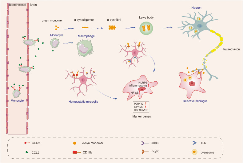
“The microglial activation trajectory extends from P2RY12high cells to two activation branches, one with highly expressed HSP90AA1 or IL1B241 cells and the other containing GPNMBhigh cells.”
§ Microglia in neurodegenerative diseases: mechanism and potential therapeutic targets > Microglial dysfunction in neurodegenerative diseases > Parkinson’s disease
What the sources say
- Not checked yetcites [215] Smajic, S. et al. Single-cell sequencing of human midbrain reveals glial activation and a Parkinson-specific neuronal state. Brain 145, 964–978 (2022). source
plaindirect- Asserted: passes — The authors put it forward as true.
- Checkable: passes — It can be checked against evidence.
- Load-bearing: passes — The paper’s conclusions depend on it.
- Based: passes — The paper gives a reason why it holds.
It may state two separate claims as one.
In the paper
“Peripheral inflammation can also affect the brain through the blood-brain barrier or the autonomic nervous system, triggering microglial activation and contributing to neurodegeneration.”
§ Microglia in neurodegenerative diseases: mechanism and potential therapeutic targets > Microglial dysfunction in neurodegenerative diseases > Parkinson’s disease
What the sources say
- Not checked yetcites [225] Ferrari, C. C. & Tarelli, R. Parkinson’s disease and systemic inflammation. Parkinsons Dis. 2011, 436813 (2011).
plaindirect- Asserted: passes — The authors put it forward as true.
- Checkable: passes — It can be checked against evidence.
- Load-bearing: passes — The paper’s conclusions depend on it.
- Based: passes — The paper gives a reason why it holds.
It may state two separate claims as one.
A conclusion the paper draws from its own results — it cites no other source for it, so there is none to check. The quote shows it is in the paper.
In the paper
“Responding to tau pathology, in the early stages of AD, TREM2 may suppress tau seeding, but later in AD, it may aggravate tau propagation.”
§ Microglia in neurodegenerative diseases: mechanism and potential therapeutic targets > Microglial dysfunction in neurodegenerative diseases > Alzheimer’s disease
hedgedconditional- Asserted: passes — The authors put it forward as true.
- Checkable: passes — It can be checked against evidence.
- Load-bearing: passes — The paper’s conclusions depend on it.
- Based: passes — The paper gives a reason why it holds.
It may state two separate claims as one.
In the paper
“Fibrinogen leaked from the site of cerebrovascular damage into the brain and subsequently bound to the receptor CD11b on the surface of microglia. The interaction of fibrinogen and CD11b mediated microglial activation led to spine loss and promoted cognitive deficits in the 5×FAD mice.”
§ Microglia in neurodegenerative diseases: mechanism and potential therapeutic targets > Microglial dysfunction in neurodegenerative diseases > Alzheimer’s disease
What the sources say
- Not checked yetcites [200] Merlini, M. et al. Fibrinogen induces microglia-mediated spine elimination and cognitive impairment in an Alzheimer’s disease model. Neuron 101, 1099–1108.e1096 (2019). source
plaindirect- Asserted: passes — The authors put it forward as true.
- Checkable: passes — It can be checked against evidence.
- Load-bearing: passes — The paper’s conclusions depend on it.
- Based: passes — The paper gives a reason why it holds.
It may state two separate claims as one.
None of the sources cited for this claim have been checked yet.
In the paper
“A higher level of NLRP3 inflammasome has been observed in microglial cells in the SN of PD patients, which has been linked to the secretion of pro-inflammatory cytokines associated with neurodegeneration.”
§ Microglia in neurodegenerative diseases: mechanism and potential therapeutic targets > Microglial dysfunction in neurodegenerative diseases > Parkinson’s disease
What the sources say
- Not checked yetcites [223] Gordon, R. et al. Inflammasome inhibition prevents alpha-synuclein pathology and dopaminergic neurodegeneration in mice. Sci. Transl. Med. 10, eaah4066 (2018). source
- Not checked yetcites [224] Haque, M. E. et al. Targeting the microglial NLRP3 inflammasome and its role in Parkinson’s disease. Mov. Disord. 35, 20–33 (2020). source
plaincorrelational- Asserted: passes — The authors put it forward as true.
- Checkable: passes — It can be checked against evidence.
- Load-bearing: passes — The paper’s conclusions depend on it.
- Based: passes — The paper gives a reason why it holds.
It may state two separate claims as one.
None of the sources cited for this claim have been checked yet.
In the paper
“other studies found that TREM2 deficiency significantly reduced brain atrophy and prevented microglial activation in tau transgenic mice.”
§ Microglia in neurodegenerative diseases: mechanism and potential therapeutic targets > Microglial dysfunction in neurodegenerative diseases > Alzheimer’s disease
What the sources say
- Not checked yetcites [178] Leyns, C. E. G. et al. TREM2 deficiency attenuates neuroinflammation and protects against neurodegeneration in a mouse model of tauopathy. Proc. Natl Acad. Sci. USA 114, 11524–11529 (2017). source
- Not checked yetcites [179] Sayed, F. A. et al. Differential effects of partial and complete loss of TREM2 on microglial injury response and tauopathy. Proc. Natl Acad. Sci. USA 115, 10172–10177 (2018). source
plaindirect- Asserted: passes — The authors put it forward as true.
- Checkable: passes — It can be checked against evidence.
- Load-bearing: unclear — It is unclear whether the paper’s conclusions depend on it.
- Based: passes — The paper gives a reason why it holds.
It may state two separate claims as one.
The paper states this without a source or data behind it here, so there is nothing to check it against. The quote shows it is in the paper.
In the paper
“Pathologically, PD is characterized by the loss of dopaminergic neurons in the substantia nigra pars compacta (SNpc) as well as the accumulation of misfolded α-syn in Lewy bodies.”
§ Microglia in neurodegenerative diseases: mechanism and potential therapeutic targets > Microglial dysfunction in neurodegenerative diseases > Parkinson’s disease
plaincorrelational- Asserted: passes — The authors put it forward as true.
- Checkable: passes — It can be checked against evidence.
- Load-bearing: fails — The paper’s conclusions may not depend on it.
- Based: passes — The paper gives a reason why it holds.
Probably not a substantial claim. The paper’s conclusions may not depend on it. It may state two separate claims as one.
A conclusion the paper draws from its own results — it cites no other source for it, so there is none to check. The quote shows it is in the paper.
In the paper
“Overall, microglia are a double-edged sword in AD. Microglia phagocytose Aβ and tau, limit propagation of Aβ and tau pathology, and can also accelerate Aβ and tau spreading and lead to neurodegeneration.”
§ Microglia in neurodegenerative diseases: mechanism and potential therapeutic targets > Microglial dysfunction in neurodegenerative diseases > Alzheimer’s disease
plaindirect- Asserted: passes — The authors put it forward as true.
- Checkable: passes — It can be checked against evidence.
- Load-bearing: passes — The paper’s conclusions depend on it.
- Based: passes — The paper gives a reason why it holds.
It may state two separate claims as one.
A conclusion the paper draws from its own results — it cites no other source for it, so there is none to check. The quote shows it is in the paper.
In the paper
“which supports the hypothesis that microglia and sTREM2 play a protective role in AD.”
§ Microglia in neurodegenerative diseases: mechanism and potential therapeutic targets > Microglial dysfunction in neurodegenerative diseases > Alzheimer’s disease
hedgeddirect- Asserted: passes — The authors put it forward as true.
- Checkable: passes — It can be checked against evidence.
- Load-bearing: passes — The paper’s conclusions depend on it.
- Based: passes — The paper gives a reason why it holds.
It may state two separate claims as one.
In the paper
“Chronically depleting microglia before and during plaque development in two AD transgenic mouse models significantly reduced PNN loss, indicating that microglia promote plaque-dependent PNN loss.”
§ Microglia in neurodegenerative diseases: mechanism and potential therapeutic targets > Microglial dysfunction in neurodegenerative diseases > Alzheimer’s disease
What the sources say
- Not checked yetcites [202] Crapser, J. D. et al. Microglia facilitate loss of perineuronal nets in the Alzheimer’s disease brain. EBioMedicine 58, 102919 (2020). source
hedgeddirect- Asserted: passes — The authors put it forward as true.
- Checkable: passes — It can be checked against evidence.
- Load-bearing: passes — The paper’s conclusions depend on it.
- Based: passes — The paper gives a reason why it holds.
It may state two separate claims as one.
None of the sources cited for this claim have been checked yet.
In the paper
“the mutation p.H157Y located at the cleavage site of TREM2 extracellular domain significantly increased TREM2 shedding with elevated sTREM2 levels in the brain and serum but associated with increased AD risk.”
§ Microglia in neurodegenerative diseases: mechanism and potential therapeutic targets > Microglial dysfunction in neurodegenerative diseases > Alzheimer’s disease
What the sources say
- Not checked yetcites [192] Schlepckow, K. et al. An Alzheimer-associated TREM2 variant occurs at the ADAM cleavage site and affects shedding and phagocytic function. EMBO Mol. Med. 9, 1356–1365 (2017). source
- Not checked yetcites [193] Thornton, P. et al. TREM2 shedding by cleavage at the H157-S158 bond is accelerated for the Alzheimer’s disease-associated H157Y variant. EMBO Mol. Med. 9, 1366–1378 (2017). source
plaindirect- Asserted: passes — The authors put it forward as true.
- Checkable: passes — It can be checked against evidence.
- Load-bearing: passes — The paper’s conclusions depend on it.
- Based: passes — The paper gives a reason why it holds.
It may state two separate claims as one.
In the paper
“APP/PS1 mice treated with Trem2 knockdown antisense oligonucleotides (ASOs) through the ventricles at late stages exhibited a 50% reduction in plaque load. In contrast, administration of ASOs at early stages did not affect plaque load.”
§ Microglia in neurodegenerative diseases: mechanism and potential therapeutic targets > Microglial dysfunction in neurodegenerative diseases > Alzheimer’s disease
What the sources say
- Not checked yetcites [180] Schoch, K. M. et al. Acute Trem2 reduction triggers increased microglial phagocytosis, slowing amyloid deposition in mice. Proc. Natl Acad. Sci. USA 118, e2100356118 (2021). source
plaindirect- Asserted: passes — The authors put it forward as true.
- Checkable: passes — It can be checked against evidence.
- Load-bearing: passes — The paper’s conclusions depend on it.
- Based: passes — The paper gives a reason why it holds.
It may state two separate claims as one.
In the paper
“Notably, microglial activation was discovered not only in individuals with long-term illnesses but also in patients who were just diagnosed.”
§ Microglia in neurodegenerative diseases: mechanism and potential therapeutic targets > Microglial dysfunction in neurodegenerative diseases > Parkinson’s disease
What the sources say
- Not checked yetcites [214] Gerhard, A. et al. In vivo imaging of microglial activation with [11C](R)-PK11195 PET in idiopathic Parkinson’s disease. Neurobiol. Dis. 21, 404–412 (2006). source
plaincorrelational- Asserted: passes — The authors put it forward as true.
- Checkable: passes — It can be checked against evidence.
- Load-bearing: unclear — It is unclear whether the paper’s conclusions depend on it.
- Based: passes — The paper gives a reason why it holds.
It may state two separate claims as one.
- Rests on an assumption: Increases in TSPO-PET ligand binding in AD and PD brains reflect microglial activation.
In the paper
“In 1988, reactive microglia were first observed in the substantia nigra (SN) of postmortem brain tissue from PD patients.”
§ Microglia in neurodegenerative diseases: mechanism and potential therapeutic targets > Microglial dysfunction in neurodegenerative diseases > Parkinson’s disease
What the sources say
- Not checked yetcites [210] McGeer, P. L., Itagaki, S., Boyes, B. E. & McGeer, E. G. Reactive microglia are positive for HLA-DR in the substantia nigra of Parkinson’s and Alzheimer’s disease brains. Neurology 38, 1285–1291 (1988). source
plain- Asserted: passes — The authors put it forward as true.
- Checkable: passes — It can be checked against evidence.
- Load-bearing: fails — The paper’s conclusions may not depend on it.
- Based: passes — The paper gives a reason why it holds.
Probably not a substantial claim. The paper’s conclusions may not depend on it.
In the paper
“Microglial activation begins early and persists throughout the course of PD.”
§ Microglia in neurodegenerative diseases: mechanism and potential therapeutic targets > Microglial dysfunction in neurodegenerative diseases > Parkinson’s disease
What the sources say
- Not checked yetcites [209] Janda, E., Boi, L. & Carta, A. R. Microglial phagocytosis and its regulation: a therapeutic target in Parkinson’s disease? Front. Mol. Neurosci. 11, 144 (2018). source
plaincorrelational- Asserted: passes — The authors put it forward as true.
- Checkable: passes — It can be checked against evidence.
- Load-bearing: passes — The paper’s conclusions depend on it.
- Based: passes — The paper gives a reason why it holds.
It may state two separate claims as one.
In the paper
“In the 5×FAD mouse model and human cortical tissue, extensive loss of PNNs has been observed, and the loss of PNNs was proportional to plaque burden.”
§ Microglia in neurodegenerative diseases: mechanism and potential therapeutic targets > Microglial dysfunction in neurodegenerative diseases > Alzheimer’s disease
What the sources say
- Not checked yetcites [202] Crapser, J. D. et al. Microglia facilitate loss of perineuronal nets in the Alzheimer’s disease brain. EBioMedicine 58, 102919 (2020). source
plaincorrelational- Asserted: passes — The authors put it forward as true.
- Checkable: passes — It can be checked against evidence.
- Load-bearing: passes — The paper’s conclusions depend on it.
- Based: passes — The paper gives a reason why it holds.
It may state two separate claims as one.
A conclusion the paper draws from its own results — it cites no other source for it, so there is none to check. The quote shows it is in the paper.
In the paper
“The above studies indicated that dysfunctional microglia in AD promoted the clearance of synapses and PNNs and impaired neuronal plasticity and activity”
§ Microglia in neurodegenerative diseases: mechanism and potential therapeutic targets > Microglial dysfunction in neurodegenerative diseases > Alzheimer’s disease
hedgeddirect- Asserted: passes — The authors put it forward as true.
- Checkable: passes — It can be checked against evidence.
- Load-bearing: passes — The paper’s conclusions depend on it.
- Based: passes — The paper gives a reason why it holds.
It may state two separate claims as one.
None of the sources cited for this claim have been checked yet.
In the paper
“experimental evidence revealed that sTREM2 directly bound to neurons in mouse models of AD194 and inhibited LTP induction.”
§ Microglia in neurodegenerative diseases: mechanism and potential therapeutic targets > Microglial dysfunction in neurodegenerative diseases > Alzheimer’s disease
What the sources say
- Not checked yetcites [194] Song, W. M. et al. Humanized TREM2 mice reveal microglia-intrinsic and -extrinsic effects of R47H polymorphism. J. Exp. Med. 215, 745–760 (2018). source
- Not checked yetcites [195] Moutinho, M. et al. TREM2 splice isoforms generate soluble TREM2 species that disrupt long-term potentiation. Genome Med. 15, 11 (2023). source
plaindirect- Asserted: passes — The authors put it forward as true.
- Checkable: passes — It can be checked against evidence.
- Load-bearing: unclear — It is unclear whether the paper’s conclusions depend on it.
- Based: passes — The paper gives a reason why it holds.
It may state two separate claims as one.
In the paper
“Using scRNA-seq and immunofluorescence analyses in a murine model, the researchers identified a distinct subset of microglia in the midbrain that displayed an intrinsic transcriptional immune alerted signature. Notably, some genes, including Casp4, Ccl4, Cd83, and H2-ab1, were exclusively overexpressed in the immune alerted subset. Interestingly, this subset was absent in other brain regions, such”
§ Microglia in neurodegenerative diseases: mechanism and potential therapeutic targets > Microglial dysfunction in neurodegenerative diseases > Parkinson’s disease
What the sources say
- Not checked yetcites [218] Uriarte Huarte, O. et al. Single-cell transcriptomics and in situ morphological analyses reveal microglia heterogeneity across the nigrostriatal pathway. Front. Immunol. 12, 639613 (2021). source
plain- Asserted: passes — The authors put it forward as true.
- Checkable: passes — It can be checked against evidence.
- Load-bearing: unclear — It is unclear whether the paper’s conclusions depend on it.
- Based: passes — The paper gives a reason why it holds.
It may state two separate claims as one.
The paper states this without a source or data behind it here, so there is nothing to check it against. The quote shows it is in the paper.
In the paper
“Extracellular α-syn exposure increases pro-inflammatory cytokine production in microglial cells.”
§ Microglia in neurodegenerative diseases: mechanism and potential therapeutic targets > Microglial dysfunction in neurodegenerative diseases > Parkinson’s disease
plaindirect- Asserted: passes — The authors put it forward as true.
- Checkable: passes — It can be checked against evidence.
- Load-bearing: fails — The paper’s conclusions may not depend on it.
- Based: fails — The paper may give no reason why it holds.
Probably not a substantial claim. The paper’s conclusions may not depend on it. The paper may give no reason why it holds.
In the paper
“Studies have suggested that the CX3CL1-CX3CR1 signaling pathway is essential for maintaining a healthy balance in microglial activity, regulating chemoattraction and synaptic plasticity, and reducing microglia-mediated inflammation and neurotoxicity.”
§ Microglia in neurodegenerative diseases: mechanism and potential therapeutic targets > Microglial dysfunction in neurodegenerative diseases > Parkinson’s disease
What the sources say
- Not checked yetcites [252] Pabon, M. M., Bachstetter, A. D., Hudson, C. E., Gemma, C. & Bickford, P. C. CX3CL1 reduces neurotoxicity and microglial activation in a rat model of Parkinson’s disease. J. Neuroinflammation 8, 9 (2011). source
hedgeddirect- Asserted: passes — The authors put it forward as true.
- Checkable: passes — It can be checked against evidence.
- Load-bearing: unclear — It is unclear whether the paper’s conclusions depend on it.
- Based: passes — The paper gives a reason why it holds.
It may state two separate claims as one.
The paper states this without a source or data behind it here, so there is nothing to check it against. The quote shows it is in the paper.
In the paper
“Compared to healthy controls, PD patients had increased frequencies of Th1 cells and higher levels of IL10 and IL17A in their serum.”
§ Microglia in neurodegenerative diseases: mechanism and potential therapeutic targets > Microglial dysfunction in neurodegenerative diseases > Parkinson’s disease
plaincorrelational- Asserted: passes — The authors put it forward as true.
- Checkable: passes — It can be checked against evidence.
- Load-bearing: unclear — It is unclear whether the paper’s conclusions depend on it.
- Based: passes — The paper gives a reason why it holds.
It may state two separate claims as one.
In the paper
“Another protein that regulates microglial activation is CD200, which is expressed on the surface of neurons and interacts with microglial CD200R. Dysregulation of the CD200-CD200R pathway has been linked to increased microglial activation and degeneration of dopaminergic neurons.”
§ Microglia in neurodegenerative diseases: mechanism and potential therapeutic targets > Microglial dysfunction in neurodegenerative diseases > Parkinson’s disease
What the sources say
- Not checked yetcites [253] Zhang, S. et al. CD200-CD200R dysfunction exacerbates microglial activation and dopaminergic neurodegeneration in a rat model of Parkinson’s disease. J. Neuroinflammation 8, 154 (2011). source
plaincorrelational- Asserted: passes — The authors put it forward as true.
- Checkable: passes — It can be checked against evidence.
- Load-bearing: unclear — It is unclear whether the paper’s conclusions depend on it.
- Based: passes — The paper gives a reason why it holds.
It may state two separate claims as one.
None of the sources cited for this claim have been checked yet.
In the paper
“Additionally, LPS/IFN-γ induces microglial activation, increases exosome release,235 and microglia-derived exosomes facilitate the transmission of α-syn to neurons, accelerating neuronal death.”
§ Microglia in neurodegenerative diseases: mechanism and potential therapeutic targets > Microglial dysfunction in neurodegenerative diseases > Parkinson’s disease
What the sources say
- Not checked yetcites [235] Tsutsumi, R. et al. Involvement of exosomes in dopaminergic neurodegeneration by microglial activation in midbrain slice cultures. Biochem. Biophys. Res. Commun. 511, 427–433 (2019). source
- Not checked yetcites [236] George, S. et al. Microglia affect alpha-synuclein cell-to-cell transfer in a mouse model of Parkinson’s disease. Mol. Neurodegener. 14, 34 (2019). source
plaindirect- Asserted: passes — The authors put it forward as true.
- Checkable: passes — It can be checked against evidence.
- Load-bearing: unclear — It is unclear whether the paper’s conclusions depend on it.
- Based: passes — The paper gives a reason why it holds.
It may state two separate claims as one.
In the paper
“Production of neuroinflammation may trigger α-syn oligomerization, leading to a harmful cycle of microglial activation.”
§ Microglia in neurodegenerative diseases: mechanism and potential therapeutic targets > Microglial dysfunction in neurodegenerative diseases > Parkinson’s disease
What the sources say
- Not checked yetcites [237] Allen Reish, H. E. & Standaert, D. G. Role of alpha-synuclein in inducing innate and adaptive immunity in Parkinson disease. J. Parkinsons Dis. 5, 1–19 (2015). source
hedgedconditional- Asserted: passes — The authors put it forward as true.
- Checkable: passes — It can be checked against evidence.
- Load-bearing: unclear — It is unclear whether the paper’s conclusions depend on it.
- Based: passes — The paper gives a reason why it holds.
It may state two separate claims as one.
None of the sources cited for this claim have been checked yet.
In the paper
“Previous studies have shown that LRRK2 expression is particularly high in various immune cells, including microglia, macrophages, and monocytes. In contrast, its expression in T cells is relatively low.258,259”
§ Microglia in neurodegenerative diseases: mechanism and potential therapeutic targets > Microglial dysfunction in neurodegenerative diseases > Parkinson’s disease
What the sources say
- Not checked yetcites [258] Thevenet, J., Pescini Gobert, R., Hooft van Huijsduijnen, R., Wiessner, C. & Sagot, Y. J. Regulation of LRRK2 expression points to a functional role in human monocyte maturation. PLoS ONE 6, e21519 (2011). source
- Not checked yetcites [259] Moehle, M. S. et al. LRRK2 inhibition attenuates microglial inflammatory responses. J. Neurosci. 32, 1602–1611 (2012). source
plaincorrelational- Asserted: passes — The authors put it forward as true.
- Checkable: passes — It can be checked against evidence.
- Load-bearing: passes — The paper’s conclusions depend on it.
- Based: passes — The paper gives a reason why it holds.
It may state two separate claims as one.
None of the sources cited for this claim have been checked yet.
In the paper
“Several animal models of PD show that microglial CB2 receptor activation is neuroprotective and improves motor symptoms.”
§ Microglia in neurodegenerative diseases: mechanism and potential therapeutic targets > Microglial dysfunction in neurodegenerative diseases > Parkinson’s disease
What the sources say
- Not checked yetcites [254] Price, D. A. et al. WIN55,212-2, a cannabinoid receptor agonist, protects against nigrostriatal cell loss in the 1-methyl-4-phenyl-1,2,3,6-tetrahydropyridine mouse model of Parkinson’s disease. Eur. J. Neurosci. 29, 2177–2186 (2009). source
- Not checked yetcites [255] Chung, Y. C. et al. CB2 receptor activation prevents glial-derived neurotoxic mediator production, BBB leakage and peripheral immune cell infiltration and rescues dopamine neurons in the MPTP model of Parkinson’s disease. Exp. Mol. Med. 48, e205 (2016). source
plaindirect- Asserted: passes — The authors put it forward as true.
- Checkable: passes — It can be checked against evidence.
- Load-bearing: unclear — It is unclear whether the paper’s conclusions depend on it.
- Based: passes — The paper gives a reason why it holds.
It may state two separate claims as one.
In the paper
“Studies in PD mouse models and patients have shown upregulation and activation of CCR2, suggesting a detrimental role of infiltrating monocytes in PD.”
§ Microglia in neurodegenerative diseases: mechanism and potential therapeutic targets > Microglial dysfunction in neurodegenerative diseases > Parkinson’s disease
What the sources say
- Not checked yetcites [230] Hall, S. et al. Cerebrospinal fluid concentrations of inflammatory markers in Parkinson’s disease and atypical parkinsonian disorders. Sci. Rep. 8, 13276 (2018). source
hedgedconditional- Asserted: passes — The authors put it forward as true.
- Checkable: passes — It can be checked against evidence.
- Load-bearing: passes — The paper’s conclusions depend on it.
- Based: passes — The paper gives a reason why it holds.
It may state two separate claims as one.
In the paper
“Extracellular α-syn is taken up by microglia, perhaps via the autophagy receptor p62, and degraded by selective autophagy.”
§ Microglia in neurodegenerative diseases: mechanism and potential therapeutic targets > Microglial dysfunction in neurodegenerative diseases > Parkinson’s disease
What the sources say
- Not checked yetcites [239] Choi, I. et al. Microglia clear neuron-released alpha-synuclein via selective autophagy and prevent neurodegeneration. Nat. Commun. 11, 1386 (2020). source
hedgedconditional- Asserted: passes — The authors put it forward as true.
- Checkable: passes — It can be checked against evidence.
- Load-bearing: unclear — It is unclear whether the paper’s conclusions depend on it.
- Based: passes — The paper gives a reason why it holds.
It may state two separate claims as one.
In the paper
“The phagocytic ability of microglia is crucial for α-syn clearance and thus plays a role in the development of PD.”
§ Microglia in neurodegenerative diseases: mechanism and potential therapeutic targets > Microglial dysfunction in neurodegenerative diseases > Parkinson’s disease
What the sources say
- Not checked yetcites [241] Zhang, W. et al. Aggregated alpha-synuclein activates microglia: a process leading to disease progression in Parkinson’s disease. FASEB J. 19, 533–542 (2005). source
plaindirect- Asserted: passes — The authors put it forward as true.
- Checkable: passes — It can be checked against evidence.
- Load-bearing: passes — The paper’s conclusions depend on it.
- Based: passes — The paper gives a reason why it holds.
It may state two separate claims as one.
In the paper
“The researchers identified microglia-specific chromatin regions that regulated LRRK2 expression and showed that a specific regulatory DNA element containing the PD-associated genetic variant rs6581593 modulates LRRK2 expression in microglia.”
§ Microglia in neurodegenerative diseases: mechanism and potential therapeutic targets > Microglial dysfunction in neurodegenerative diseases > Parkinson’s disease
What the sources say
- Not checked yetcites [261] Langston, R. G. et al. Association of a common genetic variant with Parkinson’s disease is mediated by microglia. Sci. Transl. Med. 14, eabp8869 (2022). source
plaindirect- Asserted: passes — The authors put it forward as true.
- Checkable: passes — It can be checked against evidence.
- Load-bearing: passes — The paper’s conclusions depend on it.
- Based: passes — The paper gives a reason why it holds.
It may state two separate claims as one.
None of the sources cited for this claim have been checked yet.
In the paper
“As CNS monitors, microglia take up and degrade α-syn, whose internalization can activate microglia and cause neuroinflammation.”
§ Microglia in neurodegenerative diseases: mechanism and potential therapeutic targets > Microglial dysfunction in neurodegenerative diseases > Parkinson’s disease
What the sources say
- Not checked yetcites [238] Grozdanov, V. & Danzer, K. M. Release and uptake of pathologic alpha-synuclein. Cell Tissue Res. 373, 175–182 (2018). source
- Not checked yetcites [239] Choi, I. et al. Microglia clear neuron-released alpha-synuclein via selective autophagy and prevent neurodegeneration. Nat. Commun. 11, 1386 (2020). source
plaindirect- Asserted: passes — The authors put it forward as true.
- Checkable: passes — It can be checked against evidence.
- Load-bearing: passes — The paper’s conclusions depend on it.
- Based: passes — The paper gives a reason why it holds.
It may state two separate claims as one.
In the paper
“DJ-1 dysfunction is implicated in a small percentage (1–2%) of inherited forms of early-onset PD.256”
§ Microglia in neurodegenerative diseases: mechanism and potential therapeutic targets > Microglial dysfunction in neurodegenerative diseases > Parkinson’s disease
What the sources say
- Not checked yetcites [256] Blauwendraat, C., Nalls, M. A. & Singleton, A. B. The genetic architecture of Parkinson’s disease. Lancet Neurol. 19, 170–178 (2020). source
plaincorrelational- Asserted: passes — The authors put it forward as true.
- Checkable: passes — It can be checked against evidence.
- Load-bearing: fails — The paper’s conclusions may not depend on it.
- Based: passes — The paper gives a reason why it holds.
Probably not a substantial claim. The paper’s conclusions may not depend on it.
None of the sources cited for this claim have been checked yet.
In the paper
“α-syn overexpression has been shown to drive microglia polarization towards a pro-inflammatory phenotype, resulting in increased production of inflammatory cytokines such as IL-1β, IL-6, and TNF-α, as well as enzymes such as COX-2 and iNOS, and the generation of free radicals.”
§ Microglia in neurodegenerative diseases: mechanism and potential therapeutic targets > Microglial dysfunction in neurodegenerative diseases > Parkinson’s disease
What the sources say
- Not checked yetcites [242] Ardah, M. T., Merghani, M. M. & Haque, M. E. Thymoquinone prevents neurodegeneration against MPTP in vivo and modulates alpha-synuclein aggregation in vitro. Neurochem. Int. 128, 115–126 (2019). source
- Not checked yetcites [243] Sarkar, S. et al. Molecular signatures of neuroinflammation induced by αSynuclein aggregates in microglial cells. Front. Immunol. 11, 33 (2020). source
plaindirect- Asserted: passes — The authors put it forward as true.
- Checkable: passes — It can be checked against evidence.
- Load-bearing: passes — The paper’s conclusions depend on it.
- Based: passes — The paper gives a reason why it holds.
It may state two separate claims as one.
In the paper
“Furthermore, inhibition of LRRK2 kinase activity can lead to the phosphorylation of NF-κB inhibitory subunit p50 at the protein kinase A (PKA) -specific phosphorylation site S337. This results in an abnormally higher proportion of nuclear P-p50, which may inhibit the function of NF-κB, preventing efficient DNA binding and gene transcription activation in response to inflammation.262”
§ Microglia in neurodegenerative diseases: mechanism and potential therapeutic targets > Microglial dysfunction in neurodegenerative diseases > Parkinson’s disease
What the sources say
- Not checked yetcites [262] Russo, I. et al. Leucine-rich repeat kinase 2 positively regulates inflammation and down-regulates NF-kappaB p50 signaling in cultured microglia cells. J. Neuroinflammation 12, 230 (2015). source
hedgeddirect- Asserted: passes — The authors put it forward as true.
- Checkable: passes — It can be checked against evidence.
- Load-bearing: unclear — It is unclear whether the paper’s conclusions depend on it.
- Based: passes — The paper gives a reason why it holds.
It may state two separate claims as one.
None of the sources cited for this claim have been checked yet.
In the paper
“TLRs, including TLR2 and TLR4, are receptors expressed on microglia, which were demonstrated to play a role in the uptake of α-syn and the subsequent activation of microglia, boosting α-syn clearance by microglia.”
§ Microglia in neurodegenerative diseases: mechanism and potential therapeutic targets > Microglial dysfunction in neurodegenerative diseases > Parkinson’s disease
What the sources say
- Not checked yetcites [246] Fellner, L. et al. Toll-like receptor 4 is required for alpha-synuclein dependent activation of microglia and astroglia. Glia 61, 349–360 (2013). source
- Not checked yetcites [247] Kim, C. et al. Neuron-released oligomeric alpha-synuclein is an endogenous agonist of TLR2 for paracrine activation of microglia. Nat. Commun. 4, 1562 (2013). source
boosteddirect- Asserted: passes — The authors put it forward as true.
- Checkable: passes — It can be checked against evidence.
- Load-bearing: unclear — It is unclear whether the paper’s conclusions depend on it.
- Based: passes — The paper gives a reason why it holds.
It may state two separate claims as one.
In the paper
“Different conformation states of α-syn can activate microglia in PD, leading to increased secretion of IL-6, IL-1β, and NO.”
§ Microglia in neurodegenerative diseases: mechanism and potential therapeutic targets > Microglial dysfunction in neurodegenerative diseases > Parkinson’s disease
What the sources say
- Not checked yetcites [240] Calabresi, P. et al. Alpha-synuclein in Parkinson’s disease and other synucleinopathies: from overt neurodegeneration back to early synaptic dysfunction. Cell Death Dis. 14, 176 (2023). source
plaindirect- Asserted: passes — The authors put it forward as true.
- Checkable: passes — It can be checked against evidence.
- Load-bearing: unclear — It is unclear whether the paper’s conclusions depend on it.
- Based: passes — The paper gives a reason why it holds.
It may state two separate claims as one.
The paper states this without a source or data behind it here, so there is nothing to check it against. The quote shows it is in the paper.
In the paper
“α-syn-triggered phagocytic activity of microglia is critical for α-syn clearance and prevents accumulation of misfolded α-syn.”
§ Microglia in neurodegenerative diseases: mechanism and potential therapeutic targets > Microglial dysfunction in neurodegenerative diseases > Parkinson’s disease
plaindirect- Asserted: passes — The authors put it forward as true.
- Checkable: passes — It can be checked against evidence.
- Load-bearing: passes — The paper’s conclusions depend on it.
- Based: passes — The paper gives a reason why it holds.
It may state two separate claims as one.
In the paper
“Astrocytes convert MPTP into MPP+. Dopamine receptors in neurons take up MPP+, which inhibits the mitochondrial complex 1 of the electron transport chain, resulting in ATP depletion and oxidative stress. Ultimately, this leads to the death of dopaminergic cells and the activation of pro-inflammatory microglia.”
§ Microglia in neurodegenerative diseases: mechanism and potential therapeutic targets > Microglial dysfunction in neurodegenerative diseases > Parkinson’s disease
What the sources say
- Not checked yetcites [232] Tieu, K. A guide to neurotoxic animal models of Parkinson’s disease. Cold Spring Harb. Perspect. Med. 1, a009316 (2011). source
plaindirect- Asserted: passes — The authors put it forward as true.
- Checkable: passes — It can be checked against evidence.
- Load-bearing: unclear — It is unclear whether the paper’s conclusions depend on it.
- Based: passes — The paper gives a reason why it holds.
It may state two separate claims as one.
In the paper
“Several studies have revealed that the CX3CR1 receptor expressed on microglia is critical in neuron-microglia communication. This receptor specifically recognizes the protein CX3CL1.”
§ Microglia in neurodegenerative diseases: mechanism and potential therapeutic targets > Microglial dysfunction in neurodegenerative diseases > Parkinson’s disease
What the sources say
- Not checked yetcites [251] Harrison, J. K. et al. Role for neuronally derived fractalkine in mediating interactions between neurons and CX3CR1-expressing microglia. Proc. Natl Acad. Sci. USA 95, 10896–10901 (1998). source
plaindirect- Asserted: passes — The authors put it forward as true.
- Checkable: passes — It can be checked against evidence.
- Load-bearing: unclear — It is unclear whether the paper’s conclusions depend on it.
- Based: passes — The paper gives a reason why it holds.
It may state two separate claims as one.
None of the sources cited for this claim have been checked yet.
In the paper
“The α-syn-induced microglial activation depends on TLRs 1/2 signaling294 and TLR4 signaling.246”
§ Microglia in neurodegenerative diseases: mechanism and potential therapeutic targets > Microglial dysfunction in neurodegenerative diseases > Multiple system atrophy
What the sources say
- Not checked yetcites [294] Daniele, S. G. et al. Activation of MyD88-dependent TLR1/2 signaling by misfolded alpha-synuclein, a protein linked to neurodegenerative disorders. Sci. Signal 8, ra45 (2015). source
- Not checked yetcites [246] Fellner, L. et al. Toll-like receptor 4 is required for alpha-synuclein dependent activation of microglia and astroglia. Glia 61, 349–360 (2013). source
plaindirect- Asserted: passes — The authors put it forward as true.
- Checkable: passes — It can be checked against evidence.
- Load-bearing: unclear — It is unclear whether the paper’s conclusions depend on it.
- Based: passes — The paper gives a reason why it holds.
It may state two separate claims as one.
None of the sources cited for this claim have been checked yet.
In the paper
“The above evidence indicates that via a specific interaction with TLRs, the misfolded α-syn directly activates microglia and promotes the production and release of pro-inflammatory cytokines.”
§ Microglia in neurodegenerative diseases: mechanism and potential therapeutic targets > Microglial dysfunction in neurodegenerative diseases > Multiple system atrophy
What the sources say
- Not checked yetcites [294] Daniele, S. G. et al. Activation of MyD88-dependent TLR1/2 signaling by misfolded alpha-synuclein, a protein linked to neurodegenerative disorders. Sci. Signal 8, ra45 (2015). source
- Not checked yetcites [246] Fellner, L. et al. Toll-like receptor 4 is required for alpha-synuclein dependent activation of microglia and astroglia. Glia 61, 349–360 (2013). source
hedgeddirect- Asserted: passes — The authors put it forward as true.
- Checkable: passes — It can be checked against evidence.
- Load-bearing: passes — The paper’s conclusions depend on it.
- Based: passes — The paper gives a reason why it holds.
It may state two separate claims as one.
In the paper
“In addition, microglia lacking DJ-1 showed increased mitochondrial activity, leading to elevated levels of ROS compared to normal microglia, and this effect was further increased by treatment with LPS.265”
§ Microglia in neurodegenerative diseases: mechanism and potential therapeutic targets > Microglial dysfunction in neurodegenerative diseases > Parkinson’s disease
What the sources say
- Not checked yetcites [265] Trudler, D., Weinreb, O., Mandel, S. A., Youdim, M. B. & Frenkel, D. DJ-1 deficiency triggers microglia sensitivity to dopamine toward a pro-inflammatory phenotype that is attenuated by rasagiline. J. Neurochem. 129, 434–447 (2014). source
plaindirect- Asserted: passes — The authors put it forward as true.
- Checkable: passes — It can be checked against evidence.
- Load-bearing: unclear — It is unclear whether the paper’s conclusions depend on it.
- Based: passes — The paper gives a reason why it holds.
It may state two separate claims as one.
In the paper
“Evidence consistently showed that microglia could phagocytize α-syn, but excessive uptake of α-syn by microglia led to neurodegeneration.”
§ Microglia in neurodegenerative diseases: mechanism and potential therapeutic targets > Microglial dysfunction in neurodegenerative diseases > Multiple system atrophy
What the sources say
- Not checked yetcites [295] Stefanova, N. et al. Toll-like receptor 4 promotes alpha-synuclein clearance and survival of nigral dopaminergic neurons. Am. J. Pathol. 179, 954–963 (2011). source
boosteddirect- Asserted: passes — The authors put it forward as true.
- Checkable: passes — It can be checked against evidence.
- Load-bearing: passes — The paper’s conclusions depend on it.
- Based: passes — The paper gives a reason why it holds.
It may state two separate claims as one.
None of the sources cited for this claim have been checked yet.
In the paper
“TLR4-mediated α-syn phagocytosis by microglia245,246,295 and induced NF-κB translocation, ROS production, and the release of pro-inflammatory cytokines such as TNF-α.246,296”
§ Microglia in neurodegenerative diseases: mechanism and potential therapeutic targets > Microglial dysfunction in neurodegenerative diseases > Multiple system atrophy
What the sources say
- Not checked yetcites [245] Venezia, S. et al. Toll-like receptor 4 stimulation with monophosphoryl lipid A ameliorates motor deficits and nigral neurodegeneration triggered by extraneuronal alpha-synucleinopathy. Mol. Neurodegener. 12, 52 (2017). source
- Not checked yetcites [246] Fellner, L. et al. Toll-like receptor 4 is required for alpha-synuclein dependent activation of microglia and astroglia. Glia 61, 349–360 (2013). source
- Not checked yetcites [295] Stefanova, N. et al. Toll-like receptor 4 promotes alpha-synuclein clearance and survival of nigral dopaminergic neurons. Am. J. Pathol. 179, 954–963 (2011). source
plaindirect- Asserted: passes — The authors put it forward as true.
- Checkable: passes — It can be checked against evidence.
- Load-bearing: passes — The paper’s conclusions depend on it.
- Based: passes — The paper gives a reason why it holds.
It may state two separate claims as one.
In the paper
“In DJ-1 KO mice, microglia exhibited increased expression of phosphorylation levels of STAT1, as well as inflammatory mediators COX-2, iNOS, and TNF-α.264”
§ Microglia in neurodegenerative diseases: mechanism and potential therapeutic targets > Microglial dysfunction in neurodegenerative diseases > Parkinson’s disease
What the sources say
- Not checked yetcites [264] Kim, J. H. et al. DJ-1 facilitates the interaction between STAT1 and its phosphatase, SHP-1, in brain microglia and astrocytes: a novel anti-inflammatory function of DJ-1. Neurobiol. Dis. 60, 1–10 (2013). source
plaincorrelational- Asserted: passes — The authors put it forward as true.
- Checkable: passes — It can be checked against evidence.
- Load-bearing: unclear — It is unclear whether the paper’s conclusions depend on it.
- Based: passes — The paper gives a reason why it holds.
It may state two separate claims as one.
In the paper
“In contrast, TLR stimulation with a TLR4 selective agonist (monophosphoryl lipid A) in the same MSA mouse model ameliorated motor deficits and rescued nigral neurodegeneration.245”
§ Microglia in neurodegenerative diseases: mechanism and potential therapeutic targets > Microglial dysfunction in neurodegenerative diseases > Multiple system atrophy
What the sources say
- Not checked yetcites [245] Venezia, S. et al. Toll-like receptor 4 stimulation with monophosphoryl lipid A ameliorates motor deficits and nigral neurodegeneration triggered by extraneuronal alpha-synucleinopathy. Mol. Neurodegener. 12, 52 (2017). source
plaindirect- Asserted: passes — The authors put it forward as true.
- Checkable: passes — It can be checked against evidence.
- Load-bearing: passes — The paper’s conclusions depend on it.
- Based: passes — The paper gives a reason why it holds.
It may state two separate claims as one.
In the paper
“Mechanistic studies have found that α-syn aggregated in microglia led to a significant decrease in the phagocytic ability of microglia and triggered an inflammatory response, increasing the release of pro-inflammatory factors, ROS, and nitric oxide by microglia, which created a toxic environment eventually led to neurodegeneration295”
§ Microglia in neurodegenerative diseases: mechanism and potential therapeutic targets > Microglial dysfunction in neurodegenerative diseases > Multiple system atrophy
What the sources say
- Not checked yetcites [295] Stefanova, N. et al. Toll-like receptor 4 promotes alpha-synuclein clearance and survival of nigral dopaminergic neurons. Am. J. Pathol. 179, 954–963 (2011). source
plaindirect- Asserted: passes — The authors put it forward as true.
- Checkable: passes — It can be checked against evidence.
- Load-bearing: passes — The paper’s conclusions depend on it.
- Based: passes — The paper gives a reason why it holds.
It may state two separate claims as one.
None of the sources cited for this claim have been checked yet.
In the paper
“Mechanismly, various factors including the abnormal accumulation of α-syn, microglial activation and neuroinflammation,276 autophagic impairment,277–279 mitochondrial,280 and proteasomal dysfunction281,282 are involved in the pathogenesis of MSA.”
§ Microglia in neurodegenerative diseases: mechanism and potential therapeutic targets > Microglial dysfunction in neurodegenerative diseases > Multiple system atrophy
What the sources say
- Not checked yetcites [276] Stefanova, N. et al. Microglial activation mediates neurodegeneration related to oligodendroglial alpha-synucleinopathy: implications for multiple system atrophy. Mov. Disord. 22, 2196–2203 (2007). source
- Not checked yetcites [277] Monzio Compagnoni, G. et al. Mitochondrial dysregulation and impaired autophagy in iPSC-derived dopaminergic neurons of multiple system atrophy. Stem Cell Rep. 11, 1185–1198 (2018). source
- Not checked yetcites [278] Schwarz, L., Goldbaum, O., Bergmann, M., Probst-Cousin, S. & Richter-Landsberg, C. Involvement of macroautophagy in multiple system atrophy and protein aggregate formation in oligodendrocytes. J. Mol. Neurosci. 47, 256–266 (2012). source
- Not checked yetcites [279] Tanji, K. et al. Alteration of autophagosomal proteins in the brain of multiple system atrophy. Neurobiol. Dis. 49, 190–198 (2013). source
- Not checked yetcites [280] Nakamoto, F. K. et al. The pathogenesis linked to coenzyme Q10 insufficiency in iPSC-derived neurons from patients with multiple-system atrophy. Sci. Rep. 8, 14215 (2018). source
plaindirect- Asserted: passes — The authors put it forward as true.
- Checkable: passes — It can be checked against evidence.
- Load-bearing: unclear — It is unclear whether the paper’s conclusions depend on it.
- Based: passes — The paper gives a reason why it holds.
It may state two separate claims as one.
In the paper
“Mutations in the SOD1 gene (encoding superoxide dismutase 1) occur in ~20% of cases,305”
§ Microglia in neurodegenerative diseases: mechanism and potential therapeutic targets > Microglial dysfunction in neurodegenerative diseases > Amyotrophic lateral sclerosis and frontotemporal dementia
What the sources say
- Not checked yetcites [305] Rosen, D. R. Mutations in Cu/Zn superoxide dismutase gene are associated with familial amyotrophic lateral sclerosis. Nature 364, 362 (1993). source
plain- Asserted: passes — The authors put it forward as true.
- Checkable: passes — It can be checked against evidence.
- Load-bearing: fails — The paper’s conclusions may not depend on it.
- Based: passes — The paper gives a reason why it holds.
Probably not a substantial claim. The paper’s conclusions may not depend on it.
A conclusion the paper draws from its own results — it cites no other source for it, so there is none to check. The quote shows it is in the paper.
In the paper
“Upregulated TLRs are a double-edged sword. It may have a protective effect by increasing the phagocytosis of α-syn by microglia, but as a result, it induces neuroinflammation and promotes neurodegeneration.”
§ Microglia in neurodegenerative diseases: mechanism and potential therapeutic targets > Microglial dysfunction in neurodegenerative diseases > Multiple system atrophy
hedgedconditional- Asserted: passes — The authors put it forward as true.
- Checkable: passes — It can be checked against evidence.
- Load-bearing: passes — The paper’s conclusions depend on it.
- Based: passes — The paper gives a reason why it holds.
It may state two separate claims as one.
In the paper
“Meanwhile, in the same MSA mouse model, the proportion of Th1 T cells and the level of INF-γ cytokine in the striatal tissue were significantly increased.301”
§ Microglia in neurodegenerative diseases: mechanism and potential therapeutic targets > Microglial dysfunction in neurodegenerative diseases > Multiple system atrophy
What the sources say
- Not checked yetcites [301] Williams, G. P. et al. T cell infiltration in both human multiple system atrophy and a novel mouse model of the disease. Acta Neuropathol. 139, 855–874 (2020). source
plaincorrelational- Asserted: passes — The authors put it forward as true.
- Checkable: passes — It can be checked against evidence.
- Load-bearing: passes — The paper’s conclusions depend on it.
- Based: passes — The paper gives a reason why it holds.
It may state two separate claims as one.
None of the sources cited for this claim have been checked yet.
In the paper
“Postmortem studies308–310 and in vivo PET studies311–313 revealed widespread cerebral microglial activation in patients with ALS,308–312 and even in pre-symptomatic ALS-related mutation carriers.309,313”
§ Microglia in neurodegenerative diseases: mechanism and potential therapeutic targets > Microglial dysfunction in neurodegenerative diseases > Amyotrophic lateral sclerosis and frontotemporal dementia
What the sources say
- Not checked yetcites [308] Henkel, J. S. et al. Presence of dendritic cells, MCP-1, and activated microglia/macrophages in amyotrophic lateral sclerosis spinal cord tissue. Ann. Neurol. 55, 221–235 (2004). source
- Not checked yetcites [309] Brettschneider, J. et al. Microglial activation correlates with disease progression and upper motor neuron clinical symptoms in amyotrophic lateral sclerosis. PLoS ONE 7, e39216 (2012). source
- Not checked yetcites [310] Nolan, M. et al. Quantitative patterns of motor cortex proteinopathy across ALS genotypes. Acta Neuropathol. Commun. 8, 98 (2020). source
- Not checked yetcites [311] Turner, M. R. et al. Evidence of widespread cerebral microglial activation in amyotrophic lateral sclerosis: an [11C](R)-PK11195 positron emission tomography study. Neurobiol. Dis. 15, 601–609 (2004). source
- Not checked yetcites [312] Zurcher, N. R. et al. Increased in vivo glial activation in patients with amyotrophic lateral sclerosis: assessed with [(11)C]-PBR28. Neuroimage Clin. 7, 409–414 (2015). source
- Not checked yetcites [313] Tondo, G. et al. (11) C-PK11195 PET-based molecular study of microglia activation in SOD1 amyotrophic lateral sclerosis. Ann. Clin. Transl. Neurol. 7, 1513–1523 (2020). source
plaincorrelational- Asserted: passes — The authors put it forward as true.
- Checkable: passes — It can be checked against evidence.
- Load-bearing: passes — The paper’s conclusions depend on it.
- Based: passes — The paper gives a reason why it holds.
It may state two separate claims as one.
None of the sources cited for this claim have been checked yet.
In the paper
“Mutation in C9orf72 accounts for 40% of familial ALS and FTD cases.303,304”
§ Microglia in neurodegenerative diseases: mechanism and potential therapeutic targets > Microglial dysfunction in neurodegenerative diseases > Amyotrophic lateral sclerosis and frontotemporal dementia
What the sources say
- Not checked yetcites [303] Renton, A. E., Chio, A. & Traynor, B. J. State of play in amyotrophic lateral sclerosis genetics. Nat. Neurosci. 17, 17–23 (2014). source
- Not checked yetcites [304] DeJesus-Hernandez, M. et al. Expanded GGGGCC hexanucleotide repeat in noncoding region of C9ORF72 causes chromosome 9p-linked FTD and ALS. Neuron 72, 245–256 (2011). source
plain- Asserted: passes — The authors put it forward as true.
- Checkable: passes — It can be checked against evidence.
- Load-bearing: fails — The paper’s conclusions may not depend on it.
- Based: passes — The paper gives a reason why it holds.
Probably not a substantial claim. The paper’s conclusions may not depend on it.
In the paper
“Ninety-seven percent of patients with ALS and 50% of patients with FTD have cytoplasmic TDP-43 aggregates within neurons,332”
§ Microglia in neurodegenerative diseases: mechanism and potential therapeutic targets > Microglial dysfunction in neurodegenerative diseases > Amyotrophic lateral sclerosis and frontotemporal dementia
What the sources say
- Not checked yetcites [332] Irwin, D. J. et al. Frontotemporal lobar degeneration: defining phenotypic diversity through personalized medicine. Acta Neuropathol. 129, 469–491 (2015). source
plain- Asserted: passes — The authors put it forward as true.
- Checkable: passes — It can be checked against evidence.
- Load-bearing: passes — The paper’s conclusions depend on it.
- Based: passes — The paper gives a reason why it holds.
It may state two separate claims as one.
None of the sources cited for this claim have been checked yet.
In the paper
“Microglial activation was observed in SOD1 models, and microglial activation was detected before the onset of clinical disease in some studies.330,331”
§ Microglia in neurodegenerative diseases: mechanism and potential therapeutic targets > Microglial dysfunction in neurodegenerative diseases > Amyotrophic lateral sclerosis and frontotemporal dementia
What the sources say
- Not checked yetcites [330] Alexianu, M. E., Kozovska, M. & Appel, S. H. Immune reactivity in a mouse model of familial ALS correlates with disease progression. Neurology 57, 1282–1289 (2001). source
- Not checked yetcites [331] Hall, E. D., Oostveen, J. A. & Gurney, M. E. Relationship of microglial and astrocytic activation to disease onset and progression in a transgenic model of familial ALS. Glia 23, 249–256 (1998). source
plaincorrelational- Asserted: passes — The authors put it forward as true.
- Checkable: passes — It can be checked against evidence.
- Load-bearing: unclear — It is unclear whether the paper’s conclusions depend on it.
- Based: passes — The paper gives a reason why it holds.
It may state two separate claims as one.
In the paper
“Immunohistochemical staining of postmortem brains found that the NLRP3 inflammasome was significantly upregulated compared with controls and was significantly correlated with the deposition of GCIs and neurodegeneration in the putamen of MSA299”
§ Microglia in neurodegenerative diseases: mechanism and potential therapeutic targets > Microglial dysfunction in neurodegenerative diseases > Multiple system atrophy
What the sources say
- Not checked yetcites [299] Li, F., Ayaki, T., Maki, T., Sawamoto, N. & Takahashi, R. NLRP3 inflammasome-related proteins are upregulated in the putamen of patients with multiple system atrophy. J. Neuropathol. Exp. Neurol. 77, 1055–1065 (2018). source
plaincorrelational- Asserted: passes — The authors put it forward as true.
- Checkable: passes — It can be checked against evidence.
- Load-bearing: passes — The paper’s conclusions depend on it.
- Based: passes — The paper gives a reason why it holds.
It may state two separate claims as one.
None of the sources cited for this claim have been checked yet.
In the paper
“while mutations in TARDBP (encoding TAR DNA- binding protein 43 [TDP43])306 and FUS (encoding the fused in sarcoma protein) have a lower frequency (<5%).307”
§ Microglia in neurodegenerative diseases: mechanism and potential therapeutic targets > Microglial dysfunction in neurodegenerative diseases > Amyotrophic lateral sclerosis and frontotemporal dementia
What the sources say
- Not checked yetcites [306] Sreedharan, J. et al. TDP-43 mutations in familial and sporadic amyotrophic lateral sclerosis. Science 319, 1668–1672 (2008). source
- Not checked yetcites [307] Vance, C. et al. Mutations in FUS, an RNA processing protein, cause familial amyotrophic lateral sclerosis type 6. Science 323, 1208–1211 (2009). source
plain- Asserted: passes — The authors put it forward as true.
- Checkable: passes — It can be checked against evidence.
- Load-bearing: fails — The paper’s conclusions may not depend on it.
- Based: passes — The paper gives a reason why it holds.
Probably not a substantial claim. The paper’s conclusions may not depend on it.
None of the sources cited for this claim have been checked yet.
In the paper
“The human SOD1 mutant mice (mSOD1) are widely used as ALS models because they exhibit progressive paralysis, pathological protein aggregation, motor neuron degeneration, and gliosis, which are hallmarks of ALS patients.325–327”
§ Microglia in neurodegenerative diseases: mechanism and potential therapeutic targets > Microglial dysfunction in neurodegenerative diseases > Amyotrophic lateral sclerosis and frontotemporal dementia
What the sources say
- Not checked yetcites [325] Gurney, M. E. et al. Motor neuron degeneration in mice that express a human Cu,Zn superoxide dismutase mutation. Science 264, 1772–1775 (1994). source
- Not checked yetcites [326] Philips, T. & Rothstein, J. D. Rodent models of amyotrophic lateral sclerosis. Curr. Protoc. Pharmacol. 69, 5.67.1–5.67.21 (2015). source
- Not checked yetcites [327] Turner, B. J. & Talbot, K. Transgenics, toxicity and therapeutics in rodent models of mutant SOD1-mediated familial ALS. Prog. Neurobiol. 85, 94–134 (2008). source
plaincorrelational- Asserted: passes — The authors put it forward as true.
- Checkable: passes — It can be checked against evidence.
- Load-bearing: passes — The paper’s conclusions depend on it.
- Based: passes — The paper gives a reason why it holds.
It may state two separate claims as one.
None of the sources cited for this claim have been checked yet.
In the paper
“Pathological studies have shown the deposition of abnormal proteins in the brain tissues of FTD patients, including TDP-43 (50% of cases), tau (40% of cases), and FUS (5–10% of cases).314,315”
§ Microglia in neurodegenerative diseases: mechanism and potential therapeutic targets > Microglial dysfunction in neurodegenerative diseases > Amyotrophic lateral sclerosis and frontotemporal dementia
What the sources say
- Not checked yetcites [314] Neumann, M. & Mackenzie, I. R. A. Review: Neuropathology of non-tau frontotemporal lobar degeneration. Neuropathol. Appl. Neurobiol. 45, 19–40 (2019). source
- Not checked yetcites [315] Spillantini, M. G. & Goedert, M. Tau pathology and neurodegeneration. Lancet Neurol. 12, 609–622 (2013). source
plain- Asserted: passes — The authors put it forward as true.
- Checkable: passes — It can be checked against evidence.
- Load-bearing: fails — The paper’s conclusions may not depend on it.
- Based: passes — The paper gives a reason why it holds.
Probably not a substantial claim. The paper’s conclusions may not depend on it.
In the paper
“so it was speculated that these microglia acted as a mobile vehicle to propagate α-syn in MSA after uptaking of α-syn instead of degrading these misfolded proteins in place.300”
§ Microglia in neurodegenerative diseases: mechanism and potential therapeutic targets > Microglial dysfunction in neurodegenerative diseases > Multiple system atrophy
What the sources say
- Not checked yetcites [300] Valdinocci, D., Grant, G. D., Dickson, T. C. & Pountney, D. L. Epothilone D inhibits microglia-mediated spread of alpha-synuclein aggregates. Mol. Cell Neurosci. 89, 80–94 (2018). source
hedgeddirect- Asserted: passes — The authors put it forward as true.
- Checkable: passes — It can be checked against evidence.
- Load-bearing: passes — The paper’s conclusions depend on it.
- Based: passes — The paper gives a reason why it holds.
Probably not a substantial claim.
In the paper
“It is estimated that 60% of patients with FTD have autosomal dominant mutations in C9orf72, GRN, or MAPT genes.316”
§ Microglia in neurodegenerative diseases: mechanism and potential therapeutic targets > Microglial dysfunction in neurodegenerative diseases > Amyotrophic lateral sclerosis and frontotemporal dementia
What the sources say
- Not checked yetcites [316] Panza, F. et al. Development of disease-modifying drugs for frontotemporal dementia spectrum disorders. Nat. Rev. Neurol. 16, 213–228 (2020). source
hedged- Asserted: passes — The authors put it forward as true.
- Checkable: passes — It can be checked against evidence.
- Load-bearing: fails — The paper’s conclusions may not depend on it.
- Based: passes — The paper gives a reason why it holds.
Probably not a substantial claim. The paper’s conclusions may not depend on it.
A conclusion the paper draws from its own results — it cites no other source for it, so there is none to check. The quote shows it is in the paper.
In the paper
“In addition, microglia increase synaptic pruning through interaction with complement, which exacerbates neuronal loss and behavioral abnormalities.”
§ Microglia in neurodegenerative diseases: mechanism and potential therapeutic targets > Microglial dysfunction in neurodegenerative diseases > Amyotrophic lateral sclerosis and frontotemporal dementia
plaindirect- Asserted: passes — The authors put it forward as true.
- Checkable: passes — It can be checked against evidence.
- Load-bearing: passes — The paper’s conclusions depend on it.
- Based: passes — The paper gives a reason why it holds.
It may state two separate claims as one.
In the paper
“Heterozygous GRN mutations cause the deficiency in progranulin (PGRN), a lysosomal and secreted protein, and contribute to the development of FTD,337”
§ Microglia in neurodegenerative diseases: mechanism and potential therapeutic targets > Microglial dysfunction in neurodegenerative diseases > Amyotrophic lateral sclerosis and frontotemporal dementia
What the sources say
- Not checked yetcites [337] Baker, M. et al. Mutations in progranulin cause tau-negative frontotemporal dementia linked to chromosome 17. Nature 442, 916–919 (2006). source
plaindirect- Asserted: passes — The authors put it forward as true.
- Checkable: passes — It can be checked against evidence.
- Load-bearing: unclear — It is unclear whether the paper’s conclusions depend on it.
- Based: passes — The paper gives a reason why it holds.
It may state two separate claims as one.
In the paper
“Grn deficiency led to microglial lysosomal dysfunction and a reduced ability to degrade myelin debris, thus leading to the accumulation of myelin debris in microglial lysosomes and increased microgliosis and myelin debris accumulation in the white matter of Grn−/− mice and patients with GRN-associated FTD.349”
§ Microglia in neurodegenerative diseases: mechanism and potential therapeutic targets > Microglial dysfunction in neurodegenerative diseases > Amyotrophic lateral sclerosis and frontotemporal dementia
What the sources say
- Not checked yetcites [349] Wu, Y. et al. Microglial lysosome dysfunction contributes to white matter pathology and TDP-43 proteinopathy in GRN-associated FTD. Cell Rep. 36, 109581 (2021). source
plaindirect- Asserted: passes — The authors put it forward as true.
- Checkable: passes — It can be checked against evidence.
- Load-bearing: passes — The paper’s conclusions depend on it.
- Based: passes — The paper gives a reason why it holds.
It may state two separate claims as one.
In the paper
“Recently, Xie et al. found that TDP-43 interacted with microglial TREM2 and induced TREM2-dependent microglia with phagocytic ability that facilitated clearance of TDP-43.347”
§ Microglia in neurodegenerative diseases: mechanism and potential therapeutic targets > Microglial dysfunction in neurodegenerative diseases > Amyotrophic lateral sclerosis and frontotemporal dementia
What the sources say
- Not checked yetcites [347] Xie, M. et al. TREM2 interacts with TDP-43 and mediates microglial neuroprotection against TDP-43-related neurodegeneration. Nat. Neurosci. 25, 26–38 (2022). source
plaindirect- Asserted: passes — The authors put it forward as true.
- Checkable: passes — It can be checked against evidence.
- Load-bearing: passes — The paper’s conclusions depend on it.
- Based: passes — The paper gives a reason why it holds.
It may state two separate claims as one.
None of the sources cited for this claim have been checked yet.
In the paper
“Grn loss of function causes microglial activation,339,340 cytosolic phosphorylated TDP-43 aggregation,339,340 myelin debris accumulation in white matter, and spatial learning and memory impairment in Grn -/- mice.340”
§ Microglia in neurodegenerative diseases: mechanism and potential therapeutic targets > Microglial dysfunction in neurodegenerative diseases > Amyotrophic lateral sclerosis and frontotemporal dementia
What the sources say
- Not checked yetcites [339] Yin, F. et al. Exaggerated inflammation, impaired host defense, and neuropathology in progranulin-deficient mice. J. Exp. Med. 207, 117–128 (2010). source
- Not checked yetcites [340] Yin, F. et al. Behavioral deficits and progressive neuropathology in progranulin-deficient mice: a mouse model of frontotemporal dementia. FASEB J. 24, 4639–4647 (2010).
plaindirect- Asserted: passes — The authors put it forward as true.
- Checkable: passes — It can be checked against evidence.
- Load-bearing: unclear — It is unclear whether the paper’s conclusions depend on it.
- Based: passes — The paper gives a reason why it holds.
It may state two separate claims as one.
In the paper
“Deficiency of Grn caused synaptic loss by microglia-mediated synaptic pruning via complement involvement as deletion of the C1qa gene (which encodes complement C1q subcomponent subunit A) significantly reduced synaptic pruning by Grn-/-microglia, and ameliorated behavioral deficits, neuronal cell loss and increased survival in Grn-/- mice.348”
§ Microglia in neurodegenerative diseases: mechanism and potential therapeutic targets > Microglial dysfunction in neurodegenerative diseases > Amyotrophic lateral sclerosis and frontotemporal dementia
What the sources say
- Not checked yetcites [348] Lui, H. et al. Progranulin deficiency promotes circuit-specific synaptic pruning by microglia via complement activation. Cell 165, 921–935 (2016). source
plaindirect- Asserted: passes — The authors put it forward as true.
- Checkable: passes — It can be checked against evidence.
- Load-bearing: passes — The paper’s conclusions depend on it.
- Based: passes — The paper gives a reason why it holds.
It may state two separate claims as one.
None of the sources cited for this claim have been checked yet.
In the paper
“In the CNS, Grn loss of function profoundly impacts the microglial state and function, which plays a critical role in FTD pathogenesis.”
§ Microglia in neurodegenerative diseases: mechanism and potential therapeutic targets > Microglial dysfunction in neurodegenerative diseases > Amyotrophic lateral sclerosis and frontotemporal dementia
What the sources say
- Not checked yetcites [348] Lui, H. et al. Progranulin deficiency promotes circuit-specific synaptic pruning by microglia via complement activation. Cell 165, 921–935 (2016). source
- Not checked yetcites [349] Wu, Y. et al. Microglial lysosome dysfunction contributes to white matter pathology and TDP-43 proteinopathy in GRN-associated FTD. Cell Rep. 36, 109581 (2021). source
plaindirect- Asserted: passes — The authors put it forward as true.
- Checkable: passes — It can be checked against evidence.
- Load-bearing: passes — The paper’s conclusions depend on it.
- Based: passes — The paper gives a reason why it holds.
It may state two separate claims as one.
- Rests on an assumption: Findings from animal models of neurodegenerative diseases can be extrapolated to human disease mechanisms.
In the paper
“In mice lacking TREM2, microglia were locked in a homeostatic state, and TDP-43 clearance by microglia was reduced, leading to increased neuronal damage and motor impairments.347”
§ Microglia in neurodegenerative diseases: mechanism and potential therapeutic targets > Microglial dysfunction in neurodegenerative diseases > Amyotrophic lateral sclerosis and frontotemporal dementia
What the sources say
- Not checked yetcites [347] Xie, M. et al. TREM2 interacts with TDP-43 and mediates microglial neuroprotection against TDP-43-related neurodegeneration. Nat. Neurosci. 25, 26–38 (2022). source
plaindirect- Asserted: passes — The authors put it forward as true.
- Checkable: passes — It can be checked against evidence.
- Load-bearing: passes — The paper’s conclusions depend on it.
- Based: passes — The paper gives a reason why it holds.
It may state two separate claims as one.
None of the sources cited for this claim have been checked yet.
In the paper
“Overall, microglia could phagocytize pathological protein deposits such as TDP-43, and the interaction of TDP-43 with microglial TREM2 promotes the phagocytosis and clearance of TDP-43 by microglia.”
§ Microglia in neurodegenerative diseases: mechanism and potential therapeutic targets > Microglial dysfunction in neurodegenerative diseases > Amyotrophic lateral sclerosis and frontotemporal dementia
What the sources say
- Not checked yetcites [341] Svahn, A. J. et al. Nucleo-cytoplasmic transport of TDP-43 studied in real time: impaired microglia function leads to axonal spreading of TDP-43 in degenerating motor neurons. Acta Neuropathol. 136, 445–459 (2018). source
- Not checked yetcites [342] Spiller, K. J. et al. Microglia-mediated recovery from ALS-relevant motor neuron degeneration in a mouse model of TDP-43 proteinopathy. Nat. Neurosci. 21, 329–340 (2018). source
- Not checked yetcites [347] Xie, M. et al. TREM2 interacts with TDP-43 and mediates microglial neuroprotection against TDP-43-related neurodegeneration. Nat. Neurosci. 25, 26–38 (2022). source
hedgeddirect- Asserted: passes — The authors put it forward as true.
- Checkable: passes — It can be checked against evidence.
- Load-bearing: passes — The paper’s conclusions depend on it.
- Based: passes — The paper gives a reason why it holds.
It may state two separate claims as one.
A conclusion the paper draws from its own results — it cites no other source for it, so there is none to check. The quote shows it is in the paper.
In the paper
“In the disease state, microglial lysosomes undergo dysfunction, which reduces the clearance of deposited protein and myelin debris and accumulates myelin debris in the white matter.”
§ Microglia in neurodegenerative diseases: mechanism and potential therapeutic targets > Microglial dysfunction in neurodegenerative diseases > Amyotrophic lateral sclerosis and frontotemporal dementia
plaindirect- Asserted: passes — The authors put it forward as true.
- Checkable: passes — It can be checked against evidence.
- Load-bearing: passes — The paper’s conclusions depend on it.
- Based: passes — The paper gives a reason why it holds.
It may state two separate claims as one.
None of the sources cited for this claim have been checked yet.
In the paper
“and mutations in TARDBP, the gene encoding TDP-43, have been identified in patients with ALS or FTD,306,333 indicating a mechanistic link between TDP-43 and ALS/FTD and also suggesting a common underlying mechanism”
§ Microglia in neurodegenerative diseases: mechanism and potential therapeutic targets > Microglial dysfunction in neurodegenerative diseases > Amyotrophic lateral sclerosis and frontotemporal dementia
What the sources say
- Not checked yetcites [306] Sreedharan, J. et al. TDP-43 mutations in familial and sporadic amyotrophic lateral sclerosis. Science 319, 1668–1672 (2008). source
- Not checked yetcites [333] Benajiba, L. et al. TARDBP mutations in motoneuron disease with frontotemporal lobar degeneration. Ann. Neurol. 65, 470–473 (2009). source
hedgedcorrelational- Asserted: passes — The authors put it forward as true.
- Checkable: passes — It can be checked against evidence.
- Load-bearing: passes — The paper’s conclusions depend on it.
- Based: passes — The paper gives a reason why it holds.
It may state two separate claims as one.
In the paper
“Reduced mutant levels in microglia did not have much effect on early disease progression, but substantially slowed it later on,306”
§ Microglia in neurodegenerative diseases: mechanism and potential therapeutic targets > Microglial dysfunction in neurodegenerative diseases > Amyotrophic lateral sclerosis and frontotemporal dementia
What the sources say
- Not checked yetcites [306] Sreedharan, J. et al. TDP-43 mutations in familial and sporadic amyotrophic lateral sclerosis. Science 319, 1668–1672 (2008). source
plaindirect- Asserted: passes — The authors put it forward as true.
- Checkable: passes — It can be checked against evidence.
- Load-bearing: passes — The paper’s conclusions depend on it.
- Based: passes — The paper gives a reason why it holds.
It may state two separate claims as one.
In the paper
“Recently, interleukin-18 receptor accessory protein (IL18RAP) 3′ untranslated region (3′UTR) variants were identified as protective factors which reduced the risk of developing ALS fivefold, delayed onset, and therefore the age of death in individuals with ALS.”
§ Microglia in neurodegenerative diseases: mechanism and potential therapeutic targets > Microglial dysfunction in neurodegenerative diseases > Amyotrophic lateral sclerosis and frontotemporal dementia
What the sources say
- Not checked yetcites [358] Eitan, C. et al. Whole-genome sequencing reveals that variants in the Interleukin 18 Receptor Accessory Protein 3’UTR protect against ALS. Nat. Neurosci. 25, 433–445 (2022). source
plaindirect- Asserted: passes — The authors put it forward as true.
- Checkable: passes — It can be checked against evidence.
- Load-bearing: passes — The paper’s conclusions depend on it.
- Based: passes — The paper gives a reason why it holds.
It may state two separate claims as one.
None of the sources cited for this claim have been checked yet.
In the paper
“PGRN is expressed in microglia and neurons and shows higher expression once microglia respond to the changes in their CNS environment.”
§ Microglia in neurodegenerative diseases: mechanism and potential therapeutic targets > Microglial dysfunction in neurodegenerative diseases > Amyotrophic lateral sclerosis and frontotemporal dementia
What the sources say
- Not checked yetcites [339] Yin, F. et al. Exaggerated inflammation, impaired host defense, and neuropathology in progranulin-deficient mice. J. Exp. Med. 207, 117–128 (2010). source
- Not checked yetcites [340] Yin, F. et al. Behavioral deficits and progressive neuropathology in progranulin-deficient mice: a mouse model of frontotemporal dementia. FASEB J. 24, 4639–4647 (2010).
plaindirect- Asserted: passes — The authors put it forward as true.
- Checkable: passes — It can be checked against evidence.
- Load-bearing: unclear — It is unclear whether the paper’s conclusions depend on it.
- Based: passes — The paper gives a reason why it holds.
It may state two separate claims as one.
A conclusion the paper draws from its own results — it cites no other source for it, so there is none to check. The quote shows it is in the paper.
In the paper
“Therefore, in the context of the TDP-43 mouse model, microglia can phagocytize pathological TDP-43 to play a protective role and drive neuroinflammation, resulting in neuronal damage.”
§ Microglia in neurodegenerative diseases: mechanism and potential therapeutic targets > Microglial dysfunction in neurodegenerative diseases > Amyotrophic lateral sclerosis and frontotemporal dementia
plaindirect- Asserted: passes — The authors put it forward as true.
- Checkable: passes — It can be checked against evidence.
- Load-bearing: passes — The paper’s conclusions depend on it.
- Based: passes — The paper gives a reason why it holds.
It may state two separate claims as one.
- Rests on an assumption: Findings from animal models of neurodegenerative diseases can be extrapolated to human disease mechanisms.
In the paper
“Grn deficiency leads to NF-κB activation in microglia and elevation of TNFα, leading to hyperexcitability of medium spiny neurons and obsessive-compulsive disorder-like behavior.357”
§ Microglia in neurodegenerative diseases: mechanism and potential therapeutic targets > Microglial dysfunction in neurodegenerative diseases > Amyotrophic lateral sclerosis and frontotemporal dementia
What the sources say
- Not checked yetcites [357] Krabbe, G. et al. Microglial NFkappaB-TNFalpha hyperactivation induces obsessive-compulsive behavior in mouse models of progranulin-deficient frontotemporal dementia. Proc. Natl Acad. Sci. USA 114, 5029–5034 (2017). source
plaindirect- Asserted: passes — The authors put it forward as true.
- Checkable: passes — It can be checked against evidence.
- Load-bearing: unclear — It is unclear whether the paper’s conclusions depend on it.
- Based: passes — The paper gives a reason why it holds.
It may state two separate claims as one.
In the paper
“while the homozygous GRN mutation leads to neuronal ceroid lipofuscinosis.338”
§ Microglia in neurodegenerative diseases: mechanism and potential therapeutic targets > Microglial dysfunction in neurodegenerative diseases > Amyotrophic lateral sclerosis and frontotemporal dementia
What the sources say
- Not checked yetcites [338] Smith, K. R. et al. Strikingly different clinicopathological phenotypes determined by progranulin-mutation dosage. Am. J. Hum. Genet. 90, 1102–1107 (2012). source
plaindirect- Asserted: passes — The authors put it forward as true.
- Checkable: passes — It can be checked against evidence.
- Load-bearing: fails — The paper’s conclusions may not depend on it.
- Based: passes — The paper gives a reason why it holds.
Probably not a substantial claim. The paper’s conclusions may not depend on it.
In the paper
“Another study also found that microglia presented an anti-inflammatory phenotype in SOD1G93A mice at disease onset and were neuroprotective to motor neurons. In contrast, microglia presented a pro-inflammatory phenotype at end-stage disease and were neurotoxic to motor neurons.356”
§ Microglia in neurodegenerative diseases: mechanism and potential therapeutic targets > Microglial dysfunction in neurodegenerative diseases > Amyotrophic lateral sclerosis and frontotemporal dementia
What the sources say
- Not checked yetcites [356] Liao, B., Zhao, W., Beers, D. R., Henkel, J. S. & Appel, S. H. Transformation from a neuroprotective to a neurotoxic microglial phenotype in a mouse model of ALS. Exp. Neurol. 237, 147–152 (2012). source
plaindirect- Asserted: passes — The authors put it forward as true.
- Checkable: passes — It can be checked against evidence.
- Load-bearing: passes — The paper’s conclusions depend on it.
- Based: passes — The paper gives a reason why it holds.
It may state two separate claims as one.
In the paper
“In vitro studies found when WT or various forms of mutant TDP43 protein or TDP43 aggregates were added to primary microglial cultures, they were internalized, leading to microglial activation, NLRP3 inflammasome and pro-inflammatory markers upregulation, in which mutant TDP43 eliciting a more pronounced response.353”
§ Microglia in neurodegenerative diseases: mechanism and potential therapeutic targets > Microglial dysfunction in neurodegenerative diseases > Amyotrophic lateral sclerosis and frontotemporal dementia
What the sources say
- Not checked yetcites [353] Leal-Lasarte, M. M., Franco, J. M., Labrador-Garrido, A., Pozo, D. & Roodveldt, C. Extracellular TDP-43 aggregates target MAPK/MAK/MRK overlapping kinase (MOK) and trigger caspase-3/IL-18 signaling in microglia. FASEB J. 31, 2797–2816 (2017). source
plaindirect- Asserted: passes — The authors put it forward as true.
- Checkable: passes — It can be checked against evidence.
- Load-bearing: passes — The paper’s conclusions depend on it.
- Based: passes — The paper gives a reason why it holds.
It may state two separate claims as one.
In the paper
“the loss of function of C9ORF72 may lead to microglial dysfunction, which may accelerate disease progression in C9ALS/FTD patients.”
§ Microglia in neurodegenerative diseases: mechanism and potential therapeutic targets > Microglial dysfunction in neurodegenerative diseases > Amyotrophic lateral sclerosis and frontotemporal dementia
What the sources say
- Not checked yetcites [363] Koppers, M. et al. C9orf72 ablation in mice does not cause motor neuron degeneration or motor deficits. Ann. Neurol. 78, 426–438 (2015). source
hedgedconditional- Asserted: passes — The authors put it forward as true.
- Checkable: passes — It can be checked against evidence.
- Load-bearing: passes — The paper’s conclusions depend on it.
- Based: passes — The paper gives a reason why it holds.
It may state two separate claims as one.
In the paper
“Neuropathologically, PSP is defined by the accumulation of pTau in both neurons and glial cells.369”
§ Microglia in neurodegenerative diseases: mechanism and potential therapeutic targets > Microglial dysfunction in neurodegenerative diseases > Progressive supranuclear palsy
What the sources say
- Not checked yetcites [369] Boxer, A. L. et al. Advances in progressive supranuclear palsy: new diagnostic criteria, biomarkers, and therapeutic approaches. Lancet Neurol. 16, 552–563 (2017). source
plain- Asserted: passes — The authors put it forward as true.
- Checkable: passes — It can be checked against evidence.
- Load-bearing: fails — The paper’s conclusions may not depend on it.
- Based: passes — The paper gives a reason why it holds.
Probably not a substantial claim. The paper’s conclusions may not depend on it.
None of the sources cited for this claim have been checked yet.
In the paper
“Additionally, subcortical tau pathology and microglial activation positively correlated with clinical severity376,378,379 and could predict disease progression in PSP.379”
§ Microglia in neurodegenerative diseases: mechanism and potential therapeutic targets > Microglial dysfunction in neurodegenerative diseases > Progressive supranuclear palsy
What the sources say
- Not checked yetcites [376] Passamonti, L. et al. [(11)C]PK11195 binding in Alzheimer disease and progressive supranuclear palsy. Neurology 90, e1989–e1996 (2018). source
- Not checked yetcites [378] Malpetti, M. et al. Neuroinflammation and tau colocalize in vivo in progressive supranuclear palsy. Ann. Neurol. 88, 1194–1204 (2020). source
- Not checked yetcites [379] Malpetti, M. et al. Neuroinflammation predicts disease progression in progressive supranuclear palsy. J. Neurol. Neurosurg. Psychiatry 92, 769–775 (2021). source
hedgedcorrelational- Asserted: passes — The authors put it forward as true.
- Checkable: passes — It can be checked against evidence.
- Load-bearing: passes — The paper’s conclusions depend on it.
- Based: passes — The paper gives a reason why it holds.
It may state two separate claims as one.
None of the sources cited for this claim have been checked yet.
In the paper
“Transcriptome and histologic analyses of postmortem tissue from patients with ALS carrying the C9orf72 expansion (C9-ALS) found that decreased C9orf72 expression in C9-ALS led to altered microglial function and increased microglial activation.309,361”
§ Microglia in neurodegenerative diseases: mechanism and potential therapeutic targets > Microglial dysfunction in neurodegenerative diseases > Amyotrophic lateral sclerosis and frontotemporal dementia
What the sources say
- Not checked yetcites [309] Brettschneider, J. et al. Microglial activation correlates with disease progression and upper motor neuron clinical symptoms in amyotrophic lateral sclerosis. PLoS ONE 7, e39216 (2012). source
- Not checked yetcites [361] O’Rourke, J. G. et al. C9orf72 is required for proper macrophage and microglial function in mice. Science 351, 1324–1329 (2016). source
plaindirect- Asserted: passes — The authors put it forward as true.
- Checkable: passes — It can be checked against evidence.
- Load-bearing: passes — The paper’s conclusions depend on it.
- Based: passes — The paper gives a reason why it holds.
It may state two separate claims as one.
In the paper
“Co-cultured C9orf72+/+ or C9orf72-/- microglia with WT cortical neurons found that loss of C9orf72 in microglial cells promoted microglial phagocytosis and neuronal synaptic loss.”
§ Microglia in neurodegenerative diseases: mechanism and potential therapeutic targets > Microglial dysfunction in neurodegenerative diseases > Amyotrophic lateral sclerosis and frontotemporal dementia
What the sources say
- Not checked yetcites [362] Lall, D. et al. C9orf72 deficiency promotes microglial-mediated synaptic loss in aging and amyloid accumulation. Neuron 109, 2275–2291.e2278 (2021). source
plaindirect- Asserted: passes — The authors put it forward as true.
- Checkable: passes — It can be checked against evidence.
- Load-bearing: passes — The paper’s conclusions depend on it.
- Based: passes — The paper gives a reason why it holds.
It may state two separate claims as one.
In the paper
“There was a downregulation of homeostatic genes and an increase in neurodegenerative genes in microglia after antibiotic treatment, and the microglia signature change preceded changes in motor function.359”
§ Microglia in neurodegenerative diseases: mechanism and potential therapeutic targets > Microglial dysfunction in neurodegenerative diseases > Amyotrophic lateral sclerosis and frontotemporal dementia
What the sources say
- Not checked yetcites [359] Cox, L. M. et al. The microbiota restrains neurodegenerative microglia in a model of amyotrophic lateral sclerosis. Microbiome 10, 47 (2022). source
plaindirect- Asserted: passes — The authors put it forward as true.
- Checkable: passes — It can be checked against evidence.
- Load-bearing: passes — The paper’s conclusions depend on it.
- Based: passes — The paper gives a reason why it holds.
It may state two separate claims as one.
In the paper
“The above evidence suggests that peripheral immune cells shape microglia into a pro-inflammatory phenotype and accelerate disease progression in ALS365”
§ Microglia in neurodegenerative diseases: mechanism and potential therapeutic targets > Microglial dysfunction in neurodegenerative diseases > Amyotrophic lateral sclerosis and frontotemporal dementia
What the sources say
- Not checked yetcites [365] Garofalo, S. et al. Natural killer cells modulate motor neuron-immune cell cross talk in models of Amyotrophic Lateral Sclerosis. Nat. Commun. 11, 1773 (2020). source
hedgeddirect- Asserted: passes — The authors put it forward as true.
- Checkable: passes — It can be checked against evidence.
- Load-bearing: passes — The paper’s conclusions depend on it.
- Based: passes — The paper gives a reason why it holds.
It may state two separate claims as one.
- Rests on an assumption: Findings from animal models of neurodegenerative diseases can be extrapolated to human disease mechanisms.
In the paper
“There have been over 30 drugs that target tau aggregation, processing, and accumulation reaching the clinic over the past 15 years, but the results were disappointing.370”
§ Microglia in neurodegenerative diseases: mechanism and potential therapeutic targets > Microglial dysfunction in neurodegenerative diseases > Progressive supranuclear palsy
What the sources say
- Not checked yetcites [370] Imbimbo, B. P., Ippati, S., Watling, M. & Balducci, C. A critical appraisal of tau-targeting therapies for primary and secondary tauopathies. Alzheimers Dement. 18, 1008–1037 (2022). source
plain- Asserted: passes — The authors put it forward as true.
- Checkable: passes — It can be checked against evidence.
- Load-bearing: fails — The paper’s conclusions may not depend on it.
- Based: passes — The paper gives a reason why it holds.
Probably not a substantial claim. The paper’s conclusions may not depend on it. It may state two separate claims as one.
In the paper
“These variants in the IL18RAP 3′UTR reduce mRNA stability and the binding of double-stranded RNA (dsRNA)-binding proteins.”
§ Microglia in neurodegenerative diseases: mechanism and potential therapeutic targets > Microglial dysfunction in neurodegenerative diseases > Amyotrophic lateral sclerosis and frontotemporal dementia
What the sources say
- Not checked yetcites [358] Eitan, C. et al. Whole-genome sequencing reveals that variants in the Interleukin 18 Receptor Accessory Protein 3’UTR protect against ALS. Nat. Neurosci. 25, 433–445 (2022). source
plaindirect- Asserted: passes — The authors put it forward as true.
- Checkable: passes — It can be checked against evidence.
- Load-bearing: passes — The paper’s conclusions depend on it.
- Based: passes — The paper gives a reason why it holds.
It may state two separate claims as one.
In the paper
“Mechanistically, IL18RAP is upstream of NF-κB; thus, the variants downregulate IL18RAP–NF-κB signaling in microglia.”
§ Microglia in neurodegenerative diseases: mechanism and potential therapeutic targets > Microglial dysfunction in neurodegenerative diseases > Amyotrophic lateral sclerosis and frontotemporal dementia
What the sources say
- Not checked yetcites [358] Eitan, C. et al. Whole-genome sequencing reveals that variants in the Interleukin 18 Receptor Accessory Protein 3’UTR protect against ALS. Nat. Neurosci. 25, 433–445 (2022). source
plaindirect- Asserted: passes — The authors put it forward as true.
- Checkable: passes — It can be checked against evidence.
- Load-bearing: passes — The paper’s conclusions depend on it.
- Based: passes — The paper gives a reason why it holds.
It may state two separate claims as one.
In the paper
“Grn loss of function promoted the microglial transition from a homeostatic to a disease-specific state, aggravated TDP-43 pathology and neurodegeneration through the synergy of microglia and the complement system since deleting the genes for C1qa and C3 mitigated microglial toxicity and rescued TDP-43 proteinopathy and neurodegeneration.360”
§ Microglia in neurodegenerative diseases: mechanism and potential therapeutic targets > Microglial dysfunction in neurodegenerative diseases > Amyotrophic lateral sclerosis and frontotemporal dementia
What the sources say
- Not checked yetcites [360] Zhang, J. et al. Neurotoxic microglia promote TDP-43 proteinopathy in progranulin deficiency. Nature 588, 459–465 (2020). source
plaindirect- Asserted: passes — The authors put it forward as true.
- Checkable: passes — It can be checked against evidence.
- Load-bearing: passes — The paper’s conclusions depend on it.
- Based: passes — The paper gives a reason why it holds.
It may state two separate claims as one.
In the paper
“Replacing peripheral nerve macrophages at disease onset suppressed peripheral macrophage inflammation, reduced microglial pro-inflammatory responses, and prolonged survival in mice.364”
§ Microglia in neurodegenerative diseases: mechanism and potential therapeutic targets > Microglial dysfunction in neurodegenerative diseases > Amyotrophic lateral sclerosis and frontotemporal dementia
What the sources say
- Not checked yetcites [364] Chiot, A. et al. Modifying macrophages at the periphery has the capacity to change microglial reactivity and to extend ALS survival. Nat. Neurosci. 23, 1339–1351 (2020). source
plaindirect- Asserted: passes — The authors put it forward as true.
- Checkable: passes — It can be checked against evidence.
- Load-bearing: passes — The paper’s conclusions depend on it.
- Based: passes — The paper gives a reason why it holds.
It may state two separate claims as one.
In the paper
“NK cell depletion induced a protective microglial phenotype and increased survival in the ALS mouse models (hSOD1G93A and TDP43A315T).”
§ Microglia in neurodegenerative diseases: mechanism and potential therapeutic targets > Microglial dysfunction in neurodegenerative diseases > Amyotrophic lateral sclerosis and frontotemporal dementia
What the sources say
- Not checked yetcites [365] Garofalo, S. et al. Natural killer cells modulate motor neuron-immune cell cross talk in models of Amyotrophic Lateral Sclerosis. Nat. Commun. 11, 1773 (2020). source
plaindirect- Asserted: passes — The authors put it forward as true.
- Checkable: passes — It can be checked against evidence.
- Load-bearing: passes — The paper’s conclusions depend on it.
- Based: passes — The paper gives a reason why it holds.
It may state two separate claims as one.
A conclusion the paper draws from its own results — it cites no other source for it, so there is none to check. The quote shows it is in the paper.
In the paper
“It has been hypothesized that tauopathy is the primary cause of microglial activation.”
§ Microglia in neurodegenerative diseases: mechanism and potential therapeutic targets > Microglial dysfunction in neurodegenerative diseases > Progressive supranuclear palsy
hedgeddirect- Asserted: fails — The authors may not put it forward as true.
- Checkable: passes — It can be checked against evidence.
- Load-bearing: passes — The paper’s conclusions depend on it.
- Based: passes — The paper gives a reason why it holds.
Probably not a substantial claim. The authors may not put it forward as true.
In the paper
“In the C9orf72–/– mice, progressive splenomegaly, lymphadenopathy, and upregulation of pro-inflammatory cytokines were observed at the periphery, while brain and spinal cord microglia showed an inflammatory state.361”
§ Microglia in neurodegenerative diseases: mechanism and potential therapeutic targets > Microglial dysfunction in neurodegenerative diseases > Amyotrophic lateral sclerosis and frontotemporal dementia
What the sources say
- Not checked yetcites [361] O’Rourke, J. G. et al. C9orf72 is required for proper macrophage and microglial function in mice. Science 351, 1324–1329 (2016). source
plaincorrelational- Asserted: passes — The authors put it forward as true.
- Checkable: passes — It can be checked against evidence.
- Load-bearing: passes — The paper’s conclusions depend on it.
- Based: passes — The paper gives a reason why it holds.
It may state two separate claims as one.
In the paper
“Antibiotic administration to SOD1 mice altered gut microbiota composition, weakened motor function, and caused death in the SOD1 mice.”
§ Microglia in neurodegenerative diseases: mechanism and potential therapeutic targets > Microglial dysfunction in neurodegenerative diseases > Amyotrophic lateral sclerosis and frontotemporal dementia
What the sources say
- Not checked yetcites [359] Cox, L. M. et al. The microbiota restrains neurodegenerative microglia in a model of amyotrophic lateral sclerosis. Microbiome 10, 47 (2022). source
plaindirect- Asserted: passes — The authors put it forward as true.
- Checkable: passes — It can be checked against evidence.
- Load-bearing: passes — The paper’s conclusions depend on it.
- Based: passes — The paper gives a reason why it holds.
It may state two separate claims as one.
A conclusion the paper draws from its own results — it cites no other source for it, so there is none to check. The quote shows it is in the paper.
In the paper
“Taken together, the above pathological and neuroimaging evidence consistently showed that microglial activation was closely related to tau pathology and neurodegeneration, suggesting that microglia is likely to play a crucial role in the pathogenesis of PSP.”
§ Microglia in neurodegenerative diseases: mechanism and potential therapeutic targets > Microglial dysfunction in neurodegenerative diseases > Progressive supranuclear palsy
hedgedconditional- Asserted: passes — The authors put it forward as true.
- Checkable: passes — It can be checked against evidence.
- Load-bearing: passes — The paper’s conclusions depend on it.
- Based: passes — The paper gives a reason why it holds.
It may state two separate claims as one.
In the paper
“Pathologically pTau leads to depolymerization of microtubules, neuronal damage, microgliosis, and astrogliosis, ultimately leading to irreversible neurodegeneration.370”
§ Microglia in neurodegenerative diseases: mechanism and potential therapeutic targets > Microglial dysfunction in neurodegenerative diseases > Progressive supranuclear palsy
What the sources say
- Not checked yetcites [370] Imbimbo, B. P., Ippati, S., Watling, M. & Balducci, C. A critical appraisal of tau-targeting therapies for primary and secondary tauopathies. Alzheimers Dement. 18, 1008–1037 (2022). source
plaindirect- Asserted: passes — The authors put it forward as true.
- Checkable: passes — It can be checked against evidence.
- Load-bearing: passes — The paper’s conclusions depend on it.
- Based: passes — The paper gives a reason why it holds.
It may state two separate claims as one.
A conclusion the paper draws from its own results — it cites no other source for it, so there is none to check. The quote shows it is in the paper.
In the paper
“Despite clearing tau aggregation, microglia also lead to the loss of synapses and stressed-but-viable neurons, which disrupt neuronal networks and may lead to cognitive”
§ Microglia in neurodegenerative diseases: mechanism and potential therapeutic targets > Microglial dysfunction in neurodegenerative diseases > Progressive supranuclear palsy
hedgeddirect- Asserted: passes — The authors put it forward as true.
- Checkable: passes — It can be checked against evidence.
- Load-bearing: passes — The paper’s conclusions depend on it.
- Based: passes — The paper gives a reason why it holds.
It may state two separate claims as one.
In the paper
“Reactive gliosis is routinely found in the striatum of HD patients, together with reactive fibrillary astrocytosis and reactive microglia.”
§ Microglia in neurodegenerative diseases: mechanism and potential therapeutic targets > Microglial dysfunction in neurodegenerative diseases > Huntington’s disease
What the sources say
- Not checked yetcites [441] Vonsattel, J. P., Keller, C. & Del Pilar Amaya, M. Neuropathology of Huntington’s disease. Handb. Clin. Neurol. 89, 599–618 (2008). source
boostedcorrelational- Asserted: passes — The authors put it forward as true.
- Checkable: passes — It can be checked against evidence.
- Load-bearing: unclear — It is unclear whether the paper’s conclusions depend on it.
- Based: passes — The paper gives a reason why it holds.
It may state two separate claims as one.
In the paper
“From 2 months onward, there was significant cortical pTau deposition, and several changes were observed in microglia following pTau accumulation, including morphological changes, an increase in microglial lysosomal volume, and a significant loss of homeostatic marker Tmem119.381”
§ Microglia in neurodegenerative diseases: mechanism and potential therapeutic targets > Microglial dysfunction in neurodegenerative diseases > Progressive supranuclear palsy
What the sources say
- Not checked yetcites [381] van Olst, L. et al. Microglial activation arises after aggregation of phosphorylated-tau in a neuron-specific P301S tauopathy mouse model. Neurobiol. Aging 89, 89–98 (2020). source
plaindirect- Asserted: passes — The authors put it forward as true.
- Checkable: passes — It can be checked against evidence.
- Load-bearing: passes — The paper’s conclusions depend on it.
- Based: passes — The paper gives a reason why it holds.
It may state two separate claims as one.
A conclusion the paper draws from its own results — it cites no other source for it, so there is none to check. The quote shows it is in the paper.
In the paper
“Together, microglia could phagocytize free extracellular tau, synapses of neurons containing pTau, and even live neurons containing Tau inclusion.”
§ Microglia in neurodegenerative diseases: mechanism and potential therapeutic targets > Microglial dysfunction in neurodegenerative diseases > Progressive supranuclear palsy
hedgeddirect- Asserted: passes — The authors put it forward as true.
- Checkable: passes — It can be checked against evidence.
- Load-bearing: passes — The paper’s conclusions depend on it.
- Based: passes — The paper gives a reason why it holds.
It may state two separate claims as one.
None of the sources cited for this claim have been checked yet.
In the paper
“Microglial activation led to the release of pro-inflammatory factors and induced neuroinflammation, further accelerating the accumulation of hyperphosphorylated tau.”
§ Microglia in neurodegenerative diseases: mechanism and potential therapeutic targets > Microglial dysfunction in neurodegenerative diseases > Progressive supranuclear palsy
What the sources say
- Not checked yetcites [393] Lee, D. C. et al. LPS- induced inflammation exacerbates phospho-tau pathology in rTg4510 mice. J. Neuroinflammation 7, 56 (2010). source
- Not checked yetcites [394] Gorlovoy, P., Larionov, S., Pham, T. T. & Neumann, H. Accumulation of tau induced in neurites by microglial proinflammatory mediators. FASEB J. 23, 2502–2513 (2009). source
plaindirect- Asserted: passes — The authors put it forward as true.
- Checkable: passes — It can be checked against evidence.
- Load-bearing: passes — The paper’s conclusions depend on it.
- Based: passes — The paper gives a reason why it holds.
It may state two separate claims as one.
In the paper
“These findings indicate that aggregated tau activates the ASC inflammasome via the NLRP3-ASC axis, which exacerbates exogenously seeded and non-exogenously seeded tau pathology in vivo.”
§ Microglia in neurodegenerative diseases: mechanism and potential therapeutic targets > Microglial dysfunction in neurodegenerative diseases > Progressive supranuclear palsy
What the sources say
- Not checked yetcites [399] Stancu, I. C. et al. Aggregated Tau activates NLRP3-ASC inflammasome exacerbating exogenously seeded and non-exogenously seeded Tau pathology in vivo. Acta Neuropathol. 137, 599–617 (2019). source
hedgeddirect- Asserted: passes — The authors put it forward as true.
- Checkable: passes — It can be checked against evidence.
- Load-bearing: passes — The paper’s conclusions depend on it.
- Based: passes — The paper gives a reason why it holds.
It may state two separate claims as one.
A conclusion the paper draws from its own results — it cites no other source for it, so there is none to check. The quote shows it is in the paper.
In the paper
“Overall, microglia and their secreted exosomes contribute to tauopathy progression and the inhibition of exosome synthesis or secretion could be a therapeutic target.”
§ Microglia in neurodegenerative diseases: mechanism and potential therapeutic targets > Microglial dysfunction in neurodegenerative diseases > Progressive supranuclear palsy
hedgeddirect- Asserted: passes — The authors put it forward as true.
- Checkable: passes — It can be checked against evidence.
- Load-bearing: passes — The paper’s conclusions depend on it.
- Based: passes — The paper gives a reason why it holds.
It may state two separate claims as one.
None of the sources cited for this claim have been checked yet.
In the paper
“Although these studies of human postmortem brain tissue did not demonstrate microglial activation in DLB patients, they consistently showed increased cortical recruitment of T lymphocytes in DLB,420,424 suggesting that adaptive immunity may be involved in the pathogenesis of DLB.”
§ Microglia in neurodegenerative diseases: mechanism and potential therapeutic targets > Microglial dysfunction in neurodegenerative diseases > Dementia with lewy bodies
What the sources say
- Not checked yetcites [420] Amin, J. et al. Neuroinflammation in dementia with Lewy bodies: a human post-mortem study. Transl. Psychiatry 10, 267 (2020). source
- Not checked yetcites [424] Castellani, R. J. et al. CD3 in Lewy pathology: does the abnormal recall of neurodevelopmental processes underlie Parkinson’s disease. J. Neural Transm. (Vienna) 118, 23–26 (2011). source
hedgedconditional- Asserted: passes — The authors put it forward as true.
- Checkable: passes — It can be checked against evidence.
- Load-bearing: passes — The paper’s conclusions depend on it.
- Based: passes — The paper gives a reason why it holds.
It may state two separate claims as one.
In the paper
“Importantly, immunosuppression with FK506 significantly decreased tau pathology and increased lifespan in young P301S Tg mice, thereby demonstrating that microglial activation and neuroinflammation promoted the early progression of tauopathies.”
§ Microglia in neurodegenerative diseases: mechanism and potential therapeutic targets > Microglial dysfunction in neurodegenerative diseases > Progressive supranuclear palsy
What the sources say
- Not checked yetcites [395] Yoshiyama, Y. et al. Synapse loss and microglial activation precede tangles in a P301S tauopathy mouse model. Neuron 53, 337–351 (2007). source
boosteddirect- Asserted: passes — The authors put it forward as true.
- Checkable: passes — It can be checked against evidence.
- Load-bearing: passes — The paper’s conclusions depend on it.
- Based: passes — The paper gives a reason why it holds.
It may state two separate claims as one.
None of the sources cited for this claim have been checked yet.
In the paper
“the vast majority of these studies found that DLB patients and non-demented controls had similar microglial activation and neuroinflammation levels,416–420 except two studies that examined only 5 cases of DLB each.”
§ Microglia in neurodegenerative diseases: mechanism and potential therapeutic targets > Microglial dysfunction in neurodegenerative diseases > Dementia with lewy bodies
What the sources say
- Not checked yetcites [416] Shepherd, C. E., Thiel, E., McCann, H., Harding, A. J. & Halliday, G. M. Cortical inflammation in Alzheimer disease but not dementia with Lewy bodies. Arch. Neurol. 57, 817–822 (2000). source
- Not checked yetcites [417] Streit, W. J. & Xue, Q. S. Microglia in dementia with Lewy bodies. Brain Behav. Immun. 55, 191–201 (2016). source
- Not checked yetcites [418] Bachstetter, A. D. et al. Disease-related microglia heterogeneity in the hippocampus of Alzheimer’s disease, dementia with Lewy bodies, and hippocampal sclerosis of aging. Acta Neuropathol. Commun. 3, 32 (2015). source
- Not checked yetcites [419] Erskine, D. et al. Molecular changes in the absence of severe pathology in the pulvinar in dementia with Lewy bodies. Mov. Disord. 33, 982–991 (2018). source
- Not checked yetcites [420] Amin, J. et al. Neuroinflammation in dementia with Lewy bodies: a human post-mortem study. Transl. Psychiatry 10, 267 (2020). source
plainn = 5- Asserted: passes — The authors put it forward as true.
- Checkable: passes — It can be checked against evidence.
- Load-bearing: unclear — It is unclear whether the paper’s conclusions depend on it.
- Based: passes — The paper gives a reason why it holds.
It may state two separate claims as one.
None of the sources cited for this claim have been checked yet.
In the paper
“Microglia could phagocytize different tau species,386,387 and this process was partly dependent on microglial fractalkine receptor (CX3CR1) receptor.388”
§ Microglia in neurodegenerative diseases: mechanism and potential therapeutic targets > Microglial dysfunction in neurodegenerative diseases > Progressive supranuclear palsy
What the sources say
- Not checked yetcites [386] Majerova, P. et al. Microglia display modest phagocytic capacity for extracellular tau oligomers. J. Neuroinflammation 11, 161 (2014). source
- Not checked yetcites [387] Bolos, M. et al. Direct evidence of internalization of tau by microglia in vitro and in vivo. J. Alzheimers Dis. 50, 77–87 (2016). source
- Not checked yetcites [388] Bolos, M. et al. Absence of CX3CR1 impairs the internalization of Tau by microglia. Mol. Neurodegener. 12, 59 (2017). source
hedgedconditional- Asserted: passes — The authors put it forward as true.
- Checkable: passes — It can be checked against evidence.
- Load-bearing: unclear — It is unclear whether the paper’s conclusions depend on it.
- Based: passes — The paper gives a reason why it holds.
It may state two separate claims as one.
A conclusion the paper draws from its own results — it cites no other source for it, so there is none to check. The quote shows it is in the paper.
In the paper
“These results show that senescent glial cells directly contribute to neuronal tau pathology and cognitive impairment.”
§ Microglia in neurodegenerative diseases: mechanism and potential therapeutic targets > Microglial dysfunction in neurodegenerative diseases > Progressive supranuclear palsy
boosteddirect- Asserted: passes — The authors put it forward as true.
- Checkable: passes — It can be checked against evidence.
- Load-bearing: passes — The paper’s conclusions depend on it.
- Based: passes — The paper gives a reason why it holds.
It may state two separate claims as one.
In the paper
“Although both PSP and CBD brains showed microglial activation, a distinctive pattern of microglial activation and tau pathology was found in CBD and PSP, with CBD showing more pathology in supratentorial structures. In comparison, PSP showed more pathology in infratentorial structures.”
§ Microglia in neurodegenerative diseases: mechanism and potential therapeutic targets > Microglial dysfunction in neurodegenerative diseases > Corticalbasal degeneration
What the sources say
- Not checked yetcites [374] Ishizawa, K. & Dickson, D. W. Microglial activation parallels system degeneration in progressive supranuclear palsy and corticobasal degeneration. J. Neuropathol. Exp. Neurol. 60, 647–657 (2001). source
plaincorrelational- Asserted: passes — The authors put it forward as true.
- Checkable: passes — It can be checked against evidence.
- Load-bearing: unclear — It is unclear whether the paper’s conclusions depend on it.
- Based: passes — The paper gives a reason why it holds.
It may state two separate claims as one.
In the paper
“In vivo PET investigations have demonstrated significant microglial activation in affected areas of the HD brain, and this pathological phenomenon is more pronounced in severe cases of HD.”
§ Microglia in neurodegenerative diseases: mechanism and potential therapeutic targets > Microglial dysfunction in neurodegenerative diseases > Huntington’s disease
What the sources say
- Not checked yetcites [442] Politis, M. et al. Microglial activation in regions related to cognitive function predicts disease onset in Huntington’s disease: a multimodal imaging study. Hum. Brain Mapp. 32, 258–270 (2011). source
boostedcorrelational- Asserted: passes — The authors put it forward as true.
- Checkable: passes — It can be checked against evidence.
- Load-bearing: unclear — It is unclear whether the paper’s conclusions depend on it.
- Based: passes — The paper gives a reason why it holds.
It may state two separate claims as one.
A conclusion the paper draws from its own results — it cites no other source for it, so there is none to check. The quote shows it is in the paper.
In the paper
“suggesting that T cell infiltration is involved in the pathological mechanism of tauopathies, although the exact mechanism remains unclear.”
§ Microglia in neurodegenerative diseases: mechanism and potential therapeutic targets > Microglial dysfunction in neurodegenerative diseases > Progressive supranuclear palsy
hedgedconditional- Asserted: passes — The authors put it forward as true.
- Checkable: passes — It can be checked against evidence.
- Load-bearing: unclear — It is unclear whether the paper’s conclusions depend on it.
- Based: passes — The paper gives a reason why it holds.
It may state two separate claims as one.
In the paper
“The deficiency of the CX3CR1 enhanced tau phosphorylation and aggregation and memory impairment in a transgenic mouse model of tauopathy in which the WT human MAPT gene replaced the endogenous mouse Mapt gene (hTau mice).389”
§ Microglia in neurodegenerative diseases: mechanism and potential therapeutic targets > Microglial dysfunction in neurodegenerative diseases > Progressive supranuclear palsy
What the sources say
- Not checked yetcites [389] Bhaskar, K. et al. Regulation of tau pathology by the microglial fractalkine receptor. Neuron 68, 19–31 (2010). source
plaindirect- Asserted: passes — The authors put it forward as true.
- Checkable: passes — It can be checked against evidence.
- Load-bearing: passes — The paper’s conclusions depend on it.
- Based: passes — The paper gives a reason why it holds.
It may state two separate claims as one.
In the paper
“Similar to PSP, increased microglial activation was also found in CBD brains compared with controls, and that microglial activation in most regions correlated with tau burden.”
§ Microglia in neurodegenerative diseases: mechanism and potential therapeutic targets > Microglial dysfunction in neurodegenerative diseases > Corticalbasal degeneration
What the sources say
- Not checked yetcites [374] Ishizawa, K. & Dickson, D. W. Microglial activation parallels system degeneration in progressive supranuclear palsy and corticobasal degeneration. J. Neuropathol. Exp. Neurol. 60, 647–657 (2001). source
plaincorrelational- Asserted: passes — The authors put it forward as true.
- Checkable: passes — It can be checked against evidence.
- Load-bearing: unclear — It is unclear whether the paper’s conclusions depend on it.
- Based: passes — The paper gives a reason why it holds.
It may state two separate claims as one.
In the paper
“A C1q-blocking antibody could inhibit microglial synapse removal and rescue synapse density in cultured neurons and Tau-P301S mice.”
§ Microglia in neurodegenerative diseases: mechanism and potential therapeutic targets > Microglial dysfunction in neurodegenerative diseases > Progressive supranuclear palsy
What the sources say
- Not checked yetcites [390] Dejanovic, B. et al. Changes in the synaptic proteome in tauopathy and rescue of tau-induced synapse loss by C1q antibodies. Neuron 100, 1322–1336.e1327 (2018). source
hedgedconditional- Asserted: passes — The authors put it forward as true.
- Checkable: passes — It can be checked against evidence.
- Load-bearing: unclear — It is unclear whether the paper’s conclusions depend on it.
- Based: passes — The paper gives a reason why it holds.
It may state two separate claims as one.
In the paper
“Mechanically, microglia are activated when live neurons with tau aggregation expose phosphatidylserines as a “eat-me” signal. This is followed by opsonin milk-fat-globule EGF-factor-8 (MFGE8) and nitric oxide released from the microglia, resulting in the engulfment of the live neuron by the microglia.391”
§ Microglia in neurodegenerative diseases: mechanism and potential therapeutic targets > Microglial dysfunction in neurodegenerative diseases > Progressive supranuclear palsy
What the sources say
- Not checked yetcites [391] Brelstaff, J., Tolkovsky, A. M., Ghetti, B., Goedert, M. & Spillantini, M. G. Living neurons with tau filaments aberrantly expose phosphatidylserine and are phagocytosed by microglia. Cell Rep. 24, 1939–1948.e1934 (2018). source
plaindirect- Asserted: passes — The authors put it forward as true.
- Checkable: passes — It can be checked against evidence.
- Load-bearing: passes — The paper’s conclusions depend on it.
- Based: passes — The paper gives a reason why it holds.
It may state two separate claims as one.
In the paper
“An MPO inhibitor, when applied in the MSA mouse model in the early stage of the disease, suppressed microglial activation and neuroinflammation, reduced intracellular aggregates of α-syn, and rescued neuronal loss and improved motor deficits.”
§ Microglia in neurodegenerative diseases: mechanism and potential therapeutic targets > Preclinical and clinical evidence target on microglia in neurodegenerative diseases treatment > Multiple system atrophy
What the sources say
- Not checked yetcites [495] Stefanova, N., Georgievska, B., Eriksson, H., Poewe, W. & Wenning, G. K. Myeloperoxidase inhibition ameliorates multiple system atrophy-like degeneration in a transgenic mouse model. Neurotox. Res. 21, 393–404 (2012). source
plaindirect- Asserted: passes — The authors put it forward as true.
- Checkable: passes — It can be checked against evidence.
- Load-bearing: passes — The paper’s conclusions depend on it.
- Based: passes — The paper gives a reason why it holds.
It may state two separate claims as one.
A conclusion the paper draws from its own results — it cites no other source for it, so there is none to check. The quote shows it is in the paper.
In the paper
“suggesting that the timing of treatment is important, and that inhibition of microglial activation should be implemented early in the disease.”
§ Microglia in neurodegenerative diseases: mechanism and potential therapeutic targets > Preclinical and clinical evidence target on microglia in neurodegenerative diseases treatment > Multiple system atrophy
hedgeddirect- Asserted: passes — The authors put it forward as true.
- Checkable: passes — It can be checked against evidence.
- Load-bearing: passes — The paper’s conclusions depend on it.
- Based: passes — The paper gives a reason why it holds.
It may state two separate claims as one.
In the paper
“AD patients and mouse models have elevated levels of inflammatory markers, and several AD risk genes associated with innate immune function have been identified, suggesting that neuroinflammation plays a critical role in AD pathogenesis.”
§ Microglia in neurodegenerative diseases: mechanism and potential therapeutic targets > Preclinical and clinical evidence target on microglia in neurodegenerative diseases treatment > Alzheimer’s disease
What the sources say
- Not checked yetcites [457] Leng, F. & Edison, P. Neuroinflammation and microglial activation in Alzheimer disease: where do we go from here? Nat. Rev. Neurol. 17, 157–172 (2021). source
hedgedcorrelational- Asserted: passes — The authors put it forward as true.
- Checkable: passes — It can be checked against evidence.
- Load-bearing: passes — The paper’s conclusions depend on it.
- Based: passes — The paper gives a reason why it holds.
It may state two separate claims as one.
None of the sources cited for this claim have been checked yet.
In the paper
“Minocycline belongs to second‐generation tetracyclines, which can cross the blood‐brain barrier and inhibit microglial activation and pro‐inflammatory cytokines production.497–499”
§ Microglia in neurodegenerative diseases: mechanism and potential therapeutic targets > Preclinical and clinical evidence target on microglia in neurodegenerative diseases treatment > Multiple system atrophy
What the sources say
- Not checked yetcites [497] Kobayashi, K. et al. Minocycline selectively inhibits M1 polarization of microglia. Cell Death Dis. 4, e525 (2013). source
- Not checked yetcites [498] Moller, T. et al. Critical data-based re-evaluation of minocycline as a putative specific microglia inhibitor. Glia 64, 1788–1794 (2016). source
- Not checked yetcites [499] Tikka, T., Fiebich, B. L., Goldsteins, G., Keinanen, R. & Koistinaho, J. Minocycline, a tetracycline derivative, is neuroprotective against excitotoxicity by inhibiting activation and proliferation of microglia. J. Neurosci. 21, 2580–2588 (2001). source
plaindirect- Asserted: passes — The authors put it forward as true.
- Checkable: passes — It can be checked against evidence.
- Load-bearing: passes — The paper’s conclusions depend on it.
- Based: passes — The paper gives a reason why it holds.
It may state two separate claims as one.
In the paper
“Another study also demonstrated that inhibition of NF-κB activation by daily oral administration of A Nitroalkene Benzoic Acid Derivative (BANA) could reduce reactive microglia and prolong survival in SOD1G93A rats.505”
§ Microglia in neurodegenerative diseases: mechanism and potential therapeutic targets > Preclinical and clinical evidence target on microglia in neurodegenerative diseases treatment > Amyotrophic lateral sclerosis and frontotemporal dementia
What the sources say
- Not checked yetcites [505] Ibarburu, S. et al. A nitroalkene benzoic acid derivative targets reactive microglia and prolongs survival in an inherited model of ALS via NF-kappaB inhibition. Neurotherapeutics 18, 309–325 (2021). source
boosteddirect- Asserted: passes — The authors put it forward as true.
- Checkable: passes — It can be checked against evidence.
- Load-bearing: passes — The paper’s conclusions depend on it.
- Based: passes — The paper gives a reason why it holds.
It may state two separate claims as one.
In the paper
“Additionally, NLRP3 inhibitor MCC950 significantly inhibited inflammasome activation, accumulation of α-synuclein aggregates, and nigrostriatal dopaminergic degeneration, improving motor performance in PD mouse models.”
§ Microglia in neurodegenerative diseases: mechanism and potential therapeutic targets > Preclinical and clinical evidence target on microglia in neurodegenerative diseases treatment > Parkinson’s disease
What the sources say
- Not checked yetcites [223] Gordon, R. et al. Inflammasome inhibition prevents alpha-synuclein pathology and dopaminergic neurodegeneration in mice. Sci. Transl. Med. 10, eaah4066 (2018). source
plaindirect- Asserted: passes — The authors put it forward as true.
- Checkable: passes — It can be checked against evidence.
- Load-bearing: passes — The paper’s conclusions depend on it.
- Based: passes — The paper gives a reason why it holds.
It may state two separate claims as one.
In the paper
“When applied in the transgenic MSA mouse model, minocycline suppressed microglial activation and prevented loss of dopaminergic substantia nigra pars compacta neurons and striatal dopaminergic terminals.276”
§ Microglia in neurodegenerative diseases: mechanism and potential therapeutic targets > Preclinical and clinical evidence target on microglia in neurodegenerative diseases treatment > Multiple system atrophy
What the sources say
- Not checked yetcites [276] Stefanova, N. et al. Microglial activation mediates neurodegeneration related to oligodendroglial alpha-synucleinopathy: implications for multiple system atrophy. Mov. Disord. 22, 2196–2203 (2007). source
plaindirect- Asserted: passes — The authors put it forward as true.
- Checkable: passes — It can be checked against evidence.
- Load-bearing: passes — The paper’s conclusions depend on it.
- Based: passes — The paper gives a reason why it holds.
It may state two separate claims as one.
In the paper
“Myeloperoxidase (MPO) is a key enzyme that produces ROS by phagocytic cells such as microglia and is a mediator of inflammatory processes in many diseases.”
§ Microglia in neurodegenerative diseases: mechanism and potential therapeutic targets > Preclinical and clinical evidence target on microglia in neurodegenerative diseases treatment > Parkinson’s disease
What the sources say
- Not checked yetcites [490] Aratani, Y. Myeloperoxidase: Its role for host defense, inflammation, and neutrophil function. Arch. Biochem. Biophys. 640, 47–52 (2018). source
plaindirect- Asserted: passes — The authors put it forward as true.
- Checkable: passes — It can be checked against evidence.
- Load-bearing: passes — The paper’s conclusions depend on it.
- Based: passes — The paper gives a reason why it holds.
It may state two separate claims as one.
In the paper
“Exenatide, one of the most extensively studied GLP-1 receptor agonists, provided both motor and cognitive benefits in PD patients and was well tolerated.”
§ Microglia in neurodegenerative diseases: mechanism and potential therapeutic targets > Preclinical and clinical evidence target on microglia in neurodegenerative diseases treatment > Parkinson’s disease
What the sources say
- Not checked yetcites [493] Aviles-Olmos, I. et al. Exenatide and the treatment of patients with Parkinson’s disease. J. Clin. Invest. 123, 2730–2736 (2013). source
plaindirect- Asserted: passes — The authors put it forward as true.
- Checkable: passes — It can be checked against evidence.
- Load-bearing: unclear — It is unclear whether the paper’s conclusions depend on it.
- Based: passes — The paper gives a reason why it holds.
It may state two separate claims as one.
In the paper
“Progranulin reduction in microglia alone is sufficient to recapitulate inflammation, and lysosomal dysfunction, both of which are important pathogenic drivers in FTD-GRN.510”
§ Microglia in neurodegenerative diseases: mechanism and potential therapeutic targets > Preclinical and clinical evidence target on microglia in neurodegenerative diseases treatment > Amyotrophic lateral sclerosis and frontotemporal dementia
What the sources say
- Not checked yetcites [510] Telpoukhovskaia, M. A. et al. Discovery of small molecules that normalize the transcriptome and enhance cysteine cathepsin activity in progranulin-deficient microglia. Sci. Rep. 10, 13688 (2020). source
plaindirect- Asserted: passes — The authors put it forward as true.
- Checkable: passes — It can be checked against evidence.
- Load-bearing: passes — The paper’s conclusions depend on it.
- Based: passes — The paper gives a reason why it holds.
It may state two separate claims as one.
In the paper
“Additionally, pathological proteins of ALS can be secreted into the extracellular matrix, and these pathological proteins are sensed by microglia, causing microglial activation and pro-inflammatory effect that are harmful to co-cultured motor neurons.”
§ Microglia in neurodegenerative diseases: mechanism and potential therapeutic targets > Preclinical and clinical evidence target on microglia in neurodegenerative diseases treatment > Amyotrophic lateral sclerosis and frontotemporal dementia
What the sources say
- Not checked yetcites [507] Zhao, W. et al. Extracellular mutant SOD1 induces microglial-mediated motoneuron injury. Glia 58, 231–243 (2010). source
plaindirect- Asserted: passes — The authors put it forward as true.
- Checkable: passes — It can be checked against evidence.
- Load-bearing: passes — The paper’s conclusions depend on it.
- Based: passes — The paper gives a reason why it holds.
It may state two separate claims as one.
In the paper
“Besides, Fluoxetine, a selective serotonin reuptake inhibitor, significantly reduces LPS-induced pro-inflammatory cytokines production and oxidative stress in microglia and enhances microglia phagocytosis and autophagy.501”
§ Microglia in neurodegenerative diseases: mechanism and potential therapeutic targets > Preclinical and clinical evidence target on microglia in neurodegenerative diseases treatment > Multiple system atrophy
What the sources say
- Not checked yetcites [501] Park, S. H., Lee, Y. S., Yang, H. J. & Song, G. J. Fluoxetine potentiates phagocytosis and autophagy in microglia. Front. Pharmacol. 12, 770610 (2021). source
plaindirect- Asserted: passes — The authors put it forward as true.
- Checkable: passes — It can be checked against evidence.
- Load-bearing: unclear — It is unclear whether the paper’s conclusions depend on it.
- Based: passes — The paper gives a reason why it holds.
It may state two separate claims as one.
In the paper
“In addition, LPS-treated mSOD1G93A microglia caused microglial activation and released nitric oxide, which induced co-cultured motor neuron injury.”
§ Microglia in neurodegenerative diseases: mechanism and potential therapeutic targets > Preclinical and clinical evidence target on microglia in neurodegenerative diseases treatment > Amyotrophic lateral sclerosis and frontotemporal dementia
What the sources say
- Not checked yetcites [506] Xiao, Q. et al. Mutant SOD1(G93A) microglia are more neurotoxic relative to wild-type microglia. J. Neurochem. 102, 2008–2019 (2007). source
plaindirect- Asserted: passes — The authors put it forward as true.
- Checkable: passes — It can be checked against evidence.
- Load-bearing: passes — The paper’s conclusions depend on it.
- Based: passes — The paper gives a reason why it holds.
It may state two separate claims as one.
In the paper
“In the following study, MPO inhibitor therapy was initiated in the advanced stage of the disease after the onset of severe MSA-like neuropathology. It reduced microglial activation and decreased α-syn accumulation in degenerating brain areas. However, these effects failed to improve motor impairments and rescue neuronal loss,496”
§ Microglia in neurodegenerative diseases: mechanism and potential therapeutic targets > Preclinical and clinical evidence target on microglia in neurodegenerative diseases treatment > Multiple system atrophy
What the sources say
- Not checked yetcites [496] Kaindlstorfer, C. et al. Failure of neuroprotection despite microglial suppression by delayed-start myeloperoxidase inhibition in a model of advanced multiple system atrophy: clinical implications. Neurotox. Res. 28, 185–194 (2015). source
plaindirect- Asserted: passes — The authors put it forward as true.
- Checkable: passes — It can be checked against evidence.
- Load-bearing: passes — The paper’s conclusions depend on it.
- Based: passes — The paper gives a reason why it holds.
It may state two separate claims as one.
None of the sources cited for this claim have been checked yet.
In the paper
“Emerging evidence has shown that TREM2 activation increased Aβ phagocytosis, relieved neuroinflammation, and improved cognitive behavior in AD mouse models.”
§ Microglia in neurodegenerative diseases: mechanism and potential therapeutic targets > Preclinical and clinical evidence target on microglia in neurodegenerative diseases treatment > Alzheimer’s disease
What the sources say
- Not checked yetcites [171] Schlepckow, K. et al. Enhancing protective microglial activities with a dual function TREM2 antibody to the stalk region. EMBO Mol. Med. 12, e11227 (2020). source
- Not checked yetcites [172] Wang, S. et al. Anti-human TREM2 induces microglia proliferation and reduces pathology in an Alzheimer’s disease model. J. Exp. Med. 217, e20200785 (2020). source
- Not checked yetcites [173] Zhao, P. et al. A tetravalent TREM2 agonistic antibody reduced amyloid pathology in a mouse model of Alzheimer’s disease. Sci. Transl. Med. 14, eabq0095 (2022). source
hedgeddirect- Asserted: passes — The authors put it forward as true.
- Checkable: passes — It can be checked against evidence.
- Load-bearing: passes — The paper’s conclusions depend on it.
- Based: passes — The paper gives a reason why it holds.
It may state two separate claims as one.
In the paper
“For example, the organoselenium compound diphenyl diselenide (DPDS) improved motor deficit, prolonged survival, and reduced motor neuronal loss in hSOD1G93A transgenic mouse through suppressing microglia activation by inhibiting NLRP3 inflammasome activation and IΚb/NF-κB pathway.504”
§ Microglia in neurodegenerative diseases: mechanism and potential therapeutic targets > Preclinical and clinical evidence target on microglia in neurodegenerative diseases treatment > Amyotrophic lateral sclerosis and frontotemporal dementia
What the sources say
- Not checked yetcites [504] Zhang, C. et al. Diphenyl diselenide protects motor neurons through inhibition of microglia-mediated inflammatory injury in amyotrophic lateral sclerosis. Pharmacol. Res. 165, 105457 (2021). source
plaindirect- Asserted: passes — The authors put it forward as true.
- Checkable: passes — It can be checked against evidence.
- Load-bearing: passes — The paper’s conclusions depend on it.
- Based: passes — The paper gives a reason why it holds.
It may state two separate claims as one.
In the paper
“Fluoxetine ameliorated motor deficits and decreased neurodegenerative pathology in MSA mice.502”
§ Microglia in neurodegenerative diseases: mechanism and potential therapeutic targets > Preclinical and clinical evidence target on microglia in neurodegenerative diseases treatment > Multiple system atrophy
What the sources say
- Not checked yetcites [502] Ubhi, K. et al. Fluoxetine ameliorates behavioral and neuropathological deficits in a transgenic model mouse of alpha-synucleinopathy. Exp. Neurol. 234, 405–416 (2012). source
plaindirect- Asserted: passes — The authors put it forward as true.
- Checkable: passes — It can be checked against evidence.
- Load-bearing: unclear — It is unclear whether the paper’s conclusions depend on it.
- Based: passes — The paper gives a reason why it holds.
It may state two separate claims as one.
A conclusion the paper draws from its own results — it cites no other source for it, so there is none to check. The quote shows it is in the paper.
In the paper
“The above studies suggest that controlling microglia fate towards a beneficial phenotype is feasible and worth exploring to treat AD.”
§ Microglia in neurodegenerative diseases: mechanism and potential therapeutic targets > Preclinical and clinical evidence target on microglia in neurodegenerative diseases treatment > Alzheimer’s disease
hedgedconditional- Asserted: passes — The authors put it forward as true.
- Checkable: unclear — It is unclear whether it could be checked against evidence.
- Load-bearing: passes — The paper’s conclusions depend on it.
- Based: passes — The paper gives a reason why it holds.
It may state two separate claims as one.
In the paper
“Pharmacologic blockade of P2RX7 at an early stage in tau mice significantly impaired exosome secretion from microglia, decreased tau accumulation in the brain, and improved working and contextual memory.”
§ Microglia in neurodegenerative diseases: mechanism and potential therapeutic targets > Preclinical and clinical evidence target on microglia in neurodegenerative diseases treatment > Alzheimer’s disease
What the sources say
- Not checked yetcites [161] Ruan, Z. et al. P2RX7 inhibitor suppresses exosome secretion and disease phenotype in P301S tau transgenic mice. Mol. Neurodegener. 15, 47 (2020). source
plaindirect- Asserted: passes — The authors put it forward as true.
- Checkable: passes — It can be checked against evidence.
- Load-bearing: passes — The paper’s conclusions depend on it.
- Based: passes — The paper gives a reason why it holds.
It may state two separate claims as one.
None of the sources cited for this claim have been checked yet.
In the paper
“In the CNS, microglia express the highest progranulin levels and show higher expression in reactive states when responding to aging,360 or disease pathology.509”
§ Microglia in neurodegenerative diseases: mechanism and potential therapeutic targets > Preclinical and clinical evidence target on microglia in neurodegenerative diseases treatment > Amyotrophic lateral sclerosis and frontotemporal dementia
What the sources say
- Not checked yetcites [360] Zhang, J. et al. Neurotoxic microglia promote TDP-43 proteinopathy in progranulin deficiency. Nature 588, 459–465 (2020). source
- Not checked yetcites [509] Chen-Plotkin, A. S. et al. Brain progranulin expression in GRN-associated frontotemporal lobar degeneration. Acta Neuropathol. 119, 111–122 (2010). source
plaincorrelational- Asserted: passes — The authors put it forward as true.
- Checkable: passes — It can be checked against evidence.
- Load-bearing: passes — The paper’s conclusions depend on it.
- Based: passes — The paper gives a reason why it holds.
It may state two separate claims as one.
In the paper
“AZD3241, a selective and irreversible MPO inhibitor, reduced microglial activation assessed by PET imaging in PD patients and was well tolerated.”
§ Microglia in neurodegenerative diseases: mechanism and potential therapeutic targets > Preclinical and clinical evidence target on microglia in neurodegenerative diseases treatment > Parkinson’s disease
What the sources say
- Not checked yetcites [491] Jucaite, A. et al. Effect of the myeloperoxidase inhibitor AZD3241 on microglia: a PET study in Parkinson’s disease. Brain 138, 2687–2700 (2015). source
plaindirect- Asserted: passes — The authors put it forward as true.
- Checkable: passes — It can be checked against evidence.
- Load-bearing: unclear — It is unclear whether the paper’s conclusions depend on it.
- Based: passes — The paper gives a reason why it holds.
It may state two separate claims as one.
None of the sources cited for this claim have been checked yet.
In the paper
“It has been shown that soluble mHTT activate IκB Kinase (IKK), triggering the NF-kB signaling pathway,446 and leads to the increase in the gene expression of pro-inflammatory cytokines.”
§ Microglia in neurodegenerative diseases: mechanism and potential therapeutic targets > Microglial dysfunction in neurodegenerative diseases > Huntington’s disease
What the sources say
- Not checked yetcites [446] Khoshnan, A. et al. Activation of the IkappaB kinase complex and nuclear factor-kappaB contributes to mutant huntingtin neurotoxicity. J. Neurosci. 24, 7999–8008 (2004). source
- Not checked yetcites [447] Hacker, H. & Karin, M. Regulation and function of IKK and IKK-related kinases. Sci. STKE 2006, re13 (2006). source
- Not checked yetcites [448] Khoshnan, A. & Patterson, P. H. The role of IkappaB kinase complex in the neurobiology of Huntington’s disease. Neurobiol. Dis. 43, 305–311 (2011). source
plaindirect- Asserted: passes — The authors put it forward as true.
- Checkable: passes — It can be checked against evidence.
- Load-bearing: unclear — It is unclear whether the paper’s conclusions depend on it.
- Based: passes — The paper gives a reason why it holds.
It may state two separate claims as one.
None of the sources cited for this claim have been checked yet.
In the paper
“The HTT-targeting ASO IONIS-HTTRx (also known as ISIS 443139 and RG6042), which is thought to act through the RNase H1 mechanism, has completed a Phase 1/2a clinical trial (NCT02519036, NCT03342053) and is currently undergoing a large multicenter worldwide efficacy study.”
§ Microglia in neurodegenerative diseases: mechanism and potential therapeutic targets > Preclinical and clinical evidence target on microglia in neurodegenerative diseases treatment > Huntington’s disease
What the sources say
- Not checked yetcites NCT02519036
- Not checked yetcites NCT03342053
plain- Asserted: passes — The authors put it forward as true.
- Checkable: passes — It can be checked against evidence.
- Load-bearing: unclear — It is unclear whether the paper’s conclusions depend on it.
- Based: passes — The paper gives a reason why it holds.
It may state two separate claims as one.
In the paper
“In a traumatic brain injury mouse model, administration of a PARP inhibitor for 12 days reduced inflammation, increased neuronal survival, and improved performance.524”
§ Microglia in neurodegenerative diseases: mechanism and potential therapeutic targets > Preclinical and clinical evidence target on microglia in neurodegenerative diseases treatment > Huntington’s disease
What the sources say
- Not checked yetcites [524] d’Avila, J. C. et al. Microglial activation induced by brain trauma is suppressed by post-injury treatment with a PARP inhibitor. J. Neuroinflammation 9, 31 (2012). source
plaindirect- Asserted: passes — The authors put it forward as true.
- Checkable: passes — It can be checked against evidence.
- Load-bearing: unclear — It is unclear whether the paper’s conclusions depend on it.
- Based: passes — The paper gives a reason why it holds.
It may state two separate claims as one.
A conclusion the paper draws from its own results — it cites no other source for it, so there is none to check. The quote shows it is in the paper.
In the paper
“Among these, modulating neuroinflammation is the most widely used therapeutic strategy.”
§ Microglia in neurodegenerative diseases: mechanism and potential therapeutic targets > Conclusion
plain- Asserted: passes — The authors put it forward as true.
- Checkable: passes — It can be checked against evidence.
- Load-bearing: fails — The paper’s conclusions may not depend on it.
- Based: fails — The paper may give no reason why it holds.
Probably not a substantial claim. The paper’s conclusions may not depend on it. The paper may give no reason why it holds.
In the paper
“Its upregulation in microglia occurred prior to the onset of motor impairment and contributed to inflammation through NF-κB- and NLRP3 inflammasome-dependent pathways.”
§ Microglia in neurodegenerative diseases: mechanism and potential therapeutic targets > Preclinical and clinical evidence target on microglia in neurodegenerative diseases treatment > Huntington’s disease
What the sources say
- Not checked yetcites [522] Siew, J. J. et al. Galectin-3 is required for the microglia-mediated brain inflammation in a model of Huntington’s disease. Nat. Commun. 10, 3473 (2019). source
plaindirect- Asserted: passes — The authors put it forward as true.
- Checkable: passes — It can be checked against evidence.
- Load-bearing: passes — The paper’s conclusions depend on it.
- Based: passes — The paper gives a reason why it holds.
It may state two separate claims as one.
A conclusion the paper draws from its own results — it cites no other source for it, so there is none to check. The quote shows it is in the paper.
In the paper
“In spite of significant advances in the understanding of microglial functions in neurodegenerative diseases in recent years, microglial heterogeneity and dynamics during the progression of disease still need to be better clarified.”
§ Microglia in neurodegenerative diseases: mechanism and potential therapeutic targets > Conclusion
plain- Asserted: passes — The authors put it forward as true.
- Checkable: fails — It may be too vague to check against evidence.
- Load-bearing: passes — The paper’s conclusions depend on it.
- Based: fails — The paper may give no reason why it holds.
Probably not a substantial claim. It may be too vague to check against evidence. The paper may give no reason why it holds.
In the paper
“Knockdown of Gal3 suppressed neuroinflammation, reduced the aggregation of mHTT, alleviated motor dysfunction, and increased survival in HD mice.522”
§ Microglia in neurodegenerative diseases: mechanism and potential therapeutic targets > Preclinical and clinical evidence target on microglia in neurodegenerative diseases treatment > Huntington’s disease
What the sources say
- Not checked yetcites [522] Siew, J. J. et al. Galectin-3 is required for the microglia-mediated brain inflammation in a model of Huntington’s disease. Nat. Commun. 10, 3473 (2019). source
plaindirect- Asserted: passes — The authors put it forward as true.
- Checkable: passes — It can be checked against evidence.
- Load-bearing: passes — The paper’s conclusions depend on it.
- Based: passes — The paper gives a reason why it holds.
It may state two separate claims as one.
None of the sources cited for this claim have been checked yet.
In the paper
“It is worth mentioning that administration of 5-LO blocker at both early (age of 3 months)516 and late (age of 12 months)517 stages showed a beneficial effect in the transgenic tau mice, which suggests that the inhibition of 5-LO has a protective effect throughout the disease course.”
§ Microglia in neurodegenerative diseases: mechanism and potential therapeutic targets > Preclinical and clinical evidence target on microglia in neurodegenerative diseases treatment > Progressive supranuclear palsy
What the sources say
- Not checked yetcites [516] Giannopoulos, P. F., Chiu, J. & Pratico, D. Antileukotriene therapy by reducing tau phosphorylation improves synaptic integrity and cognition of P301S transgenic mice. Aging Cell 17, e12759 (2018). source
- Not checked yetcites [517] Giannopoulos, P. F., Chiu, J. & Pratico, D. Learning impairments, memory deficits, and neuropathology in aged tau transgenic mice are dependent on leukotrienes biosynthesis: role of the cdk5 kinase pathway. Mol. Neurobiol. 56, 1211–1220 (2019). source
hedgeddirect- Asserted: passes — The authors put it forward as true.
- Checkable: passes — It can be checked against evidence.
- Load-bearing: passes — The paper’s conclusions depend on it.
- Based: passes — The paper gives a reason why it holds.
It may state two separate claims as one.
In the paper
“Genetic ablation of CB2 receptors in R6/2 mice enhanced microglial activation, aggravated disease symptoms, and reduced lifespan.”
§ Microglia in neurodegenerative diseases: mechanism and potential therapeutic targets > Preclinical and clinical evidence target on microglia in neurodegenerative diseases treatment > Huntington’s disease
What the sources say
- Not checked yetcites [452] Palazuelos, J. et al. Microglial CB2 cannabinoid receptors are neuroprotective in Huntington’s disease excitotoxicity. Brain 132, 3152–3164 (2009). source
plaindirect- Asserted: passes — The authors put it forward as true.
- Checkable: passes — It can be checked against evidence.
- Load-bearing: passes — The paper’s conclusions depend on it.
- Based: passes — The paper gives a reason why it holds.
It may state two separate claims as one.
In the paper
“One previous study found that BFT reduced phosphorylated tau levels, ameliorated Aβ load, and improved cognitive function in an AD mouse model APP/PS1 mice.519”
§ Microglia in neurodegenerative diseases: mechanism and potential therapeutic targets > Preclinical and clinical evidence target on microglia in neurodegenerative diseases treatment > Progressive supranuclear palsy
What the sources say
- Not checked yetcites [519] Pan, X. et al. Powerful beneficial effects of benfotiamine on cognitive impairment and beta-amyloid deposition in amyloid precursor protein/presenilin-1 transgenic mice. Brain 133, 1342–1351 (2010). source
plaindirect- Asserted: passes — The authors put it forward as true.
- Checkable: passes — It can be checked against evidence.
- Load-bearing: passes — The paper’s conclusions depend on it.
- Based: passes — The paper gives a reason why it holds.
It may state two separate claims as one.
In the paper
“In the YAC128 transgenic HD mouse model, treatment with an anti-SEMA4D monoclonal antibody reduced brain atrophy, improved cognition, and reduced anxiety-like behavior.528”
§ Microglia in neurodegenerative diseases: mechanism and potential therapeutic targets > Preclinical and clinical evidence target on microglia in neurodegenerative diseases treatment > Huntington’s disease
What the sources say
- Not checked yetcites [528] Southwell, A. L. et al. Anti-semaphorin 4D immunotherapy ameliorates neuropathology and some cognitive impairment in the YAC128 mouse model of Huntington disease. Neurobiol. Dis. 76, 46–56 (2015). source
plaindirect- Asserted: passes — The authors put it forward as true.
- Checkable: passes — It can be checked against evidence.
- Load-bearing: passes — The paper’s conclusions depend on it.
- Based: passes — The paper gives a reason why it holds.
It may state two separate claims as one.
In the paper
“Galectin-3 (Gal3) is a lectin that was upregulated in the plasma and brain of patients and mice with HD, and their plasma Gal3 levels correlated with disease severity.”
§ Microglia in neurodegenerative diseases: mechanism and potential therapeutic targets > Preclinical and clinical evidence target on microglia in neurodegenerative diseases treatment > Huntington’s disease
What the sources say
- Not checked yetcites [522] Siew, J. J. et al. Galectin-3 is required for the microglia-mediated brain inflammation in a model of Huntington’s disease. Nat. Commun. 10, 3473 (2019). source
plaincorrelational- Asserted: passes — The authors put it forward as true.
- Checkable: passes — It can be checked against evidence.
- Load-bearing: passes — The paper’s conclusions depend on it.
- Based: passes — The paper gives a reason why it holds.
It may state two separate claims as one.
A conclusion the paper draws from its own results — it cites no other source for it, so there is none to check. The quote shows it is in the paper.
In the paper
“The recent advent of high throughput omic data analyses help identify and determine the functional roles of microglia that are in specific states.”
§ Microglia in neurodegenerative diseases: mechanism and potential therapeutic targets > Conclusion
plaindirect- Asserted: passes — The authors put it forward as true.
- Checkable: passes — It can be checked against evidence.
- Load-bearing: passes — The paper’s conclusions depend on it.
- Based: fails — The paper may give no reason why it holds.
Probably not a substantial claim. The paper may give no reason why it holds.
None of the sources cited for this claim have been checked yet.
In the paper
“In contrast, genetic deletion of 5-LO in P301S tau mice515 or pharmacologic inhibition of 5-LO with zileuton,512,516,517 both resulted in significant reduction of tau phosphorylation and glial cell activation and thus ameliorated cognitive deficits.”
§ Microglia in neurodegenerative diseases: mechanism and potential therapeutic targets > Preclinical and clinical evidence target on microglia in neurodegenerative diseases treatment > Progressive supranuclear palsy
What the sources say
- Not checked yetcites [515] Vagnozzi, A. N., Giannopoulos, P. F. & Pratico, D. The direct role of 5-lipoxygenase on tau pathology, synaptic integrity and cognition in a mouse model of tauopathy. Transl. Psychiatry 7, 1288 (2017). source
- Not checked yetcites [512] Giannopoulos, P. F. et al. Pharmacologic inhibition of 5-lipoxygenase improves memory, rescues synaptic dysfunction, and ameliorates tau pathology in a transgenic model of tauopathy. Biol. Psychiatry 78, 693–701 (2015). source
- Not checked yetcites [516] Giannopoulos, P. F., Chiu, J. & Pratico, D. Antileukotriene therapy by reducing tau phosphorylation improves synaptic integrity and cognition of P301S transgenic mice. Aging Cell 17, e12759 (2018). source
- Not checked yetcites [517] Giannopoulos, P. F., Chiu, J. & Pratico, D. Learning impairments, memory deficits, and neuropathology in aged tau transgenic mice are dependent on leukotrienes biosynthesis: role of the cdk5 kinase pathway. Mol. Neurobiol. 56, 1211–1220 (2019). source
plaindirect- Asserted: passes — The authors put it forward as true.
- Checkable: passes — It can be checked against evidence.
- Load-bearing: passes — The paper’s conclusions depend on it.
- Based: passes — The paper gives a reason why it holds.
It may state two separate claims as one.
In the paper
“Microglial activation in HD is caused by neuronal mHTT-mediated excitotoxicity or mHTT expression in microglia.529”
§ Microglia in neurodegenerative diseases: mechanism and potential therapeutic targets > Preclinical and clinical evidence target on microglia in neurodegenerative diseases treatment > Huntington’s disease
What the sources say
- Not checked yetcites [529] Yang, H. M., Yang, S., Huang, S. S., Tang, B. S. & Guo, J. F. Microglial activation in the pathogenesis of Huntington’s disease. Front. Aging Neurosci. 9, 193 (2017). source
plaindirect- Asserted: passes — The authors put it forward as true.
- Checkable: passes — It can be checked against evidence.
- Load-bearing: passes — The paper’s conclusions depend on it.
- Based: passes — The paper gives a reason why it holds.
It may state two separate claims as one.
A conclusion the paper draws from its own results — it cites no other source for it, so there is none to check. The quote shows it is in the paper.
In the paper
“Several therapeutic approaches are being investigated to reduce the expression of mutant huntingtin RNA (Htt RNA), which could potentially attenuate microglial activation, alleviate inflammatory processes, and provide neuroprotection.”
§ Microglia in neurodegenerative diseases: mechanism and potential therapeutic targets > Preclinical and clinical evidence target on microglia in neurodegenerative diseases treatment > Huntington’s disease
hedgedconditional- Asserted: passes — The authors put it forward as true.
- Checkable: passes — It can be checked against evidence.
- Load-bearing: passes — The paper’s conclusions depend on it.
- Based: passes — The paper gives a reason why it holds.
It may state two separate claims as one.
In the paper
“Olaparib, a PARP-1 inhibitor commercially available as an anti-neoplastic drug, was found to have a neuroprotective effect in the R6/2 mouse model of HD. Administering Olaparib from the pre-symptomatic stage increased survival and improved clinical outcomes mainly by modulating the inflammasome activation.525”
§ Microglia in neurodegenerative diseases: mechanism and potential therapeutic targets > Preclinical and clinical evidence target on microglia in neurodegenerative diseases treatment > Huntington’s disease
What the sources say
- Not checked yetcites [525] Paldino, E. et al. Modulation of Inflammasome and Pyroptosis by Olaparib, a PARP-1 Inhibitor, in the R6/2 Mouse Model of Huntington’s Disease. Cells 9, 2286 (2020). source
plaindirect- Asserted: passes — The authors put it forward as true.
- Checkable: passes — It can be checked against evidence.
- Load-bearing: passes — The paper’s conclusions depend on it.
- Based: passes — The paper gives a reason why it holds.
It may state two separate claims as one.
A conclusion the paper draws from its own results — it cites no other source for it, so there is none to check. The quote shows it is in the paper.
In the paper
“Regulation of neuroinflammation, inhibition of microglial exosome synthesis and secretion, altering microglial metabolism, altering the microglial phenotype, and TREM2 activation are potential therapeutic strategies in treating neurodegenerative diseases.”
§ Microglia in neurodegenerative diseases: mechanism and potential therapeutic targets > Preclinical and clinical evidence target on microglia in neurodegenerative diseases treatment > Huntington’s disease
hedgedconditional- Asserted: passes — The authors put it forward as true.
- Checkable: passes — It can be checked against evidence.
- Load-bearing: passes — The paper’s conclusions depend on it.
- Based: passes — The paper gives a reason why it holds.
It may state two separate claims as one.
A conclusion the paper draws from its own results — it cites no other source for it, so there is none to check. The quote shows it is in the paper.
In the paper
“Dysfunctional microglia may also promote the clearance of synapses and perineuronal nets and impair neuronal plasticity and activity.”
§ Microglia in neurodegenerative diseases: mechanism and potential therapeutic targets > Conclusion
hedgedconditional- Asserted: passes — The authors put it forward as true.
- Checkable: passes — It can be checked against evidence.
- Load-bearing: passes — The paper’s conclusions depend on it.
- Based: fails — The paper may give no reason why it holds.
Probably not a substantial claim. The paper may give no reason why it holds. It may state two separate claims as one.
In the paper
“5-lipoxygenase (5-LO), broadly expressed in the CNS, is a potent pro-inflammatory mediator regulating microglial activation.511”
§ Microglia in neurodegenerative diseases: mechanism and potential therapeutic targets > Preclinical and clinical evidence target on microglia in neurodegenerative diseases treatment > Progressive supranuclear palsy
What the sources say
- Not checked yetcites [511] Zhang, X. Y. et al. Regulation of rotenone-induced microglial activation by 5-lipoxygenase and cysteinyl leukotriene receptor 1. Brain Res. 1572, 59–71 (2014). source
plaindirect- Asserted: passes — The authors put it forward as true.
- Checkable: passes — It can be checked against evidence.
- Load-bearing: passes — The paper’s conclusions depend on it.
- Based: passes — The paper gives a reason why it holds.
It may state two separate claims as one.
In the paper
“Experimental evidence revealed that in progranulin‑deficient primary mouse microglia, these two compounds promoted microglial transcriptional profiles transition from the Grn KO signature toward the Grn WT state and could also partially rescue lysosomal dysfunction.”
§ Microglia in neurodegenerative diseases: mechanism and potential therapeutic targets > Preclinical and clinical evidence target on microglia in neurodegenerative diseases treatment > Amyotrophic lateral sclerosis and frontotemporal dementia
What the sources say
- Not checked yetcites [510] Telpoukhovskaia, M. A. et al. Discovery of small molecules that normalize the transcriptome and enhance cysteine cathepsin activity in progranulin-deficient microglia. Sci. Rep. 10, 13688 (2020). source
hedgeddirect- Asserted: passes — The authors put it forward as true.
- Checkable: passes — It can be checked against evidence.
- Load-bearing: passes — The paper’s conclusions depend on it.
- Based: passes — The paper gives a reason why it holds.
It may state two separate claims as one.
A conclusion the paper draws from its own results — it cites no other source for it, so there is none to check. The quote shows it is in the paper.
In the paper
“Microglia can limit the propagation of Aβ and tau by phagocytosis of these proteins, but it can also contribute to neurodegeneration by accelerating their spreading.”
§ Microglia in neurodegenerative diseases: mechanism and potential therapeutic targets > Conclusion
plaindirect- Asserted: passes — The authors put it forward as true.
- Checkable: passes — It can be checked against evidence.
- Load-bearing: passes — The paper’s conclusions depend on it.
- Based: passes — The paper gives a reason why it holds.
It may state two separate claims as one.
None of the sources cited for this claim have been checked yet.
In the paper
“By employing an AAV vector system to over-express 5-LO in the brain of the same transgenic model of human tauopathy, 5-LO increased tau phosphorylation and glial activation and enhanced behavioral impairments.513,514”
§ Microglia in neurodegenerative diseases: mechanism and potential therapeutic targets > Preclinical and clinical evidence target on microglia in neurodegenerative diseases treatment > Progressive supranuclear palsy
What the sources say
- Not checked yetcites [513] Giannopoulos, P. F. & Pratico, D. Overexpression of 5-Lipoxygenase Worsens the Phenotype of a Mouse Model of Tauopathy. Mol. Neurobiol. 55, 5926–5936 (2018). source
- Not checked yetcites [514] Vagnozzi, A. N., Giannopoulos, P. F. & Pratico, D. Brain 5-lipoxygenase over-expression worsens memory, synaptic integrity, and tau pathology in the P301S mice. Aging Cell 17, e12695 (2018). source
plaindirect- Asserted: passes — The authors put it forward as true.
- Checkable: passes — It can be checked against evidence.
- Load-bearing: passes — The paper’s conclusions depend on it.
- Based: passes — The paper gives a reason why it holds.
It may state two separate claims as one.
A conclusion the paper draws from its own results — it cites no other source for it, so there is none to check. The quote shows it is in the paper.
In the paper
“Dysregulation of microglia may cause impaired phagocytosis of pathological deposits, pathological deposits propagation, neuroinflammation, and microglial phenotype switching, and thus lead to pathology progression and neurodegeneration.”
§ Microglia in neurodegenerative diseases: mechanism and potential therapeutic targets > Conclusion
hedgedconditional- Asserted: passes — The authors put it forward as true.
- Checkable: passes — It can be checked against evidence.
- Load-bearing: passes — The paper’s conclusions depend on it.
- Based: passes — The paper gives a reason why it holds.
It may state two separate claims as one.
In the paper
“This therapy has been deemed safe, with no significant side effects reported in the treatment group.”
§ Microglia in neurodegenerative diseases: mechanism and potential therapeutic targets > Preclinical and clinical evidence target on microglia in neurodegenerative diseases treatment > Huntington’s disease
What the sources say
- Not checked yetcites [530] Tabrizi, S. J. et al. Targeting huntingtin expression in patients with Huntington’s disease. N. Engl. J. Med. 380, 2307–2316 (2019). source
plain- Asserted: passes — The authors put it forward as true.
- Checkable: passes — It can be checked against evidence.
- Load-bearing: unclear — It is unclear whether the paper’s conclusions depend on it.
- Based: passes — The paper gives a reason why it holds.
It may state two separate claims as one.
None of the sources cited for this claim have been checked yet.
In the paper
“Two selective ASOs, WVE-120101 and WVE-120102, which target the most often occurring SNPs in HD patients, have been investigated in human studies (NCT04617847, NCT04617860). However, results showed no significant change in mHTT protein in CSF.”
§ Microglia in neurodegenerative diseases: mechanism and potential therapeutic targets > Preclinical and clinical evidence target on microglia in neurodegenerative diseases treatment > Huntington’s disease
What the sources say
- Not checked yetcites NCT04617847
- Not checked yetcites NCT04617860
plain- Asserted: passes — The authors put it forward as true.
- Checkable: passes — It can be checked against evidence.
- Load-bearing: unclear — It is unclear whether the paper’s conclusions depend on it.
- Based: passes — The paper gives a reason why it holds.
It may state two separate claims as one.
In the paper
“In contrast, activation of CB2 receptors with selective agonists reduced neuroinflammation, striatal neuronal loss, and motor symptoms in WT mice subjected to excitotoxicity.452”
§ Microglia in neurodegenerative diseases: mechanism and potential therapeutic targets > Preclinical and clinical evidence target on microglia in neurodegenerative diseases treatment > Huntington’s disease
What the sources say
- Not checked yetcites [452] Palazuelos, J. et al. Microglial CB2 cannabinoid receptors are neuroprotective in Huntington’s disease excitotoxicity. Brain 132, 3152–3164 (2009). source
plaindirect- Asserted: passes — The authors put it forward as true.
- Checkable: passes — It can be checked against evidence.
- Load-bearing: passes — The paper’s conclusions depend on it.
- Based: passes — The paper gives a reason why it holds.
It may state two separate claims as one.
The authors state this as an assumption.
The M1/M2 and A1/A2 classifications, although inaccurate, are helpful for elucidating microglia-astrocyte interaction.
“Although the canonical M1/M2 (microglia) and A1/A2 (astrocyte) classifications are not accurate in describing the states of microglia and astrocytes, this classification is helpful to elucidate the interaction of microglia and astrocytes and will be adopted here.”
The paper relies on this without support.
Increases in TSPO-PET ligand binding in AD and PD brains reflect microglial activation.
“Consistent evidence using PET imaging indicated microglial activation in the brains of AD patients.”
“Additionally, PET scans of PD patients revealed widespread microglial activation.”
The paper relies on this without support.
Findings from animal models of neurodegenerative diseases can be extrapolated to human disease mechanisms.
“Transgenetic mouse models of MSA overexpressing α-syn in oligodendrocytes also presented early and significant microglial activation and related neuroinflammation accompanying the α-syn accumulation in oligodendrocytes.292,293”
The paper relies on this without support.
Findings from animal models of neurodegenerative diseases are relevant to human disease.
“These findings suggest that modulation of microglial bioenergetic pathways and metabolic state might be promising strategies to treat AD.”
The paper relies on this without support.
Microglial activation observed by PET or immunohistochemistry reflects functional microglial state.
“In vivo brain imaging allows the investigation of inflammatory processes in the early stages of the disease and facilitates longitudinal monitoring of microglial activation during disease progression.”
The paper relies on this without support.
Modulating microglial phenotype or reducing neuroinflammation will be safe and effective in humans.
“The above evidence suggests that modulating microglia-mediated neuroinflammation is a key therapeutic target in treating AD.”
The paper relies on this without support.
The M1/M2 polarization classification is valid and meaningful for microglia.
“Furthermore, stimulation of CB1 and/or CB2 receptors in microglia was demonstrated to induce M2 polarization, while knocking out CB2 receptors in mice inhibited M2 polarization.”
The paper relies on this without support.
Exosome-mediated tau propagation is a significant mechanism in human tauopathies.
“Overall, microglia and their secreted exosomes contribute to tauopathy progression and the inhibition of exosome synthesis or secretion could be a therapeutic target.”
The paper relies on this without support.
Heterozygous GRN mutations cause progranulin haploinsufficiency, resulting in patients with FTD (FTD-GRN).
“Heterozygous GRN mutations cause progranulin haploinsufficiency, resulting in patients with FTD (FTD-GRN).”
The paper relies on this without support.
GRN encodes a secreted protein (progranulin) expressed in microglia and neurons in human brains.
“GRN encodes a secreted protein (progranulin) expressed in microglia and neurons in human brains.”
The paper relies on this without support.
There is no disease-modifying therapy for HD, so current treatment focuses on chorea and mental symptoms.
“There is no disease-modifying therapy for HD, so current treatment focuses on chorea and mental symptoms.”
The paper relies on this without support.
GWAS have demonstrated that most AD risk genes were highly or exclusively in microglia.
“Recently, GWAS have demonstrated that most AD risk genes were highly or exclusively in microglia, suggesting that microglia play an important role in the development of AD.”
Claims other papers make about this one
Each is checked against this paper’s own text: does it say what the citing paper says it does?
The source backs part of the claim, not all of it — or backs it for a different population, condition or comparison.
In the citing paper
“However, when microglia become dysfunctional, they can release inflammatory factors that damage neurons and synapses, ultimately contributing to disease pathogenesis [11].”
From Targeting Microglial Transcriptional Reprogramming as a Therapy Strategy for Alzheimer's Disease (2026) · cites this paper as [11]
Experimental — accuracy not yet measuredThe source backs part of the claim, not all of it — or backs it for a different population, condition or comparison.
In the citing paper
“However, perturbations in microglial activity, often associated with aging, can affect the effective clearance of these neuron-derived Aβ fibrils, leading to increased plaque formation and the progression of cognitive and behavioral impairments (17–20).”
From Reducing microglial lipid load enhances β amyloid phagocytosis in an Alzheimer’s disease mouse model (2025) · cites this paper as [17]
Experimental — accuracy not yet measuredThe source backs part of the claim, not all of it — or backs it for a different population, condition or comparison.
In the citing paper
“Prior work shows that E4 impairs microglial metabolism/immune reactivity and can exaggerate cytokine reactivity to LPS”
From Older adult APOE4 mice exhibit modest immunometabolic adaptation following microglial APOE2 replacement (2027) · cites this paper as Gao et al., 2023
Experimental — accuracy not yet measuredThe cited source says what the claim attributes to it.
In the citing paper
“Numerous studies have highlighted the crucial contribution of microglia to brain homeostasis during aging and neurodegeneration (17, 18, 28, 30).”
From Reducing microglial lipid load enhances β amyloid phagocytosis in an Alzheimer’s disease mouse model (2025) · cites this paper as [17]
The cited source says what the claim attributes to it.
In the citing paper
“Similarly, pharmacological targeting of metabolic pathways to modulate microglial activation has shown promise in preclinical studies (Amo-Aparicio et al., 2024; Gao et al., 2023; Lepiarz-Raba et al., 2023).”
From Older adult APOE4 mice exhibit modest immunometabolic adaptation following microglial APOE2 replacement (2027) · cites this paper as Gao et al., 2023
The cited source says what the claim attributes to it.
In the citing paper
“Dysfunctional microglial activation has been implicated in AD, contributing to synaptic loss, neuronal damage, and chronic inflammation [1, 38, 39].”
From microRNA-132 attenuates inflammation in induced pluripotent stem cell-derived microglia from Alzheimer’s disease patients (2026) · cites this paper as [39]
The check wasn’t confident either way, so it gives no verdict — never disputed.
In the citing paper
“These findings align with studies showing that E2 supports efficient lipid turnover, mitochondrial function, and resilience to inflammatory stress”
From Older adult APOE4 mice exhibit modest immunometabolic adaptation following microglial APOE2 replacement (2027) · cites this paper as Gao et al., 2023
Experimental — accuracy not yet measuredIn the citing paper
“Similarly, the involvement of microglia in frontotemporal dementia (FTD) and Huntington’s disease (HD) is also well documented.90–92”
From Microbiota–gut–brain axis and its therapeutic applications in neurodegenerative diseases (2024) · cites this paper as [92]
Self-check
Percentages, sums and reported differences re-computed from the paper’s own numbers.
Each contradiction between two passages listed here was confirmed by an independent check.
Rated major: it would touch a result — a value a result rests on, reported and recomputed p-values on opposite sides of the significance level, two passages that conflict with no sign which is right, or a cited work retracted before this paper was published.
In the paper
§ Microglia in neurodegenerative diseases: mechanism and potential therapeutic targets > Ontogeny of microglia
“Thus, primary EMPs, secondary EMP-derived fetal liver monocytes, and HSC-derived fetal liver monocytes contribute to all tissue macrophages, with microglia arising solely from primary EMP.41”
§ Microglia in neurodegenerative diseases: mechanism and potential therapeutic targets > Origin and development of brain microglia
“Hoxb8 microglia progenitors appear to arise during the second wave of YS hematopoiesis and then enter the AGM region and fetal liver, where their number is greatly increased before they migrate into the developing brain at E12.5.”
Citations
Cites
4 retracted
Items 201–400 of 556
TGFβ produced by IL‐10 redirected astrocytes attenuates microglial activation
Norden, D. M., Fenn, A. M., Dugan, A., & Godbout, J. P. (2014) · Glia · doi:10.1002/glia.22647 · Cited by 332
TREM2 function impedes tau seeding in neuritic plaques
Leyns, C. E. G., Gratuze, M., Narasimhan, S., Jain, N., Koscal, L. J., Jiang, H., Manis, M., Colonna, M., Lee, V. M. Y., Ulrich, J. D., et al. (2019) · Nature Neuroscience · doi:10.1038/s41593-019-0433-0 · Cited by 332
Neuron-glia signaling in synapse elimination
Wilton, D. K., Dissing-Olesen, L., & Stevens, B. (2019) · Annual Review of Neuroscience · doi:10.1146/annurev-neuro-070918-050306 · Cited by 332
Microglia amplify inflammatory activation of astrocytes in manganese neurotoxicity
Kirkley, K. S., Popichak, K. A., Afzali, M. F., Legare, M. E., & Tjalkens, R. B. (2017) · Journal of Neuroinflammation · doi:10.1186/s12974-017-0871-0 · Cited by 331
The Neuropathology of Huntington’s Disease
Waldvogel, H. J., Kim, E. H., Tippett, L. J., Vonsattel, J., & Faull, R. L. M. (2014) · Current topics in behavioral neurosciences · doi:10.1007/7854_2014_354 · Cited by 327
Cytokines in Parkinson’s disease
Nagatsu, T., Mogi, M., Ichinose, H., & Togari, A. (2000) · Springer eBooks · doi:10.1007/978-3-7091-6284-2_12 · Cited by 327
Ewers, M., Franzmeier, N., Suárez-Calvet, M., Morenas-Rodriguez, E., Caballero, M. A. A., Kleinberger, G., Piccio, L., Cruchaga, C., Deming, Y., Dichgans, M., et al. (2019) · Science Translational Medicine · doi:10.1126/scitranslmed.aav6221 · Cited by 327
Enhancing protective microglial activities with a dual function TREM2 antibody to the stalk region
Schlepckow, K., Monroe, K. M., Kleinberger, G., Cantuti‐Castelvetri, L., Parhizkar, S., Xia, D., Willem, M., Werner, G., Pettkus, N., Brunner, B., et al. (2020) · EMBO Molecular Medicine · doi:10.15252/emmm.201911227 · Cited by 324
TARDBP mutations in motoneuron disease with frontotemporal lobar degeneration
Benajiba, L., Ber, I. L., Camuzat, A., Lacoste, M., Thomas-Antérion, C., Couratier, P., Legallic, S., Salachas, F., Hannequin, D., Décousus, M., et al. (2009) · Annals of Neurology · doi:10.1002/ana.21612 · Cited by 322
Astrocytic interleukin-3 programs microglia and limits Alzheimer’s disease
McAlpine, C. S., Park, J., Griciuc, A., Kim, E., Choi, S. H., Iwamoto, Y., Kiss, M. G., Christie, K. A., Vinegoni, C., Poller, W. C., et al. (2021) · Nature · doi:10.1038/s41586-021-03734-6 · Cited by 322
Alzheimer's disease: synapses gone cold
Koffie, R. M., Hyman, B. T., & Spires‐Jones, T. L. (2011) · Molecular Neurodegeneration · doi:10.1186/1750-1326-6-63 · Cited by 316
Coding variants in TREM2 increase risk for Alzheimer's disease
Jin, S. C., Benitez, B. A., Karch, C. M., Cooper, B., Skorupa, T., Carrell, D., Norton, J. B., Hsu, S., Harari, O., Cai, Y., et al. (2014) · Human Molecular Genetics · doi:10.1093/hmg/ddu277 · Cited by 314
Rodent Models of Amyotrophic Lateral Sclerosis
Philips, T., & Rothstein, J. D. (2015) · Current Protocols in Pharmacology · doi:10.1002/0471141755.ph0567s69 · Cited by 311
Piccio, L. M., Buonsanti, C., Cella, M., Tassi, I., Schmidt, R. E., Fenoglio, C., Rinker, J. R., Naismith, R. T., Panina‐Bordignon, P., Passini, N., et al. (2008) · Brain · doi:10.1093/brain/awn217 · Cited by 311
Yolk sac macrophage progenitors traffic to the embryo during defined stages of development
Stremmel, C., Schuchert, R., Wagner, F., Thaler, R., Weinberger, T., Pick, R., Mass, E., Ishikawa-Ankerhold, H. C., Margraf, A., Hutter, S., et al. (2018) · Nature Communications · doi:10.1038/s41467-017-02492-2 · Cited by 308
Therapies targeting DNA and RNA in Huntington's disease
Wild, E. J., & Tabrizi, S. J. (2017) · The Lancet Neurology · doi:10.1016/s1474-4422(17)30280-6 · Cited by 307
Humanized TREM2 mice reveal microglia-intrinsic and -extrinsic effects of R47H polymorphism
Song, W. M., Joshita, S., Zhou, Y., Ulland, T. K., Gilfillan, S., & Colonna, M. (2018) · Journal of Experimental Medicine · doi:10.1084/jem.20171529 · Cited by 305
Daniele, S. G., Béraud, D., Davenport, C., Cheng, K., Yin, H., & Maguire-Zeiss, K. A. (2015) · Science Signaling · doi:10.1126/scisignal.2005965 · Cited by 303
Increased cerebrospinal fluid soluble TREM2 concentration in Alzheimer’s disease
Heslegrave, A., Heywood, W., Paterson, R., Magdalinou, N., Svensson, J., Johansson, P., Öhrfelt, A., Blennow, K., Hardy, J., Schott, J., et al. (2016) · Molecular Neurodegeneration · doi:10.1186/s13024-016-0071-x · Cited by 303
Laurent, C., Dorothée, G., Hunot, S., Martin, E., Monnet, Y., Duchamp, M., Dong, Y., Légeron, F.-P., Leboucher, A., Burnouf, S., et al. (2017) · Brain · doi:10.1093/brain/aww270 · Cited by 303
C 9orf72 ablation in mice does not cause motor neuron degeneration or motor deficits
Koppers, M., Blokhuis, A. M., Westeneng, H., Terpstra, M. L., Zundel, C. A. C., de Sá, R. V., Schellevis, R. D., Waite, A. J., Blake, D. J., Veldink, J. H., et al. (2015) · Annals of Neurology · doi:10.1002/ana.24453 · Cited by 301
Spiller, K. J., Restrepo, C. R., Khan, T., Dominique, M. A., Fang, T. C., Canter, R. G., Roberts, C. J., Miller, K. R., Ransohoff, R. M., Trojanowski, J. Q., et al. (2018) · Nature Neuroscience · doi:10.1038/s41593-018-0083-7 · Cited by 301
Motor and Cognitive Advantages Persist 12 Months After Exenatide Exposure in Parkinson’s Disease
Avilés-Olmos, I., Dickson, J. C., Kefalopoulou, Z. M., Djamshidian, A., Kahan, J., Ell, P. J., Whitton, P. S., Wyse, R. K. H., Isaacs, T., Lees, A. J., et al. (2014) · Journal of Parkinson s Disease · doi:10.3233/jpd-140364 · Cited by 300
Trem2 restrains the enhancement of tau accumulation and neurodegeneration by β-amyloid pathology
Lee, S.-H., Meilandt, W. J., Xie, L., Gandham, V. D., Ngu, H., Barck, K. H., Rezzonico, M. G., Imperio, J., Lalehzadeh, G., Huntley, M. A., et al. (2021) · Neuron · doi:10.1016/j.neuron.2021.02.010 · Cited by 300
Bemiller, S. M., McCray, T. J., Allan, K., Formica, S. V., Xu, G., Wilson, G., Kokiko-Cochran, O. N., Crish, S. D., Lasagna-Reeves, C. A., Ransohoff, R. M., et al. (2017) · Molecular Neurodegeneration · doi:10.1186/s13024-017-0216-6 · Cited by 299
Lonnemann, N., Hosseini, S., Marchetti, C., Skouras, D. B., Stefanoni, D., D’Alessandro, A., Dinarello, C. A., & Korte, M. (2020) · Proceedings of the National Academy of Sciences · doi:10.1073/pnas.2009680117 · Cited by 298
Wunderlich, P., Glebov, K., Kemmerling, N., Tien, N. T., Neumann, H., & Walter, J. (2013) · Journal of Biological Chemistry · doi:10.1074/jbc.m113.517540 · Cited by 296
LPS- induced inflammation exacerbates phospho-tau pathology in rTg4510 mice
Lee, D. C., Rizer, J., Selenica, M.-L. B., Reid, P., Kraft, C., Johnson, A., Blair, L., Gordon, M. N., Dickey, C. A., & Morgan, D. (2010) · Journal of Neuroinflammation · doi:10.1186/1742-2094-7-56 · Cited by 293
Pan, R., Ma, J., Kong, X.-X., Wang, X., Li, S., Qi, X., Yan, Y., Cheng, J., Liu, Q., Jin, W., et al. (2019) · Science Advances · doi:10.1126/sciadv.aau6328 · Cited by 293
Entry and distribution of microglial cells in human embryonic and fetal cerebral cortex
Monier, A., Adle-Biassette, H., Delezoide, A.-L., Evrard, P., Gressens, P., & Verney, C. (2007) · Journal of Neuropathology and Experimental Neurology · doi:10.1097/nen.0b013e3180517b46 · Cited by 291
Role of Microglia TLRs in Neurodegeneration
Fiebich, B. L., Batista, C. R. A., Saliba, S. W., Yousif, N. M., & de Oliveira, A. C. P. (2018) · Frontiers in Cellular Neuroscience · doi:10.3389/fncel.2018.00329 · Cited by 291
Periphery and brain, innate and adaptive immunity in Parkinson’s disease
Harms, A. S., Ferreira, S. A., & Romero‐Ramos, M. (2021) · Acta Neuropathologica · doi:10.1007/s00401-021-02268-5 · Cited by 291
Extracellular mutant SOD1 induces microglial‐mediated motoneuron injury
Zhao, W., Beers, D. R., Henkel, J. S., Zhang, W., Urushitani, M., Julien, J., & Appel, S. H. (2010) · Glia · doi:10.1002/glia.20919 · Cited by 288
Brain macrophages synthesize interleukin‐1 and interleukin‐1 mRNAs in vitro
Hetier, E., Ayala‐Sanmartín, J., Denèfle, P., Bousseau, A., Rouget, P., Mallat, M., & Prochiantz, A. (1988) · Journal of Neuroscience Research · doi:10.1002/jnr.490210230 · Cited by 287
Activation of Neuronal NMDA Receptors Triggers Transient ATP-Mediated Microglial Process Outgrowth
Dissing-Olesen, L., LeDue, J. M., Rungta, R. L., Hefendehl, J. K., Choi, H. B., & MacVicar, B. A. (2014) · Journal of Neuroscience · doi:10.1523/jneurosci.0405-14.2014 · Cited by 286
Microglia facilitate loss of perineuronal nets in the Alzheimer's disease brain
Crapser, J. D., Spangenberg, E. E., Barahona, R. A., Arreola, M. A., Hohsfield, L. A., & Green, K. N. (2020) · EBioMedicine · doi:10.1016/j.ebiom.2020.102919 · Cited by 280
Arcuri, C., Mecca, C., Bianchi, R., Giambanco, I., & Donato, R. (2017) · Frontiers in Molecular Neuroscience · doi:10.3389/fnmol.2017.00191 · Cited by 279
Frontotemporal lobar degeneration: defining phenotypic diversity through personalized medicine
Irwin, D. J., Cairns, N. J., Grossman, M., McMillan, C. T., Lee, E. B., Deerlin, V. M. V., Lee, V. M. ‐., & Trojanowski, J. Q. (2014) · Acta Neuropathologica · doi:10.1007/s00401-014-1380-1 · Cited by 278
Simmons, D. A., Casale, M., Alcon, B., Pham, N., Narayan, N., & LYNCH, G. S. (2007) · Glia · doi:10.1002/glia.20526 · Cited by 277
Huntington's DiseasePreprint
Finkbeiner, S. M. (2011) · Cold Spring Harbor Perspectives in Biology · doi:10.1101/cshperspect.a007476 · Cited by 274
Pisanu, A., Lecca, D., Mulas, G., Wardas, J., Simbula, G., Spiga, S., & Carta, A. R. (2014) · Neurobiology of Disease · doi:10.1016/j.nbd.2014.08.011 · Cited by 273
Toll-Like Receptor 4 Promotes α-Synuclein Clearance and Survival of Nigral Dopaminergic Neurons
Stefanova, N., Fellner, L., Reindl, M., Masliah, E., Poewe, W., & Wenning, G. K. (2011) · The American Journal of Pathology · doi:10.1016/j.ajpath.2011.04.013 · Cited by 272
Microglial activation correlates in vivo with both tau and amyloid in Alzheimer’s disease
Dani, M., Wood, M., Mizoguchi, R., Fan, Z., Walker, Z., Morgan, R., Hinz, R., Biju, M., Kuruvilla, T., Brooks, D. J., et al. (2018) · Brain · doi:10.1093/brain/awy188 · Cited by 270
Brettschneider, J., Toledo, J. B., Van Deerlin, V. M., Elman, L., McCluskey, L., Lee, V. M.-Y., & Trojanowski, J. Q. (2012) · PLoS ONE · doi:10.1371/journal.pone.0039216 · Cited by 268
Verney, C., Monier, A., Fallet‐Bianco, C., & Gressèns, P. (2010) · Journal of Anatomy · doi:10.1111/j.1469-7580.2010.01245.x · Cited by 266
Sierra, A., de Castro, F., del Río‐Hortega, J., Rafael Iglesias‐Rozas, J., Garrosa, M., & Kettenmann, H. (2016) · Glia · doi:10.1002/glia.23046 · Cited by 266
Schlepckow, K., Kleinberger, G., Fukumori, A., Feederle, R., Lichtenthaler, S. F., Steiner, H., & Haass, C. (2017) · EMBO Molecular Medicine · doi:10.15252/emmm.201707672 · Cited by 266
Genetics and Neuropathology of Huntington's Disease
Reiner, A. J., Dragatsis, I., & Dietrich, P. (2011) · International review of neurobiology · doi:10.1016/b978-0-12-381328-2.00014-6 · Cited by 262
The Ischemic Environment Drives Microglia and Macrophage Function
Fumagalli, S., Perego, C., Pischiutta, F., Zanier, E. R., & Simoni, M. G. D. (2015) · Frontiers in Neurology · doi:10.3389/fneur.2015.00081 · Cited by 261
PU.1 is a major downstream target of AML1 (RUNX1) in adult mouse hematopoiesis.
Huang, G., Zhang, P., Hirai, H., Elf, S., Yan, X., Chen, Z., Koschmieder, S., Okuno, Y., Dayaram, T., Growney, J. D., et al. (2008) · Nature Genetics · doi:10.1038/ng.2007.7 · Cited by 259
Lyons, A., Downer, E. J., Crotty, S., Nolan, Y. M., Mills, K. H. G., & Lynch, M. A. (2007) · The Journal of Neuroscience · doi:10.1523/jneurosci.1781-07.2007 · Cited by 258
Bachstetter, A. D., Van Eldik, L. J., Schmitt, F. A., Neltner, J. H., Ighodaro, E. T., Webster, S. J., Patel, E., Abner, E. L., Kryscio, R. J., & Nelson, P. T. (2015) · Acta Neuropathologica Communications · doi:10.1186/s40478-015-0209-z · Cited by 258
Hu, Y., Mai, W., Chen, L., Cao, K., Zhang, B., Zhang, Z., Liu, Y., Lou, H., Duan, S., & Gao, Z. (2019) · Glia · doi:10.1002/glia.23760 · Cited by 258
Doorn, K. J., Moors, T., Drukarch, B., van de Berg, W. D., Lucassen, P. J., & van Dam, A.-M. (2014) · Acta Neuropathologica Communications · doi:10.1186/s40478-014-0090-1 · Cited by 254
Impact of TREM2R47H variant on tau pathology–induced gliosis and neurodegeneration
Gratuze, M., Leyns, C. E. G., Sauerbeck, A. D., St-Pierre, M.-K., Xiong, M., Kim, N., Serrano, J. R., Tremblay, M.-È., Kummer, T. T., Colonna, M., et al. (2020) · Journal of Clinical Investigation · doi:10.1172/jci138179 · Cited by 252
Targeting the Microglial NLRP3 Inflammasome and Its Role in Parkinson's Disease
Haque, M. E., Akther, M., Jakaria, M., Kim, I., Azam, S., & Choi, D. (2019) · Movement Disorders · doi:10.1002/mds.27874 · Cited by 251
Neural progenitor cells orchestrate microglia migration and positioning into the developing cortex
Arnò, B., Grassivaro, F., Rossi, C., Bergamaschi, A., Castiglioni, V., Furlan, R., Greter, M., Favaro, R., Comı, G., Becher, B., et al. (2014) · Nature Communications · doi:10.1038/ncomms6611 · Cited by 250
The role of the LRRK2 gene in Parkinsonism
Li, J.-Q., Tan, L., & Yu, J.-T. (2014) · Molecular Neurodegeneration · doi:10.1186/1750-1326-9-47 · Cited by 249
Impaired Synaptic Function in the Microglial KARAP/DAP12-Deficient Mouse
Roumier, A., Béchade, C., Poncer, J. C., Smalla, K., Tomasello, E., Vivier, É., Gundelfinger, E. D., Triller, A., & Bessis, A. (2004) · Journal of Neuroscience · doi:10.1523/jneurosci.2251-04.2004 · Cited by 248
The microglial NLRP3 inflammasome is activated by amyotrophic lateral sclerosis proteins
Deora, V., Lee, J. D., Albornoz, E. A., McAlary, L., Jagaraj, C. J., Robertson, A. A. B., Atkin, J. D., Cooper, M. A., Schroder, K., Yerbury, J. J., et al. (2020) · Glia · doi:10.1002/glia.23728 · Cited by 245
Clayton, K., Delpech, J. C., Herron, S., Iwahara, N., Ericsson, M., Saito, T., Saido, T. C., Ikezu, S., & Ikezu, T. (2021) · Molecular Neurodegeneration · doi:10.1186/s13024-021-00440-9 · Cited by 245
Role of α-Synuclein in Inducing Innate and Adaptive Immunity in Parkinson Disease
Allen Reish, H. E., & Standaert, D. G. (2015) · Journal of Parkinson’s Disease · doi:10.3233/jpd-140491 · Cited by 244
Cien Años de Microglía: Milestones in a Century of Microglial Research
Sierra, A., Paolicelli, R. C., & Kettenmann, H. (2019) · Trends in Neurosciences · doi:10.1016/j.tins.2019.09.004 · Cited by 244
TDP-43 activates microglia through NF-κB and NLRP3 inflammasome
Zhao, W., Beers, D. R., Bell, S., Wang, J., Wen, S., Baloh, R. H., & Appel, S. H. (2015) · Experimental Neurology · doi:10.1016/j.expneurol.2015.07.019 · Cited by 241
Expanding the spectrum of neuronal pathology in multiple system atrophy
Cykowski, M. D., Coon, E. A., Powell, S. Z., Jenkins, S. M., Benarroch, E. E., Low, P. A., Schmeichel, A. M., & Parisi, J. E. (2015) · Brain · doi:10.1093/brain/awv114 · Cited by 241
CX3CL1 reduces neurotoxicity and microglial activation in a rat model of Parkinson’s disease
Pabon, M. M., Bachstetter, A. D., Hudson, C. E., Gemma, C., & Bickford, P. C. (2011) · Journal of Neuroinflammation · doi:10.1186/1742-2094-8-9 · Cited by 241
APOE and TREM2 regulate amyloid-responsive microglia in Alzheimer’s disease
Nguyen, A. T., Wang, K., Hu, G., Wang, X., Miao, Z., Azevedo, J. A., Suh, E., Van Deerlin, V. M., Choi, D., Roeder, K., et al. (2020) · Acta Neuropathologica · doi:10.1007/s00401-020-02200-3 · Cited by 241
The methodology of TSPO imaging with positron emission tomography
Turkheimer, F. E., Rizzo, G., Bloomfield, P. S., Howes, O., Zanotti-Fregonara, P., Bertoldo, A., & Veronese, M. (2015) · Biochemical Society Transactions · doi:10.1042/bst20150058 · Cited by 238
Endocannabinoids drive the acquisition of an alternative phenotype in microglia
Mecha, M., Feliú, A., Carrillo-Salinas, F. J., Rueda-Zubiaurre, A., Ortega-Gutiérrez, S., de Sola, R. G., & Guaza, C. (2015) · Brain, Behavior, and Immunity · doi:10.1016/j.bbi.2015.06.002 · Cited by 234
Direct Evidence of Internalization of Tau by Microglia In Vitro and In Vivo
Bolós, M., Llorens-Martín, M., Jurado-Arjona, J., Hernández, F., Rábano, A., & Avila, J. (2016) · Journal of Alzheimer's Disease · doi:10.3233/jad-150704 · Cited by 234
Mora, J. S., Genge, A. L., Chiò, A., Estol, C. J., Chaverri, D., Hernández, M., Marín, S., Cadavid, J. M., Rodriguez, G. E., Povedano, M., et al. (2019) · Amyotrophic Lateral Sclerosis and Frontotemporal Degeneration · doi:10.1080/21678421.2019.1632346 · Cited by 233
Morphology of Microglia Across Contexts of Health and Disease
Savage, J. C., Carrier, M., & Tremblay, M.-È. (2019) · Methods in Molecular Biology · doi:10.1007/978-1-4939-9658-2_2 · Cited by 232
Absence of CX3CR1 impairs the internalization of Tau by microglia
Bolós, M., Llorens-Martín, M., Perea, J. R., Jurado-Arjona, J., Rábano, A., Hernández, F., & Avila, J. (2017) · Molecular Neurodegeneration · doi:10.1186/s13024-017-0200-1 · Cited by 231
Price, D. A., Martinez, A. A., Seillier, A., Koek, W., Acosta, Y. P. V., Fernández, E., Strong, R., Lutz, B., Marsicano, G., Roberts, J. L., et al. (2009) · European Journal of Neuroscience · doi:10.1111/j.1460-9568.2009.06764.x · Cited by 231
Kettenmann, H., Hoppe, D., Gottmann, K., Banati, R. B., & Kreutzberg, G. W. (1990) · Journal of Neuroscience Research · doi:10.1002/jnr.490260303 · Cited by 229
Sayed, F. A., Telpoukhovskaia, M., Kodama, L., Li, Y., Zhou, Y., Le, D., Hauduc, A., Ludwig, C., Gao, F., Clelland, C., et al. (2018) · Proceedings of the National Academy of Sciences · doi:10.1073/pnas.1811411115 · Cited by 229 · cited 2×
“178,179 Notably, the impact of TREM2 on A β and tau pathology may vary at different disease stages.”
BackgroundSiew, J. J., Chen, H., Chen, H., Chen, H.-L., Chen, C., Soong, B., Wu, Y., Chang, C., Chan, Y., Lin, C., et al. (2019) · Nature Communications · doi:10.1038/s41467-019-11441-0 · Cited by 229
Microglia affect α-synuclein cell-to-cell transfer in a mouse model of Parkinson’s disease
George, S., Rey, N. L., Tyson, T., Esquibel, C., Meyerdirk, L., Schulz, E., Pierce, S., Burmeister, A. R., Madaj, Z., Steiner, J. A., et al. (2019) · Molecular Neurodegeneration · doi:10.1186/s13024-019-0335-3 · Cited by 229
Swinnen, N., Smolders, S., Ávila, A., Notelaers, K., Paesen, R., Ameloot, M. M., Brône, B., Legendre, P., & Rigo, J. (2012) · Glia · doi:10.1002/glia.22421 · Cited by 226
Mizuno, T., Kurotani, T., Komatsu, Y., Kawanokuchi, J., Kato, H., Mitsuma, N., & Suzumura, A. (2003) · Neuropharmacology · doi:10.1016/j.neuropharm.2003.09.009 · Cited by 226
Zürcher, N. R., Loggia, M. L., Lawson, R., Chonde, D. B., Izquierdo-Garcia, D., Yasek, J. E., Akeju, O., Catana, C., Rosen, B. R., Cudkowicz, M. E., et al. (2015) · NeuroImage: Clinical · doi:10.1016/j.nicl.2015.01.009 · Cited by 226
Membrane recruitment of endogenous LRRK2 precedes its potent regulation of autophagy
Schapansky, J., Nardozzi, J. D., Felizia, F., & LaVoie, M. J. (2014) · Human Molecular Genetics · doi:10.1093/hmg/ddu138 · Cited by 226
Effect of the myeloperoxidase inhibitor AZD3241 on microglia: a PET study in Parkinson’s disease
Jučaitė, A., Svenningsson, P., Rinne, J. O., Cselényi, Z., Varnäs, K., Johnström, P., Amini, N., Kirjavainen, A. K., Helin, S., Minkwitz, M. C., et al. (2015) · Brain · doi:10.1093/brain/awv184 · Cited by 224
Khoshnan, A., Ko, J., Watkin, E. E., Paige, L. A., Reinhart, P. H., & Patterson, P. H. (2004) · Journal of Neuroscience · doi:10.1523/jneurosci.2675-04.2004 · Cited by 223
Parkinson's Disease and Systemic Inflammation
Ferrari, C. C., & Tarelli, R. (2011) · Parkinson s Disease · doi:10.4061/2011/436813 · Cited by 223
Stefanova, N., Reindl, M., Neumann, M., Kahle, P. J., Poewe, W. H., & Wenning, G. K. (2007) · Movement Disorders · doi:10.1002/mds.21671 · Cited by 222
Iannaccone, S., Cerami, C., Alessio, M., Garibotto, V., Panzacchi, A., Olivieri, S., Gelsomino, G., Moresco, R. M., & Perani, D. (2013) · Parkinsonism & Related Disorders · doi:10.1016/j.parkreldis.2012.07.002 · Cited by 221
Inflammatory microglia are glycolytic and iron retentive and typify the microglia in APP/PS1 mice
Holland, R., McIntosh, A. L., Finucane, O. M., Mela, V., Rubio‐Araiz, A., Timmons, G. A., McCarthy, S. A., Gun’ko, Y. K., & Lynch, M. A. (2017) · Brain Behavior and Immunity · doi:10.1016/j.bbi.2017.10.017 · Cited by 221
The Prevalence and Incidence of Dementia with Lewy Bodies: a Systematic Review
Hogan, D. B., Fiest, K. M., Roberts, J. I., Maxwell, C. J., Dykeman, J., Pringsheim, T. M., Steeves, T. D. L., Smith, E. E., Pearson, D. M., & Jetté, N. (2016) · Canadian Journal of Neurological Sciences / Journal Canadien des Sciences Neurologiques · doi:10.1017/cjn.2016.2 · Cited by 221
Passamonti, L., Tsvetanov, K. A., Jones, P. S., Bevan-Jones, W. R., Arnold, R., Borchert, R. J., Mak, E., Su, L., O'Brien, J. T., & Rowe, J. B. (2019) · The Journal of Neuroscience · doi:10.1523/jneurosci.2574-18.2019 · Cited by 220
Three-Dimensional Analysis of Vascular Development in the Mouse Embryo
Walls, J. R., Coultas, L., Rossant, J., & Henkelman, R. M. (2008) · PLoS ONE · doi:10.1371/journal.pone.0002853 · Cited by 217
Marinova‐Mutafchieva, L., Sadeghian, M., Broom, L., Davis, J. B., Medhurst, A. D., & Dexter, D. T. (2009) · Journal of Neurochemistry · doi:10.1111/j.1471-4159.2009.06189.x · Cited by 217
Activated microglia mitigate Aβ-associated tau seeding and spreading
Gratuze, M., Chen, Y., Parhizkar, S., Jain, N., Strickland, M. R., Serrano, J. R., Colonna, M., Ulrich, J. D., & Holtzman, D. M. (2021) · Journal of Experimental Medicine · doi:10.1084/jem.20210542 · Cited by 215
Microglia contribute to the propagation of Aβ into unaffected brain tissue
d’Errico, P., Ziegler-Waldkirch, S., Aires, V., Hoffmann, P., Mezö, C., Erny, D., Monasor, L. S., Liebscher, S., Ravi, V. M., Joseph, K., et al. (2022) · Nature Neuroscience · doi:10.1038/s41593-021-00951-0 · Cited by 214
Cell loss in the motor and cingulate cortex correlates with symptomatology in Huntington’s disease
Thu, D. C. V., Oorschot, D. E., Tippett, L. J., Nana, A. L., Hogg, V. M., Synek, B. J., Luthi‐Carter, R., Waldvogel, H. J., & Faull, R. L. M. (2010) · Brain · doi:10.1093/brain/awq047 · Cited by 213
Fan, Z., Aman, Y., Ahmed, I., Chetelat, G., Landeau, B., Ray Chaudhuri, K., Brooks, D. J., & Edison, P. (2015) · Alzheimer's & Dementia · doi:10.1016/j.jalz.2014.06.016 · Cited by 212
Politis, M., Pavese, N., Tai, Y. F., Kiferle, L., Mason, S. L., Brooks, D. J., Tabrizi, S. J., Barker, R. A., & Piccini, P. (2011) · Human Brain Mapping · doi:10.1002/hbm.21008 · Cited by 212
Microglia in Brain Development, Homeostasis, and Neurodegeneration
Bohlen, C. J., Friedman, B. A., Dejanovic, B., & Sheng, M. (2019) · Annual Review of Genetics · doi:10.1146/annurev-genet-112618-043515 · Cited by 212
Increased TDP-43 protein in cerebrospinal fluid of patients with amyotrophic lateral sclerosis
Kasai, T., Tokuda, T., Ishigami, N., Sasayama, H., Foulds, P., Mitchell, D. J., Mann, D. M. A., Allsop, D., & Nakagawa, M. (2009) · Acta Neuropathologica · doi:10.1007/s00401-008-0456-1 · Cited by 211
Ishizawa, K., & Dickson, D. W. (2001) · Journal of Neuropathology & Experimental Neurology · doi:10.1093/jnen/60.6.647 · Cited by 209
Diverse human astrocyte and microglial transcriptional responses to Alzheimer’s pathology
Smith, A. M., Davey, K., Tsartsalis, S., Khozoie, C., Fancy, N., Tang, S. S., Liaptsi, E., Weinert, M., McGarry, A., Muirhead, R. C. J., et al. (2022) · Acta Neuropathologica · doi:10.1007/s00401-021-02372-6 · Cited by 209 · cited 1×
“145 These results suggest that microglia respond to the pathology of AD.”
Stefanova, N., Reindl, M., Neumann, M., Haass, C., Poewe, W., Kahle, P. J., & Wenning, G. K. (2005) · The American Journal of Pathology · doi:10.1016/s0002-9440(10)62307-3 · Cited by 208
The brain insulin signal transduction system and sporadic (type II) Alzheimer disease: an update
Hoyer, S. (2002) · Journal of Neural Transmission · doi:10.1007/s007020200028 · Cited by 207
Sánchez‐Pernaute, R., Ferree, A. W., Cooper, O., Yu, M., Brownell, A., & Ole Isacson, A. (2004) · Journal of Neuroinflammation · doi:10.1186/1742-2094-1-6 · Cited by 205
Sagredo, O., González, S., Aroyo, I., Pazos, M. R., Benito, C., Lastres‐Becker, I., Romero, J. P., Tolón, R. M., Mechoulam, R., Brouillet, E., et al. (2009) · Glia · doi:10.1002/glia.20838 · Cited by 201
Thornton, P., Sevalle, J., Deery, M. J., Fraser, G., Zhou, Y., Ståhl, S., Franssen, E. H., Dodd, R. B., Qamar, S., Gomez Perez‐Nievas, B., et al. (2017) · EMBO Molecular Medicine · doi:10.15252/emmm.201707673 · Cited by 200
Cassano, T., Calcagnini, S., Pace, L., Marco, F. D., Romano, A., & Gaetani, S. (2017) · Frontiers in Neuroscience · doi:10.3389/fnins.2017.00030 · Cited by 200
Steinacker, P., Hendrich, C., Sperfeld, A. D., Jesse, S., von Arnim, C. A. F., Lehnert, S., Pabst, A., Uttner, I., Tumani, H., Lee, V. M.-Y., et al. (2008) · Archives of Neurology · doi:10.1001/archneur.65.11.1481 · Cited by 199
Parbo, P., Ismail, R., Hansen, K. V., Amidi, A., Mårup, F. H., Gottrup, H., Brændgaard, H., Eriksson, B. O., Eskildsen, S. F., Lund, T. E., et al. (2017) · Brain · doi:10.1093/brain/awx120 · Cited by 199
HTT-lowering reverses Huntington’s disease immune dysfunction caused by NFκB pathway dysregulation
Träger, U., André, R., Lahiri, N., Magnusson, A., Weiss, A., Grueninger, S., McKinnon, C., Sirinathsinghji, E. C., Kahlon, S., Pfister, E. L., et al. (2014) · Brain · doi:10.1093/brain/awt355 · Cited by 196
Incidence and Pathology of Synucleinopathies and Tauopathies Related to Parkinsonism
Savica, R., Grossardt, B. R., Bower, J. H., Ahlskog, J. E., & Rocca, W. A. (2013) · JAMA Neurology · doi:10.1001/jamaneurol.2013.114 · Cited by 196
Critical data‐based re‐evaluation of minocycline as a putative specific microglia inhibitor
Möller, T., Bard, F., Bhattacharya, A., Biber, K. P. H., Campbell, B., Dale, E., Eder, C., Gan, L., Garden, G. A., Hughes, Z. A., et al. (2016) · Glia · doi:10.1002/glia.23007 · Cited by 196
Nemes-Baran, A. D., White, D. R., & DeSilva, T. M. (2020) · Cell Reports · doi:10.1016/j.celrep.2020.108047 · Cited by 196
Brelstaff, J., Tolkovsky, A. M., Ghetti, B., Goedert, M., & Spillantini, M. G. (2018) · Cell Reports · doi:10.1016/j.celrep.2018.07.072 · Cited by 195
Recent developments on PET radiotracers for TSPO and their applications in neuroimaging
Zhang, L., Hu, K., Shao, T., Hou, L., Zhang, S., Ye, W., Josephson, L., Meyer, J. H., Zhang, M., Vasdev, N., et al. (2020) · Acta Pharmaceutica Sinica B · doi:10.1016/j.apsb.2020.08.006 · Cited by 195
Neurotoxic microglia promote TDP-43 proteinopathy in progranulin deficiency
Zhang, J., Velmeshev, D., Hashimoto, K., Huang, Y., Hofmann, J. W., Shi, X., Chen, J., Leidal, A. M., Dishart, J. G., Cahill, M. K., et al. (2020) · Nature · doi:10.1038/s41586-020-2709-7 · Cited by 192
Cellular sources of TSPO expression in healthy and diseased brain
Nutma, E., Ceyzériat, K., Amor, S., Tsartsalis, S., Millet, P., Owen, D. R., Papadopoulos, V., & Tournier, B. B. (2021) · European Journal of Nuclear Medicine and Molecular Imaging · doi:10.1007/s00259-020-05166-2 · Cited by 192
Regulation of LRRK2 Expression Points to a Functional Role in Human Monocyte Maturation
Thévenet, J., Gobert, R. P., van Huijsduijnen, R. H., Wießner, C., & Sagot, Y. (2011) · PLoS ONE · doi:10.1371/journal.pone.0021519 · Cited by 191
Microglial Phagocytosis and Its Regulation: A Therapeutic Target in Parkinson’s Disease?
Janda, E., Boi, L., & Carta, A. R. (2018) · Frontiers in Molecular Neuroscience · doi:10.3389/fnmol.2018.00144 · Cited by 190
Dodel, R. C., Spottke, A. E., Gerhard, A., Reuß, A., Reinecker, S., Schimke, N., Trenkwalder, C., Sixel‐Döring, F., Herting, B., Kamm, C., et al. (2009) · Movement Disorders · doi:10.1002/mds.22732 · Cited by 187
In vivo detection of microglial activation in frontotemporal dementia
Cagnin, A., Rossor, M., Sampson, E. L., MacKinnon, T., & Banati, R. B. (2004) · Annals of Neurology · doi:10.1002/ana.20332 · Cited by 187 · cited 1×
“…in TBK1 are associated with developing FTD, 317,318 and variants in TREM2 increase an individual ’ s risk of developing FTD. 319 In vivo PET studies 320,321 and immunohistochemistry studies 322 – 324 have found increased microglial activation in the frontal and temporal cortices of patients with…”
BackgroundYin, F., Dumont, M., Banerjee, R., Ma, Y., Li, H., Lin, M. T., Beal, M. F., Nathan, C., Thomas, B., & Ding, A. (2010) · The FASEB Journal · doi:10.1096/fj.10-161471 · Cited by 180
Microglia and C9orf72 in neuroinflammation and ALS and frontotemporal dementia
Lall, D., & Baloh, R. H. (2017) · Journal of Clinical Investigation · doi:10.1172/jci90607 · Cited by 175 · cited 1×
“The mechanism by which these repeat expansions may cause disease is outlined in three hypotheses: first, downregulation of gene expression (haploinsufficiency) leads to a loss of C9orf72’s normal cellular function; second, the presence of sense and antisense RNA foci could sequester RNA-binding proteins, resulting in…”
BackgroundZinc Triggers Microglial Activation
Kauppinen, T. M., Higashi, Y., Suh, S. W., Escartin, C., Nagasawa, K., & Swanson, R. A. (2008) · The Journal of Neuroscience · doi:10.1523/jneurosci.1236-08.2008 · Cited by 174
Mutant SOD1G93A microglia are more neurotoxic relative to wild‐type microglia
Xiao, Q., Zhao, W., Beers, D. R., Yen, A. A., Xie, W., Henkel, J. S., & Appel, S. H. (2007) · Journal of Neurochemistry · doi:10.1111/j.1471-4159.2007.04677.x · Cited by 174
Yokokura, M., Mori, N., Yagi, S., Yoshikawa, E., Kikuchi, M., Yoshihara, Y., Wakuda, T., Sugihara, G., Takebayashi, K., Suda, S., et al. (2011) · European Journal of Nuclear Medicine and Molecular Imaging · doi:10.1007/s00259-010-1612-0 · Cited by 174
Young microglia restore amyloid plaque clearance of aged microglia
Daria, A., Colombo, A., Llovera, G., Hampel, H., Willem, M., Liesz, A., Haass, C., & Tahirovic, S. (2017) · The EMBO Journal · doi:10.15252/embj.201694591 · Cited by 174
Zhang, L., Zhang, L., Li, L., & Hölscher, C. (2019) · Journal of Parkinson's Disease · doi:10.3233/jpd-181503 · Cited by 173
Microglial Activation Parallels System Degeneration in Multiple System Atrophy
Ishizawa, K., Komori, T., Sasaki, S., Arai, N., Mizutani, T., & Hirose, T. (2004) · Journal of Neuropathology & Experimental Neurology · doi:10.1093/jnen/63.1.43 · Cited by 172
Calvello, R., Cianciulli, A., Nicolardi, G., Nuccio, F. D., Giannotti, L., Salvatore, R., Porro, C., Trotta, T., Panaro, M. A., & Lofrumento, D. D. (2016) · Journal of Neuroimmune Pharmacology · doi:10.1007/s11481-016-9720-7 · Cited by 172
In vivo imaging of microglial activation with [11C](R)‐PK11195 PET in progressive supranuclear palsy
Gerhard, A., Trender‐Gerhard, I., Turkheimer, F., Quinn, N. P., Bhatia, K. P., & Brooks, D. J. (2006) · Movement Disorders · doi:10.1002/mds.20668 · Cited by 171
Selective activation of microglia facilitates synaptic strength
Clark, A. K., Gruber-Schoffnegger, D., Drdla-Schutting, R., Gerhold, K. J., Malcangio, M., & Sandkühler, J. (2015) · The Journal of Neuroscience · doi:10.1523/jneurosci.2061-14.2015 · Cited by 171
Xu, Y., Propson, N. E., Du, S., Xiong, W., & Zheng, H. (2021) · Proceedings of the National Academy of Sciences · doi:10.1073/pnas.2023418118 · Cited by 171
Garofalo, S., Cocozza, G., Porzia, A., Inghilleri, M., Raspa, M., Scavizzi, F., Aronica, E., Bernardini, G., Peng, L., Ransohoff, R. M., et al. (2020) · Nature Communications · doi:10.1038/s41467-020-15644-8 · Cited by 170
Microglia Elimination Increases Neural Circuit Connectivity and Activity in Adult Mouse Cortex
Liu, Y., Spangenberg, E. E., Tang, B., Holmes, T. C., Green, K. N., & Xu, X. (2020) · Journal of Neuroscience · doi:10.1523/jneurosci.2140-20.2020 · Cited by 169
Ojha, S. K., Javed, H., Azimullah, S., & Haque, M. E. (2016) · Molecular and Cellular Biochemistry · doi:10.1007/s11010-016-2733-y · Cited by 165
Trías, E., Ibarburu, S., Barreto‐Núñez, R., Babdor, J., Maciel, T. T., Guillo, M., Gros, L., Dubreuil, P. C., Díaz‐Amarilla, P., Cassina, P., et al. (2016) · Journal of Neuroinflammation · doi:10.1186/s12974-016-0620-9 · Cited by 164
Lü, Y., Guo, Z., Zhang, Y., Li, C., Zhang, Y., Guo, Q., Chen, Q., Chen, X., He, X., Liu, L., et al. (2018) · Advanced Science · doi:10.1002/advs.201801586 · Cited by 164
Zusso, M., Methot, L., Lo, R., Greenhalgh, A. D., David, S., & Stifani, S. (2012) · The Journal of Neuroscience · doi:10.1523/jneurosci.6182-11.2012 · Cited by 163
Tau Oligomers and Fibrils Induce Activation of Microglial Cells
Morales, I., Jiménez, J. M., Mancilla, M., & Maccioni, R. B. (2013) · Journal of Alzheimer’s Disease · doi:10.3233/jad-131843 · Cited by 160
Lee, J., Kim, D. E., Griffin, P. Y., Sheehan, P. W., Kim, D., Musiek, E. S., & Yoon, S. (2019) · Aging Cell · doi:10.1111/acel.13078 · Cited by 158
Hall, S., Janelidze, S., Surova, Y., Widner, H., Zetterberg, H., & Hansson, O. (2018) · Scientific Reports · doi:10.1038/s41598-018-31517-z · Cited by 157
Crapser, J. D., Ochaba, J., Soni, N., Reidling, J. C., Thompson, L. M., & Green, K. N. (2020) · Brain · doi:10.1093/brain/awz363 · Cited by 157
Xie, M., Liu, Y. U., Zhao, S., Zhang, L., Bosco, D. B., Pang, Y.-P., Zhong, J., Sheth, U., Martens, Y. A., Zhao, N., et al. (2022) · Nature Neuroscience · doi:10.1038/s41593-021-00975-6 · Cited by 157
Accumulation of tau induced in neurites by microglial proinflammatory mediators
Gorlovoy, P., Larionov, S., Pham, T. T. H., & Neumann, H. (2009) · The FASEB Journal · doi:10.1096/fj.08-123877 · Cited by 156
Pattern of brain atrophy rates in autopsy-confirmed dementia with Lewy bodies
Nedelska, Z., Ferman, T. J., Boeve, B. F., Przybelski, S. A., Lesnick, T. G., Murray, M. E., Gunter, J. L., Senjem, M. L., Vemuri, P., Smith, G. E., et al. (2015) · Neurobiology of Aging · doi:10.1016/j.neurobiolaging.2014.07.005 · Cited by 155
Zhang, S., Wang, X.-J., Tian, L.-P., Pan, J., Lu, G.-Q., Zhang, Y.-J., Ding, J.-Q., & Chen, S.-D. (2011) · Journal of Neuroinflammation · doi:10.1186/1742-2094-8-154 · Cited by 154 · cited 1×
“253 Besides, CB1 receptors are abundant in neurons, while CB2 receptors are primarily expressed in microglia in the brain.”
BackgroundSelf-citationEarly microglial activation and peripheral inflammation in dementia with Lewy bodies
Surendranathan, A., Su, L., Mak, E., Passamonti, L., Hong, Y. T., Arnold, R., Vázquez Rodríguez, P., Bevan-Jones, W. R., Brain, S. A. E., Fryer, T. D., et al. (2018) · Brain · doi:10.1093/brain/awy265 · Cited by 154
Microglial Gi-dependent dynamics regulate brain network hyperexcitability
Merlini, M., Rafalski, V. A., Ma, K., Keun-young Kim, A., Bushong, E. A., Coronado, P. E. R., Yan, Z., Mendiola, A. S., Sözmen, E. G., Ryu, J. K., et al. (2020) · Nature Neuroscience · doi:10.1038/s41593-020-00756-7 · Cited by 153
d’Avila, J. C., Lam, T. I., Bingham, D., Shi, J., Won, S. J., Kauppinen, T. M., Massa, S. M., Liu, J., & Swanson, R. A. (2012) · Journal of Neuroinflammation · doi:10.1186/1742-2094-9-31 · Cited by 151
Microglial activation in the pathogenesis of Huntington's disease
Yang, H.-M., Yang, S., Huang, S.-S., Tang, B.-S., & Guo, J.-F. (2017) · Frontiers in Aging Neuroscience · doi:10.3389/fnagi.2017.00193 · Cited by 151
Kodali, M., Attaluri, S., Madhu, L. N., Shuai, B., Upadhya, R., Gonzalez, J. J., Rao, X., & Shetty, A. K. (2021) · Aging Cell · doi:10.1111/acel.13277 · Cited by 151
P2RX7 inhibitor suppresses exosome secretion and disease phenotype in P301S tau transgenic mice
Ruan, Z., Delpech, J.-C., Venkatesan Kalavai, S., Van Enoo, A. A., Hu, J., Ikezu, S., & Ikezu, T. (2020) · Molecular Neurodegeneration · doi:10.1186/s13024-020-00396-2 · Cited by 150
Yasuno, F., Kosaka, J., Ota, M., Higuchi, M., Ito, H., Fujimura, Y., Nozaki, S., Takahashi, S., Mizukami, K., Asada, T., et al. (2012) · Psychiatry Research: Neuroimaging · doi:10.1016/j.pscychresns.2011.08.013 · Cited by 148
Genetics of FTLD : overview and what else we can expect from genetic studies
Pottier, C., Ravenscroft, T. A., Sanchez‐Contreras, M., & Rademakers, R. (2016) · Journal of Neurochemistry · doi:10.1111/jnc.13622 · Cited by 148
Giraldo, M., Lopera, F., Siniard, A. L., Corneveaux, J. J., Schrauwen, I., Carvajal, J., Muñoz, C., Ramirez-Restrepo, M., Gaiteri, C., Myers, A. J., et al. (2013) · Neurobiology of Aging · doi:10.1016/j.neurobiolaging.2013.02.016 · Cited by 147
Non-neuronal cells in amyotrophic lateral sclerosis — from pathogenesis to biomarkers
Vahsen, B. F., Gray, E., Thompson, A. G., Ansorge, O., Anthony, D. C., Cowley, S. A., Talbot, K., & Turner, M. R. (2021) · Nature Reviews Neurology · doi:10.1038/s41582-021-00487-8 · Cited by 147
C9orf72 deficiency promotes microglial-mediated synaptic loss in aging and amyloid accumulation
Lall, D., Lorenzini, I., Mota, T. A., Bell, S., Mahan, T. E., Ulrich, J. D., Davtyan, H., Rexach, J. E., Muhammad, A. K. M. G., Shelest, O., et al. (2021) · Neuron · doi:10.1016/j.neuron.2021.05.020 · Cited by 147
Mutant α-Synuclein Overexpression Mediates Early Proinflammatory Activity
Su, X., Federoff, H. J., & Maguire-Zeiss, K. A. (2009) · Neurotoxicity Research · doi:10.1007/s12640-009-9053-x · Cited by 145
In vivo imaging of microglial activation with [11C](R)‐PK11195 PET in corticobasal degeneration
Gerhard, A., Watts, J., Trender‐Gerhard, I., Turkheimer, F., Banati, R. B., Bhatia, K. P., & Brooks, D. J. (2004) · Movement Disorders · doi:10.1002/mds.20162 · Cited by 145
Review: neuropathology of non-tau frontotemporal lobar degeneration
Neumann, M., & Mackenzie, I. R. A. (2019) · Neuropathology and Applied Neurobiology · doi:10.1111/nan.12526 · Cited by 144
Palis, J. (2016) · FEBS Letters · doi:10.1002/1873-3468.12459 · Cited by 143
Regulation of microglia development and homeostasis.
Greter, M., & Merad, M. (2013) · Glia · doi:10.1002/glia.22408 · Cited by 141
Zhao, P., Xu, Y., Jiang, L., Fan, X., Li, L., Li, X., Arase, H., Zhao, Y., Cao, W., Zheng, H., et al. (2022) · Science Translational Medicine · doi:10.1126/scitranslmed.abq0095 · Cited by 141
Quinti, L., Dayalan Naidu, S., Träger, U., Chen, X., Kegel-Gleason, K., Llères, D., Connolly, C., Chopra, V., Low, C., Moniot, S., et al. (2017) · Proceedings of the National Academy of Sciences · doi:10.1073/pnas.1614943114 · Cited by 140
Desai Bradaric, B., Patel, A., Schneider, J. A., Carvey, P. M., & Hendey, B. (2012) · Journal of Neural Transmission · doi:10.1007/s00702-011-0684-8 · Cited by 139
Two distinct ontogenies confer heterogeneity to mouse brain microglia
De, S., Deren, D. A. V., Peden, E., Hockin, M., Boulet, A. M., Titen, S. W. A., & Capecchi, M. R. (2018) · Development · doi:10.1242/dev.152306 · Cited by 139
Krabbe, G., Minami, S., Etchegaray, J. I., Taneja, P., Djukic, B., Davalos, D., Le, D., Lo, I., Zhan, L., Reichert, M. C., et al. (2017) · Proceedings of the National Academy of Sciences · doi:10.1073/pnas.1700477114 · Cited by 137
Prevalence of Parkinson's Disease: A Community‐Based Study in China
Qi, S., Yin, P., Wang, L., Qu, M., Kan, G. L., Zhang, H., Zhang, Q., Xiao, Y., Deng, Y., Dong, Z., et al. (2021) · Movement Disorders · doi:10.1002/mds.28762 · Cited by 137
Alpha-Synuclein: Mechanisms of Release and Pathology Progression in Synucleinopathies
Brás, I. C., & Outeiro, T. F. (2021) · Cells · doi:10.3390/cells10020375 · Cited by 136
Pan, X., Gong, N., Zhao, J., Yu, Z., Gu, F., Chen, J., Sun, X., Zhao, L., Yu, M., Xu, Z., et al. (2010) · Brain · doi:10.1093/brain/awq069 · Cited by 135
Immune Cell Activation in the Cerebrospinal Fluid of Patients With Parkinson's Disease
Schröder, J. B., Pawlowski, M., Meyer zu Hörste, G., Gross, C. C., Wiendl, H., Meuth, S. G., Ruck, T., & Warnecke, T. (2018) · Frontiers in Neurology · doi:10.3389/fneur.2018.01081 · Cited by 134
Imamura, K., Hishikawa, N., Ono, K., Suzuki, H., Sawada, M., Nagatsu, T., Yoshida, M., & Hashizume, Y. (2004) · Acta Neuropathologica · doi:10.1007/s00401-004-0919-y · Cited by 133
Russo, I., Berti, G., Plotegher, N., Bernardo, G., Filograna, R., Bubacco, L., & Greggio, E. (2015) · Journal of Neuroinflammation · doi:10.1186/s12974-015-0449-7 · Cited by 133
Ewers, M., Biechele, G., Suárez‐Calvet, M., Sacher, C., Blume, T., Morenas‐Rodriguez, E., Deming, Y., Piccio, L., Cruchaga, C., Kleinberger, G., et al. (2020) · EMBO Molecular Medicine · doi:10.15252/emmm.202012308 · Cited by 133
Tao, L., Liu, Q., Zhang, F., Fu, Y., Zhu, X. D., Weng, X., Han, H., Huang, Y., Suo, Y., Chen, L., et al. (2021) · Light Science & Applications · doi:10.1038/s41377-021-00617-3 · Cited by 133
Suridjan, I., Pollock, B. G., Verhoeff, N. P. L. G., Voineskos, A. N., Chow, T., Rusjan, P. M., Lobaugh, N. J., Houle, S., Mulsant, B. H., & Mizrahi, R. (2015) · Molecular Psychiatry · doi:10.1038/mp.2015.1 · Cited by 132
Takata, M., Tanaka, H., Kimura, M., Nagahara, Y., Tanaka, K., Kawasaki, K., Seto, M., Tsuruma, K., Shimazawa, M., & Hara, H. (2013) · British Journal of Pharmacology · doi:10.1111/bph.12277 · Cited by 131
Frequency of nuclear mutant huntingtin inclusion formation in neurons and glia is cell‐type‐specific
Jansen, A. H. P., van Hal, M., den Kelder, I. C. O., Meier, R. T., Ruiter, A. A. D., Schut, M. H., Smith, D. L., Grit, C., Brouwer, N., Kamphuis, W., et al. (2016) · Glia · doi:10.1002/glia.23050 · Cited by 129
Fitz, N. F., Nam, K. N., Wolfe, C. M., Letronne, F., Playso, B. E., Iordanova, B. E., Kozai, T. D. Y., Biedrzycki, R. J., Kagan, V. E., Tyurina, Y. Y., et al. (2021) · Nature Communications · doi:10.1038/s41467-021-23762-0 · Cited by 129
Pey, P., Pearce, R. K., Kalaitzakis, M. E., Griffin, W. S. T., & Gentleman, S. M. (2014) · Acta Neuropathologica Communications · doi:10.1186/2051-5960-2-21 · Cited by 128
Activated microglia in dementia with Lewy bodies
Mackenzie, I. R. (2000) · Neurology · doi:10.1212/wnl.55.1.132 · Cited by 126
Microglia-derived extracellular vesicles in Alzheimer’s Disease: A double-edged sword
Trotta, T., Antonietta Panaro, M., Cianciulli, A., Mori, G., Di Benedetto, A., & Porro, C. (2018) · Biochemical Pharmacology · doi:10.1016/j.bcp.2017.12.020 · Cited by 126
Chung, Y. C., Shin, W.-H., Baek, J. Y., Cho, E. J., Baik, H. H., Kim, S. R., Won, S.-Y., & Jin, B. K. (2016) · Experimental & Molecular Medicine · doi:10.1038/emm.2015.100 · Cited by 126
Nishie, M., Mori, F., Yoshimoto, M., Takahashi, H., & Wakabayashi, K. (2004) · Neuropathology and Applied Neurobiology · doi:10.1111/j.1365-2990.2004.00564.x · Cited by 123
Multiple sclerosis and Alzheimer's disease
Dal Bianco, A., Bradl, M., Frischer, J., Kutzelnigg, A., Jellinger, K., & Lassmann, H. (2008) · Annals of Neurology · doi:10.1002/ana.21240 · Cited by 123
Opposing Effects of Membrane-Anchored CX3CL1 on Amyloid and Tau Pathologies via the p38 MAPK Pathway
Lee, S., Xu, G., Jay, T. R., Bhatta, S., Kim, K.-W., Jung, S., Landreth, G. E., Ransohoff, R. M., & Lamb, B. T. (2014) · The Journal of Neuroscience · doi:10.1523/jneurosci.0853-14.2014 · Cited by 123
Development of disease-modifying drugs for frontotemporal dementia spectrum disorders
Panza, F., Lozupone, M., Seripa, D., Daniele, A., Watling, M., Giannelli, G., & Imbimbo, B. P. (2020) · Nature Reviews Neurology · doi:10.1038/s41582-020-0330-x · Cited by 122
Chiot, A., Zaïdi, S., Iltis, C., Ribon, M., Berriat, F., Schiaffino, L., Jolly, A., de la Grange, P., Mallat, M., Bohl, D., et al. (2020) · Nature Neuroscience · doi:10.1038/s41593-020-00718-z · Cited by 122
Okuno, T., Nakatsuji, Y., Moriya, M., Takamatsu, H., Nojima, S., Takegahara, N., Toyofuku, T., Nakagawa, Y., Kang, S., Friedel, R. H., et al. (2010) · The Journal of Immunology · doi:10.4049/jimmunol.0903302 · Cited by 119
Khasnavis, S., Jana, A., Roy, A., Mazumder, M., Bhushan, B., Wood, T., Ghosh, S., Watson, R. L., & Pahan, K. (2012) · Journal of Biological Chemistry · doi:10.1074/jbc.m111.338012 · Cited by 117
Gravel, M., Béland, L.-C., Soucy, G., Abdelhamid, E., Rahimian, R., Gravel, C., & Kriz, J. (2016) · The Journal of Neuroscience · doi:10.1523/jneurosci.0854-15.2016 · Cited by 117
GENESIS OF MICROGLIA IN THE HUMAN BRAIN
KERSHMAN, J. (1939) · Archives of Neurology And Psychiatry · doi:10.1001/archneurpsyc.1939.02270130034002 · Cited by 116
Guilarte, T. R., Rodichkin, A. N., McGlothan, J. L., de la Rocha, A. A., & Azzam, D. J. (2021) · Pharmacology & Therapeutics · doi:10.1016/j.pharmthera.2021.108048 · Cited by 116
[ 11 C]PK11195 binding in Alzheimer disease and progressive supranuclear palsy
Passamonti, L., Rodríguez, P. V., Hong, Y. T., Allinson, K. S. J., Bevan-Jones, W. R., Williamson, D., Jones, P. S., Arnold, R., Borchert, R. J., Surendranathan, A., et al. (2018) · Neurology · doi:10.1212/wnl.0000000000005610 · Cited by 115
Kamei, Y., Inagaki, N., Nishizawa, M., Tsutsumi, O., Taketani, Y., & Inagaki, M. (1998) · Glia · doi:10.1002/(sici)1098-1136(199807)23:3 · Cited by 114
Trudler, D., Weinreb, O., Mandel, S. A., Youdim, M. B. H., & Frenkel, D. (2013) · Journal of Neurochemistry · doi:10.1111/jnc.12633 · Cited by 111
Stefanova, N., Georgievska, B., Eriksson, H., Poewe, W., & Wenning, G. K. (2012) · Neurotoxicity Research · doi:10.1007/s12640-011-9294-3 · Cited by 111
High resolution dissection of reactive glial nets in Alzheimer’s disease
Bouvier, D. S., Jones, E. V., Quesseveur, G., Davoli, M. A., A. Ferreira, T., Quirion, R., Mechawar, N., & Murai, K. K. (2016) · Scientific Reports · doi:10.1038/srep24544 · Cited by 111
A rare coding variant in TREM2 increases risk for Alzheimer's disease in Han Chinese
Jiang, T., Tan, L., Chen, Q., Tan, M.-S., Zhou, J.-S., Zhu, X.-C., Lu, H., Wang, H.-F., Zhang, Y.-D., & Yu, J.-T. (2016) · Neurobiology of Aging · doi:10.1016/j.neurobiolaging.2016.02.023 · Cited by 110
Research works printed but not linked: 4
- Rı´o-Hortega, P. El “Tercer Elemento” de los Centros Nerviosos. I. La Microglı´a en Estado Normal, Boletı´n de la Sociedad Espan˜ola de Biologı´a VIII, pp. 67–82, (in Spanish). (1919).
- Rı´o-Hortega, P. El “Tercer Elemento de los Centros Nerviosos”. II. Intervencio´n de la Microglı´a en los Procesos Patolo´gicos (Ce´lulas en Bastoncitoy Cuerpos Gra´nuloadiposos), Boletı´n de la Sociedad Espan˜ ola de Biologı´a VIII, pp. 91–103, (in Spanish). (1919).
- Rı´o-Hortega, P. El “Tercer Elemento” de los Centros Nerviosos. III. Naturaleza Probable de la Microglı´a, Boletı´n de la Sociedad Espan˜ ola de Biologı´a VIII, pp. 108–121, (in Spanish). (1919).
- Rı´o-Hortega, P. El “Tercer Elemento de los Centros Nerviosos”. IV. Poder Fagocitarioy Movilidad de la Microglı´a, Boletı´n de la Sociedad Espan˜ ola de Biologı´a VIII, pp. 154–171, in Spanish. (1919).
Cited by
Sorted by citations
Microbiota–gut–brain axis and its therapeutic applications in neurodegenerative diseases
Loh, J. S., Mak, W. Q., Tan, L. K. S., Ng, C. X., Chan, H. H., Yeow, S. H., Foo, J. B., Ong, Y. S., How, C. W., & Khaw, K. Y. (2024) · Signal Transduction and Targeted Therapy · doi:10.1038/s41392-024-01743-1 · Cited by 1,193
Targeting Microglia in Alzheimer’s Disease: Pathogenesis and Potential Therapeutic Strategies
Sun, Z., Zhang, X., So, K.-F., Jiang, W., & Chiu, K. (2024) · Biomolecules · doi:10.3390/biom14070833 · Cited by 71
Lei, W., Cheng, Y., Liu, X., Gao, J., Zhu, Z., Ding, W., Xu, X., Li, Y., Ling, Z., Jiang, R., et al. (2025) · Frontiers in Immunology · doi:10.3389/fimmu.2025.1582119 · Cited by 68
Reducing microglial lipid load enhances β amyloid phagocytosis in an Alzheimer’s disease mouse model
Wu, X., Miller, J. A., Lee, B. T. K., Wang, Y., & Ruedl, C. (2025) · Science Advances · doi:10.1126/sciadv.adq6038 · Cited by 67
“However, perturbations in microglial activity, often associated with aging, can affect the effective clearance of these neuron-derived A β fibrils, leading to increased plaque formation and the progression of cognitive and behavioral impairments ( 17 – 20 ).”
Gao, L., Wang, J., & Bi, Y. (2025) · International Journal of Nanomedicine · doi:10.2147/ijn.s549893 · Cited by 52
“129 A critical aspect in the construction of such systems lies in balancing chemical stability with acid-sensitivity.”
Metadata
- Authors
- Gao, C., Jiang, J., Tan, Y., & Chen, S.
- Affiliations
- Shanghai Jiao Tong University (CN)
- Corresponding author
- Tan, Yuyan
- Chen, Shengdi
- Venue
- Signal Transduction and Targeted Therapy
- Published
- 2023-09-22
- Volume
- 8
- Issue
- 1
- Pages
- 359-359
- ISSN
- 2059-3635 (ISSN-L)
- PMC
- PMC10514343
- OpenAlex
- W4386913845
- Semantic Scholar
- 6232709d2c4c65345a5d72e7e13f5f923c50302f
- Cited by
- 1,616
- References
- 556
- Citations by year
- 2026: 656 · 2025: 672 · 2024: 280 · 2023: 8
- FWCI
- 42.17
- Type
- journal article
- Language
- en
- Open access
- gold
- doi.org
- Locations
- Signal Transduction and Targeted Therapy (publishedVersion, cc-by)
- PubMed Central (publishedVersion, cc-by)
- PubMed (publishedVersion)
- DOAJ (DOAJ: Directory of Open Access Journals) (submittedVersion, cc-by-sa)
- Funders
- National Natural Science Foundation of China (2017-01-07-00-01-E00046, 2018SHZDZX05, 81971183, 201701, 82101477, 81971187, 82171401)
- Shanghai Municipal Education Commission (2017-01-07-00-01-E00046, 81971187, 2018SHZDZX05)
- Topics
- Life Sciences › Neuroscience › Neurology › Neuroinflammation and Neurodegeneration Mechanisms
- Life Sciences › Neuroscience › Neurology › Neurological Disease Mechanisms and Treatments
- Life Sciences › Neuroscience › Cellular and Molecular Neuroscience › Alzheimer's disease research and treatments
- Keywords
- microglia, neurodegenerative diseases, microglial activation, microglial heterogeneity, phagocytosis, protein aggregate clearance, neuroinflammation, peripheral immune cell infiltration, extracellular vesicles, autophagy, microglia-targeted therapy, Alzheimer's disease, Parkinson's disease, multiple system atrophy, amyotrophic lateral sclerosis, frontotemporal dementia, progressive supranuclear palsy, corticobasal degeneration, dementia with Lewy bodies, Huntington's disease
- MeSH terms
- Neuroinflammatory Diseases, Alzheimer Disease, Animals, Microglia, Neurodegenerative Diseases, Protein Aggregates
About these checks
Registry, retraction, Cites and statistics results are computed, not judged, with one exception: a printed reference that no index lists is read once by a model before it counts as a citation error. Claims are extracted by a model and kept only when their quote appears verbatim in the text; each cited source is then checked, with a model’s help, for whether it says what the claim relies on. Those verdicts can be wrong — read the claim’s quote and the cited source before relying on one.
A language model drew the claims from the paper on 2026-10-06, under version 3 of the claim definition (model deepseek-v4.1-flash, prompt extract-v3).
A second model labelled each claim on 2026-10-06 (model jev-1.13-20260917, definition revision gold-guideline-v0.2).
Cited sources are checked with a language model’s help (gpt-6-sol).
Statistics and arithmetic are recomputed, not judged.
Confidence bands are shown only for fields calibrated on our annotated test set. None is calibrated yet, so no band is shown.
What counts as a claim ›How we measure confidence ›How we check this ›
Are you an author, or do you see a problem with this page? Request a review
