

The DOI matches its Crossref record; 268 of the 268 references it cites were found in a registry or an index.
Recent advances on Dirofilaria repens in dogs and humans in Europe
Open accessAbstract
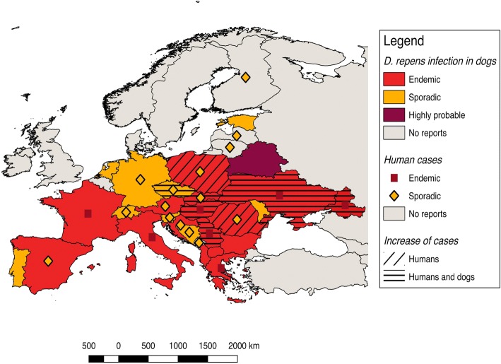
Dirofilaria repens is a nematode affecting domestic and wild canids, transmitted by several species of mosquitoes. It usually causes a non-pathogenic subcutaneous infection in dogs and is the principal agent of human dirofilariosis in the Old World. In the last decades, D. repens has increased in prevalence in areas where it has already been reported and its distribution range has expanded into new areas of Europe, representing a paradigmatic example of an emergent pathogen. Despite its emergence and zoonotic impact, D. repens has received less attention by scientists compared to Dirofilaria immitis. In this review we report the recent advances of D. repens infection in dogs and humans, and transmission by vectors, and discuss possible factors that influence the spread and increase of this zoonotic parasite in Europe. There is evidence that D. repens has spread faster than D. immitis from the endemic areas of southern Europe to northern Europe. Climate change affecting mosquito vectors and the facilitation of pet travel seem to have contributed to this expansion; however, in the authors' opinion, the major factor is likely the rate of undiagnosed dogs continuing to perpetuate the life-cycle of D. repens. Many infected dogs remain undetected due to the subclinical nature of the disease, the lack of rapid and reliable diagnostic tools and the poor knowledge and still low awareness of D. repens in non-endemic areas. Improved diagnostic tools are warranted to bring D. repens diagnosis to the state of D. immitis diagnosis, as well as improved screening of imported dogs and promotion of preventative measures among veterinarians and dog owners. For vector-borne diseases involving pets, veterinarians play a significant role in prevention and should be more aware of their responsibility in reducing the impact of the zoonotic agents. In addition, they should enhance multisectorial collaboration with medical entomologists and the public health experts, under the concept and the actions of One Health-One Medicine.
Overview
The claims and statistics of this paper haven’t been checked yet.
Run the check — free, no account needed. It reads the open-access text and checks the sources it cites.
Integrity signals
Registry
Cites
Statistics
Figures
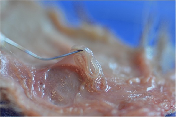
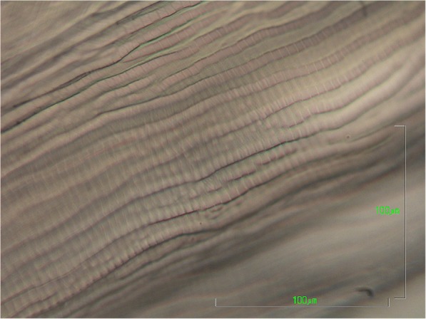
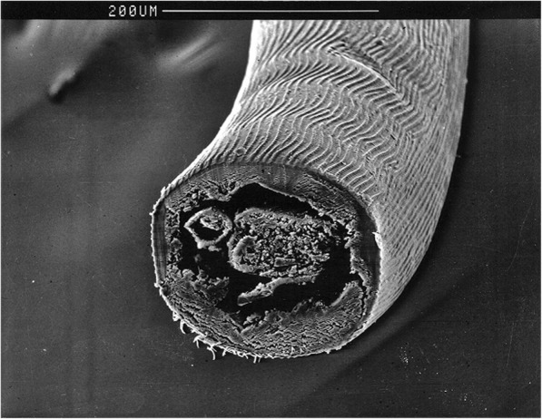
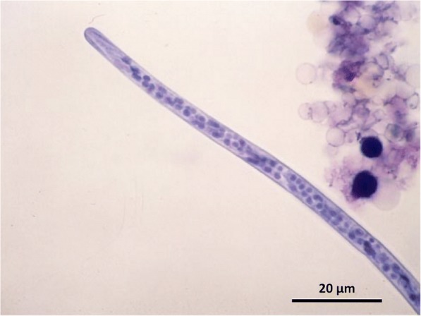
Verification
Claims the paper makes
The claims and statistics of this paper haven’t been checked yet.
Run the check — free, no account needed. It reads the open-access text and checks the sources it cites.
Claims other papers make about this one
Each is checked against this paper’s own text: does it say what the citing paper says it does?
In the citing paper
“In Serbia, humidity and temperature conditions during a large proportion of the year allow for the transmission of dirofilariasis, with seroepidemiological data revealing noteworthy prevalence rates in the country's dog populations and human clinical cases caused by both D. immitis and D. repens (1, 3, 24).”
From Seroepidemiological Study of Canine and Human Dirofilariasis in the Endemic Region of Northern Serbia (2020) · cites this paper as [3]
In the citing paper
“Dirofilariasis is on the rise in the European population of dogs and humans (1, 3, 11, 12).”
From Seroepidemiological Study of Canine and Human Dirofilariasis in the Endemic Region of Northern Serbia (2020) · cites this paper as [3]
In the citing paper
“A possible explanation for the difference in diagnostic methods used between the two filarial species would be the lack of a rapid screening system designed specifically for D. repens, as there exists for D. immitis and other common travel diseases (Capelli et al. 2018).”
From Current trends in canine dirofilariosis in Austria-do we face a pre-endemic status? (2020) · cites this paper as Capelli et al. 2018
In the citing paper
“It also appears that D. immitis is establishing itself more slowly in areas where D. repens is already present (Genchi et al. 2005; Capelli et al. 2018).”
From Current trends in canine dirofilariosis in Austria-do we face a pre-endemic status? (2020) · cites this paper as Capelli et al. 2018
In the citing paper
“D repens is currently considered an emerging zoonotic agent in Europe.”
From Dirofilarioses in cats: European guidelines from the ABCD on prevention and management (2020) · cites this paper as [14]
Self-check
Percentages, sums and reported differences re-computed from the paper’s own numbers.
The self-check needs the paper’s full text.
Citations
Cites
World map of the Köppen-Geiger climate classification updated
Kottek, M., Grieser, J., Beck, C., Rudolf, B., & Rubel, F. (2006) · Meteorologische Zeitschrift · doi:10.1127/0941-2948/2006/0130 · Cited by 11,456
Human and animal dirofilariasis: The emergence of a zoonotic mosaic
Simón, F., Siles-Lucas, M., Morchón, R., González-Miguel, J., Mellado, I., Carretón, E., & Montoya-Alonso, J. A. (2012) · Clinical Microbiology Reviews · doi:10.1128/cmr.00012-12 · Cited by 834
The Biology of Mosquitoes, Volume 1: Development, Nutrition and Reproduction
Clements, A. N. (1992) · CABI eBooks · doi:10.1079/9780851993744.0000 · Cited by 715
Climate change and infectious diseases in Europe
Semenza, J. C., & Menne, B. (2009) · The Lancet Infectious Diseases · doi:10.1016/s1473-3099(09)70104-5 · Cited by 679
Chapter 4 Heartworm Disease in Animals and Humans
McCall, J. W., Genchi, C., Kramer, L. H., Guerrero, J., & Venco, L. (2008) · Advances in Parasitology · doi:10.1016/s0065-308x(08)00204-2 · Cited by 609
Self-citationClimate and Dirofilaria infection in Europe
Genchi, C., Rinaldi, L., Mortarino, M., Genchi, M., & Cringoli, G. (2009) · Veterinary parasitology · doi:10.1016/j.vetpar.2009.03.026 · Cited by 349
Self-citationMelanization immune responses in mosquito vectors
Christensen, B. M., Li, J., Chen, C.-C., & Nappi, A. J. (2005) · Trends in Parasitology · doi:10.1016/j.pt.2005.02.007 · Cited by 346
Vector-borne helminths of dogs and humans in Europe
Otranto, D., Dantas-Torres, F., Brianti, E., Traversa, D., Petrić, D., Genchi, C., & Capelli, G. (2013) · Parasites & Vectors · doi:10.1186/1756-3305-6-16 · Cited by 336
Self-citationDiscrimination between six species of canine microfilariae by a single polymerase chain reaction
Rishniw, M., Barr, S. C., Simpson, K. W., Frongillo, M. F., Franz, M., & Dominguez Alpizar, J. L. (2006) · Veterinary Parasitology · doi:10.1016/j.vetpar.2005.10.013 · Cited by 307
Dengue and dengue vectors in the WHO European region: past, present, and scenarios for the future
Schaffner, F., & Mathis, A. (2014) · The Lancet Infectious Diseases · doi:10.1016/s1473-3099(14)70834-5 · Cited by 271
Pampiglione, S., & Rivasi, F. (2000) · PubMed · Cited by 264
Is heartworm disease really spreading in Europe?
Genchi, C., Rinaldi, L., Cascone, C., Mortarino, M., & Cringoli, G. (2005) · Veterinary Parasitology · doi:10.1016/j.vetpar.2005.04.009 · Cited by 222
Self-citationHuman dirofilariasis due to Dirofilaria (Nochtiella) repens: a review of world literature.
Pampiglione, S., Trotti, G. C., & Rivasi, F. (1995) · PubMed · Cited by 218
Genchi, C., Mortarino, M., Rinaldi, L., Cringoli, G., Traldi, G., & Genchi, M. (2011) · Veterinary Parasitology · doi:10.1016/j.vetpar.2011.01.012 · Cited by 209
Self-citationAedes albopictus is a natural vector of Dirofilaria immitis in Italy
Cancrini, G., Frangipane di Regalbono, A., Ricci, I., Tessarin, C., Gabrielli, S., & Pietrobelli, M. (2003) · Veterinary Parasitology · doi:10.1016/j.vetpar.2003.10.011 · Cited by 206
Zoonotic helminths affecting the human eye
Otranto, D., & Eberhard, M. L. (2011) · Parasites & Vectors · doi:10.1186/1756-3305-4-41 · Cited by 203
Self-citationDirofilariasis due to Dirofilaria repens in Italy, an emergent zoonosis: report of 60 new cases
Pampiglione, S., Rivasi, F., Angeli, G., Boldorini, R., Incensati, R. M., Pastormerlo, M., Pavesi, M., & Ramponi, A. (2001) · Histopathology · doi:10.1046/j.1365-2559.2001.01099.x · Cited by 202
Otranto, D., Cantacessi, C., Dantas‐Torres, F., Brianti, E., Pfeffer, M., Genchi, C., Gubertì, V., Capelli, G., & Deplazes, P. (2015) · Veterinary Parasitology · doi:10.1016/j.vetpar.2015.04.020 · Cited by 194
Self-citationFerri, E., Bain, O., Barbuto, M., Martin, C., Lo, N. C., Uni, S., Landmann, F., Baccei, S. G., Guerrero, R., Lima, S. D. S., et al. (2011) · PLoS ONE · doi:10.1371/journal.pone.0020843 · Cited by 185
Otranto, D., Capelli, G., & Genchi, C. (2009) · Parasites & Vectors · doi:10.1186/1756-3305-2-s1-s2 · Cited by 177
Self-citationMagnis, J., Lorentz, S., Guardone, L., Grimm, F., Magi, M., Naucke, T. J., & Deplazes, P. (2013) · Parasites & Vectors · doi:10.1186/1756-3305-6-48 · Cited by 169
Dirofilariosis in the Americas: a more virulent Dirofilaria immitis?
Dantas‐Torres, F., & Otranto, D. (2013) · Parasites & Vectors · doi:10.1186/1756-3305-6-288 · Cited by 165
Self-citation[Aedes albopictus in Italy and possible diffusion of the species into the Mediterranean area].
Sabatini, A., Raineri, V., Trovato, G., & Coluzzi, M. (1990) · PubMed · Cited by 164
(1992) · BRILL eBooks · doi:10.1163/9789004630437 · Cited by 159
Subcutaneous dirofilariosis (Dirofilaria repens): an infection spreading throughout the old world
Genchi, C., & Kramer, L. (2017) · Parasites & Vectors · doi:10.1186/s13071-017-2434-8 · Cited by 159
Self-citationTrotz‐William, L. A., & Trees, A. J. (2003) · Veterinary Record · doi:10.1136/vr.152.4.97 · Cited by 129
Hodžić, A., Alić, A., Fuehrer, H., Harl, J., Wille-Piazzai, W., & Duscher, G. G. (2015) · Parasites & Vectors · doi:10.1186/s13071-015-0692-x · Cited by 125
Self-citationDiagnosis of Canine Vector-Borne Diseases in Young Dogs: a Longitudinal Study
Otranto, D., Testini, G., Dantas‐Torres, F., Latrofa, M. S., Diniz, P. P. V. D. P., de Caprariis, D., Lia, R. P., Mencke, N., Stanneck, D., Capelli, G., et al. (2010) · Journal of Clinical Microbiology · doi:10.1128/jcm.00379-10 · Cited by 120
Self-citationSałamatin, R., Pavlikovska, T., Sagach, O., Nikolayenko, S., Kornyushin, V. V., Kharchenko, V., Masny, A., Cielecka, D., Konieczna-Sałamatin, J., Conn, D. B., et al. (2013) · Acta Parasitologica · doi:10.2478/s11686-013-0187-x · Cited by 118
Kronefeld, M., Kampen, H., Sassnau, R., & Werner, D. (2014) · Parasites & Vectors · doi:10.1186/1756-3305-7-30 · Cited by 107
Natural vectors of dirofilariasis in rural and urban areas of the Tuscan region, central Italy
Cancrini, G., Magi, M., Gabrielli, S., Arispici, M., Tolari, F., Dell’Omodarme, M., & Prati, M. C. (2006) · Journal of Medical Entomology · doi:10.1093/jmedent/43.3.574 · Cited by 105
Experimental maintenance of Dirofilaria repens and D. immitis in dogs
Webber, W. A. F., & Hawking, F. (1955) · Experimental Parasitology · doi:10.1016/0014-4894(55)90007-2 · Cited by 101
Fuehrer, H.-P., Auer, H., Leschnik, M., Silbermayr, K., Duscher, G., & Joachim, A. (2016) · PLOS Neglected Tropical Diseases · doi:10.1371/journal.pntd.0004547 · Cited by 100
Self-citationDirofilarial human cases in the Old World, attributed to Dirofilaria immitis : a critical analysis
Pampiglione, S., Rivasi, F., & Gustinelli, A. (2009) · Histopathology · doi:10.1111/j.1365-2559.2008.03197_a.x · Cited by 96
Dirofilaria immitis: An emerging parasite in dogs, red foxes and golden jackals in Hungary
Tolnai, Z., Széll, Z., Sproch, Á., Szeredi, L., & Sréter, T. (2014) · Veterinary Parasitology · doi:10.1016/j.vetpar.2014.04.004 · Cited by 95
Cancrini, G., Scaramozzino, P., Gabrielli, S., Paolo, M. D., Toma, L., & Romi, R. (2007) · Journal of Medical Entomology · doi:10.1603/0022-2585(2007)44[1064:aaacpi]2.0.co;2 · Cited by 90
A Novel Dirofilaria Species Causing Human and Canine Infections in Hong Kong
To, K. K., Wong, S. S. Y., Poon, R. W., Trendell-Smith, N. J., Ngan, A. H. Y., Lam, W. K., Tang, T. H., AhChong, A.-K., Kan, J. C.-H., Chan, K., et al. (2012) · Journal of Clinical Microbiology · doi:10.1128/jcm.01590-12 · Cited by 87
Gioia, G., Lecová, L., Genchi, M., Ferri, E., Genchi, C., & Mortarino, M. (2010) · Veterinary Parasitology · doi:10.1016/j.vetpar.2010.04.027 · Cited by 82
Self-citationDevelopment of Dirofilaria immitis and Dirofilaria repens in Aedes japonicus and Aedes geniculatus
Silaghi, C., Beck, R., Capelli, G., Montarsi, F., & Mathis, A. (2017) · Parasites & Vectors · doi:10.1186/s13071-017-2015-x · Cited by 80
Self-citationBaneth, G., Bourdeau, P., Bourdoiseau, G., Bowman, D., Breitschwerdt, E., Capelli, G., Cardoso, L., Dantas-Torres, F., Day, M., Dedet, J.-P., et al. (2012) · Parasites & Vectors · doi:10.1186/1756-3305-5-55 · Cited by 78
Self-citationLatrofa, M. S., Montarsi, F., Ciocchetta, S., Annoscia, G., Dantas-Torres, F., Ravagnan, S., Capelli, G., & Otranto, D. (2012) · Parasites & Vectors · doi:10.1186/1756-3305-5-76 · Cited by 77
Self-citationPantchev, N., Schaper, R., Limousin, S., Norden, N., Weise, M., & Lorentzen, L. (2009) · Parasitology Research · doi:10.1007/s00436-009-1501-2 · Cited by 76
Human dirofilariosis in Hungary: an emerging zoonosis in central Europe
Szénási, Z., Kovács, A. H., Pampiglione, S., Fioravanti, M. L., Kucsera, I., Tánczos, B., & Tiszlavicz, L. (2008) · Wiener klinische Wochenschrift · doi:10.1007/s00508-008-0928-2 · Cited by 74
Traversa, D., Aste, G., Milillo, P., Capelli, G., Pampurini, F., Tunesi, C., Santori, D., Paoletti, B., & Boari, A. (2010) · Veterinary Parasitology · doi:10.1016/j.vetpar.2009.12.034 · Cited by 71
Self-citationIonică, A. M., Matei, I. A., Mircean, V., Dumitrache, M. O., D’Amico, G., Győrke, A., Pantchev, N., Annoscia, G., Albrechtová, K., Otranto, D., et al. (2015) · Parasitology Research · doi:10.1007/s00436-014-4263-4 · Cited by 70
Self-citationA multiplex PCR for the simultaneous detection of species of filarioids infesting dogs
Latrofa, M. S., Weigl, S., Dantas-Torres, F., Annoscia, G., Traversa, D., Brianti, E., & Otranto, D. (2012) · Acta Tropica · doi:10.1016/j.actatropica.2012.01.006 · Cited by 70
Self-citationDirofilaria repens Infection and Concomitant Meningoencephalitis
Poppert, S., Hodapp, M., Krueger, A., Hegasy, G., Niesen, W.-D., Kern, W. V., & Tannich, E. (2009) · Emerging Infectious Diseases · doi:10.3201/eid1511.090936 · Cited by 70
Self-citationBazzocchi, C., Genchi, C., Paltrinieri, S., Lecchi, C., Mortarino, M., & Bandi, C. (2003) · Veterinary Parasitology · doi:10.1016/j.vetpar.2003.07.013 · Cited by 70
Self-citationDiagnosis of Imported Canine Filarial Infections in Germany 2008 – 2010
Pantchev, N., Etzold, M., Daugschies, A., & Dyachenko, V. (2011) · Parasitology Research · doi:10.1007/s00436-011-2403-7 · Cited by 69
Survey of canine dirofilariasis in Vojvodina, Serbia
Tasić, A., Rossi, L., Tasić, S., Miladinović-Tasić, N., Ilić, T., & Dimitrijević, S. (2008) · Parasitology Research · doi:10.1007/s00436-008-1132-z · Cited by 67
Canine and human Dirofilaria infections in the Balkan Peninsula
Otašević, S., Trenkić-Božinović, M., Gabrielli, S., & Genchi, C. (2015) · Veterinary Parasitology · doi:10.1016/j.vetpar.2015.02.016 · Cited by 66
Self-citationYılmaz, E., Fritzenwanker, M., Pantchev, N., Lendner, M., Wongkamchai, S., Otranto, D., Kroidl, I., Dennebaum, M., Le, T. H., Le, T. A., et al. (2016) · PLoS neglected tropical diseases · doi:10.1371/journal.pntd.0005028 · Cited by 66
Self-citationVulpes vulpes: A Possible Wild Reservoir for Zoonotic Filariae
Magi, M., Calderini, P., Gabrielli, S., Dell'Omodarme, M., Macchioni, F., Prati, M. C., & Cancrini, G. (2008) · Vector-Borne and Zoonotic Diseases · doi:10.1089/vbz.2007.0207 · Cited by 65
Venco, L., McCall, J. W., Guerrero, J., & Genchi, C. (2004) · Veterinary Parasitology · doi:10.1016/j.vetpar.2004.06.024 · Cited by 65
Self-citationHuman Subcutaneous Dirofilariasis, Russia
Kramer, L. H., Kartashev, V. V., Grandi, G., Morchón, R., Nagornii, S. A., Karanis, P., & Simón, F. (2007) · Emerging Infectious Diseases · doi:10.3201/eid1301.060920 · Cited by 64
Nodular lesions due to infestation by D irofilaria repens in dogs from I taly
Albanese, F., Abramo, F., Braglia, C., Caporali, C., Venco, L., Vercelli, A., Ghibaudo, G., Leone, F., Carrani, F., Giannelli, A., et al. (2013) · Veterinary Dermatology · doi:10.1111/vde.12009 · Cited by 64
Self-citationHuman dirofilariosis in Poland: the first cases of autochthonous infections with Dirofilaria repens.
Cielecka, D., Hanna, Ż.-P., Masny, A., Sałamatin, R., Wesołowska, M., & Gołąb, E. (2012) · PubMed · Cited by 60
An Atlas of Protozoan Parasites in Animal Tissues
Bruckner, D. A., Gardiner, C. H., Fayer, R., et al. (1991) · Journal of Parasitology · doi:10.2307/3283088 · Cited by 60
Intravitreal Dirofilariasis: A Rare Ocular Infection
Gorezis, S., Psilla, M., Asproudis, I., Peschos, D., Papadopoulou, C., & Stefaniotou, M. I. (2006) · Orbit · doi:10.1080/01676830500499224 · Cited by 59
Development of Dirofilaria and Setaria nematodes in Aedes albopictus.
Cancrini, G., Pietrobelli, M., di Regalbono, A. F., Mp, T., & della Torre, A. (1995) · PubMed · Cited by 59
Evidences of increasing risk of dirofilarioses in southern Italy
Giangaspero, A., Marangi, M., Latrofa, M. S., Martinelli, D., Traversa, D., Otranto, D., & Genchi, C. (2013) · Parasitology Research · doi:10.1007/s00436-012-3206-1 · Cited by 58
Self-citationErmakova, L. A., Nagorny, S. A., Криворотова, Е. Ю., Pshenichnaya, N. Y., & Матина, О. Н. (2014) · International Journal of Infectious Diseases · doi:10.1016/j.ijid.2014.02.008 · Cited by 56
Dirofilaria infections in dogs in different areas of Greece
Diakou, A., Kapantaidakis, E., Tamvakis, A., Giannakis, V., & Strus, N. (2016) · Parasites & Vectors · doi:10.1186/s13071-016-1797-6 · Cited by 56
Mircean, M. V., Ionică, A. M., Mircean, V., Györke, A., Codea, A. R., Tăbăran, F., Taulescu, M. A., & Dumitrache, M. O. (2017) · Parasitology International · doi:10.1016/j.parint.2017.02.003 · Cited by 56
Self-citationFirst autochthonous case of canine ocular Dirofilaria repens infection in Germany
Hermosilla, C. R., Pantchev, N., Dyachenko, V., Gutmann, M., & Bauer, C. (2006) · Veterinary Record · doi:10.1136/vr.158.4.134 · Cited by 55
The first report of authochthonous dirofilariosis in dogs in the Czech Republic
Svobodová, Z., Svobodová, V., Genchi, C., & Forejtek, P. (2006) · Helminthologia · doi:10.2478/s11687-006-0046-5 · Cited by 55
Self-citationStable Transmission ofDirofilaria repensNematodes, Northern Germany
Czajka, C., Becker, N., Jöst, H., Poppert, S., Schmidt‐Chanasit, J., Krüger, A., & Tannich, E. (2014) · Emerging infectious diseases · doi:10.3201/eid2002.131003 · Cited by 52
Self-citationCzajka, C., Becker, N., Poppert, S., Jöst, H., Schmidt-Chanasit, J., & Krüger, A. (2012) · Parasites & Vectors · doi:10.1186/1756-3305-5-14 · Cited by 51
Dirofilaria repens transmission in southeastern Finland
Pietikäinen, R., Nordling, S., Jokiranta, S. T., Saari, S. A., Heikkinen, P., Gardiner, C. H., Kerttula, A.-M., Kantanen, T., Михайловна, Н. А., Laaksonen, S., et al. (2017) · Parasites & Vectors · doi:10.1186/s13071-017-2499-4 · Cited by 51
Otranto, D., Brianti, E., Gaglio, G., Dantas‐Torres, F., Azzaro, S., & Giannetto, S. (2011) · American Journal of Tropical Medicine and Hygiene · doi:10.4269/ajtmh.2011.10-0719 · Cited by 50
Self-citationScreening blood-fed mosquitoes for the diagnosis of filarioid helminths and avian malaria
Zittra, C., Kocziha, Z., Pinnyei, S., Harl, J., Kieser, K., Laciny, A., Eigner, B., Silbermayr, K., Duscher, G. G., Fok, É., et al. (2015) · Parasites & Vectors · doi:10.1186/s13071-015-0637-4 · Cited by 49
Self-citationCanine and human dirofilariosis in the rostov region (southern Russia)
Kartashev, V., Batashova, I., Kartashov, S., Ermakov, A., Mironova, A., Kuleshova, Y., Ilyasov, B., Kolodiy, I., Klyuchnikov, A., Ryabikina, E., et al. (2011) · Veterinary Medicine International · doi:10.4061/2011/685713 · Cited by 49
Hamel, D., Silaghi, C., Zapadynska, S., Kudrin, A., & Pfister, K. (2013) · Ticks and Tick-borne Diseases · doi:10.1016/j.ttbdis.2012.08.005 · Cited by 48
Dirofilaria repens microfilariae in Aedes vexans mosquitoes in Slovakia
Bocková, E., Rudolf, I., Kočišová, A., Betášová, L., Venclíková, K., Mendel, J., & Hubálek, Z. (2013) · Parasitology Research · doi:10.1007/s00436-013-3526-9 · Cited by 48
Dirofilaria infections in working dogs in Slovakia
Miterpáková, M., Antolová, D., Hurníková, Z., Dubinsky, P., Pavlačka, A., & Németh, J. (2009) · Journal of Helminthology · doi:10.1017/s0022149x09990496 · Cited by 48
[A case of subcutaneous dirofilaria (Nochtiella) repens with microfilaremia originating in Corsica].
Jp, N., Bain, O., & Gentilini, M. (1994) · PubMed · Cited by 47
Dirofilaria in the anterior chamber: A rare occurrence
Chopra, R., Bhatti, S., Mohan, S., & Taneja, N. (2012) · Middle East African Journal of Ophthalmology · doi:10.4103/0974-9233.97965 · Cited by 44
Vector-borne pathogens in dogs and red foxes from the federal state of Brandenburg, Germany
Liesner, J. M., Krücken, J., Schaper, R., Pachnicke, S., Kohn, B., Müller, E., Schulze, C., & von Samson-Himmelstjerna, G. (2016) · Veterinary Parasitology · doi:10.1016/j.vetpar.2016.05.012 · Cited by 44
Self-citationDirofilaria repens infection in a dog: diagnosis and treatment with melarsomine and doramectin
Baneth, G., Volansky, Z., Anug, Y., Favia, G., Bain, O., Goldstein, R. E., & Harrus, S. (2002) · Veterinary Parasitology · doi:10.1016/s0304-4017(02)00006-7 · Cited by 43
Self-citationRivasi, F., Boldorini, R. L., Criante, P., Leutner, M., & Pampiglione, S. (2006) · Apmis · doi:10.1111/j.1600-0463.2006.apm_423.x · Cited by 43
Dirofilaria immitis and D. repens show circadian co-periodicity in naturally co-infected dogs
Ionică, A. M., Matei, I. A., D’Amico, G., Bel, L. V., Dumitrache, M. O., Modrý, D., & Mihalca, A. D. (2017) · Parasites & Vectors · doi:10.1186/s13071-017-2055-2 · Cited by 43
Self-citationAutochthonous Dirofilaria repens in Austria
Silbermayr, K., Eigner, B., Joachim, A., Duscher, G. G., Seidel, B., Allerberger, F., Indra, A., Hufnagl, P., & Fuehrer, H.-P. (2014) · Parasites & Vectors · doi:10.1186/1756-3305-7-226 · Cited by 42
Self-citationClinical and laboratory features of human dirofilariasis in Russia
Ermakova, L., Nagorny, S., Pshenichnaya, N., Ambalov, Y., & Boltachiev, K. (2017) · IDCases · doi:10.1016/j.idcr.2017.07.006 · Cited by 42
Kartashev, V. V., Afonin, A. N., González‐Miguel, J., Correa, R. A. S., Simón, L., Morchón, R., & Simón, F. (2014) · BioMed Research International · doi:10.1155/2014/858936 · Cited by 41
Canine dirofilariosis in two cities of southeastern Spain
Cancrini, G., Allende, E., Favia, G., Bornay, F., Antón, F., & Simón, F. (2000) · Veterinary Parasitology · doi:10.1016/s0304-4017(00)00270-3 · Cited by 41
Tahir, D., Bittar, F., Barré-Cardi, H., Sow, D., Dahmani, M., Mediannikov, O., Raoult, D., Davoust, B., & Parola, P. (2017) · Veterinary Parasitology · doi:10.1016/j.vetpar.2017.01.002 · Cited by 41
Canine filariasis in Salamanca (northwest Spain)
Perez-Sanchez, R., Gomez-Bautista, M., & Grandes, A. E. (1989) · Annals of Tropical Medicine & Parasitology · doi:10.1080/00034983.1989.11812322 · Cited by 40
Increasing Risks of Human Dirofilariasis in Travelers
Diaz, J. H. (2014) · Journal of Travel Medicine · doi:10.1111/jtm.12174 · Cited by 40
Harizanov, R., Jordanova, D. P., & Bikov, I. S. (2014) · Parasitology Research · doi:10.1007/s00436-014-3802-3 · Cited by 40
Dirofilaria repens: emergence of autochthonous human infections in the Czech Republic (case reports)
Matějů, J., Chanová, M., Modrý, D., Mitková, B., Hrazdilová, K., Žampachová, V., & Kolářová, L. (2016) · BMC Infectious Diseases · doi:10.1186/s12879-016-1505-3 · Cited by 40
Self-citationCirculation of Dirofilaria repens and Dirofilaria immitis in Moldova
Șuleșco, T., von Thien, H., Toderaș, L., Toderaș, I., Lühken, R., & Tannich, E. (2016) · Parasites & Vectors · doi:10.1186/s13071-016-1916-4 · Cited by 40
PCR identification and distribution of Anopheles daciae (Diptera, Culicidae) in Germany
Kronefeld, M., Werner, D., & Kampen, H. (2014) · Parasitology Research · doi:10.1007/s00436-014-3857-1 · Cited by 39
Der erste autochthone Fall einer subkutanen Dirofilariose in Österreich
Auer, H., & Susani, M. (2008) · Wiener klinische Wochenschrift · doi:10.1007/s00508-008-1031-4 · Cited by 38
[Detection of Dirofilaria in Austrian dogs].
Duscher, G. G., Feiler, A., Wille-Piazzai, W., Bakonyi, T., Leschnik, M. W., Miterpáková, M., Kolodziejek, J., Nowotny, N. R., & Joachim, A. (2009) · PubMed · Cited by 38
Dóczi, I., Bereczki, L., Gyetvai, T., Fejes, I., Skribek, Á., Szabó, Á., Berkes, S., Tiszlavicz, L., Bartha, N., Bende, B., et al. (2015) · Wiener klinische Wochenschrift · doi:10.1007/s00508-015-0825-4 · Cited by 38
Petry, G., Genchi, M., Schmidt, H., Schaper, R., Lawrenz, B., & Genchi, C. (2015) · Parasitology Research · doi:10.1007/s00436-015-4519-7 · Cited by 38
Self-citationA case of autochthonous human Dirofilaria infection, Germany, March 2014
Tappe, D., Plauth, M., Bauer, T., Muntau, B., Dießel, L., Tannich, E., & Herrmann-Trost, P. (2014) · Eurosurveillance · doi:10.2807/1560-7917.es2014.19.17.20790 · Cited by 38
Dirofilaria immitis: Effect on the longevity of Aedes trivittatus
Christensen, B. M. (1978) · Experimental Parasitology · doi:10.1016/0014-4894(78)90087-5 · Cited by 37
Subconjunctival Infection With Dirofilaria repens
Ruiz‐Moreno, J. M. (1998) · Archives of Ophthalmology · doi:10.1001/archopht.116.10.1370 · Cited by 37
Human Dirofilariasis Due to Dirofilaria repens Mimicking a Scrotal Tumor
Fleck, R., Kurz, W., Quade, B., Geginat, G., & Hof, H. (2008) · Urology · doi:10.1016/j.urology.2008.02.015 · Cited by 37
Rossi, A., Péix, Á., Pavlikovskaya, T., Sagach, O., Nikolaenko, S., Васильевна, Ч. Н., Kartashev, V. V., Simón, F., & Siles‐Lucas, M. (2014) · Acta Tropica · doi:10.1016/j.actatropica.2014.10.021 · Cited by 37
Čabanová, V., Miterpáková, M., Valentová, D., Blažejová, H., Rudolf, I., Stloukal, E., Hurníková, Z., & Dzidová, M. (2018) · Parasites & Vectors · doi:10.1186/s13071-018-2845-1 · Cited by 37
Seroepidemiological survey of human exposure to Dirofilaria spp. in Romania and Moldova
Ciuca, L., Simòn, F., Rinaldi, L., Kramer, L., Genchi, M., Cringoli, G., Acatrinei, D., Miron, L., & Morchon, R. (2018) · Acta Tropica · doi:10.1016/j.actatropica.2018.07.012 · Cited by 37
Adult Dirofilaria repens in a subcutaneous granuloma on the chest of a dog
Bredal, W. P., Gjerde, B., Eberhard, M. L., Aleksandersen, M., Wilhelmsen, D. K., & Mansfield, L. S. (1998) · Journal of Small Animal Practice · doi:10.1111/j.1748-5827.1998.tb03715.x · Cited by 36
Sassnau, R., Czajka, C., Kronefeld, M., Werner, D., Genchi, C., Tannich, E., & Kampen, H. (2014) · Parasitology Research · doi:10.1007/s00436-014-3970-1 · Cited by 36
Self-citationDirofilaria repens Diagnosed by the Presence of Microfilariae in Fine Needle Aspirates
Negahban, S., Daneshbod, Y., Atefi, S., et al. (2007) · Acta Cytologica · doi:10.1159/000325796 · Cited by 35
Microfilarial periodicity of Dirofilaria repens in naturally infested dogs
Cesare, A. D., Otranto, D., Giulio, E. D., Simonato, G., Latrofa, M. S., Torre, F. L., Coccia, G., & Traversa, D. (2013) · Parasitology Research · doi:10.1007/s00436-013-3619-5 · Cited by 34
Self-citationBajer, A., Rodo, A., Mierzejewska, E. J., Tołkacz, K., & Welc-Faleciak, R. (2016) · BMC Veterinary Research · doi:10.1186/s12917-016-0816-3 · Cited by 34
Dermatitis associated with microfilariae (Filarioidea) in 10 dogs
Hargis, Lewis, Duclos, Loeffler, & Rausch (1999) · Veterinary Dermatology · doi:10.1046/j.1365-3164.1999.00136.x · Cited by 33
Albonico, F., Loiacono, M., Gioia, G., Genchi, C., Genchi, M., & Mortarino, M. (2013) · Veterinary Parasitology · doi:10.1016/j.vetpar.2013.11.027 · Cited by 33
Self-citationFirst records of Dirofilaria repens in wild canids from the region of Central Balkan
Ćirović, D., Penezić, A., Pavlović, I., Kulišić, Z., Ćosić, N., Burazerović, J., & Maletić, V. (2014) · Acta Veterinaria Hungarica · doi:10.1556/avet.2014.021 · Cited by 33
Winkler, S., Pollreisz, A., Georgopoulos, M., Bagó-Horváth, Z., Auer, H., To, K. K., Krücken, J., Poppert, S., & Walochnik, J. (2017) · Emerging infectious diseases · doi:10.3201/eid2308.170423 · Cited by 33
Self-citationFirst case of cutaneous human dirofilariosis in Slovak Republic
Babal, P., Kobzová, D., Novak, I., Dubinsky, P., & Jalili, N. (2008) · Bratislavské lekárske listy/Bratislava medical journal · doi:10.1007/bf03657113 · Cited by 33
Molecular Study of Dirofilaria immitis and Dirofilaria repens in Dogs from Tunisia
Rjeibi, M. R., Rouatbi, M., Mabrouk, M., Tabib, I., Rekik, M., & Gharbi, M. (2016) · Transboundary and Emerging Diseases · doi:10.1111/tbed.12541 · Cited by 32
(1995) · PubMed · Cited by 31
Tasić, A., Tasić-Otašević, S., Gabrielli, S., Miladinović-Tasić, N., Ignjatović, A., Đorđević, J., Dimitrijević, S., & Cancrini, G. (2012) · Vector-Borne and Zoonotic Diseases · doi:10.1089/vbz.2011.0949 · Cited by 31
Genchi, M., Pengo, G., & Genchi, C. (2010) · Veterinary Parasitology · doi:10.1016/j.vetpar.2010.01.034 · Cited by 31
Self-citationFirst cases of canine dirofilariosis inSlovakia: a case report
Svobodová, V., Svobodová, Z., Beladicova, V., & Valentová, D. (2005) · Veterinární Medicína · doi:10.17221/5656-vetmed · Cited by 31
Mirahmadi, H., Maleki, A., Hasanzadeh, R., Ahoo, M. B., Mobedi, I., & Rostami, A. (2016) · Parasitology International · doi:10.1016/j.parint.2016.10.022 · Cited by 31
Allergic dermatitis by Dirofilaria repens in a dog: clinical picture and treatment
Rocconi, F., Tommaso, M. D., Traversa, D., Palmieri, C., Pampurini, F., & Boari, A. (2012) · Parasitology Research · doi:10.1007/s00436-012-2833-x · Cited by 30
Fok, É., Jacsó, O., Szebeni, Z., Győrffy, A., Sükösd, L., Lukács, Z., & Schaper, R. (2010) · Parasitology Research · doi:10.1007/s00436-010-1783-4 · Cited by 30
Intra‐oral Dirofilaria repens infection: report of seven cases
Tilakaratne, W. M., & Pitakotuwage, T. N. (2003) · Journal of Oral Pathology and Medicine · doi:10.1034/j.1600-0714.2003.00183.x · Cited by 30
Human subcutaneous/ocular dirofilariasis in the Russian Federation and Belarus, 1997-2013
Kartashev, V., Tverdokhlebova, T., Korzan, A., Vedenkov, A., Simón, L., González-Miguel, J., Morchón, R., Siles-Lucas, M., & Simón, F. (2015) · International Journal of Infectious Diseases · doi:10.1016/j.ijid.2015.02.017 · Cited by 30
Human Subcutaneous Dirofilariasis: A Report of Three Cases Manifesting as Breast or Axillary Nodules
Maltezos, E. S., Sivridis, E., Giatromanolaki, A. N., & Simopoulos, C. (2002) · Scottish Medical Journal · doi:10.1177/003693300204700404 · Cited by 30
Potters, I., Vanfraechem, G., & Bottieau, E. (2018) · Emerging infectious diseases · doi:10.3201/eid2409.180462 · Cited by 29
Periorbital dirofilariasis-Clinical and imaging findings: Live worm on ultrasound
Gopinath, T., Lakshmi, K., Shaji, P., et al. (2013) · Indian Journal of Ophthalmology · doi:10.4103/0301-4738.114111 · Cited by 28
Cardot‐Leccia, N., Patouraux, S., Carpentier, X., Boissy, C., Giudice, P. D., Parks, S. K., Michiels, J. F., & Ambrosetti, D. (2012) · Pathogens and Global Health · doi:10.1179/2047773212y.0000000029 · Cited by 28
Papazahariadou, M. G., Koutinas, A. F., Rallis, T. S., & Haralabidis, S. T. (1994) · Journal of Helminthology · doi:10.1017/s0022149x00014413 · Cited by 28
Dirofilaria-repens-Befall in einem Schlittenhunde-Rudel im Land Brandenburg
Dyachenko, V., Pantchev, N., Stöckel, F., Dittmar, K., Daugschies, A., & Sassnau, R. (2009) · Tierärztliche Praxis Ausgabe K Kleintiere / Heimtiere · doi:10.1055/s-0038-1622777 · Cited by 28
Microfilaremia from a Dirofilaria‐like parasite in Greece
Petrocheilou, V., THEODORAKIS, M., Williams, J. F., et al. (1998) · Apmis · doi:10.1111/j.1699-0463.1998.tb01352.x · Cited by 28
Abdel-Rahman, S. M., Mahmoud, A. E.-R. A. E.-R., Galal, L., Gustinelli, A., & Pampiglione, S. (2008) · Annals of Tropical Medicine and Parasitology · doi:10.1179/136485908x300904 · Cited by 27
Genchi, C., Genchi, M., Petry, G., Kruedewagen, E. M., & Schaper, R. (2013) · Parasitology Research · doi:10.1007/s00436-013-3283-9 · Cited by 27
Self-citationCanine filarial infections in Tuscany, central Italy
Magi, M., Guardone, L., Prati, M. C., Tozzini, G., Torracca, B., Monni, G., & Macchioni, F. (2012) · Journal of Helminthology · doi:10.1017/s0022149x11000113 · Cited by 27
HumanDirofilaria repensInfection in Romania: A Case Report
Popescu, I., Tudose, I., Rácz, P., Muntau, B., Giurcăneanu, C., & Poppert, S. (2012) · Case Reports in Infectious Diseases · doi:10.1155/2012/472976 · Cited by 27
Self-citationBiology of filarial worms parasitizing dogs and cats.
Manfredi, M. T., Cerbo, A. D., & Genchi, M. (2007) · Cited by 26
Demiaszkiewicz, A. W., Polanczyk, G., Pyziel, A. M., Kuligowska, I., & Lachowicz, J. (2009) · PubMed · Cited by 26
Subcutaneous Infection Caused by Dirofilaria repens Imported to Slovenia
Logar, J., Novšak, V., Rakovec, S., & Staniša, O. (2001) · Journal of Infection · doi:10.1053/jinf.2000.0759 · Cited by 26
Krstić, M., Gabrielli, S., Ignjatović, M., Savić, S., Cancrini, G., Ranđelović, G., Momčilović, S., Stojnev, S., & Otašević, S. (2017) · Molecular and Cellular Probes · doi:10.1016/j.mcp.2016.08.005 · Cited by 26
Self-citationGiannelli, A., Ramos, R. A. N., Traversa, D., Brianti, E., Annoscia, G., Bastelli, F., Dantas‐Torres, F., & Otranto, D. (2013) · Veterinary Parasitology · doi:10.1016/j.vetpar.2013.05.012 · Cited by 25
Self-citationIlyasov, B., Kartashev, V. V., Bastrikov, N., Madjugina, L., González‐Miguel, J., Morchón, R., & Simón, F. (2014) · Enfermedades Infecciosas y Microbiología Clínica · doi:10.1016/j.eimc.2014.04.002 · Cited by 25
Effect of cibarial armature of mosquitoes (Diptera, Culicidae) on blood-meal haemolysis
Coluzzi, M., Concetti, A., & Ascoli, F. (1982) · Journal of Insect Physiology · doi:10.1016/0022-1910(82)90103-2 · Cited by 25
Intradural dirofilariasis mimicking a Langerhans cell histiocytosis tumor
Perret-Court, A., Coulibaly, B., Ranque, S., Bouvier, C., Lena, G., Coze, C., & Verschuur, A. C. (2009) · Pediatric Blood & Cancer · doi:10.1002/pbc.22114 · Cited by 25
Case of human Dirofilaria repens infection manifested by cutaneous larva migrans syndrome
Antolová, D., Miterpáková, M., & Paraličová, Z. (2015) · Parasitology Research · doi:10.1007/s00436-015-4499-7 · Cited by 25
Human dirofilariosis in the Slovak Republic - a case report.
Ondriska, F., Lengyel, D., Miterpáková, M., Lengyelova, B., Strehárová, A., & Dubinsky, P. (2010) · PubMed · Cited by 25
Human Subcutaneous Dirofilariasis Caused by Dirofilaria repens in Ibiza, Spain
Fuentes, I., Cascales, A., Ros, J. M., et al. (1994) · American Journal of Tropical Medicine and Hygiene · doi:10.4269/ajtmh.1994.51.401 · Cited by 25
Autochthonous case of Dirofilaria repens in a dog in the Netherlands
Overgaauw, P. A. M., & van Dijk, E. (2009) · Veterinary Record · doi:10.1136/vr.164.5.158 · Cited by 24
The detection of different Dirofilaria species using direct PCR technique
Silbermayr, K., Eigner, B., Duscher, G. G., Joachim, A., & Fuehrer, H. (2013) · Parasitology Research · doi:10.1007/s00436-013-3682-y · Cited by 24
Self-citationIs Dirofilaria repens Endemic in the Havelland District in Brandenburg, Germany?
Sassnau, R., Kohn, M., Demeler, J., Kohn, B., Müller, E., Krücken, J., & von Samson‐Himmelstjerna, G. (2013) · Vector-Borne and Zoonotic Diseases · doi:10.1089/vbz.2012.1293 · Cited by 23
Self-citationSvobodova, V., & Mišoňova, P. (2005) · Veterinary Parasitology · doi:10.1016/j.vetpar.2004.11.018 · Cited by 23
Recently Recognized Cases of Ophthalmofilariasis in Hungary
Fodor, E., Fok, É., Maka, E., Lukáts, O., & Tóth*, J. (2009) · European Journal of Ophthalmology · doi:10.1177/112067210901900424 · Cited by 23
Epidemiological aspects of canine dirofilariasis in the north of iran.
Ranjbar-Bahadori, S., Veshgini, A., Shirani, D., Eslami, A., Mohieddin, H., Shemshadi, B., & Masooleh, R. (2011) · Europe PMC (PubMed Central) · Cited by 23
Subconjunctival Dirofilaria repens infection in a dog resident in the UK
Agapito, D., Aziz, N. A. A., Wang, T., Morgan, E. R., & Wright, I. P. (2017) · Journal of Small Animal Practice · doi:10.1111/jsap.12795 · Cited by 23
Grassi, B., & Noe, G. (1900) · BMJ · doi:10.1136/bmj.2.2079.1306 · Cited by 22
The incrimination of Aedes (stegomyia) aegypti as the vector of Dirofilaria repens in Nigeria
Anyanwu, I. N., Agbede, R. I. S., Ajanusi, O. J., Umoh, J. U., & Ibrahim, N. D. G. (2000) · Veterinary Parasitology · doi:10.1016/s0304-4017(00)00311-3 · Cited by 22
Dirofilaria repens Microfilaremia in Three Dogs in Estonia
Jokelainen, P., Mõtsküla, P. F., Heikkinen, P., Ülevaino, E., Oksanen, A., & Lassen, B. (2016) · Vector-Borne and Zoonotic Diseases · doi:10.1089/vbz.2015.1833 · Cited by 22
Human Dirofilaria repens infections diagnosed in Slovakia in the last 10 years (2007–2017)
Miterpáková, M., Antolová, D., Ondriska, F., & Gál, V. (2017) · Wiener klinische Wochenschrift · doi:10.1007/s00508-017-1233-8 · Cited by 22
Scaramozzino, P., Gabrielli, S., Di Paolo, M., Sala, M., Scholl, F., & Cancrini, G. (2005) · BMC Infectious Diseases · doi:10.1186/1471-2334-5-75 · Cited by 21
Dirofilariose humaine: 3 cas autochtones dans le sud de la France
Pomares-Estran, C., Marty, P., Blanc, V., Faure, O., Leccia, M. ‐., Pelloux, H., Diebolt, E., Ambrosetti, D., & Cardot‐Leccia, N. (2007) · La Presse Médicale · doi:10.1016/j.lpm.2006.11.030 · Cited by 21
[Dirofilariasis--Dirofilaria repens--first time described in Polish patients].
Hanna, Ż.-P., Cielecka, D., & Sałamatin, R. (2008) · PubMed · Cited by 21
Tiškina, V., & Jokelainen, P. (2017) · Veterinary Parasitology · doi:10.1016/j.vetpar.2017.07.012 · Cited by 21
Masny, A., Sałamatin, R., Rozej-Bielicka, W., & Golab, E. (2016) · Parasitology Research · doi:10.1007/s00436-015-4767-6 · Cited by 20
Subconjunctival ocular filariasis -Case report-
Macarie, S. S., Dobre, C., Suciu, M.-C., Ionică, A. M., CERNEA, M. S., Tarcău, P., & Bodea, F. I. (2017) · Romanian Journal of Ophthalmology · doi:10.22336/rjo.2017.14 · Cited by 20
Self-citationAn Atlas of Metazoan Parasites in Animal Tissues
Esch, G. W., Gardiner, C. H., Fayer, R., et al. (2001) · Journal of Parasitology · doi:10.2307/3285215 · Cited by 20
[Ocular dirofilariasis: a case report].
Janjetović, Ž., Arar, Ž. V., Paradžik, M. T., et al. (2010) · PubMed · Cited by 19
Dirofilariasis – an emergent human parasitosis in Romania
Arbune, M., & Dobre, M. (2015) · Acta Parasitologica · doi:10.1515/ap-2015-0068 · Cited by 19
Dirofilariasis of breast: Report of two cases diagnosed by fine‐needle aspiration biopsy
Ng, W. K., Siu, T. H., Fung, B. T., et al. (2002) · Diagnostic Cytopathology · doi:10.1002/dc.10009 · Cited by 19
Prevalence ofDirofilaria immitisin dogs from Hatay province, Turkey
Yaman, M., Guzel, M., Koltaş, İ. S., Demirkazık, M., & Aktas, H. (2009) · Journal of Helminthology · doi:10.1017/s0022149x08198832 · Cited by 19
Ashrafi, K., Golchai, J., & Geranmayeh, S. (2010) · Europe PMC (PubMed Central) · Cited by 19
Subcutaneous Dirofilariosis in South‐East Serbia‐Case Report
Tasić, S., Stoiljković, N., Miladinović‐Tasić, N., Tasić, A., Mihailović, D., Rossi, L., Gabrielli, S., & Cancrini, G. (2011) · Zoonoses and Public Health · doi:10.1111/j.1863-2378.2010.01379.x · Cited by 18
Hellmann, K., Heine, J., Braun, G., Paran-Dobesova, R., & Svobodova, V. (2011) · Parasitology Research · doi:10.1007/s00436-011-2404-6 · Cited by 18
Hrčková, G., Kuchtová, H., Miterpáková, M., Ondriska, F., Cibíček, J., & Kovacs, Š. (2012) · Journal of Helminthology · doi:10.1017/s0022149x12000077 · Cited by 18
Canine dirofilariosis under specific environmental conditions of the Eastern Slovak Lowland.
Iglódyová, A., Miterpáková, M., Hurníková, Z., Antolová, D., Dubinsky, P., & Letková, V. (2012) · PubMed · Cited by 17
Marušić, Z., Stastny, T., Kirac, I., Stojčević, D., Krušlin, B., & Tomas, D. (2008) · PubMed · Cited by 17
Detection of Dirofilaria repens microfilariae in a dog from Portugal
Maia, C., Lorentz, S., Cardoso, L., Otranto, D., & Naucke, T. J. (2016) · Parasitology Research · doi:10.1007/s00436-015-4796-1 · Cited by 16
Self-citationDang, T. C. T., Nguyen, T. H., Do, T. D., Uga, S., Morishima, Y., Sugiyama, H., & Yamasaki, H. (2010) · Parasitology Research · doi:10.1007/s00436-010-1961-4 · Cited by 16
Dirofilariasis by Dirofilaria repens : an imported case and a brief review
Baptista-Fernandes, T., Rodrigues, M. G., Domingues, D., Monteiro, L., Paixão, P., Pereira, P., Tavares, R., Rodrigues, P., Maurício, I. L., Belo, S., et al. (2015) · Parasitology International · doi:10.1016/j.parint.2015.03.001 · Cited by 16
Dirofilaria repens presenting as a premasseteric nodule
Latıfoǧlu, O., Özmen, S., Sezer, C., et al. (2002) · Oral Surgery Oral Medicine Oral Pathology Oral Radiology and Endodontology · doi:10.1067/moe.2002.125275 · Cited by 16
EXPERIMENTAL INFECTION OF MOSQUITOES WITH DIROFILARIA REPENS (NEMATODA, FILARIOIDEA) LARVAE
Kuzmin, Y., Varodi, E. I., Vasylyk, N., & Kononko, G. (2005) · The scientific electronic library of periodicals of the National Academy of Sciences of Ukraine (National Academy of Sciences of Ukraine) · Cited by 16
(2001) · PubMed · Cited by 16
McCall, J. W., Hodgkins, E., Varloud, M., et al. (2017) · Parasites & Vectors · doi:10.1186/s13071-017-2439-3 · Cited by 16
What is your diagnosis? Fine‐needle aspirate from a subcutaneous mass in a dog
Giori, L., Garbagnoli, V., Venco, L., Genchi, M., Bazzocchi, C., & Bertazzolo, W. (2010) · Veterinary Clinical Pathology · doi:10.1111/j.1939-165x.2009.00212.x · Cited by 15
Cesare, A. D., Braun, G., Giulio, E. D., Paoletti, B., Aquilino, V., Bartolini, R., Torre, F. L., Meloni, S., Drake, J., Pandolfi, F., et al. (2014) · Parasites & Vectors · doi:10.1186/1756-3305-7-347 · Cited by 15
Dirofilaria repens infection case in Hungary: a case report
Pónyai, K., Wikonkál, N., Bottlik, G., Hársing, J., Kucsera, I., Horváth, A., & Kárpáti, S. (2006) · JDDG: Journal der Deutschen Dermatologischen Gesellschaft · doi:10.1111/j.1610-0387.2006.06056.x · Cited by 15
Dirofilaria Repens in the Eyelid: Case Report of Subcutaneous Manifestation
Mannino, G., Contestabile, M. T., Medori, E. M., Mannino, C., Enrici, M. M., Marangi, M., & Rossi, P. D. (2009) · European Journal of Ophthalmology · doi:10.1177/112067210901900325 · Cited by 15
Subcutaneous Dirofilaria repens infection of the eyelid in Serbia and Montenegro.
Džamić, A., Arsić-Arsenijević, V. S., Radonjic, I., Mitrović, S., Marty, P., & If, K.-Z. (2004) · PubMed · Cited by 15
Desai, R., Pai, N., Nehete, A., & Singh, J. (2015) · Indian Journal of Medical Microbiology · doi:10.4103/0255-0857.167342 · Cited by 15
Human dirofilariasis in Italy: a new case in the spermatic cord.
Pampiglione, S., Fioravanti, M., Piccolotti, D., Pizzicannella, G., & Reale, D. (2002) · PubMed · Cited by 14
[Human pulmonary dirofilariasis: the first case observed in Greece].
Pampiglione, S., Rivasi, F., & Vakalis, N. C. (2000) · PubMed · Cited by 14
Skri︠a︡bin, K. I., laboratorii︠a︡, G., & Raveh, M. (1969) · Israel Program for Scientific Translations eBooks · Cited by 14
Rossi, L., Ferroglio, E., & Agostini, A. (2004) · Veterinary Record · doi:10.1136/vr.154.1.26 · Cited by 14
SubconjunctivalDirofilaria repens
Khoramnia, R., & Wegner, A. (2010) · New England Journal of Medicine · doi:10.1056/nejmicm1003006 · Cited by 14
A Case of Orbital Dirofilariasis in Northern Turkey
Beden, Ü., Hökelek, M., AÇICI, M., et al. (2007) · Ophthalmic Plastic and Reconstructive Surgery · doi:10.1097/iop.0b013e318073cca3 · Cited by 14
Use of moxidectin tablets in the control of canine subcutaneous dirofilariosis
Rossi, L., Ferroglio, E., & Agostini, A. (2002) · Veterinary Record · doi:10.1136/vr.150.12.383-a · Cited by 14
Kramer, L. H., & Genchi, C. (2014) · Veterinary Parasitology · doi:10.1016/j.vetpar.2014.03.028 · Cited by 14
Self-citationHuman Ocular Dirofilariasis in Hong Kong
Kwok, R. P. W., Chow, P. P. C., Lam, J. K. M., et al. (2015) · Optometry and Vision Science · doi:10.1097/opx.0000000000000770 · Cited by 14
Subconjunctival infection with Dirofilaria repens.
Raniel, Y., Machamudov, Z., & Garzozi, H. J. (2006) · PubMed · Cited by 14
The prevalence of parasitic helminths in stray dogs in the Baghdad area, Iraq
Tarish, J. H., Al-Saqur, I. M., Al-Abbassy, S. N., et al. (1986) · Annals of Tropical Medicine and Parasitology · doi:10.1080/00034983.1986.11812024 · Cited by 14
Tippelt, L., Walther, D., & Kampen, H. (2017) · Parasitology Research · doi:10.1007/s00436-017-5652-2 · Cited by 14
Révész, E., Markovics, G., Darabos, Z., Tóth, I., & Fok, É. (2008) · Magyar Sebészet · doi:10.1556/maseb.61.2008.5.6 · Cited by 13
Male and female filarial worms Dirofilaria (Nochtiella) repens recovered from the scrotum
Fernando, S., Ihalamulla, R., & Silva, W. D. (2015) · Ceylon Medical Journal · doi:10.4038/cmj.v45i3.8114 · Cited by 13
Research works printed but not linked: 31
- Lusitano A. Curationum Medicinalium Centuria Septima. Venetiis: apud Vincentium Valgresium, curatio. 1566;63:106.
- Addario GG. A nematode of the human eye. Ann Ottalmol. 1885;14:133–7 (in Italian).
- Ercolani G. Helmintological observations on the dimorbiosis of nematodes: on the Filaria immitis and on a new species of dog’s distoma. Mem R Acad Sci Ist Bol. 1874;3:390–441 (in Italian).
- Fulleborn F. Über Versuche an Hundefilarien und deren übertrangung durch Müncken. Arch Schiffs Tropenhyg. 1908;8:313–59.
- Lanzillotti-Bonsanti A. On the alterations that produce the embryos of Filaria immitis and on a cyst with Filaria immitis in the intermuscular connective of a dog. Clin Vet. 1881;4:212–5 (In Italian).
- Bonvicini A. Of a case of filariasis in a dog suffering from demodectic mange. Mod Zoiatra Parte Sci. 1910;42:55 (In Italian).
- Bernard P, Bauche J. Conditions de propagation de la filariose sous-cutanée du chien. Stegomyia fasciata hote intermédiaire de Dirofilaria repems. Bull Soc Pathol Exot. 1913;7:89–99.
- Rivolta S. Herpes form in the dog produced by embryos of filaria. Med Vet G Teor. 1868;3:300–2 (In Italian).
- Rosso G. Dog’s cutaneous helminthiasis. A case of general pustular dermatitis caused by embryos of Filaria immitis. Mod Zooiatro. 1897;8:185–8 (In Italian).
- Mazzanti E. Two zooparasitological observations. Observation first on a herpetic affection in a dog produced by nematodes. G R Soc Vet. 1900;50:626–9 (In Italian).
- Balbo T, Panichi M. Filariosis in the dog. Nuova Vet. 1968;44:18–32 (In Italian).
- Locatelli A. On the animal filariosis in the province of Pavia. Parassitologia. 1971;13:197–202 (In Italian).
- Rojo-Vázquez F, Valcárcel F, Guerrero J, Gómez M. Prevalencia de la dirofilariosis canina en cuatro áreas geográficas de España. Med Vet Barcelona. 1990;7:297–305.
- Bourdeau P, Roussel A. Canine infection with Dirofilaria repens in France: distribution and prevalence from a national survey with veterinary clinics. Melbourne: Proceedings ICOPA XII; 2010.
- Fok É, Szaboó Z, Farkas R. The first autochthonous case of a dog infected with Dirofilaria repens. Kisállatorvoslás. 1998;4:218–9 (In Hungarian).
- Fok É, Kiss G, Majoros G, Jacsó O, Farkas R, Gyurkovszky M. Preliminary results of an epidemiological survey on dirofilariosis of dogs and cats in Hungary. Mappe Parassitol. 2007;8:195–6.
- Holler D, Racz A, Bošnir J, Petrak O. The prevalence of dirofilariasis in the hinterland of the Istrian Peninsula. Medica Jadertina. 2011;40:67–74.
- Jurhar-Pavlova M, Cvetković D, Duma H, Kochevski Z. Dirofilariasis in R. Macedonia - an overview of literature data and our expirience, vol. 2012. Niš: 46th Days of Preventive Medicine, International Congress, Book of Abstracts; 2012. p. 130–2.
- Kanev I, Kamenov I, Ganchev G, Prelezov P, Tzvetkov Y, Tomcheva V, et al. Dirofilaria repens and Dirofilaria immitis in animals and humans in Bulgaria. Parassitologia. 1996;38:358.
- Olteanu G. Dirofilariosis in man and animals in Romania. Parassitologia. 1996;38(Suppl. 1–2):360.
- Paşca S, Miron L, Acatrinei D, Mihalachi S. Both cardio-vasculary and subcutaneous forms of dirofilariosis in dog: a case report. Sci Papers Vet Med Iași. 2008;51:123–7.
- Popescu I, Tudose I, Racz P, Muntau B, Giurcaneanu C, Poppert S. Human Dirofilaria repens infection in Romania: a case report. Case Rep Infect Dis. 2012;2012:472976.
- Bain O. Développment en Camargue de la filaire du chien, Dirofilaria repens Railliet at Henry, 1911, chez les Aedes halophile. Bull Mus Nat Hist Nat. 1978;351:19–27.
- Cancrini G, Yanchang S, Della Torre A, Coluzzi M. Influence of temperature on the larval development of Dirofilaria repens in different species of mosquitoes. Parassitologia. 1988;30:38 (In Italian).
- Khudaverdiev T. The biology Dirofilaria repens repens Railliet and Henry, 1911, the causal agent of dirofilariasis of dogs in the Nakhichevan Republic, ASSR. Mater Nauchnoi Konf Vsesoyuznogo Obs Gelmintol Russ. 1976;28:163–9 (In Russian).
- Mišić S, Stajković N, Tešić M, Mišić Z, Lj L. Human dirofilariosis in Yugoslavia: report on three cases of Dirofilaria repens infection. Parassitologia. 1996;38:358.
- Sergiev V, Suprjaga V, Morozov E, LA J. Human dirofilariasis: diagnosis and character of the relationship between causative agent and host. Med Parasitol Parasit Dis. 2009;3:3–6.
- Euzeby J. Diagnostic expérimental des helminthoses animales: animaux domestiques, animaux de laboratoire, primates; travaux pratiques d'helminthologie vétérinaire: livre 1 généralités, diagnostic ante-mortem. Paris: Informations Techniques des Services Vétérinaires; 1981. p. 349.
- Orihel T, Ash L. Dirofilaria species - Parasites in Human Tissues. 1st ed. Chicago: American Society of Clinical Pathologists; 1995.
- Genchi C, Poglayen G, Kramer L, Casiraghi M, Venco L, Brianti E. Efficacia di selamectin nella profilassi delle infestazioni da Dirofilaria repens del cane. Veterinaria. 2002;16:69–71.
- Petrić D, Zgomba M, Bellini R, Becker N. Surveillance of mosquito populations - a key element to understanding the spread of invasive vector species and vector-borne diseases in Europe. In: Hauppauge MD, editor. Essays on Fundamental and Applied Environmental Topics. New York: Nova Science Publishers; 2012. p. 192–224.
Cited by
Sorted by citations
Overgaauw, P. A. M., Vinke, C. M., van Hagen, M. A. E., & Lipman, L. J. A. (2020) · International Journal of Environmental Research and Public Health · doi:10.3390/ijerph17113789 · Cited by 301
The prevalence of Dirofilaria immitis and D. repens in the Old World
Genchi, C., & Kramer, L. H. (2020) · Veterinary Parasitology · doi:10.1016/j.vetpar.2019.108995 · Cited by 187
Self-citationFuehrer, H.-P., Morelli, S., Unterköfler, M. S., Bajer, A., Bakran-Lebl, K., Dwużnik-Szarek, D., Farkas, R., Grandi, G., Heddergott, M., Jokelainen, P., et al. (2021) · Pathogens · doi:10.3390/pathogens10101268 · Cited by 96
Self-citationDirofilaria repens microfilaremia in humans: Case description and literature review
Pupić-Bakrač, A., Pupić-Bakrač, J., Beck, A., Jurković, D., Polkinghorne, A., & Beck, R. (2021) · One Health · doi:10.1016/j.onehlt.2021.100306 · Cited by 58
The current situation of canine dirofilariosis in Hungary
Farkas, R., Mag, V., Gyurkovszky, M., Takács, N., Vörös, K., & Solymosi, N. (2020) · Parasitology Research · doi:10.1007/s00436-019-06478-5 · Cited by 55
Metadata
- Authors
- Capelli, G., Genchi, C., Baneth, G., Bourdeau, P., Brianti, E., Cardoso, L., Danesi, P., Fuehrer, H.-P., Giannelli, A., Ionică, A. M., Maia, C., Modrý, D., Montarsi, F., Krücken, J., Papadopoulos, E., Petrić, D., Pfeffer, M., Savić, S., Otranto, D., Poppert, S., & Silaghi, C.
- Affiliations
- Istituto Zooprofilattico Sperimentale delle Venezie (IT)
- University of Milan (IT)
- Hebrew University of Jerusalem (IL)
- Université Nantes Angers Le Mans (FR)
- University of Messina (IT)
- University of Veterinary Medicine Vienna (AT)
- University of Bari Aldo Moro (IT)
- University of Agricultural Sciences and Veterinary Medicine of Cluj-Napoca (RO)
- Universidade Nova de Lisboa (PT)
- University of Veterinary Sciences Brno (CZ)
- Freie Universität Berlin (DE)
- Aristotle University of Thessaloniki (GR)
- University of Novi Sad (RS)
- Leipzig University (DE)
- Scientific Veterinary Institute "Novi Sad" (RS)
- Swiss Tropical and Public Health Institute (CH)
- Friedrich-Loeffler-Institut (DE)
- Corresponding author
- Capelli, Gioia
- ORCID
- Venue
- Parasites & Vectors
- Published
- 2018-12
- Volume
- 11
- Issue
- 1
- Pages
- 663-663
- ISSN
- 1756-3305 (ISSN-L)
- PMC
- PMC6299983
- OpenAlex
- W2905090683
- Cited by
- 298
- References
- 268
- Citations by year
- 2026: 20 · 2025: 60 · 2024: 33 · 2023: 42 · 2022: 42 · 2021: 45 · 2020: 41 · 2019: 13 · 2018: 1
- FWCI
- 3.67
- Type
- journal article
- Language
- en
- Open access
- gold
- doi.org
- Licence
- Locations
- Parasites & Vectors (publishedVersion, cc-by)
- Europe PMC (PubMed Central) (publishedVersion, other-oa)
- PubMed Central (publishedVersion, cc-by)
- PubMed (publishedVersion)
- edoc (University of Basel) (acceptedVersion)
- Zurich Open Repository and Archive (University of Zurich) (acceptedVersion, cc-by)
- Portuguese National Funding Agency for Science, Research and Technology (RCAAP Project by FCT) (submittedVersion, other-oa)
- DOAJ (DOAJ: Directory of Open Access Journals) (submittedVersion, cc-by-sa)
- Refubium (Universitätsbibliothek der Freien Universität Berlin) (submittedVersion, cc-by)
- CINECA IRIS Institutional Research Information System (University of Bari Aldo Moro) (submittedVersion, other-oa)
- Zurich Open Repository and Archive (University of Zurich) (submittedVersion, cc-by)
- Universitätsbibliothek der FU Berlin Hochschulschriftenstelle u. Dokumentenserver (cc-by)
- Zurich Open Repository and Archive (University of Zurich)
- edoc (University of Basel)
- Funders
- Regione del Veneto
- Topics
- Health Sciences › Medicine › Infectious Diseases › Parasitic Diseases Research and Treatment
- Life Sciences › Immunology and Microbiology › Parasitology › Helminth infection and control
- Health Sciences › Medicine › Public Health, Environmental and Occupational Health › Research on Leishmaniasis Studies
- Keywords
- Dirofilaria repens, canine dirofilariasis, zoonotic nematodes, emerging pathogens, geographic range expansion, mosquito vectors, climate change, imported dogs, asymptomatic infection, diagnostic tools, One Health, Dirofilaria immitis
- MeSH terms
- Animals, Dirofilariasis, Dog Diseases, Dogs, Europe, Humans, Insect Vectors, Culicidae, Dirofilaria repens
About these checks
Registry, retraction, Cites and statistics results are computed, not judged, with one exception: a printed reference that no index lists is read once by a model before it counts as a citation error. Claims are extracted by a model and kept only when their quote appears verbatim in the text; each cited source is then checked, with a model’s help, for whether it says what the claim relies on. Those verdicts can be wrong — read the claim’s quote and the cited source before relying on one.
Statistics and arithmetic are recomputed, not judged.
Confidence bands are shown only for fields calibrated on our annotated test set. None is calibrated yet, so no band is shown.
What counts as a claim ›How we measure confidence ›How we check this ›
Are you an author, or do you see a problem with this page? Request a review
