

The DOI matches its Crossref record; 298 of the 314 references it cites were found in a registry or an index; 13 of the 236 sources its claims cite were checked for support.
Mitochondrial Uncoupling: A Key Controller of Biological Processes in Physiology and Diseases
Open accessAbstract
Mitochondrial uncoupling can be defined as a dissociation between mitochondrial membrane potential generation and its use for mitochondria-dependent ATP synthesis. Although this process was originally considered a mitochondrial dysfunction, the identification of UCP-1 as an endogenous physiological uncoupling protein suggests that the process could be involved in many other biological processes. In this review, we first compare the mitochondrial uncoupling agents available in term of mechanistic and non-specific effects. Proteins regulating mitochondrial uncoupling, as well as chemical compounds with uncoupling properties are discussed. Second, we summarize the most recent findings linking mitochondrial uncoupling and other cellular or biological processes, such as bulk and specific autophagy, reactive oxygen species production, protein secretion, cell death, physical exercise, metabolic adaptations in adipose tissue, and cell signaling. Finally, we show how mitochondrial uncoupling could be used to treat several human diseases, such as obesity, cardiovascular diseases, or neurological disorders.
Overview
AI snapshot
Written from the paper’s full text.
TL;DR
Mitochondrial uncoupling is a dissociation between mitochondrial membrane potential generation and ATP synthesis, and this review argues it is not merely dysfunction but a regulator of autophagy, ROS, secretion, cell death, exercise adaptation, adipose biology, and disease. It surveys natural and synthetic uncouplers and their therapeutic potential, while cautioning that most mechanistic evidence comes from non-specific agents such as DNP and FCCP.
Key findings
- Basal proton leak can account for up to 30–50% of resting cellular metabolic rate, and ANTs can account for up to 50% of basal mitochondrial membrane proton conductance.
- UCP-1 is a well-established uncoupling protein, but uncoupling properties of other UCPs remain debated; UCP-3 expression is 200–700-fold lower than UCP-1 in mouse skeletal muscle, so overexpression models may be artefactual.
- Mitochondrial uncoupling is linked to bulk and specific autophagy/mitophagy, ROS production, protein secretion, cell death, physical exercise, adipose tissue browning, and cell signaling; mitochondrial ROS release can account for 0.1–0.2% of O₂ consumed in rat tissues.
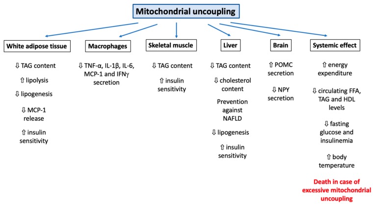
- Therapeutic potential is discussed for obesity, diabetes, cardiovascular disease, and neurological disorders; controlled-release DNP in rats decreased fasting plasma glucose, fatty acids by 30%, TAG, HDL by 30%, and hyperinsulinemia by 50%, with 50–90% decreases in TAG in plasma, liver, and skeletal muscle. DNP at 30 °C decreased energy expenditure by 17% and weight loss by 23% in female C57BL/6J mice, while no significant effect was observed at 22 °C.
Methods
- Narrative review comparing mitochondrial uncoupling agents, uncoupling proteins (UCPs and ANTs), and chemical compounds with uncoupling properties.
- Synthesizes cited literature on cellular and biological processes including autophagy, mitophagy, ROS production, protein secretion, cell death, physical exercise, adipose tissue browning, and cell signaling.
- Discusses possible use of mitochondrial uncouplers in human diseases such as obesity, cardiovascular diseases, and neurological disorders; no primary experimental data or systematic search strategy is reported.
Limitations
- The authors state that most mechanistic studies have focused on classical but non-specific uncouplers such as DNP or FCCP, making it difficult to discriminate effects directly imputable to mitochondrial uncoupling from unspecific effects.
- The authors state that cellular consequences have not been studied rigorously and that continuous development of mitochondria-specific uncoupling agents is needed to improve knowledge and safety.
- Evident from the text: this is a narrative review without stated systematic search or inclusion criteria, and many findings are described as conflicting or dependent on species, cell type, temperature, membrane composition, and uncoupler used.
Model-written summary. Check it against the paper before relying on it.
Integrity signals
Registry
Cites
Statistics
Figures
Verification
Claims the paper makes
159 Not checked yet · 10 Own interpretation · 4 No basis given · 3 Supported · 2 Partly supported · 1 Supported so far · 1 Mixed support · accuracy not yet measured
Some cited sources support this claim and some don’t — read each source’s verdict below.
In the paper
“Of note, some forms of mitophagy triggered by mitochondrial uncoupling do not seem to require Parkin binding to mitochondria [106,119,120].”
§ Mitochondrial Uncoupling: A Key Controller of Biological Processes in Physiology and Diseases > 3. Cellular Responses to Mitochondrial Uncoupling > 3.2. Mitophagy
What the sources say
- Partly supportedcites [106] Pickles, 2018, Mitophagy and Quality Control Mechanisms in Mitochondrial Maintenance, Curr. Biol. sourceExperimental — accuracy not yet measured
The “Parkin and PINK1-dependent mitophagy” section states that low levels of mitophagy persist even in the absence of Parkin, supporting the point that mitochondrial Parkin binding is not always required. The passage does not establish that this Parkin-independent mitophagy was triggered specifically by mitochondrial uncoupling; no page number is available in our copy. (LLM, conf=0.82)
- Disputedcites [119] Xiao, 2017, Superoxide drives progression of Parkin/PINK1-dependent mitophagy following translocation of Parkin to mitochondria, Cell Death Dis. sourceExperimental — accuracy not yet measured
The Results section on ROS-facilitated mitophagy and the Discussion describe Parkin translocation as a prerequisite in the CCCP-triggered process studied here. The paper does not establish a general rule for every form of mitophagy, but its finding runs contrary to the claim attributed to it. (LLM, conf=0.84)
Checked by a reasoning model — may be wrong.
- Disputedcites [120] Rakovic, 2018, PINK1-dependent mitophagy is driven by the UPS and can occur independently of LC3 conversion, Cell Death Differ. sourceExperimental — accuracy not yet measured
The Introduction and Fig. 10 model describe uncoupler-induced mitochondrial depolarization as leading to Parkin recruitment to mitochondria, not as bypassing it. The Results section ‘The UPS is essential for removal of depolarized mitochondria’ observes diffuse cytosolic Parkin after Valinomycin-induced clearance, but that endpoint does not establish that recruitment was unnecessary earlier in the process. (LLM, conf=0.82)
Checked by a reasoning model — may be wrong.
hedged- Asserted: passes — The authors put it forward as true.
- Checkable: passes — It can be checked against evidence.
- Load-bearing: unclear — It is unclear whether the paper’s conclusions depend on it.
- Based: passes — The paper gives a reason why it holds.
- Partly supported
The sources with a verdict support this claim; the rest weren’t checked yet, have no full text we could read, or their verdicts aren’t rated yet.
In the paper
“The positive effect of mitochondrial uncoupling on autophagy has been established in many cellular and animal models, including adipocytes [91], MEFs [92], HeLa cells [93], HepG2 cells [90], SH-SY5Y neuroblastoma cells [93], or C57BL6 mice [94].”
§ Mitochondrial Uncoupling: A Key Controller of Biological Processes in Physiology and Diseases > 3. Cellular Responses to Mitochondrial Uncoupling > 3.1. Autophagy
What the sources say
- Supportedcites [91] Demine, 2018, Mild mitochondrial uncoupling induces HSL/ATGL-independent lipolysis relying on a form of autophagy in 3T3-L1 adipocytes, J. Cell. Physiol. source
The citation to this paper is attached specifically to “adipocytes,” not the other listed models. In the section “Macroautophagy is stimulated in adipocytes exposed to mitochondrial uncoupling,” the authors report marker changes suggesting increased macroautophagy in uncoupled adipocytes. (LLM, conf=0.93)
- Not checked yetcites [92] Kwon, 2011, CCCP induces autophagy in an AMPK-independent manner, Biochem. Biophys. Res. Commun. source
- Supportedcites [93] Chan, 2011, Broad activation of the ubiquitin–proteasome system by Parkin is critical for mitophagy, Hum. Mol. Genet. source
The Results section ('The UPS is essential for Parkin-mediated mitophagy') reports CCCP-induced mitophagy in Parkin-expressing SH-SY5Y cells and describes the corresponding response in HeLa cells. Those are the two cell models for which this paper is cited. The evidence concerns mitophagy under Parkin-expressing conditions, rather than autophagy generally. (LLM, conf=0.88)
boosteddirect- Asserted: passes — The authors put it forward as true.
- Checkable: passes — It can be checked against evidence.
- Load-bearing: unclear — It is unclear whether the paper’s conclusions depend on it.
- Based: passes — The paper gives a reason why it holds.
- Supported
Every source the paper cites for this claim was checked and supports it.
In the paper
“If both mouse models are overweight at the basal state but protected against diet-induced obesity, only IL-18r KO mice can increase thermogenesis and white browning in response to HFD or cold [241].”
§ Mitochondrial Uncoupling: A Key Controller of Biological Processes in Physiology and Diseases > 3. Cellular Responses to Mitochondrial Uncoupling > 3.7. Adipose Tissue Browning
What the sources say
- Supportedcites [241] Pazos, 2015, Divergent responses to thermogenic stimuli in BAT and subcutaneous adipose tissue from interleukin 18 and interleukin 18 receptor 1-deficient mice, Sci. Rep. source
Among the two knockout models, the Abstract reports that Il18-KO mice failed to develop diet-induced thermogenesis, whereas Il18r1-KO mice showed increased iWAT Ucp1 expression after HFD and stronger browning responses to cold. The Discussion likewise contrasts blocked inducible browning in Il18-KO mice with beige reprogramming in Il18r1-KO mice. The preceding statement about both models being protected against diet-induced obesity is not part of the quoted claim. (LLM, conf=0.90)
plaindirect- Asserted: passes — The authors put it forward as true.
- Checkable: passes — It can be checked against evidence.
- Load-bearing: unclear — It is unclear whether the paper’s conclusions depend on it.
- Based: passes — The paper gives a reason why it holds.
- Supported
The cited sources support only part of this claim — read each source’s verdict below.
In the paper
“By decreasing the Ca2+ buffering capacity of mitochondria, CCCP or FCCP increases global exocytosis in chromaffin cells [164,165,166].”
§ Mitochondrial Uncoupling: A Key Controller of Biological Processes in Physiology and Diseases > 3. Cellular Responses to Mitochondrial Uncoupling > 3.8. Cell Signaling
What the sources say
- Partly supportedcites [164] Giovannucci, 1999, Mitochondria regulate the Ca(2+)-exocytosis relationship of bovine adrenal chromaffin cells, J. Neurosci. sourceExperimental — accuracy not yet measured
In “Effect of FCCP on the stimulus-evoked Cm response,” the paper reports that FCCP reduces mitochondrial Ca2+ import and increases stimulus-evoked exocytosis in bovine chromaffin cells. It does not establish the same effect for CCCP, and it reports no significant effect of FCCP on basal membrane capacitance. The supported finding is therefore an increase in evoked, rather than unqualified, exocytosis. (LLM, conf=0.86)
- Partly supportedcites [165] Jurkiewicz, 2013, Novel features on the regulation by mitochondria of calcium and secretion transients in chromaffin cells challenged with acetylcholine at 37°C, Physiol. Rep. sourceExperimental — accuracy not yet measured
The Introduction says CCCP and FCCP suppress mitochondrial Ca2+ buffering and increase exocytosis in bovine chromaffin cells; the Results also report that FCCP potentiates ACh-evoked secretion. But the Introduction notes that protonophores reduce K+-evoked secretory responses in mouse chromaffin cells, so the unqualified claim about chromaffin cells is too broad. (LLM, conf=0.87)
- Not checked yetcites [166] Jurkiewicz, 2009, Role of the Endoplasmic Reticulum and Mitochondria on Quantal Catecholamine Release from Chromaffin Cells of Control and Hypertensive Rats, J. Pharmacol. Exp. Ther. source
plaindirect- Asserted: passes — The authors put it forward as true.
- Checkable: passes — It can be checked against evidence.
- Load-bearing: unclear — It is unclear whether the paper’s conclusions depend on it.
- Based: passes — The paper gives a reason why it holds.
- Partly supported
None of the sources cited for this claim have been checked yet.
In the paper
“Regarding UCP-3, several groups reported that UCP-3 has no clear uncoupling effect as it does, for instance, in CHO cells overexpressing this protein [19] or in rat skeletal muscles [20].”
§ Mitochondrial Uncoupling: A Key Controller of Biological Processes in Physiology and Diseases > 2. Mitochondrial Uncouplers > 2.1. UCPs and ANTs
What the sources say
- Not checked yetcites [19] Mozo, 2006, Expression of UCP3 in CHO cells does not cause uncoupling, but controls mitochondrial activity in the presence of glucose, Biochem. J. source
- Not checked yetcites [20] Cadenas, 1999, UCP2 and UCP3 rise in starved rat skeletal muscle but mitochondrial proton conductance is unchanged, FEBS Lett. source
plain- Asserted: passes — The authors put it forward as true.
- Checkable: passes — It can be checked against evidence.
- Load-bearing: unclear — It is unclear whether the paper’s conclusions depend on it.
- Based: passes — The paper gives a reason why it holds.
In the paper
“First, purine nucleotides, such as GDP, directly bind UCP-1 and lead to its inactivation [8].”
§ Mitochondrial Uncoupling: A Key Controller of Biological Processes in Physiology and Diseases > 2. Mitochondrial Uncouplers > 2.1. UCPs and ANTs
What the sources say
- Not checked yetcites [8] Rial, 1983, Brown-adipose-tissue mitochondria: The regulation of the 32000-Mr uncoupling protein by fatty acids and purine nucleotides, JBIC J. Biol. Inorg. Chem.
plaindirect- Asserted: passes — The authors put it forward as true.
- Checkable: passes — It can be checked against evidence.
- Load-bearing: unclear — It is unclear whether the paper’s conclusions depend on it.
- Based: passes — The paper gives a reason why it holds.
In the paper
“UCP-2 could then only be activated under some circumstances, such as a massive entry of FFAs into mitochondria [18].”
§ Mitochondrial Uncoupling: A Key Controller of Biological Processes in Physiology and Diseases > 2. Mitochondrial Uncouplers > 2.1. UCPs and ANTs
What the sources say
- Not checked yetcites [18] Berardi, 2014, Fatty Acid Flippase Activity of UCP2 Is Essential for Its Proton Transport in Mitochondria, Cell Metab. source
hedgedconditional- Asserted: passes — The authors put it forward as true.
- Checkable: passes — It can be checked against evidence.
- Load-bearing: unclear — It is unclear whether the paper’s conclusions depend on it.
- Based: passes — The paper gives a reason why it holds.
In the paper
“Underlining their importance, ANTs can account for up to 50% of basal mitochondrial membrane proton conductance [25].”
§ Mitochondrial Uncoupling: A Key Controller of Biological Processes in Physiology and Diseases > 2. Mitochondrial Uncouplers > 2.1. UCPs and ANTs
What the sources say
- Not checked yetcites [25] Brand, 2005, The basal proton conductance of mitochondria depends on adenine nucleotide translocase content, Biochem. J. source
plaindirect- Asserted: passes — The authors put it forward as true.
- Checkable: passes — It can be checked against evidence.
- Load-bearing: unclear — It is unclear whether the paper’s conclusions depend on it.
- Based: passes — The paper gives a reason why it holds.
None of the sources cited for this claim have been checked yet.
In the paper
“Second, free fatty acids (FFAs) are well-known to promote UCP-1-dependent protein leak [8,9].”
§ Mitochondrial Uncoupling: A Key Controller of Biological Processes in Physiology and Diseases > 2. Mitochondrial Uncouplers > 2.1. UCPs and ANTs
What the sources say
- Not checked yetcites [8] Rial, 1983, Brown-adipose-tissue mitochondria: The regulation of the 32000-Mr uncoupling protein by fatty acids and purine nucleotides, JBIC J. Biol. Inorg. Chem.
- Not checked yetcites [9] Strieleman, 1985, Fatty acid activation of the reconstituted brown adipose tissue mitochondria uncoupling protein, J. Biol. Chem. source
boosteddirect- Asserted: passes — The authors put it forward as true.
- Checkable: passes — It can be checked against evidence.
- Load-bearing: unclear — It is unclear whether the paper’s conclusions depend on it.
- Based: passes — The paper gives a reason why it holds.
None of the sources cited for this claim have been checked yet.
In the paper
“First, they stimulate directly mitochondrial respiration, as seen in intact isolated brown adipocytes or mitochondria isolated from these cells [28,29].”
§ Mitochondrial Uncoupling: A Key Controller of Biological Processes in Physiology and Diseases > 2. Mitochondrial Uncouplers > 2.2. Natural Uncouplers
What the sources say
- Not checked yetcites [28] Bukowiecki, 1981, Metabolic relationships between lipolysis and respiration in rat brown adipocytes. The role of long chain fatty acids as regulators of mitochondrial respiration and feedback inhibitors of lipolysis, J. Biol. Chem. source
- Not checked yetcites [29] Wojtczak, 1993, Effect of fatty acids on energy coupling processes in mitochondria, Biochim. Biophys. Acta source
plaindirect- Asserted: passes — The authors put it forward as true.
- Checkable: passes — It can be checked against evidence.
- Load-bearing: passes — The paper’s conclusions depend on it.
- Based: passes — The paper gives a reason why it holds.
- It may state two separate claims as one. 63
- Probably not a substantial claim. 14
- The quote only partly states this claim. 1
The cited sources support only part of this claim — read each source’s verdict below.
In the paper
“Importantly, although strong evidence of the mitochondrial uncoupling property is clear for UCP-1 [7,12], it is still heavily debated for the other UCPs [13,14,15].”
§ Mitochondrial Uncoupling: A Key Controller of Biological Processes in Physiology and Diseases > 2. Mitochondrial Uncouplers > 2.1. UCPs and ANTs
What the sources say
- Partly supportedcites [7] Jacobsson, 1985, Mitochondrial uncoupling protein from mouse brown fat. Molecular cloning, genetic mapping, and mRNA expression, J. Biol. Chem. sourceExperimental — accuracy not yet measured
The Discussion (page unavailable) identifies the brown-fat UCP as a mitochondrial protein and says it is believed to produce heat by uncoupling respiration from ATP production. That supports the UCP-1 mechanism, but the paper hedges it rather than establishing the claim’s stronger assertion that the evidence is clear. The debate about other UCPs is attached to different citations, not this source. (LLM, conf=0.84)
- Supportedcites [12] Nedergaard, 2001, UCP1: The only protein able to mediate adaptive non-shivering thermogenesis and metabolic inefficiency, Biochim. Biophys. Acta source
Section 5.3 reports that mitochondria from UCP1-ablated mice are fully coupled, supporting UCP1’s role in uncoupled respiration. Section 5.4 contrasts UCP1’s clear effect with the lack of consistent evidence for uncoupling by UCP2/UCP3 when expressed at normal levels; the Conclusions likewise call evidence for their uncoupling function weak. (LLM, conf=0.93)
- Supportedcites [13] Bouillaud, 2009, UCP2, not a physiologically relevant uncoupler but a glucose sparing switch impacting ROS production and glucose sensing, Biochim. Biophys. Acta source
Section 2 states that UCP1 clearly uncouples mitochondria, while evidence for uncoupling by UCP2 and UCP3 remains ambiguous. The Conclusion (section 8) says conclusive evidence for the transport activity underlying UCP2/3’s effects is still lacking and that the uncoupling hypothesis needs further support. (LLM, conf=0.93)
- Not checked yetcites [14] Shabalina, 2011, Mitochondrial (‘mild’) uncoupling and ROS production: Physiologically relevant or not?, Biochem. Soc. Trans. source
- Not checked yetcites [15] Stuart, 2001, Physiological Levels of Mammalian Uncoupling Protein 2 Do Not Uncouple Yeast Mitochondria, J. Biol. Chem. source
boosted- Asserted: passes — The authors put it forward as true.
- Checkable: passes — It can be checked against evidence.
- Load-bearing: unclear — It is unclear whether the paper’s conclusions depend on it.
- Based: passes — The paper gives a reason why it holds.
It may state two separate claims as one.
- Partly supported
Every source the paper cites for this claim was checked and supports it.
In the paper
“Indeed, mice treated with DNP present a decrease in mTOR activity, in the insulin-PI3K-MAPK signaling pathways, and a stimulation of the Ca2+-CREB axis accompanied by an increase in autophagy [94].”
§ Mitochondrial Uncoupling: A Key Controller of Biological Processes in Physiology and Diseases > 3. Cellular Responses to Mitochondrial Uncoupling > 3.1. Autophagy
What the sources say
- Supportedcites [94] Liu, 2015, The Mitochondrial Uncoupler DNP Triggers Brain Cell mTOR Signaling Network Reprogramming and CREB Pathway Upregulation, J. Neurochem. source
The Results sections report reduced mTOR and phosphorylated mTOR in DNP-treated mouse cortex, suppressed insulin/PI3K–AKT and MAPK–ERK signaling, and up-regulated calcium–calmodulin–CREB signaling. The autophagy Results section reports increased autophagy-related genes and protein markers; it does not directly measure autophagic flux. Page numbers were not available in our copy. (LLM, conf=0.88)
plaindirect- Asserted: passes — The authors put it forward as true.
- Checkable: passes — It can be checked against evidence.
- Load-bearing: unclear — It is unclear whether the paper’s conclusions depend on it.
- Based: passes — The paper gives a reason why it holds.
It may state two separate claims as one.
- Supported
Every source the paper cites for this claim was checked and supports it.
In the paper
“IL-18 expression seems to be tightly correlated to other proinflammatory markers, such as TNFα, and a linear correlation between IL-18 and BMI was found in humans [237].”
§ Mitochondrial Uncoupling: A Key Controller of Biological Processes in Physiology and Diseases > 3. Cellular Responses to Mitochondrial Uncoupling > 3.7. Adipose Tissue Browning
What the sources say
- Supportedcites [237] Ahmad, 2017, Increased adipose tissue expression of IL-18R and its ligand IL-18 associates with inflammation and insulin resistance in obesity, Immun. Inflamm. Dis. source
The Results section, under “Association of IL‐18R/IL‐18 adipose tissue expression with local inflammation and insulin resistance,” reports a positive correlation between adipose-tissue IL‐18 and TNF‐α expression (r = 0.60, P = 0.04). The Discussion also states that IL‐18 mRNA correlated positively with TNF‐α gene expression. (LLM, conf=0.90)
hedgedcorrelational- Asserted: passes — The authors put it forward as true.
- Checkable: passes — It can be checked against evidence.
- Load-bearing: fails — The paper’s conclusions may not depend on it.
- Based: passes — The paper gives a reason why it holds.
Probably not a substantial claim. The paper’s conclusions may not depend on it.
- Supported
In the paper
“UCP-1 is a transmembrane protein localized in the inner mitochondrial membrane catalyzing the transport of protons across the mitochondrial membrane and thereby inducing mitochondrial uncoupling [7].”
§ Mitochondrial Uncoupling: A Key Controller of Biological Processes in Physiology and Diseases > 2. Mitochondrial Uncouplers > 2.1. UCPs and ANTs
What the sources say
- Not checked yetcites [7] Jacobsson, 1985, Mitochondrial uncoupling protein from mouse brown fat. Molecular cloning, genetic mapping, and mRNA expression, J. Biol. Chem. source
plaindirect- Asserted: passes — The authors put it forward as true.
- Checkable: passes — It can be checked against evidence.
- Load-bearing: unclear — It is unclear whether the paper’s conclusions depend on it.
- Based: passes — The paper gives a reason why it holds.
It may state two separate claims as one.
In the paper
“In addition, UCP-1 activity is also tightly modulated by phosphorylation, a phenomenon shown to increase in response to cold in rats [10].”
§ Mitochondrial Uncoupling: A Key Controller of Biological Processes in Physiology and Diseases > 2. Mitochondrial Uncouplers > 2.1. UCPs and ANTs
What the sources say
- Not checked yetcites [10] Carroll, 2008, Identification of serine phosphorylation in mitochondrial uncoupling protein 1, Biochim. Biophys. Acta source
plaindirect- Asserted: passes — The authors put it forward as true.
- Checkable: passes — It can be checked against evidence.
- Load-bearing: unclear — It is unclear whether the paper’s conclusions depend on it.
- Based: passes — The paper gives a reason why it holds.
It may state two separate claims as one.
In the paper
“The same authors suggested that UCP-2 activity could be completely blocked by GDP at the basal state (100 µM, which corresponds to the basal intracellular GDP/GTP concentration) [18].”
§ Mitochondrial Uncoupling: A Key Controller of Biological Processes in Physiology and Diseases > 2. Mitochondrial Uncouplers > 2.1. UCPs and ANTs
What the sources say
- Not checked yetcites [18] Berardi, 2014, Fatty Acid Flippase Activity of UCP2 Is Essential for Its Proton Transport in Mitochondria, Cell Metab. source
hedgeddirect- Asserted: passes — The authors put it forward as true.
- Checkable: passes — It can be checked against evidence.
- Load-bearing: fails — The paper’s conclusions may not depend on it.
- Based: passes — The paper gives a reason why it holds.
Probably not a substantial claim. The paper’s conclusions may not depend on it.
In the paper
“This process essentially depends on the composition of the inner mitochondrial membrane and could account for up to 30–50% of the resting cellular metabolic rate [3].”
§ Mitochondrial Uncoupling: A Key Controller of Biological Processes in Physiology and Diseases > 1. Introduction
What the sources say
- Not checked yetcites [3] Jastroch, 2010, Mitochondrial proton and electron leaks, Essays Biochem. source
hedgeddirect- Asserted: passes — The authors put it forward as true.
- Checkable: passes — It can be checked against evidence.
- Load-bearing: unclear — It is unclear whether the paper’s conclusions depend on it.
- Based: passes — The paper gives a reason why it holds.
It may state two separate claims as one.
None of the sources cited for this claim have been checked yet.
In the paper
“The molecular mechanism is not entirely clear but has been suggested to depend on the transport of fatty acids [23,24].”
§ Mitochondrial Uncoupling: A Key Controller of Biological Processes in Physiology and Diseases > 2. Mitochondrial Uncouplers > 2.1. UCPs and ANTs
What the sources say
- Not checked yetcites [23] Andreyev, 1989, The ATP/ADP-antiporter is involved in the uncoupling effect of fatty acids on mitochondria, JBIC J. Biol. Inorg. Chem.
- Not checked yetcites [24] Sparks, 2016, ANT1-mediated fatty acid-induced uncoupling as a target for improving myocellular insulin sensitivity, Diabetologia source
hedgedconditional- Asserted: passes — The authors put it forward as true.
- Checkable: passes — It can be checked against evidence.
- Load-bearing: fails — The paper’s conclusions may not depend on it.
- Based: passes — The paper gives a reason why it holds.
Probably not a substantial claim. The paper’s conclusions may not depend on it.
In the paper
“For instance, BAM15 ((2-fluorophenyl)6-[(2-fluorophenyl)amino](1,2,5-oxadiazolo[3,4-e]pyrazin-5-yl)amine) is a novel mitochondria-specific protonophore uncoupler that possesses a similar potency to FCCP or DNP [45].”
§ Mitochondrial Uncoupling: A Key Controller of Biological Processes in Physiology and Diseases > 2. Mitochondrial Uncouplers > 2.3. Synthetic Mitochondrial Uncouplers
What the sources say
- Not checked yetcites [45] Kenwood, 2014, Identification of a novel mitochondrial uncoupler that does not depolarize the plasma membrane, Mol. Metab. source
plaincorrelational- Asserted: passes — The authors put it forward as true.
- Checkable: passes — It can be checked against evidence.
- Load-bearing: unclear — It is unclear whether the paper’s conclusions depend on it.
- Based: passes — The paper gives a reason why it holds.
It may state two separate claims as one.
In the paper
“For instance, CCCP can interfere with lysosomal function and inhibit autophagic cargo degradation in both yeast and HeLa cells [83].”
§ Mitochondrial Uncoupling: A Key Controller of Biological Processes in Physiology and Diseases > 2. Mitochondrial Uncouplers > 2.4. Mitochondrial Uncouplers Affect Lysosomal Ion Homeostasis
What the sources say
- Not checked yetcites [83] Padman, 2013, The protonophore CCCP interferes with lysosomal degradation of autophagic cargo in yeast and mammalian cells, Autophagy source
plaindirect- Asserted: passes — The authors put it forward as true.
- Checkable: passes — It can be checked against evidence.
- Load-bearing: unclear — It is unclear whether the paper’s conclusions depend on it.
- Based: passes — The paper gives a reason why it holds.
It may state two separate claims as one.
In the paper
“In this model, a low concentration (0.5 μM) in this mitochondrial uncoupler is sufficient to reduce adipocyte triacylglycerol (TAG) content, resulting from a lipolysis activation that is independent of the classical cytosolic lipases HSL (Hormone Sensitive Lipase) and ATGL (Adipose Triglyceride Lipase) [91].”
§ Mitochondrial Uncoupling: A Key Controller of Biological Processes in Physiology and Diseases > 3. Cellular Responses to Mitochondrial Uncoupling > 3.1. Autophagy
What the sources say
- Not checked yetcites [91] Demine, 2018, Mild mitochondrial uncoupling induces HSL/ATGL-independent lipolysis relying on a form of autophagy in 3T3-L1 adipocytes, J. Cell. Physiol. source
plaindirect- Asserted: passes — The authors put it forward as true.
- Checkable: passes — It can be checked against evidence.
- Load-bearing: passes — The paper’s conclusions depend on it.
- Based: passes — The paper gives a reason why it holds.
It may state two separate claims as one.
None of the sources cited for this claim have been checked yet.
In the paper
“These molecules are also known to upregulate UCP-1 expression in many models, including mouse [40,41], rat [42,43], and human [44] brown adipose tissues.”
§ Mitochondrial Uncoupling: A Key Controller of Biological Processes in Physiology and Diseases > 2. Mitochondrial Uncouplers > 2.2. Natural Uncouplers
What the sources say
- Not checked yetcites [40] Rehnmark, 1990, Alpha- and beta-adrenergic induction of the expression of the uncoupling protein thermogenin in brown adipocytes differentiated in culture, J. Biol. Chem. source
- Not checked yetcites [41] Champigny, 1992, Regulation of UCP gene expression in brown adipocytes differentiated in primary culture. Effects of a new beta-adrenoceptor agonist, Mol. Cell. Endocrinol. source
- Not checked yetcites [42] Silva, 1988, Full Expression of Uncoupling Protein Gene Requires the Concurrence of Norepinephrine and Triiodothyronine, Mol. Endocrinol. source
- Not checked yetcites [43] Herron, 1990, Norepinephrine-induced synthesis of the uncoupling protein thermogenin (UCP) and its mitochondrial targeting in brown adipocytes differentiated in culture, FEBS Lett. source
- Not checked yetcites [44] Champigny, 1996, Evidence from in vitro differentiating cells that adrenoceptor agonists can increase uncoupling protein mRNA level in adipocytes of adult humans: An RT-PCR study, J. Lipid Res. source
plaindirect- Asserted: passes — The authors put it forward as true.
- Checkable: passes — It can be checked against evidence.
- Load-bearing: unclear — It is unclear whether the paper’s conclusions depend on it.
- Based: passes — The paper gives a reason why it holds.
It may state two separate claims as one.
In the paper
“In summary, FR58P1 mildly uncouples mitochondria, leading to a mitochondrial clearance by mitophagy, activation of the cytoprotective AMPK/Sirt1 axis, and a glycolytic shift.”
§ Mitochondrial Uncoupling: A Key Controller of Biological Processes in Physiology and Diseases > 2. Mitochondrial Uncouplers > 2.3. Synthetic Mitochondrial Uncouplers
What the sources say
- Not checked yetcites [54] Urra, 2018, FR58P1a; a new uncoupler of OXPHOS that inhibits migration in triple-negative breast cancer cells via Sirt1/AMPK/β1-integrin pathway, Sci. Rep. source
plaindirect- Asserted: passes — The authors put it forward as true.
- Checkable: passes — It can be checked against evidence.
- Load-bearing: unclear — It is unclear whether the paper’s conclusions depend on it.
- Based: passes — The paper gives a reason why it holds.
It may state two separate claims as one.
In the paper
“Indeed, mice deficient for LAL (Lysosomal Acid Lipase, lysosomal lipase) exhibit BAT with lysosome and lipid droplet accumulation, reduced UCP-1 expression, and mitochondrial dysfunction [97].”
§ Mitochondrial Uncoupling: A Key Controller of Biological Processes in Physiology and Diseases > 3. Cellular Responses to Mitochondrial Uncoupling > 3.1. Autophagy
What the sources say
- Not checked yetcites [97] Sachdev, 2018, Lysosomal acid lipase regulates fatty acid channeling in brown adipose tissue to maintain thermogenesis, Biochim. Biophys. Acta Mol. Cell. Biol. Lipids
plaindirect- Asserted: passes — The authors put it forward as true.
- Checkable: passes — It can be checked against evidence.
- Load-bearing: passes — The paper’s conclusions depend on it.
- Based: passes — The paper gives a reason why it holds.
It may state two separate claims as one.
In the paper
“For instance, low doses of CCCP (5 μM) or C12TPP (0.2 nM) do not upregulate macroautophagy in human EAhy926 endothelial cells but still induce an anti-inflammatory response, including reduction in the expression of E-selectin, ICAM1, and VCAM1 and in the adhesion of neutrophils to endothelium induced by TNFα (Tumor Necrosis Factor α) [95].”
§ Mitochondrial Uncoupling: A Key Controller of Biological Processes in Physiology and Diseases > 3. Cellular Responses to Mitochondrial Uncoupling > 3.1. Autophagy
What the sources say
- Not checked yetcites [95] Romaschenko, 2015, Low concentrations of uncouplers of oxidative phosphorylation prevent inflammatory activation of endothelial cells by tumor necrosis factor, Biochemistry
plaindirect- Asserted: passes — The authors put it forward as true.
- Checkable: passes — It can be checked against evidence.
- Load-bearing: unclear — It is unclear whether the paper’s conclusions depend on it.
- Based: passes — The paper gives a reason why it holds.
It may state two separate claims as one.
In the paper
“Indeed, the mitochondrial uncoupler cloxyquin (5-chloro-8-hydroxyquinoline) protects mouse cardiomyocytes against ischemia-reperfusion, an effect requiring autophagy [99].”
§ Mitochondrial Uncoupling: A Key Controller of Biological Processes in Physiology and Diseases > 3. Cellular Responses to Mitochondrial Uncoupling > 3.1. Autophagy
What the sources say
- Not checked yetcites [99] Zhang, 2016, The cardioprotective compound cloxyquin uncouples mitochondria and induces autophagy, Am. J. Physiol. Heart Circ. Physiol. source
plaindirect- Asserted: passes — The authors put it forward as true.
- Checkable: passes — It can be checked against evidence.
- Load-bearing: unclear — It is unclear whether the paper’s conclusions depend on it.
- Based: passes — The paper gives a reason why it holds.
It may state two separate claims as one.
None of the sources cited for this claim have been checked yet.
In the paper
“T3 regulates mitochondrial uncoupling by different mechanisms: (1) by sympathetic stimulation [61], (2) by increasing acylcarnitine production [61], thereby activating mitochondrial respiration/uncoupling, and (3) by directly stimulating the transcription of the Ucp1 gene [37].”
§ Mitochondrial Uncoupling: A Key Controller of Biological Processes in Physiology and Diseases > 2. Mitochondrial Uncouplers > 2.2. Natural Uncouplers
What the sources say
plaindirect- Asserted: passes — The authors put it forward as true.
- Checkable: passes — It can be checked against evidence.
- Load-bearing: unclear — It is unclear whether the paper’s conclusions depend on it.
- Based: passes — The paper gives a reason why it holds.
It may state two separate claims as one.
None of the sources cited for this claim have been checked yet.
In the paper
“PUFAs such as eicosapentaenoic acid (EPA) and docosahexaenoic acid (DHA) also stimulate UCP1 expression and thus adaptive non-shivering or diet-induced thermogenesis in brown adipocytes, as well as the browning of the white adipose tissue (WAT) in C57BL/6 mice [67,68], Sprague-Dawley rats [69,70], and overweight humans [71].”
§ Mitochondrial Uncoupling: A Key Controller of Biological Processes in Physiology and Diseases > 2. Mitochondrial Uncouplers > 2.2. Natural Uncouplers
What the sources say
- Not checked yetcites [67] Bargut, T.C.L., Martins, F.F., Santos, L.P., Aguila, M.B., and Mandarim-de-Lacerda, C.A. (2018). Administration of eicosapentaenoic and docosahexaenoic acids may improve the remodeling and browning in subcutaneous white adipose tissue and thermogenic markers in brown adipose tissue in mice. Mol. Cell. Endocrinol. source
- Not checked yetcites [68] Zhao, 2014, Eicosapentaenoic acid promotes thermogenic and fatty acid storage capacity in mouse subcutaneous adipocytes, Biochem. Biophys. Res. Commun. source
- Not checked yetcites [69] Oudart, 1997, Brown fat thermogenesis in rats fed high-fat diets enriched with n-3 polyunsaturated fatty acids, Int. J. Obes. source
- Not checked yetcites [70] Takahashi, 2000, Dietary n-3 fatty acids affect mRNA level of brown adipose tissue uncoupling protein 1, and white adipose tissue leptin and glucose transporter 4 in the rat, Br. J. Nutr. source
- Not checked yetcites [71] Laiglesia, 2016, Eicosapentaenoic acid promotes mitochondrial biogenesis and beige-like features in subcutaneous adipocytes from overweight subjects, J. Nutr. Biochem. source
plaindirect- Asserted: passes — The authors put it forward as true.
- Checkable: passes — It can be checked against evidence.
- Load-bearing: unclear — It is unclear whether the paper’s conclusions depend on it.
- Based: passes — The paper gives a reason why it holds.
It may state two separate claims as one.
In the paper
“However, BAM15 has low or no effect on plasma membrane polarization, as seen in rat L6 myoblasts [45].”
§ Mitochondrial Uncoupling: A Key Controller of Biological Processes in Physiology and Diseases > 2. Mitochondrial Uncouplers > 2.3. Synthetic Mitochondrial Uncouplers
What the sources say
- Not checked yetcites [45] Kenwood, 2014, Identification of a novel mitochondrial uncoupler that does not depolarize the plasma membrane, Mol. Metab. source
plainSingle case- Asserted: passes — The authors put it forward as true.
- Checkable: passes — It can be checked against evidence.
- Load-bearing: unclear — It is unclear whether the paper’s conclusions depend on it.
- Based: passes — The paper gives a reason why it holds.
It may state two separate claims as one. The quote only partly states this claim.
In the paper
“Indeed, hyperthyroidic Atg5KO mice are characterized by a lower body temperature and lower mitochondrial uncoupling than WT littermates [61].”
§ Mitochondrial Uncoupling: A Key Controller of Biological Processes in Physiology and Diseases > 2. Mitochondrial Uncouplers > 2.2. Natural Uncouplers
What the sources say
- Not checked yetcites [61] Yau, 2019, Thyroid hormone (T3) stimulates brown adipose tissue activation via mitochondrial biogenesis and MTOR-mediated mitophagy, Autophagy source
plainconditional- Asserted: passes — The authors put it forward as true.
- Checkable: passes — It can be checked against evidence.
- Load-bearing: unclear — It is unclear whether the paper’s conclusions depend on it.
- Based: passes — The paper gives a reason why it holds.
It may state two separate claims as one.
In the paper
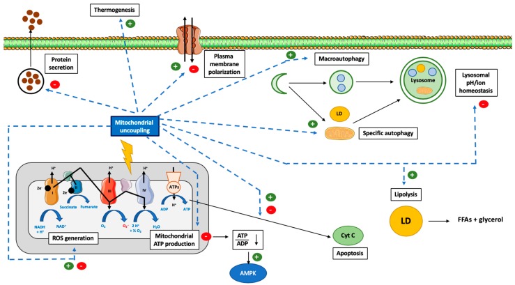
“Mitochondrial uncoupling has often been associated with autophagy activation, and more specifically, with mitophagy (specific degradation of mitochondria by macroautophagy) (Figure 1).”
§ Mitochondrial Uncoupling: A Key Controller of Biological Processes in Physiology and Diseases > 3. Cellular Responses to Mitochondrial Uncoupling > 3.1. Autophagy
What the sources say
- Not checked yetcites [90] Lyamzaev, 2018, Induction of autophagy by depolarization of mitochondria, Autophagy source
plaincorrelational- Asserted: passes — The authors put it forward as true.
- Checkable: passes — It can be checked against evidence.
- Load-bearing: unclear — It is unclear whether the paper’s conclusions depend on it.
- Based: passes — The paper gives a reason why it holds.
It may state two separate claims as one.
In the paper
“Interestingly, rapamycin treatment is accompanied by an increase in mitochondrial potential membrane [100], suggesting, again, a link between mitochondrial (un)coupling and autophagy.”
§ Mitochondrial Uncoupling: A Key Controller of Biological Processes in Physiology and Diseases > 3. Cellular Responses to Mitochondrial Uncoupling > 3.1. Autophagy
What the sources say
- Not checked yetcites [100] Radad, 2015, Rapamycin protects dopaminergic neurons against rotenone-induced cell death in primary mesencephalic cell culture, Folia Neuropathol. source
hedgeddirect- Asserted: passes — The authors put it forward as true.
- Checkable: passes — It can be checked against evidence.
- Load-bearing: unclear — It is unclear whether the paper’s conclusions depend on it.
- Based: passes — The paper gives a reason why it holds.
It may state two separate claims as one.
In the paper
“In humans, physical exercise increases total mitochondrial mass and uncoupling in skeletal muscular cells [192].”
§ Mitochondrial Uncoupling: A Key Controller of Biological Processes in Physiology and Diseases > 3. Cellular Responses to Mitochondrial Uncoupling > 3.6. Physical Exercise
What the sources say
- Not checked yetcites [192] Phielix, 2010, Exercise training increases mitochondrial content and ex vivo mitochondrial function similarly in patients with type 2 diabetes and in control individuals, Diabetologia source
plaindirect- Asserted: passes — The authors put it forward as true.
- Checkable: passes — It can be checked against evidence.
- Load-bearing: unclear — It is unclear whether the paper’s conclusions depend on it.
- Based: passes — The paper gives a reason why it holds.
It may state two separate claims as one.
None of the sources cited for this claim have been checked yet.
In the paper
“In accordance with this theory, use of CCCP or FCCP decreases the Ca2+ buffering capacity of mitochondria and increases global exocytosis in bovine [164,165] and rat chromaffin cells [166] but not in the same cell type in mice [167], suggesting species differences.”
§ Mitochondrial Uncoupling: A Key Controller of Biological Processes in Physiology and Diseases > 3. Cellular Responses to Mitochondrial Uncoupling > 3.4. Protein Secretion
What the sources say
- Not checked yetcites [164] Giovannucci, 1999, Mitochondria regulate the Ca(2+)-exocytosis relationship of bovine adrenal chromaffin cells, J. Neurosci. source
- Not checked yetcites [165] Jurkiewicz, 2013, Novel features on the regulation by mitochondria of calcium and secretion transients in chromaffin cells challenged with acetylcholine at 37°C, Physiol. Rep. source
- Not checked yetcites [166] Jurkiewicz, 2009, Role of the Endoplasmic Reticulum and Mitochondria on Quantal Catecholamine Release from Chromaffin Cells of Control and Hypertensive Rats, J. Pharmacol. Exp. Ther. source
- Not checked yetcites [167] Fuentealba, 2005, Depolarization evokes different patterns of calcium signals and exocytosis in bovine and mouse chromaffin cells: The role of mitochondria, Eur. J. Neurosci. source
hedgeddirect- Asserted: passes — The authors put it forward as true.
- Checkable: passes — It can be checked against evidence.
- Load-bearing: unclear — It is unclear whether the paper’s conclusions depend on it.
- Based: passes — The paper gives a reason why it holds.
It may state two separate claims as one.
None of the sources cited for this claim have been checked yet.
In the paper
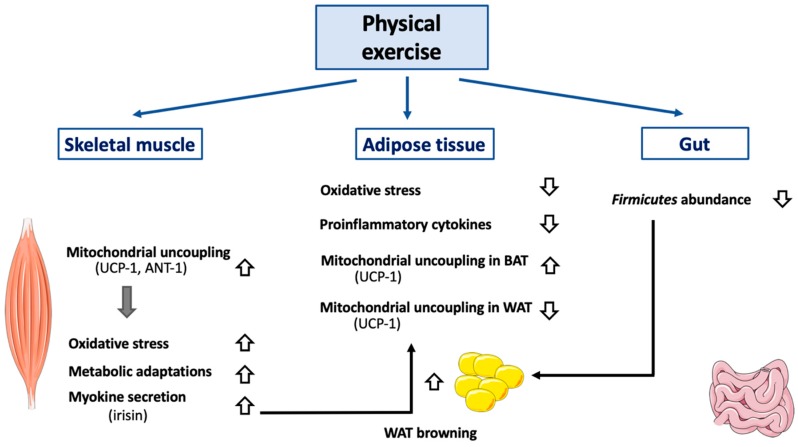
“First, physical exercise by itself directly reduces proton leak and ROS production and increases mitochondrial membrane potential, suggesting an improvement of mitochondrial coupling (Figure 3).”
§ Mitochondrial Uncoupling: A Key Controller of Biological Processes in Physiology and Diseases > 3. Cellular Responses to Mitochondrial Uncoupling > 3.6. Physical Exercise
hedgeddirect- Asserted: passes — The authors put it forward as true.
- Checkable: passes — It can be checked against evidence.
- Load-bearing: unclear — It is unclear whether the paper’s conclusions depend on it.
- Based: passes — The paper gives a reason why it holds.
It may state two separate claims as one.
None of the sources cited for this claim have been checked yet.
In the paper
“Irisin is mainly secreted by skeletal muscle cells, although some secretion was also reported in WAT [204] and, to a lesser extent, in BAT [204,205].”
§ Mitochondrial Uncoupling: A Key Controller of Biological Processes in Physiology and Diseases > 3. Cellular Responses to Mitochondrial Uncoupling > 3.6. Physical Exercise
What the sources say
- Not checked yetcites [204] Roca-Rivada, A., Castelao, C., Senin, L.L., Landrove, M.O., Baltar, J., Crujeiras, A.B., Seoane, L.M., Casanueva, F.F., and Pardo, M. (2013). FNDC5/Irisin Is Not Only a Myokine but Also an Adipokine. PLoS ONE, 8. source
- Not checked yetcites [205] 2018, Irisin a Novel Metabolic biomarker: Present knowledge and future directions, Int. J. Endocrinol.
plain- Asserted: passes — The authors put it forward as true.
- Checkable: passes — It can be checked against evidence.
- Load-bearing: unclear — It is unclear whether the paper’s conclusions depend on it.
- Based: passes — The paper gives a reason why it holds.
It may state two separate claims as one.
The paper states this without a source or data behind it here, so there is nothing to check it against. The quote shows it is in the paper.
In the paper
“Another hallmark of this tissue is the expression of UCP-1, allowing these cells to oxidize fatty acids in order to dissipate and produce energy as heat, a phenomenon called “non-shivering adaptive thermogenesis.””
§ Mitochondrial Uncoupling: A Key Controller of Biological Processes in Physiology and Diseases > 3. Cellular Responses to Mitochondrial Uncoupling > 3.7. Adipose Tissue Browning
plaindirect- Asserted: passes — The authors put it forward as true.
- Checkable: passes — It can be checked against evidence.
- Load-bearing: fails — The paper’s conclusions may not depend on it.
- Based: passes — The paper gives a reason why it holds.
Probably not a substantial claim. The paper’s conclusions may not depend on it. It may state two separate claims as one.
In the paper
“Contrary to UCP-1, the effect of the overexpression of ANT-1 (but not ANT2 [184]) is well-known to promote apoptosis.”
§ Mitochondrial Uncoupling: A Key Controller of Biological Processes in Physiology and Diseases > 3. Cellular Responses to Mitochondrial Uncoupling > 3.5. Cell Death
What the sources say
- Not checked yetcites [184] Zamora, 2004, Recruitment of NF-κB into Mitochondria Is Involved in Adenine Nucleotide Translocase 1 (ANT1)-induced Apoptosis, J. Biol. Chem. source
boosteddirect- Asserted: passes — The authors put it forward as true.
- Checkable: passes — It can be checked against evidence.
- Load-bearing: unclear — It is unclear whether the paper’s conclusions depend on it.
- Based: passes — The paper gives a reason why it holds.
It may state two separate claims as one.
None of the sources cited for this claim have been checked yet.
In the paper
“Three types of adipose tissues have been identified so far: The WAT [218], the BAT [219,220], and the beige or brite (brown in white) adipose tissue (BIWAT) [220].”
§ Mitochondrial Uncoupling: A Key Controller of Biological Processes in Physiology and Diseases > 3. Cellular Responses to Mitochondrial Uncoupling > 3.7. Adipose Tissue Browning
What the sources say
- Not checked yetcites [218] Rutkowski, 2015, The cell biology of fat expansion, J. Cell Biol. source
- Not checked yetcites [219] Schulz, 2013, Brown adipose tissue: Development, metabolism and beyond, Biochem. J. source
- Not checked yetcites [220] Harms, 2013, Brown and beige fat: Development, function and therapeutic potential, Nat. Med. source
plain- Asserted: passes — The authors put it forward as true.
- Checkable: passes — It can be checked against evidence.
- Load-bearing: fails — The paper’s conclusions may not depend on it.
- Based: passes — The paper gives a reason why it holds.
Probably not a substantial claim. The paper’s conclusions may not depend on it.
In the paper
“To explain this effect, it was suggested that mitochondrial uncoupling, by decreasing ATP content, could slow down the energy-demanding steps in protein secretion [160].”
§ Mitochondrial Uncoupling: A Key Controller of Biological Processes in Physiology and Diseases > 3. Cellular Responses to Mitochondrial Uncoupling > 3.4. Protein Secretion
What the sources say
- Not checked yetcites [160] Argon, 1989, Two steps in the intracellular transport of IgD are sensitive to energy depletion, J. Immunol. source
hedgedconditional- Asserted: passes — The authors put it forward as true.
- Checkable: passes — It can be checked against evidence.
- Load-bearing: fails — The paper’s conclusions may not depend on it.
- Based: passes — The paper gives a reason why it holds.
Probably not a substantial claim. The paper’s conclusions may not depend on it.
A conclusion the paper draws from its own results — it cites no other source for it, so there is none to check. The quote shows it is in the paper.
In the paper
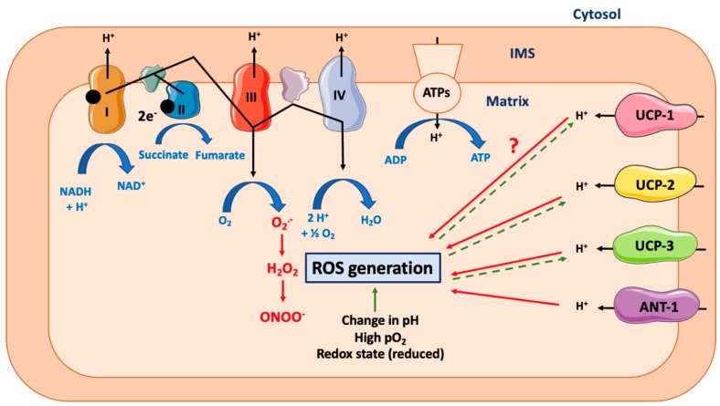
“Based on the simple model described above, the induction of mitochondrial uncoupling would theoretically lead to a decrease in ROS production (Figure 2).”
§ Mitochondrial Uncoupling: A Key Controller of Biological Processes in Physiology and Diseases > 3. Cellular Responses to Mitochondrial Uncoupling > 3.3. Reactive Oxygen Species Production
hedgedconditional- Asserted: passes — The authors put it forward as true.
- Checkable: passes — It can be checked against evidence.
- Load-bearing: passes — The paper’s conclusions depend on it.
- Based: passes — The paper gives a reason why it holds.
Probably not a substantial claim.
In the paper
“In fine, excessive ROS production damages mitochondria and induces secondary mitochondrial respiration uncoupling [158].”
§ Mitochondrial Uncoupling: A Key Controller of Biological Processes in Physiology and Diseases > 3. Cellular Responses to Mitochondrial Uncoupling > 3.3. Reactive Oxygen Species Production
What the sources say
- Not checked yetcites [158] Ma, 2015, Mitochondrial targeted β-lapachone induces mitochondrial dysfunction and catastrophic vacuolization in cancer cells, Bioorg. Med. Chem. Lett. source
plaindirect- Asserted: passes — The authors put it forward as true.
- Checkable: passes — It can be checked against evidence.
- Load-bearing: unclear — It is unclear whether the paper’s conclusions depend on it.
- Based: passes — The paper gives a reason why it holds.
It may state two separate claims as one.
In the paper
“However, this coupling is not complete and leads in fine to the production of ROS, a process called electron leak [3] (Figure 2).”
§ Mitochondrial Uncoupling: A Key Controller of Biological Processes in Physiology and Diseases > 3. Cellular Responses to Mitochondrial Uncoupling > 3.3. Reactive Oxygen Species Production
What the sources say
- Not checked yetcites [3] Jastroch, 2010, Mitochondrial proton and electron leaks, Essays Biochem. source
plaindirect- Asserted: passes — The authors put it forward as true.
- Checkable: passes — It can be checked against evidence.
- Load-bearing: unclear — It is unclear whether the paper’s conclusions depend on it.
- Based: passes — The paper gives a reason why it holds.
It may state two separate claims as one.
In the paper
“Indeed, no increase in the mitochondrial uncoupling rate in subcutaneous inguinal WAT of rats exposed to cold was found, even though UCP-1 protein expression was elevated as expected [223].”
§ Mitochondrial Uncoupling: A Key Controller of Biological Processes in Physiology and Diseases > 3. Cellular Responses to Mitochondrial Uncoupling > 3.7. Adipose Tissue Browning
What the sources say
- Not checked yetcites [223] Jani, 2019, Cold acclimation enhances UCP1 content, lipolysis, and triacylglycerol resynthesis, but not mitochondrial uncoupling and fat oxidation, in rat white adipocytes, Am. J. Physiol. Physiol. source
plain- Asserted: passes — The authors put it forward as true.
- Checkable: passes — It can be checked against evidence.
- Load-bearing: passes — The paper’s conclusions depend on it.
- Based: passes — The paper gives a reason why it holds.
It may state two separate claims as one.
None of the sources cited for this claim have been checked yet.
In the paper
“In the adipose tissue, the latest cytokine is released by both macrophages and adipocytes in obese mice and humans [237,238].”
§ Mitochondrial Uncoupling: A Key Controller of Biological Processes in Physiology and Diseases > 3. Cellular Responses to Mitochondrial Uncoupling > 3.7. Adipose Tissue Browning
What the sources say
- Not checked yetcites [237] Ahmad, 2017, Increased adipose tissue expression of IL-18R and its ligand IL-18 associates with inflammation and insulin resistance in obesity, Immun. Inflamm. Dis. source
- Not checked yetcites [238] Skurk, 2005, The proatherogenic cytokine interleukin-18 is secreted by human adipocytes, Eur. J. Endocrinol. source
plain- Asserted: passes — The authors put it forward as true.
- Checkable: passes — It can be checked against evidence.
- Load-bearing: fails — The paper’s conclusions may not depend on it.
- Based: passes — The paper gives a reason why it holds.
Probably not a substantial claim. The paper’s conclusions may not depend on it.
In the paper
“Indeed, C57BL7 mice deficient for IL-18 are extremely sensitive to obesity but also exhibit larger brown adipocytes and a higher UCP-1 expression in BAT compared to wild-type littermates [240].”
§ Mitochondrial Uncoupling: A Key Controller of Biological Processes in Physiology and Diseases > 3. Cellular Responses to Mitochondrial Uncoupling > 3.7. Adipose Tissue Browning
What the sources say
- Not checked yetcites [240] Yamanishi, 2018, Deficiency in interleukin-18 promotes differentiation of brown adipose tissue resulting in fat accumulation despite dyslipidemia, J. Transl. Med. source
plaincorrelational- Asserted: passes — The authors put it forward as true.
- Checkable: passes — It can be checked against evidence.
- Load-bearing: unclear — It is unclear whether the paper’s conclusions depend on it.
- Based: passes — The paper gives a reason why it holds.
It may state two separate claims as one.
In the paper
“The effect of IL-1β on white adipocytes includes an increase in ROS production, a decrease in SOD activity, and mitochondrial depolarization [229].”
§ Mitochondrial Uncoupling: A Key Controller of Biological Processes in Physiology and Diseases > 3. Cellular Responses to Mitochondrial Uncoupling > 3.7. Adipose Tissue Browning
What the sources say
- Not checked yetcites [229] Okla, 2018, Inhibitory effects of toll-like receptor 4, NLRP3 inflammasome, and interleukin-1β on white adipocyte browning, Inflammation source
plaindirect- Asserted: passes — The authors put it forward as true.
- Checkable: passes — It can be checked against evidence.
- Load-bearing: unclear — It is unclear whether the paper’s conclusions depend on it.
- Based: passes — The paper gives a reason why it holds.
It may state two separate claims as one.
In the paper
“If both mouse models are overweight at the basal state but protected against diet-induced obesity, only IL-18r KO mice can increase thermogenesis and white browning in response to HFD or cold [241].”
§ Mitochondrial Uncoupling: A Key Controller of Biological Processes in Physiology and Diseases > 3. Cellular Responses to Mitochondrial Uncoupling > 3.7. Adipose Tissue Browning
What the sources say
- Not checked yetcites [241] Pazos, 2015, Divergent responses to thermogenic stimuli in BAT and subcutaneous adipose tissue from interleukin 18 and interleukin 18 receptor 1-deficient mice, Sci. Rep. source
plaincorrelational- Asserted: passes — The authors put it forward as true.
- Checkable: passes — It can be checked against evidence.
- Load-bearing: passes — The paper’s conclusions depend on it.
- Based: passes — The paper gives a reason why it holds.
It may state two separate claims as one.
A conclusion the paper draws from its own results — it cites no other source for it, so there is none to check. The quote shows it is in the paper.
In the paper
“The precise action of IL-18 is likely to be complex and possibly independent from the IL-18 receptor (IL-18r).”
§ Mitochondrial Uncoupling: A Key Controller of Biological Processes in Physiology and Diseases > 3. Cellular Responses to Mitochondrial Uncoupling > 3.7. Adipose Tissue Browning
hedgedconditional- Asserted: passes — The authors put it forward as true.
- Checkable: passes — It can be checked against evidence.
- Load-bearing: unclear — It is unclear whether the paper’s conclusions depend on it.
- Based: passes — The paper gives a reason why it holds.
It may state two separate claims as one.
None of the sources cited for this claim have been checked yet.
In the paper
“Sestrins are a family of stress-inducible proteins involved in the negative regulation of the AMPK/TORC1 axis [227] and in ROS detoxifying by restoration of the activity of oxidized peroxiredoxins [227,228].”
§ Mitochondrial Uncoupling: A Key Controller of Biological Processes in Physiology and Diseases > 3. Cellular Responses to Mitochondrial Uncoupling > 3.7. Adipose Tissue Browning
What the sources say
plaindirect- Asserted: passes — The authors put it forward as true.
- Checkable: passes — It can be checked against evidence.
- Load-bearing: passes — The paper’s conclusions depend on it.
- Based: passes — The paper gives a reason why it holds.
It may state two separate claims as one.
In the paper
“This effect seems to be due to IL-1β release by macrophages, as IL-1β blocking antibodies protected the human white adipocytes.”
§ Mitochondrial Uncoupling: A Key Controller of Biological Processes in Physiology and Diseases > 3. Cellular Responses to Mitochondrial Uncoupling > 3.7. Adipose Tissue Browning
What the sources say
- Not checked yetcites [229] Okla, 2018, Inhibitory effects of toll-like receptor 4, NLRP3 inflammasome, and interleukin-1β on white adipocyte browning, Inflammation source
hedgedconditional- Asserted: passes — The authors put it forward as true.
- Checkable: passes — It can be checked against evidence.
- Load-bearing: unclear — It is unclear whether the paper’s conclusions depend on it.
- Based: passes — The paper gives a reason why it holds.
It may state two separate claims as one.
In the paper
“For instance, in combination with IL-2, IL-18 can promote a Th2 response, while a Th1 response will be promoted in response to IL-12 (reviewed here [239]).”
§ Mitochondrial Uncoupling: A Key Controller of Biological Processes in Physiology and Diseases > 3. Cellular Responses to Mitochondrial Uncoupling > 3.7. Adipose Tissue Browning
What the sources say
- Not checked yetcites [239] Seljeflot, 2010, The role of interleukin-18 in the metabolic syndrome, Cardiovasc. Diabetol. source
plaindirect- Asserted: passes — The authors put it forward as true.
- Checkable: passes — It can be checked against evidence.
- Load-bearing: fails — The paper’s conclusions may not depend on it.
- Based: passes — The paper gives a reason why it holds.
Probably not a substantial claim. The paper’s conclusions may not depend on it. It may state two separate claims as one.
In the paper
“IL-18 is known to enhance both T cell and natural killer cell maturation and promote proinflammatory cytokine release [239].”
§ Mitochondrial Uncoupling: A Key Controller of Biological Processes in Physiology and Diseases > 3. Cellular Responses to Mitochondrial Uncoupling > 3.7. Adipose Tissue Browning
What the sources say
- Not checked yetcites [239] Seljeflot, 2010, The role of interleukin-18 in the metabolic syndrome, Cardiovasc. Diabetol. source
boosteddirect- Asserted: passes — The authors put it forward as true.
- Checkable: passes — It can be checked against evidence.
- Load-bearing: fails — The paper’s conclusions may not depend on it.
- Based: passes — The paper gives a reason why it holds.
Probably not a substantial claim. The paper’s conclusions may not depend on it. It may state two separate claims as one.
None of the sources cited for this claim have been checked yet.
In the paper
“Oxidative stress is usually observed in adipocytes during obesity.”
§ Mitochondrial Uncoupling: A Key Controller of Biological Processes in Physiology and Diseases > 3. Cellular Responses to Mitochondrial Uncoupling > 3.7. Adipose Tissue Browning
What the sources say
plaincorrelational- Asserted: passes — The authors put it forward as true.
- Checkable: passes — It can be checked against evidence.
- Load-bearing: fails — The paper’s conclusions may not depend on it.
- Based: passes — The paper gives a reason why it holds.
Probably not a substantial claim. The paper’s conclusions may not depend on it.
In the paper
“To support the role of IL-18 in this process, exogenous administration of IL-18 in these mice restores the adipocyte size as well as the expression of UCP-1 [240].”
§ Mitochondrial Uncoupling: A Key Controller of Biological Processes in Physiology and Diseases > 3. Cellular Responses to Mitochondrial Uncoupling > 3.7. Adipose Tissue Browning
What the sources say
- Not checked yetcites [240] Yamanishi, 2018, Deficiency in interleukin-18 promotes differentiation of brown adipose tissue resulting in fat accumulation despite dyslipidemia, J. Transl. Med. source
plaindirect- Asserted: passes — The authors put it forward as true.
- Checkable: passes — It can be checked against evidence.
- Load-bearing: unclear — It is unclear whether the paper’s conclusions depend on it.
- Based: passes — The paper gives a reason why it holds.
It may state two separate claims as one.
In the paper
“Though most studies often focus on WAT adipokine secretion, BAT is also characterized by its own set of (non-BAT-specific) adipokines, including, for instance, IL-6, IL-8, and MCP-1 [242].”
§ Mitochondrial Uncoupling: A Key Controller of Biological Processes in Physiology and Diseases > 3. Cellular Responses to Mitochondrial Uncoupling > 3.7. Adipose Tissue Browning
What the sources say
- Not checked yetcites [242] Veress, 2019, Interleukin-6 released from differentiating human beige adipocytes improves browning, Exp. Cell Res. source
plain- Asserted: passes — The authors put it forward as true.
- Checkable: passes — It can be checked against evidence.
- Load-bearing: fails — The paper’s conclusions may not depend on it.
- Based: passes — The paper gives a reason why it holds.
Probably not a substantial claim. The paper’s conclusions may not depend on it.
In the paper
“IL-18 expression seems to be tightly correlated to other proinflammatory markers, such as TNFα, and a linear correlation between IL-18 and BMI was found in humans [237].”
§ Mitochondrial Uncoupling: A Key Controller of Biological Processes in Physiology and Diseases > 3. Cellular Responses to Mitochondrial Uncoupling > 3.7. Adipose Tissue Browning
What the sources say
- Not checked yetcites [237] Ahmad, 2017, Increased adipose tissue expression of IL-18R and its ligand IL-18 associates with inflammation and insulin resistance in obesity, Immun. Inflamm. Dis. source
plaincorrelational- Asserted: passes — The authors put it forward as true.
- Checkable: passes — It can be checked against evidence.
- Load-bearing: fails — The paper’s conclusions may not depend on it.
- Based: passes — The paper gives a reason why it holds.
Probably not a substantial claim. The paper’s conclusions may not depend on it.
None of the sources cited for this claim have been checked yet.
In the paper
“The reported side effects of DNP include acute cataract [261,262], sensory axonal polyneuropathy [263], fever, tachycardia, sweating, nausea, rash, breathing difficulties, abdominal pain, agitation, headache, and death [264,265].”
§ Mitochondrial Uncoupling: A Key Controller of Biological Processes in Physiology and Diseases > 4. Possible Use of Mitochondrial Uncouplers for Human Diseases
What the sources say
- Not checked yetcites [261] Margo, 2014, Diet pills and the cataract outbreak of 1935: Reflections on the evolution of consumer protection legislation, Surv. Ophthalmol. source
- Not checked yetcites [262] Horner, 1936, Cataract following di-nitrophenol treatment for obesity, Arch. Ophthalmol. source
- Not checked yetcites [263] Izumoto, 2017, Sensory axonal polyneuropathy due to 2,4-dinitrophenol, Rinsho Shinkeigaku source
- Not checked yetcites [264] Kamour, 2015, Increasing frequency of severe clinical toxicity after use of 2,4-dinitrophenol in the UK: A report from the National Poisons Information Service, Emerg. Med. J. source
- Not checked yetcites [265] Grundlingh, 2011, 2,4-Dinitrophenol (DNP): A Weight Loss Agent with Significant Acute Toxicity and Risk of Death, J. Med. Toxicol. source
plaindirect- Asserted: passes — The authors put it forward as true.
- Checkable: passes — It can be checked against evidence.
- Load-bearing: passes — The paper’s conclusions depend on it.
- Based: passes — The paper gives a reason why it holds.
It may state two separate claims as one.
In the paper
“Moreover, these mice also showed a similar intolerance to physical activity and a degeneration of smooth muscle cells [259].”
§ Mitochondrial Uncoupling: A Key Controller of Biological Processes in Physiology and Diseases > 3. Cellular Responses to Mitochondrial Uncoupling > 3.8. Cell Signaling
What the sources say
- Not checked yetcites [259] Ost, M., Werner, F., Dokas, J., Klaus, S., and Voigt, A. (2014). Activation of AMPKα2 Is Not Crucial for Mitochondrial Uncoupling-Induced Metabolic Effects but Required to Maintain Skeletal Muscle Integrity. PLoS ONE, 9. source
plaincorrelational- Asserted: passes — The authors put it forward as true.
- Checkable: passes — It can be checked against evidence.
- Load-bearing: unclear — It is unclear whether the paper’s conclusions depend on it.
- Based: passes — The paper gives a reason why it holds.
It may state two separate claims as one.
In the paper
“Interestingly, the effects of UCP-1 overexpression are fat depot-dependent and can be explained by differences in term of AMPK subunit activation (reviewed here [258]).”
§ Mitochondrial Uncoupling: A Key Controller of Biological Processes in Physiology and Diseases > 3. Cellular Responses to Mitochondrial Uncoupling > 3.8. Cell Signaling
What the sources say
- Not checked yetcites [258] Klaus, 2012, Augmenting energy expenditure by mitochondrial uncoupling: A role of AMP-activated protein kinase, Genes Nutr. source
plaindirect- Asserted: passes — The authors put it forward as true.
- Checkable: passes — It can be checked against evidence.
- Load-bearing: unclear — It is unclear whether the paper’s conclusions depend on it.
- Based: passes — The paper gives a reason why it holds.
It may state two separate claims as one.
In the paper
“Indeed, DNP induces a decrease in energy expenditure by 17% and weight loss by 23% at 30 °C in female C57BL/6J, while no significant effect has been observed at 22 °C [269].”
§ Mitochondrial Uncoupling: A Key Controller of Biological Processes in Physiology and Diseases > 4. Possible Use of Mitochondrial Uncouplers for Human Diseases
What the sources say
- Not checked yetcites [269] Goldgof, 2014, The Chemical Uncoupler 2,4-Dinitrophenol (DNP) Protects against Diet-induced Obesity and Improves Energy Homeostasis in Mice at Thermoneutrality, J. Biol. Chem. source
plaindirect- Asserted: passes — The authors put it forward as true.
- Checkable: passes — It can be checked against evidence.
- Load-bearing: unclear — It is unclear whether the paper’s conclusions depend on it.
- Based: passes — The paper gives a reason why it holds.
It may state two separate claims as one.
In the paper
“Indeed, intermittent fasting is associated with body weight decrease and WAT browning, also by increasing the Firmicutes level [248].”
§ Mitochondrial Uncoupling: A Key Controller of Biological Processes in Physiology and Diseases > 3. Cellular Responses to Mitochondrial Uncoupling > 3.7. Adipose Tissue Browning
What the sources say
- Not checked yetcites [248] Li, 2017, Intermittent Fasting Promotes White Adipose Browning and Decreases Obesity by Shaping the Gut Microbiota, Cell Metab. source
plaincorrelational- Asserted: passes — The authors put it forward as true.
- Checkable: passes — It can be checked against evidence.
- Load-bearing: unclear — It is unclear whether the paper’s conclusions depend on it.
- Based: passes — The paper gives a reason why it holds.
It may state two separate claims as one.
None of the sources cited for this claim have been checked yet.
In the paper
“More recently, an increase in the use of DNP as a self-medication has been observed in China [266] and the United Kingdom [267], leading to new cases of DNP-associated fatalities.”
§ Mitochondrial Uncoupling: A Key Controller of Biological Processes in Physiology and Diseases > 4. Possible Use of Mitochondrial Uncouplers for Human Diseases
What the sources say
- Not checked yetcites [266] Lee, 2014, 2,4-Dinitrophenol: A threat to Chinese body-conscious groups, J. Chin. Med. Assoc. source
- Not checked yetcites [267] Holborow, A., Purnell, R.M., and Wong, J.F. (2016). Beware the yellow slimming pill: Fatal 2,4-dinitrophenol overdose. BMJ Case Rep., 2016. source
plaindirect- Asserted: passes — The authors put it forward as true.
- Checkable: passes — It can be checked against evidence.
- Load-bearing: unclear — It is unclear whether the paper’s conclusions depend on it.
- Based: passes — The paper gives a reason why it holds.
It may state two separate claims as one.
In the paper
“For instance, FCCP leads to an increase in intracellular/cytosolic Ca2+ concentration and to the secondary opening of Ca2+-activated K+ channels, causing a plasma membrane hyperpolarization in mouse sensory neurons [77].”
§ Mitochondrial Uncoupling: A Key Controller of Biological Processes in Physiology and Diseases > 3. Cellular Responses to Mitochondrial Uncoupling > 3.8. Cell Signaling
What the sources say
- Not checked yetcites [77] Duchen, 1990, Effects of metabolic inhibition on the membrane properties of isolated mouse primary sensory neurones, J. Physiol. source
plaindirect- Asserted: passes — The authors put it forward as true.
- Checkable: passes — It can be checked against evidence.
- Load-bearing: unclear — It is unclear whether the paper’s conclusions depend on it.
- Based: passes — The paper gives a reason why it holds.
It may state two separate claims as one.
In the paper
“Moreover, interindividual sensitivity largely differs, i.e., side effects could be observed in some patients but not in others, even at therapeutically efficient doses, thereby complicating its medical use.”
§ Mitochondrial Uncoupling: A Key Controller of Biological Processes in Physiology and Diseases > 4. Possible Use of Mitochondrial Uncouplers for Human Diseases
What the sources say
- Not checked yetcites [260] Ingbe, S.Z., and Carlson-Lynch, H. (1995). Toxicological Profile for Dinitrophenols.
hedgedcorrelational- Asserted: passes — The authors put it forward as true.
- Checkable: passes — It can be checked against evidence.
- Load-bearing: passes — The paper’s conclusions depend on it.
- Based: passes — The paper gives a reason why it holds.
It may state two separate claims as one.
None of the sources cited for this claim have been checked yet.
In the paper
“In terms of cell signaling, the best-known effect of mitochondrial uncouplers is probably a drop in ATP/AMP levels, which induces the activation of the AMPK signaling pathway (Figure 1).”
§ Mitochondrial Uncoupling: A Key Controller of Biological Processes in Physiology and Diseases > 3. Cellular Responses to Mitochondrial Uncoupling > 3.8. Cell Signaling
What the sources say
- Not checked yetcites [91] Demine, 2018, Mild mitochondrial uncoupling induces HSL/ATGL-independent lipolysis relying on a form of autophagy in 3T3-L1 adipocytes, J. Cell. Physiol. source
- Not checked yetcites [251] Matejkova, 2004, Possible involvement of AMP-activated protein kinase in obesity resistance induced by respiratory uncoupling in white fat, FEBS Lett. source
- Not checked yetcites [252] Neschen, 2008, Uncoupling protein 1 expression in murine skeletal muscle increases AMPK activation, glucose turnover, and insulin sensitivity in vivo, Physiol. Genom. source
- Not checked yetcites [253] Han, 2004, UCP-mediated energy depletion in skeletal muscle increases glucose transport despite lipid accumulation and mitochondrial dysfunction, Am. J. Physiol. Metab.
- Not checked yetcites [54] Urra, 2018, FR58P1a; a new uncoupler of OXPHOS that inhibits migration in triple-negative breast cancer cells via Sirt1/AMPK/β1-integrin pathway, Sci. Rep. source
- Not checked yetcites [254] Figarola, 2015, SR4 Uncouples Mitochondrial Oxidative Phosphorylation, Modulates AMP-dependent Kinase (AMPK)-Mammalian Target of Rapamycin (mTOR) Signaling, and Inhibits Proliferation of HepG2 Hepatocarcinoma Cells, J. Biol. Chem. source
- Not checked yetcites [255] Singhal, 2012, 1,3-bis(3,5-dichlorophenyl) Urea Compound ‘COH-SR4’ Inhibits Proliferation and Activates Apoptosis in Melanoma, Biochem. Pharmacol. source
- Not checked yetcites [256] Singhal, 2013, Novel Compound 1, 3-bis (3, 5-dichlorophenyl) urea Inhibits Lung Cancer Progression, Biochem. Pharmacol. source
hedgeddirect- Asserted: passes — The authors put it forward as true.
- Checkable: passes — It can be checked against evidence.
- Load-bearing: unclear — It is unclear whether the paper’s conclusions depend on it.
- Based: passes — The paper gives a reason why it holds.
It may state two separate claims as one.
In the paper
“MCP-1 release was also decreased by lower concentrations (1.5 μM) of DNP in murine 3T3-L1 adipocytes, an effect which relies on endoplasmic reticulum stress induction and AMPK activation [272].”
§ Mitochondrial Uncoupling: A Key Controller of Biological Processes in Physiology and Diseases > 4. Possible Use of Mitochondrial Uncouplers for Human Diseases
What the sources say
- Not checked yetcites [272] Takahashi, 2012, Induction of mitochondrial uncoupling enhances VEGF₁₂₀ but reduces MCP-1 release in mature 3T3-L1 adipocytes: Possible regulatory mechanism through endogenous ER stress and AMPK-related pathways, Biochem. Biophys. Res. Commun. source
plaindirect- Asserted: passes — The authors put it forward as true.
- Checkable: passes — It can be checked against evidence.
- Load-bearing: unclear — It is unclear whether the paper’s conclusions depend on it.
- Based: passes — The paper gives a reason why it holds.
It may state two separate claims as one.
In the paper
“Rats exposed to 1 mg/kg DNP using an orally available, controlled-release formulation of DNP exhibit a decrease in fasting plasma glucose, fatty acids (by 30%), TAG concentrations, HDL concentrations (by 30%) and hyperinsulinemia (50%) [277].”
§ Mitochondrial Uncoupling: A Key Controller of Biological Processes in Physiology and Diseases > 4. Possible Use of Mitochondrial Uncouplers for Human Diseases
What the sources say
- Not checked yetcites [277] Perry, 2015, Controlled-release mitochondrial protonophore reverses diabetes and steatohepatitis in rats, Science source
plaindirect- Asserted: passes — The authors put it forward as true.
- Checkable: passes — It can be checked against evidence.
- Load-bearing: passes — The paper’s conclusions depend on it.
- Based: passes — The paper gives a reason why it holds.
It may state two separate claims as one.
In the paper
“The mechanism seems to depend on (1) a direct activation of AMPK via the β1 subunit by salsalate and (2) an AMPK-independent increase in mitochondrial proton conductance [281].”
§ Mitochondrial Uncoupling: A Key Controller of Biological Processes in Physiology and Diseases > 4. Possible Use of Mitochondrial Uncouplers for Human Diseases
What the sources say
- Not checked yetcites [281] Smith, 2016, Salsalate (Salicylate) Uncouples Mitochondria, Improves Glucose Homeostasis, and Reduces Liver Lipids Independent of AMPK-β1, Diabetes source
hedgedconditional- Asserted: passes — The authors put it forward as true.
- Checkable: passes — It can be checked against evidence.
- Load-bearing: unclear — It is unclear whether the paper’s conclusions depend on it.
- Based: passes — The paper gives a reason why it holds.
It may state two separate claims as one.
In the paper
“Indeed, mitochondrial dysfunction, a common hallmark in neurodegenerative diseases, points especially to the mitochondrial uncoupling process as a critical player [288].”
§ Mitochondrial Uncoupling: A Key Controller of Biological Processes in Physiology and Diseases > 4. Possible Use of Mitochondrial Uncouplers for Human Diseases
What the sources say
- Not checked yetcites [288] Montesanto, 2018, Uncoupling protein 4 (UCP4) gene variability in neurodegenerative disorders: Further evidence of association in Frontotemporal dementia, Aging source
boosteddirect- Asserted: passes — The authors put it forward as true.
- Checkable: passes — It can be checked against evidence.
- Load-bearing: passes — The paper’s conclusions depend on it.
- Based: passes — The paper gives a reason why it holds.
It may state two separate claims as one.
In the paper
“When used in rats, the treatment reduces hepatic steatosis development and lowers TAG liver levels as well as total cholesterol content, while body temperature is not increased [279].”
§ Mitochondrial Uncoupling: A Key Controller of Biological Processes in Physiology and Diseases > 4. Possible Use of Mitochondrial Uncouplers for Human Diseases
What the sources say
- Not checked yetcites [279] Wei, 2017, Sustained-release mitochondrial protonophore reverses nonalcoholic fatty liver disease in rats, Int. J. Pharm. source
plaindirect- Asserted: passes — The authors put it forward as true.
- Checkable: passes — It can be checked against evidence.
- Load-bearing: passes — The paper’s conclusions depend on it.
- Based: passes — The paper gives a reason why it holds.
It may state two separate claims as one.
In the paper
“For instance, a long-chain PUFA (PolyUnsaturated Fatty Acids)-enriched HFD protects C57BL/6J mice against obesity, while an isocaloric diet strongly induces weight gain in the same mouse model [282].”
§ Mitochondrial Uncoupling: A Key Controller of Biological Processes in Physiology and Diseases > 4. Possible Use of Mitochondrial Uncouplers for Human Diseases
What the sources say
- Not checked yetcites [282] Worsch, 2018, Dietary n-3 long-chain polyunsaturated fatty acids upregulate energy dissipating metabolic pathways conveying anti-obesogenic effects in mice, Nutr. Metab. source
boosteddirect- Asserted: passes — The authors put it forward as true.
- Checkable: passes — It can be checked against evidence.
- Load-bearing: passes — The paper’s conclusions depend on it.
- Based: passes — The paper gives a reason why it holds.
It may state two separate claims as one.
In the paper
“Interestingly, food consumption of mice housed at 22 °C was higher than at 30 °C, which could counter-balance the effect of DNP [269].”
§ Mitochondrial Uncoupling: A Key Controller of Biological Processes in Physiology and Diseases > 4. Possible Use of Mitochondrial Uncouplers for Human Diseases
What the sources say
- Not checked yetcites [269] Goldgof, 2014, The Chemical Uncoupler 2,4-Dinitrophenol (DNP) Protects against Diet-induced Obesity and Improves Energy Homeostasis in Mice at Thermoneutrality, J. Biol. Chem. source
hedgedconditional- Asserted: passes — The authors put it forward as true.
- Checkable: passes — It can be checked against evidence.
- Load-bearing: unclear — It is unclear whether the paper’s conclusions depend on it.
- Based: passes — The paper gives a reason why it holds.
It may state two separate claims as one.
In the paper
“Molecular mechanisms include diminished de novo lipogenesis and increased hepatic and intestinal fatty acid oxidation [282].”
§ Mitochondrial Uncoupling: A Key Controller of Biological Processes in Physiology and Diseases > 4. Possible Use of Mitochondrial Uncouplers for Human Diseases
What the sources say
- Not checked yetcites [282] Worsch, 2018, Dietary n-3 long-chain polyunsaturated fatty acids upregulate energy dissipating metabolic pathways conveying anti-obesogenic effects in mice, Nutr. Metab. source
plaindirect- Asserted: passes — The authors put it forward as true.
- Checkable: passes — It can be checked against evidence.
- Load-bearing: passes — The paper’s conclusions depend on it.
- Based: passes — The paper gives a reason why it holds.
It may state two separate claims as one.
In the paper
“Indeed, although HFD supplemented with n-3 PUFA protects C57BL/6J mice against weight gain, no effect on the expression and/or activation of the gene encoding the thermogenic UCP-1 could be found [286].”
§ Mitochondrial Uncoupling: A Key Controller of Biological Processes in Physiology and Diseases > 4. Possible Use of Mitochondrial Uncouplers for Human Diseases
What the sources say
- Not checked yetcites [286] Flachs, 2005, Polyunsaturated fatty acids of marine origin upregulate mitochondrial biogenesis and induce β-oxidation in white fat, Diabetologia source
plain- Asserted: passes — The authors put it forward as true.
- Checkable: passes — It can be checked against evidence.
- Load-bearing: unclear — It is unclear whether the paper’s conclusions depend on it.
- Based: passes — The paper gives a reason why it holds.
It may state two separate claims as one.
In the paper
“Mutations in VCP (Valosin-Containing Protein), a type II AAA+ ATPase family, could also cause mitochondrial uncoupling and play a role in frontotemporal dementia (IBMPFD) and some familial cases of amyotrophic lateral sclerosis, as it could sensitize deficient neuronal cells to subsequent stress, such as ischemia and other conditions requiring high energy demand [290].”
§ Mitochondrial Uncoupling: A Key Controller of Biological Processes in Physiology and Diseases > 4. Possible Use of Mitochondrial Uncouplers for Human Diseases
What the sources say
- Not checked yetcites [290] Wu, 2013, Pathogenic VCP Mutations Induce Mitochondrial Uncoupling and Reduced ATP Levels, Neuron source
hedgedconditional- Asserted: passes — The authors put it forward as true.
- Checkable: passes — It can be checked against evidence.
- Load-bearing: unclear — It is unclear whether the paper’s conclusions depend on it.
- Based: passes — The paper gives a reason why it holds.
It may state two separate claims as one.
In the paper
“Mechanistically, the treatment seems to induce AMPKα phosphorylation (possibly due to UCP-1 uncoupling/thermogenic activity and a decrease in ATP/AMP level), PPARα activation, and the expression of Fgf21 [282].”
§ Mitochondrial Uncoupling: A Key Controller of Biological Processes in Physiology and Diseases > 4. Possible Use of Mitochondrial Uncouplers for Human Diseases
What the sources say
- Not checked yetcites [282] Worsch, 2018, Dietary n-3 long-chain polyunsaturated fatty acids upregulate energy dissipating metabolic pathways conveying anti-obesogenic effects in mice, Nutr. Metab. source
hedgedconditional- Asserted: passes — The authors put it forward as true.
- Checkable: passes — It can be checked against evidence.
- Load-bearing: unclear — It is unclear whether the paper’s conclusions depend on it.
- Based: passes — The paper gives a reason why it holds.
It may state two separate claims as one.
In the paper
“DNP treatment also prevents NAFLD (Non-Alcoholic Fatty Liver Disease) development in rats by reducing fasting plasma glucose as well as concentrations of non-esterified fatty acids and insulin, accompanied by a 50–90% decrease in TAG concentrations in plasma, liver, and skeletal muscle [277].”
§ Mitochondrial Uncoupling: A Key Controller of Biological Processes in Physiology and Diseases > 4. Possible Use of Mitochondrial Uncouplers for Human Diseases
What the sources say
- Not checked yetcites [277] Perry, 2015, Controlled-release mitochondrial protonophore reverses diabetes and steatohepatitis in rats, Science source
plaindirect- Asserted: passes — The authors put it forward as true.
- Checkable: passes — It can be checked against evidence.
- Load-bearing: passes — The paper’s conclusions depend on it.
- Based: passes — The paper gives a reason why it holds.
It may state two separate claims as one.
In the paper
“For instance, DNP can affect melanocortin-secreting neuron activity by inhibiting orexigenic NPY (Neuropeptide Y) and activating anorexigenic POMC (Proopiomelanocortin) neurons [270].”
§ Mitochondrial Uncoupling: A Key Controller of Biological Processes in Physiology and Diseases > 4. Possible Use of Mitochondrial Uncouplers for Human Diseases
What the sources say
- Not checked yetcites [270] Michael, 2017, Mitochondrial uncoupling in the melanocortin system differentially regulates NPY and POMC neurons to promote weight-loss, Mol. Metab. source
plaindirect- Asserted: passes — The authors put it forward as true.
- Checkable: passes — It can be checked against evidence.
- Load-bearing: unclear — It is unclear whether the paper’s conclusions depend on it.
- Based: passes — The paper gives a reason why it holds.
It may state two separate claims as one.
In the paper
“Global glucose tolerance was also improved in DNP-treated rats, an effect resulting from an increase in liver- and muscle-insulin sensitivity [277].”
§ Mitochondrial Uncoupling: A Key Controller of Biological Processes in Physiology and Diseases > 4. Possible Use of Mitochondrial Uncouplers for Human Diseases
What the sources say
- Not checked yetcites [277] Perry, 2015, Controlled-release mitochondrial protonophore reverses diabetes and steatohepatitis in rats, Science source
plaindirect- Asserted: passes — The authors put it forward as true.
- Checkable: passes — It can be checked against evidence.
- Load-bearing: passes — The paper’s conclusions depend on it.
- Based: passes — The paper gives a reason why it holds.
It may state two separate claims as one.
In the paper
“Indeed, a putative role in stroke, dementia, and depression has been suggested.”
§ Mitochondrial Uncoupling: A Key Controller of Biological Processes in Physiology and Diseases > 4. Possible Use of Mitochondrial Uncouplers for Human Diseases
What the sources say
- Not checked yetcites [288] Montesanto, 2018, Uncoupling protein 4 (UCP4) gene variability in neurodegenerative disorders: Further evidence of association in Frontotemporal dementia, Aging source
hedgedconditional- Asserted: passes — The authors put it forward as true.
- Checkable: passes — It can be checked against evidence.
- Load-bearing: unclear — It is unclear whether the paper’s conclusions depend on it.
- Based: passes — The paper gives a reason why it holds.
It may state two separate claims as one.
In the paper
“Cholesterol synthesis was also reduced, while its reabsorption was increased [287].”
§ Mitochondrial Uncoupling: A Key Controller of Biological Processes in Physiology and Diseases > 4. Possible Use of Mitochondrial Uncouplers for Human Diseases
What the sources say
- Not checked yetcites [287] Van Schothorst, E.M., Flachs, P., Hal, N.L.F.-V., Kuda, O., Bunschoten, A., Molthoff, J., Vink, C., Hooiveld, G.J.E.J., Kopecky, J., and Keijer, J. (2009). Induction of lipid oxidation by polyunsaturated fatty acids of marine origin in small intestine of mice fed a high-fat diet. BMC Genom., 10. source
plaindirect- Asserted: passes — The authors put it forward as true.
- Checkable: passes — It can be checked against evidence.
- Load-bearing: unclear — It is unclear whether the paper’s conclusions depend on it.
- Based: passes — The paper gives a reason why it holds.
It may state two separate claims as one.
A conclusion the paper draws from its own results — it cites no other source for it, so there is none to check. The quote shows it is in the paper.
In the paper
“These responses are complex and often rely on the combination of several biological features (species, cell type), physical (temperature), and/or chemical (ion concentration, lipid composition of membranes, nature of the uncoupler) parameters.”
§ Mitochondrial Uncoupling: A Key Controller of Biological Processes in Physiology and Diseases > 5. Conclusions
plaincorrelational- Asserted: passes — The authors put it forward as true.
- Checkable: passes — It can be checked against evidence.
- Load-bearing: passes — The paper’s conclusions depend on it.
- Based: fails — The paper may give no reason why it holds.
Probably not a substantial claim. The paper may give no reason why it holds.
A conclusion the paper draws from its own results — it cites no other source for it, so there is none to check. The quote shows it is in the paper.
In the paper
“However, the induction of such a condition not only impacts mitochondrial respiration but can also activate or hamper multiple cellular mechanisms, including bulk and specific forms of autophagy, ROS production regulation, protein secretion, physical exercise capacity, and adipose tissue biology (Figure 1).”
§ Mitochondrial Uncoupling: A Key Controller of Biological Processes in Physiology and Diseases > 5. Conclusions
plaindirect- Asserted: passes — The authors put it forward as true.
- Checkable: passes — It can be checked against evidence.
- Load-bearing: passes — The paper’s conclusions depend on it.
- Based: passes — The paper gives a reason why it holds.
It may state two separate claims as one.
The paper relies on this without support.
During browning, UCP-1 must be expressed, imported into mitochondria, and activated; during whitening, UCP-1 protein must be cleared from the mitochondrial population.
“During browning, UCP-1 must be expressed, imported in mitochondria, and activated; while, conversely, UCP-1 protein must be cleared from the mitochondrial population during whitening (Figure 4).”
The paper relies on this without support.
The beige-to-white adipocyte transition is often associated with mitochondrial disposal by mitophagy.
“The beige-to-white adipocyte transition is often associated with a mitochondrial disposal by mitophagy.”
The paper relies on this without support.
The cellular consequences of mitochondrial uncoupling have not been studied rigorously.
“Unfortunately, the cellular consequences have not been studied rigorously.”
Claims other papers make about this one
Each is checked against this paper’s own text: does it say what the citing paper says it does?
The source backs part of the claim, not all of it — or backs it for a different population, condition or comparison.
In the citing paper
“Under high-workload conditions, mitochondria exhibit an increased electrochemical (proton) gradient across the inner mitochondrial membrane, which may initially reduce electron leakage per unit of oxygen consumed.”
From Exercise-Induced Muscle–Fat Crosstalk: Molecular Mediators and Their Pharmacological Modulation for the Maintenance of Metabolic Flexibility in Aging (2025) · cites this paper as [31]
Experimental — accuracy not yet measuredThe source backs part of the claim, not all of it — or backs it for a different population, condition or comparison.
In the citing paper
“However, because of the marked increase in total mitochondrial respiration, the absolute number of electrons escaping the electron transport chain (ETC) increases, even if the percentage of leakage is lower [31].”
From Exercise-Induced Muscle–Fat Crosstalk: Molecular Mediators and Their Pharmacological Modulation for the Maintenance of Metabolic Flexibility in Aging (2025) · cites this paper as [31]
Experimental — accuracy not yet measuredThe source backs part of the claim, not all of it — or backs it for a different population, condition or comparison.
In the citing paper
“Higher oxygen consumption together with an increased glycolysis while having reduced ATP production is a sign of mitochondrial uncoupling.”
From APOE genotype-dependent differences in human astrocytic energy metabolism (2025) · cites this paper as Demine et al., 2019
Experimental — accuracy not yet measuredThe source backs part of the claim, not all of it — or backs it for a different population, condition or comparison.
In the citing paper
“Increased mitochondrial respiration together with decreased ATP levels can also be induced by treating cells with mitochondrial uncouplers supporting our hypothesis of APOE4 may induce mitochondrial uncoupling (Demine et al., 2019; Shrestha et al.,”
From APOE genotype-dependent differences in human astrocytic energy metabolism (2025) · cites this paper as Demine et al., 2019
Experimental — accuracy not yet measured
Self-check
Percentages, sums and reported differences re-computed from the paper’s own numbers.
Each contradiction between two passages listed here was confirmed by an independent check.
Rated minor: it would not touch a result — a value no result rests on, a p-value that stays on the same side of the significance level, or a conflict that is clerical, resolved elsewhere in the paper, or outside its own findings.
In the paper
§ Mitochondrial Uncoupling: A Key Controller of Biological Processes in Physiology and Diseases > 3. Cellular Responses to Mitochondrial Uncoupling > 3.2. Mitophagy
“both the increase in mitochondrial fission and the decrease in mitochondrial fission are a prerequisite for mitochondrial uncoupling-induced mitophagy.”
§ Mitochondrial Uncoupling: A Key Controller of Biological Processes in Physiology and Diseases > 3. Cellular Responses to Mitochondrial Uncoupling > 3.2. Mitophagy
“The overexpression of SIRT4 is also sufficient to counteract the effect of CCCP in HEK293 cells [126]”
Rated minor: it would not touch a result — a value no result rests on, a p-value that stays on the same side of the significance level, or a conflict that is clerical, resolved elsewhere in the paper, or outside its own findings.
In the paper
§ Mitochondrial Uncoupling: A Key Controller of Biological Processes in Physiology and Diseases > 2. Mitochondrial Uncouplers > 2.3. Synthetic Mitochondrial Uncouplers
“However, exposure of MDA-MB-231 cells to 30 μM FR58P1 still leads to a rapid decrease in intracellular ATP content, a drop in NAD+/NADH ratio, AMPK activation, and Sirt1 inhibition”
§ Mitochondrial Uncoupling: A Key Controller of Biological Processes in Physiology and Diseases > 2. Mitochondrial Uncouplers > 2.3. Synthetic Mitochondrial Uncouplers
“FR58P1 mildly uncouples mitochondria, leading to a mitochondrial clearance by mitophagy, activation of the cytoprotective AMPK/Sirt1 axis, and a glycolytic shift.”
Citations
Cites
1 retracted
Mitochondrial protein UCP1 mediates liver injury induced by LPS through EKR signaling pathway.
Liu, P., Yang, J., Chen, Z.-Y., et al. (2017) · PubMed · doi:10.26355/eurrev_202007_21863 · Cited by 4 · cited 2×
“Similar results were obtained in the liver tissue of sepsis patients [182].”
BackgroundResultRetractedA PGC1-α-dependent myokine that drives brown-fat-like development of white fat and thermogenesis
Boström, P., Wu, J., Jedrychowski, M. P., Korde, A., Ye, L., Lo, J. C., Rasbach, K. A., Boström, E. A., Choi, J. H., Long, J. Z., et al. (2012) · Nature · doi:10.1038/nature10777 · Cited by 4,810 · cited 3×
“Indeed, physical exercise would reduce UCP-1 and PGC-1α (Peroxisome Proliferator-Activated Receptor γ Coactivator 1α) protein expression in sub-cutaneous WAT of HFD-treated C57BL/6 mice [196].”
BackgroundAdipokines in inflammation and metabolic disease
Ouchi, N., Parker, J. L., Lugus, J. J., & Walsh, K. (2011) · Nature Reviews Immunology · doi:10.1038/nri2921 · Cited by 4,625 · cited 1×
“By acting on white adipocyte mass, mitochondrial uncouplers could protect against diabetes, as limiting hyperaccumulation of triacyglycerol (TAG) in these cells might limit organelle stress [91,273–275] and modifications in the expression pattern of genes encoding adipokines, which are known to communicate the level o…”
BackgroundParkin is recruited selectively to impaired mitochondria and promotes their autophagy
Narendra, D., Tanaka, A., Suen, D.-F., & Youle, R. J. (2008) · The Journal of Cell Biology · doi:10.1083/jcb.200809125 · Cited by 4,026 · cited 2×
“Indeed, CCCP is known to rapidly activate mitochondrial degradation by mitophagy [83,85] via a mechanism discussed later in this review, and it has been suggested that this could be due to the generation of a negative lysosomal membrane potential (by decreasing the lysosomal pH gradient without affecting ion counter-g…”
BackgroundBoveris, A., & Chance, B. (1973) · Biochemical Journal · doi:10.1042/bj1340707 · Cited by 2,626 · cited 12×
“Indeed, skeletal muscles from ANT-1-deficient mice exhibit a 3- to 4-fold increase in H2O2 levels [156].”
BackgroundInfluentialBrown and beige fat: Development, function and therapeutic potential
Harms, M., & Seale, P. (2013) · Nature Medicine · doi:10.1038/nm.3361 · Cited by 2,328
The machinery of macroautophagy
Feng, Y., He, D., Yao, Z., & Klionsky, D. J. (2014) · Cell Research · doi:10.1038/cr.2013.168 · Cited by 2,083 · cited 3×
“CMA seems to only exist in mammals, while both macro- and micro-autophagy have been reported in mammals and yeast [86,87].”
BackgroundMitophagy and Quality Control Mechanisms in Mitochondrial Maintenance
Pickles, S., Vigié, P., & Youle, R. J. (2018) · Current Biology · doi:10.1016/j.cub.2018.01.004 · Cited by 2,081
Molecular definitions of autophagy and related processes
Galluzzi, L., Baehrecke, E. H., Ballabio, A., Boya, P., Bravo‐San Pedro, J. M., Cecconi, F., Choi, A. M., Chu, C. T., Codogno, P., Colombo, M. I., et al. (2017) · The EMBO Journal · doi:10.15252/embj.201796697 · Cited by 1,646 · cited 1×
“We will not discuss here the molecular actors of autophagy, but we invite the reader to continue reading with a dedicated review [89].”
BackgroundInflammation, Oxidative Stress, and Obesity
Fernández-Sánchez, A., Madrigal‐Santillán, E., Bautista, M., Esquivel-Soto, J., Morales‐González, Á., Esquivel-Chirino, C., Durante-Montiel, I., Sánchez-Rivera, G., Valadez-Vega, M. D. C., & Morales‐González, J. A. (2011) · International Journal of Molecular Sciences · doi:10.3390/ijms12053117 · Cited by 1,536
Research works printed but not linked: 2
- Gnaiger, E., Aasander Frostner, E., Abdul Karim, N., Abdel-Rahman, E.A., Abumrad, N.A., Acuna-Castroviejo, D., Adiele, R.C., Ahn, B., Alencar, M.B., and Ali, S.S. (2019). Mitochondrial respiratory states and rates. MitoFit Preprint Arch.
- Garlid, 2018, Mitochondrial uncoupling proteins: Subtle regulators of cellular redox signaling, Antioxid. Redox Signal., 29, 667
Cited by
Sorted by citations
Nolfi-Donegan, D., Braganza, A., & Shiva, S. (2020) · Redox Biology · doi:10.1016/j.redox.2020.101674 · Cited by 1,488
“Free fatty acids can also act as uncouplers, though this function appears to be distinct from their ability to act as substrate to fuel respiration [15].”
BackgroundMitochondrial dysfunction: mechanisms and advances in therapy
Yao, Z., Li, H., Liao, P., Chen, L., Pan, Y., Zheng, Y., Zhang, C., Liu, D., Zheng, M., & Gao, J. (2024) · Signal Transduction and Targeted Therapy · doi:10.1038/s41392-024-01839-8 · Cited by 1,382
“368 This led to the development of safer alternatives with higher therapeutic indexes, such as DNP derivatives and controlled-release formulations like DNP-methyl ether (DNP-ME) and controlled-release mitochondrial protonophore (CRMP).”
BackgroundKang, Q., & Yang, C. (2020) · Redox Biology · doi:10.1016/j.redox.2020.101799 · Cited by 1,134
“which can be mediated by overexpressing uncoupling protein (UCP) to collapse voltage gradient [39,40].”
BackgroundAnti-obesity drug discovery: advances and challenges
Müller, T. D., Blüher, M., Tschöp, M. H., & DiMarchi, R. D. (2022) · Nature Reviews Drug Discovery · doi:10.1038/s41573-021-00337-8 · Cited by 1,021
Mitochondria in health, disease, and aging
Harrington, J. S., Ryter, S. W., Plataki, M. E., Price, D. R., & Choi, A. M. K. (2023) · Physiological Reviews · doi:10.1152/physrev.00058.2021 · Cited by 641
Arroyave-Ospina, J. C., Wu, Z., Geng, Y., & Moshage, H. J. (2021) · Antioxidants · doi:10.3390/antiox10020174 · Cited by 560
ROS: Basic Concepts, Sources, Cellular Signaling, and its Implications in Aging Pathways
de Almeida, A. J. P. O., de Oliveira, J. C. P. L., Pontes, L. V. D. S., Júnior, J. F. D. S., Gonçalves, T. A. F., Dantas, S. H., Feitosa, M. S. D. A., Silva, A. O., & de Medeiros, I. A. (2022) · Oxidative Medicine and Cellular Longevity · doi:10.1155/2022/1225578 · Cited by 557
“Five UCPs (UCP1-5) have been discovered in mammals [46], in which UCP2 and UCP3 act to protect against mitochondrial oxidative damage [47].”
BackgroundJamwal, S., Blackburn, J. K., & Elsworth, J. D. (2021) · Pharmacology & Therapeutics · doi:10.1016/j.pharmthera.2020.107705 · Cited by 236
Zhang, Z., Huang, Q., Zhao, D., Lian, F., Li, X., & Qi, W. (2023) · Frontiers in Endocrinology · doi:10.3389/fendo.2023.1112363 · Cited by 236
“In addition, UCPs lower the membrane potential by transferring protons from the IMS to the matrix and uncouple from the ATP synthesis process, forming a switch for ATP synthesis with ATP synthase (81) (Figure 2).”
BackgroundNrf2 as a regulator of mitochondrial function: Energy metabolism and beyond
Esteras, N., & Abramov, A. Y. (2022) · Free Radical Biology and Medicine · doi:10.1016/j.freeradbiomed.2022.07.013 · Cited by 225
“Mitochondrial uncoupling can occur through different mechanisms with physiological and pathological roles [5].”
Gut microbiota in overweight and obesity: crosstalk with adipose tissue
Cani, P. D., & Hul, M. V. (2023) · Nature Reviews Gastroenterology & Hepatology · doi:10.1038/s41575-023-00867-z · Cited by 230
“…can be induced within adipose tissue to regulate energy homeostasis and combat the development of obesity has led to high interest in the identification of so-called browning agents (that is, conditions or agents that can increase the amount or activity of UCP1 in adipose tissues) 35 .”
Inter and Intracellular mitochondrial trafficking in health and disease
Shanmughapriya, S., Langford, D., & Natarajaseenivasan, K. (2020) · Ageing Research Reviews · doi:10.1016/j.arr.2020.101128 · Cited by 171
AMPK protects against alcohol-induced liver injury through UQCRC2 to up-regulate mitophagy
Lü, X., Xuan, W., Li, J., Yao, H., Huang, C., & Li, J. (2021) · Autophagy · doi:10.1080/15548627.2021.1886829 · Cited by 143
Mahoney, H., da Silva, F. C., Roberts, C., Schultz, M., Ji, X., Alcaraz, A. J., Montgomery, D. W., Selinger, S., Challis, J. K., Giesy, J. P., et al. (2022) · Environmental Science & Technology Letters · doi:10.1021/acs.estlett.2c00431 · Cited by 144
Wang, G., Wang, Y., Yang, Q., Xu, C., Zheng, Y., Wang, L., Wu, J., Zeng, M., & Luo, M. (2022) · Cell Death and Disease · doi:10.1038/s41419-021-04478-x · Cited by 139
“The generation of ROS, disruption of MMP, and mitochondrial dysfunction are characteristic features of intracellular apoptosis [51].”
BackgroundInfluentialPhysical Exercise and Selective Autophagy: Benefit and Risk on Cardiovascular Health
Wu, N. N., Tian, H., Chen, P., Wang, D., Ren, J. T., & Zhang, Y. (2019) · Cells · doi:10.3390/cells8111436 · Cited by 139
Exploring the therapeutic potential of mitochondrial uncouplers in cancer
Shrestha, R., Johnson, E., & Byrne, F. L. (2021) · Molecular Metabolism · doi:10.1016/j.molmet.2021.101222 · Cited by 129
The Role of GDF15 as a Myomitokine
Johann, K., Kleinert, M., & Klaus, S. (2021) · Cells · doi:10.3390/cells10112990 · Cited by 125
“For example, mitohormesis can be triggered by a mild mitochondrial uncoupling via chemical uncouplers or uncoupling proteins, which was shown to improve metabolic health, thereby affecting aging and longevity [3,10].”
BackgroundDeterminants, maintenance, and function of organellar pH
Freeman, S. A., Grinstein, S., & Orlowski, J. (2022) · Physiological Reviews · doi:10.1152/physrev.00009.2022 · Cited by 127
Pokharel, M. D., Flores, A. G., Marciano, D. P., Franco, M. C., Fineman, J. R., Aggarwal, S., Wang, T., & Black, S. M. (2024) · Redox Biology · doi:10.1016/j.redox.2024.103049 · Cited by 124
“One such theory is that it provides a mechanism to alleviate oxidative stress and stabilize membrane potential [249].”
BackgroundSirtuins, resveratrol and the intertwining cellular pathways connecting them
Ungurianu, A., Zanfirescu, A., & Margina, D. M. (2023) · Ageing Research Reviews · doi:10.1016/j.arr.2023.101936 · Cited by 114
Muzio, G., Barrera, G., & Pizzimenti, S. (2021) · Antioxidants · doi:10.3390/antiox10111734 · Cited by 104
“Uncoupling proteins (UCPs) contribute to the maintenance of intracellular redox homeostasis by decreasing the ROS production in mitochondria [143].”
BackgroundImmunometabolic rewiring of tubular epithelial cells in kidney disease
van der Rijt, S., Leemans, J. C., Florquin, S., Houtkooper, R. H., & Tammaro, A. (2022) · Nature Reviews Nephrology · doi:10.1038/s41581-022-00592-x · Cited by 105
Pureza, I. R. D. O. M., Júnior, A. E. D. S., Praxedes, D. R. S., Vasconcelos, L. G. L., Macêna, M. D. L., de Melo, I. S. V., Florêncio, T. M. D. M. T., & Bueno, N. B. (2020) · Clinical Nutrition · doi:10.1016/j.clnu.2020.06.036 · Cited by 102
Goedeke, L., & Shulman, G. I. (2021) · Molecular Metabolism · doi:10.1016/j.molmet.2021.101178 · Cited by 102
Futile cycles: Emerging utility from apparent futility
Sharma, A. K., Khandelwal, R., & Wolfrum, C. (2024) · Cell Metabolism · doi:10.1016/j.cmet.2024.03.008 · Cited by 91
Jia, P., Wu, X., Pan, T., Xu, S., Hu, J., & Ding, X. (2019) · EBioMedicine · doi:10.1016/j.ebiom.2019.10.023 · Cited by 85
Mitochondrial uncoupling and longevity – A role for mitokines?
Klaus, S., & Ost, M. (2019) · Experimental Gerontology · doi:10.1016/j.exger.2019.110796 · Cited by 80
Dantas, W. S., Zunica, E. R. M., Heintz, E. C., Vandanmagsar, B., Floyd, Z. E., Yu, Y., Fujioka, H., Hoppel, C. L., Belmont, K. P., Axelrod, C. L., et al. (2022) · Journal of Cachexia Sarcopenia and Muscle · doi:10.1002/jcsm.12982 · Cited by 84
Oxidative Stress, Mitochondrial Function and Adaptation to Exercise: New Perspectives in Nutrition
Vargas-Mendoza, N., Ángeles-Valencia, M., Morales‐González, Á., Madrigal‐Santillán, E., Morales-Martínez, M., Madrigal‐Bujaidar, E., Álvarez‐González, I., Gutiérrez‐Salinas, J., Esquivel-Chirino, C., Chamorro, G., et al. (2021) · Life · doi:10.3390/life11111269 · Cited by 88
Nesci, S., Trombetti, F., Pagliarani, A., Ventrella, V., Algieri, C., Tioli, G., & Lenaz, G. (2021) · Life · doi:10.3390/life11030242 · Cited by 84
Current Knowledge on Functionality and Potential Therapeutic Uses of Donkey Milk
Martini, M., Altomonte, I., Tricò, D., Lapenta, R., & Salari, F. (2021) · Animals · doi:10.3390/ani11051382 · Cited by 86
Zhang, J., & Feng, Q. (2022) · Frontiers in Pharmacology · doi:10.3389/fphar.2022.854674 · Cited by 79
“High levels of UCPs may lead to high levels of protein leakage and uncoupling of substrate oxidation from ADP phosphorylation, thereby limiting ATP synthesis and energy loss (Demine et al., 2019).”
BackgroundFélix, L. M., Carreira, P., & Peixoto, F. (2022) · Chemosphere · doi:10.1016/j.chemosphere.2022.136895 · Cited by 77
Paoli, A., & Cerullo, G. (2023) · Antioxidants · doi:10.3390/antiox12051065 · Cited by 79
“Mitochondrial UCPs are a protein family (UCP1–UCP5 in humans) involved in several functions that, due to their pivotal role in regulating the proton gradient across the inner mitochondrial membrane, basically protect mitochondria from oxidative injury and dysfunction [63,64].”
BackgroundBays, H. E., Fitch, A., Christensen, S., Burridge, K., & Tondt, J. (2022) · Obesity Pillars · doi:10.1016/j.obpill.2022.100018 · Cited by 68
Effect of Garlic’s Active Constituents in Inflammation, Obesity and Cardiovascular Disease
Quesada, I., de Paola, M., Palazzolo, C. T., Camargo, A. B., Ferder, L., Manucha, W. A., & Castro, C. (2020) · Current Hypertension Reports · doi:10.1007/s11906-019-1009-9 · Cited by 67
Perivascular adipose tissue: Fine-tuner of vascular redox status and inflammation
Cheng, C. K., Ding, H., Jiang, M., Yin, H., Gollasch, M., & Huang, Y. (2023) · Redox Biology · doi:10.1016/j.redox.2023.102683 · Cited by 72
“UCP1 (BAT > BeAT > WAT), where UCP1 upregulation often accounts for a decreased ROS production in adipocytes [49], implying that the hierarchies of ROS production in adipose tissues are as follows: WAT > BeAT > BAT.”
BackgroundThe Role of T Cells in Obesity-Associated Inflammation and Metabolic Disease
Park, C., & Shastri, N. (2022) · Immune Network · doi:10.4110/in.2022.22.e13 · Cited by 76
How did antibiotic growth promoters increase growth and feed efficiency in poultry?
Fernández-Miyakawa, M. E., Casanova, N. A., & Kogut, M. H. (2023) · Poultry Science · doi:10.1016/j.psj.2023.103278 · Cited by 77
“…role in the energy metabolism, homeostasis, and energy balance in the gut, but it is also an important source of cellular reactive oxygen species ( ROS ) because electrons can leak from the respiratory chain and react with molecular oxygen to produce a superoxide anion (Demine et al., 2019).”
BackgroundAnti-obesity properties and mechanism of action of flavonoids: A review
Oliveira, A. K. D. S., Silva, A. M. D. O. E., Pereira, R. O., Santos, A. S., Barbosa, E. V., Carvalho, M. T. B., Barreto, R. D. S. S., Quintans‐Júnior, L. J., & Quintans, J. D. S. S. (2021) · Critical Reviews in Food Science and Nutrition · doi:10.1080/10408398.2021.1919051 · Cited by 64
“The protein uncoupling 1 gene (UCP-1) directly dissipates energy in the form of heat by decoupling the electron transport chain from ATP synthesis (Demine, Renard, and Arnould 2019).”
BackgroundMarón, F. J. M., Camargo, A. B., & Manucha, W. A. (2020) · Life Sciences · doi:10.1016/j.lfs.2020.117513 · Cited by 62
Central and peripheral regulations mediated by short-chain fatty acids on energy homeostasis
Luo, P., Lednovich, K., Xu, K., Nnyamah, C., Layden, B. T., & Xu, P. (2022) · Translational research · doi:10.1016/j.trsl.2022.06.003 · Cited by 62
Mishra, Y., & Kaundal, R. K. (2023) · Drug Discovery Today · doi:10.1016/j.drudis.2023.103583 · Cited by 63
Ravera, S., Colombo, E., Pasquale, C., Benedicenti, S., Solimei, L., Signore, A., & Amaroli, A. (2021) · International Journal of Molecular Sciences · doi:10.3390/ijms22094347 · Cited by 58
Mitochondria Encapsulation in Hydrogel‐Based Artificial Cells as ATP Producing Subunits
Westensee, I. N., Brodszkij, E., Qian, X., Marcelino, T. F., Lefkimmiatis, K., & Städler, B. (2021) · Small · doi:10.1002/smll.202007959 · Cited by 55
Jin, H., Zhao, K., Li, J., Xu, Z., Liao, S., & Sun, S. (2023) · International Immunopharmacology · doi:10.1016/j.intimp.2023.109981 · Cited by 57
Therapeutic effect of oxyberberine on obese non-alcoholic fatty liver disease rats
Li, Q., Dou, Y., Huang, Z., Chen, H., Li, Y., Chen, J.-N., Liu, Y., Huang, X., Zeng, H., Yang, X., et al. (2021) · Phytomedicine · doi:10.1016/j.phymed.2021.153550 · Cited by 55
Old, V. J., Davies, M. J., Papamargaritis, D., Choudhary, P., & Watson, E. L. (2025) · Journal of Cachexia, Sarcopenia and Muscle · doi:10.1002/jcsm.13677 · Cited by 55
Stressed mitochondria: A target to intrude alzheimer’s disease
Sharma, V., Singh, T. G., & Mehta, V. (2021) · Mitochondrion · doi:10.1016/j.mito.2021.04.004 · Cited by 53
Yang, Y., An, Y., Ren, M., Wang, H., Bai, J., Du, W., & Kong, D. (2023) · Frontiers in Pharmacology · doi:10.3389/fphar.2023.1243613 · Cited by 56
“Mito-Q induces mitochondrial uncoupling (Fedorenko et al., 2012a; Demine et al., 2019), as manifested by reduced ATP, reduced mitochondrial membrane potential (MMP) and increased mitochondrial energy production.”
BackgroundKleszczyński, K., Bilska, B., Stegemann, A., Flis, D. J., Ziolkowski, W., Pyza, E., Luger, T. A., Reiter, R. J., Böhm, M., & Slominski, A. T. (2018) · International Journal of Molecular Sciences · doi:10.3390/ijms19123786 · Cited by 54
Richardson, R. B., & Mailloux, R. J. (2023) · Antioxidants · doi:10.3390/antiox12030674 · Cited by 60
“That UCPs are a critical component of the circadian interaction between oxidative stress and the redoxome is particularly indicated by diseases where circadian rhythm disruption and hypo/hyperglycemia play a role, such as metabolic syndrome [181].”
BackgroundChuang, C.-H., Dorsch, M., Dujardin, P., Silas, S., Ueffing, K., Hölken, J. M., Yang, D., Winslow, M. M., & Grüner, B. M. (2020) · Cancer Research · doi:10.1158/0008-5472.can-20-1865 · Cited by 55
Mechanisms of drug induced liver injury
Skat‐Rørdam, J., Lykkesfeldt, J., Gluud, L. L., & Tveden‐Nyborg, P. (2025) · Cellular and Molecular Life Sciences · doi:10.1007/s00018-025-05744-3 · Cited by 64
“The repeated cycling decreases the mitochondrial membrane potential and mediates uncoupling of transmembrane proton movement resulting in heat production instead of ATP [70].”
Iqbal, M., Qian, Y., Tang, J., & Xiang, J. (2025) · Journal of Bacteriology · doi:10.1128/jb.00479-24 · Cited by 72
“Beige adipose tissue, an intermediary between WAT and BAT, possesses some thermogenic capacity, though its physiological relevance in humans remains under investigation (23).”
Naggar, Y. A., Fahmy, N. M., Alkhaibari, A. M., Al-Akeel, R. K., Alharbi, H. M., Mohamed, A. A., Eleftherianos, I., El-Seedi, H. R., Giesy, J. P., & Alharbi, H. A. (2025) · Toxics · doi:10.3390/toxics13080681 · Cited by 68
Kumar, R. K., Amruthanjali, T., Singothu, S., Singh, S. B., & Bhandari, V. (2022) · Biomedicine & Pharmacotherapy · doi:10.1016/j.biopha.2022.112656 · Cited by 48
The Adipokines in Cancer Cachexia
Mannelli, M., Gamberi, T., Magherini, F., & Fiaschi, T. (2020) · International Journal of Molecular Sciences · doi:10.3390/ijms21144860 · Cited by 53
Mohareer, K., Medikonda, J., Vadankula, G. R., & Banerjee, S. (2020) · Frontiers in Cellular and Infection Microbiology · doi:10.3389/fcimb.2020.00457 · Cited by 51
“autophagy and many related changes in cellular physiology and cell fate (Demine et al., 2019).”
BackgroundGhosh, N., Chacko, L., Bhattacharya, H., Vallamkondu, J., Nag, S., Dey, A., Karmakar, T., Reddy, P. H., Kandimalla, R. J. L., & Dewanjee, S. (2023) · Biomedicines · doi:10.3390/biomedicines11041126 · Cited by 47
Stress-mediated generation of deleterious ROS in healthy individuals - role of cytochrome c oxidase
Ramzan, R., Vogt, S. K., & Kadenbach, B. (2020) · Journal of Molecular Medicine · doi:10.1007/s00109-020-01905-y · Cited by 52
Takeda, Y., Harada, Y., Yoshikawa, T., & Dai, P. (2023) · International Journal of Molecular Sciences · doi:10.3390/ijms24021352 · Cited by 49
“Unlike UCP1, UCP2 is ubiquitously expressed in most tissues such as the liver, heart, lung, brain, and pancreas [93,94], and it differentially contributes to the regulation of mitochondrial oxidative stress and energy metabolism [95,96].”
BackgroundRai, Y., Anita, A., Kumari, N., Singh, S., Kalra, N., Soni, R., & Bhatt, A. N. (2020) · Biochimica et Biophysica Acta (BBA) - Bioenergetics · doi:10.1016/j.bbabio.2020.148325 · Cited by 45
Fouda, M. M. G., Ajarem, J. S., Maodaa, S. N., Allam, A. A., Taher, M. M., & Ahmed, M. (2020) · Synthetic Metals · doi:10.1016/j.synthmet.2020.116553 · Cited by 47
Mitochondria: A critical hub for hepatic stellate cells activation during chronic liver diseases
Ezhilarasan, D. (2021) · Hepatobiliary & pancreatic diseases international · doi:10.1016/j.hbpd.2021.04.010 · Cited by 46
Dang, C. P., Issara-Amphorn, J., Charoensappakit, A., Udompornpitak, K., Bhunyakarnjanarat, T., Saisorn, W., Sae‐khow, K., & Leelahavanichkul, A. (2021) · Journal of Innate Immunity · doi:10.1159/000516348 · Cited by 45
Burridge, K., Christensen, S. M., Golden, A. K., Ingersoll, A. B., Tondt, J., & Bays, H. E. (2022) · Obesity Pillars · doi:10.1016/j.obpill.2021.100007 · Cited by 46
“o Some investigational anti-obesity therapeutic agents upregulate uncoupling proteins or increase brown or beige adipocytes, causing cellular inefficiency by “uncoupling” mitochondrial respiration towards ATP synthesis, and, instead, increasing the amount of energy released as heat [130].”
BackgroundInfluentialMitochondrial Dysfunction in Cardiac Arrhythmias
Deng, J., Jiang, Y., Chen, Z. B., Rhee, J., Deng, Y., & Wang, Z. V. (2023) · Cells · doi:10.3390/cells12050679 · Cited by 53
“However, severe mitochondrial uncoupling may cause rapid cellular ATP depletion and excessive ROS production, leading to mitochondrial dysfunction [31].”
BackgroundGenetic Polymorphisms Complicate COVID-19 Therapy: Pivotal Role of HO-1 in Cytokine Storm
Fakhouri, E. W., Peterson, S. J., Kothari, J., Alex, R., Shapiro, J. I., & Abraham, N. G. (2020) · Antioxidants · doi:10.3390/antiox9070636 · Cited by 48
Kreiter, J., Rupprecht, A., Škulj, S., Brkljača, Z., Žuna, K., Knyazev, D. G., Bardakji, S., Vazdar, M., & Pohl, E. E. (2021) · International Journal of Molecular Sciences · doi:10.3390/ijms22052490 · Cited by 45
Mayor, E. (2023) · Frontiers in Aging · doi:10.3389/fragi.2023.1161814 · Cited by 47
“Proton slippage is partially downregulated by uncoupling proteins (UCPs; Demine and Arnould, 2019; Haas & Barnstable, 2021).”
BackgroundAnderson-Bain, K., Roberts, C., Kohlman, E., Ji, X., Alcaraz, A. J., Miller, J. G. P., Gangur-Powell, T., Weber, L. P., Janz, D. M., Hecker, M., et al. (2023) · Comparative Biochemistry and Physiology Part C Toxicology & Pharmacology · doi:10.1016/j.cbpc.2023.109697 · Cited by 48
Oxidative Stress, Proton Fluxes, and Chloroquine/Hydroxychloroquine Treatment for COVID-19
Klouda, C. B., & Stone, W. L. (2020) · Antioxidants · doi:10.3390/antiox9090894 · Cited by 42
Lipid transformation during postmortem chilled aging in Mongolian sheep using lipidomics
Zhang, M., Su, R., Corazzin, M., et al. (2022) · Food Chemistry · doi:10.1016/j.foodchem.2022.134882 · Cited by 42
Laber, S., Strobel, S., Mercader, J. M., Dashti, H., dos Santos, F. R. C., Kubitz, P., Jackson, M., Ainbinder, A., Honecker, J., Agrawal, S., et al. (2023) · Cell Genomics · doi:10.1016/j.xgen.2023.100346 · Cited by 42
Multiple Mechanisms Regulate Eukaryotic Cytochrome C Oxidase
Ramzan, R., Kadenbach, B., & Vogt, S. K. (2021) · Cells · doi:10.3390/cells10030514 · Cited by 44
“Degradation of ∆Ψm occurs predominantly via ATP synthase and “uncouplers” of OxPhos or uncoupling proteins [3].”
BackgroundDzhemileva, L. U., D'yakonov, V. A., Seitkalieva, M. M., Kulikovskaya, N. S., Egorova, K. S., & Ananikov, V. P. (2021) · Green Chemistry · doi:10.1039/d1gc01520f · Cited by 42
Tero-Vescan, A., Degens, H., Matsakas, A., Ștefănescu, R., Ősz, B. E., & Slevin, M. (2025) · Pharmaceuticals · doi:10.3390/ph18081222 · Cited by 44
“However, because of the marked increase in total mitochondrial respiration, the absolute number of electrons escaping the electron transport chain (ETC) increases, even if the percentage of leakage is lower [31].”
Childress, E. S., Salamoun, J. M., Hargett, S. R., Alexopoulos, S. J., Chen, S.-Y., Shah, D. P., Santiago-Rivera, J. A., Garcia, C. J., Dai, Y., Tucker, S. P., et al. (2020) · Journal of Medicinal Chemistry · doi:10.1021/acs.jmedchem.9b01440 · Cited by 40
New insights into muscle function in chronic kidney disease and metabolic acidosis
Chalupsky, M., Goodson, D. A., Gamboa, J. L., & Roshanravan, B. (2021) · Current Opinion in Nephrology & Hypertension · doi:10.1097/mnh.0000000000000700 · Cited by 40
“Accumulation of mitochondrial ROS from impaired mitophagy or autophagy of damaged mitochondria is associated with activation of the Copyright © 2021 Wolters Kluwer H 372 www.co-nephrolhypertens.com NLRP3 inflammasome triggering inflammation and catabolism [26].”
BackgroundInfluentialCellular mechanisms and recommended drug-based therapeutic options in diabetic cardiomyopathy
Dannenberg, L., Weske, S., Kelm, M., Levkau, B., & Polzin, A. (2021) · Pharmacology & Therapeutics · doi:10.1016/j.pharmthera.2021.107920 · Cited by 40
Behl, T., Makkar, R., Anwer, M. K., Hassani, R., Khuwaja, G., Khalid, A., Mohan, S., Alhazmi, H. A., Sachdeva, M., & Rachamalla, M. (2023) · Journal of Clinical Medicine · doi:10.3390/jcm12082882 · Cited by 38
“The family of uncoupling proteins (UCP) present in the inner mitochondrial membrane play a significant role in the thermogenesis occurring in black adipose tissue (BAT), which helps in the regulation of mitochondrial ROS disposal in other tissues [74,75].”
BackgroundBonfante, S., Giustina, A. D., Danielski, L. G., Denicol, T. L., Joaquim, L. D. S., Biehl, E., Scopel, G. E., Carli, R. J. D., Hübner, M., Cardoso, T., et al. (2019) · Microvascular Research · doi:10.1016/j.mvr.2019.103956 · Cited by 38
A cell state-specific metabolic vulnerability to GPX4-dependent ferroptosis in glioblastoma
Banu, M. A., Dovas, A., Argenziano, M., Zhao, W., Sperring, C. P., Cuervo, H., Liu, Z., Higgins, D. M. O., Amini, M., Pereira, B., et al. (2024) · The EMBO Journal · doi:10.1038/s44318-024-00176-4 · Cited by 40
“5M).”
Das, S., Mukhuty, A., Mullen, G. P., & Rudolph, M. C. (2024) · International Journal of Molecular Sciences · doi:10.3390/ijms25126681 · Cited by 43
“Exercise stimulates mitochondrial biogenesis, function, and uncoupling and enhances metabolism [157,158].”
BackgroundCheng, F., Dun, Y., Cheng, J., Ripley-Gonzalez, J. W., Jiang, W., You, B., & Liu, S. (2022) · Biochemical and Biophysical Research Communications · doi:10.1016/j.bbrc.2022.02.058 · Cited by 36
Zhang, Y., Luo, M., Jia, Y., Gao, T., Deng, L., Gong, T., Zhang, Z., Cao, X., & Fu, Y. S. (2024) · Acta Biomaterialia · doi:10.1016/j.actbio.2024.04.029 · Cited by 35
Xie, X., & Huang, C. X. (2024) · Ageing Research Reviews · doi:10.1016/j.arr.2024.102316 · Cited by 38
Macrophage and Adipocyte Mitochondrial Dysfunction in Obesity-Induced Metabolic Diseases
Wang, L., Hu, J., & Zhou, H. (2020) · The World Journal of Men s Health · doi:10.5534/wjmh.200163 · Cited by 35
“The hypertrophic adipocytes are inclined to activate endoplasmic reticulum (ER) and mitochondrial stress responses [34], which trigger cell death in adipocytes and initiate adipose tissue inflammation [35].”
BackgroundThe role of mitochondria in the pathophysiology and treatment of common metabolic diseases in humans
Georgiev, A., Granata, C., & Roden, M. (2022) · American Journal of Physiology-Cell Physiology · doi:10.1152/ajpcell.00035.2022 · Cited by 36
Mitochondrial transplantation rescues neuronal cells from ferroptosis
Chen, T., Majerníková, N., Marmolejo-Garza, A., Trombetta-Lima, M., Sabogal-Guáqueta, A. M., Zhang, Y., ten Kate, R., Zuidema, M., Mulder, P. P. M. F. A., den Dunnen, W. F. A., et al. (2023) · Free Radical Biology and Medicine · doi:10.1016/j.freeradbiomed.2023.07.034 · Cited by 37
Neuroprotective Potential of Mild Uncoupling in Mitochondria. Pros and Cons
Zorov, D. B., Andrianova, N. V., Babenko, V. A., Pevzner, I. B., Popkov, V. A., Zorov, S. D., Zorova, L. D., Plotnikov, E. Y., Sukhikh, G. T., & Silachev, D. N. (2021) · Brain Sciences · doi:10.3390/brainsci11081050 · Cited by 35
Sleep loss is a metabolic disorder
Feeney, S. P., McCarthy, J. M., Petruconis, C. R., & Tudor, J. C. (2025) · Science Signaling · doi:10.1126/scisignal.adp9358 · Cited by 38
LRRK2 at the Crossroad of Aging and Parkinson’s Disease
Hur, E., & Lee, B. D. (2021) · Genes · doi:10.3390/genes12040505 · Cited by 32
Metabolic therapy and bioenergetic analysis: The missing piece of the puzzle
Duraj, T., Carrión-Navarro, J., Seyfried, T. N., Garcia‐Romero, N., & Ayuso‐Sacido, Á. (2021) · Molecular Metabolism · doi:10.1016/j.molmet.2021.101389 · Cited by 36
Rout, S. K., Priya, V., Setia, A., Mehata, A. K., Mohan, S., Albratty, M., Najmi, A., Meraya, A. M., Makeen, H. A., Tambuwala, M. M., et al. (2022) · Biomedicine & Pharmacotherapy · doi:10.1016/j.biopha.2022.113451 · Cited by 36
Clinical Consequences of Metabolic Acidosis—Muscle
Ho, J. Q., & Abramowitz, M. K. (2022) · Advances in Chronic Kidney Disease · doi:10.1053/j.ackd.2022.04.010 · Cited by 34
Dogaru, G., Bulboacă, A. E., Gheban, D., Boarescu, P.-M., Rus, V., Festila, D. G., Sitar-Tăut, A., & Stănescu, I. (2020) · In Vivo · doi:10.21873/invivo.11809 · Cited by 32
Xiong, W., Xiong, Z., Song, A., Lei, C., Ye, C., Su, H., & Zhang, C. (2023) · Journal of Translational Medicine · doi:10.1186/s12967-023-04376-0 · Cited by 33
“Proton H + enters the mitochondrial matrix side through UCPs, the electrochemical potential energy is released in the form of heat, and ATP generation is reduced [22, 23].”
Background
Metadata
- Authors
- Demine, S., Renard, P., & Arnould, T.
- Affiliations
- Université Libre de Bruxelles (BE)
- University of Namur (BE)
- Corresponding author
- Arnould, Thierry
- Venue
- Cells
- Publisher
- MDPI AG
- Published
- 2019-07-30
- Volume
- 8
- Issue
- 8
- Pages
- 795
- ISSN
- 2073-4409 (ISSN-L)
- PMC
- PMC6721602
- OpenAlex
- W2964584752
- Semantic Scholar
- 5fe753e811e24f270976b9bc449a6cba68aac805
- Cited by
- 476
- References
- 314
- Citations by year
- 2026: 47 · 2025: 77 · 2024: 78 · 2023: 89 · 2022: 70 · 2021: 74 · 2020: 35 · 2019: 5
- FWCI
- 4.06
- Type
- journal article
- Language
- en
- Open access
- gold
- dx.doi.org
- Licence
- Locations
- Cells (publishedVersion, cc-by)
- PubMed Central (publishedVersion, cc-by)
- PubMed (publishedVersion)
- Repository of the University of Namur (publishedVersion, cc-by)
- Dépôt institutionnel de l'Université libre de Bruxelles (Université Libre de Bruxelles) (submittedVersion, other-oa)
- DOAJ (DOAJ: Directory of Open Access Journals) (submittedVersion, cc-by-sa)
- Funders
- Fonds De La Recherche Scientifique - FNRS (Crédit de Recherche J.0034.18, Crédit de Recherche 19497337)
- Topics
- Life Sciences › Biochemistry, Genetics and Molecular Biology › Molecular Biology › Mitochondrial Function and Pathology
- Health Sciences › Medicine › Physiology (medical) › Adipose Tissue and Metabolism
- Life Sciences › Biochemistry, Genetics and Molecular Biology › Cell Biology › Autophagy in Disease and Therapy
- Keywords
- mitochondrial uncoupling, UCP1, uncoupling proteins, mitochondrial membrane potential, ATP production, autophagy, reactive oxygen species, protein secretion, cell death, physical activity, adipose tissue metabolism, signal transduction, obesity, cardiovascular disease, neurological disorders, disease therapy
- MeSH terms
- Mitochondrial Uncoupling Proteins, Animals, Autophagy, Humans, Mitochondria, Oxidative Phosphorylation, Uncoupling Agents, Signal Transduction, Reactive Oxygen Species
About these checks
Registry, retraction, Cites and statistics results are computed, not judged, with one exception: a printed reference that no index lists is read once by a model before it counts as a citation error. Claims are extracted by a model and kept only when their quote appears verbatim in the text; each cited source is then checked, with a model’s help, for whether it says what the claim relies on. Those verdicts can be wrong — read the claim’s quote and the cited source before relying on one.
A language model drew the claims from the paper on 2026-10-09, under version 3 of the claim definition (model deepseek-v4.1-flash, prompt extract-v3).
A second model labelled each claim on 2026-10-09 (model jev-1.13-20260917, definition revision gold-guideline-v0.2).
Cited sources are checked with a language model’s help (gpt-6-sol).
Statistics and arithmetic are recomputed, not judged.
Confidence bands are shown only for fields calibrated on our annotated test set. None is calibrated yet, so no band is shown.
What counts as a claim ›How we measure confidence ›How we check this ›
Are you an author, or do you see a problem with this page? Request a review
