

The DOI matches its Crossref record; 144 of the 146 references it cites were found in a registry or an index; none of the 127 sources its claims cite is checked for support yet.
Overview of MicroRNA Biogenesis, Mechanisms of Actions, and Circulation
Open accessAbstract
MicroRNAs (miRNAs) are a class of non-coding RNAs that play important roles in regulating gene expression. The majority of miRNAs are transcribed from DNA sequences into primary miRNAs and processed into precursor miRNAs, and finally mature miRNAs. In most cases, miRNAs interact with the 3' untranslated region (3' UTR) of target mRNAs to induce mRNA degradation and translational repression. However, interaction of miRNAs with other regions, including the 5' UTR, coding sequence, and gene promoters, have also been reported. Under certain conditions, miRNAs can also activate translation or regulate transcription. The interaction of miRNAs with their target genes is dynamic and dependent on many factors, such as subcellular location of miRNAs, the abundancy of miRNAs and target mRNAs, and the affinity of miRNA-mRNA interactions. miRNAs can be secreted into extracellular fluids and transported to target cells via vesicles, such as exosomes, or by binding to proteins, including Argonautes. Extracellular miRNAs function as chemical messengers to mediate cell-cell communication. In this review, we provide an update on canonical and non-canonical miRNA biogenesis pathways and various mechanisms underlying miRNA-mediated gene regulations. We also summarize the current knowledge of the dynamics of miRNA action and of the secretion, transfer, and uptake of extracellular miRNAs.
Overview
AI snapshot
Written from the paper’s full text.
TL;DR
This review summarizes microRNA biogenesis, mechanisms of action, dynamic subcellular regulation, and extracellular circulation. It highlights miRNAs as both gene regulators and intercellular signals while noting unresolved questions about nuclear function, translational activation, and secretion/uptake.
Key findings
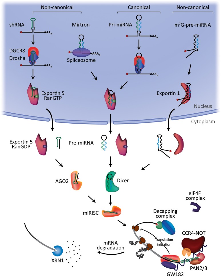
- Canonical miRNA biogenesis involves pri-miRNA processing by Drosha/DGCR8, export by XPO5/RanGTP, Dicer cleavage into a mature duplex, and loading of the 5p or 3p strand into AGO1–4; non-canonical pathways include Drosha/DGCR8-independent mirtrons and m7G-capped pre-miRNAs, and Dicer-independent AGO2-dependent processing.
- Most miRNAs bind 3′ UTR miRNA response elements to induce translational repression and mRNA deadenylation, decapping, and degradation via GW182, PAN2/3, CCR4-NOT, DCP2, and XRN1; binding to 5′ UTR/coding regions can silence, promoter binding can induce transcription, and activation occurs under specific conditions such as serum starvation, cell cycle arrest, oocytes, and amino acid starvation.
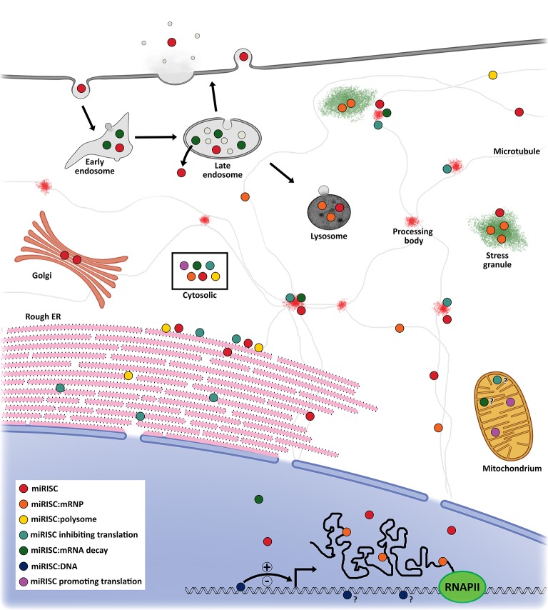
- miRNA action is dynamic: miRISC localizes to rER, P-bodies, stress granules, endosomes, multivesicular bodies, mitochondria, and the nucleus; average polysome occupancy is below 40%, and FANTOM5 data indicate that the top five expressed miRNAs represent on average ~50% of the miRNA pool in a given human cell type, with about half cell-type enriched and one quarter broadly expressed.
- Extracellular miRNAs are highly stable, resisting room-temperature degradation for up to 4 days and conditions such as boiling, freeze-thaw cycles, and high or low pH; they circulate in vesicles or bound to proteins such as AGO2, HDL, and NPM1, and can act as autocrine, paracrine, or endocrine signals, including via Toll-like receptors.
Methods
- Narrative review; no primary experimental data, systematic search strategy, or sample size is reported.
- The main analysis is a thematic synthesis of published literature on canonical and non-canonical biogenesis, miRNA-mediated gene regulation, subcellular dynamics, and extracellular miRNA circulation.
- Data/sample: not applicable; the paper synthesizes prior studies and includes proposed models in figures.
Limitations
- The paper states that understanding of when and how miRNAs regulate transcription is limited, conditions for translational activation need further exploration, and mechanisms of extracellular miRNA secretion and uptake are not well understood.
- The paper notes that many studies use in vitro transfection into immortalized or cancer cell lines, which may not reflect endogenous miRNA functions in vivo, and that chemical tags on miRNAs could affect miRNA:MRE or AGO:miRNA interactions.
- Evident from the text: as a narrative review, it does not systematically assess evidence quality or quantify effect sizes across the cited literature.
Model-written summary. Check it against the paper before relying on it.
Integrity signals
Registry
Cites
Statistics
Figures
Verification
Claims the paper makes
82 Not checked yet · 18 Own interpretation · 6 No basis given · 1 Own method
In the paper
“In serum starved cells, AGO2 and another protein related to the miRNA-protein complex (microRNPs), Fragile-x-mental retardation related protein 1 (FXR1), were associated with AU-rich elements (AREs) at 3′ UTR to activate translation (55).”
§ Overview of MicroRNA Biogenesis, Mechanisms of Actions, and Circulation > Mechanisms of miRNA-mediated gene regulation > MicroRNA-mediated translational activation
What the sources say
- Not checked yetcites [55] Vasudevan, 2007, AU-rich-element-mediated upregulation of translation by FXR1 and Argonaute 2, Cell source
plaincorrelational- Asserted: passes — The authors put it forward as true.
- Checkable: passes — It can be checked against evidence.
- Load-bearing: unclear — It is unclear whether the paper’s conclusions depend on it.
- Based: passes — The paper gives a reason why it holds.
In the paper
“In most cases, miRNAs interact with the 3′ untranslated region (3′ UTR) of target mRNAs to induce mRNA degradation and translational repression.”
§ Overview of MicroRNA Biogenesis, Mechanisms of Actions, and Circulation > Abstract
What the sources say
- Not checked yetcites [14] Ha, 2014, Regulation of microRNA biogenesis, Nat Rev Mol Cell Biol. source
plaindirect- Asserted: passes — The authors put it forward as true.
- Checkable: passes — It can be checked against evidence.
- Load-bearing: unclear — It is unclear whether the paper’s conclusions depend on it.
- Based: passes — The paper gives a reason why it holds.
None of the sources cited for this claim have been checked yet.
In the paper
“Extracellular miRNAs function as chemical messengers to mediate cell-cell communication.”
§ Overview of MicroRNA Biogenesis, Mechanisms of Actions, and Circulation > Abstract
What the sources say
- Not checked yetcites [21] Hayes, 2014, MicroRNAs in cancer: biomarkers, functions and therapy, Trends Mol Med. source
- Not checked yetcites [22] Wang, 2016, MicroRNA as Biomarkers and Diagnostics, J Cell Physiol. source
- Not checked yetcites [23] Huang, 2017, MicroRNAs: biomarkers, diagnostics, and therapeutics, Methods Mol Biol. source
plaindirect- Asserted: passes — The authors put it forward as true.
- Checkable: passes — It can be checked against evidence.
- Load-bearing: passes — The paper’s conclusions depend on it.
- Based: passes — The paper gives a reason why it holds.
In the paper
“In this pathway, pri-miRNAs are transcribed from their genes and then processed into pre-miRNAs by the microprocessor complex, consisting of an RNA binding protein DiGeorge Syndrome Critical Region 8 (DGCR8) and a ribonuclease III enzyme, Drosha (27).”
§ Overview of MicroRNA Biogenesis, Mechanisms of Actions, and Circulation > Biogenesis of miRNAs > The canonical pathway of miRNA biogenesis
What the sources say
- Not checked yetcites [27] Denli, 2004, Processing of primary microRNAs by the Microprocessor complex, Nature source
plaindirect- Asserted: passes — The authors put it forward as true.
- Checkable: passes — It can be checked against evidence.
- Load-bearing: unclear — It is unclear whether the paper’s conclusions depend on it.
- Based: passes — The paper gives a reason why it holds.
In the paper
“while miRNA interaction with promoter region has been reported to induce transcription (45).”
§ Overview of MicroRNA Biogenesis, Mechanisms of Actions, and Circulation > Mechanisms of miRNA-mediated gene regulation
What the sources say
- Not checked yetcites [45] Dharap, 2013, MicroRNA miR-324-3p induces promoter-mediated expression of RelA gene, PLoS ONE source
hedgeddirect- Asserted: passes — The authors put it forward as true.
- Checkable: passes — It can be checked against evidence.
- Load-bearing: unclear — It is unclear whether the paper’s conclusions depend on it.
- Based: passes — The paper gives a reason why it holds.
In the paper
“The minimal miRNA-induced silencing complex (miRISC) consists of the guide strand and AGO (46).”
§ Overview of MicroRNA Biogenesis, Mechanisms of Actions, and Circulation > Mechanisms of miRNA-mediated gene regulation > MicroRNA-mediated gene silencing via miRISC
What the sources say
- Not checked yetcites [46] Kawamata, 2010, Making RISC, Trends Biochem Sci. source
plain- Asserted: passes — The authors put it forward as true.
- Checkable: passes — It can be checked against evidence.
- Load-bearing: unclear — It is unclear whether the paper’s conclusions depend on it.
- Based: passes — The paper gives a reason why it holds.
In the paper
“The miRNA-mediated activation of translation involves AGO2 and FXR1 instead of GW182 (56).”
§ Overview of MicroRNA Biogenesis, Mechanisms of Actions, and Circulation > Mechanisms of miRNA-mediated gene regulation > MicroRNA-mediated translational activation
What the sources say
- Not checked yetcites [56] Truesdell, 2012, MicroRNA-mediated mRNA translation activation in quiescent cells and oocytes involves recruitment of a nuclear microRNP, Sci Rep. source
plaindirect- Asserted: passes — The authors put it forward as true.
- Checkable: passes — It can be checked against evidence.
- Load-bearing: unclear — It is unclear whether the paper’s conclusions depend on it.
- Based: passes — The paper gives a reason why it holds.
In the paper
“The selection of the 5p or 3p strand is based in part on the thermodynamic stability at the 5′ ends of the miRNA duplex or a 5′ U at nucleotide position 1 (34).”
§ Overview of MicroRNA Biogenesis, Mechanisms of Actions, and Circulation > Biogenesis of miRNAs > The canonical pathway of miRNA biogenesis
What the sources say
- Not checked yetcites [34] Khvorova, 2003, Functional siRNAs and miRNAs exhibit strand bias, Cell source
plainconditional- Asserted: passes — The authors put it forward as true.
- Checkable: passes — It can be checked against evidence.
- Load-bearing: unclear — It is unclear whether the paper’s conclusions depend on it.
- Based: passes — The paper gives a reason why it holds.
In the paper
“Both strands derived from the mature miRNA duplex can be loaded into the Argonaute (AGO) family of proteins (AGO1-4 in humans) in an ATP-dependent manner (32).”
§ Overview of MicroRNA Biogenesis, Mechanisms of Actions, and Circulation > Biogenesis of miRNAs > The canonical pathway of miRNA biogenesis
What the sources say
- Not checked yetcites [32] Yoda, 2010, ATP-dependent human RISC assembly pathways, Nat Struct Mol Biol. source
plaindirect- Asserted: passes — The authors put it forward as true.
- Checkable: passes — It can be checked against evidence.
- Load-bearing: unclear — It is unclear whether the paper’s conclusions depend on it.
- Based: passes — The paper gives a reason why it holds.
In the paper
“However, interaction of miRNAs with other regions, including the 5′ UTR, coding sequence, and gene promoters, have also been reported.”
§ Overview of MicroRNA Biogenesis, Mechanisms of Actions, and Circulation > Abstract
What the sources say
- Not checked yetcites [15] Broughton, 2016, Pairing beyond the Seed Supports MicroRNA Targeting Specificity, Mol Cell. source
plaincorrelational- Asserted: passes — The authors put it forward as true.
- Checkable: passes — It can be checked against evidence.
- Load-bearing: unclear — It is unclear whether the paper’s conclusions depend on it.
- Based: passes — The paper gives a reason why it holds.
- It may state two separate claims as one. 29
- Probably not a substantial claim. 13
In the paper
“Under certain conditions, miRNAs can also activate translation or regulate transcription.”
§ Overview of MicroRNA Biogenesis, Mechanisms of Actions, and Circulation > Abstract
What the sources say
- Not checked yetcites [16] Vasudevan, 2012, Posttranscriptional upregulation by microRNAs, Wiley Interdiscip Rev RNA source
plainconditional- Asserted: passes — The authors put it forward as true.
- Checkable: passes — It can be checked against evidence.
- Load-bearing: unclear — It is unclear whether the paper’s conclusions depend on it.
- Based: passes — The paper gives a reason why it holds.
It may state two separate claims as one.
None of the sources cited for this claim have been checked yet.
In the paper
“The formation of a silencing miRISC complex starts with the recruitment of the GW182 family of proteins by miRISC; GW182 provides the scaffolding needed to recruit other effector proteins (52), such as the poly(A)-deadenylase complexes PAN2-PAN3 and CCR4-NOT, following miRNA:target mRNA interaction (50, 53).”
§ Overview of MicroRNA Biogenesis, Mechanisms of Actions, and Circulation > Mechanisms of miRNA-mediated gene regulation > MicroRNA-mediated gene silencing via miRISC
What the sources say
- Not checked yetcites [52] Behm-Ansmant, 2006, mRNA degradation by miRNAs and GW182 requires both CCR4:NOT deadenylase and DCP1:DCP2 decapping complexes, Genes Dev. source
- Not checked yetcites [50] Jonas, 2015, Towards a molecular understanding of microRNA-mediated gene silencing, Nat Rev Genet. source
- Not checked yetcites [53] Christie, 2013, Structure of the PAN3 pseudokinase reveals the basis for interactions with the PAN2 deadenylase and the GW182 proteins, Mol Cell. source
plaindirect- Asserted: passes — The authors put it forward as true.
- Checkable: passes — It can be checked against evidence.
- Load-bearing: unclear — It is unclear whether the paper’s conclusions depend on it.
- Based: passes — The paper gives a reason why it holds.
It may state two separate claims as one.
None of the sources cited for this claim have been checked yet.
In the paper
“An example of such pre-miRNAs is mirtrons, which are produced from the introns of mRNA during splicing (35, 36).”
§ Overview of MicroRNA Biogenesis, Mechanisms of Actions, and Circulation > Biogenesis of miRNAs > Non-canonical miRNA biogenesis pathways
What the sources say
plaindirect- Asserted: passes — The authors put it forward as true.
- Checkable: passes — It can be checked against evidence.
- Load-bearing: fails — The paper’s conclusions may not depend on it.
- Based: passes — The paper gives a reason why it holds.
Probably not a substantial claim. The paper’s conclusions may not depend on it.
In the paper
“On the other hand, Dicer-independent miRNAs are processed by Drosha from endogenous short hairpin RNA (shRNA) transcripts (38). These pre-miRNAs require AGO2 to complete their maturation within the cytoplasm because they are of insufficient length to be Dicer-substrates (38).”
§ Overview of MicroRNA Biogenesis, Mechanisms of Actions, and Circulation > Biogenesis of miRNAs > Non-canonical miRNA biogenesis pathways
What the sources say
- Not checked yetcites [38] Yang, 2010, Conserved vertebrate mir-451 provides a platform for Dicer-independent, Ago2-mediated microRNA biogenesis, Proc Natl Acad Sci USA. source
plaindirect- Asserted: passes — The authors put it forward as true.
- Checkable: passes — It can be checked against evidence.
- Load-bearing: unclear — It is unclear whether the paper’s conclusions depend on it.
- Based: passes — The paper gives a reason why it holds.
It may state two separate claims as one.
In the paper
“Several miRNAs, including let-7, were found to be associated with AGO2 and FXR1 to activate translation during cell cycle arrest, but they inhibit translation in proliferating cells (55).”
§ Overview of MicroRNA Biogenesis, Mechanisms of Actions, and Circulation > Mechanisms of miRNA-mediated gene regulation > MicroRNA-mediated translational activation
What the sources say
- Not checked yetcites [55] Vasudevan, 2007, AU-rich-element-mediated upregulation of translation by FXR1 and Argonaute 2, Cell source
plaindirect- Asserted: passes — The authors put it forward as true.
- Checkable: passes — It can be checked against evidence.
- Load-bearing: unclear — It is unclear whether the paper’s conclusions depend on it.
- Based: passes — The paper gives a reason why it holds.
It may state two separate claims as one.
None of the sources cited for this claim have been checked yet.
In the paper
“About half of all currently identified miRNAs are intragenic and processed mostly from introns and relatively few exons of protein coding genes, while the remaining are intergenic, transcribed independently of a host gene and regulated by their own promoters (13, 24).”
§ Overview of MicroRNA Biogenesis, Mechanisms of Actions, and Circulation > Biogenesis of miRNAs
What the sources say
plain- Asserted: passes — The authors put it forward as true.
- Checkable: passes — It can be checked against evidence.
- Load-bearing: unclear — It is unclear whether the paper’s conclusions depend on it.
- Based: passes — The paper gives a reason why it holds.
It may state two separate claims as one.
A conclusion the paper draws from its own results — it cites no other source for it, so there is none to check. The quote shows it is in the paper.
In the paper
“The interaction of miRNAs with their target genes is dynamic and dependent on many factors, such as subcellular location of miRNAs, the abundancy of miRNAs and target mRNAs, and the affinity of miRNA-mRNA interactions.”
§ Overview of MicroRNA Biogenesis, Mechanisms of Actions, and Circulation > Abstract
plaincorrelational- Asserted: passes — The authors put it forward as true.
- Checkable: passes — It can be checked against evidence.
- Load-bearing: fails — The paper’s conclusions may not depend on it.
- Based: fails — The paper may give no reason why it holds.
Probably not a substantial claim. The paper’s conclusions may not depend on it. The paper may give no reason why it holds.
None of the sources cited for this claim have been checked yet.
In the paper
“Once pre-miRNAs are generated, they are exported to the cytoplasm by an exportin 5 (XPO5)/RanGTP complex and then processed by the RNase III endonuclease Dicer (27, 30).”
§ Overview of MicroRNA Biogenesis, Mechanisms of Actions, and Circulation > Biogenesis of miRNAs > The canonical pathway of miRNA biogenesis
What the sources say
plaindirect- Asserted: passes — The authors put it forward as true.
- Checkable: passes — It can be checked against evidence.
- Load-bearing: unclear — It is unclear whether the paper’s conclusions depend on it.
- Based: passes — The paper gives a reason why it holds.
It may state two separate claims as one.
In the paper
“miRNAs are critical for normal animal development and are involved in a variety of biological processes (18).”
§ Overview of MicroRNA Biogenesis, Mechanisms of Actions, and Circulation > Introduction
What the sources say
- Not checked yetcites [18] Fu, 2013, MicroRNAs in human placental development and pregnancy complications, Int J Mol Sci. source
plaindirect- Asserted: passes — The authors put it forward as true.
- Checkable: passes — It can be checked against evidence.
- Load-bearing: unclear — It is unclear whether the paper’s conclusions depend on it.
- Based: passes — The paper gives a reason why it holds.
It may state two separate claims as one.
None of the sources cited for this claim have been checked yet.
In the paper
“miRNAs have also been shown to interact at genomic loci, where enhancer-derived RNA (eRNA) are transcribed, and increase mRNA levels of adjacent genes by promoting a transcriptionally active chromatin state (67) while altering alternative splicing profiles (64).”
§ Overview of MicroRNA Biogenesis, Mechanisms of Actions, and Circulation > Mechanisms of miRNA-mediated gene regulation > MicroRNA-mediated transcriptional and post-transcriptional gene regulation within the nucleus
What the sources say
- Not checked yetcites [67] Xiao, 2017, MicroRNAs activate gene transcription epigenetically as an enhancer trigger, RNA Biol. source
- Not checked yetcites [64] Allo, 2014, Argonaute-1 binds transcriptional enhancers and controls constitutive and alternative splicing in human cells, Proc Natl Acad Sci USA. source
boosteddirect- Asserted: passes — The authors put it forward as true.
- Checkable: passes — It can be checked against evidence.
- Load-bearing: unclear — It is unclear whether the paper’s conclusions depend on it.
- Based: passes — The paper gives a reason why it holds.
It may state two separate claims as one.
None of the sources cited for this claim have been checked yet.
In the paper
“Cooperation of multiple, proximal MREs on target mRNA has been shown to increase RNAi activity and increase retention times of miRISC complexes on those mRNA, most likely through the three AGO binding sites on the GW182 family proteins (94, 100).”
§ Overview of MicroRNA Biogenesis, Mechanisms of Actions, and Circulation > Dynamics of miRNA actions > A global view of miRNA at the cellular level
What the sources say
hedgeddirect- Asserted: passes — The authors put it forward as true.
- Checkable: passes — It can be checked against evidence.
- Load-bearing: unclear — It is unclear whether the paper’s conclusions depend on it.
- Based: passes — The paper gives a reason why it holds.
It may state two separate claims as one.
None of the sources cited for this claim have been checked yet.
In the paper
“Alternative splicing and alternative polyadenylation affecting 3′ UTRs, and cell type-specific RNA binding proteins that affect target mRNA secondary structures, change the available pool of MREs (63, 68, 69).”
§ Overview of MicroRNA Biogenesis, Mechanisms of Actions, and Circulation > Dynamics of miRNA actions
What the sources say
- Not checked yetcites [63] Bottini, 2017, Post-transcriptional gene silencing mediated by microRNAs is controlled by nucleoplasmic Sfpq, Nat Commun. source
- Not checked yetcites [68] Nam, 2014, Global analyses of the effect of different cellular contexts on microRNA targeting, Mol Cell. source
- Not checked yetcites [69] Blazie, 2017, Alternative polyadenylation directs tissue-specific miRNA targeting in Caenorhabditis elegans Somatic Tissues, Genetics source
plaindirect- Asserted: passes — The authors put it forward as true.
- Checkable: passes — It can be checked against evidence.
- Load-bearing: unclear — It is unclear whether the paper’s conclusions depend on it.
- Based: passes — The paper gives a reason why it holds.
It may state two separate claims as one.
A conclusion the paper draws from its own results — it cites no other source for it, so there is none to check. The quote shows it is in the paper.
In the paper
“Studies have revealed that miRNA-mediated gene regulation is dynamic and helps to buffer gene expression to a steady state.”
§ Overview of MicroRNA Biogenesis, Mechanisms of Actions, and Circulation > Dynamics of miRNA actions
plaindirect- Asserted: passes — The authors put it forward as true.
- Checkable: passes — It can be checked against evidence.
- Load-bearing: unclear — It is unclear whether the paper’s conclusions depend on it.
- Based: unclear — It is unclear why the paper says it holds.
It may state two separate claims as one.
None of the sources cited for this claim have been checked yet.
In the paper
“Nuclear localized miRISC was found to regulate both transcriptional rates and post-transcriptional levels of mRNA (59–61) and associate with euchromatin at gene loci with active transcription (62).”
§ Overview of MicroRNA Biogenesis, Mechanisms of Actions, and Circulation > Mechanisms of miRNA-mediated gene regulation > MicroRNA-mediated transcriptional and post-transcriptional gene regulation within the nucleus
What the sources say
- Not checked yetcites [59] Nishi, 2013, Human TNRC6A is an Argonaute-navigator protein for microRNA-mediated gene silencing in the nucleus, RNA source
- Not checked yetcites [60] Benhamed, 2012, Senescence is an endogenous trigger for microRNA-directed transcriptional gene silencing in human cells, Nat Cell Biol. source
- Not checked yetcites [61] Pitchiaya, 2017, Resolving subcellular miRNA trafficking and turnover at single-molecule resolution, Cell Rep. source
- Not checked yetcites [62] Cernilogar, 2011, Chromatin-associated RNA interference components contribute to transcriptional regulation in Drosophila, Nature source
plaindirect- Asserted: passes — The authors put it forward as true.
- Checkable: passes — It can be checked against evidence.
- Load-bearing: unclear — It is unclear whether the paper’s conclusions depend on it.
- Based: passes — The paper gives a reason why it holds.
It may state two separate claims as one.
In the paper
“Moreover, approximately half of the expressed miRNAs are cell type enriched, one quarter are broadly expressed, and the remaining had low-level expression regardless of cell type.”
§ Overview of MicroRNA Biogenesis, Mechanisms of Actions, and Circulation > Dynamics of miRNA actions > A global view of miRNA at the cellular level
What the sources say
- Not checked yetcites [13] de Rie, 2017, An integrated expression atlas of miRNAs and their promoters in human and mouse, Nat Biotechnol. source
plain- Asserted: passes — The authors put it forward as true.
- Checkable: passes — It can be checked against evidence.
- Load-bearing: unclear — It is unclear whether the paper’s conclusions depend on it.
- Based: passes — The paper gives a reason why it holds.
It may state two separate claims as one.
In the paper
“miRISC has also been found to copurify with polysomes, and the miRISC:mRNA complexes bound to these polysomes were associated with increased levels of translational inhibition and mRNA degradation (83).”
§ Overview of MicroRNA Biogenesis, Mechanisms of Actions, and Circulation > Dynamics of miRNA actions > Subcellular compartmentalization of miRNAs
What the sources say
- Not checked yetcites [83] Pillai, 2005, Inhibition of translational initiation by Let-7 MicroRNA in human cells, Science source
plaincorrelational- Asserted: passes — The authors put it forward as true.
- Checkable: passes — It can be checked against evidence.
- Load-bearing: unclear — It is unclear whether the paper’s conclusions depend on it.
- Based: passes — The paper gives a reason why it holds.
It may state two separate claims as one.
In the paper
“A recent study showed that miRNAs, AGO2, and target mRNAs are associated with rER-membrane-bound polysomes, and this occurs before miRNA-mediated translational repression (70).”
§ Overview of MicroRNA Biogenesis, Mechanisms of Actions, and Circulation > Dynamics of miRNA actions > Subcellular compartmentalization of miRNAs
What the sources say
- Not checked yetcites [70] Barman, 2015, mRNA targeting to endoplasmic reticulum precedes ago protein interaction and microRNA (miRNA)-mediated translation repression in mammalian cells, J Biol Chem. source
plaincorrelational- Asserted: passes — The authors put it forward as true.
- Checkable: passes — It can be checked against evidence.
- Load-bearing: unclear — It is unclear whether the paper’s conclusions depend on it.
- Based: passes — The paper gives a reason why it holds.
It may state two separate claims as one.
None of the sources cited for this claim have been checked yet.
In the paper
“For example, cyclin-dependent kinase inhibitor 1A (CDKN1A), a tumor suppressor that is downregulated in multiple cancers (106), is targeted by at least 28 miRNAs and many of which are upregulated together in cancers where CDKN1A has been implicated (107).”
§ Overview of MicroRNA Biogenesis, Mechanisms of Actions, and Circulation > Dynamics of miRNA actions > A global view of miRNA at the cellular level
What the sources say
plaincorrelational- Asserted: passes — The authors put it forward as true.
- Checkable: passes — It can be checked against evidence.
- Load-bearing: unclear — It is unclear whether the paper’s conclusions depend on it.
- Based: passes — The paper gives a reason why it holds.
It may state two separate claims as one.
In the paper
“Additionally, the degree of polysome occupancy of target mRNAs is dependent on the affinity between miRNA seed region and MRE, but not on miRNA abundance (84).”
§ Overview of MicroRNA Biogenesis, Mechanisms of Actions, and Circulation > Dynamics of miRNA actions > Subcellular compartmentalization of miRNAs
What the sources say
- Not checked yetcites [84] Molotski, 2012, Differential association of microRNAs with polysomes reflects distinct strengths of interactions with their mRNA targets, RNA source
plaindirect- Asserted: passes — The authors put it forward as true.
- Checkable: passes — It can be checked against evidence.
- Load-bearing: unclear — It is unclear whether the paper’s conclusions depend on it.
- Based: passes — The paper gives a reason why it holds.
It may state two separate claims as one.
In the paper
“The extracellular miRNAs can be delivered to target cells and they may act as autocrine, paracrine, and/or endocrine regulators to modulate cellular activities (116).”
§ Overview of MicroRNA Biogenesis, Mechanisms of Actions, and Circulation > Circulation of miRNAs
What the sources say
- Not checked yetcites [116] Iftikhar, 2016, Evidence and potential in vivo functions for biofluid miRNAs: from expression profiling to functional testing: potential roles of extracellular miRNAs as indicators of physiological change and as agents of intercellular information exchange, Bioessays source
hedgedconditional- Asserted: passes — The authors put it forward as true.
- Checkable: passes — It can be checked against evidence.
- Load-bearing: unclear — It is unclear whether the paper’s conclusions depend on it.
- Based: passes — The paper gives a reason why it holds.
It may state two separate claims as one.
In the paper
“It has been shown that the secretion of exosomal miRNAs is mediated by a ceramide-dependent pathway and the secreted miRNAs exert growth regulatory effects in target cells (129).”
§ Overview of MicroRNA Biogenesis, Mechanisms of Actions, and Circulation > Circulation of miRNAs > Secretion and uptake of microRNAs
What the sources say
- Not checked yetcites [129] Kosaka, 2010, Secretory mechanisms and intercellular transfer of microRNAs in living cells, J Biol Chem. source
plaindirect- Asserted: passes — The authors put it forward as true.
- Checkable: passes — It can be checked against evidence.
- Load-bearing: unclear — It is unclear whether the paper’s conclusions depend on it.
- Based: passes — The paper gives a reason why it holds.
It may state two separate claims as one.
None of the sources cited for this claim have been checked yet.
In the paper
“Both cell stress induced by heat shock and translation inhibition following treatment with hippuristanol or cycloheximide induced translocation of miRISC from the nucleus and cytoplasm to transient, cytoplasmic SG (72, 111).”
§ Overview of MicroRNA Biogenesis, Mechanisms of Actions, and Circulation > Dynamics of miRNA actions > A global view of miRNA at the cellular level
What the sources say
- Not checked yetcites [72] Detzer, 2011, Cell stress is related to re-localization of Argonaute 2 and to decreased RNA interference in human cells, Nucleic Acids Res. source
- Not checked yetcites [111] Leung, 2006, Quantitative analysis of Argonaute protein reveals microRNA-dependent localization to stress granules, Proc Natl Acad Sci USA. source
plaindirect- Asserted: passes — The authors put it forward as true.
- Checkable: passes — It can be checked against evidence.
- Load-bearing: unclear — It is unclear whether the paper’s conclusions depend on it.
- Based: passes — The paper gives a reason why it holds.
It may state two separate claims as one.
None of the sources cited for this claim have been checked yet.
In the paper
“While several studies found that the majority of extracellular miRNAs are not associated with exosomes/microvesicles but are instead bound to AGO2 (118, 125), another study reported that extracellular miRNAs are present predominantly in exosomes in human serum and saliva (120).”
§ Overview of MicroRNA Biogenesis, Mechanisms of Actions, and Circulation > Circulation of miRNAs > MicroRNAs in biological fluids
What the sources say
- Not checked yetcites [118] Arroyo, 2011, Argonaute2 complexes carry a population of circulating microRNAs independent of vesicles in human plasma, Proc Natl Acad Sci USA source
- Not checked yetcites [125] Turchinovich, 2011, Characterization of extracellular circulating microRNA, Nucleic Acids Res. source
plaincorrelational- Asserted: passes — The authors put it forward as true.
- Checkable: passes — It can be checked against evidence.
- Load-bearing: fails — The paper’s conclusions may not depend on it.
- Based: passes — The paper gives a reason why it holds.
Probably not a substantial claim. The paper’s conclusions may not depend on it.
None of the sources cited for this claim have been checked yet.
In the paper
“Extracellular miRNAs have also been reported to bind to Toll-like receptors (137), activate downstream signaling events, and eventually lead to biological responses, such as tumor growth and metastasis (138), and neurodegeneration (139).”
§ Overview of MicroRNA Biogenesis, Mechanisms of Actions, and Circulation > Circulation of miRNAs > Secretion and uptake of microRNAs
What the sources say
- Not checked yetcites [137] Fabbri, 2018, MicroRNAs and miRceptors: a new mechanism of action for intercellular communication, Philos Trans R Soc Lond B Biol Sci. source
- Not checked yetcites [138] Fabbri, 2012, MicroRNAs bind to Toll-like receptors to induce prometastatic inflammatory response, Proc Natl Acad Sci USA. source
- Not checked yetcites [139] Lehmann, 2012, An unconventional role for miRNA: let-7 activates Toll-like receptor 7 and causes neurodegeneration, Nat Neurosci. source
hedgeddirect- Asserted: passes — The authors put it forward as true.
- Checkable: passes — It can be checked against evidence.
- Load-bearing: unclear — It is unclear whether the paper’s conclusions depend on it.
- Based: passes — The paper gives a reason why it holds.
It may state two separate claims as one.
In the paper
“For example, exosome mediated transfer of miR-105 from metastatic breast cancer cells to endothelial cells directly targeted a tight junction protein, zonula occludens 1 (ZO-1), and this led to the destruction of the barrier function of endothelium and promoted metastasis (134).”
§ Overview of MicroRNA Biogenesis, Mechanisms of Actions, and Circulation > Circulation of miRNAs > Secretion and uptake of microRNAs
What the sources say
- Not checked yetcites [134] Zhou, 2014, Cancer-secreted miR-105 destroys vascular endothelial barriers to promote metastasis, Cancer Cell source
boosteddirect- Asserted: passes — The authors put it forward as true.
- Checkable: passes — It can be checked against evidence.
- Load-bearing: unclear — It is unclear whether the paper’s conclusions depend on it.
- Based: passes — The paper gives a reason why it holds.
It may state two separate claims as one.
In the paper
“The presence of miRNAs in vesicles or with accompanying proteins is generally thought to protect extracellular miRNAs and increase their stability in the extracellular milieu (120).”
§ Overview of MicroRNA Biogenesis, Mechanisms of Actions, and Circulation > Circulation of miRNAs > MicroRNAs in biological fluids
What the sources say
- Not checked yetcites [120] Gallo, 2012, The majority of microRNAs detectable in serum and saliva is concentrated in exosomes, PLoS ONE source
hedgeddirect- Asserted: passes — The authors put it forward as true.
- Checkable: passes — It can be checked against evidence.
- Load-bearing: unclear — It is unclear whether the paper’s conclusions depend on it.
- Based: passes — The paper gives a reason why it holds.
It may state two separate claims as one.
In the paper
“This process has been reported to be dependent on clathrin, but not on caveolae or lipid rafts in PC12 cells (142).”
§ Overview of MicroRNA Biogenesis, Mechanisms of Actions, and Circulation > Circulation of miRNAs > Secretion and uptake of microRNAs
What the sources say
- Not checked yetcites [142] Tian, 2014, Exosome uptake through clathrin-mediated endocytosis and macropinocytosis and mediating miR-21 delivery, J Biol Chem. source
plaindirect- Asserted: passes — The authors put it forward as true.
- Checkable: passes — It can be checked against evidence.
- Load-bearing: unclear — It is unclear whether the paper’s conclusions depend on it.
- Based: passes — The paper gives a reason why it holds.
It may state two separate claims as one.
In the paper
“In neuroendocrine cells, miRNAs in large dense-core vesicles (LDCVs) are released by exocytosis through vesicle fusion, and this process is mediated by the SNARE complex and accelerated by Ca2+ stimulus (131).”
§ Overview of MicroRNA Biogenesis, Mechanisms of Actions, and Circulation > Circulation of miRNAs > Secretion and uptake of microRNAs
What the sources say
- Not checked yetcites [131] Gumurdu, 2017, MicroRNA exocytosis by large dense-core vesicle fusion, Sci Rep. source
plaindirect- Asserted: passes — The authors put it forward as true.
- Checkable: passes — It can be checked against evidence.
- Load-bearing: unclear — It is unclear whether the paper’s conclusions depend on it.
- Based: passes — The paper gives a reason why it holds.
It may state two separate claims as one.
In the paper
“another study reported that extracellular miRNAs are present predominantly in exosomes in human serum and saliva (120).”
§ Overview of MicroRNA Biogenesis, Mechanisms of Actions, and Circulation > Circulation of miRNAs > MicroRNAs in biological fluids
What the sources say
- Not checked yetcites [120] Gallo, 2012, The majority of microRNAs detectable in serum and saliva is concentrated in exosomes, PLoS ONE source
plaincorrelational- Asserted: passes — The authors put it forward as true.
- Checkable: passes — It can be checked against evidence.
- Load-bearing: fails — The paper’s conclusions may not depend on it.
- Based: passes — The paper gives a reason why it holds.
Probably not a substantial claim. The paper’s conclusions may not depend on it.
None of the sources cited for this claim have been checked yet.
In the paper
“Two populations of extracellular miRNAs exist in biological fluids. One can be found in vesicles such as exosomes, microvesicles, and apoptotic bodies (116, 120) while the other is associated with proteins, especially AGO2 (120, 125).”
§ Overview of MicroRNA Biogenesis, Mechanisms of Actions, and Circulation > Circulation of miRNAs > MicroRNAs in biological fluids
What the sources say
- Not checked yetcites [116] Iftikhar, 2016, Evidence and potential in vivo functions for biofluid miRNAs: from expression profiling to functional testing: potential roles of extracellular miRNAs as indicators of physiological change and as agents of intercellular information exchange, Bioessays source
- Not checked yetcites [120] Gallo, 2012, The majority of microRNAs detectable in serum and saliva is concentrated in exosomes, PLoS ONE source
- Not checked yetcites [125] Turchinovich, 2011, Characterization of extracellular circulating microRNA, Nucleic Acids Res. source
plaincorrelational- Asserted: passes — The authors put it forward as true.
- Checkable: passes — It can be checked against evidence.
- Load-bearing: unclear — It is unclear whether the paper’s conclusions depend on it.
- Based: passes — The paper gives a reason why it holds.
It may state two separate claims as one.
In the paper
“Moreover, exosomes from umbilical cord blood were found to be enriched in miR-21-3p, which promoted the proliferation and migration of fibroblasts, and induced the angiogenic activities of endothelial cells, leading to accelerated wound healing (135).”
§ Overview of MicroRNA Biogenesis, Mechanisms of Actions, and Circulation > Circulation of miRNAs > Secretion and uptake of microRNAs
What the sources say
- Not checked yetcites [135] Hu, 2018, Exosomes from human umbilical cord blood accelerate cutaneous wound healing through miR-21-3p-mediated promotion of angiogenesis and fibroblast function, Theranostics source
plaindirect- Asserted: passes — The authors put it forward as true.
- Checkable: passes — It can be checked against evidence.
- Load-bearing: unclear — It is unclear whether the paper’s conclusions depend on it.
- Based: passes — The paper gives a reason why it holds.
It may state two separate claims as one.
In the paper
“In a study by Wang et al. (110), upon serum starvation of multiple human cell lines, there was an immediate exportation of stable, vesicular or vesicle-free miRNA and a concomitant decrease of intracellular miRNA levels (110).”
§ Overview of MicroRNA Biogenesis, Mechanisms of Actions, and Circulation > Dynamics of miRNA actions > A global view of miRNA at the cellular level
What the sources say
- Not checked yetcites [110] Wang, 2010, Export of microRNAs and microRNA-protective protein by mammalian cells, Nucleic Acids Res. source
plaindirect- Asserted: passes — The authors put it forward as true.
- Checkable: passes — It can be checked against evidence.
- Load-bearing: unclear — It is unclear whether the paper’s conclusions depend on it.
- Based: passes — The paper gives a reason why it holds.
It may state two separate claims as one.
A conclusion the paper draws from its own results — it cites no other source for it, so there is none to check. The quote shows it is in the paper.
In the paper
“Mechanisms by which miRNAs are secreted and taken up by cells are not well understood and require further investigation.”
§ Overview of MicroRNA Biogenesis, Mechanisms of Actions, and Circulation > Concluding remarks
plain- Asserted: passes — The authors put it forward as true.
- Checkable: passes — It can be checked against evidence.
- Load-bearing: passes — The paper’s conclusions depend on it.
- Based: passes — The paper gives a reason why it holds.
Probably not a substantial claim.
A conclusion the paper draws from its own results — it cites no other source for it, so there is none to check. The quote shows it is in the paper.
In the paper
“It is now clear that miRNAs are powerful gene regulators, and that they not only help control mRNA stability and translation but are also involved in transcription.”
§ Overview of MicroRNA Biogenesis, Mechanisms of Actions, and Circulation > Concluding remarks
boosted- Asserted: passes — The authors put it forward as true.
- Checkable: passes — It can be checked against evidence.
- Load-bearing: passes — The paper’s conclusions depend on it.
- Based: passes — The paper gives a reason why it holds.
Probably not a substantial claim.
A conclusion the paper draws from its own results — it cites no other source for it, so there is none to check. The quote shows it is in the paper.
In the paper
“The assaying of miRNA activity within a test tube may not be recapitulated within the cellular environment and thus should be viewed with caution.”
§ Overview of MicroRNA Biogenesis, Mechanisms of Actions, and Circulation > Concluding remarks
hedged- Asserted: passes — The authors put it forward as true.
- Checkable: passes — It can be checked against evidence.
- Load-bearing: passes — The paper’s conclusions depend on it.
- Based: passes — The paper gives a reason why it holds.
Probably not a substantial claim.
A conclusion the paper draws from its own results — it cites no other source for it, so there is none to check. The quote shows it is in the paper.
In the paper
“Also, the addition of chemical tags to miRNA could also impact miRNA:MRE interaction or AGO:miRNA interactions, specifically with 5′ and 3′ miRNA nucleotide modifications.”
§ Overview of MicroRNA Biogenesis, Mechanisms of Actions, and Circulation > Concluding remarks
hedgedconditional- Asserted: passes — The authors put it forward as true.
- Checkable: passes — It can be checked against evidence.
- Load-bearing: fails — The paper’s conclusions may not depend on it.
- Based: fails — The paper may give no reason why it holds.
Probably not a substantial claim. The paper’s conclusions may not depend on it. The paper may give no reason why it holds.
The paper states this without a source or data behind it here, so there is nothing to check it against. The quote shows it is in the paper.
In the paper
“miRNAs regulate the activity of host cells, and they are also secreted and transferred to recipient cells.”
§ Overview of MicroRNA Biogenesis, Mechanisms of Actions, and Circulation > Concluding remarks
plaindirect- Asserted: passes — The authors put it forward as true.
- Checkable: passes — It can be checked against evidence.
- Load-bearing: passes — The paper’s conclusions depend on it.
- Based: passes — The paper gives a reason why it holds.
It may state two separate claims as one.
A conclusion the paper draws from its own results — it cites no other source for it, so there is none to check. The quote shows it is in the paper.
In the paper
“However, our understanding of when and how miRNAs can exert regulatory effects on transcription is limited.”
§ Overview of MicroRNA Biogenesis, Mechanisms of Actions, and Circulation > Concluding remarks
plain- Asserted: passes — The authors put it forward as true.
- Checkable: passes — It can be checked against evidence.
- Load-bearing: passes — The paper’s conclusions depend on it.
- Based: fails — The paper may give no reason why it holds.
Probably not a substantial claim. The paper may give no reason why it holds.
A conclusion the paper draws from its own results — it cites no other source for it, so there is none to check. The quote shows it is in the paper.
In the paper
“Many factors contribute to the activity of miRNAs, including subcellular location, miRNA/mRNA abundance, miRNA:MRE affinity, cell type/state, and the availability of various miRISC components.”
§ Overview of MicroRNA Biogenesis, Mechanisms of Actions, and Circulation > Concluding remarks
plaincorrelational- Asserted: passes — The authors put it forward as true.
- Checkable: passes — It can be checked against evidence.
- Load-bearing: passes — The paper’s conclusions depend on it.
- Based: fails — The paper may give no reason why it holds.
Probably not a substantial claim. The paper may give no reason why it holds.
A conclusion the paper draws from its own results — it cites no other source for it, so there is none to check. The quote shows it is in the paper.
In the paper
“Cytosolic miRISC components shuttle between different compartments.”
§ Overview of MicroRNA Biogenesis, Mechanisms of Actions, and Circulation > Concluding remarks
plain- Asserted: passes — The authors put it forward as true.
- Checkable: passes — It can be checked against evidence.
- Load-bearing: fails — The paper’s conclusions may not depend on it.
- Based: fails — The paper may give no reason why it holds.
Probably not a substantial claim. The paper’s conclusions may not depend on it. The paper may give no reason why it holds.
What the paper did — it cites no source for this, so there is none to check. The quote shows it is in the paper.
In the paper
“However, most studies conducted so far were done in vitro using co-culture of different cell types.”
§ Overview of MicroRNA Biogenesis, Mechanisms of Actions, and Circulation > Concluding remarks
plain- Asserted: passes — The authors put it forward as true.
- Checkable: passes — It can be checked against evidence.
- Load-bearing: passes — The paper’s conclusions depend on it.
- Based: fails — The paper may give no reason why it holds.
Probably not a substantial claim. The paper may give no reason why it holds.
A conclusion the paper draws from its own results — it cites no other source for it, so there is none to check. The quote shows it is in the paper.
In the paper
“miRNAs in the nucleus play a role in regulating transcription and alternative splicing.”
§ Overview of MicroRNA Biogenesis, Mechanisms of Actions, and Circulation > Concluding remarks
plaindirect- Asserted: passes — The authors put it forward as true.
- Checkable: passes — It can be checked against evidence.
- Load-bearing: fails — The paper’s conclusions may not depend on it.
- Based: fails — The paper may give no reason why it holds.
Probably not a substantial claim. The paper’s conclusions may not depend on it. The paper may give no reason why it holds.
The paper relies on this without support.
The first and last miRNA nucleotides clamp the miRNA within AGO proteins.
“The first and last miRNA nucleotides clamp the miRNA within AGO proteins”
Claims other papers make about this one
Each is checked against this paper’s own text: does it say what the citing paper says it does?
The source backs part of the claim, not all of it — or backs it for a different population, condition or comparison.
In the citing paper
“Additionally, translation of any synthetic mRNA released into the cytoplasm may be blocked by micro ribonucleic acid (microRNA) [44].”
From Relative risk reduction: Misinformative measure in clinical trials and COVID-19 vaccine efficacy (2022) · cites this paper as [44]
Experimental — accuracy not yet measuredThe source backs part of the claim, not all of it — or backs it for a different population, condition or comparison.
In the citing paper
“A large volume of evidence implicates microRNAs playing crucial roles in multiple biological functions such as proliferation, differentiation, or apoptosis.3,21”
From MiR-128-3p – a gray eminence of the human central nervous system (2024) · cites this paper as [3]
Experimental — accuracy not yet measuredIn the citing paper
“Although this study is focused on identifying CREs, there are other non-coding regulatory mechanisms that could alter gene expression in AD including microRNAs (miRNAs),90 long non-coding RNAs (lncRNAs),91 transposable elements,92 etc., that are not assessed.”
From Single nucleus multiomics identifies ZEB1 and MAFB as candidate regulators of Alzheimer’s disease-specific cis-regulatory elements (2023) · cites this paper as [90]
Self-check
Percentages, sums and reported differences re-computed from the paper’s own numbers.
Each contradiction between two passages listed here was confirmed by an independent check.
Rated minor: it would not touch a result — a value no result rests on, a p-value that stays on the same side of the significance level, or a conflict that is clerical, resolved elsewhere in the paper, or outside its own findings.
In the paper
§ Overview of MicroRNA Biogenesis, Mechanisms of Actions, and Circulation > Mechanisms of miRNA-mediated gene regulation > MicroRNA-mediated transcriptional and post-transcriptional gene regulation within the nucleus
“A more recent study by Miao et al. (66) found nuclear miR-522 interacting with a DNA cruciform structure (a stem-loop on sense and antisense DNA strands) within the promoter of CYP2E1 and suppressing its transcription (66).”
§ Overview of MicroRNA Biogenesis, Mechanisms of Actions, and Circulation > References
“66. Miao L Yao H Li C Pu M Yao X Yang H. A dual inhibition: microRNA-552 suppresses both transcription and translation of cytochrome P450 2E1.”
Rated minor: it would not touch a result — a value no result rests on, a p-value that stays on the same side of the significance level, or a conflict that is clerical, resolved elsewhere in the paper, or outside its own findings.
In the paper
§ Overview of MicroRNA Biogenesis, Mechanisms of Actions, and Circulation > Circulation of miRNAs > Secretion and uptake of microRNAs
“Recently, it was demonstrated that atheroprotective laminar shear stress induced the release of vesicle-free miR-126-3p and other miRNAs, as well as AGO2, from endothelial cells by activating vesicle-associated membrane protein 3 (VAMP3) and synaptosomal-associated protein 23 (SNAP23) (130).”
§ Overview of MicroRNA Biogenesis, Mechanisms of Actions, and Circulation > References
“130. Zhu JJ Liu YF Zhang YP Zhao CR Yao WJ Li YS. VAMP3 and SNAP23 mediate the disturbed flow-induced endothelial microRNA secretion and smooth muscle hyperplasia.”
Citations
Cites
The C. elegans heterochronic gene lin-4 encodes small RNAs with antisense complementarity to lin-14
Lee, R. C., Feinbaum, R. L., & Ambros, V. (1993) · Cell · doi:10.1016/0092-8674(93)90529-y · Cited by 12,948
Circulating microRNAs as stable blood-based markers for cancer detection
Mitchell, P. S., Parkin, R. K., Kroh, E. M., Fritz, B. R., Wyman, S. K., Pogosova-Agadjanyan, E. L., Peterson, A., Noteboom, J., O'Briant, K. C., Allen, A., et al. (2008) · Proceedings of the National Academy of Sciences · doi:10.1073/pnas.0804549105 · Cited by 7,969
Global quantification of mammalian gene expression control
Schwanhäusser, B., Busse, D., Li, N., Dittmar, G., Schuchhardt, J., Wolf, J., Chen, W., & Selbach, M. (2011) · Nature · doi:10.1038/nature10098 · Cited by 6,566 · cited 1×
“for any given gene is the rate of translation, followed by mRNA levels (93).”
BackgroundRegulation of microRNA biogenesis
Ha, M., & Kim, V. N. (2014) · Nature Reviews Molecular Cell Biology · doi:10.1038/nrm3838 · Cited by 5,462
Chen, X., Ba, Y., Ma, L., Cai, X., Yin, Y., Wang, K., Guo, J., Zhang, Y., Chen, J., Guo, X., et al. (2008) · Cell Research · doi:10.1038/cr.2008.282 · Cited by 4,560
Wightman, B., Ha, I., & Ruvkun, G. (1993) · Cell · doi:10.1016/0092-8674(93)90530-4 · Cited by 4,292
Silencing of microRNAs in vivo with ‘antagomirs’
Krützfeldt, J., Rajewsky, N., Braich, R., Rajeev, K. G., Tuschl, T., Manoharan, M., & Stoffel, M. (2005) · Nature · doi:10.1038/nature04303 · Cited by 3,949
Arroyo, J. D., Chevillet, J. R., Kroh, E. M., Ruf, I. K., Pritchard, C. C., Gibson, D. F., Mitchell, P. S., Bennett, C. F., Pogosova-Agadjanyan, E. L., Stirewalt, D. L., et al. (2011) · Proceedings of the National Academy of Sciences · doi:10.1073/pnas.1019055108 · Cited by 3,294
Processing of primary microRNAs by the Microprocessor complex
Denli, A. M., Tops, B. B. J., Plasterk, R. H. A., Ketting, R. F., & Hannon, G. J. (2004) · Nature · doi:10.1038/nature03049 · Cited by 2,784
MicroRNAs are transported in plasma and delivered to recipient cells by high-density lipoproteins
Vickers, K. C., Palmisano, B. T., Shoucri, B. M., Shamburek, R. D., & Remaley, A. T. (2011) · Nature Cell Biology · doi:10.1038/ncb2210 · Cited by 2,741
p21 in cancer: Intricate networks and multiple activities
Abbas, T., & Dutta, A. (2009) · Nature Reviews Cancer · doi:10.1038/nrc2657 · Cited by 2,712
Conservation of the sequence and temporal expression of let-7 heterochronic regulatory RNA
Pasquinelli, A. E., Reinhart, B. J., Slack, F. J., Martindale, M. Q., Kuroda, M. I., Maller, B., Hayward, D. C., Ball, E. E., Degnan, B. M., Müller, P., et al. (2000) · Nature · doi:10.1038/35040556 · Cited by 2,671
Functional siRNAs and miRNAs exhibit strand bias
Khvorova, A., Reynolds, A., & Jayasena, S. D. (2003) · Cell · doi:10.1016/s0092-8674(03)00801-8 · Cited by 2,645
The MicroRNA Spectrum in 12 Body Fluids
Weber, J. A., Baxter, D. H., Zhang, S., Huang, D. Y., How Huang, K., Jen Lee, M., Galas, D. J., & Wang, K. (2010) · Clinical Chemistry · doi:10.1373/clinchem.2010.147405 · Cited by 2,477
Gene silencing by microRNAs: contributions of translational repression and mRNA decay
Huntzinger, E., & Izaurralde, E. (2011) · Nature Reviews Genetics · doi:10.1038/nrg2936 · Cited by 2,339
The Drosha-DGCR8 complex in primary microRNA processingPreprint
Han, J., Lee, Y., Yeom, K.-H., Kim, Y. K., Jin, H., & Kim, V. N. (2004) · Genes & Development · doi:10.1101/gad.1262504 · Cited by 2,320
MicroRNAs in cancer: biomarkers, functions and therapy
Hayes, J., Peruzzi, P. P., & Lawler, S. (2014) · Trends in Molecular Medicine · doi:10.1016/j.molmed.2014.06.005 · Cited by 2,049
Characterization of extracellular circulating microRNA
Turchinovich, A., Weiz, L., Langheinz, A., & Burwinkel, B. (2011) · Nucleic Acids Research · doi:10.1093/nar/gkr254 · Cited by 1,941
Secretory Mechanisms and Intercellular Transfer of MicroRNAs in Living Cells
Kosaka, N., Iguchi, H., Yoshioka, Y., Takeshita, F., Matsuki, Y., & Ochiya, T. (2010) · Journal of Biological Chemistry · doi:10.1074/jbc.m110.107821 · Cited by 1,934
Towards a molecular understanding of microRNA-mediated gene silencing
Jonas, S., & Izaurralde, E. (2015) · Nature Reviews Genetics · doi:10.1038/nrg3965 · Cited by 1,872
miRTarBase update 2018: a resource for experimentally validated microRNA-target interactions
Chou, C., Shrestha, S., Yang, C.-D., Chang, N.-W., Lin, Y., Liao, K., Huang, W.-C., Sun, T.-H., Tu, S.-J., Lee, W.-H., et al. (2017) · Nucleic Acids Research · doi:10.1093/nar/gkx1067 · Cited by 1,812
Principles and Properties of Stress Granules
Protter, D. S. W., & Parker, R. (2016) · Trends in Cell Biology · doi:10.1016/j.tcb.2016.05.004 · Cited by 1,738
Intronic microRNA precursors that bypass Drosha processing
Ruby, J. G., Jan, C. H., & Bartel, D. P. (2007) · Nature · doi:10.1038/nature05983 · Cited by 1,722
Roles for MicroRNAs in Conferring Robustness to Biological Processes
Ebert, M. S., & Sharp, P. A. (2012) · Cell · doi:10.1016/j.cell.2012.04.005 · Cited by 1,564
Inhibition of Translational Initiation by Let-7 MicroRNA in Human Cells
Pillai, R. S., Bhattacharyya, S. N., Artus-Revel, C. G., Zoller, T., Cougot, N., Basyuk, E. I., Bertrand, É., & Filipowicz, W. (2005) · Science · doi:10.1126/science.1115079 · Cited by 1,489
Cancer-secreted miR-105 destroys vascular endothelial barriers to promote metastasis
Zhou, W., Fong, M. Y., Min, Y., Somlo, G., Liu, L., Palomares, M. R., Yu, Y., Chow, A., O’Connor, S. T. F., Chin, A. R., et al. (2014) · Cancer Cell · doi:10.1016/j.ccr.2014.03.007 · Cited by 1,476
MicroRNAs bind to Toll-like receptors to induce prometastatic inflammatory response
Fabbri, M., Paone, A., Calore, F., Galli, R., Gaudio, E., Santhanam, R., Lovat, F., Fadda, P., Mao, C., Nuovo, G. J., et al. (2012) · Proceedings of the National Academy of Sciences · doi:10.1073/pnas.1209414109 · Cited by 1,434
N6-methyladenosine marks primary microRNAs for processing
Alarcón, C. R., Lee, H., Goodarzi, H., Halberg, N., & Tavazoie, S. F. (2015) · Nature · doi:10.1038/nature14281 · Cited by 1,420
MicroRNA-10a binds the 5’UTR of ribosomal protein mRNAs and enhances their translation
Ørom, U. A., Nielsen, F. C., & Lund, A. H. (2008) · Molecular Cell · doi:10.1016/j.molcel.2008.05.001 · Cited by 1,384
P bodies and the control of mRNA translation and degradation
Parker, R., & Sheth, U. (2007) · Molecular Cell · doi:10.1016/j.molcel.2007.02.011 · Cited by 1,335
MicroRNA-dependent localization of targeted mRNAs to mammalian P-bodies
Liu, J., Valencia-Sanchez, M. A., Hannon, G. J., & Parker, R. (2005) · Nature Cell Biology · doi:10.1038/ncb1274 · Cited by 1,234
A dicer-independent miRNA biogenesis pathway that requires Ago catalysis
Cheloufi, S., Dos Santos, C. O., Chong, M. M. W., & Hannon, G. J. (2010) · Nature · doi:10.1038/nature09092 · Cited by 1,149
Behm‐Ansmant, I., Rehwinkel, J., Doerks, T., Stark, A., Bork, P., & Izaurralde, E. (2006) · Genes & Development · doi:10.1101/gad.1424106 · Cited by 1,113
The majority of microRNAs detectable in serum and saliva is concentrated in exosomes
Gallo, A., Tandon, M., Alevizos, I., & Illei, G. G. (2012) · PLoS ONE · doi:10.1371/journal.pone.0030679 · Cited by 1,060
Single Processing Center Models for Human Dicer and Bacterial RNase III
Zhang, H., Kolb, F. A., Jaśkiewicz, Ł., Westhof, É., & Filipowicz, W. (2004) · Cell · doi:10.1016/j.cell.2004.06.017 · Cited by 1,043
Gibbings, D., Ciaudo, C., Erhardt, M., & Voinnet, O. (2009) · Nature Cell Biology · doi:10.1038/ncb1929 · Cited by 1,037
Export of microRNAs and microRNA-protective protein by mammalian cells
Wang, K., Zhang, S., Weber, J., Baxter, D., & Galas, D. J. (2010) · Nucleic Acids Research · doi:10.1093/nar/gkq601 · Cited by 1,002
Babiarz, J., Ruby, J. G., Wang, Y., Bartel, D. P., & Blelloch, R. H. (2008) · Genes & Development · doi:10.1101/gad.1705308 · Cited by 964
Cogswell, J. P., Ward, J., Taylor, I. A., Waters, M., Shi, Y., Cannon, B., Kelnar, K., Kemppainen, J., Brown, D., Chen, C., et al. (2008) · Journal of Alzheimer s Disease · doi:10.3233/jad-2008-14103 · Cited by 943
Circular RNAs: analysis, expression and potential functions
Barrett, S. P., & Salzman, J. (2016) · Development · doi:10.1242/dev.128074 · Cited by 859
MicroRNA as Biomarkers and Diagnostics
Wang, J., Chen, J., & Sen, S. (2016) · Journal of Cellular Physiology · doi:10.1002/jcp.25056 · Cited by 793
An unconventional role for miRNA: let-7 activates toll-like receptor 7 and causes neurodegeneration
Lehmann, S. M., Krüger, C., Park, B., Derkow, K., Rosenberger, K., Baumgart, J., Trimbuch, T., Eom, G., Hinz, M., Kaul, D., et al. (2012) · Nature Neuroscience · doi:10.1038/nn.3113 · Cited by 786
Processing of intronic microRNAs
Kim, Y., & Kim, V. N. (2007) · The EMBO Journal · doi:10.1038/sj.emboj.7601512 · Cited by 754
Tian, T., Zhu, Y.-L., Zhou, Y.-Y., Liang, G.-F., Wang, Y.-Y., Hu, F.-H., & Xiao, Z.-D. (2014) · Journal of Biological Chemistry · doi:10.1074/jbc.m114.588046 · Cited by 726
P-body formation is a consequence, not the cause, of RNA-mediated gene silencing
Eulalio, A., Behm-Ansmant, I., Schweizer, D., & Izaurralde, E. (2007) · Molecular and Cellular Biology · doi:10.1128/mcb.00128-07 · Cited by 710
Yang, M., Chen, J., Su, F., Yu, B., Su, F., Lin, L., Liu, Y., Huang, J.-D., & Song, E. (2011) · Molecular Cancer · doi:10.1186/1476-4598-10-117 · Cited by 693
Secreted microRNAs: a new form of intercellular communication
Chen, X., Liang, H., Zhang, J., Zen, K., & Zhang, C.-Y. (2012) · Trends in Cell Biology · doi:10.1016/j.tcb.2011.12.001 · Cited by 690
AU-rich-element-mediated upregulation of translation by FXR1 and Argonaute 2
Vasudevan, S., & Steitz, J. A. (2007) · Cell · doi:10.1016/j.cell.2007.01.038 · Cited by 656
Molecular Evolution of a MicroRNA Cluster
Andrea Tanzer, A., & Stadler, P. F. (2004) · Journal of Molecular Biology · doi:10.1016/j.jmb.2004.03.065 · Cited by 650
Target RNA–Directed Trimming and Tailing of Small Silencing RNAs
Ameres, S. L., Horwich, M. D., Hung, J.-H., Xu, J., Ghildiyal, M., Weng, Z., & Zamore, P. D. (2010) · Science · doi:10.1126/science.1187058 · Cited by 628
Immune-related MicroRNAs are Abundant in Breast Milk Exosomes
Zhou, Q. G., Li, M., Wang, X., Li, Q., Wang, T., Zhu, Q., Zhou, X., Wang, X., Gao, X., & Li, X. (2012) · International Journal of Biological Sciences · doi:10.7150/ijbs.8.118 · Cited by 615
Forman, J. J., Legesse-Miller, A., & Coller, H. A. (2008) · Proceedings of the National Academy of Sciences · doi:10.1073/pnas.0803230105 · Cited by 581
Hu, Y., Rao, S.-S., Wang, Z.-X., Cao, J., Tan, Y.-J., Luo, J., Li, H.-M., Zhang, W.-S., Chen, C.-Y., & Xie, H. (2018) · Theranostics · doi:10.7150/thno.21234 · Cited by 574
The role of microRNAs in human diseases
Tüfekci, K. U., Öner, M. G., Meuwissen, R. L. J., & Genç, Ş. (2014) · Methods in molecular biology · doi:10.1007/978-1-62703-748-8_3 · Cited by 566
An integrated expression atlas of miRNAs and their promoters in human and mouse
The FANTOM Consortium, de Rie, D., Abugessaisa, I., Alam, T., Arner, E., Arner, P., Ashoor, H., Åström, G., Babina, M., Bertin, N., et al. (2017) · Nature Biotechnology · doi:10.1038/nbt.3947 · Cited by 561
Mutations that lead to reiterations in the cell lineages of C. elegans.
Chalfie, M., Horvitz, H., & Sulston, J. (1981) · Cell · doi:10.1016/0092-8674(81)90501-8 · Cited by 541
Posttranscriptional Upregulation by MicroRNAs
Vasudevan, S. (2011) · Wiley Interdisciplinary Reviews - RNA · doi:10.1002/wrna.121 · Cited by 536
Extracellular miRNAs: the mystery of their origin and function
Turchinovich, A. A., Weiz, L., & Burwinkel, B. (2012) · Trends in Biochemical Sciences · doi:10.1016/j.tibs.2012.08.003 · Cited by 534
A High-Resolution Structure of the Pre-microRNA Nuclear Export Machinery
岡田, 千., Yamashita, E., Lee, S. J., Shibata, S., Katahira, J., Nakagawa, A., Yoneda, Y., & Tomitake Tsukihara, A. (2009) · Science · doi:10.1126/science.1178705 · Cited by 525
MicroRNA Directly Enhances Mitochondrial Translation during Muscle Differentiation
Zhang, X., Zuo, X., Yang, B., Li, Z., Xue, Y., Zhou, Y., Huang, J. S., Zhao, X., Zhou, J., Yan, Y., et al. (2014) · Cell · doi:10.1016/j.cell.2014.05.047 · Cited by 505
Pairing beyond the Seed Supports MicroRNA Targeting Specificity
Broughton, J. P., Lovci, M. T., Huang, J. L., Yeo, G. W., & Pasquinelli, A. E. (2016) · Molecular Cell · doi:10.1016/j.molcel.2016.09.004 · Cited by 487
Yang, J.-S., Maurin, T., Robine, N., Rasmussen, K. D., Jeffrey, K. L., Chandwani, R., Papapetrou, E. P., Sadelain, M., O’Carroll, D., & Lai, E. C. (2010) · Proceedings of the National Academy of Sciences · doi:10.1073/pnas.1006432107 · Cited by 481
Kawamata, T., & Tomari, Y. (2010) · Trends in Biochemical Sciences · doi:10.1016/j.tibs.2010.03.009 · Cited by 481
MicroRNA history: Discovery, recent applications, and next frontiers
Almeida, M. I., Reis, R. M., & Calin, G. A. (2011) · Mutation Research - Fundamental and Molecular Mechanisms of Mutagenesis · doi:10.1016/j.mrfmmm.2011.03.009 · Cited by 466
da Silveira, J. C., Veeramachaneni, D. N. R., Winger, Q. A., Carnevale, E. M., & Bouma, G. J. (2011) · Biology of Reproduction · doi:10.1095/biolreprod.111.093252 · Cited by 450
Serum exosomal microRNAs as novel biomarkers for hepatocellular carcinoma
Sohn, W., Kim, J., Kang, S. H., Yang, S. R., Cho, J., Cho, H. C., Shim, S. G., & Paik, Y. (2015) · Experimental & Molecular Medicine · doi:10.1038/emm.2015.68 · Cited by 434
Horvitz, H., & Sulston, J. (1980) · Genetics · doi:10.1093/genetics/96.2.435 · Cited by 429
HDL-transferred microRNA-223 regulates ICAM-1 expression in endothelial cells
Tabet, F., Vickers, K. C., Cuesta Torres, L. F., Wiese, C. B., Shoucri, B. M., Lambert, G., Catherinet, C., Prado-Lourenco, L., Levin, M. G., Thacker, S., et al. (2014) · Nature Communications · doi:10.1038/ncomms4292 · Cited by 428
Interplay between miRNAs and human diseases
Paul, P., Chakraborty, A., Sarkar, D., Langthasa, M., Rahman, M., Bari, M., Singha, R. S., Malakar, A. K., & Chakraborty, S. (2018) · Journal of Cellular Physiology · doi:10.1002/jcp.25854 · Cited by 420 · cited 1×
“Aberrant expression of miRNAs is associated with many human diseases (19, 20).”
BackgroundLeung, A. K. L., Calabrese, J. M., & Sharp, P. A. (2006) · Proceedings of the National Academy of Sciences · doi:10.1073/pnas.0608845103 · Cited by 414
Competing endogenous RNAs (ceRNAs): new entrants to the intricacies of gene regulation
Kartha, R. V., & Subramanian, S. (2014) · Frontiers in Genetics · doi:10.3389/fgene.2014.00008 · Cited by 408
Argonaute Divides Its RNA Guide into Domains with Distinct Functions and RNA-Binding Properties
Wee, L. M., Flores-Jasso, C. F., Salomon, W. E., & Zamore, P. D. (2012) · Cell · doi:10.1016/j.cell.2012.10.036 · Cited by 398
MicroRNAs activate gene transcription epigenetically as an enhancer trigger
Xiao, M., Li, J., Li, W., Wang, Y., Wu, F., Xi, Y., Zhang, L., Ding, C., Luo, H., Li, Y., et al. (2016) · RNA Biology · doi:10.1080/15476286.2015.1112487 · Cited by 396
MicroRNA control of protein expression noise
Schmiedel, J. M., Klemm, S. L., Zheng, Y., Sahay, A., Blüthgen, N., Marks, D. S., & van Oudenaarden, A. (2015) · Science · doi:10.1126/science.aaa1738 · Cited by 395
ATP-dependent human RISC assembly pathways
Yoda, M., Kawamata, T., Paroo, Z., Ye, X., Iwasaki, S., Liu, Q., & Tomari, Y. (2010) · Nature Structural & Molecular Biology · doi:10.1038/nsmb.1733 · Cited by 380
RNAi Factors Are Present and Active in Human Cell Nuclei
Gagnon, K. T., Li, L., Chu, Y., Janowski, B. A., & Corey, D. R. (2014) · Cell Reports · doi:10.1016/j.celrep.2013.12.013 · Cited by 372
Multiple microRNAs modulate p21Cip1/Waf1 expression by directly targeting its 3′ untranslated region
Wu, S., Huang, S., Ding, J., Zhao, Y., Liang, L., Liu, T., Zhan, R., & He, X. (2010) · Oncogene · doi:10.1038/onc.2010.34 · Cited by 346
Denzler, R., McGeary, S. E., Title, A. C., Agarwal, V., Bartel, D. P., & Stoffel, M. (2016) · Molecular Cell · doi:10.1016/j.molcel.2016.09.027 · Cited by 340
Global Analyses of the Effect of Different Cellular Contexts on MicroRNA Targeting
Nam, J., Rissland, O. S., Koppstein, D., Abreu‐Goodger, C., Jan, C. H., Agarwal, V., Yıldırım, M. A., Rodríguez, A., & Bartel, D. P. (2014) · Molecular Cell · doi:10.1016/j.molcel.2014.02.013 · Cited by 337
The circular RNA circBIRC6 participates in the molecular circuitry controlling human pluripotency
Yu, C.-Y., Li, T.-C., Wu, Y.-Y., Yeh, C.-H., Chiang, W., Chuang, C.-Y., & Kuo, H.-C. (2017) · Nature Communications · doi:10.1038/s41467-017-01216-w · Cited by 336
A genetic pathway for the specification of the vulval cell lineages of Caenorhabditis elegans
Ferguson, E. L., Sternberg, P. W., & Horvitz, H. R. (1987) · Nature · doi:10.1038/326259a0 · Cited by 334
Pre-microRNA and Mature microRNA in Human Mitochondria
Barrey, É., Saint‐Auret, G., Bonnamy, B., Damas, D., Boyer, O., & Gidrol, X. (2011) · PLoS ONE · doi:10.1371/journal.pone.0020220 · Cited by 317
Mechanisms of control of microRNA biogenesis
Davis‐Dusenbery, B. N., & Hata, A. (2010) · The Journal of Biochemistry · doi:10.1093/jb/mvq096 · Cited by 314
MicroRNAs in Human Placental Development and Pregnancy Complications
Fu, G., Brkić, J., Hayder, H., & Peng, C. (2013) · International Journal of Molecular Sciences · doi:10.3390/ijms14035519 · Cited by 293
Self-citationFrom guide to target: molecular insights into eukaryotic RNA-interference machinery
Ipsaro, J. J., & Joshua-Tor, L. (2015) · Nature Structural & Molecular Biology · doi:10.1038/nsmb.2931 · Cited by 281
Intracellular and extracellular microRNA: An update on localization and biological role
Makarova, J. A., Shkurnikov, M. Y., Wicklein, D., Lange, T., Samatov, T. R., Turchinovich, A. A., & Tonevitsky, A. (2016) · Progress in Histochemistry and Cytochemistry · doi:10.1016/j.proghi.2016.06.001 · Cited by 274
Zhang, J., Fu, W., He, M., et al. (2011) · RNA Biology · doi:10.4161/rna.8.5.16043 · Cited by 270
Evidence for the biogenesis of more than 1,000 novel human microRNAs
Friedländer, M. R., Lizano, E., Houben, A. J. S., Bezdan, D., Báñez-Coronel, M., Kudla, G., Mateu-Huertas, E., Kagerbauer, B., González, J., Chen, K. C., et al. (2014) · Genome biology · doi:10.1186/gb-2014-15-4-r57 · Cited by 265
Mammalian 5′-Capped MicroRNA Precursors that Generate a Single MicroRNA
Xie, M., Li, M., Vilborg, A., Lee, N., Shu, M.-D., Yartseva, V., Šestan, N., & STEITZ, J. A. (2013) · Cell · doi:10.1016/j.cell.2013.11.027 · Cited by 256
Benhamed, M., Herbig, U., Ye, T., Dejean, A., & Bischof, O. (2012) · Nature Cell Biology · doi:10.1038/ncb2443 · Cited by 252
MicroRNAs transfer from human macrophages to hepato-carcinoma cells and inhibit proliferation
Aucher, A., Rudnicka, D., & Davis, D. M. (2013) · The Journal of Immunology · doi:10.4049/jimmunol.1301728 · Cited by 247
Wei, F., Ma, C., Zhou, T., Dong, X., Luo, Q., Geng, L., Ding, L., Zhang, Y., Zhang, L., Li, N., et al. (2017) · Molecular Cancer · doi:10.1186/s12943-017-0694-8 · Cited by 245
Cernilogar, F. M., Onorati, M. C., Kothe, G. O., Burroughs, A. M., Parsi, K. M., Breiling, A., Sardo, F. L., Saxena, A., Miyoshi, K., Siomi, H., et al. (2011) · Nature · doi:10.1038/nature10492 · Cited by 243
MicroRNAs: biomarkers, diagnostics, and therapeutics
Huang, W. (2017) · Methods in molecular biology · doi:10.1007/978-1-4939-7046-9_4 · Cited by 237
Regulation of miRNA strand selection: follow the leader?
Meijer, H. A., Smith, E. M., & Bushell, M. (2014) · Biochemical Society Transactions · doi:10.1042/bst20140142 · Cited by 233
A short history of a short RNA
Lee, R., Feinbaum, R. L., & Ambros, V. (2004) · Cell · doi:10.1016/s0092-8674(04)00035-2 · Cited by 224
Hannafon, B. N., Carpenter, K. J., Berry, W. L., Janknecht, R., Dooley, W. C., & Ding, W.-Q. (2015) · Molecular Cancer · doi:10.1186/s12943-015-0400-7 · Cited by 218
Human Argonaute 2 Has diverse reaction pathways on Target RNAs
Jo, M. H., Shin, S., Jung, S.-R., Kim, E., Song, J.-J., & Hohng, S. (2015) · Molecular Cell · doi:10.1016/j.molcel.2015.04.027 · Cited by 207
Sakha, S., Muramatsu, T., Ueda, K., & Inazawa, J. (2016) · Scientific Reports · doi:10.1038/srep38750 · Cited by 201
Ambros, V., & Horvitz, H. R. (1987) · Genes & Development · doi:10.1101/gad.1.4.398 · Cited by 197
Understanding microRNA-mediated gene regulatory networks through mathematical modelling
Lai, X., Wolkenhauer, O., & Vera, J. (2016) · Nucleic Acids Research · doi:10.1093/nar/gkw550 · Cited by 196
Human TNRC6A is an Argonaute-navigator protein for microRNA-mediated gene silencing in the nucleus
Nishi, K., Nishi, A., Nagasawa, T., & Ui-Tei, K. (2013) · RNA · doi:10.1261/rna.034769.112 · Cited by 188
Activity-dependent spatially localized miRNA maturation in neuronal dendrites
Sambandan, S., Akbalık, G., Kochen, L., Rinne, J. S., Kahlstatt, J., Glock, C., Tushev, G., Alvarez‐Castelao, B., Heckel, A., & Schuman, E. M. (2017) · Science · doi:10.1126/science.aaf8995 · Cited by 187
A direct interaction between DCP1 and XRN1 couples mRNA decapping to 5′ exonucleolytic degradation
Braun, J. E., Truffault, V., Boland, A., Huntzinger, E., Chang, C.-T., Haas, G., Weichenrieder, O., Coles, M. P., & Izaurralde, E. (2012) · Nature Structural & Molecular Biology · doi:10.1038/nsmb.2413 · Cited by 180
Evolutionary Convergence of Pathway-Specific Enzyme Expression Stoichiometry
Lalanne, J., Taggart, J., Guo, M. S., Herzel, L., Schieler, A., & Li, G. (2018) · Cell · doi:10.1016/j.cell.2018.03.007 · Cited by 175
Truesdell, S. S., Mortensen, R. D., Seo, M., Schroeder, J. C., Lee, J. H., LeTonqueze, O., & Vasudevan, S. (2012) · Scientific Reports · doi:10.1038/srep00842 · Cited by 173
MiRNAs confer phenotypic robustness to gene networks by suppressing biological noise
Siciliano, V., Garzilli, I., Fracassi, C., Criscuolo, S., Ventre, S., & di Bernardo, D. (2013) · Nature Communications · doi:10.1038/ncomms3364 · Cited by 166
MicroRNAs: crucial regulators of placental development
Hayder, H., O’Brien, J. A., Nadeem, U., & Peng, C. (2018) · Reproduction · doi:10.1530/rep-17-0603 · Cited by 160
Self-citationIdentifying microRNA targets in different gene regions
Xu, W., San Lucas, A., Wang, Z., & Liu, Y. (2014) · BMC Bioinformatics · doi:10.1186/1471-2105-15-s7-s4 · Cited by 159
MicroRNA miR-324-3p induces promoter-mediated expression of RelA gene
Dharap, A., Pokrzywa, C., Murali, S., Pandi, G., & Vemuganti, R. (2013) · PLoS ONE · doi:10.1371/journal.pone.0079467 · Cited by 154
Identification of homologous microRNAs in 56 animal genomes
Li, S., Chan, W., Hu, L., Lai, C.-H., Hsu, C., & Lin, W. (2010) · Genomics · doi:10.1016/j.ygeno.2010.03.009 · Cited by 145
Circulating microRNAs as markers of liver inflammation, fibrosis and cancer
Roderburg, C., & Luedde, T. (2014) · Journal of Hepatology · doi:10.1016/j.jhep.2014.07.017 · Cited by 121
The sufficient minimal set of miRNA seed types
Ellwanger, D. C., Büttner, F. A., Mewes, H.-W., & Stümpflen, V. (2011) · Bioinformatics · doi:10.1093/bioinformatics/btr149 · Cited by 112
Oncogenic role of microRNA-532-5p in human colorectal cancer via targeting of the 5′ UTR of RUNX3
Zhang, J., Zhou, W., Liu, Y., Liu, T., Li, C., & Wang, L. (2018) · Oncology Letters · doi:10.3892/ol.2018.8217 · Cited by 112
A Specialized Mechanism of Translation Mediated by FXR1a-Associated MicroRNP in Cellular Quiescence
Bukhari, S. I. A., Truesdell, S. S., Lee, S., Kollu, S., Classon, A., Boukhali, M., Jain, E., Mortensen, R. D., Yanagiya, A., Sadreyev, R. I., et al. (2016) · Molecular Cell · doi:10.1016/j.molcel.2016.02.013 · Cited by 111
Alló, M., Agirre, E., Bessonov, S., Bertucci, P., Acuña, L. I. G., Buggiano, V., Bellora, N., Singh, B., Petrillo, E., Blaustein, M., et al. (2014) · Proceedings of the National Academy of Sciences · doi:10.1073/pnas.1416858111 · Cited by 107
Christie, M., Boland, A., Huntzinger, E., Weichenrieder, O., & Izaurralde, E. (2013) · Molecular Cell · doi:10.1016/j.molcel.2013.07.011 · Cited by 106
Multivalent Recruitment of Human Argonaute by GW182
Elkayam, E., Faehnle, C. R., Morales, M., Sun, J., Li, H., & Joshua‐Tor, L. (2017) · Molecular Cell · doi:10.1016/j.molcel.2017.07.007 · Cited by 101
Resolving subcellular miRNA trafficking and turnover at single-molecule resolution
Pitchiaya, S., Heinicke, L. A., Park, J. I., Cameron, E. L., & Walter, N. G. (2017) · Cell Reports · doi:10.1016/j.celrep.2017.03.075 · Cited by 98
Dynamic and Modularized MicroRNA Regulation and Its Implication in Human Cancers
Shu, J., Silva, B. V. R. E., Gao, T. X., Xu, Z. Q., & Cui, J. (2017) · Scientific Reports · doi:10.1038/s41598-017-13470-5 · Cited by 98
A dual inhibition: microRNA-552 suppresses both transcription and translation of cytochrome P450 2E1
Miao, L., Yao, H., Li, C., Pu, M., Yao, X., Yang, H., Qi, X., Ren, J., & Wang, Y. (2016) · Biochimica et Biophysica Acta (BBA) - Gene Regulatory Mechanisms · doi:10.1016/j.bbagrm.2016.02.016 · Cited by 98
Microtubule disruption targets HIF-1α mRNA to cytoplasmic P-bodies for translational repression
Carbonaro, M., O’Brate, A., & Giannakakou, P. (2011) · The Journal of Cell Biology · doi:10.1083/jcb.201004145 · Cited by 95
Post-transcriptional gene silencing mediated by microRNAs is controlled by nucleoplasmic Sfpq
Bottini, S., Hamouda-Tekaya, N., Mategot, R., Zaragosi, L.-E., Audebert, S., Pisano, S., Grandjean, V., Mauduit, C., Benahmed, M., Barbry, P., et al. (2017) · Nature Communications · doi:10.1038/s41467-017-01126-x · Cited by 89
Iftikhar, H., & Carney, G. E. (2016) · BioEssays · doi:10.1002/bies.201500130 · Cited by 87
Barman, B., & Bhattacharyya, S. N. (2015) · Journal of Biological Chemistry · doi:10.1074/jbc.c115.661868 · Cited by 86
Mirsynergy: detecting synergistic miRNA regulatory modules by overlapping neighbourhood expansion
Li, Y., Liang, C., Wong, K.-C., Luo, J., & Zhang, Z. (2014) · Bioinformatics · doi:10.1093/bioinformatics/btu373 · Cited by 83
Detzer, A., Engel, C., Wünsche, W., & Sczakiel, G. (2011) · Nucleic Acids Research · doi:10.1093/nar/gkq1216 · Cited by 74
Blazie, S. M., Geissel, H. C., Wilky, H., Joshi, R., Newbern, J. M., & Mangone, M. (2017) · Genetics · doi:10.1534/genetics.116.196774 · Cited by 74
MicroRNAs and miRceptors: a new mechanism of action for intercellular communication
Fabbri, M. (2018) · Philosophical Transactions of the Royal Society B Biological Sciences · doi:10.1098/rstb.2016.0486 · Cited by 65
Drosha Promotes Splicing of a Pre-microRNA-like Alternative Exon
Havens, M. A., Reich, A. A., & Hastings, M. L. (2014) · PLoS Genetics · doi:10.1371/journal.pgen.1004312 · Cited by 62
Xu, J., Chen, Q., Zen, K., Zhang, C., & Zhang, Q. (2013) · Journal of Neurochemistry · doi:10.1111/jnc.12057 · Cited by 61
Synergic functions of miRNAs determine neuronal fate of adult neural stem cells
Pons-Espinal, M., de Luca, E., Marzi, M. J., Beckervordersandforth, R., Armirotti, A., Nicassio, F., Fabel, K., Kempermann, G., & De Pietri Tonelli, D. (2017) · Stem Cell Reports · doi:10.1016/j.stemcr.2017.02.012 · Cited by 59
Zhu, J.-J., Liu, Y.-F., Zhang, Y.-P., Zhao, C.-R., Yao, W.-J., Li, Y.-S., Wang, K.-C., Huang, T.-S., Pang, W., Wang, X.-F., et al. (2017) · Proceedings of the National Academy of Sciences · doi:10.1073/pnas.1700561114 · Cited by 56
Kucherenko, M. M., & Shcherbata, H. R. (2018) · Journal of Cell Science · doi:10.1242/jcs.202002 · Cited by 55
Pereira‐da‐Silva, T., Cruz, M. C., Carrusca, C., Ferreira, F., Napoleão, P., & Carmo, M. M. (2018) · Europe PMC (PubMed Central) · Cited by 53
Bose, M., Barman, B., Goswami, A., & Bhattacharyya, S. N. (2016) · Molecular and Cellular Biology · doi:10.1128/mcb.00464-16 · Cited by 48
Control of the localization and function of a miRNA silencing component TNRC6A by Argonaute protein
Nishi, K., Takahashi, T., Suzawa, M., Miyakawa, T., Nagasawa, T., Ming, Y., Tanokura, M., & Ui-Tei, K. (2015) · Nucleic Acids Research · doi:10.1093/nar/gkv1026 · Cited by 43
Target-dependent biogenesis of cognate microRNAs in human cells
Bose, M., & Bhattacharyya, S. N. (2016) · Nature Communications · doi:10.1038/ncomms12200 · Cited by 38
Yao, W., Guo, G., Zhang, Q., Fan, L., Wu, N., & Bo, Y. (2014) · Virology · doi:10.1016/j.virol.2014.04.007 · Cited by 36
Mammalian microtubule P-body dynamics are mediated by nesprin-1
Rajgor, D., Mellad, J. A., Soong, D., Rattner, J. B., Fritzler, M. J., & Shanahan, C. M. (2014) · Journal of Cell Biology · doi:10.1083/jcb.201306076 · Cited by 26
Molotski, N., & Soen, Y. (2012) · RNA · doi:10.1261/rna.033142.112 · Cited by 26
MicroRNA exocytosis by large dense-core vesicle fusion
Gümürdü, A., Yildiz, R., Eren, E., Karakülah, G., Ünver, T., Genç, Ş., & Park, Y. (2017) · Scientific Reports · doi:10.1038/srep45661 · Cited by 21
Dissecting miRNA gene repression on single cell level with an advanced fluorescent reporter system
Lemus-Diaz, N., Böker, K. O., Rodriguez-Polo, I., Mitter, M., Preis, J., Arlt, M., & Gruber, J. (2017) · Scientific Reports · doi:10.1038/srep45197 · Cited by 18
Deciphering synergistic regulatory networks of microRNAs in hESCs and fibroblasts
Sahu, M., & Mallick, B. (2018) · International Journal of Biological Macromolecules · doi:10.1016/j.ijbiomac.2018.03.061 · Cited by 16
Cited by
Sorted by citations
NRF2, a Transcription Factor for Stress Response and Beyond
He, F., Ru, X., & Wen, T. (2020) · International Journal of Molecular Sciences · doi:10.3390/ijms21134777 · Cited by 1,820
miRNAs as Biomarkers in Disease: Latest Findings Regarding Their Role in Diagnosis and Prognosis
Condrat, C. E., Thompson, D. C., Barbu, M. G., Bugnar, O. L., Boboc, A. E., Crețoiu, D., Suciu, N., Creţoiu, S. M., & Voinea, S. C. (2020) · Cells · doi:10.3390/cells9020276 · Cited by 1,405
Regulatory Mechanism of MicroRNA Expression in Cancer
Syeda, Z. A., Langden, S. S. S., Munkhzul, C., Lee, M., & Song, S. J. (2020) · International Journal of Molecular Sciences · doi:10.3390/ijms21051723 · Cited by 1,029
Targeting the progression of chronic kidney disease
Ruiz-Ortega, M., Rayego-Mateos, S., Lamas, S., Ortiz, A., & Rodrigues-Diez, R. R. (2020) · Nature Reviews Nephrology · doi:10.1038/s41581-019-0248-y · Cited by 979
Trends in insulin resistance: insights into mechanisms and therapeutic strategy
Li, M., Chi, X., Wang, Y., Setrerrahmane, S., Xie, W., & Xu, H. (2022) · Signal Transduction and Targeted Therapy · doi:10.1038/s41392-022-01073-0 · Cited by 939
Metadata
- Authors
- O'Brien, J., Hayder, H., Zayed, Y., & Peng, C.
- Affiliations
- York University (CA)
- Corresponding author
- Peng, Chun
- Venue
- Frontiers in Endocrinology
- Publisher
- Frontiers Media SA
- Published
- 2018-08-03
- Volume
- 9
- Pages
- 402-402
- ISSN
- 1664-2392 (ISSN-L)
- PMC
- PMC6085463
- OpenAlex
- W2811212358
- Cited by
- 5,287
- References
- 146
- Citations by year
- 2026: 502 · 2025: 781 · 2024: 872 · 2023: 930 · 2022: 906 · 2021: 696 · 2020: 421 · 2019: 149 · 2018: 6
- FWCI
- 33.05
- Type
- journal article
- Language
- en
- Open access
- gold
- doi.org
- Locations
- Frontiers in Endocrinology (publishedVersion, cc-by)
- Europe PMC (PubMed Central) (publishedVersion, cc-by)
- PubMed Central (publishedVersion, cc-by)
- PubMed (publishedVersion)
- DOAJ (DOAJ: Directory of Open Access Journals) (submittedVersion, cc-by-sa)
- Funders
- Canadian Institutes of Health Research (153146_1, MOP89931, 132565_1, 92222_1, MOP-81370)
- Topics
- Life Sciences › Biochemistry, Genetics and Molecular Biology › Cancer Research › MicroRNA in disease regulation
- Life Sciences › Agricultural and Biological Sciences › Plant Science › Plant Molecular Biology Research
- Life Sciences › Biochemistry, Genetics and Molecular Biology › Molecular Biology › RNA Interference and Gene Delivery
- Keywords
- microRNA biogenesis, microRNA regulation of gene expression, 3' untranslated region, mRNA degradation, protein synthesis inhibition, Argonaute proteins, exosomes, extracellular miRNAs, intercellular communication
About these checks
Registry, retraction, Cites and statistics results are computed, not judged, with one exception: a printed reference that no index lists is read once by a model before it counts as a citation error. Claims are extracted by a model and kept only when their quote appears verbatim in the text; each cited source is then checked, with a model’s help, for whether it says what the claim relies on. Those verdicts can be wrong — read the claim’s quote and the cited source before relying on one.
A language model drew the claims from the paper on 2026-10-07, under version 3 of the claim definition (model deepseek-v4.1-flash, prompt extract-v3).
A second model labelled each claim on 2026-10-07 (model jev-1.13-20260917, definition revision gold-guideline-v0.2).
Statistics and arithmetic are recomputed, not judged.
Confidence bands are shown only for fields calibrated on our annotated test set. None is calibrated yet, so no band is shown.
What counts as a claim ›How we measure confidence ›How we check this ›
Are you an author, or do you see a problem with this page? Request a review
