

The DOI matches its Crossref record; 212 of the 212 references it cites were found in a registry or an index; 18 of the 245 sources its claims cite were checked for support.
Glutamine reliance in cell metabolism
Open accessAbstract
As knowledge of cell metabolism has advanced, glutamine has been considered an important amino acid that supplies carbon and nitrogen to fuel biosynthesis. A recent study provided a new perspective on mitochondrial glutamine metabolism, offering mechanistic insights into metabolic adaptation during tumor hypoxia, the emergence of drug resistance, and glutaminolysis-induced metabolic reprogramming and presenting metabolic strategies to target glutamine metabolism in cancer cells. In this review, we introduce the various biosynthetic and bioenergetic roles of glutamine based on the compartmentalization of glutamine metabolism to explain why cells exhibit metabolic reliance on glutamine. Additionally, we examined whether glutamine derivatives contribute to epigenetic regulation associated with tumorigenesis. In addition, in discussing glutamine transporters, we propose a metabolic target for therapeutic intervention in cancer.
Overview
AI snapshot
Written from the paper’s full text.
TL;DR
This review synthesizes how glutamine's compartmentalized metabolism supplies carbon and nitrogen for biosynthesis, bioenergetics, redox control, and epigenetic regulation, explaining why many cancer cells become "glutamine addicted." It also surveys oncogenic drivers of glutamine dependence and the therapeutic strategies (transporters, GLS inhibitors, prodrugs) being pursued against it.
Key findings
- Glutamine is conditionally essential in transformed cells because consumption exceeds endogenous synthesis; it feeds TCA anaplerosis, nucleotide/NEAA/glutathione synthesis, and NADPH production. In K-Ras/Akt-transformed cells, ~60% of total FADH2 and NADH2 was reported to derive from glutamine versus ~30% from glucose.
- Nucleotide biosynthesis consumes glutamine-derived nitrogen at defined stoichiometries: two glutamines to generate IMP, one more for IMP→GMP, one for the CPS step of pyrimidine synthesis, and one more for UTP→CTP; in K-Ras/LKB1-mutant lung cancer, mitochondrial CPS1 is the major rate-limiting enzyme.
- Glutamine-derived α-KG is a cofactor for 2-OGDDs (JmjC histone demethylases, TET DNA demethylases), linking glutaminolysis to epigenetic state; mutant IDH1/2 converts α-KG to R-2-HG, and succinate/fumarate accumulation similarly inhibits these enzymes, producing hypermethylation phenotypes.
- Under hypoxia, HIF-2α (not HIF-1α) drives expression of the mitochondrial SLC1A5 variant and shifts glutamine toward reductive carboxylation to citrate for lipid synthesis; L-2-HG generated by LDHA/MDH under hypoxia or acidic pH further modulates methylation and stabilizes HIF-1α.
Methods
- Narrative review; no primary experiments, datasets, or systematic search protocol are reported.
- The authors organize the literature around glutamine metabolic pathways (transport, glutaminolysis, nucleotide/NEAA/glutathione synthesis), hypoxia adaptation, epigenetic control, oncometabolites (R-2-HG, succinate, fumarate), energy production, oncogenic signaling (MYC, K-Ras, PIK3CA, mTORC1), and therapeutic targeting.
- Evidence cited is drawn from prior in vitro, in vivo, and clinical studies, including the authors' own prior work on the SLC1A5 mitochondrial variant (ref. 16).
Limitations
- Stated by the paper: the mechanism by which asparagine supplementation rescues glutamine-deprived cells "is still unknown"; the extent to which glutamine supports tumor growth in vivo "remains elusive" given reports that K-Ras lung tumors and glioblastoma rely more on glucose/glutamate; no compound yet inhibits SLC1A5 with good sensitivity and specificity (GPNA also inhibits SLC38A1; V-9302 works even in SLC1A5 knockouts); and no glutaminolysis-targeting agent has yet been used clinically.
- Evident from the text: as a review, it presents no new data and its claims rest on cited primary studies of varying models; several proposed links (e.g., SLC1A5-variant control of epigenetic regulation, glutamine–autophagy–metabolic remodeling connections) are explicitly flagged as requiring further investigation.
Model-written summary. Check it against the paper before relying on it.
Integrity signals
Registry
Cites
Statistics
Figures
Verification
Claims the paper makes
170 Not checked yet · 23 Own interpretation · 11 No basis given · 8 Partly supported · 2 Supported · accuracy not yet measured
The cited sources support only part of this claim — read each source’s verdict below.
In the paper
“In the context of constitutive HIF-2α stabilization75 or a defective mitochondrial electron transport chain76, glutamine-derived α-KG is reductively carboxylated through the consumption of NADPH by IDH2 to generate citrate.”
§ Glutamine reliance in cell metabolism > Glutamine metabolic pathways > Control of glutamine metabolism by hypoxia
What the sources say
- Partly supportedcites [75] Metallo, C. M. et al. Reductive glutamine metabolism by IDH1 mediates lipogenesis under hypoxia. Nature 481, 380–384 (2011). sourceExperimental — accuracy not yet measured
In the passage on VHL-deficient renal carcinoma cells (section labeled Abstract; page unavailable), the paper reports increased reductive glutamine metabolism and shows that HIF-2α knockdown partially reverses the metabolic switch. But its IDH experiments implicate IDH1, not IDH2: IDH2 knockdown produced no significant change in reductive flux in the tested cell lines. The defective-electron-transport-chain context is attached to citation 76, not this source. (LLM, conf=0.88)
- Supportedcites [76] Mullen, A. R. et al. Reductive carboxylation supports growth in tumour cells with defective mitochondria. Nature 481, 385–388 (2011). source
For the defective-electron-transport-chain portion cited to this paper, the main text reports that cells with ETC defects produce citrate by reductive carboxylation of glutamine-derived α-ketoglutarate (Fig. 1c). It identifies IDH2 as a contributing NADP+/NADPH-dependent enzyme (Fig. 2a,b), though IDH1 also contributes. The HIF-2α portion is attached to a different citation and is not judged here. (LLM, conf=0.92)
plaindirect- Asserted: passes — The authors put it forward as true.
- Checkable: passes — It can be checked against evidence.
- Load-bearing: passes — The paper’s conclusions depend on it.
- Based: passes — The paper gives a reason why it holds.
- Partly supported
The cited sources support only part of this claim — read each source’s verdict below.
In the paper
“Glutamine is considered the rate-limiting factor in glutathione synthesis58,59.”
§ Glutamine reliance in cell metabolism > Glutamine metabolic pathways > Redox control of glutamine
What the sources say
- Not checked yetcites [58] Welbourne, T. C. Ammonia production and glutamine incorporation into glutathione in the functioning rat kidney. Can. J. Biochem. 57, 233–237 (1979). source
- Partly supportedcites [59] Sappington, D. R. et al. Glutamine drives glutathione synthesis and contributes to radiation sensitivity of A549 and H460 lung cancer cell lines. Biochim. Biophys. Acta 1860, 836–843 (2016). sourceExperimental — accuracy not yet measured
The Discussion reports that removing glutamine abolished glutathione synthesis and excretion in the studied lung cancer cells. That supports glutamine dependence in those cells, but the paper does not establish glutamine as the rate-limiting factor in glutathione synthesis generally. (LLM, conf=0.86)
plaindirect- Asserted: passes — The authors put it forward as true.
- Checkable: passes — It can be checked against evidence.
- Load-bearing: passes — The paper’s conclusions depend on it.
- Based: passes — The paper gives a reason why it holds.
The cited sources support only part of this claim — read each source’s verdict below.
In the paper
“Under these conditions, several studies have reported that glutamine supplementation can offer a therapeutic approach for these critical illnesses145–147.”
§ Glutamine reliance in cell metabolism > Glutamine metabolic pathways > Glutamine metabolism upon cellular stresses
What the sources say
- Partly supportedcites [145] Ferreira, C. et al. Glutamine supplementation improves the efficacy of miltefosine treatment for visceral leishmaniasis. PLoS Negl. Trop. Dis. 14, e0008125 (2020). sourceExperimental — accuracy not yet measured
The Abstract Conclusions propose dietary glutamine as a promising adjuvant for visceral leishmaniasis, and the Discussion reports improved miltefosine treatment in infected mice. This supports a potential therapeutic approach for that disease, but not the broader claim about critical illnesses generally. (LLM, conf=0.85)
- Not checked yetcites [146] Gianotti, L., Alexander, J. W., Gennari, R., Pyles, T. & Babcock, G. F. Oral glutamine decreases bacterial translocation and improves survival in experimental gut-origin sepsis. J. Parenter. Enter. Nutr. 19, 69–74 (1995). source
- Not checked yetcites [147] Bakalar, B. et al. Parenterally administered dipeptide alanyl-glutamine prevents worsening of insulin sensitivity in multiple-trauma patients. Crit. Care Med. 34, 381–386 (2006). source
plainconditional- Asserted: passes — The authors put it forward as true.
- Checkable: passes — It can be checked against evidence.
- Load-bearing: passes — The paper’s conclusions depend on it.
- Based: passes — The paper gives a reason why it holds.
- Partly supported
The cited sources support only part of this claim — read each source’s verdict below.
In the paper
“Nevertheless, the specific metabolic importance of glutamine in tumorigenesis and tumor growth has also been reported194–196”
§ Glutamine reliance in cell metabolism > Targeting glutamine metabolism and therapeutic implications
What the sources say
- Partly supportedcites [194] Le, A. et al. Glucose-independent glutamine metabolism via TCA cycling for proliferation and survival in B cells. Cell Metab. 15, 110–121 (2012). sourceExperimental — accuracy not yet measured
The Discussion (page unavailable) reports that blocking glutamine metabolism inhibited tumor-cell growth and reduced tumor xenograft growth in vivo. The paper also describes glutamine metabolism as important for cancer-cell survival and proliferation, but these findings do not directly establish a role in tumorigenesis. (LLM, conf=0.82)
- Supportedcites [195] Xiang, Y. et al. Targeted inhibition of tumor-specific glutaminase diminishes cell-autonomous tumorigenesis. J. Clin. Invest. 125, 2293–2306 (2015). source
The Introduction describes glutamine as a source of carbon and nitrogen for proliferating cancer cells and states that glutamine metabolism has a central role in cancer cell growth. It also reports that reducing glutaminase activity blunted tumor progression in a mouse model, supporting the claim about tumorigenesis and growth. (LLM, conf=0.91)
- Supportedcites [196] Shroff, E. H. et al. MYC oncogene overexpression drives renal cell carcinoma in a mouse model through glutamine metabolism. Proc. Natl Acad. Sci. USA 112, 6539–6544 (2015). source
The Discussion reports that MYC-induced renal carcinoma cells were dependent on glutamine metabolism and that inhibiting glutaminase halted tumor growth in vitro and in vivo. The results section also links initiation of RCC in the mouse model to activation of glutaminolysis. These findings support the cited statement as an example of glutamine’s metabolic importance in tumorigenesis and growth. (LLM, conf=0.92)
plaincorrelational- Asserted: passes — The authors put it forward as true.
- Checkable: passes — It can be checked against evidence.
- Load-bearing: passes — The paper’s conclusions depend on it.
- Based: passes — The paper gives a reason why it holds.
- Partly supported
The cited sources support only part of this claim — read each source’s verdict below.
In the paper
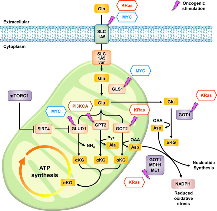
“MYC-driven cancer cells exhibit enhanced glutamine utilization accompanied by increased expression of key glutaminolysis enzymes, including GLS1/GLS2 and GLUD1178–180.”
§ Glutamine reliance in cell metabolism > Crosstalk between glutamine metabolism and oncogenic signaling
What the sources say
- Partly supportedcites [178] Gao, P. et al. c-Myc suppression of miR-23a/b enhances mitochondrial glutaminase expression and glutamine metabolism. Nature 458, 762–765 (2009). sourceExperimental — accuracy not yet measured
The main text reports that Myc increases glutaminase expression and glutamine catabolism, supporting the enhanced-utilization and GLS1 parts of the claim. But it says only GLS1 was predominantly expressed in the studied cells (passage 6), while glutamate dehydrogenase mRNA appeared unaltered (passage 2). It does not support increased GLS2 or GLUD1 expression. (LLM, conf=0.91)
- Partly supportedcites [179] Yuneva, M. O. et al. The metabolic profile of tumors depends on both the responsible genetic lesion and tissue type. Cell Metab. 15, 157–170 (2012). sourceExperimental — accuracy not yet measured
The Results section, “MYC and MET Differently Affect Glutamine Metabolism in the Liver” (page unavailable), reports increased glutamine transport and catabolism in MYC-induced liver tumors, with GLS1 replacing GLS2. The Results section on tissue context reports increased GLS1 in MYC-induced lung tumors but no change in GLS2. The claim is therefore supported for glutamine utilization and GLS1, not for increased GLS2; no passage found supports increased GLUD1 expression. (LLM, conf=0.91)
- Partly supportedcites [180] Xiao, D. et al. Myc promotes glutaminolysis in human neuroblastoma through direct activation of glutaminase 2. Oncotarget 6, 40655–40666 (2015). sourceExperimental — accuracy not yet measured
The Results section reports that N-Myc promotes glutamine metabolism through GLS2 in MYCN-amplified neuroblastoma cells. It also reports that GLS1 expression was reduced, not increased, in MYCN-amplified tumors; the Introduction describes c-Myc-associated GLS1 expression in other cancer cells. The passages do not support the attributed increase in GLUD1. (LLM, conf=0.90)
plaincorrelational- Asserted: passes — The authors put it forward as true.
- Checkable: passes — It can be checked against evidence.
- Load-bearing: passes — The paper’s conclusions depend on it.
- Based: passes — The paper gives a reason why it holds.
- Partly supported
None of the sources cited for this claim have been checked yet.
In the paper
“The major findings are that aerobic glycolysis is not a symptom of impaired mitochondrial function, and that glutamine supports mitochondrial oxidative metabolism when pyruvate derived from glucose is converted into lactate and secreted2–4.”
§ Glutamine reliance in cell metabolism > Introduction
What the sources say
- Not checked yetcites [2] DeBerardinis, R. J. & Cheng, T. Q’s next: the diverse functions of glutamine in metabolism, cell biology and cancer. Oncogene 29, 313–324 (2010). source
- Not checked yetcites [3] Hensley, C. T., Wasti, A. T. & DeBerardinis, R. J. Glutamine and cancer: cell biology, physiology, and clinical opportunities. J. Clin. Invest. 123, 3678–3684 (2013). source
- Not checked yetcites [4] Altman, B. J., Stine, Z. E. & Dang, C. V. From Krebs to clinic: glutamine metabolism to cancer therapy. Nat. Rev. Cancer 16, 749 (2016). source
plaindirect- Asserted: passes — The authors put it forward as true.
- Checkable: passes — It can be checked against evidence.
- Load-bearing: passes — The paper’s conclusions depend on it.
- Based: passes — The paper gives a reason why it holds.
In the paper
“specific genetic interference with glutaminase (GLS) inhibits tumor cell growth13.”
§ Glutamine reliance in cell metabolism > Introduction
What the sources say
- Not checked yetcites [13] Lobo, C. et al. Inhibition of glutaminase expression by antisense mRNA decreases growth and tumourigenicity of tumour cells. Biochem. J. 348, 257–261 (2000). source
plaindirect- Asserted: passes — The authors put it forward as true.
- Checkable: passes — It can be checked against evidence.
- Load-bearing: passes — The paper’s conclusions depend on it.
- Based: passes — The paper gives a reason why it holds.
In the paper
“Glutamine, which is a nonessential amino acid (NEAA) due to the endogenous glutamine biosynthesis pathway, is currently considered essential in cancer cells because transformed cells consume glutamine at a rate exceeding that of glutamine biosynthesis5.”
§ Glutamine reliance in cell metabolism > Introduction
What the sources say
- Not checked yetcites [5] Zhang, J., Pavlova, N. N. & Thompson, C. B. Cancer cell metabolism: the essential role of the nonessential amino acid, glutamine. Embo J. 36, 1302–1315 (2017). source
plaindirect- Asserted: passes — The authors put it forward as true.
- Checkable: passes — It can be checked against evidence.
- Load-bearing: passes — The paper’s conclusions depend on it.
- Based: passes — The paper gives a reason why it holds.
None of the sources cited for this claim have been checked yet.
In the paper
“The major findings are that aerobic glycolysis is not a symptom of impaired mitochondrial function, and that glutamine supports mitochondrial oxidative metabolism when pyruvate derived from glucose is converted into lactate and secreted2–4.”
§ Glutamine reliance in cell metabolism > Introduction
What the sources say
- Not checked yetcites [2] DeBerardinis, R. J. & Cheng, T. Q’s next: the diverse functions of glutamine in metabolism, cell biology and cancer. Oncogene 29, 313–324 (2010). source
- Not checked yetcites [3] Hensley, C. T., Wasti, A. T. & DeBerardinis, R. J. Glutamine and cancer: cell biology, physiology, and clinical opportunities. J. Clin. Invest. 123, 3678–3684 (2013). source
- Not checked yetcites [4] Altman, B. J., Stine, Z. E. & Dang, C. V. From Krebs to clinic: glutamine metabolism to cancer therapy. Nat. Rev. Cancer 16, 749 (2016). source
plain- Asserted: passes — The authors put it forward as true.
- Checkable: passes — It can be checked against evidence.
- Load-bearing: passes — The paper’s conclusions depend on it.
- Based: passes — The paper gives a reason why it holds.
None of the sources cited for this claim have been checked yet.
In the paper
“glutamine has been confirmed to be a major nutrient source for oxidative metabolism in some cancer cell lines10–12”
§ Glutamine reliance in cell metabolism > Introduction
What the sources say
- Not checked yetcites [10] Eagle, H. Nutrition needs of mammalian cells in tissue culture. Science 122, 501–514 (1955). source
- Not checked yetcites [11] Kovacevic, Z. & Morris, H. P. The role of glutamine in the oxidative metabolism of malignant cells. Cancer Res. 32, 326–333 (1972).
- Not checked yetcites [12] Reitzer, L. J., Wice, B. M. & Kennell, D. Evidence that glutamine, not sugar, is the major energy source for cultured HeLa cells. J. Biol. Chem. 254, 2669–2676 (1979). source
boostedcorrelational- Asserted: passes — The authors put it forward as true.
- Checkable: passes — It can be checked against evidence.
- Load-bearing: passes — The paper’s conclusions depend on it.
- Based: passes — The paper gives a reason why it holds.
- It may state two separate claims as one. 85
- Probably not a substantial claim. 4
- The quote only partly states this claim. 1
The cited sources support only part of this claim — read each source’s verdict below.
In the paper
“Although glutamate is the major downstream product of glutamine, glutamate supplementation during glutamine deprivation cannot rescue the impaired cell growth or mitochondrial respiration16,43–45, indicating that mitochondrial GLS-catalyzed cleavage of the gamma-nitrogen of glutamine is essential for glutaminolysis.”
§ Glutamine reliance in cell metabolism > Glutamine metabolic pathways > Glutamate
What the sources say
- Not checked yetcites [16] Yoo, H. C. et al. A variant of SLC1A5 is a mitochondrial glutamine transporter for metabolic reprogramming in cancer cells. Cell Metab. 31, 267–283 (2020). source
- Partly supportedcites [43] Zhang, J. et al. Asparagine plays a critical role in regulating cellular adaptation to glutamine depletion. Mol. Cell 56, 205–218 (2014). sourceExperimental — accuracy not yet measured
The Results section, “Asparagine Restores Viability, but not Anaplerosis, Other Nonessential Amino Acids, or Proliferation” (page unavailable), reports that glutamate substitution failed to suppress apoptosis during glutamine deprivation. It does not establish that glutamate failed to rescue cell growth or mitochondrial respiration, or that mitochondrial GLS-catalyzed cleavage is essential for glutaminolysis. (LLM, conf=0.82)
- Partly supportedcites [44] Huang, H. et al. Role of glutamine and interlinked asparagine metabolism in vessel formation. Embo J. 36, 2334–2352 (2017). sourceExperimental — accuracy not yet measured
The Introduction identifies glutamate as a product of GLS, and the section on rescuing glutamine-deprived endothelial cells reports impaired proliferation and incomplete rescue by several supplements. But the paper does not report that glutamate supplementation fails to rescue growth or mitochondrial respiration during glutamine deprivation. In fact, the section on endothelial-cell sprouting reports that exogenous glutamate reversed the anti-proliferative effect of GLS1 inhibition—a different condition that does not establish the claimed necessity of GLS-catalyzed cleavage. (LLM, conf=0.88)
- Partly supportedcites [45] Pavlova, N. N. et al. As extracellular glutamine levels decline, asparagine becomes an essential amino acid. Cell Metab. 27, 428 (2018). sourceExperimental — accuracy not yet measured
The Results section (‘Asparagine facilitates the expression of GLUL under glutamine deprivation’; page unavailable) reports that replacing glutamine with glutamate did not rescue proliferation of glutamine-depleted cells. Our copy does not report whether glutamate supplementation rescues mitochondrial respiration. The statement about essential mitochondrial GLS-catalyzed cleavage follows the citations as the citing authors’ inference, rather than a finding this citation directly supports. (LLM, conf=0.87)
hedgeddirect- Asserted: passes — The authors put it forward as true.
- Checkable: passes — It can be checked against evidence.
- Load-bearing: passes — The paper’s conclusions depend on it.
- Based: passes — The paper gives a reason why it holds.
It may state two separate claims as one.
The cited sources support only part of this claim — read each source’s verdict below.
In the paper
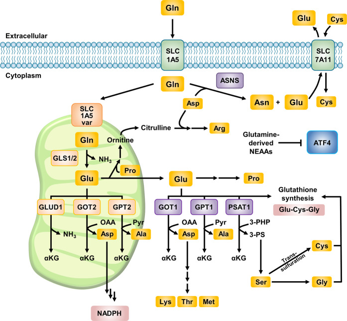
“In cells with NRF2 activation, most glutamate is secreted, and cystine is imported by the SLC7A11 (xCT) antiporter mechanism47 (Fig. 3).”
§ Glutamine reliance in cell metabolism > Glutamine metabolic pathways > Glutamate
What the sources say
- Partly supportedcites [47] Sayin, V. I. et al. Activation of the NRF2 antioxidant program generates an imbalance in central carbon metabolism in cancer. eLife 6, e28083 (2017). sourceExperimental — accuracy not yet measured
The Results section reports higher extracellular and lower intracellular glutamate in KEAP1-mutant cells and describes xCT/SLC7A11 exchanging glutamate for cystine. The Discussion attributes glutamate secretion under increased NRF2 to this transporter. Neither passage establishes that most glutamate is secreted; the evidence is also drawn chiefly from the studied cancer-cell models, not all cells with NRF2 activation. (LLM, conf=0.88)
plaindirect- Asserted: passes — The authors put it forward as true.
- Checkable: passes — It can be checked against evidence.
- Load-bearing: unclear — It is unclear whether the paper’s conclusions depend on it.
- Based: passes — The paper gives a reason why it holds.
It may state two separate claims as one.
- Partly supported
Every source the paper cites for this claim was checked and supports it.
In the paper
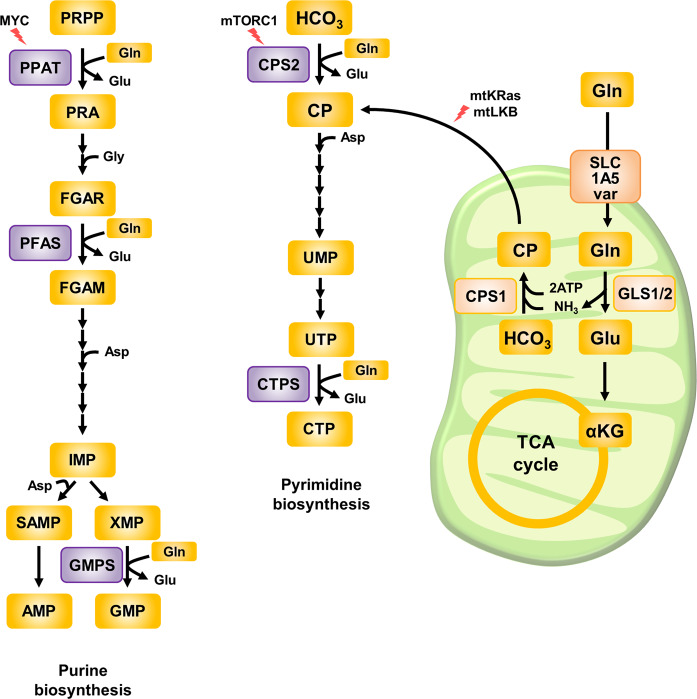
“PPAT transfers glutamine-derived nitrogen to 5-phosphoribosyl pyrophosphate (PRPP), and this step is considered the initial step in purine biosynthesis37.”
§ Glutamine reliance in cell metabolism > Glutamine metabolic pathways > Nucleotides synthesized from glutamine
What the sources say
- Supportedcites [37] Lane, A. N. & Fan, T. W. Regulation of mammalian nucleotide metabolism and biosynthesis. Nucleic Acids Res. 43, 2466–2485 (2015). source
Figure 3 in “Bioenergetics of nucleotide biosynthesis” identifies PPAT as enzyme 1 in the purine-biosynthesis pathway and says glutamine’s amido nitrogen contributes to the purine ring. Table 1 likewise lists PPAT as catalyzing reaction 1. Together, these support the description of PPAT’s glutamine-to-PRPP reaction as the initial step. (LLM, conf=0.90)
plaindirect- Asserted: passes — The authors put it forward as true.
- Checkable: passes — It can be checked against evidence.
- Load-bearing: passes — The paper’s conclusions depend on it.
- Based: passes — The paper gives a reason why it holds.
It may state two separate claims as one.
- Supported
The cited sources support only part of this claim — read each source’s verdict below.
In the paper
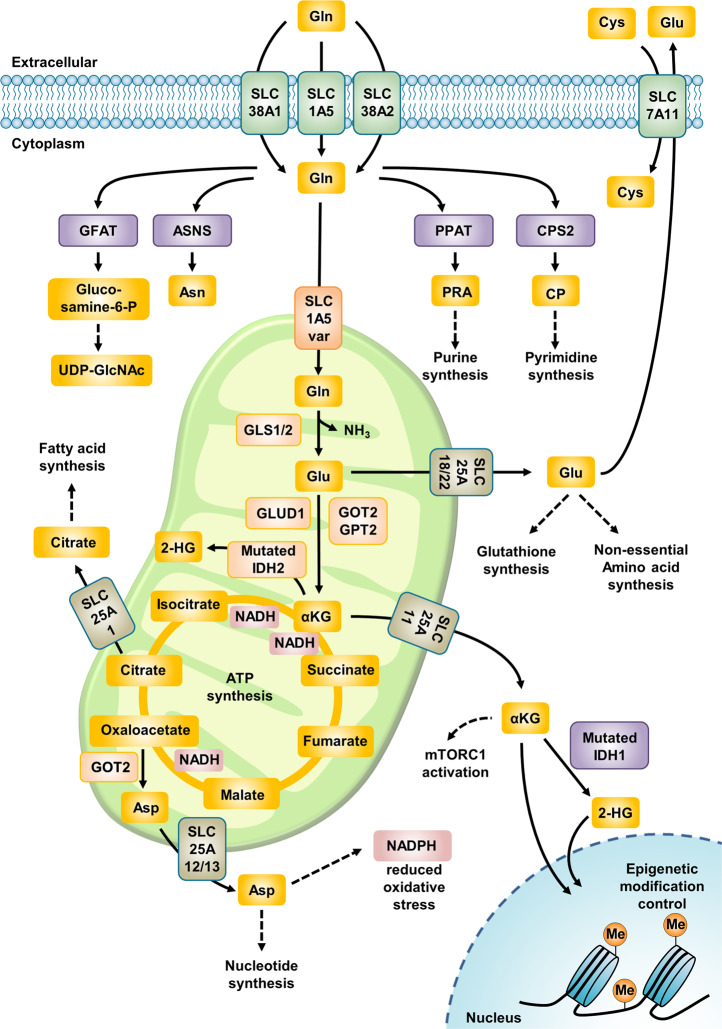
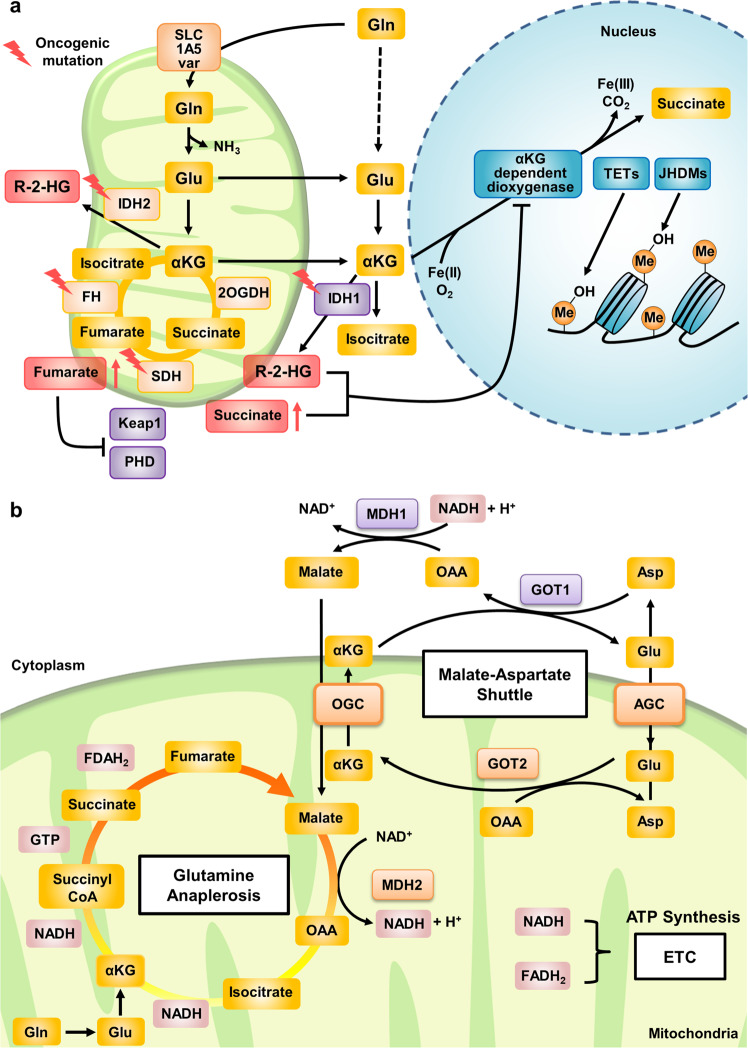
“In addition, α-KG is exported from mitochondria through SLC25A11 to the cytosol21 and then participates in fatty acid biosynthesis and NADH generation22 (Fig. 1).”
§ Glutamine reliance in cell metabolism > Glutamine metabolic pathways
What the sources say
- Could not verifycites [21] Stine, Z. E. & Dang, C. V. Glutamine skipping the Q into mitochondria. Trends Mol. Med. 26, 6–7 (2020). sourceExperimental — accuracy not yet measured
The section labeled Abstract says glutamate is converted to α-ketoglutarate in mitochondria, but does not address the claimed export to the cytosol. Searches for SLC25A11, fatty acid biosynthesis, and NADH generation found no passage supporting those claims; the paper discusses SLC1A5_var-mediated glutamine import and NADPH instead. (LLM, conf=0.75)
- Partly supportedcites [22] Mullen, A. R. et al. Oxidation of alpha-ketoglutarate is required for reductive carboxylation in cancer cells with mitochondrial defects. Cell Rep. 7, 1679–1690 (2014). sourceExperimental — accuracy not yet measured
The Introduction (page unavailable) says AKG-derived citrate can supply acetyl-CoA for fatty acid biosynthesis, and the Results section on AKG dehydrogenase and NNT (page unavailable) says AKG oxidation generates NADH. However, the paper locates that NADH-generating activity in mitochondria; it does not establish that exported cytosolic AKG generates NADH. The SLC25A11 export clause is attached to citation 21, not this source’s citation 22. (LLM, conf=0.84)
plaindirect- Asserted: passes — The authors put it forward as true.
- Checkable: passes — It can be checked against evidence.
- Load-bearing: unclear — It is unclear whether the paper’s conclusions depend on it.
- Based: passes — The paper gives a reason why it holds.
It may state two separate claims as one.
- Could not verify
Every source the paper cites for this claim was checked and supports it.
In the paper
“This severe glutamine deprivation leads to dramatic histone hypermethylation due to decreased α-KG levels subsequent to decreased activity of Jumonji domain-containing histone demethylases and results in cancer cell dedifferentiation and resistance to BRAF inhibitors103.”
§ Glutamine reliance in cell metabolism > Glutamine metabolic pathways > Control of epigenetic changes by glutamine
What the sources say
- Supportedcites [103] Pan, M. et al. Regional glutamine deficiency in tumours promotes dedifferentiation through inhibition of histone demethylation. Nat. Cell Biol. 18, 1090–1101 (2016). source
Split into 2 atoms — 2 confirmed, 0 partially supported, 0 disputed, 0 misattributed, 0 unverifiable. Expand the claim to see per-atom verdicts.
plaindirect- Asserted: passes — The authors put it forward as true.
- Checkable: passes — It can be checked against evidence.
- Load-bearing: passes — The paper’s conclusions depend on it.
- Based: passes — The paper gives a reason why it holds.
It may state two separate claims as one.
- Rests on an assumption: The core region of solid tumors exhibits a deficiency of glutamine compared with other amino acids.
- Supported
None of the sources cited for this claim have been checked yet.
In the paper
“Moreover, CB-839, the first glutaminase inhibitor, has entered several clinical trials14,15.”
§ Glutamine reliance in cell metabolism > Introduction
What the sources say
- Not checked yetcites [14] Gross, M. I. et al. Antitumor activity of the glutaminase inhibitor CB-839 in triple-negative breast cancer. Mol. Cancer Ther. 13, 890–901 (2014). source
- Not checked yetcites [15] Kizilbash, S. H. et al. The addition of CB-839 to temozolomide significantly reduces glioma aspartate and glutamate in an IDH mutated patient derived glioma xenograft model. Cancer Res. https://doi.org/10.1158/1538-7445.AM2019-3870 (2019). source
plain- Asserted: passes — The authors put it forward as true.
- Checkable: passes — It can be checked against evidence.
- Load-bearing: fails — The paper’s conclusions may not depend on it.
- Based: passes — The paper gives a reason why it holds.
Probably not a substantial claim. The paper’s conclusions may not depend on it.
None of the sources cited for this claim have been checked yet.
In the paper
“Thus, glutamine deprivation suppresses cancer growth and even induces cell death in several cancers6,7.”
§ Glutamine reliance in cell metabolism > Introduction
What the sources say
- Not checked yetcites [6] Qing, G. et al. ATF4 regulates MYC-mediated neuroblastoma cell death upon glutamine deprivation. Cancer Cell 22, 631–644 (2012). source
- Not checked yetcites [7] Chen, L. & Cui, H. Targeting glutamine induces apoptosis: a cancer therapy approach. Int. J. Mol. Sci. 16, 22830–22855 (2015). source
plaindirect- Asserted: passes — The authors put it forward as true.
- Checkable: passes — It can be checked against evidence.
- Load-bearing: passes — The paper’s conclusions depend on it.
- Based: passes — The paper gives a reason why it holds.
It may state two separate claims as one.
In the paper
“In addition to pyruvate-derived acetyl-CoA, α-KG-derived metabolites (e.g., succinate and fumarate) generated via glutaminolysis are considered oncometabolites contributing to tumorigenesis23.”
§ Glutamine reliance in cell metabolism > Glutamine metabolic pathways
What the sources say
- Not checked yetcites [23] Yang, L. F., Venneti, S. & Nagrath, D. Glutaminolysis: a hallmark of cancer metabolism. Annu. Rev. Biomed. Eng. 19, 163–194 (2017). source
plaindirect- Asserted: passes — The authors put it forward as true.
- Checkable: passes — It can be checked against evidence.
- Load-bearing: fails — The paper’s conclusions may not depend on it.
- Based: passes — The paper gives a reason why it holds.
Probably not a substantial claim. The paper’s conclusions may not depend on it.
In the paper
“Cytosolic glutamine supports nucleotide biosynthesis, which is essential for rapidly proliferating cells30.”
§ Glutamine reliance in cell metabolism > Glutamine metabolic pathways > Nucleotides synthesized from glutamine
What the sources say
- Not checked yetcites [30] Tong, X., Zhao, F. & Thompson, C. B. The molecular determinants of de novo nucleotide biosynthesis in cancer cells. Curr. Opin. Genet. Dev. 19, 32–37 (2009). source
plaindirect- Asserted: passes — The authors put it forward as true.
- Checkable: passes — It can be checked against evidence.
- Load-bearing: passes — The paper’s conclusions depend on it.
- Based: passes — The paper gives a reason why it holds.
It may state two separate claims as one.
The paper states this without a source or data behind it here, so there is nothing to check it against. The quote shows it is in the paper.
In the paper
“Glutamine-derived carbon is an important substrate that supports the TCA cycle and the synthesis of glutathione.”
§ Glutamine reliance in cell metabolism > Glutamine metabolic pathways > NEAAs synthesized from glutamine
plaindirect- Asserted: passes — The authors put it forward as true.
- Checkable: passes — It can be checked against evidence.
- Load-bearing: passes — The paper’s conclusions depend on it.
- Based: passes — The paper gives a reason why it holds.
It may state two separate claims as one.
None of the sources cited for this claim have been checked yet.
In the paper
“In cancer cells, the use of glutamate-derived nitrogen for NEAA production may be favored in various types of cancer cells to preserve nitrogen for anabolic reactions48 and may prevent apoptosis induced by ATF4 activation upon glutamine deprivation6.”
§ Glutamine reliance in cell metabolism > Glutamine metabolic pathways > Glutamate
What the sources say
- Not checked yetcites [48] Coloff, J. L. et al. Differential glutamate metabolism in proliferating and quiescent mammary epithelial cells. Cell Metab. 23, 867–880 (2016). source
- Not checked yetcites [6] Qing, G. et al. ATF4 regulates MYC-mediated neuroblastoma cell death upon glutamine deprivation. Cancer Cell 22, 631–644 (2012). source
hedgedconditional- Asserted: passes — The authors put it forward as true.
- Checkable: passes — It can be checked against evidence.
- Load-bearing: unclear — It is unclear whether the paper’s conclusions depend on it.
- Based: passes — The paper gives a reason why it holds.
It may state two separate claims as one.
In the paper
“In purine biosynthesis, two glutamines are used to generate inosine monophosphate (IMP), a precursor of both adenosine monophosphate (AMP) and guanosine monophosphate (GMP). Then, one glutamine molecule is needed for the conversion of IMP to GMP31.”
§ Glutamine reliance in cell metabolism > Glutamine metabolic pathways > Nucleotides synthesized from glutamine
What the sources say
- Not checked yetcites [31] Moffatt, B. A. & Ashihara, H. Purine and pyrimidine nucleotide synthesis and metabolism. Arabidopsis Book 1, e0018 (2002). source
plaindirect- Asserted: passes — The authors put it forward as true.
- Checkable: passes — It can be checked against evidence.
- Load-bearing: unclear — It is unclear whether the paper’s conclusions depend on it.
- Based: passes — The paper gives a reason why it holds.
It may state two separate claims as one.
In the paper
“Mitochondrial α-KG can then participate in the TCA cycle, supporting the oxidative phosphorylation (OXPHOS) pathway or the reductive carboxylation pathway23.”
§ Glutamine reliance in cell metabolism > Glutamine metabolic pathways
What the sources say
- Not checked yetcites [23] Yang, L. F., Venneti, S. & Nagrath, D. Glutaminolysis: a hallmark of cancer metabolism. Annu. Rev. Biomed. Eng. 19, 163–194 (2017). source
plaindirect- Asserted: passes — The authors put it forward as true.
- Checkable: passes — It can be checked against evidence.
- Load-bearing: unclear — It is unclear whether the paper’s conclusions depend on it.
- Based: passes — The paper gives a reason why it holds.
It may state two separate claims as one.
In the paper
“The gamma-nitrogen of glutamine is used in five reactions in de novo nucleotide synthesis, and its bioavailability controls de novo biosynthesis of pyrimidines and purines (Fig. 2)5.”
§ Glutamine reliance in cell metabolism > Glutamine metabolic pathways > Nucleotides synthesized from glutamine
What the sources say
- Not checked yetcites [5] Zhang, J., Pavlova, N. N. & Thompson, C. B. Cancer cell metabolism: the essential role of the nonessential amino acid, glutamine. Embo J. 36, 1302–1315 (2017). source
plaindirect- Asserted: passes — The authors put it forward as true.
- Checkable: passes — It can be checked against evidence.
- Load-bearing: passes — The paper’s conclusions depend on it.
- Based: passes — The paper gives a reason why it holds.
It may state two separate claims as one.
In the paper
“In pyrimidine biosynthesis, one glutamine molecule is consumed by a carbamoyl phosphate synthetase enzyme (CPS1 or CPS2, which are localized in the mitochondria and cytosol, respectively). One more glutamine molecule is used to synthesize cytidine triphosphate (CTP) from uridine triphosphate (UTP)31.”
§ Glutamine reliance in cell metabolism > Glutamine metabolic pathways > Nucleotides synthesized from glutamine
What the sources say
- Not checked yetcites [31] Moffatt, B. A. & Ashihara, H. Purine and pyrimidine nucleotide synthesis and metabolism. Arabidopsis Book 1, e0018 (2002). source
plaindirect- Asserted: passes — The authors put it forward as true.
- Checkable: passes — It can be checked against evidence.
- Load-bearing: unclear — It is unclear whether the paper’s conclusions depend on it.
- Based: passes — The paper gives a reason why it holds.
It may state two separate claims as one.
In the paper
“Instead, asparagine supplementation enhances the expression of glutamine synthetase (GLUL) and increases intracellular glutamine usage via glutaminolysis, resulting in the recovery of global protein translation that is blocked by glutamine deprivation45.”
§ Glutamine reliance in cell metabolism > Glutamine metabolic pathways > Asparagine
What the sources say
- Not checked yetcites [45] Pavlova, N. N. et al. As extracellular glutamine levels decline, asparagine becomes an essential amino acid. Cell Metab. 27, 428 (2018). source
plaindirect- Asserted: passes — The authors put it forward as true.
- Checkable: passes — It can be checked against evidence.
- Load-bearing: passes — The paper’s conclusions depend on it.
- Based: passes — The paper gives a reason why it holds.
It may state two separate claims as one.
None of the sources cited for this claim have been checked yet.
In the paper
“GLSs have at least three isoforms, GLS1, GLS2, and GAC (a splicing isoform of GLS1), all of which were recently reported to be localized in mitochondria18–20.”
§ Glutamine reliance in cell metabolism > Glutamine metabolic pathways
What the sources say
- Not checked yetcites [18] Wang, J. B. et al. Targeting mitochondrial glutaminase activity inhibits oncogenic transformation. Cancer Cell 18, 397–397 (2010). source
- Not checked yetcites [19] Li, B. et al. Targeting glutaminase 1 attenuates stemness properties in hepatocellular carcinoma by increasing reactive oxygen species and suppressing Wnt/beta-catenin pathway. EBioMedicine 39, 239–254 (2019). source
- Not checked yetcites [20] Lukey, M. J. et al. Liver-type glutaminase GLS2 is a druggable metabolic node in luminal-subtype breast cancer. Cell Rep. 29, 76–88 e77 (2019). source
plain- Asserted: passes — The authors put it forward as true.
- Checkable: passes — It can be checked against evidence.
- Load-bearing: unclear — It is unclear whether the paper’s conclusions depend on it.
- Based: passes — The paper gives a reason why it holds.
It may state two separate claims as one.
In the paper
“This finding is surprising because asparagine supplementation does not restore the levels of other NEAAs (alanine, proline, and glutamate) or any TCA cycle intermediates (α-KG, malate, and fumarate).”
§ Glutamine reliance in cell metabolism > Glutamine metabolic pathways > Asparagine
What the sources say
- Not checked yetcites [43] Zhang, J. et al. Asparagine plays a critical role in regulating cellular adaptation to glutamine depletion. Mol. Cell 56, 205–218 (2014). source
plain- Asserted: passes — The authors put it forward as true.
- Checkable: passes — It can be checked against evidence.
- Load-bearing: passes — The paper’s conclusions depend on it.
- Based: passes — The paper gives a reason why it holds.
It may state two separate claims as one.
In the paper
“In pancreatic cancer cells, oncogenic K-Ras maintains the nucleotide pool via the MAPK-dependent signaling pathway, leading to MYC upregulation, and the use of MEK inhibitors reduces the incorporation of glutamine-derived nitrogen into purine nucleotides38.”
§ Glutamine reliance in cell metabolism > Glutamine metabolic pathways > Nucleotides synthesized from glutamine
What the sources say
- Not checked yetcites [38] Santana-Codina, N. et al. Oncogenic KRAS supports pancreatic cancer through regulation of nucleotide synthesis. Nat. Commun. 9, 4945 (2018). source
plaindirect- Asserted: passes — The authors put it forward as true.
- Checkable: passes — It can be checked against evidence.
- Load-bearing: unclear — It is unclear whether the paper’s conclusions depend on it.
- Based: passes — The paper gives a reason why it holds.
It may state two separate claims as one.
In the paper
“In addition, IDH1-dependent reductive glutamine metabolism produces NADPH, which decreases mitochondrial ROS during anchorage-independent growth64.”
§ Glutamine reliance in cell metabolism > Glutamine metabolic pathways > Redox control of glutamine
What the sources say
- Not checked yetcites [64] Jiang, L. et al. Reductive carboxylation supports redox homeostasis during anchorage-independent growth. Nature 532, 255–258 (2016). source
plaindirect- Asserted: passes — The authors put it forward as true.
- Checkable: passes — It can be checked against evidence.
- Load-bearing: unclear — It is unclear whether the paper’s conclusions depend on it.
- Based: passes — The paper gives a reason why it holds.
It may state two separate claims as one.
In the paper
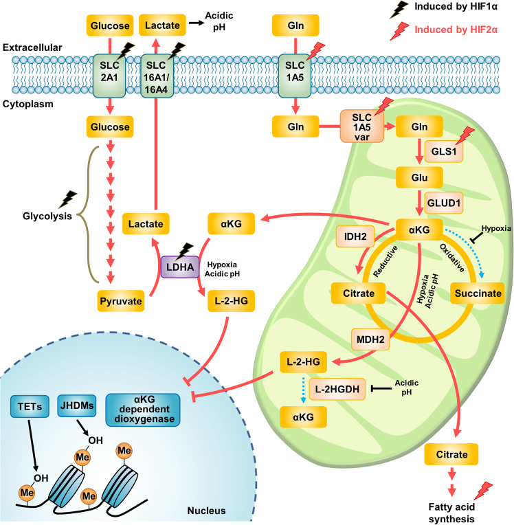
“In addition, extracellular lactate stabilizes HIF-2α, and HIF-2α then transactivates MYC, increasing the levels of glutamine transporters and GLS1, in turn resulting in increased glutamine catabolism72.”
§ Glutamine reliance in cell metabolism > Glutamine metabolic pathways > Control of glutamine metabolism by hypoxia
What the sources say
- Not checked yetcites [72] Perez-Escuredo, J. et al. Lactate promotes glutamine uptake and metabolism in oxidative cancer cells. Cell Cycle 15, 72–83 (2016). source
plaindirect- Asserted: passes — The authors put it forward as true.
- Checkable: passes — It can be checked against evidence.
- Load-bearing: passes — The paper’s conclusions depend on it.
- Based: passes — The paper gives a reason why it holds.
It may state two separate claims as one.
In the paper
“Consistent with this observation, glutamine starvation has been associated with impaired uptake of cystine through xCT and decreased intracellular glutathione levels62.”
§ Glutamine reliance in cell metabolism > Glutamine metabolic pathways > Redox control of glutamine
What the sources say
- Not checked yetcites [62] Timmerman, L. A. et al. Glutamine sensitivity analysis identifies the xCT antiporter as a common triple-negative breast tumor therapeutic target. Cancer Cell 24, 450–465 (2013). source
plaincorrelational- Asserted: passes — The authors put it forward as true.
- Checkable: passes — It can be checked against evidence.
- Load-bearing: passes — The paper’s conclusions depend on it.
- Based: passes — The paper gives a reason why it holds.
It may state two separate claims as one.
In the paper
“Recently, hypoxia-induced expression of the SLC1A5 variant was shown to be mediated by HIF-2α and to lead to metabolic reprogramming toward glutamine metabolism in pancreatic cancer cells16.”
§ Glutamine reliance in cell metabolism > Glutamine metabolic pathways > Control of glutamine metabolism by hypoxia
What the sources say
- Not checked yetcites [16] Yoo, H. C. et al. A variant of SLC1A5 is a mitochondrial glutamine transporter for metabolic reprogramming in cancer cells. Cell Metab. 31, 267–283 (2020). source
plaindirect- Asserted: passes — The authors put it forward as true.
- Checkable: passes — It can be checked against evidence.
- Load-bearing: passes — The paper’s conclusions depend on it.
- Based: passes — The paper gives a reason why it holds.
It may state two separate claims as one.
In the paper
“Recent studies suggest that cancer cells are under increased oxidative stress caused by oncogenic transformation, leading to metabolic alterations that result in ROS production56.”
§ Glutamine reliance in cell metabolism > Glutamine metabolic pathways > Redox control of glutamine
What the sources say
- Not checked yetcites [56] Gorrini, C., Harris, I. S. & Mak, T. W. Modulation of oxidative stress as an anticancer strategy. Nat. Rev. Drug Discov. 12, 931–947 (2013). source
hedgeddirect- Asserted: passes — The authors put it forward as true.
- Checkable: passes — It can be checked against evidence.
- Load-bearing: passes — The paper’s conclusions depend on it.
- Based: passes — The paper gives a reason why it holds.
It may state two separate claims as one.
None of the sources cited for this claim have been checked yet.
In the paper
“Given that HIF-2α is an important transcription factor in cancer progression and leads to poor prognosis69,70”
§ Glutamine reliance in cell metabolism > Glutamine metabolic pathways > Control of glutamine metabolism by hypoxia
What the sources say
- Not checked yetcites [69] Zhao, J., Du, F., Shen, G., Zheng, F. & Xu, B. The role of hypoxia-inducible factor-2 in digestive system cancers. Cell Death Dis. 6, e1600 (2015). source
- Not checked yetcites [70] Li, Z. et al. Hypoxia-inducible factors regulate tumorigenic capacity of glioma stem cells. Cancer Cell 15, 501–513 (2009). source
plaindirect- Asserted: passes — The authors put it forward as true.
- Checkable: passes — It can be checked against evidence.
- Load-bearing: passes — The paper’s conclusions depend on it.
- Based: passes — The paper gives a reason why it holds.
It may state two separate claims as one.
In the paper
“A low level of reactive oxygen species (ROS) activates tumorigenic growth signaling; however, when the level exceeds the cellular redox capacity, ROS can damage macromolecules such as proteins, lipids and nucleotides55.”
§ Glutamine reliance in cell metabolism > Glutamine metabolic pathways > Redox control of glutamine
What the sources say
- Not checked yetcites [55] Weinberg, F. et al. Mitochondrial metabolism and ROS generation are essential for Kras-mediated tumorigenicity. Proc. Natl Acad. Sci. USA 107, 8788–8793 (2010). source
plaindirect- Asserted: passes — The authors put it forward as true.
- Checkable: passes — It can be checked against evidence.
- Load-bearing: passes — The paper’s conclusions depend on it.
- Based: passes — The paper gives a reason why it holds.
It may state two separate claims as one.
In the paper
“Hypoxia increases the deoxycytidine triphosphate (dCTP) level through the pentose phosphate pathway (PPP) via glucose metabolism and results in resistance to gemcitabine, a dCTP analog87.”
§ Glutamine reliance in cell metabolism > Glutamine metabolic pathways > Control of glutamine metabolism by hypoxia
What the sources say
- Not checked yetcites [87] Shukla, S. K. et al. MUC1 and HIF-1alpha signaling crosstalk induces anabolic glucose metabolism to impart gemcitabine resistance to pancreatic cancer. Cancer Cell 32, 392 (2017). source
plaindirect- Asserted: passes — The authors put it forward as true.
- Checkable: passes — It can be checked against evidence.
- Load-bearing: passes — The paper’s conclusions depend on it.
- Based: passes — The paper gives a reason why it holds.
It may state two separate claims as one.
In the paper
“Furthermore, HIF-2α represses the transcription of carnitine palmitoyltransferase 1A (CPT1A), which is responsible for mitochondrial β-oxidation by transporting fatty acids and results in lipid deposition78.”
§ Glutamine reliance in cell metabolism > Glutamine metabolic pathways > Control of glutamine metabolism by hypoxia
What the sources say
- Not checked yetcites [78] Du, W. et al. HIF drives lipid deposition and cancer in ccRCC via repression of fatty acid metabolism. Nat. Commun. 8, 1769 (2017). source
plaindirect- Asserted: passes — The authors put it forward as true.
- Checkable: passes — It can be checked against evidence.
- Load-bearing: passes — The paper’s conclusions depend on it.
- Based: passes — The paper gives a reason why it holds.
It may state two separate claims as one.
None of the sources cited for this claim have been checked yet.
In the paper
“Recently, asparagine has been reported to function as an exchange factor needed for the uptake of other amino acids that are required for mTORC1 activation53 and for enhanced nucleotide biosynthesis under mitochondrial electron chain transport system impairment54.”
§ Glutamine reliance in cell metabolism > Glutamine metabolic pathways > Asparagine
What the sources say
- Not checked yetcites [53] Krall, A. S., Xu, S., Graeber, T. G., Braas, D. & Christofk, H. R. Asparagine promotes cancer cell proliferation through use as an amino acid exchange factor. Nat. Commun. 7, 11457 (2016). source
- Not checked yetcites [54] Krall, A. S. et al. Asparagine signals mitochondrial respiration and can be targeted to impair tumour growth. bioRxiv, 2020.2003.2017.995670 (2020). source
plaindirect- Asserted: passes — The authors put it forward as true.
- Checkable: passes — It can be checked against evidence.
- Load-bearing: unclear — It is unclear whether the paper’s conclusions depend on it.
- Based: passes — The paper gives a reason why it holds.
It may state two separate claims as one.
In the paper
“This study suggested that l-asparaginase treatment alone can reduce the incidence of breast cancer metastasis to the lung without affecting primary tumor growth.”
§ Glutamine reliance in cell metabolism > Glutamine metabolic pathways > Asparagine
What the sources say
- Not checked yetcites [51] Knott, S. R. V. et al. Asparagine bioavailability governs metastasis in a model of breast cancer. Nature https://doi.org/10.1038/nature26162 (2018). source
hedgeddirect- Asserted: passes — The authors put it forward as true.
- Checkable: passes — It can be checked against evidence.
- Load-bearing: passes — The paper’s conclusions depend on it.
- Based: passes — The paper gives a reason why it holds.
It may state two separate claims as one.
A conclusion the paper draws from its own results — it cites no other source for it, so there is none to check. The quote shows it is in the paper.
In the paper
“In summary, glutamine maintains cellular redox homeostasis by supplying fuels for glutathione synthesis and endowing reducing power in the form of NADPH for sustaining tumor growth.”
§ Glutamine reliance in cell metabolism > Glutamine metabolic pathways > Redox control of glutamine
plaindirect- Asserted: passes — The authors put it forward as true.
- Checkable: passes — It can be checked against evidence.
- Load-bearing: passes — The paper’s conclusions depend on it.
- Based: passes — The paper gives a reason why it holds.
It may state two separate claims as one.
The paper states this without a source or data behind it here, so there is nothing to check it against. The quote shows it is in the paper.
In the paper
“This mechanism is very important in clear cell renal cell carcinoma (ccRCC) in which HIF-2α signaling is constitutively activated and intracellular lipid droplets are abundant.”
§ Glutamine reliance in cell metabolism > Glutamine metabolic pathways > Control of glutamine metabolism by hypoxia
boostedcorrelational- Asserted: passes — The authors put it forward as true.
- Checkable: passes — It can be checked against evidence.
- Load-bearing: passes — The paper’s conclusions depend on it.
- Based: passes — The paper gives a reason why it holds.
It may state two separate claims as one.
In the paper
“Furthermore, cells in several types of cancers are characterized by significant enhancement of glutathione biosynthesis, and this metabolic vulnerability has been targeted to sensitize these cancer cells to ROS-induced drugs63.”
§ Glutamine reliance in cell metabolism > Glutamine metabolic pathways > Redox control of glutamine
What the sources say
- Not checked yetcites [63] Okazaki, K., Papagiannakopoulos, T. & Motohashi, H. Metabolic features of cancer cells in NRF2 addiction status. Biophys. Rev. https://doi.org/10.1007/s12551-020-00659-8 (2020). source
plaindirect- Asserted: passes — The authors put it forward as true.
- Checkable: passes — It can be checked against evidence.
- Load-bearing: unclear — It is unclear whether the paper’s conclusions depend on it.
- Based: passes — The paper gives a reason why it holds.
It may state two separate claims as one.
In the paper
“As shown in Fig. 3, glutamine is a direct fuel for the use of glutathione as a source of glutamate and is indirectly responsible for cystine uptake via the xCT antiporter system, which takes up cystine and simultaneously secretes glutamate61.”
§ Glutamine reliance in cell metabolism > Glutamine metabolic pathways > Redox control of glutamine
What the sources say
- Not checked yetcites [61] Muir, A. et al. Environmental cystine drives glutamine anaplerosis and sensitizes cancer cells to glutaminase inhibition. eLife https://doi.org/10.7554/eLife.27713 (2017). source
plaindirect- Asserted: passes — The authors put it forward as true.
- Checkable: passes — It can be checked against evidence.
- Load-bearing: passes — The paper’s conclusions depend on it.
- Based: passes — The paper gives a reason why it holds.
It may state two separate claims as one.
None of the sources cited for this claim have been checked yet.
In the paper
“Indeed, recent studies have shown that HIF-2α can be targeted by selective inhibitors and have indicated that these molecules effectively suppress cancer cell growth and tumor angiogenesis characteristics in ccRCC79–82.”
§ Glutamine reliance in cell metabolism > Glutamine metabolic pathways > Control of glutamine metabolism by hypoxia
What the sources say
- Not checked yetcites [79] Scheuermann, T. H. et al. Allosteric inhibition of hypoxia inducible factor-2 with small molecules. Nat. Chem. Biol. 9, 271–276 (2013). source
- Not checked yetcites [80] Wallace, E. M. et al. A small-molecule antagonist of HIF2alpha Is efficacious in preclinical models of renal cell carcinoma. Cancer Res. 76, 5491–5500 (2016). source
- Not checked yetcites [81] Chen, W. et al. Targeting renal cell carcinoma with a HIF-2 antagonist. Nature 539, 112–117 (2016). source
- Not checked yetcites [82] Cho, H. et al. On-target efficacy of a HIF-2alpha antagonist in preclinical kidney cancer models. Nature 539, 107–111 (2016). source
hedgeddirect- Asserted: passes — The authors put it forward as true.
- Checkable: passes — It can be checked against evidence.
- Load-bearing: unclear — It is unclear whether the paper’s conclusions depend on it.
- Based: passes — The paper gives a reason why it holds.
It may state two separate claims as one.
In the paper
“HIF-1α is activated due to blockade of its degradation pathway mediated by low oxygen levels, thereby increasing the expression of target genes, including those encoding glycolytic enzymes and glucose transporters, and increasing lactate secretion67.”
§ Glutamine reliance in cell metabolism > Glutamine metabolic pathways > Control of glutamine metabolism by hypoxia
What the sources say
- Not checked yetcites [67] Denko, N. C. Hypoxia, HIF1 and glucose metabolism in the solid tumour. Nat. Rev. Cancer 8, 705–713 (2008). source
plaindirect- Asserted: passes — The authors put it forward as true.
- Checkable: passes — It can be checked against evidence.
- Load-bearing: unclear — It is unclear whether the paper’s conclusions depend on it.
- Based: passes — The paper gives a reason why it holds.
It may state two separate claims as one.
In the paper
“Acidic pH impairs the activity of the mitochondrial L-2-HG removal enzyme L-2-hydroxyglutarate dehydrogenase (L2HGDH) and enhances the protein stabilization of HIF-1α, leading to its escape from the degradation pathway90.”
§ Glutamine reliance in cell metabolism > Glutamine metabolic pathways > Control of glutamine metabolism by hypoxia
What the sources say
- Not checked yetcites [90] Nadtochiy, S. M. et al. Acidic pH is a metabolic switch for 2-hydroxyglutarate generation and signaling. J. Biol. Chem. 291, 20188–20197 (2016). source
plaindirect- Asserted: passes — The authors put it forward as true.
- Checkable: passes — It can be checked against evidence.
- Load-bearing: unclear — It is unclear whether the paper’s conclusions depend on it.
- Based: passes — The paper gives a reason why it holds.
It may state two separate claims as one.
In the paper
“L-2-HGA is an autosomal recessive encephalopathy with an onset in childhood that causes developmental delays, epilepsy and cerebellar ataxia, the traditional clinical signs of this condition.”
§ Glutamine reliance in cell metabolism > Glutamine metabolic pathways > Control of glutamine metabolism by hypoxia
What the sources say
- Not checked yetcites [92] Kranendijk, M., Struys, E. A., Salomons, G. S., Van der Knaap, M. S. & Jakobs, C. Progress in understanding 2-hydroxyglutaric acidurias. J. Inherit. Metab. Dis. 35, 571–587 (2012). source
plaindirect- Asserted: passes — The authors put it forward as true.
- Checkable: passes — It can be checked against evidence.
- Load-bearing: fails — The paper’s conclusions may not depend on it.
- Based: passes — The paper gives a reason why it holds.
Probably not a substantial claim. The paper’s conclusions may not depend on it. It may state two separate claims as one.
In the paper
“In this study, glutamine flux was directed toward the generation of R-2-HG in IDH1/2 mutant cells, and the kinetics of R-2-HG formation were proportionally of the same order of magnitude as those of glutamate or α-KG formation via glutaminolysis.”
§ Glutamine reliance in cell metabolism > Glutamine metabolic pathways > R-2-HG
What the sources say
- Not checked yetcites [121] Salamanca-Cardona, L. et al. In vivo imaging of glutamine metabolism to the oncometabolite 2-hydroxyglutarate in IDH1/2 mutant tumors. Cell Metab. 26, 830–841 e833 (2017). source
plaindirect- Asserted: passes — The authors put it forward as true.
- Checkable: passes — It can be checked against evidence.
- Load-bearing: passes — The paper’s conclusions depend on it.
- Based: passes — The paper gives a reason why it holds.
It may state two separate claims as one.
In the paper
“The researchers discovered that glutamine metabolism is impaired in the obese state, leading to increased chromatin O-GlcNAcylation and activation of genes in proinflammatory pathways.”
§ Glutamine reliance in cell metabolism > Glutamine metabolic pathways > Control of epigenetic changes by glutamine
What the sources say
- Not checked yetcites [110] Petrus, P. et al. Glutamine links obesity to inflammation in human white adipose tissue. Cell Metab. 31, 375 (2020). source
plaindirect- Asserted: passes — The authors put it forward as true.
- Checkable: passes — It can be checked against evidence.
- Load-bearing: unclear — It is unclear whether the paper’s conclusions depend on it.
- Based: passes — The paper gives a reason why it holds.
It may state two separate claims as one.
In the paper
“Consistent with these findings, while NRF2 induces chemoresistance in KRAS-driven cancers, suppressing glutamine metabolism leads to weakened chemoresistance in these cancer cells85.”
§ Glutamine reliance in cell metabolism > Glutamine metabolic pathways > Control of glutamine metabolism by hypoxia
What the sources say
- Not checked yetcites [85] Mukhopadhyay, S. et al. Undermining glutaminolysis bolsters chemotherapy while NRF2 promotes chemoresistance in KRAS-driven pancreatic cancers. Cancer Res. https://doi.org/10.1158/0008-5472.CAN-19-1363 (2020). source
plaindirect- Asserted: passes — The authors put it forward as true.
- Checkable: passes — It can be checked against evidence.
- Load-bearing: passes — The paper’s conclusions depend on it.
- Based: passes — The paper gives a reason why it holds.
It may state two separate claims as one.
None of the sources cited for this claim have been checked yet.
In the paper
“In cancer cells, mutations in succinate dehydrogenase subunit B (SDHB) cause susceptibility to familial pheochromocytoma99 and familial paraganglioma100 as well as gastrointestinal stromal tumors101.”
§ Glutamine reliance in cell metabolism > Glutamine metabolic pathways > Control of epigenetic changes by glutamine
What the sources say
- Not checked yetcites [99] Astuti, D. et al. Gene mutations in the succinate dehydrogenase subunit SDHB cause susceptibility to familial pheochromocytoma and to familial paraganglioma. Am. J. Hum. Genet. 69, 640–640 (2001). source
- Not checked yetcites [100] Letouze, E. et al. SDH mutations establish a hypermethylator phenotype in paraganglioma. Cancer Cell 23, 739–752 (2013). source
- Not checked yetcites [101] Janeway, K. A. et al. Defects in succinate dehydrogenase in gastrointestinal stromal tumors lacking KIT and PDGFRA mutations. Proc. Natl Acad. Sci. USA 108, 314–318 (2011). source
plaindirect- Asserted: passes — The authors put it forward as true.
- Checkable: passes — It can be checked against evidence.
- Load-bearing: unclear — It is unclear whether the paper’s conclusions depend on it.
- Based: passes — The paper gives a reason why it holds.
It may state two separate claims as one.
In the paper
“In addition, at later stages of pluripotency, α-KG derived from glutamine can promote early differentiation, suggesting that the stage of cellular maturity can alter the effect of α-KG105.”
§ Glutamine reliance in cell metabolism > Glutamine metabolic pathways > Control of epigenetic changes by glutamine
What the sources say
- Not checked yetcites [105] TeSlaa, T. et al. alpha-Ketoglutarate accelerates the initial differentiation of primed human pluripotent stem cells. Cell Metab. 24, 485–493 (2016). source
hedgeddirect- Asserted: passes — The authors put it forward as true.
- Checkable: passes — It can be checked against evidence.
- Load-bearing: unclear — It is unclear whether the paper’s conclusions depend on it.
- Based: passes — The paper gives a reason why it holds.
It may state two separate claims as one.
None of the sources cited for this claim have been checked yet.
In the paper
“In 2008, mutation of SDH was discovered in cancers such as paraganglioma and pheochromocytoma cells124. Later, similar observations were made in gastrointestinal tumors, neuroblastomas, renal tumors, thyroid tumors, and testicular tumors125,126.”
§ Glutamine reliance in cell metabolism > Glutamine metabolic pathways > Succinate
What the sources say
- Not checked yetcites [124] Richter, S. et al. Krebs cycle metabolite profiling for identification and stratification of pheochromocytomas/paragangliomas due to succinate dehydrogenase deficiency. J. Clin. Endocrinol. Metab. 99, 3903–3911 (2014). source
- Not checked yetcites [125] Bardella, C., Pollard, P. J. & Tomlinson, I. SDH mutations in cancer. Biochim Biophys. Acta 1807, 1432–1443 (2011). source
- Not checked yetcites [126] Müller, S. N. U. Mutations in SDHC cause autosomal dominant paraganglioma, type 3. Nat. Genet. 26, 268–270 (2000). source
plaincorrelational- Asserted: passes — The authors put it forward as true.
- Checkable: passes — It can be checked against evidence.
- Load-bearing: unclear — It is unclear whether the paper’s conclusions depend on it.
- Based: passes — The paper gives a reason why it holds.
It may state two separate claims as one.
None of the sources cited for this claim have been checked yet.
In the paper
“Under normal physiological conditions, LDHA catalyzes the conversion of pyruvate to lactate. However, under hypoxic conditions, LDHA can produce L-2-HG.”
§ Glutamine reliance in cell metabolism > Glutamine metabolic pathways > Control of glutamine metabolism by hypoxia
plaindirect- Asserted: passes — The authors put it forward as true.
- Checkable: passes — It can be checked against evidence.
- Load-bearing: passes — The paper’s conclusions depend on it.
- Based: passes — The paper gives a reason why it holds.
It may state two separate claims as one.
In the paper
“In these reactions, α-KG is oxidized to succinate, and increasing levels of succinate can suppress the progression of α-KG-dependent histone or DNA demethylase reactions98.”
§ Glutamine reliance in cell metabolism > Glutamine metabolic pathways > Control of epigenetic changes by glutamine
What the sources say
- Not checked yetcites [98] Schvartzman, J. M., Thompson, C. B. & Finley, L. W. S. Metabolic regulation of chromatin modifications and gene expression. J. Cell Biol. 217, 2247–2259 (2018). source
plaindirect- Asserted: passes — The authors put it forward as true.
- Checkable: passes — It can be checked against evidence.
- Load-bearing: passes — The paper’s conclusions depend on it.
- Based: passes — The paper gives a reason why it holds.
It may state two separate claims as one.
In the paper
“Indeed, glutamine-derived R-2-HG accumulates and prevents the differentiation of myeloblasts, resulting in uncontrolled growth of blood cells122.”
§ Glutamine reliance in cell metabolism > Glutamine metabolic pathways > R-2-HG
What the sources say
- Not checked yetcites [122] Gonsalves, W. I. et al. Glutamine-derived 2-hydroxyglutarate is associated with disease progression in plasma cell malignancies. JCI Insight https://doi.org/10.1172/jci.insight.94543 (2018). source
plaindirect- Asserted: passes — The authors put it forward as true.
- Checkable: passes — It can be checked against evidence.
- Load-bearing: passes — The paper’s conclusions depend on it.
- Based: passes — The paper gives a reason why it holds.
It may state two separate claims as one.
In the paper
“Several studies have shown that SDH-deficient cells exhibit increased tumorigenesis and that this increase is reversed by the addition of α-KG, supporting the idea that succinate accumulation contributes to tumorigenesis through epigenetic modification100.”
§ Glutamine reliance in cell metabolism > Glutamine metabolic pathways > Succinate
What the sources say
- Not checked yetcites [100] Letouze, E. et al. SDH mutations establish a hypermethylator phenotype in paraganglioma. Cancer Cell 23, 739–752 (2013). source
hedgeddirect- Asserted: passes — The authors put it forward as true.
- Checkable: passes — It can be checked against evidence.
- Load-bearing: passes — The paper’s conclusions depend on it.
- Based: passes — The paper gives a reason why it holds.
It may state two separate claims as one.
In the paper
“An increased ratio of succinate to α-KG in cancers resulting from impaired succinate dehydrogenase (SDH) activity is related to pervasive DNA hypermethylation, which contributes to the downregulation of key genes implicated in cell differentiation and cancer stages102.”
§ Glutamine reliance in cell metabolism > Glutamine metabolic pathways > Control of epigenetic changes by glutamine
What the sources say
- Not checked yetcites [102] Killian, J. K. et al. Succinate dehydrogenase mutation underlies global epigenomic divergence in gastrointestinal stromal tumor. Cancer Discov. 3, 648–657 (2013). source
plaincorrelational- Asserted: passes — The authors put it forward as true.
- Checkable: passes — It can be checked against evidence.
- Load-bearing: unclear — It is unclear whether the paper’s conclusions depend on it.
- Based: passes — The paper gives a reason why it holds.
It may state two separate claims as one.
The paper states this without a source or data behind it here, so there is nothing to check it against. The quote shows it is in the paper.
In the paper
“These hydroxylation reactions also require Fe2+ as a cofactor, O2 as a cosubstrate and ascorbic acid (vitamin C) as a reductase, which restore the activity of 2-OGDD enzymes (Fig. 5a).”
§ Glutamine reliance in cell metabolism > Glutamine metabolic pathways > Control of epigenetic changes by glutamine
plaindirect- Asserted: passes — The authors put it forward as true.
- Checkable: passes — It can be checked against evidence.
- Load-bearing: unclear — It is unclear whether the paper’s conclusions depend on it.
- Based: passes — The paper gives a reason why it holds.
It may state two separate claims as one.
In the paper
“In T cell activation, glutamine deprivation has been shown to alter the activation of naive CD4+ T cells and result in their differentiation into forkhead box P3-positive (Foxp3+) regulatory T (Treg) cells, which have suppressor functions109.”
§ Glutamine reliance in cell metabolism > Glutamine metabolic pathways > Control of epigenetic changes by glutamine
What the sources say
- Not checked yetcites [109] Klysz, D. et al. Glutamine-dependent alpha-ketoglutarate production regulates the balance between T helper 1 cell and regulatory T cell generation. Sci. Signal. 8, ra97 (2015). source
plaindirect- Asserted: passes — The authors put it forward as true.
- Checkable: passes — It can be checked against evidence.
- Load-bearing: unclear — It is unclear whether the paper’s conclusions depend on it.
- Based: passes — The paper gives a reason why it holds.
It may state two separate claims as one.
A conclusion the paper draws from its own results — it cites no other source for it, so there is none to check. The quote shows it is in the paper.
In the paper
“These studies indicate an oncogenic effect of L-2-HG and the association of L-2-HG with tumorigenesis under hypoxic conditions.”
§ Glutamine reliance in cell metabolism > Glutamine metabolic pathways > Control of glutamine metabolism by hypoxia
hedgedcorrelational- Asserted: passes — The authors put it forward as true.
- Checkable: passes — It can be checked against evidence.
- Load-bearing: passes — The paper’s conclusions depend on it.
- Based: passes — The paper gives a reason why it holds.
It may state two separate claims as one.
None of the sources cited for this claim have been checked yet.
In the paper
“The role of R-2-HG as an oncometabolite has been implicated in epigenetic modifications through the inhibition of α-KG-dependent dioxygenases and demethylases”
§ Glutamine reliance in cell metabolism > Glutamine metabolic pathways > R-2-HG
What the sources say
- Not checked yetcites [117] Figueroa, M. E. et al. Leukemic IDH1 and IDH2 mutations result in a hypermethylation phenotype, disrupt TET2 function, and impair hematopoietic differentiation. Cancer Cell 18, 553–567 (2010). source
- Not checked yetcites [118] Lu, C. et al. IDH mutation impairs histone demethylation and results in a block to cell differentiation. Nature 483, 474–478 (2012). source
hedgeddirect- Asserted: passes — The authors put it forward as true.
- Checkable: passes — It can be checked against evidence.
- Load-bearing: passes — The paper’s conclusions depend on it.
- Based: passes — The paper gives a reason why it holds.
It may state two separate claims as one.
- The authors state this as an assumption: R-2-HG inhibition of α-KG-dependent dioxygenases and demethylases is a driver of tumorigenesis.
A conclusion the paper draws from its own results — it cites no other source for it, so there is none to check. The quote shows it is in the paper.
In the paper
“These events mitigate cellular reductive stress by suppressing key metabolic pathways, indicating a crucial role of L-2-HG.”
§ Glutamine reliance in cell metabolism > Glutamine metabolic pathways > Control of glutamine metabolism by hypoxia
hedgeddirect- Asserted: passes — The authors put it forward as true.
- Checkable: passes — It can be checked against evidence.
- Load-bearing: passes — The paper’s conclusions depend on it.
- Based: passes — The paper gives a reason why it holds.
It may state two separate claims as one.
In the paper
“Glutamate and α-KG serve as important exchangers in the MAS, and since GLS1 knockdown significantly suppresses NADH and ATP production in cancer cells143, the supply of glutamate and α-KG for the induction of MAS activity is evidently critical for ATP production in cancer cells”
§ Glutamine reliance in cell metabolism > Glutamine metabolic pathways > Glutamine-derived energy production
What the sources say
- Not checked yetcites [143] Lee, J. S. et al. Dual targeting of glutaminase 1 and thymidylate synthase elicits death synergistically in NSCLC. Cell Death Dis. 7, e2511 (2016). source
boosteddirect- Asserted: passes — The authors put it forward as true.
- Checkable: passes — It can be checked against evidence.
- Load-bearing: passes — The paper’s conclusions depend on it.
- Based: passes — The paper gives a reason why it holds.
It may state two separate claims as one.
In the paper
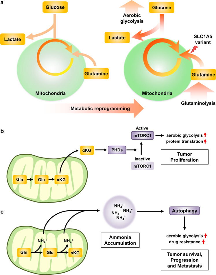
“Hence, the facilitation of glutaminolysis leads to the accumulation of excess ammonia within cells, and a high concentration of ammonia is a potent inducer of autophagy167”
§ Glutamine reliance in cell metabolism > Metabolic reprogramming induced by glutamine metabolism
What the sources say
- Not checked yetcites [167] Eng, C. H., Yu, K., Lucas, J., White, E. & Abraham, R. T. Ammonia derived from glutaminolysis is a diffusible regulator of autophagy. Sci. Signal. 3, ra31 (2010).
plaindirect- Asserted: passes — The authors put it forward as true.
- Checkable: passes — It can be checked against evidence.
- Load-bearing: passes — The paper’s conclusions depend on it.
- Based: passes — The paper gives a reason why it holds.
It may state two separate claims as one.
In the paper
“Upon glutamine starvation, p53 activity is induced and can help cancer cells adapt to nutrient starvation through diverse mechanisms148.”
§ Glutamine reliance in cell metabolism > Glutamine metabolic pathways > Glutamine metabolism upon cellular stresses
What the sources say
- Not checked yetcites [148] Reid, M. A. et al. The B55alpha subunit of PP2A drives a p53-dependent metabolic adaptation to glutamine deprivation. Mol. Cell 50, 200–211 (2013). source
plaindirect- Asserted: passes — The authors put it forward as true.
- Checkable: passes — It can be checked against evidence.
- Load-bearing: unclear — It is unclear whether the paper’s conclusions depend on it.
- Based: passes — The paper gives a reason why it holds.
It may state two separate claims as one.
None of the sources cited for this claim have been checked yet.
In the paper
“In addition, fumarate inhibits TET enzymes, which stimulate EMT, leading to cancer metastasis131,132.”
§ Glutamine reliance in cell metabolism > Glutamine metabolic pathways > Fumarate
What the sources say
- Not checked yetcites [131] Sciacovelli, M. et al. Fumarate is an epigenetic modifier that elicits epithelial-to-mesenchymal transition. Nature 537, 544–547 (2016). source
- Not checked yetcites [132] Laukka, T. et al. Fumarate and succinate regulate expression of hypoxia-inducible genes via TET enzymes. J. Biol. Chem. 291, 4256–4265 (2016). source
plaindirect- Asserted: passes — The authors put it forward as true.
- Checkable: passes — It can be checked against evidence.
- Load-bearing: unclear — It is unclear whether the paper’s conclusions depend on it.
- Based: passes — The paper gives a reason why it holds.
It may state two separate claims as one.
None of the sources cited for this claim have been checked yet.
In the paper
“In addition, mTORC1 activation via glutaminolysis suppresses autophagy and the DNA damage response164,165.”
§ Glutamine reliance in cell metabolism > Metabolic reprogramming induced by glutamine metabolism
What the sources say
- Not checked yetcites [164] Sanli, T., Steinberg, G. R., Singh, G. & Tsakiridis, T. AMP-activated protein kinase (AMPK) beyond metabolism: a novel genomic stress sensor participating in the DNA damage response pathway. Cancer Biol. Ther. 15, 156–169 (2014). source
- Not checked yetcites [165] Shen, C. & Houghton, P. J. The mTOR pathway negatively controls ATM by upregulating miRNAs. Proc. Natl Acad. Sci. USA 110, 11869–11874 (2013). source
plaindirect- Asserted: passes — The authors put it forward as true.
- Checkable: passes — It can be checked against evidence.
- Load-bearing: passes — The paper’s conclusions depend on it.
- Based: passes — The paper gives a reason why it holds.
It may state two separate claims as one.
In the paper
“Specifically, autophagy suppresses anoikis induced by the detachment of cancer cells from the extracellular matrix (ECM) and hence promotes metastasis169.”
§ Glutamine reliance in cell metabolism > Metabolic reprogramming induced by glutamine metabolism
What the sources say
- Not checked yetcites [169] Debnath, J. Detachment-induced autophagy during anoikis and lumen formation in epithelial acini. Autophagy 4, 351–353 (2008). source
plaindirect- Asserted: passes — The authors put it forward as true.
- Checkable: passes — It can be checked against evidence.
- Load-bearing: unclear — It is unclear whether the paper’s conclusions depend on it.
- Based: passes — The paper gives a reason why it holds.
It may state two separate claims as one.
None of the sources cited for this claim have been checked yet.
In the paper
“Unsurprisingly, glutamine metabolism plays a critical role in tumor progression since it not only supports mitochondrial oxidative phosphorylation but also supplies metabolic intermediates for the TCA cycle, glutathione synthesis, and NEAA synthesis and simultaneously produces NADPH157–159.”
§ Glutamine reliance in cell metabolism > Metabolic reprogramming induced by glutamine metabolism
What the sources say
- Not checked yetcites [157] Szeliga, M. & Obara-Michlewska, M. Glutamine in neoplastic cells: focus on the expression and roles of glutaminases. Neurochem. Int. 55, 71–75 (2009). source
- Not checked yetcites [158] Eng, C. H. & Abraham, R. T. Glutaminolysis yields a metabolic by-product that stimulates autophagy. Autophagy 6, 968–970 (2010). source
- Not checked yetcites [159] Rabinowitz, J. D. & White, E. Autophagy and metabolism. Science 330, 1344–1348 (2010). source
boosteddirect- Asserted: passes — The authors put it forward as true.
- Checkable: passes — It can be checked against evidence.
- Load-bearing: passes — The paper’s conclusions depend on it.
- Based: passes — The paper gives a reason why it holds.
It may state two separate claims as one.
None of the sources cited for this claim have been checked yet.
In the paper
“Accordingly, high mTORC1 activity in cancer cells promotes aerobic glycolysis and drives glucose addiction162,163”
§ Glutamine reliance in cell metabolism > Metabolic reprogramming induced by glutamine metabolism
What the sources say
- Not checked yetcites [162] Poulain, L. et al. High mTORC1 activity drives glycolysis addiction and sensitivity to G6PD inhibition in acute myeloid leukemia cells. Leukemia 31, 2326–2335 (2017). source
- Not checked yetcites [163] Dutchak, P. A. et al. Loss of a negative regulator of mTORC1 induces aerobic glycolysis and altered fiber composition in skeletal muscle. Cell Rep. 23, 1907–1914 (2018). source
plaindirect- Asserted: passes — The authors put it forward as true.
- Checkable: passes — It can be checked against evidence.
- Load-bearing: passes — The paper’s conclusions depend on it.
- Based: passes — The paper gives a reason why it holds.
It may state two separate claims as one.
A conclusion the paper draws from its own results — it cites no other source for it, so there is none to check. The quote shows it is in the paper.
In the paper
“Therefore, glutaminolysis can suppress autophagy by activating the mTORC1 pathway but, on the other hand, can stimulate autophagy in the context of excess ammonia production.”
§ Glutamine reliance in cell metabolism > Metabolic reprogramming induced by glutamine metabolism
plaindirect- Asserted: passes — The authors put it forward as true.
- Checkable: passes — It can be checked against evidence.
- Load-bearing: passes — The paper’s conclusions depend on it.
- Based: passes — The paper gives a reason why it holds.
It may state two separate claims as one.
In the paper
“The other mechanism by which succinate supports tumorigenesis acts through the inhibition of hypoxia-inducible factor prolyl hydroxylase (PHD). PHD activates the pseudohypoxic response by stabilizing HIF-1α, which is a well-known tumorigenesis enhancer”
§ Glutamine reliance in cell metabolism > Glutamine metabolic pathways > Succinate
What the sources say
- Not checked yetcites [127] Selak, M. A. et al. Succinate links TCA cycle dysfunction to oncogenesis by inhibiting HIF-alpha prolyl hydroxylase. Cancer Cell 7, 77–85 (2005). source
boosteddirect- Asserted: passes — The authors put it forward as true.
- Checkable: passes — It can be checked against evidence.
- Load-bearing: unclear — It is unclear whether the paper’s conclusions depend on it.
- Based: passes — The paper gives a reason why it holds.
It may state two separate claims as one. The quote only partly states this claim.
None of the sources cited for this claim have been checked yet.
In the paper
“Notably, cancer cells driven by oncogenic MYC, K-Ras, and PIK3CA require glutamine for their survival and display extensive anabolic utilization of glutamine29,174,175”
§ Glutamine reliance in cell metabolism > Crosstalk between glutamine metabolism and oncogenic signaling
What the sources say
- Not checked yetcites [29] Son, J. et al. Glutamine supports pancreatic cancer growth through a KRAS-regulated metabolic pathway. Nature https://doi.org/10.1038/nature12317 (2013). source
- Not checked yetcites [174] Dang, C. V. Rethinking the Warburg effect with Myc micromanaging glutamine metabolism. Cancer Res. 70, 859–862 (2010). source
- Not checked yetcites [175] Hao, Y. et al. Oncogenic PIK3CA mutations reprogram glutamine metabolism in colorectal cancer. Nat. Commun. 7, 11971 (2016). source
plaindirect- Asserted: passes — The authors put it forward as true.
- Checkable: passes — It can be checked against evidence.
- Load-bearing: unclear — It is unclear whether the paper’s conclusions depend on it.
- Based: passes — The paper gives a reason why it holds.
It may state two separate claims as one.
In the paper
“Therefore, enhanced glutaminolysis might eventually contribute to the initiation and progression of cancer by stimulating cell growth via the mTORC1 pathway and enhancing aerobic glycolysis while disrupting the proper elimination of misfolded proteins, damaged DNA and organelles through the inhibition of autophagy and the DNA damage response166.”
§ Glutamine reliance in cell metabolism > Metabolic reprogramming induced by glutamine metabolism
What the sources say
- Not checked yetcites [166] Villar, V. H., Merhi, F., Djavaheri-Mergny, M. & Duran, R. V. Glutaminolysis and autophagy in cancer. Autophagy 11, 1198–1208 (2015). source
hedgedconditional- Asserted: passes — The authors put it forward as true.
- Checkable: passes — It can be checked against evidence.
- Load-bearing: passes — The paper’s conclusions depend on it.
- Based: passes — The paper gives a reason why it holds.
It may state two separate claims as one.
A conclusion the paper draws from its own results — it cites no other source for it, so there is none to check. The quote shows it is in the paper.
In the paper
“Collectively, cancer glutamine metabolism shows extraordinary flexibility and is intertwined with diverse metabolic pathways.”
§ Glutamine reliance in cell metabolism > Glutamine metabolic pathways > Glutamine metabolism upon cellular stresses
plaincorrelational- Asserted: passes — The authors put it forward as true.
- Checkable: passes — It can be checked against evidence.
- Load-bearing: passes — The paper’s conclusions depend on it.
- Based: passes — The paper gives a reason why it holds.
It may state two separate claims as one.
In the paper
“Moreover, evidence has shown that during the DNA damage response, normal cells show a decrease in glutaminolysis controlled by SIRT4 protein suppressing GLUD1. In the absence of SIRT4, a failure to undergo cell cycle arrest induced by DNA damage causes a delay in DNA repair and increased chromosomal instability, suggesting a tumor suppressor effect of SIRT4151.”
§ Glutamine reliance in cell metabolism > Glutamine metabolic pathways > Glutamine metabolism upon cellular stresses
What the sources say
- Not checked yetcites [151] Jeong, S. M. et al. SIRT4 has tumor-suppressive activity and regulates the cellular metabolic response to DNA damage by inhibiting mitochondrial glutamine metabolism. Cancer Cell 23, 450–463 (2013). source
hedgeddirect- Asserted: passes — The authors put it forward as true.
- Checkable: passes — It can be checked against evidence.
- Load-bearing: unclear — It is unclear whether the paper’s conclusions depend on it.
- Based: passes — The paper gives a reason why it holds.
It may state two separate claims as one.
In the paper
“Under DNA damage such as radiation, glutamine is conditionally essential to support the synthesis of nucleotides and redox homeostasis. It has recently been demonstrated that radioresistant cancer cells reprogram metabolic flux toward glutamine anabolism. Under these conditions, cancer cells highly express glutamine synthetase, facilitating cancer cell growth under radiation stress150.”
§ Glutamine reliance in cell metabolism > Glutamine metabolic pathways > Glutamine metabolism upon cellular stresses
What the sources say
- Not checked yetcites [150] Fu, S. J. et al. Glutamine synthetase promotes radiation resistance via facilitating nucleotide metabolism and subsequent DNA damage repair. Cell Rep. 28, 1136 (2019). source
boosteddirect- Asserted: passes — The authors put it forward as true.
- Checkable: passes — It can be checked against evidence.
- Load-bearing: unclear — It is unclear whether the paper’s conclusions depend on it.
- Based: passes — The paper gives a reason why it holds.
It may state two separate claims as one.
In the paper
“Intriguingly, this study regarding elevated glutamine metabolism in cancer cells also showed that glutaminolysis could in turn reinforce metabolic reprogramming, thus implying that glutamine metabolism plays a crucial role in tumorigenesis and tumor progression16”
§ Glutamine reliance in cell metabolism > Metabolic reprogramming induced by glutamine metabolism
What the sources say
- Not checked yetcites [16] Yoo, H. C. et al. A variant of SLC1A5 is a mitochondrial glutamine transporter for metabolic reprogramming in cancer cells. Cell Metab. 31, 267–283 (2020). source
hedgedconditional- Asserted: passes — The authors put it forward as true.
- Checkable: passes — It can be checked against evidence.
- Load-bearing: passes — The paper’s conclusions depend on it.
- Based: passes — The paper gives a reason why it holds.
It may state two separate claims as one.
In the paper
“Additionally, the level of the mitochondrial glutamine transporter controls the cellular ATP level stimulated by glutamine, suggesting that glutamine is an important energy source via mitochondrial glutaminolysis16.”
§ Glutamine reliance in cell metabolism > Glutamine metabolic pathways > Glutamine-derived energy production
What the sources say
- Not checked yetcites [16] Yoo, H. C. et al. A variant of SLC1A5 is a mitochondrial glutamine transporter for metabolic reprogramming in cancer cells. Cell Metab. 31, 267–283 (2020). source
hedgeddirect- Asserted: passes — The authors put it forward as true.
- Checkable: passes — It can be checked against evidence.
- Load-bearing: passes — The paper’s conclusions depend on it.
- Based: passes — The paper gives a reason why it holds.
It may state two separate claims as one.
None of the sources cited for this claim have been checked yet.
In the paper
“In 2001, mutation of fumarate hydratase leading to its inactivation was discovered in renal cell cancer129. Mutation of this enzyme leads to fumarate accumulation not only in skin cancer and uterine leiomyomas but also in breast, bladder, and Leydig cell tumors130.”
§ Glutamine reliance in cell metabolism > Glutamine metabolic pathways > Fumarate
What the sources say
- Not checked yetcites [129] Virpi Launonen, O. V. et al. Inherited susceptibility to uterine leiomyomas and renal cell cancer. PNAS 98, 3387–3392 (2001). source
- Not checked yetcites [130] Tomlinson, I. P. et al. Germline mutations in FH predispose to dominantly inherited uterine fibroids, skin leiomyomata and papillary renal cell cancer. Nat. Genet. 30, 406–410 (2002). source
plaindirect- Asserted: passes — The authors put it forward as true.
- Checkable: passes — It can be checked against evidence.
- Load-bearing: unclear — It is unclear whether the paper’s conclusions depend on it.
- Based: passes — The paper gives a reason why it holds.
It may state two separate claims as one.
A conclusion the paper draws from its own results — it cites no other source for it, so there is none to check. The quote shows it is in the paper.
In the paper
“In conclusion, metabolic reliance on glutamine arises via the intrinsic functional diversity of glutamine, supporting macromolecule biosynthesis and reinforcing the TCA cycle.”
§ Glutamine reliance in cell metabolism > Conclusion
plaindirect- Asserted: passes — The authors put it forward as true.
- Checkable: passes — It can be checked against evidence.
- Load-bearing: passes — The paper’s conclusions depend on it.
- Based: unclear — It is unclear why the paper says it holds.
It may state two separate claims as one.
In the paper
“Furthermore, human glioblastoma cells do not rely much on circulating glutamine for proliferation but rather more on glutamate to synthesize glutamine via glutamine synthetase to fuel purine biosynthesis193.”
§ Glutamine reliance in cell metabolism > Targeting glutamine metabolism and therapeutic implications
What the sources say
- Not checked yetcites [193] Tardito, S. et al. Glutamine synthetase activity fuels nucleotide biosynthesis and supports growth of glutamine-restricted glioblastoma. Nat. Cell Biol. 17, 1556–1568 (2015). source
plaindirect- Asserted: passes — The authors put it forward as true.
- Checkable: passes — It can be checked against evidence.
- Load-bearing: unclear — It is unclear whether the paper’s conclusions depend on it.
- Based: passes — The paper gives a reason why it holds.
It may state two separate claims as one.
In the paper
“Consistent with this observation, K-Ras sensitizes cells to glutamine deprivation, and K-Ras knockdown rescues cells from apoptosis induced by low glutamine levels187.”
§ Glutamine reliance in cell metabolism > Crosstalk between glutamine metabolism and oncogenic signaling
What the sources say
- Not checked yetcites [187] Gwinn, D. M. et al. Oncogenic KRAS regulates amino acid homeostasis and asparagine biosynthesis via ATF4 and alters sensitivity to L-asparaginase. Cancer Cell 33, 91–107 (2018). source
plaindirect- Asserted: passes — The authors put it forward as true.
- Checkable: passes — It can be checked against evidence.
- Load-bearing: passes — The paper’s conclusions depend on it.
- Based: passes — The paper gives a reason why it holds.
It may state two separate claims as one.
In the paper
“The researchers showed that blocking glutamine metabolism through JHU083 not only suppressed tumor cell metabolism but also mitigated the tumor microenvironment, which is hostile to the immune response due to its hypoxic, acidic, and nutrient-depleted conditions, unleashing the natural antitumor T cell response.”
§ Glutamine reliance in cell metabolism > Targeting glutamine metabolism and therapeutic implications
What the sources say
- Not checked yetcites [200] Leone, R. D. et al. Glutamine blockade induces divergent metabolic programs to overcome tumor immune evasion. Science 366, 1013 (2019). source
boosteddirect- Asserted: passes — The authors put it forward as true.
- Checkable: passes — It can be checked against evidence.
- Load-bearing: passes — The paper’s conclusions depend on it.
- Based: passes — The paper gives a reason why it holds.
It may state two separate claims as one.
A conclusion the paper draws from its own results — it cites no other source for it, so there is none to check. The quote shows it is in the paper.
In the paper
“In the context of tumorigenesis, glutamine-derived 2-HG alters the epigenetic landscape of chromosomes and induces oncogenic transformation.”
§ Glutamine reliance in cell metabolism > Conclusion
plaindirect- Asserted: passes — The authors put it forward as true.
- Checkable: passes — It can be checked against evidence.
- Load-bearing: passes — The paper’s conclusions depend on it.
- Based: unclear — It is unclear why the paper says it holds.
It may state two separate claims as one.
- The authors state this as an assumption: R-2-HG inhibition of α-KG-dependent dioxygenases and demethylases is a driver of tumorigenesis.
In the paper
“According to another study, glutaminolysis increases the level of α-KG production, resulting in GTP loading of RagB and lysosomal translocation of the mTORC1 complex in human cancer cell lines160.”
§ Glutamine reliance in cell metabolism > Crosstalk between glutamine metabolism and oncogenic signaling
What the sources say
- Not checked yetcites [160] Duran, R. V. & Hall, M. N. Glutaminolysis feeds mTORC1. Cell Cycle 11, 4107–4108 (2012). source
plaindirect- Asserted: passes — The authors put it forward as true.
- Checkable: passes — It can be checked against evidence.
- Load-bearing: unclear — It is unclear whether the paper’s conclusions depend on it.
- Based: passes — The paper gives a reason why it holds.
It may state two separate claims as one.
In the paper
“They also confirmed that concurrent treatment with JHU083 and anti-PD-1 checkpoint inhibitor improved the antitumor effects compared with anti-PD-1 treatment alone, suggesting the presence of metabolic plasticity between cancer cells and effector T cells, which could be exploited as a metabolic checkpoint for cancer immunotherapy200.”
§ Glutamine reliance in cell metabolism > Targeting glutamine metabolism and therapeutic implications
What the sources say
- Not checked yetcites [200] Leone, R. D. et al. Glutamine blockade induces divergent metabolic programs to overcome tumor immune evasion. Science 366, 1013 (2019). source
hedgeddirect- Asserted: passes — The authors put it forward as true.
- Checkable: passes — It can be checked against evidence.
- Load-bearing: passes — The paper’s conclusions depend on it.
- Based: passes — The paper gives a reason why it holds.
It may state two separate claims as one.
In the paper
“Accordingly, loss of SIRT4 enhances glutamine-dependent proliferation and genomic instability, which simultaneously contribute to tumorigenesis151.”
§ Glutamine reliance in cell metabolism > Crosstalk between glutamine metabolism and oncogenic signaling
What the sources say
- Not checked yetcites [151] Jeong, S. M. et al. SIRT4 has tumor-suppressive activity and regulates the cellular metabolic response to DNA damage by inhibiting mitochondrial glutamine metabolism. Cancer Cell 23, 450–463 (2013). source
plaindirect- Asserted: passes — The authors put it forward as true.
- Checkable: passes — It can be checked against evidence.
- Load-bearing: passes — The paper’s conclusions depend on it.
- Based: passes — The paper gives a reason why it holds.
It may state two separate claims as one.
In the paper
“In addition, SLC1A5 was shown to have clinical importance, and it is considered the most critical plasma membrane glutamine transporter in cancer cells201.”
§ Glutamine reliance in cell metabolism > Targeting glutamine metabolism and therapeutic implications
What the sources say
- Not checked yetcites [201] Liu, Y. et al. The role of ASCT2 in cancer: a review. Eur. J. Pharm. 837, 81–87 (2018). source
plain- Asserted: passes — The authors put it forward as true.
- Checkable: passes — It can be checked against evidence.
- Load-bearing: unclear — It is unclear whether the paper’s conclusions depend on it.
- Based: passes — The paper gives a reason why it holds.
It may state two separate claims as one.
In the paper
“GLS overexpression has been observed in different tumor cells, and these enzymes are found to function in the metabolic reprogramming of glutamine addiction in cancer198.”
§ Glutamine reliance in cell metabolism > Targeting glutamine metabolism and therapeutic implications
What the sources say
- Not checked yetcites [198] Lobo, C. et al. Inhibition of glutaminase expression by antisense mRNA decreases growth and tumourigenicity of tumour cells. Biochem. J. 348, 257–261 (2000). source
plaindirect- Asserted: passes — The authors put it forward as true.
- Checkable: passes — It can be checked against evidence.
- Load-bearing: passes — The paper’s conclusions depend on it.
- Based: passes — The paper gives a reason why it holds.
It may state two separate claims as one.
A conclusion the paper draws from its own results — it cites no other source for it, so there is none to check. The quote shows it is in the paper.
In the paper
“SLC1A5 might not be an appropriate target for suppressing glutamine uptake by cancer cells because it is not the only plasma membrane glutamine transporter, and its function would therefore be compensated by other redundant glutamine transporters such as SLC38A1 and SLC38A2.”
§ Glutamine reliance in cell metabolism > Targeting glutamine metabolism and therapeutic implications
hedgedconditional- Asserted: passes — The authors put it forward as true.
- Checkable: passes — It can be checked against evidence.
- Load-bearing: passes — The paper’s conclusions depend on it.
- Based: passes — The paper gives a reason why it holds.
It may state two separate claims as one.
None of the sources cited for this claim have been checked yet.
In the paper
“However, other researchers have argued that controversial issues exist because GPNA also inhibits other glutamine transporters, such as SLC38A1, and V-9302 is effective even in SLC1A5 knockout models206,207.”
§ Glutamine reliance in cell metabolism > Targeting glutamine metabolism and therapeutic implications
What the sources say
- Not checked yetcites [206] Broer, A., Fairweather, S. & Broer, S. Disruption of amino acid homeostasis by novel ASCT2 inhibitors involves multiple targets. Front. Pharm. 9, 785 (2018). source
- Not checked yetcites [207] Chiu, M. et al. GPNA inhibits the sodium-independent transport system L for neutral amino acids. Amino Acids 49, 1365–1372 (2017). source
plaindirect- Asserted: passes — The authors put it forward as true.
- Checkable: passes — It can be checked against evidence.
- Load-bearing: passes — The paper’s conclusions depend on it.
- Based: passes — The paper gives a reason why it holds.
Probably not a substantial claim. It may state two separate claims as one.
In the paper
“Among these agents, CB-839 is the only one to proceed to clinical trials; however, its selectivity toward GLS1 and failure to inhibit the compensatory effect of GLS2 require in-depth study14.”
§ Glutamine reliance in cell metabolism > Targeting glutamine metabolism and therapeutic implications
What the sources say
- Not checked yetcites [14] Gross, M. I. et al. Antitumor activity of the glutaminase inhibitor CB-839 in triple-negative breast cancer. Mol. Cancer Ther. 13, 890–901 (2014). source
plain- Asserted: passes — The authors put it forward as true.
- Checkable: passes — It can be checked against evidence.
- Load-bearing: unclear — It is unclear whether the paper’s conclusions depend on it.
- Based: passes — The paper gives a reason why it holds.
It may state two separate claims as one.
In the paper
“Importantly, activation of K-Ras in cancer cells results in bypass of the late G1 glutamine-dependent checkpoint. Specifically, glutamine deprivation in K-Ras-driven cancer cells leads to growth arrest in S or G2/M phase instead of in G1 phase.”
§ Glutamine reliance in cell metabolism > Crosstalk between glutamine metabolism and oncogenic signaling
What the sources say
- Not checked yetcites [185] Saqcena, M. et al. Blocking anaplerotic entry of glutamine into the TCA cycle sensitizes K-Ras mutant cancer cells to cytotoxic drugs. Oncogene 34, 2672–2680 (2015). source
plaindirect- Asserted: passes — The authors put it forward as true.
- Checkable: passes — It can be checked against evidence.
- Load-bearing: passes — The paper’s conclusions depend on it.
- Based: passes — The paper gives a reason why it holds.
It may state two separate claims as one.
In the paper
“Moreover, glutamine also acts as a precursor for the synthesis of various NEAAs, including asparagine and arginine, implicated in mTORC1 activation39.”
§ Glutamine reliance in cell metabolism > Crosstalk between glutamine metabolism and oncogenic signaling
What the sources say
- Not checked yetcites [39] Choi, B. H. & Coloff, J. L. The diverse functions of non-essential amino acids in cancer. Cancers 11, 675 (2019). source
plaincorrelational- Asserted: passes — The authors put it forward as true.
- Checkable: passes — It can be checked against evidence.
- Load-bearing: unclear — It is unclear whether the paper’s conclusions depend on it.
- Based: passes — The paper gives a reason why it holds.
It may state two separate claims as one.
The paper relies on this without support.
The core region of solid tumors exhibits a deficiency of glutamine compared with other amino acids.
“Moreover, the core region of solid tumors exhibits a deficiency of glutamine compared with other amino acids.”
The authors state this as an assumption.
R-2-HG inhibition of α-KG-dependent dioxygenases and demethylases is a driver of tumorigenesis.
“which has been assumed to be a driver of tumorigenesis117,118”
The paper relies on this without support.
Therapeutic effects of targeting glutamine metabolism observed in preclinical cancer models will translate to human cancers.
“Certain types of cancer, including renal cell carcinoma, hematologic malignancies, glioblastoma, pancreatic cancer, and those reported to depend on HIF-2α, seem to depend on glutamine; hence, targeting glutamine metabolism may show therapeutic effects in these cancers.”
Claims other papers make about this one
Each is checked against this paper’s own text: does it say what the citing paper says it does?
The source backs part of the claim, not all of it — or backs it for a different population, condition or comparison.
In the citing paper
“Glutamine (Gln), a nonessential amino acid (NEAA), is primarily produced by the enzyme Gln synthetase (GS) using glutamate (Glu) and ammonia (NH3) as substrates. It has been recognized as a significant regulator of osteoblasts due to the increased demand for amino acids resulting from the augmented synthesis of the bone matrix during bone formation [61].”
From Mesenchymal stromal cells in bone marrow niche of patients with multiple myeloma: a double-edged sword (2025) · cites this paper as [61]
Experimental — accuracy not yet measuredIn the citing paper
“Glutamine is also a major source of nitrogen for proliferating cells52,54.”
From The metabolic landscape of RAS-driven cancers from biology to therapy (2021) · cites this paper as [54]
Self-check
Percentages, sums and reported differences re-computed from the paper’s own numbers.
Each contradiction between two passages listed here was confirmed by an independent check.
Rated minor: it would not touch a result — a value no result rests on, a p-value that stays on the same side of the significance level, or a conflict that is clerical, resolved elsewhere in the paper, or outside its own findings.
In the paper
§ Glutamine reliance in cell metabolism > Glutamine metabolic pathways > Succinate
“The other mechanism by which succinate supports tumorigenesis acts through the inhibition of hypoxia-inducible factor prolyl hydroxylase (PHD).”
§ Glutamine reliance in cell metabolism > Glutamine metabolic pathways > Succinate
“PHD activates the pseudohypoxic response by stabilizing HIF-1α, which is a well-known tumorigenesis enhancer, and as a transcription factor, maintains the metabolic reprogramming of cancer cells to support their survival127.”
Rated minor: it would not touch a result — a value no result rests on, a p-value that stays on the same side of the significance level, or a conflict that is clerical, resolved elsewhere in the paper, or outside its own findings.
In the paper
§ Glutamine reliance in cell metabolism > Targeting glutamine metabolism and therapeutic implications
“The plasma membrane glutamine transporters SLC6A14, SLC7A11, and SLC38A1 have been targeted and found to be inhibited by erastin, α-Me-Trp, and MeAIB, respectively (Fig. 8).”
§ Glutamine reliance in cell metabolism > Targeting glutamine metabolism and therapeutic implications
“SLC6A14 and SLC38A1 are inhibited by α-Me-Trp and MeIAB, respectively. The most intensely researched topic is inhibitors of SLC1A5, a major glutamine transporter, which include substrate analog competitive inhibitors such as GPNA, benzylserine, and V-9302 and the inhibitory antibody MEDI7247. Although they exhibit low potency, inhibitors of SLC7A11 include erastin and SSZ.”
Rated minor: it would not touch a result — a value no result rests on, a p-value that stays on the same side of the significance level, or a conflict that is clerical, resolved elsewhere in the paper, or outside its own findings.
In the paper
§ Glutamine reliance in cell metabolism > Glutamine metabolic pathways > Nucleotides synthesized from glutamine
“One more glutamine molecule is used to synthesize cytidine triphosphate (CTP) from uridine triphosphate (UTP)31.”
§ Glutamine reliance in cell metabolism > Glutamine metabolic pathways > Nucleotides synthesized from glutamine
“two glutamine molecules are spent to convert UTP into CTP.”
Rated minor: it would not touch a result — a value no result rests on, a p-value that stays on the same side of the significance level, or a conflict that is clerical, resolved elsewhere in the paper, or outside its own findings.
In the paper
§ Glutamine reliance in cell metabolism > Glutamine metabolic pathways > Fumarate
“Fumarate is another example of an oncometabolite produced by the action of fumarate hydratase on succinate.”
§ Glutamine reliance in cell metabolism > Glutamine metabolic pathways > Succinate
“The normally functioning SDH enzyme is localized in the inner mitochondrial membrane and plays a role in the electron transport chain as well as the conversion of succinate into fumarate.”
Rated minor: it would not touch a result — a value no result rests on, a p-value that stays on the same side of the significance level, or a conflict that is clerical, resolved elsewhere in the paper, or outside its own findings.
In the paper
§ Glutamine reliance in cell metabolism > Glutamine metabolic pathways > Control of epigenetic changes by glutamine
“Mutation of IDH1 and IDH2 produces R-2-HG from α-KG, which, when accumulated, leads to the inhibition of dioxygenases, in turn leading to the activation of TET and JHDM enzymes inside the nucleus.”
§ Glutamine reliance in cell metabolism > Glutamine metabolic pathways > R-2-HG
“The role of R-2-HG as an oncometabolite has been implicated in epigenetic modifications through the inhibition of α-KG-dependent dioxygenases and demethylases”
Citations
Cites
Understanding the Warburg Effect: The Metabolic Requirements of Cell Proliferation
Vander Heiden, M. G., Cantley, L. C., & Thompson, C. B. (2009) · Science · doi:10.1126/science.1160809 · Cited by 16,197
Warburg, O. (1956) · Science · doi:10.1126/science.123.3191.309 · Cited by 13,425
Cancer-associated IDH1 mutations produce 2-hydroxyglutarate
Dang, L., White, D. W., Gross, S., Bennett, B. D., Bittinger, M. A., Driggers, E. M., Fantin, V. R., Jang, H. G., Jin, S., Keenan, M. C., et al. (2009) · Nature · doi:10.1038/nature08617 · Cited by 4,154
Kim, J.-W., Tchernyshyov, I., Semenza, G. L., & Dang, C. V. (2006) · Cell Metabolism · doi:10.1016/j.cmet.2006.02.002 · Cited by 3,905
Modulation of oxidative stress as an anticancer strategy
Gorrini, C., Harris, I. S., & Mak, T. W. (2013) · Nature Reviews Drug Discovery · doi:10.1038/nrd4002 · Cited by 3,579
Mitochondrial TCA cycle metabolites control physiology and disease
Martínez-Reyes, I., & Chandel, N. S. (2020) · Nature Communications · doi:10.1038/s41467-019-13668-3 · Cited by 2,813
Figueroa, M. E., Abdel-Wahab, O., Lu, C., Ward, P. S., Patel, J., Shih, A., Li, Y., Bhagwat, N., Vasanthakumar, A., Fernandez, H. F., et al. (2010) · Cancer Cell · doi:10.1016/j.ccr.2010.11.015 · Cited by 2,766
Gao, P., Tchernyshyov, I., Chang, T.-C., Lee, Y.-S., Kita, K., Ochi, T., Zeller, K. I., De Marzo, A. M., Van Eyk, J. E., Mendell, J. T., et al. (2009) · Nature · doi:10.1038/nature07823 · Cited by 2,124
Cellular Fatty Acid Metabolism and Cancer
Currie, E., Schulze, A., Zechner, R., Walther, T. C., & Farese, R. V. (2013) · Cell Metabolism · doi:10.1016/j.cmet.2013.05.017 · Cited by 2,123
From Krebs to clinic: glutamine metabolism to cancer therapy
Altman, B. J., Stine, Z. E., & Dang, C. V. (2016) · Nature Reviews Cancer · doi:10.1038/nrc.2016.71 · Cited by 2,119
Succinate links TCA cycle dysfunction to oncogenesis by inhibiting HIF-α prolyl hydroxylase
Selak, M. A., Armour, S. M., MacKenzie, E. D., Boulahbel, H., Watson, D. G., Mansfield, K. D., Pan, Y., Simon, M. C., Thompson, C. B., & Gottlieb, E. (2005) · Cancer Cell · doi:10.1016/j.ccr.2004.11.022 · Cited by 2,108
Ward, P. S., Patel, J., Wise, D. R., Abdel-Wahab, O., Bennett, B. D., Coller, H. A., Cross, J. R., Fantin, V. R., Hedvat, C. V., Perl, A. E., et al. (2010) · Cancer Cell · doi:10.1016/j.ccr.2010.01.020 · Cited by 2,020
Wise, D. R., DeBerardinis, R. J., Mancuso, A., Sayed, N., Zhang, X.-Y., Pfeiffer, H. K., Nissim, I., Daikhin, E., Yudkoff, M., McMahon, S. B., et al. (2008) · Proceedings of the National Academy of Sciences · doi:10.1073/pnas.0810199105 · Cited by 2,001
Glutamine supports pancreatic cancer growth through a KRAS-regulated metabolic pathway
Son, J., Lyssiotis, C. A., Ying, H., Wang, X., Hua, S., Ligorio, M., Perera, R. M., Ferrone, C. R., Mullarky, E., Shyh-Chang, N., et al. (2013) · Nature · doi:10.1038/nature12040 · Cited by 1,990
IDH mutation impairs histone demethylation and results in a block to cell differentiation
Lu, C., Ward, P. S., Kapoor, G. S., Rohle, D., Turcan, S., Abdel-Wahab, O., Edwards, C. R., Khanin, R., Figueroa, M. E., Melnick, A., et al. (2012) · Nature · doi:10.1038/nature10860 · Cited by 1,985
Rabinowitz, J. D., & White, E. (2010) · Science · doi:10.1126/science.1193497 · Cited by 1,982
Bidirectional Transport of Amino Acids Regulates mTOR and Autophagy
Nicklin, P., Bergman, P., Zhang, B., Triantafellow, E., Wang, H., Nyfeler, B., Yang, H., Hild, M., Kung, C., Wilson, C., et al. (2009) · Cell · doi:10.1016/j.cell.2008.11.044 · Cited by 1,798
Reductive glutamine metabolism by IDH1 mediates lipogenesis under hypoxia
Metallo, C. M., Gameiro, P. A., Bell, E. L., Mattaini, K. R., Yang, J., Hiller, K., Jewell, C. M., Johnson, Z. R., Irvine, D. J., Guarente, L., et al. (2012) · Nature · doi:10.1038/nature10602 · Cited by 1,791
Glutamine addiction: a new therapeutic target in cancer
Wise, D. R., & Thompson, C. B. (2010) · Trends in Biochemical Sciences · doi:10.1016/j.tibs.2010.05.003 · Cited by 1,770
Mitochondrial metabolism and ROS generation are essential for Kras-mediated tumorigenicity
Weinberg, F., Hamanaka, R., Wheaton, W. W., Weinberg, S., Joseph, J., Lopez, M., Kalyanaraman, B., Mutlu, G. M., Budinger, G. R. S., & Chandel, N. S. (2010) · Proceedings of the National Academy of Sciences · doi:10.1073/pnas.1003428107 · Cited by 1,742
Hypoxia, HIF1 and glucose metabolism in the solid tumour
Denko, N. C. (2008) · Nature Reviews Cancer · doi:10.1038/nrc2468 · Cited by 1,707
The Multiple Leiomyoma Consortium (2002) · Nature Genetics · doi:10.1038/ng849 · Cited by 1,635
Nutrition Needs of Mammalian Cells in Tissue Culture
Eagle, H. (1955) · Science · doi:10.1126/science.122.3168.501 · Cited by 1,619
Stine, Z. E., Walton, Z. E., Altman, B. J., Hsieh, A. L., & Dang, C. V. (2015) · Cancer Discovery · doi:10.1158/2159-8290.cd-15-0507 · Cited by 1,435
Q's next: the diverse functions of glutamine in metabolism, cell biology and cancer
DeBerardinis, R. J., & Cheng, T. (2010) · Oncogene · doi:10.1038/onc.2009.358 · Cited by 1,365
Hypoxia-inducible factors regulate tumorigenic capacity of glioma stem cells
Li, Z., Bao, S., Wu, Q., Wang, H., Eyler, C., Sathornsumetee, S., Shi, Q., Cao, Y., Lathia, J., McLendon, R. E., et al. (2009) · Cancer Cell · doi:10.1016/j.ccr.2009.03.018 · Cited by 1,354
Cancer metabolism: fatty acid oxidation in the limelight
Carracedo, A., Cantley, L. C., & Pandolfi, P. P. (2013) · Nature reviews. Cancer · doi:10.1038/nrc3483 · Cited by 1,353
Reductive carboxylation supports growth in tumour cells with defective mitochondria
Mullen, A. R., Wheaton, W. W., Jin, E. S., Chen, P.-H., Sullivan, L. B., Cheng, T., Yang, Y., Linehan, W. M., Chandel, N. S., & DeBerardinis, R. J. (2012) · Nature · doi:10.1038/nature10642 · Cited by 1,328
α-ketoglutarate orchestrates macrophage activation through metabolic and epigenetic reprogramming
Liu, P.-S., Wang, H., Li, X., Chao, T., Teav, T., Christen, S., Di Conza, G., Cheng, W.-C., Chou, C.-H., Vavakova, M., et al. (2017) · Nature Immunology · doi:10.1038/ni.3796 · Cited by 1,264
Glutamine and cancer: cell biology, physiology, and clinical opportunities
Hensley, C. T., Wasti, A. T., & DeBerardinis, R. J. (2013) · Journal of Clinical Investigation · doi:10.1172/jci69600 · Cited by 1,262
Evidence that glutamine, not sugar, is the major energy source for cultured HeLa cells.
Reitzer, L. J., Wice, B. M., & Kennell, D. (1979) · Journal of Biological Chemistry · doi:10.1016/s0021-9258(17)30124-2 · Cited by 1,240
Supporting aspartate biosynthesis is an essential function of respiration in proliferating cells
Sullivan, L. B., Gui, D. Y., Hosios, A. M., Bush, L. N., Freinkman, E., & Vander Heiden, M. G. (2015) · Cell · doi:10.1016/j.cell.2015.07.017 · Cited by 1,173
Glutamine blockade induces divergent metabolic programs to overcome tumor immune evasion
Leone, R. D., Zhao, L., Englert, J. M., Sun, I.-M., Oh, M.-H., Sun, I.-H., Arwood, M. L., Bettencourt, I. A., Patel, C. H., Wen, J., et al. (2019) · Science · doi:10.1126/science.aav2588 · Cited by 1,169
Glucose-Independent Glutamine Metabolism via TCA Cycling for Proliferation and Survival in B Cells
Le, A., Lane, A. N., Hamaker, M., Bose, S., Gouw, A., Barbi, J., Tsukamoto, T., Rojas, C. J., Slusher, B. S., Zhang, H., et al. (2012) · Cell Metabolism · doi:10.1016/j.cmet.2011.12.009 · Cited by 1,095
Astuti, D., Latif, F., Dallol, A., Dahia, P. L. M., Douglas, F., George, E., Sköldberg, F., Husebye, E. S., Eng, C., & Maher, E. R. (2001) · The American Journal of Human Genetics · doi:10.1086/321282 · Cited by 1,087
Antitumor activity of the glutaminase inhibitor CB-839 in triple-negative breast cancer
Gross, M. I., Demo, S. D., Dennison, J. B., Chen, L., Chernov-Rogan, T., Goyal, B., Janes, J. R., Laidig, G. J., Lewis, E. R., Li, J., et al. (2014) · Molecular Cancer Therapeutics · doi:10.1158/1535-7163.mct-13-0870 · Cited by 1,068
Amary, M. F., Bacsi, K., Maggiani, F., Damato, S., Halai, D., Berisha, F., Pollock, R., O'Donnell, P., Grigoriadis, A., Diss, T., et al. (2011) · The Journal of Pathology · doi:10.1002/path.2913 · Cited by 1,023
Intracellular α-ketoglutarate maintains the pluripotency of embryonic stem cells
Carey, B. W., Finley, L. W. S., Cross, J. R., Allis, C. D., & Thompson, C. B. (2015) · Nature · doi:10.1038/nature13981 · Cited by 1,007
Wise, D. R., Ward, P. S., Shay, J. E. S., Cross, J. R., Gruber, J. J., Sachdeva, U. M., Platt, J. M., DeMatteo, R. G., Simon, M. C., & Thompson, C. B. (2011) · Proceedings of the National Academy of Sciences · doi:10.1073/pnas.1117773108 · Cited by 975
Regulation of mammalian nucleotide metabolism and biosynthesis
Lane, A. N., & Fan, T. W.-M. (2015) · Nucleic Acids Research · doi:10.1093/nar/gkv047 · Cited by 959
Isaacs, J. S., Jung, Y. J., Mole, D. R., Lee, S., Torres-Cabala, C., Chung, Y.-L., Merino, M., Trepel, J., Zbar, B., Toro, J., et al. (2005) · Cancer Cell · doi:10.1016/j.ccr.2005.06.017 · Cited by 958
Mutations in SDHC cause autosomal dominant paraganglioma, type 3
Niemann, S., & Müller, U. (2000) · Nature Genetics · doi:10.1038/81551 · Cited by 875
Glutaminolysis: A Hallmark of Cancer Metabolism
Yang, L., Venneti, S., & Nagrath, D. (2017) · Annual Review of Biomedical Engineering · doi:10.1146/annurev-bioeng-071516-044546 · Cited by 859
Targeting Mitochondrial Glutaminase Activity Inhibits Oncogenic Transformation
Wang, J.-B., Erickson, J. W., Fuji, R., Ramachandran, S., Gao, P., Dinavahi, R., Wilson, K. F., Ambrosio, A. L. B., Dias, S. M. G., Dang, C. V., et al. (2010) · Cancer Cell · doi:10.1016/j.ccr.2010.08.009 · Cited by 821
SDH mutations establish a hypermethylator phenotype in paraganglioma
Letouzé, E., Martinelli, C., Loriot, C., Burnichon, N., Abermil, N., Ottolenghi, C., Janin, M., Menara, M., Nguyen, A. T., Benit, P., et al. (2013) · Cancer Cell · doi:10.1016/j.ccr.2013.04.018 · Cited by 776
Differential regulation of mTORC1 by leucine and glutamine
Jewell, J. L., Kim, Y. C., Russell, R. C., Yu, F.-X., Park, H. W., Plouffe, S. W., Tagliabracci, V. S., & Guan, K.-L. (2015) · Science · doi:10.1126/science.1259472 · Cited by 757
Stimulation of de novo pyrimidine synthesis by growth signaling through mTOR and S6K1
Ben-Sahra, I., Howell, J. J., Asara, J. M., & Manning, B. D. (2013) · Science · doi:10.1126/science.1228792 · Cited by 739
Environment impacts the metabolic dependencies of Ras-driven non-small cell lung cancer
Davidson, S. M., Papagiannakopoulos, T., Olenchock, B. A., Heyman, J. E., Keibler, M. A., Luengo, A., Bauer, M. R., Jha, A. K., O’Brien, J. P., Pierce, K. A., et al. (2016) · Cell Metabolism · doi:10.1016/j.cmet.2016.01.007 · Cited by 737
Targeting renal cell carcinoma with a HIF-2 antagonist
Chen, W., Hill, H., Christie, A., Kim, M. S., Holloman, E., Pavia-Jimenez, A., Homayoun, F., Ma, Y., Patel, N., Yell, P., et al. (2016) · Nature · doi:10.1038/nature19796 · Cited by 709
( R )-2-Hydroxyglutarate Is Sufficient to Promote Leukemogenesis and Its Effects Are Reversible
Losman, J.-A., Looper, R. E., Koivunen, P., Lee, S., Schneider, R. K., McMahon, C., Cowley, G. S., Root, D. E., Ebert, B. L., & Kaelin, W. G. (2013) · Science · doi:10.1126/science.1231677 · Cited by 703
Inherited susceptibility to uterine leiomyomas and renal cell cancer
Launonen, V., Vierimaa, O., Kiuru, M., Isola, J., Roth, S., Pukkala, E., Sistonen, P., Herva, R., & Aaltonen, L. A. (2001) · Proceedings of the National Academy of Sciences · doi:10.1073/pnas.051633798 · Cited by 669
Cancer cell metabolism: the essential role of the nonessential amino acid, glutamine
Zhang, J., Pavlova, N. N., & Thompson, C. B. (2017) · The EMBO Journal · doi:10.15252/embj.201696151 · Cited by 651
The metabolic profile of tumors depends on both the responsible genetic lesion and tissue type
Yuneva, M. O., Fan, T. W. M., Allen, T. D., Higashi, R. M., Ferraris, D. V., Tsukamoto, T., Matés, J. M., Alonso, F. J., Wang, C., Seo, Y., et al. (2012) · Cell Metabolism · doi:10.1016/j.cmet.2011.12.015 · Cited by 639
Janeway, K. A., Kim, S. Y., Lodish, M., Nosé, V., Rustin, P., Gaal, J., Dahia, P. L. M., Liegl, B., Ball, E. R., Raygada, M., et al. (2011) · Proceedings of the National Academy of Sciences · doi:10.1073/pnas.1009199108 · Cited by 628
The mTORC1 Pathway Stimulates Glutamine Metabolism and Cell Proliferation by Repressing SIRT4
Csibi, A., Fendt, S.-M., Li, C., Poulogiannis, G., Choo, A. Y., Chapski, D. J., Jeong, S. M., Dempsey, J. M., Parkhitko, A., Morrison, T., et al. (2013) · Cell · doi:10.1016/j.cell.2013.04.023 · Cited by 586
Adam, J., Hatipoglu, E., O'Flaherty, L., Ternette, N., Sahgal, N., Lockstone, H., Baban, D., Nye, E., Stamp, G. W., Wolhuter, K., et al. (2011) · Cancer Cell · doi:10.1016/j.ccr.2011.09.006 · Cited by 585
Fumarate is an epigenetic modifier that elicits epithelial-to-mesenchymal transition
Sciacovelli, M., Gonçalves, E., Johnson, T. I., Zecchini, V. R., da Costa, A. S. H., Gaude, E., Drubbel, A. V., Theobald, S. J., Abbo, S. R., Tran, M. G. B., et al. (2016) · Nature · doi:10.1038/nature19353 · Cited by 584
Reductive carboxylation supports redox homeostasis during anchorage-independent growth
Jiang, L., Shestov, A. A., Swain, P., Yang, C., Parker, S. J., Wang, Q. A., Terada, L. S., Adams, N. D., McCabe, M. T., Pietrak, B., et al. (2016) · Nature · doi:10.1038/nature17393 · Cited by 582
Timmerman, L. A., Holton, T., Yuneva, M., Louie, R. J., Padró, M., Daemen, A., Hu, M., Chan, D. A., Ethier, S. P., van ‘t Veer, L. J., et al. (2013) · Cancer Cell · doi:10.1016/j.ccr.2013.08.020 · Cited by 573
Tardito, S., Oudin, A., Ahmed, S. U., Fack, F., Keunen, O., Zheng, L., Miletic, H., Sakariassen, P. Ø., Weinstock, A., Wagner, A., et al. (2015) · Nature Cell Biology · doi:10.1038/ncb3272 · Cited by 557
Shukla, S. K., Purohit, V., Mehla, K., Gunda, V., Chaika, N. V., Vernucci, E., King, R. J., Abrego, J., Goode, G. D., Dasgupta, A., et al. (2017) · Cancer Cell · doi:10.1016/j.ccell.2017.06.004 · Cited by 548
Asparagine bioavailability governs metastasis in a model of breast cancer
Knott, S., Wagenblast, E., Khan, S., Kim, S. Y., Soto, M., Wagner, M., Turgeon, M., Fish, L., Erard, N., Gable, A. L., et al. (2018) · Nature · doi:10.1038/nature25465 · Cited by 539
Quantitative Phosphoproteomics Reveal mTORC1 Activates de Novo Pyrimidine Synthesis
Robitaille, A. M., Christen, S., Shimobayashi, M., Cornu, M., Fava, L. L., Moes, S., Prescianotto-Baschong, C., Sauer, U., Jenoe, P., & Hall, M. N. (2013) · Science · doi:10.1126/science.1228771 · Cited by 525
Klysz, D., Tai, X., Robert, P. A., Craveiro, M., Cretenet, G., Oburoglu, L., Mongellaz, C., Floess, S., Fritz, V., Matias, M. I., et al. (2015) · Science Signaling · doi:10.1126/scisignal.aab2610 · Cited by 522
The impact of cellular metabolism on chromatin dynamics and epigenetics
Reid, M. A., Dai, Z., & Locasale, J. W. (2017) · Nature Cell Biology · doi:10.1038/ncb3629 · Cited by 515
Hypoxia Induces Production of L-2-Hydroxyglutarate
Intlekofer, A. M., Dematteo, R. G., Venneti, S., Finley, L. W. S., Lu, C., Judkins, A. R., Rustenburg, A. S., Grinaway, P. B., Chodera, J. D., Cross, J. R., et al. (2015) · Cell Metabolism · doi:10.1016/j.cmet.2015.06.023 · Cited by 513
Asparagine promotes cancer cell proliferation through use as an amino acid exchange factor
Krall, A. S., Xu, S., Graeber, T. G., Braas, D., & Christofk, H. R. (2016) · Nature Communications · doi:10.1038/ncomms11457 · Cited by 504
Schulte, M. L., Fu, A., Zhao, P., Li, J., Geng, L., Smith, S. T., Kondo, J., Coffey, R. J., Johnson, M. O., Rathmell, J. C., et al. (2018) · Nature Medicine · doi:10.1038/nm.4464 · Cited by 494
Pyruvate carboxylase is required for glutamine-independent growth of tumor cells
Cheng, T., Sudderth, J., Yang, C., Mullen, A. R., Jin, E. S., Matés, J. M., & DeBerardinis, R. J. (2011) · Proceedings of the National Academy of Sciences · doi:10.1073/pnas.1016627108 · Cited by 475
HIF drives lipid deposition and cancer in ccRCC via repression of fatty acid metabolism
Du, W., Zhang, L., Brett-Morris, A., Aguila, B., Kerner, J., Hoppel, C. L., Puchowicz, M., Serra, D., Herrero, L., Rini, B. I., et al. (2017) · Nature Communications · doi:10.1038/s41467-017-01965-8 · Cited by 468
Glutaminolysis as a target for cancer therapy
Jin, L., Alesi, G. N., & Kang, S. (2016) · Oncogene · doi:10.1038/onc.2015.447 · Cited by 459
On-target efficacy of a HIF-2α antagonist in preclinical kidney cancer models
Cho, H., Du, X., Rizzi, J. P., Liberzon, E., Chakraborty, A. A., Gao, W., Carvo, I., Signoretti, S., Bruick, R. K., Josey, J. A., et al. (2016) · Nature · doi:10.1038/nature19795 · Cited by 451
Yoo, H. C., Park, S. J., Nam, M., Kang, J., Kim, K., Yeo, J. H., Kim, J.-K., Heo, Y., Lee, H. S., Lee, M. Y., et al. (2020) · Cell Metabolism · doi:10.1016/j.cmet.2019.11.020 · Cited by 449
Self-citationJeong, S. M., Xiao, C., Finley, L. W. S., Lahusen, T., Souza, A. L., Pierce, K., Li, Y.-H., Wang, X., Laurent, G., German, N. J., et al. (2013) · Cancer Cell · doi:10.1016/j.ccr.2013.02.024 · Cited by 445
Asparagine Plays a Critical Role in Regulating Cellular Adaptation to Glutamine Depletion
Zhang, J., Fan, J., Venneti, S., Cross, J. R., Takagi, T., Bhinder, B., Djaballah, H., Kanai, M., Cheng, E. H., Judkins, A. R., et al. (2014) · Molecular Cell · doi:10.1016/j.molcel.2014.08.018 · Cited by 439
Extreme vulnerability of IDH1 mutant cancers to NAD+ depletion
Tateishi, K., Wakimoto, H., Iafrate, A. J., Tanaka, S., Loebel, F., Lelic, N., Wiederschain, D., Bedel, O., Deng, G., Zhang, B., et al. (2015) · Cancer Cell · doi:10.1016/j.ccell.2015.11.006 · Cited by 433
Fan, J., Kamphorst, J. J., Mathew, R., Chung, M. K., White, E., Shlomi, T., & Rabinowitz, J. D. (2013) · Molecular Systems Biology · doi:10.1038/msb.2013.65 · Cited by 428
Targeted inhibition of tumor-specific glutaminase diminishes cell-autonomous tumorigenesis
Xiang, Y., Stine, Z. E., Xia, J., Lu, Y., O’Connor, R. S., Altman, B. J., Hsieh, A. L., Gouw, A. M., Thomas, A. G., Gao, P., et al. (2015) · Journal of Clinical Investigation · doi:10.1172/jci75836 · Cited by 409
Ooi, A., Wong, J.-C., Petillo, D., Roossien, D., Perrier-Trudova, V., Whitten, D., Min, B. W. H., Tan, M.-H., Zhang, Z., Yang, X. J., et al. (2011) · Cancer Cell · doi:10.1016/j.ccr.2011.08.024 · Cited by 401
Bardella, C., Pollard, P. J., & Tomlinson, I. (2011) · Biochimica et Biophysica Acta (BBA) - Bioenergetics · doi:10.1016/j.bbabio.2011.07.003 · Cited by 395
Qiu, B., Ackerman, D., Sanchez, D. J., Li, B., Ochocki, J. D., Grazioli, A., Bobrovnikova-Marjon, E., Diehl, J. A., Keith, B., & Simon, M. C. (2015) · Cancer Discovery · doi:10.1158/2159-8290.cd-14-1507 · Cited by 394
Rethinking the Warburg Effect with Myc Micromanaging Glutamine Metabolism
Dang, C. V. (2010) · Cancer Research · doi:10.1158/0008-5472.can-09-3556 · Cited by 379
ATF4 Regulates MYC-Mediated Neuroblastoma Cell Death upon Glutamine Deprivation
Qing, G., Li, B., Vu, A., Skuli, N., Walton, Z. E., Liu, X., Mayes, P. A., Wise, D. R., Thompson, C. B., Maris, J. M., et al. (2012) · Cancer Cell · doi:10.1016/j.ccr.2012.09.021 · Cited by 375
Purine and Pyrimidine Nucleotide Synthesis and Metabolism
Moffatt, B. A., & Ashihara, H. (2002) · The Arabidopsis Book · doi:10.1199/tab.0018 · Cited by 375
Pan, M., Reid, M. A., Lowman, X. H., Kulkarni, R. P., Tran, T. Q., Liu, X., Yang, Y., Hernandez-Davies, J. E., Rosales, K. K., Li, H., et al. (2016) · Nature Cell Biology · doi:10.1038/ncb3410 · Cited by 368
Oldham, W. M., Clish, C. B., Yang, Y., & Loscalzo, J. (2015) · Cell Metabolism · doi:10.1016/j.cmet.2015.06.021 · Cited by 364
Killian, J. K., Kim, S. Y., Miettinen, M., Smith, C., Merino, M., Tsokos, M., Quezado, M., Smith, W. I., Jahromi, M. S., Xekouki, P., et al. (2013) · Cancer Discovery · doi:10.1158/2159-8290.cd-13-0092 · Cited by 356
The molecular determinants of de novo nucleotide biosynthesis in cancer cells
Tong, X., Zhao, F., & Thompson, C. B. (2009) · Current Opinion in Genetics & Development · doi:10.1016/j.gde.2009.01.002 · Cited by 351
Sun, R. C., & Denko, N. C. (2014) · Cell Metabolism · doi:10.1016/j.cmet.2013.11.022 · Cited by 344
As Extracellular Glutamine Levels Decline, Asparagine Becomes an Essential Amino Acid
Pavlova, N. N., Hui, S., Ghergurovich, J. M., Fan, J., Intlekofer, A. M., White, R. M., Rabinowitz, J. D., Thompson, C. B., & Zhang, J. (2018) · Cell Metabolism · doi:10.1016/j.cmet.2017.12.006 · Cited by 341
Ammonia Derived from Glutaminolysis Is a Diffusible Regulator of Autophagy
Eng, C. H., Yu, K., Lucas, J., White, E., & Abraham, R. T. (2010) · Science Signaling · doi:10.1126/scisignal.2000911 · Cited by 339
Erratum: From Krebs to clinic: glutamine metabolism to cancer therapy
Altman, B. J., Stine, Z. E., & Dang, C. V. (2016) · Nature Reviews Cancer · doi:10.1038/nrc.2016.114 · Cited by 339
Mullen, A. R., Hu, Z., Shi, X., Jiang, L., Boroughs, L. K., Kovacs, Z., Boriack, R., Rakheja, D., Sullivan, L. B., Linehan, W. M., et al. (2014) · Cell Reports · doi:10.1016/j.celrep.2014.04.037 · Cited by 337
A Small-Molecule Antagonist of HIF2α Is Efficacious in Preclinical Models of Renal Cell Carcinoma
Wallace, E. M., Rizzi, J. P., Han, G., Wehn, P. M., Cao, Z., Du, X., Cheng, T., Czerwinski, R. M., Dixon, D. D., Goggin, B. S., et al. (2016) · Cancer Research · doi:10.1158/0008-5472.can-16-0473 · Cited by 336
CPS1 maintains pyrimidine pools and DNA synthesis in KRAS/LKB1-mutant lung cancer cells
Kim, J., Hu, Z., Cai, L., Li, K., Choi, E., Faubert, B., Bezwada, D., Rodriguez-Canales, J., Villalobos, P., Lin, Y.-F., et al. (2017) · Nature · doi:10.1038/nature22359 · Cited by 325
L-asparaginase therapy for leukemia and other malignant neoplasms. Remission in human leukemia
Hill, J. M. (1967) · JAMA · doi:10.1001/jama.202.9.882 · Cited by 322
Role of glutamine and interlinked asparagine metabolism in vessel formation
Huang, H., Vandekeere, S., Kalucka, J., Bierhansl, L., Zecchin, A., Brüning, U., Visnagri, A., Yuldasheva, N., Goveia, J., Cruys, B., et al. (2017) · The EMBO Journal · doi:10.15252/embj.201695518 · Cited by 320
Global Regulation of Nucleotide Biosynthetic Genes by c-Myc
Liu, Y.-C., Li, F., Handler, J., Huang, C. R. L., Xiang, Y., Neretti, N., Sedivy, J. M., Zeller, K. I., & Dang, C. V. (2008) · PLoS ONE · doi:10.1371/journal.pone.0002722 · Cited by 317
Allosteric inhibition of hypoxia inducible factor-2 with small molecules
Scheuermann, T. H., Li, Q., Ma, H.-W., Key, J., Zhang, L., Chen, R., Garcia, J. A., Naidoo, J., Longgood, J., Frantz, D. E., et al. (2013) · Nature Chemical Biology · doi:10.1038/nchembio.1185 · Cited by 315
Muir, A., Danai, L. V., Gui, D. Y., Waingarten, C. Y., Lewis, C. A., & Vander Heiden, M. G. (2017) · eLife · doi:10.7554/elife.27713 · Cited by 311
Fumarate and Succinate Regulate Expression of Hypoxia-inducible Genes via TET Enzymes
Laukka, T., Mariani, C. J., Ihantola, T., Cao, J. Z., Hokkanen, J., Kaelin, W. G., Godley, L. A., & Koivunen, P. (2016) · Journal of Biological Chemistry · doi:10.1074/jbc.m115.688762 · Cited by 307
The Proto-oncometabolite Fumarate Binds Glutathione to Amplify ROS-Dependent Signaling
Sullivan, L. B., Martinez-Garcia, E., Nguyen, H., Mullen, A. R., Dufour, E., Sudarshan, S., Licht, J. D., Deberardinis, R. J., & Chandel, N. S. (2013) · Molecular Cell · doi:10.1016/j.molcel.2013.05.003 · Cited by 304
Progress in understanding 2‐hydroxyglutaric acidurias
Kranendijk, M., Struys, E. A., Salomons, G. S., Van der Knaap, M. S., & Jakobs, C. (2012) · Journal of Inherited Metabolic Disease · doi:10.1007/s10545-012-9462-5 · Cited by 292
Oncogenic PIK3CA mutations reprogram glutamine metabolism in colorectal cancer
Hao, Y., Samuels, Y., Li, Q., Krokowski, D., Guan, B.-J., Wang, C., Jin, Z., Dong, B., Cao, B., Feng, X., et al. (2016) · Nature Communications · doi:10.1038/ncomms11971 · Cited by 290
Targeting aspartate aminotransferase in breast cancer
Thornburg, J. M., Nelson, K. K., Clem, B. F., Lane, A. N., Arumugam, S., Simmons, A., Eaton, J. W., Telang, S., & Chesney, J. (2008) · Breast Cancer Research · doi:10.1186/bcr2154 · Cited by 288
Differential Glutamate Metabolism in Proliferating and Quiescent Mammary Epithelial Cells
Coloff, J. L., Murphy, J. P., Braun, C. R., Harris, I. S., Shelton, L. M., Kami, K., Gygi, S. P., Selfors, L. M., & Brugge, J. S. (2016) · Cell Metabolism · doi:10.1016/j.cmet.2016.03.016 · Cited by 288
α-Ketoglutarate Accelerates the Initial Differentiation of Primed Human Pluripotent Stem Cells
TeSlaa, T., Chaikovsky, A. C., Lipchina, I., Escobar, S. L., Hochedlinger, K., Huang, J., Graeber, T. G., Braas, D., & Teitell, M. A. (2016) · Cell Metabolism · doi:10.1016/j.cmet.2016.07.002 · Cited by 287
l -2-Hydroxyglutarate: An Epigenetic Modifier and Putative Oncometabolite in Renal Cancer
Shim, E.-H., Livi, C. B., Rakheja, D., Tan, J., Benson, D., Parekh, V., Kho, E.-Y., Ghosh, A. P., Kirkman, R., Velu, S., et al. (2014) · Cancer Discovery · doi:10.1158/2159-8290.cd-13-0696 · Cited by 280
Mukhopadhyay, S., Goswami, D., Adiseshaiah, P. P., Burgan, W., Yi, M., Guerin, T. M., Kozlov, S. V., Nissley, D. V., & McCormick, F. (2020) · Cancer Research · doi:10.1158/0008-5472.can-19-1363 · Cited by 279
L-2-Hydroxyglutarate production arises from noncanonical enzyme function at acidic pH
Intlekofer, A. M., Wang, B., Liu, H., Shah, H., Carmona-Fontaine, C., Rustenburg, A. S., Salah, S., Gunner, M. R., Chodera, J. D., Cross, J. R., et al. (2017) · Nature Chemical Biology · doi:10.1038/nchembio.2307 · Cited by 268
Glutamine, as a Precursor of Glutathione, and Oxidative Stress
Amores-Sánchez, M. I., & Medina, M. Á. (1999) · Molecular Genetics and Metabolism · doi:10.1006/mgme.1999.2857 · Cited by 261
Breast Cancer-Derived Lung Metastases Show Increased Pyruvate Carboxylase-Dependent Anaplerosis
Christen, S., Lorendeau, D., Schmieder, R., Broekaert, D., Metzger, K., Veys, K., Elia, I., Buescher, J. M., Orth, M. F., Davidson, S. M., et al. (2016) · Cell Reports · doi:10.1016/j.celrep.2016.09.042 · Cited by 258
Shroff, E. H., Eberlin, L. S., Dang, V. M., Gouw, A. M., Gabay, M., Adam, S. J., Bellovin, D. I., Tran, P. T., Philbrick, W. M., Garcia-Ocana, A., et al. (2015) · Proceedings of the National Academy of Sciences · doi:10.1073/pnas.1507228112 · Cited by 255
Glutamine Links Obesity to Inflammation in Human White Adipose Tissue
Petrus, P., Lecoutre, S., Dollet, L., Wiel, C., Sulen, A., Gao, H., Tavira, B., Laurencikiene, J., Rooyackers, O., Checa, A., et al. (2020) · Cell Metabolism · doi:10.1016/j.cmet.2019.11.019 · Cited by 254
Glutamine Metabolism Regulates Proliferation and Lineage Allocation in Skeletal Stem Cells
Yu, Y., Newman, H., Shen, L., Sharma, D., Hu, G., Mirando, A. J., Zhang, H., Knudsen, E., Zhang, G.-F., Hilton, M. J., et al. (2019) · Cell Metabolism · doi:10.1016/j.cmet.2019.01.016 · Cited by 253
Self-citationOncogenic KRAS supports pancreatic cancer through regulation of nucleotide synthesis
Santana-Codina, N., Roeth, A. A., Zhang, Y., Yang, A., Mashadova, O., Asara, J. M., Wang, X., Bronson, R. T., Lyssiotis, C. A., Ying, H., et al. (2018) · Nature Communications · doi:10.1038/s41467-018-07472-8 · Cited by 252
Li, B., Cao, Y., Meng, G., Qian, L., Xu, T., Yan, C., Luo, O., Wang, S., Wei, J., Ding, Y., et al. (2019) · EBioMedicine · doi:10.1016/j.ebiom.2018.11.063 · Cited by 245
Cappel, D. A., Deja, S., Duarte, J. A. G., Kucejova, B., Iñigo, M., Fletcher, J. A., Fu, X., Berglund, E. D., Liu, T., Elmquist, J. K., et al. (2019) · Cell Metabolism · doi:10.1016/j.cmet.2019.03.014 · Cited by 242
Metabolic regulation of chromatin modifications and gene expression
Schvartzman, J. M., Thompson, C. B., & Finley, L. W. S. (2018) · Journal of Cell Biology · doi:10.1083/jcb.201803061 · Cited by 233
Lactate promotes glutamine uptake and metabolism in oxidative cancer cells
Pérez-Escuredo, J., Dadhich, R. K., Dhup, S., Cacace, A., Van Hée, V. F., De Saedeleer, C. J., Sboarina, M., Rodriguez, F., Fontenille, M.-J., Brisson, L., et al. (2016) · Cell Cycle · doi:10.1080/15384101.2015.1120930 · Cited by 231
Sayin, V. I., LeBoeuf, S. E., Singh, S. X., Davidson, S. M., Biancur, D., Guzelhan, B. S., Alvarez, S. W., Wu, W. L., Karakousi, T. R., Zavitsanou, A. M., et al. (2017) · eLife · doi:10.7554/elife.28083 · Cited by 231
Gwinn, D. M., Lee, A. G., Briones-Martin-del-Campo, M., Conn, C. S., Simpson, D. R., Scott, A. I., Le, A., Cowan, T. M., Ruggero, D., & Sweet-Cordero, E. A. (2018) · Cancer Cell · doi:10.1016/j.ccell.2017.12.003 · Cited by 230
A Role for p53 in the Adaptation to Glutamine Starvation through the Expression of SLC1A3
Tajan, M., Hock, A. K., Blagih, J., Robertson, N. A., Labuschagne, C. F., Kruiswijk, F., Humpton, T. J., Adams, P. D., & Vousden, K. H. (2018) · Cell Metabolism · doi:10.1016/j.cmet.2018.07.005 · Cited by 230
Chang, S., Yim, S., & Park, H. (2019) · Experimental & Molecular Medicine · doi:10.1038/s12276-019-0230-6 · Cited by 228
The role of glutamine in the oxidative metabolism of malignant cells.
Kovacević, Z., & Morris, H. P. (1972) · PubMed · Cited by 225
Hypoxia-inducible factors and their roles in energy metabolism
Goda, N., & Kanai, M. (2012) · International Journal of Hematology · doi:10.1007/s12185-012-1069-y · Cited by 221
Sanli, T., Steinberg, G. R., Singh, G., & Tsakiridis, T. (2014) · Cancer Biology & Therapy · doi:10.4161/cbt.26726 · Cited by 220
Fan, Q., Yang, L., Zhang, X., Ma, Y., Li, Y., Dong, L., Zong, Z., Hua, X., Su, D., Li, H., et al. (2018) · Journal of Experimental & Clinical Cancer Research · doi:10.1186/s13046-018-0673-y · Cited by 216
The Diverse Functions of Non-Essential Amino Acids in Cancer
Choi, B.-H., & Coloff, J. L. (2019) · Cancers · doi:10.3390/cancers11050675 · Cited by 213
Gianotti, L., Alexander, J. W., Gennari, R., Pyles, T., & Babcock, G. F. (1995) · Journal of Parenteral and Enteral Nutrition · doi:10.1177/014860719501900169 · Cited by 194
Inhibiting glutaminase in acute myeloid leukemia: metabolic dependency of selected AML subtypes
Matre, P., Velez, J., Jacamo, R., Qi, Y., Su, X., Cai, T., Chan, S. M., Lodi, A., Sweeney, S. R., Ma, H., et al. (2016) · Oncotarget · doi:10.18632/oncotarget.12944 · Cited by 185
Targeting Glutamine Induces Apoptosis: A Cancer Therapy Approach
Chen, L., & Cui, H. (2015) · International Journal of Molecular Sciences · doi:10.3390/ijms160922830 · Cited by 184
Glutamine Transport and Mitochondrial Metabolism in Cancer Cell Growth
Scalise, M., Pochini, L., Galluccio, M., Console, L., & Indiveri, C. (2017) · Frontiers in Oncology · doi:10.3389/fonc.2017.00306 · Cited by 183
Targeting Glutamine Metabolism in Breast Cancer with Aminooxyacetate
Korangath, P., Teo, W. W., Sadik, H., Han, L., Mori, N., Huijts, C. M., Wildes, F., Bharti, S., Zhang, Z., Santa-Maria, C. A., et al. (2015) · Clinical Cancer Research · doi:10.1158/1078-0432.ccr-14-1200 · Cited by 182
Psat1-Dependent Fluctuations in α-Ketoglutarate Affect the Timing of ESC Differentiation
Hwang, I.-Y., Kwak, S., Lee, S., Kim, H., Lee, S. E., Kim, J.-H., Kim, Y. A., Jeon, Y. K., Chung, D. H., Jin, X., et al. (2016) · Cell Metabolism · doi:10.1016/j.cmet.2016.06.014 · Cited by 180
Regulation of Tumor Initiation by the Mitochondrial Pyruvate Carrier
Bensard, C., Wisidagama, D. R., Olson, K. A., Berg, J. A., Krah, N. M., Schell, J. C., Nowinski, S. M., Fogarty, S., Bott, A. J., Wei, P., et al. (2019) · Cell Metabolism · doi:10.1016/j.cmet.2019.11.002 · Cited by 170
Corbet, C., Draoui, N., Polet, F., Pinto, A., Drozak, X., Riant, O., & Feron, O. (2014) · Cancer Research · doi:10.1158/0008-5472.can-14-0705 · Cited by 169
Fu, S., Li, Z., Xiao, L., Hu, W., Zhang, L., Xie, B., Zhou, Q., He, J., Qiu, Y., Wen, M., et al. (2019) · Cell Reports · doi:10.1016/j.celrep.2019.07.002 · Cited by 169
The role of ASCT2 in cancer: A review
Liu, Y., Zhao, T., Li, Z., Wang, L., Yuan, S., & Sun, L. (2018) · European Journal of Pharmacology · doi:10.1016/j.ejphar.2018.07.007 · Cited by 162
Hydroxyglutaric aciduria and malignant brain tumor: a case report and literature review
Aghili, M., Zahedi, F., & Rafiee, E. (2009) · Journal of Neuro-Oncology · doi:10.1007/s11060-008-9706-2 · Cited by 159
Inhibition of Mitochondrial Aconitase by Succination in Fumarate Hydratase Deficiency
Ternette, N., Yang, M., Laroyia, M., Kitagawa, M., O’Flaherty, L., Wolhulter, K., Igarashi, K., Saito, K., Kato, K., Fischer, R., et al. (2013) · Cell Reports · doi:10.1016/j.celrep.2013.02.013 · Cited by 158
The B55α Subunit of PP2A Drives a p53-Dependent Metabolic Adaptation to Glutamine Deprivation
Reid, M. A., Wang, W.-I., Rosales, K. R., Welliver, M. X., Pan, M., & Kong, M. (2013) · Molecular Cell · doi:10.1016/j.molcel.2013.02.008 · Cited by 154
LOBO, C., RUIZ-BELLIDO, M. A., ALEDO, J. C., MÁRQUEZ, J., NÚÑEZ DE CASTRO, I., & ALONSO, F. J. (2000) · Biochemical Journal · doi:10.1042/bj3480257 · Cited by 151
Nγ-Aryl glutamine analogues as probes of the ASCT2 neutral amino acid transporter binding site
Esslinger, C. S., Cybulski, K. A., & Rhoderick, J. F. (2004) · Bioorganic & Medicinal Chemistry · doi:10.1016/j.bmc.2004.11.028 · Cited by 151
Prognostic role of tumor PIK3CA mutation in colorectal cancer: a systematic review and meta-analysis
Mei, Z., Duan, C., Li, C. B., Cui, L., & Ogino, S. (2016) · Annals of Oncology · doi:10.1093/annonc/mdw264 · Cited by 151
LeBoeuf, S. E., Wu, W. L., Karakousi, T. R., Karadal, B., Jackson, S. R., Davidson, S. M., Wong, K.-K., Koralov, S. B., Sayin, V. I., & Papagiannakopoulos, T. (2020) · Cell Metabolism · doi:10.1016/j.cmet.2019.11.012 · Cited by 150
Anaplerotic roles of pyruvate carboxylase in mammalian tissues
Jitrapakdee, S., Vidal-Puig, A., & Wallace, J. C. (2006) · Cellular and Molecular Life Sciences · doi:10.1007/s00018-005-5410-y · Cited by 149
Poulain, L., Sujobert, P., Zylbersztejn, F., Barreau, S., Stuani, L., Lambert, M., Palama, T. L., Chesnais, V, Birsen, R., Vergez, F., et al. (2017) · Leukemia · doi:10.1038/leu.2017.81 · Cited by 145
Glutaminolysis and autophagy in cancer
Villar, V. H., Merhi, F., Djavaheri-Mergny, M., & Durán, R. V. (2015) · Autophagy · doi:10.1080/15548627.2015.1053680 · Cited by 143
Sappington, D. R., Siegel, E. R., Hiatt, G., Desai, A., Penney, R. B., Jamshidi-Parsian, A., Griffin, R. J., & Boysen, G. (2016) · Biochimica et Biophysica Acta (BBA) - General Subjects · doi:10.1016/j.bbagen.2016.01.021 · Cited by 143
Acidic pH Is a Metabolic Switch for 2-Hydroxyglutarate Generation and Signaling
Nadtochiy, S. M., Schafer, X., Fu, D., Nehrke, K., Munger, J., & Brookes, P. S. (2016) · Journal of Biological Chemistry · doi:10.1074/jbc.m116.738799 · Cited by 143
HIF-independent role of prolyl hydroxylases in the cellular response to amino acids
Durán, R. V., MacKenzie, E. D., Boulahbel, H., Frezza, C., Heiserich, L., Tardito, S., Bussolati, O., Rocha, S., Hall, M. N., & Gottlieb, E. (2013) · Oncogene · doi:10.1038/onc.2012.465 · Cited by 139
Borger, D. R., Goyal, L., Yau, T., Poon, R. T., Ancukiewicz, M., Deshpande, V., Christiani, D. C., Liebman, H. M., Yang, H., Kim, H., et al. (2014) · Clinical Cancer Research · doi:10.1158/1078-0432.ccr-13-2649 · Cited by 138
Ju, H.-Q., Gocho, T., Aguilar, M., Wu, M., Zhuang, Z.-N., Fu, J., Yanaga, K., Huang, P., & Chiao, P. J. (2015) · Molecular Cancer Therapeutics · doi:10.1158/1535-7163.mct-14-0420 · Cited by 137
Bakalar, B., Duska, F., Pachl, J., Fric, M., Otahal, M., Pazout, J., & Andel, M. (2006) · Critical Care Medicine · doi:10.1097/01.ccm.0000196829.30741.d4 · Cited by 136
Richter, S., Peitzsch, M., Rapizzi, E., Lenders, J. W., Qin, N., de Cubas, A. A., Schiavi, F., Rao, J. U., Beuschlein, F., Quinkler, M., et al. (2014) · The Journal of Clinical Endocrinology & Metabolism · doi:10.1210/jc.2014-2151 · Cited by 135
Hypoxia-induced switch in SNAT2/SLC38A2 regulation generates endocrine resistance in breast cancer
Morotti, M., Bridges, E., Valli, A., Choudhry, H., Sheldon, H., Wigfield, S., Gray, N., Zois, C. E., Grimm, F., Jones, D., et al. (2019) · Proceedings of the National Academy of Sciences · doi:10.1073/pnas.1818521116 · Cited by 131
Quantitative metabolome analysis profiles activation of glutaminolysis in glioma with IDH1 mutation
Ohka, F., Ito, M., Ranjit, M., Senga, T., Motomura, A., Motomura, K., Saito, K., Kato, K., Kato, Y., Wakabayashi, T., et al. (2014) · Tumor Biology · doi:10.1007/s13277-014-1784-5 · Cited by 124
Disruption of Amino Acid Homeostasis by Novel ASCT2 Inhibitors Involves Multiple Targets
Bröer, A., Fairweather, S., & Bröer, S. (2018) · Frontiers in Pharmacology · doi:10.3389/fphar.2018.00785 · Cited by 124
The role of hypoxia-inducible factor-2 in digestive system cancers
Zhao, J., Du, F., Shen, G., Zheng, F., & Xu, B. (2015) · Cell Death & Disease · doi:10.1038/cddis.2014.565 · Cited by 122
Ammonia production and glutamine incorporation into glutathione in the functioning rat kidney
Welbourne, T. C. (1979) · Canadian Journal of Biochemistry · doi:10.1139/o79-029 · Cited by 115
Glutamine: essential for immune nutrition in the critically ill
Andrews, F. J., & Griffiths, R. D. (2002) · British Journal of Nutrition · doi:10.1079/bjn2001451 · Cited by 114
Kinch, L. N., Grishin, N. V., & Brugarolas, J. (2011) · Cancer Cell · doi:10.1016/j.ccr.2011.10.005 · Cited by 109
Salamanca-Cardona, L., Shah, H., Poot, A. J., Correa, F. M., Di Gialleonardo, V., Lui, H., Miloushev, V. Z., Granlund, K. L., Tee, S. S., Cross, J. R., et al. (2017) · Cell Metabolism · doi:10.1016/j.cmet.2017.10.001 · Cited by 106
Liver-Type Glutaminase GLS2 Is a Druggable Metabolic Node in Luminal-Subtype Breast Cancer
Lukey, M. J., Cluntun, A. A., Katt, W. P., Lin, M.-C. J., Druso, J. E., Ramachandran, S., Erickson, J. W., Le, H. H., Wang, Z.-E., Blank, B., et al. (2019) · Cell Reports · doi:10.1016/j.celrep.2019.08.076 · Cited by 103
New inhibitors for the neutral amino acid transporter ASCT2 reveal its Na+‐dependent anion leak
Grewer, C. T., & Grabsch, E. (2004) · The Journal of Physiology · doi:10.1113/jphysiol.2004.062521 · Cited by 102
Shukla, S. K., Purohit, V., Mehla, K., Gunda, V., Chaika, N. V., Vernucci, E., King, R. J., Abrego, J., Goode, G. D., Dasgupta, A., et al. (2017) · Cancer Cell · doi:10.1016/j.ccell.2017.08.008 · Cited by 98
PARTIAL PURIFICATION AND PROPERTIES OF RENAL GLUTAMINASE
Klingman, J. D., & Handler, P. (1958) · Journal of Biological Chemistry · doi:10.1016/s0021-9258(18)70403-1 · Cited by 93
Disrupting glutamine metabolic pathways to sensitize gemcitabine-resistant pancreatic cancer
Chen, R., Lai, L. A., Sullivan, Y., Wong, M., Wang, L., Riddell, J., Jung, L., Pillarisetty, V. G., Brentnall, T. A., & Pan, S. (2017) · Scientific Reports · doi:10.1038/s41598-017-08436-6 · Cited by 92
Zhu, Y., Li, T., da Silva, S. R., Lee, J. J., Lu, C., Eoh, H., Jung, J. U., & Gao, S. (2017) · mBio · doi:10.1128/mbio.01179-17 · Cited by 92
Patel, D., Menon, D., Bernfeld, E., Mroz, V., Kalan, S., Loayza, D., & Foster, D. A. (2016) · Journal of Biological Chemistry · doi:10.1074/jbc.m115.710145 · Cited by 86
Glutamine as an Essential Amino Acid for KRas-Driven Cancer Cells
Bernfeld, E., & Foster, D. A. (2019) · Trends in Endocrinology & Metabolism · doi:10.1016/j.tem.2019.03.003 · Cited by 85
Myc promotes glutaminolysis in human neuroblastoma through direct activation of glutaminase 2
Xiao, D., Ren, P., Su, H., Yue, M., Xiu, R., Hu, Y., Liu, H., & Qing, G. (2015) · Oncotarget · doi:10.18632/oncotarget.5821 · Cited by 80
Tompkins, S. C., Sheldon, R. D., Rauckhorst, A. J., Noterman, M. F., Solst, S. R., Buchanan, J. L., Mapuskar, K. A., Pewa, A. D., Gray, L. R., Oonthonpan, L., et al. (2019) · Cell Reports · doi:10.1016/j.celrep.2019.07.098 · Cited by 79
Durán, R. V., & Hall, M. N. (2012) · Cell Cycle · doi:10.4161/cc.22632 · Cited by 77
GPNA inhibits the sodium-independent transport system l for neutral amino acids
Chiu, M., Sabino, C., Taurino, G., Bianchi, M. G., Andreoli, R., Giuliani, N., & Bussolati, O. (2017) · Amino Acids · doi:10.1007/s00726-017-2436-z · Cited by 77
Clinical Role of ASCT2 (SLC1A5) in KRAS-Mutated Colorectal Cancer
Toda, K., Nishikawa, G., Iwamoto, M., Itatani, Y., Takahashi, R., Sakai, Y., & Kawada, K. (2017) · International Journal of Molecular Sciences · doi:10.3390/ijms18081632 · Cited by 77
Glutamine in neoplastic cells: Focus on the expression and roles of glutaminases
Szeliga, M., & Obara-Michlewska, M. (2009) · Neurochemistry International · doi:10.1016/j.neuint.2009.01.008 · Cited by 76
Liu, B., Jia, Y., Cao, Y., Wu, S., Jiang, H., Sun, X., Ma, J., Yin, X., Mao, A., & Shang, M. (2016) · Cellular Physiology and Biochemistry · doi:10.1159/000445633 · Cited by 75
Amino Acids and mTOR Mediate Distinct Metabolic Checkpoints in Mammalian G1 Cell Cycle
Saqcena, M., Menon, D., Patel, D., Mukhopadhyay, S., Chow, V., & Foster, D. A. (2013) · PLoS ONE · doi:10.1371/journal.pone.0074157 · Cited by 74
Kim, M., Gwak, J., Hwang, S., Yang, S., & Jeong, S. M. (2019) · Oncogene · doi:10.1038/s41388-019-0751-4 · Cited by 74
Detachment-induced autophagy during anoikis and lumen formation in epithelial acini
Debnath, J. (2008) · Autophagy · doi:10.4161/auto.5523 · Cited by 70
Hoerner, C. R., Chen, V. J., & Fan, A. C. (2019) · Kidney Cancer · doi:10.3233/kca-180043 · Cited by 68
Saqcena, M., Mukhopadhyay, S., Hosny, C., Alhamed, A., Chatterjee, A., & Foster, D. A. (2015) · Oncogene · doi:10.1038/onc.2014.207 · Cited by 67
Ammonia: A Diffusible Factor Released by Proliferating Cells That Induces Autophagy
Mariño, G., & Kroemer, G. (2010) · Science Signaling · doi:10.1126/scisignal.3124pe19 · Cited by 66
Glutaminolysis yields a metabolic by-product that stimulates autophagy
Eng, C. H., & Abraham, R. T. (2010) · Autophagy · doi:10.4161/auto.6.7.13082 · Cited by 64
Greenhouse, W. V. V., & Lehninger, A. L. (1977) · PubMed · Cited by 59
Kang, J. H., Lee, S.-H., Lee, J., Nam, B., Seong, T. W., Son, J., Jang, H., Hong, K. M., Lee, C., & Kim, S. (2016) · Oncotarget · doi:10.18632/oncotarget.10354 · Cited by 58
Gonsalves, W. I., Ramakrishnan, V., Hitosugi, T., Ghosh, T., Jevremovic, D., Dutta, T., Sakrikar, D., Petterson, X.-M., Wellik, L., Kumar, S. K., et al. (2018) · JCI Insight · doi:10.1172/jci.insight.94543 · Cited by 57
Metabolic features of cancer cells in NRF2 addiction status
Okazaki, K., Papagiannakopoulos, T., & Motohashi, H. (2020) · Biophysical Reviews · doi:10.1007/s12551-020-00659-8 · Cited by 57
Dual targeting of glutaminase 1 and thymidylate synthase elicits death synergistically in NSCLC
Lee, J.-S., Kang, J. H., Lee, S.-H., Hong, D., Son, J., Hong, K. M., Song, J., & Kim, S.-Y. (2016) · Cell Death & Disease · doi:10.1038/cddis.2016.404 · Cited by 56
Pant, A., Cao, S., & Yang, Z. (2019) · Journal of Virology · doi:10.1128/jvi.01834-18 · Cited by 56
Dutchak, P. A., Estill-Terpack, S. J., Plec, A. A., Zhao, X., Yang, C., Chen, J., Ko, B., Deberardinis, R. J., Yu, Y., & Tu, B. P. (2018) · Cell Reports · doi:10.1016/j.celrep.2018.04.058 · Cited by 53
Self-citationTargeting Mitochondrial Glutaminase Activity Inhibits Oncogenic Transformation
Wang, J.-B., Erickson, J. W., Fuji, R., Ramachandran, S., Gao, P., Dinavahi, R., Wilson, K. F., Ambrosio, A. L. B., Dias, S. M. G., Dang, C. V., et al. (2010) · Cancer Cell · doi:10.1016/j.ccr.2010.10.011 · Cited by 52
The mTOR pathway negatively controls ATM by up-regulating miRNAs
Shen, C., & Houghton, P. J. (2013) · Proceedings of the National Academy of Sciences · doi:10.1073/pnas.1220898110 · Cited by 50
Ammonia and autophagy: An emerging relationship with implications for disorders with hyperammonemia
Soria, L. R., & Brunetti‐Pierri, N. (2019) · Journal of Inherited Metabolic Disease · doi:10.1002/jimd.12061 · Cited by 43
Sasakawa, A., Hosomi, K., Sumitomo, Y., Takizawa, T., Tomura-Suruki, S., Imaizumi, M., Kasai, N., Poh, T. W., Yamano, K., Yong, W. P., et al. (2018) · Europe PMC (PubMed Central) · Cited by 41
Anglin, J., Zavareh, R. B., Sander, P. N., Haldar, D., Mullarky, E., Cantley, L. C., Kimmelman, A. C., Lyssiotis, C. A., & Lairson, L. L. (2018) · Bioorganic & Medicinal Chemistry Letters · doi:10.1016/j.bmcl.2018.04.061 · Cited by 39
Therapeutic Targeting of Cellular Metabolism in Cells with Hyperactive mTORC1: A Paradigm Shift
Medvetz, D. A., Priolo, C., & Henske, E. P. (2014) · Molecular Cancer Research · doi:10.1158/1541-7786.mcr-14-0343 · Cited by 37
Glutamine supplementation improves the efficacy of miltefosine treatment for visceral leishmaniasis
Ferreira, C., Mesquita, I., Barbosa, A. M., Osório, N. S., Torrado, E., Beauparlant, C.-J., Droit, A., Cunha, C., Carvalho, A., Saha, B., et al. (2020) · PLOS Neglected Tropical Diseases · doi:10.1371/journal.pntd.0008125 · Cited by 35
Research works printed but not linked: 4
- Metallo, C. M. et al. Reductive glutamine metabolism by IDH1 mediates lipogenesis under hypoxia. Nature 481, 380–384 (2011). · DOI · Paper page
- Astuti, D. et al. Gene mutations in the succinate dehydrogenase subunit SDHB cause susceptibility to familial pheochromocytoma and to familial paraganglioma. Am. J. Hum. Genet. 69, 49–54 (2001). · DOI · Paper page
- Patel, D. et al. Aspartate rescues S-phase arrest caused by suppression of glutamine utilization in KRas-driven cancer cells. J. Biol. Chem. 291, 9322–9329 (2016). · DOI · Paper page
- Lobo, C. et al. Inhibition of glutaminase expression by antisense mRNA decreases growth and tumourigenicity of tumour cells. Biochem. J. 348, 257–261 (2000). · DOI · Paper page
Cited by
Sorted by citations
The metabolic landscape of RAS-driven cancers from biology to therapy
Mukhopadhyay, S., Vander Heiden, M. G., & McCormick, F. (2021) · Nature Cancer · doi:10.1038/s43018-021-00184-x · Cited by 315
Cellular senescence: all roads lead to mitochondria
Martini, H., & Passos, J. F. (2023) · The FEBS Journal · doi:10.1111/febs.16361 · Cited by 334
NUPR1 inhibitor ZZW-115 induces ferroptosis in a mitochondria-dependent manner
Huang, C., Santofimia-Castaño, P., Liu, X., Xia, Y., Peng, L., Gotorbe, C., Neira, J. L., Tang, D., Pouyssegur, J., & Iovanna, J. (2021) · Cell Death Discovery · doi:10.1038/s41420-021-00662-2 · Cited by 76
The Role of Solute Carrier Transporters in Efficient Anticancer Drug Delivery and Therapy
Puris, E., Fricker, G., & Gynther, M. (2023) · Pharmaceutics · doi:10.3390/pharmaceutics15020364 · Cited by 58
Fan, Y., Xue, H., Li, Z., Huo, M., Gao, H., & Guan, X. (2024) · Frontiers in Pharmacology · doi:10.3389/fphar.2024.1345522 · Cited by 40
Metadata
- Authors
- Yoo, H. C., Yu, Y. C., Sung, Y., & Han, J. M.
- Affiliations
- Yonsei University (KR)
- Corresponding author
- Han, Jung Min
- Venue
- Experimental & Molecular Medicine
- Published
- 2020-09
- Volume
- 52
- Issue
- 9
- Pages
- 1496-1516
- ISSN
- 1226-3613 (ISSN-L)
- 2092-6413
- PMC
- PMC8080614
- OpenAlex
- W3087165503
- Semantic Scholar
- b25e30707323859ba4f4b151172c143c4b727ae6
- Cited by
- 1,021
- References
- 212
- Citations by year
- 2026: 172 · 2025: 258 · 2024: 220 · 2023: 195 · 2022: 127 · 2021: 49
- FWCI
- 9.47
- Type
- journal article
- Language
- en
- Open access
- gold
- doi.org
- Licence
- Locations
- Experimental & Molecular Medicine (publishedVersion, cc-by)
- PubMed Central (publishedVersion, cc-by)
- PubMed (publishedVersion)
- DOAJ (DOAJ: Directory of Open Access Journals) (submittedVersion, cc-by-sa)
- Funders
- National Research Foundation (2018R1A6A1A03023718)
- Yonsei University
- National Research Foundation of Korea (2018R1A6A1A03023718, 2020M3E5E2040282, 2020R1I1A1A01067423)
- Ministry of Science and ICT, South Korea (2018R1A6A1A03023718)
- Topics
- Life Sciences › Biochemistry, Genetics and Molecular Biology › Cancer Research › Cancer, Hypoxia, and Metabolism
- Life Sciences › Biochemistry, Genetics and Molecular Biology › Biochemistry › Amino Acid Enzymes and Metabolism
- Life Sciences › Biochemistry, Genetics and Molecular Biology › Cancer Research › Cancer, Lipids, and Metabolism
- Keywords
- glutamine metabolism, tumor hypoxia, drug resistance, metabolic reprogramming, glutaminolysis, cancer metabolism, glutamine transporter, epigenetic regulation, carcinogenesis, biosynthesis, bioenergetics
- MeSH terms
- Animals, Cell Transformation, Neoplastic, Disease Susceptibility, Energy Metabolism, Glutamine, Humans, Neoplasms, Oxidation-Reduction, Signal Transduction, Gene Expression Regulation, Neoplastic, Epigenesis, Genetic, Metabolic Networks and Pathways, Metabolomics, Metabolome
About these checks
Registry, retraction, Cites and statistics results are computed, not judged, with one exception: a printed reference that no index lists is read once by a model before it counts as a citation error. Claims are extracted by a model and kept only when their quote appears verbatim in the text; each cited source is then checked, with a model’s help, for whether it says what the claim relies on. Those verdicts can be wrong — read the claim’s quote and the cited source before relying on one.
A language model drew the claims from the paper on 2026-10-06, under version 3 of the claim definition (model deepseek-v4.1-flash, prompt extract-v3).
A second model labelled each claim on 2026-10-06 (model jev-1.13-20260917, definition revision gold-guideline-v0.2).
Cited sources are checked with a language model’s help (gpt-6-sol).
Statistics and arithmetic are recomputed, not judged.
Confidence bands are shown only for fields calibrated on our annotated test set. None is calibrated yet, so no band is shown.
What counts as a claim ›How we measure confidence ›How we check this ›
Are you an author, or do you see a problem with this page? Request a review
