

The DOI matches its Crossref record; 329 of the 349 references it cites were found in a registry or an index; 3 of the 293 sources its claims cite were checked for support.
Intracrine androgen biosynthesis, metabolism and action revisited
Open accessAbstract
Androgens play an important role in metabolic homeostasis and reproductive health in both men and women. Androgen signalling is dependent on androgen receptor activation, mostly by testosterone and 5α-dihydrotestosterone. However, the intracellular or intracrine activation of C 19 androgen precursors to active androgens in peripheral target tissues of androgen action is of equal importance. Intracrine androgen synthesis is often not reflected by circulating androgens but rather by androgen metabolites and conjugates. In this review we provide an overview of human C 19 steroid biosynthesis including the production of 11-oxygenated androgens, their transport in circulation and uptake into peripheral tissues. We conceptualise the mechanisms of intracrinology and review the intracrine pathways of activation and inactivation in selected human tissues. The contribution of liver and kidney as organs driving androgen inactivation and renal excretion are also highlighted. Finally, the importance of quantifying androgen metabolites and conjugates to assess intracrine androgen production is discussed.
Overview
AI snapshot
Written from the paper’s full text.
TL;DR
This review argues that intracrine activation of C19 androgen precursors in peripheral tissues is central to androgen action in both sexes, with 11-oxygenated androgens contributing substantially. It synthesizes human C19 steroid biosynthesis, transport, tissue-specific activation/inactivation, and analytical implications for assessing androgen burden.
Key findings
- Adrenal vein sampling in healthy women showed secretion of DHEAS (low micromolar range) ≫ DHEA, 11OHA4, A4 (medium nanomolar ranges) > 5-diol, 11KA4, T, 11OHT, 11KT and DHT (low nanomolar ranges), indicating the adrenal secretes mainly androgen precursors rather than active androgens.
- In healthy premenopausal women, circulating 11KT levels were 3–4-fold higher than T; 11KT and 11KDHT are full AR agonists with the same AR-activating potential as T and DHT, respectively, whereas 11OHT and 11OHDHT are partial AR agonists.
- Testicular venous blood concentrations in healthy individuals were T (high nanomolar–low micromolar) ≫ A4, DHEA (medium nanomolar), making the testes the only steroidogenic organ secreting significant amounts of an active androgen.
- Peripheral DHT generation can proceed via the alternate 5α-dione pathway bypassing T, and in castration-resistant prostate cancer this pathway is important for intratumoral DHT production; 11KT and 11KDHT were inactivated more slowly than T and DHT in prostate cancer cell lines.
Methods
- Narrative review of human C19 steroid biosynthesis, circulation, uptake, intracrine activation/inactivation, and tissue-specific pathways, including adipose tissue, skeletal muscle, liver, breast/endometrium, prostate, placenta, skin, salivary gland, and kidney.
- Data are drawn from cited literature; no primary cohort, systematic search strategy, or quantitative synthesis is stated. Tables summarize enzymes, serum C19 steroids by LC-MS/MS, and urinary C19 metabolites by GC-MS after de-conjugation.
- Main analysis is conceptual synthesis of intracrinology principles and pathways, with selected reported concentrations and tissue enzyme activities used as illustrative evidence.
Limitations
- The paper states that rodent and non-human primate models differ substantially from humans in adrenal C19 steroidogenesis, enzyme isoforms, substrate specificity, and tissue distribution, limiting their use for studying human intracrinology.
- The paper states that binding of 11-oxygenated androgens to SHBG and albumin has not been investigated, so the free 11KT fraction relative to T is unknown.
- Evident from the text: as a narrative review, it lacks stated systematic search/selection criteria; some cited evidence comes from small or non-physiological samples, such as kidney tissue from tumour nephrectomies and prostate cancer tissue from two patients.
Model-written summary. Check it against the paper before relying on it.
Integrity signals
Registry
Cites
Statistics
Figures
Verification
Claims the paper makes
178 Not checked yet · 26 No basis given · 18 Own interpretation · 2 Own result · 1 Supported so far · 1 Partly supported · accuracy not yet measured
In the paper
“Adrenal vein sampling of healthy women has shown that the adrenal secretes DHEAS (low micromolar range) » DHEA, 11OHA4, A4 (medium nanomolar ranges) > 5-diol, 11-ketoandrostenedione (11KA4), T, 11OHT, 11-ketotestosterone (11KT) and DHT (low nanomolar ranges) (Rege et al., 2013).”
§ Intracrine androgen biosynthesis, metabolism and action revisited > De-novo androgen biosynthesis in the adrenal and gonads > Adrenal androgen biosynthesis
What the sources say
- Not checked yetcites Rege et al., 2013 source
plain- Asserted: passes — The authors put it forward as true.
- Checkable: passes — It can be checked against evidence.
- Load-bearing: passes — The paper’s conclusions depend on it.
- Based: passes — The paper gives a reason why it holds.
None of the sources cited for this claim have been checked yet.
In the paper
“Concentrations of androgen in testicular venous blood of healthy individuals are T (high nanomolar-low micromolar) » A4, DHEA (medium nanomolar) (Hammond et al., 1977, Ishida et al., 1990, Weinstein et al., 1974).”
§ Intracrine androgen biosynthesis, metabolism and action revisited > De-novo androgen biosynthesis in the adrenal and gonads > Testicular C19 biosynthesis
What the sources say
plain- Asserted: passes — The authors put it forward as true.
- Checkable: passes — It can be checked against evidence.
- Load-bearing: unclear — It is unclear whether the paper’s conclusions depend on it.
- Based: passes — The paper gives a reason why it holds.
In the paper
“This results in a decline of circulating T concentrations of approximately 0.1 nmol/L per year (Camacho et al., 2013).”
§ Intracrine androgen biosynthesis, metabolism and action revisited > De-novo androgen biosynthesis in the adrenal and gonads > Testicular C19 biosynthesis
What the sources say
- Not checked yetcites Camacho et al., 2013 source
plaindirect- Asserted: passes — The authors put it forward as true.
- Checkable: passes — It can be checked against evidence.
- Load-bearing: unclear — It is unclear whether the paper’s conclusions depend on it.
- Based: passes — The paper gives a reason why it holds.
In the paper
“Significantly, our study in healthy and PCOS women showed for the first time that the circulating levels of the active androgen 11KT were 3–4fold higher than that of T in healthy premenopausal women (O'Reilly et al., 2016), highlighting the important contribution of the 11-oxygenated androgens to the circulating androgen pool in women.”
§ Intracrine androgen biosynthesis, metabolism and action revisited > C19 steroids in circulation
What the sources say
- Not checked yetcites O'Reilly et al., 2016 source
boosted- Asserted: passes — The authors put it forward as true.
- Checkable: passes — It can be checked against evidence.
- Load-bearing: passes — The paper’s conclusions depend on it.
- Based: passes — The paper gives a reason why it holds.
In the paper
“For example, we recently profiled classical and 11-oxygenated androgens in serum of healthy premenopausal women and premenopausal women with PCOS. We found that the 11-oxygenated androgens were the predominant C19 steroids in women with PCOS (O'Reilly et al., 2016).”
§ Intracrine androgen biosynthesis, metabolism and action revisited > C19 steroids in circulation
What the sources say
- Not checked yetcites O'Reilly et al., 2016 source
plain- Asserted: passes — The authors put it forward as true.
- Checkable: passes — It can be checked against evidence.
- Load-bearing: passes — The paper’s conclusions depend on it.
- Based: passes — The paper gives a reason why it holds.
A result the paper reports from its own data — it cites no other source for it, so there is none to check. The quote shows it is in the paper.
In the paper
“Importantly, men and women show comparable concentrations of circulating androgen metabolites (Table 2).”
§ Intracrine androgen biosynthesis, metabolism and action revisited > C19 steroids in circulation
plain- Asserted: passes — The authors put it forward as true.
- Checkable: passes — It can be checked against evidence.
- Load-bearing: unclear — It is unclear whether the paper’s conclusions depend on it.
- Based: passes — The paper gives a reason why it holds.
In the paper
“This mechanism has recently been shown to be of relevance also for colon cancer (Gilligan et al., 2017).”
§ Intracrine androgen biosynthesis, metabolism and action revisited > C19 steroid metabolism in peripheral target tissues – principles of intracrine androgen activation and inactivation > Cellular uptake and deconjugation
What the sources say
- Not checked yetcites Gilligan et al., 2017 source
plaincorrelational- Asserted: passes — The authors put it forward as true.
- Checkable: passes — It can be checked against evidence.
- Load-bearing: unclear — It is unclear whether the paper’s conclusions depend on it.
- Based: passes — The paper gives a reason why it holds.
In the paper
“Knock down of OATPs have also been shown to reduce the DHEAS-stimulated proliferation of prostate cancer cell lines (Arakawa et al., 2012).”
§ Intracrine androgen biosynthesis, metabolism and action revisited > C19 steroid metabolism in peripheral target tissues – principles of intracrine androgen activation and inactivation > Cellular uptake and deconjugation
What the sources say
- Not checked yetcites Arakawa et al., 2012 source
plaindirect- Asserted: passes — The authors put it forward as true.
- Checkable: passes — It can be checked against evidence.
- Load-bearing: unclear — It is unclear whether the paper’s conclusions depend on it.
- Based: passes — The paper gives a reason why it holds.
In the paper
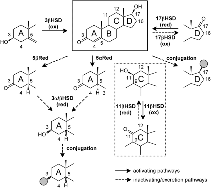
“Conversely, our group has shown that the activation of 11KA4 (Δ4, 17-keto) proceeds by the 17β-reduction to 11KT, followed by 5α-reduction to 11KDHT (5α,17β) (Pretorius et al., 2017) (Fig. 6).”
§ Intracrine androgen biosynthesis, metabolism and action revisited > C19 steroid metabolism in peripheral target tissues – principles of intracrine androgen activation and inactivation > Enzymes involved in intracrine androgen metabolism
What the sources say
- Not checked yetcites Pretorius et al., 2017 source
boosteddirect- Asserted: passes — The authors put it forward as true.
- Checkable: passes — It can be checked against evidence.
- Load-bearing: passes — The paper’s conclusions depend on it.
- Based: passes — The paper gives a reason why it holds.
The paper states this without a source or data behind it here, so there is nothing to check it against. The quote shows it is in the paper.
In the paper
“Interestingly, we also showed that in the same cohort 11KT did not correlate with BMI”
§ Intracrine androgen biosynthesis, metabolism and action revisited > Intracrinology in metabolic target tissues > Adipose tissue
plain- Asserted: passes — The authors put it forward as true.
- Checkable: passes — It can be checked against evidence.
- Load-bearing: passes — The paper’s conclusions depend on it.
- Based: passes — The paper gives a reason why it holds.
A result the paper reports from its own data — it cites no other source for it, so there is none to check. The quote shows it is in the paper.
In the paper
“We have recently shown that serum T levels correlated with BMI in both healthy women and women with PCOS.”
§ Intracrine androgen biosynthesis, metabolism and action revisited > Intracrinology in metabolic target tissues > Adipose tissue
plaincorrelational- Asserted: passes — The authors put it forward as true.
- Checkable: passes — It can be checked against evidence.
- Load-bearing: passes — The paper’s conclusions depend on it.
- Based: passes — The paper gives a reason why it holds.
The paper states this without a source or data behind it here, so there is nothing to check it against. The quote shows it is in the paper.
In the paper
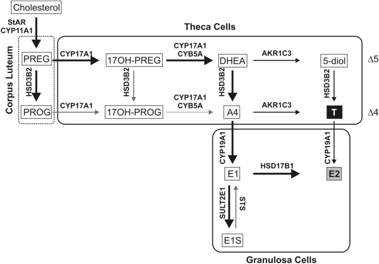
“Ovarian steroid biosynthesis can be described by the “two-cell, two-gonadotrophin” model: Two different cell types - granulosa cells of the follicle and the surrounding theca cells - perform distinct reactions due to specific enzyme expression (Fig. 3).”
§ Intracrine androgen biosynthesis, metabolism and action revisited > De-novo androgen biosynthesis in the adrenal and gonads > Ovarian C19 steroid biosynthesis
plaindirect- Asserted: passes — The authors put it forward as true.
- Checkable: passes — It can be checked against evidence.
- Load-bearing: unclear — It is unclear whether the paper’s conclusions depend on it.
- Based: passes — The paper gives a reason why it holds.
In the paper
“While the adrenal glands only produce low levels of the active androgen T, they produce significant levels of the inactive C19 androgen precursors dehydroepiandrosterone (DHEA, androst-5-ene-3βol-17-one) and its sulfate ester DHEAS, androstenedione (A4, androst-4-ene-3,17-dione) and 11β-hydroxyandrostenedione (11OHA4), which in the majority are released into circulation (Rege et al., 2013).”
§ Intracrine androgen biosynthesis, metabolism and action revisited > Introduction
What the sources say
- Not checked yetcites Rege et al., 2013 source
plain- Asserted: passes — The authors put it forward as true.
- Checkable: passes — It can be checked against evidence.
- Load-bearing: unclear — It is unclear whether the paper’s conclusions depend on it.
- Based: passes — The paper gives a reason why it holds.
The paper states this without a source or data behind it here, so there is nothing to check it against. The quote shows it is in the paper.
In the paper
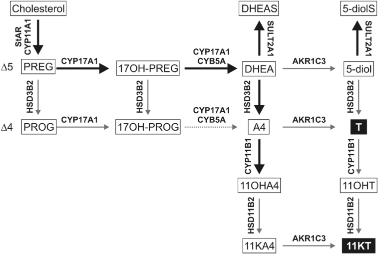
“DHEA is converted to DHEAS by DHEA sulfotransferase (sulfotransferase 2A1, SULT2A1) and as such DHEAS is the major C19 steroid secreted by the adrenal, circulating in low micromolar concentrations.”
§ Intracrine androgen biosynthesis, metabolism and action revisited > De-novo androgen biosynthesis in the adrenal and gonads > Adrenal androgen biosynthesis
plaindirect- Asserted: passes — The authors put it forward as true.
- Checkable: passes — It can be checked against evidence.
- Load-bearing: unclear — It is unclear whether the paper’s conclusions depend on it.
- Based: passes — The paper gives a reason why it holds.
None of the sources cited for this claim have been checked yet.
In the paper
“Importantly, the expression of CYP17A1 in adrenal of adult mice and rat, which are frequently used as pre-clinical models for endocrine and pharmacology studies, is minimal (Le Goascogne et al., 1991, Mostaghel et al., 2017, Rodriguez et al., 1997).”
§ Intracrine androgen biosynthesis, metabolism and action revisited > De-novo androgen biosynthesis in the adrenal and gonads > Adrenal androgen biosynthesis
What the sources say
plain- Asserted: passes — The authors put it forward as true.
- Checkable: passes — It can be checked against evidence.
- Load-bearing: passes — The paper’s conclusions depend on it.
- Based: passes — The paper gives a reason why it holds.
None of the sources cited for this claim have been checked yet.
In the paper
“Adrenal androgen biosynthesis takes place in the zona reticularis and proceeds via the classical Δ5 pathway (Miller and Auchus, 2011, Turcu and Auchus, 2015) as depicted in Fig. 2.”
§ Intracrine androgen biosynthesis, metabolism and action revisited > De-novo androgen biosynthesis in the adrenal and gonads > Adrenal androgen biosynthesis
What the sources say
plain- Asserted: passes — The authors put it forward as true.
- Checkable: passes — It can be checked against evidence.
- Load-bearing: unclear — It is unclear whether the paper’s conclusions depend on it.
- Based: passes — The paper gives a reason why it holds.
The paper states this without a source or data behind it here, so there is nothing to check it against. The quote shows it is in the paper.
In the paper
“The specific co-localisation of the cytochrome P450 17α-hydroxylase/17,20-lyase (CYP17A1) and cytochrome b5 in the zona reticularis favours the 17,20-lyase activity of CYP17A1 and thus C19 steroid production.”
§ Intracrine androgen biosynthesis, metabolism and action revisited > De-novo androgen biosynthesis in the adrenal and gonads > Adrenal androgen biosynthesis
plaindirect- Asserted: passes — The authors put it forward as true.
- Checkable: passes — It can be checked against evidence.
- Load-bearing: unclear — It is unclear whether the paper’s conclusions depend on it.
- Based: passes — The paper gives a reason why it holds.
The paper states this without a source or data behind it here, so there is nothing to check it against. The quote shows it is in the paper.
In the paper
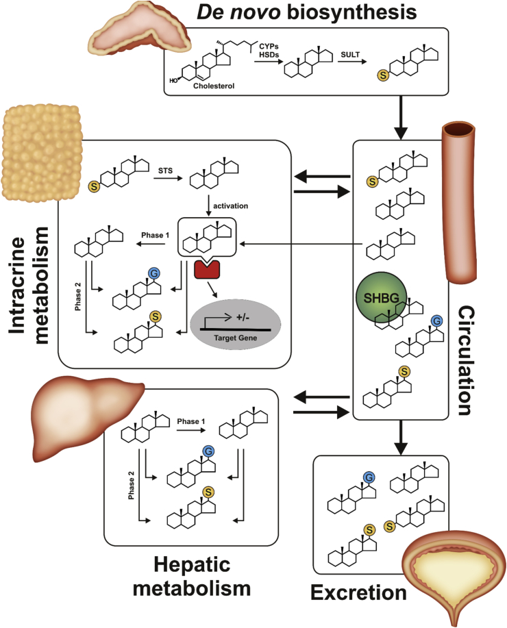
“After uptake into a peripheral tissue with the required enzyme machinery, these androgen precursors are converted into active androgens which elicit a physiological response.”
§ Intracrine androgen biosynthesis, metabolism and action revisited > Introduction
plaindirect- Asserted: passes — The authors put it forward as true.
- Checkable: passes — It can be checked against evidence.
- Load-bearing: passes — The paper’s conclusions depend on it.
- Based: passes — The paper gives a reason why it holds.
None of the sources cited for this claim have been checked yet.
In the paper
“Adrenal androgen biosynthesis takes place in the zona reticularis and proceeds via the classical Δ5 pathway (Miller and Auchus, 2011, Turcu and Auchus, 2015) as depicted in Fig. 2.”
§ Intracrine androgen biosynthesis, metabolism and action revisited > De-novo androgen biosynthesis in the adrenal and gonads > Adrenal androgen biosynthesis
What the sources say
plaindirect- Asserted: passes — The authors put it forward as true.
- Checkable: passes — It can be checked against evidence.
- Load-bearing: unclear — It is unclear whether the paper’s conclusions depend on it.
- Based: passes — The paper gives a reason why it holds.
In the paper
“A peak of adrenal C19 steroid production is reached in the third and fourth decade of human life in both men and women after which levels decline significantly (Labrie et al., 1997b).”
§ Intracrine androgen biosynthesis, metabolism and action revisited > De-novo androgen biosynthesis in the adrenal and gonads > Adrenal androgen biosynthesis
What the sources say
- Not checked yetcites Labrie et al., 1997b
plaincorrelational- Asserted: passes — The authors put it forward as true.
- Checkable: passes — It can be checked against evidence.
- Load-bearing: unclear — It is unclear whether the paper’s conclusions depend on it.
- Based: passes — The paper gives a reason why it holds.
A conclusion the paper draws from its own results — it cites no other source for it, so there is none to check. The quote shows it is in the paper.
In the paper
“However, the intracellular or intracrine activation of C19 androgen precursors to active androgens in peripheral target tissues of androgen action is of equal importance.”
§ Intracrine androgen biosynthesis, metabolism and action revisited > Abstract
plain- Asserted: passes — The authors put it forward as true.
- Checkable: passes — It can be checked against evidence.
- Load-bearing: passes — The paper’s conclusions depend on it.
- Based: unclear — It is unclear why the paper says it holds.
- Rests on an assumption: Androgen signalling is dependent on androgen receptor activation, mostly by testosterone and 5α-dihydrotestosterone.
A conclusion the paper draws from its own results — it cites no other source for it, so there is none to check. The quote shows it is in the paper.
In the paper
“Intracrine androgen synthesis is often not reflected by circulating androgens but rather by androgen metabolites and conjugates.”
§ Intracrine androgen biosynthesis, metabolism and action revisited > Abstract
plaincorrelational- Asserted: passes — The authors put it forward as true.
- Checkable: passes — It can be checked against evidence.
- Load-bearing: passes — The paper’s conclusions depend on it.
- Based: unclear — It is unclear why the paper says it holds.
In the paper
“Nonetheless, 80% of the circulating DHEA in postmenopausal women is of adrenal origin with the remainder produced by the ovary (Labrie et al., 2011).”
§ Intracrine androgen biosynthesis, metabolism and action revisited > De-novo androgen biosynthesis in the adrenal and gonads > Adrenal androgen biosynthesis
What the sources say
- Not checked yetcites Labrie et al., 2011 source
plain- Asserted: passes — The authors put it forward as true.
- Checkable: passes — It can be checked against evidence.
- Load-bearing: unclear — It is unclear whether the paper’s conclusions depend on it.
- Based: passes — The paper gives a reason why it holds.
The paper states this without a source or data behind it here, so there is nothing to check it against. The quote shows it is in the paper.
In the paper
“The C19 steroid fatty acid ester can then be transferred to other lipoproteins.”
§ Intracrine androgen biosynthesis, metabolism and action revisited > C19 steroids in circulation
plaindirect- Asserted: passes — The authors put it forward as true.
- Checkable: passes — It can be checked against evidence.
- Load-bearing: unclear — It is unclear whether the paper’s conclusions depend on it.
- Based: passes — The paper gives a reason why it holds.
The paper states this without a source or data behind it here, so there is nothing to check it against. The quote shows it is in the paper.
In the paper
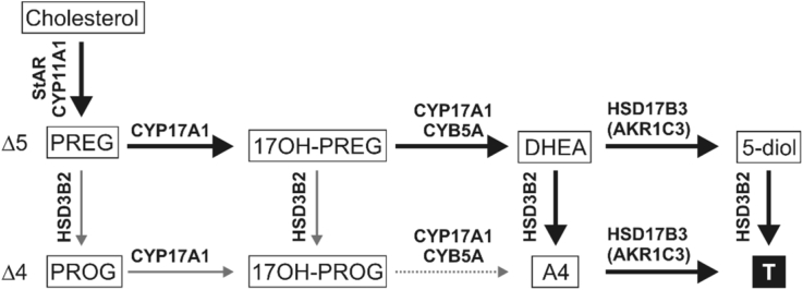
“Due to high abundance of HSD3B2 and 17β-hydroxysteroid dehydrogenase type 3 (HSD17B3) and the absence of SULTs, the final products are the 3-keto-Δ4 androgens A4 and T (Fig. 4).”
§ Intracrine androgen biosynthesis, metabolism and action revisited > De-novo androgen biosynthesis in the adrenal and gonads > Testicular C19 biosynthesis
plaindirect- Asserted: passes — The authors put it forward as true.
- Checkable: passes — It can be checked against evidence.
- Load-bearing: passes — The paper’s conclusions depend on it.
- Based: passes — The paper gives a reason why it holds.
None of the sources cited for this claim have been checked yet.
In the paper
“In men, the decline can be explained by simultaneous age-related reductions of both adrenal and testicular androgen biosynthesis (Beattie et al., 2015, Camacho et al., 2013, Golan et al., 2015, Labrie et al., 1997b).”
§ Intracrine androgen biosynthesis, metabolism and action revisited > C19 steroids in circulation
What the sources say
plaindirect- Asserted: passes — The authors put it forward as true.
- Checkable: passes — It can be checked against evidence.
- Load-bearing: unclear — It is unclear whether the paper’s conclusions depend on it.
- Based: passes — The paper gives a reason why it holds.
In the paper
“In addition to the classical C19 steroids (DHEAS, DHEA, T and A4), recent advances in mass spectrometry-based analytical methodology have allowed for the identification and quantification of non-classical androgen metabolites (Bloem et al., 2015).”
§ Intracrine androgen biosynthesis, metabolism and action revisited > C19 steroids in circulation
What the sources say
- Not checked yetcites Bloem et al., 2015 source
plaindirect- Asserted: passes — The authors put it forward as true.
- Checkable: passes — It can be checked against evidence.
- Load-bearing: passes — The paper’s conclusions depend on it.
- Based: passes — The paper gives a reason why it holds.
None of the sources cited for this claim have been checked yet.
In the paper
“The major fractions of androgen metabolites are conjugated to facilitate their excretion.”
§ Intracrine androgen biosynthesis, metabolism and action revisited > C19 steroids in circulation
plaindirect- Asserted: passes — The authors put it forward as true.
- Checkable: passes — It can be checked against evidence.
- Load-bearing: unclear — It is unclear whether the paper’s conclusions depend on it.
- Based: passes — The paper gives a reason why it holds.
In the paper
“Significantly, our study in healthy and PCOS women showed for the first time that the circulating levels of the active androgen 11KT were 3–4fold higher than that of T in healthy premenopausal women (O'Reilly et al., 2016), highlighting the important contribution of the 11-oxygenated androgens to the circulating androgen pool in women.”
§ Intracrine androgen biosynthesis, metabolism and action revisited > C19 steroids in circulation
What the sources say
- Not checked yetcites O'Reilly et al., 2016 source
boostedcorrelational- Asserted: passes — The authors put it forward as true.
- Checkable: passes — It can be checked against evidence.
- Load-bearing: passes — The paper’s conclusions depend on it.
- Based: passes — The paper gives a reason why it holds.
None of the sources cited for this claim have been checked yet.
In the paper
“In contrast, SHBG binds sex steroids (including active androgens, oestrogens, precursors and metabolites (Avvakumov et al., 2010, Cherkasov et al., 2008, Dunn et al., 1981, Grishkovskaya et al., 2002)) with high specificity and affinity (nanomolar ranges) (reviewed in (Hammond, 2016)).”
§ Intracrine androgen biosynthesis, metabolism and action revisited > C19 steroids in circulation
What the sources say
plain- Asserted: passes — The authors put it forward as true.
- Checkable: passes — It can be checked against evidence.
- Load-bearing: unclear — It is unclear whether the paper’s conclusions depend on it.
- Based: passes — The paper gives a reason why it holds.
In the paper
“These sex steroid-binding plasma proteins therefore play a crucial role in the regulation of androgen action (Laurent et al., 2016).”
§ Intracrine androgen biosynthesis, metabolism and action revisited > C19 steroids in circulation
What the sources say
- Not checked yetcites Laurent et al., 2016 source
plaindirect- Asserted: passes — The authors put it forward as true.
- Checkable: passes — It can be checked against evidence.
- Load-bearing: unclear — It is unclear whether the paper’s conclusions depend on it.
- Based: passes — The paper gives a reason why it holds.
None of the sources cited for this claim have been checked yet.
In the paper
“DHEA-fatty acid esters can be taken up by peripheral cells via lipoprotein receptors (Lavallee et al., 1996, Roy and Belanger, 1989).”
§ Intracrine androgen biosynthesis, metabolism and action revisited > C19 steroids in circulation
What the sources say
- Not checked yetcites Lavallee et al., 1996
- Not checked yetcites Roy and Belanger, 1989
plaindirect- Asserted: passes — The authors put it forward as true.
- Checkable: passes — It can be checked against evidence.
- Load-bearing: unclear — It is unclear whether the paper’s conclusions depend on it.
- Based: passes — The paper gives a reason why it holds.
The paper states this without a source or data behind it here, so there is nothing to check it against. The quote shows it is in the paper.
In the paper
“Unlike the fetal ovaries, the fetal testes have steroidogenic activity which peaks between 11 and 17 weeks of gestation.”
§ Intracrine androgen biosynthesis, metabolism and action revisited > De-novo androgen biosynthesis in the adrenal and gonads > Testicular C19 biosynthesis
plain- Asserted: passes — The authors put it forward as true.
- Checkable: passes — It can be checked against evidence.
- Load-bearing: unclear — It is unclear whether the paper’s conclusions depend on it.
- Based: passes — The paper gives a reason why it holds.
The paper states this without a source or data behind it here, so there is nothing to check it against. The quote shows it is in the paper.
In the paper
“The majority of unconjugated C19 steroids circulate bound to the plasma proteins albumin or SHBG and only a small fraction (1–2%) circulates in the free form which is accessible to the target tissue.”
§ Intracrine androgen biosynthesis, metabolism and action revisited > C19 steroids in circulation
plain- Asserted: passes — The authors put it forward as true.
- Checkable: passes — It can be checked against evidence.
- Load-bearing: passes — The paper’s conclusions depend on it.
- Based: passes — The paper gives a reason why it holds.
None of the sources cited for this claim have been checked yet.
In the paper
“HSD17B3 deficiency results in disordered sex development in genetically male children (Boehmer et al., 1999, Mendonca et al., 1999).”
§ Intracrine androgen biosynthesis, metabolism and action revisited > De-novo androgen biosynthesis in the adrenal and gonads > Testicular C19 biosynthesis
What the sources say
- Not checked yetcites Boehmer et al., 1999
- Not checked yetcites Mendonca et al., 1999
plaindirect- Asserted: passes — The authors put it forward as true.
- Checkable: passes — It can be checked against evidence.
- Load-bearing: unclear — It is unclear whether the paper’s conclusions depend on it.
- Based: passes — The paper gives a reason why it holds.
None of the sources cited for this claim have been checked yet.
In the paper
“Interestingly, DHEA and 5-diol can be acylated with fatty acids by plasma lecithin:cholesterol acyltransferase located on high density lipoproteins (Jones and James, 1985, Lavallee et al., 1996).”
§ Intracrine androgen biosynthesis, metabolism and action revisited > C19 steroids in circulation
What the sources say
- Not checked yetcites Jones and James, 1985
- Not checked yetcites Lavallee et al., 1996
plaindirect- Asserted: passes — The authors put it forward as true.
- Checkable: passes — It can be checked against evidence.
- Load-bearing: unclear — It is unclear whether the paper’s conclusions depend on it.
- Based: passes — The paper gives a reason why it holds.
None of the sources cited for this claim have been checked yet.
In the paper
“In both men and women, ageing leads to decreases in C19 steroid production (Damgaard-Olesen et al., 2016, Haring et al., 2012, Trabert et al., 2016).”
§ Intracrine androgen biosynthesis, metabolism and action revisited > C19 steroids in circulation
What the sources say
plaindirect- Asserted: passes — The authors put it forward as true.
- Checkable: passes — It can be checked against evidence.
- Load-bearing: unclear — It is unclear whether the paper’s conclusions depend on it.
- Based: passes — The paper gives a reason why it holds.
In the paper
“and OATP polymorphisms are associated with time to progression during androgen deprivation therapy (Yang et al., 2011).”
§ Intracrine androgen biosynthesis, metabolism and action revisited > C19 steroid metabolism in peripheral target tissues – principles of intracrine androgen activation and inactivation > Cellular uptake and deconjugation
What the sources say
- Not checked yetcites Yang et al., 2011 source
plaincorrelational- Asserted: passes — The authors put it forward as true.
- Checkable: passes — It can be checked against evidence.
- Load-bearing: unclear — It is unclear whether the paper’s conclusions depend on it.
- Based: passes — The paper gives a reason why it holds.
None of the sources cited for this claim have been checked yet.
In the paper
“Indeed, the conversion of A4 (Δ4, 17-keto) to DHT (5α, 17β) proceeds via the 5α-reduction of A4 to 5α-androstanedione (5α-dione, 5α-androstane-3,17-dione), followed by the conversion of 5α-dione to DHT (Chang et al., 2011' Luu-The and Labrie, 2010, Samson et al., 2010) (Fig. 6).”
§ Intracrine androgen biosynthesis, metabolism and action revisited > C19 steroid metabolism in peripheral target tissues – principles of intracrine androgen activation and inactivation > Enzymes involved in intracrine androgen metabolism
What the sources say
plaindirect- Asserted: passes — The authors put it forward as true.
- Checkable: passes — It can be checked against evidence.
- Load-bearing: unclear — It is unclear whether the paper’s conclusions depend on it.
- Based: passes — The paper gives a reason why it holds.
The paper states this without a source or data behind it here, so there is nothing to check it against. The quote shows it is in the paper.
In the paper
“The access of a conjugated steroid to a specific cell is determined by (1) the expression level of transporters in combination with (2) the substrate specificity and (3) the transport kinetics of the respective transporters.”
§ Intracrine androgen biosynthesis, metabolism and action revisited > C19 steroid metabolism in peripheral target tissues – principles of intracrine androgen activation and inactivation > Cellular uptake and deconjugation
plaindirect- Asserted: passes — The authors put it forward as true.
- Checkable: passes — It can be checked against evidence.
- Load-bearing: passes — The paper’s conclusions depend on it.
- Based: passes — The paper gives a reason why it holds.
None of the sources cited for this claim have been checked yet.
In the paper
“The potential of STS inhibition has been evaluated in promising clinical trials (Geisler et al., 2011, Purohit and Foster, 2012, Thomas and Potter, 2015).”
§ Intracrine androgen biosynthesis, metabolism and action revisited > C19 steroid metabolism in peripheral target tissues – principles of intracrine androgen activation and inactivation > Cellular uptake and deconjugation
What the sources say
plain- Asserted: passes — The authors put it forward as true.
- Checkable: passes — It can be checked against evidence.
- Load-bearing: unclear — It is unclear whether the paper’s conclusions depend on it.
- Based: passes — The paper gives a reason why it holds.
The paper states this without a source or data behind it here, so there is nothing to check it against. The quote shows it is in the paper.
In the paper
“5α-reduction is necessary to achieve maximum AR activation (DHT, 11KDHT).”
§ Intracrine androgen biosynthesis, metabolism and action revisited > C19 steroid metabolism in peripheral target tissues – principles of intracrine androgen activation and inactivation > Enzymes involved in intracrine androgen metabolism
plaindirect- Asserted: passes — The authors put it forward as true.
- Checkable: passes — It can be checked against evidence.
- Load-bearing: unclear — It is unclear whether the paper’s conclusions depend on it.
- Based: unclear — It is unclear why the paper says it holds.
A conclusion the paper draws from its own results — it cites no other source for it, so there is none to check. The quote shows it is in the paper.
In the paper
“Dysregulation of intracrine pathways can result from (1) alterations of expression leading to effects specific for the respective tissue”
§ Intracrine androgen biosynthesis, metabolism and action revisited > C19 steroid metabolism in peripheral target tissues – principles of intracrine androgen activation and inactivation > Principles of intracrine androgen steroid metabolism
plaindirect- Asserted: passes — The authors put it forward as true.
- Checkable: passes — It can be checked against evidence.
- Load-bearing: unclear — It is unclear whether the paper’s conclusions depend on it.
- Based: passes — The paper gives a reason why it holds.
The paper states this without a source or data behind it here, so there is nothing to check it against. The quote shows it is in the paper.
In the paper
“By contrast, patients with STS deficiency due to mutation show only a rather mild clinical phenotype with ichthyosis due to the accumulation of sulfated steroids in the skin.”
§ Intracrine androgen biosynthesis, metabolism and action revisited > C19 steroid metabolism in peripheral target tissues – principles of intracrine androgen activation and inactivation > Cellular uptake and deconjugation
plaindirect- Asserted: passes — The authors put it forward as true.
- Checkable: passes — It can be checked against evidence.
- Load-bearing: unclear — It is unclear whether the paper’s conclusions depend on it.
- Based: passes — The paper gives a reason why it holds.
None of the sources cited for this claim have been checked yet.
In the paper
“SRD5A2 encoding steroid 5α-reductase type 2 (SRD5A2) is expressed in male reproductive tissues (Thigpen et al., 1993) and its disruption leads to the impairment of local DHT formation and in consequence disordered sex development in 46, XY individuals (Okeigwe and Kuohung, 2014, Wilson et al., 1993).”
§ Intracrine androgen biosynthesis, metabolism and action revisited > C19 steroid metabolism in peripheral target tissues – principles of intracrine androgen activation and inactivation > Enzymes involved in intracrine androgen metabolism
What the sources say
- Not checked yetcites Okeigwe and Kuohung, 2014
- Not checked yetcites Wilson et al., 1993
plaindirect- Asserted: passes — The authors put it forward as true.
- Checkable: passes — It can be checked against evidence.
- Load-bearing: unclear — It is unclear whether the paper’s conclusions depend on it.
- Based: passes — The paper gives a reason why it holds.
None of the sources cited for this claim have been checked yet.
In the paper
“which is associated with metabolic dysfunction (Conway et al., 2014, O'Reilly et al., 2014b).”
§ Intracrine androgen biosynthesis, metabolism and action revisited > C19 steroid metabolism in peripheral target tissues – principles of intracrine androgen activation and inactivation > Enzymes involved in intracrine androgen metabolism
What the sources say
- Not checked yetcites Conway et al., 2014 source
- Not checked yetcites O'Reilly et al., 2014b
plaincorrelational- Asserted: passes — The authors put it forward as true.
- Checkable: passes — It can be checked against evidence.
- Load-bearing: unclear — It is unclear whether the paper’s conclusions depend on it.
- Based: passes — The paper gives a reason why it holds.
The paper states this without a source or data behind it here, so there is nothing to check it against. The quote shows it is in the paper.
In the paper
“Additionally, de-conjugation is required after influx before the steroid can be metabolised and or interact with its receptor.”
§ Intracrine androgen biosynthesis, metabolism and action revisited > C19 steroid metabolism in peripheral target tissues – principles of intracrine androgen activation and inactivation > Cellular uptake and deconjugation
plaindirect- Asserted: passes — The authors put it forward as true.
- Checkable: passes — It can be checked against evidence.
- Load-bearing: passes — The paper’s conclusions depend on it.
- Based: passes — The paper gives a reason why it holds.
A conclusion the paper draws from its own results — it cites no other source for it, so there is none to check. The quote shows it is in the paper.
In the paper
“and can be associated with metabolic dysfunction or sex steroid-dependent cancer as discussed below.”
§ Intracrine androgen biosynthesis, metabolism and action revisited > C19 steroid metabolism in peripheral target tissues – principles of intracrine androgen activation and inactivation > Principles of intracrine androgen steroid metabolism
plaincorrelational- Asserted: passes — The authors put it forward as true.
- Checkable: passes — It can be checked against evidence.
- Load-bearing: unclear — It is unclear whether the paper’s conclusions depend on it.
- Based: passes — The paper gives a reason why it holds.
A conclusion the paper draws from its own results — it cites no other source for it, so there is none to check. The quote shows it is in the paper.
In the paper
“or (2) mutations/polymorphisms of involved enzymes leading to systemic effects”
§ Intracrine androgen biosynthesis, metabolism and action revisited > C19 steroid metabolism in peripheral target tissues – principles of intracrine androgen activation and inactivation > Principles of intracrine androgen steroid metabolism
plaindirect- Asserted: passes — The authors put it forward as true.
- Checkable: passes — It can be checked against evidence.
- Load-bearing: unclear — It is unclear whether the paper’s conclusions depend on it.
- Based: passes — The paper gives a reason why it holds.
In the paper
“Significantly, the metabolism of DHEA to active androgens reaches saturation with increasing circulating concentrations of DHEA, thereby protecting peripheral tissues from increased intracellular levels of androgens which may result from pathologically increased biosynthesis of androgen precursors in the adrenal (Labrie et al., 2007).”
§ Intracrine androgen biosynthesis, metabolism and action revisited > C19 steroid metabolism in peripheral target tissues – principles of intracrine androgen activation and inactivation > Principles of intracrine androgen steroid metabolism
What the sources say
- Not checked yetcites Labrie et al., 2007 source
hedgedconditional- Asserted: passes — The authors put it forward as true.
- Checkable: passes — It can be checked against evidence.
- Load-bearing: passes — The paper’s conclusions depend on it.
- Based: passes — The paper gives a reason why it holds.
In the paper
“Certain polymorphisms of AKR1C3 are associated with PCOS and increased T levels in women (Ju et al., 2015).”
§ Intracrine androgen biosynthesis, metabolism and action revisited > C19 steroid metabolism in peripheral target tissues – principles of intracrine androgen activation and inactivation > Enzymes involved in intracrine androgen metabolism
What the sources say
- Not checked yetcites Ju et al., 2015
plaincorrelational- Asserted: passes — The authors put it forward as true.
- Checkable: passes — It can be checked against evidence.
- Load-bearing: unclear — It is unclear whether the paper’s conclusions depend on it.
- Based: passes — The paper gives a reason why it holds.
The paper states this without a source or data behind it here, so there is nothing to check it against. The quote shows it is in the paper.
In the paper
“Circulating, bioavailable androgens and their precursors must cross the plasma membrane of target cells to be (1) metabolised by enzymes that are located intracellularly in the cytosol or membrane of the endoplasmic reticulum and/or (2) to activate the AR, which is localised in the cytosol prior to activation by a suitable ligand.”
§ Intracrine androgen biosynthesis, metabolism and action revisited > C19 steroid metabolism in peripheral target tissues – principles of intracrine androgen activation and inactivation > Cellular uptake and deconjugation
plaindirect- Asserted: passes — The authors put it forward as true.
- Checkable: passes — It can be checked against evidence.
- Load-bearing: passes — The paper’s conclusions depend on it.
- Based: passes — The paper gives a reason why it holds.
A conclusion the paper draws from its own results — it cites no other source for it, so there is none to check. The quote shows it is in the paper.
In the paper
“STS may therefore rather function as a fine-tuning mechanism for intracellular free steroids.”
§ Intracrine androgen biosynthesis, metabolism and action revisited > C19 steroid metabolism in peripheral target tissues – principles of intracrine androgen activation and inactivation > Cellular uptake and deconjugation
hedgedconditional- Asserted: passes — The authors put it forward as true.
- Checkable: passes — It can be checked against evidence.
- Load-bearing: passes — The paper’s conclusions depend on it.
- Based: passes — The paper gives a reason why it holds.
The paper states this without a source or data behind it here, so there is nothing to check it against. The quote shows it is in the paper.
In the paper
“The 11-oxygenated androgens also require the conversion of the 11β-hydroxyl to the 11-ketone in order to obtain maximal activity (11KT and 11KDHT; dotted inset).”
§ Intracrine androgen biosynthesis, metabolism and action revisited > C19 steroid metabolism in peripheral target tissues – principles of intracrine androgen activation and inactivation > Enzymes involved in intracrine androgen metabolism
plaindirect- Asserted: passes — The authors put it forward as true.
- Checkable: passes — It can be checked against evidence.
- Load-bearing: passes — The paper’s conclusions depend on it.
- Based: passes — The paper gives a reason why it holds.
None of the sources cited for this claim have been checked yet.
In the paper
“It should also be noted that the expression of HSD11B2, which encodes the oxidative HSD11B isoform, is required for the activation of 11OHA4, which is the primary 11-oxygenated C19 androgen precursor in circulation (Rege et al., 2013, Storbeck et al., 2013, Swart et al., 2013) (Table 2).”
§ Intracrine androgen biosynthesis, metabolism and action revisited > C19 steroid metabolism in peripheral target tissues – principles of intracrine androgen activation and inactivation > Enzymes involved in intracrine androgen metabolism
What the sources say
plaindirect- Asserted: passes — The authors put it forward as true.
- Checkable: passes — It can be checked against evidence.
- Load-bearing: unclear — It is unclear whether the paper’s conclusions depend on it.
- Based: passes — The paper gives a reason why it holds.
None of the sources cited for this claim have been checked yet.
In the paper
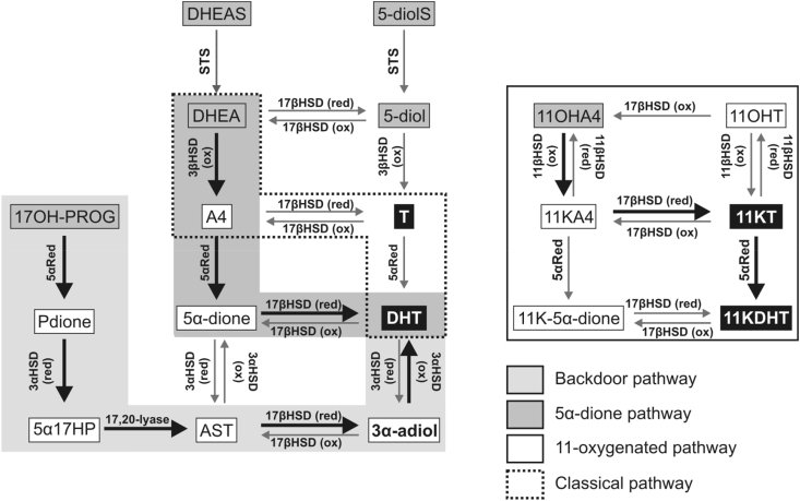
“In humans, the backdoor pathway is relevant in pathological conditions when 17OHPROG accumulates, such as in congenital adrenal hyperplasia due to cytochrome P450 oxidoreductase deficiency (PORD) or 21-hydroxylase deficiency (Arlt et al., 2004, Homma et al., 2006, Kamrath et al., 2012, Krone et al., 2012, Miller and Auchus, 2011).”
§ Intracrine androgen biosynthesis, metabolism and action revisited > Pathways of peripheral androgen activation > The backdoor pathway
What the sources say
plaincorrelational- Asserted: passes — The authors put it forward as true.
- Checkable: passes — It can be checked against evidence.
- Load-bearing: unclear — It is unclear whether the paper’s conclusions depend on it.
- Based: passes — The paper gives a reason why it holds.
None of the sources cited for this claim have been checked yet.
In the paper
“and their expression and activity correlates with obesity in women and men (Blouin et al., 2005, Blouin et al., 2006).”
§ Intracrine androgen biosynthesis, metabolism and action revisited > Intracrinology in metabolic target tissues > Adipose tissue
What the sources say
plaincorrelational- Asserted: passes — The authors put it forward as true.
- Checkable: passes — It can be checked against evidence.
- Load-bearing: unclear — It is unclear whether the paper’s conclusions depend on it.
- Based: passes — The paper gives a reason why it holds.
In the paper
“and the degree of androgen excess correlates with the severity of insulin resistance in PCOS (O'Reilly et al., 2014b).”
§ Intracrine androgen biosynthesis, metabolism and action revisited > Intracrinology in metabolic target tissues > Adipose tissue
What the sources say
- Not checked yetcites O'Reilly et al., 2014b
plaincorrelational- Asserted: passes — The authors put it forward as true.
- Checkable: passes — It can be checked against evidence.
- Load-bearing: passes — The paper’s conclusions depend on it.
- Based: passes — The paper gives a reason why it holds.
The paper states this without a source or data behind it here, so there is nothing to check it against. The quote shows it is in the paper.
In the paper
“Muscle cells exhibit 3βHSD, reductive 17βHSD and 5α-reductase activity resulting in the activation of DHEA to T and DHT.”
§ Intracrine androgen biosynthesis, metabolism and action revisited > Intracrinology in metabolic target tissues > Skeletal muscle
plaindirect- Asserted: passes — The authors put it forward as true.
- Checkable: passes — It can be checked against evidence.
- Load-bearing: passes — The paper’s conclusions depend on it.
- Based: passes — The paper gives a reason why it holds.
In the paper
“AKR1C1 (predominant 20αHSD with low reductive 3αHSD activity), AKR1C2 (reductive 3αHSD activity) and AKR1C3 (reductive 17βHSD activity) show the highest expression levels of all steroid converting enzymes expressed in adipocytes of women and men (Blouin et al., 2009b)”
§ Intracrine androgen biosynthesis, metabolism and action revisited > Intracrinology in metabolic target tissues > Adipose tissue
What the sources say
- Not checked yetcites Blouin et al., 2009b
plain- Asserted: passes — The authors put it forward as true.
- Checkable: passes — It can be checked against evidence.
- Load-bearing: unclear — It is unclear whether the paper’s conclusions depend on it.
- Based: passes — The paper gives a reason why it holds.
A conclusion the paper draws from its own results — it cites no other source for it, so there is none to check. The quote shows it is in the paper.
In the paper
“The relative activities of HSD11B2 and HSD11B1 are therefore critical in determining the activity of 11-oxygenated androgens in peripheral tissue.”
§ Intracrine androgen biosynthesis, metabolism and action revisited > Pathways of peripheral androgen activation > The 11-oxygenated androgen pathways
plaindirect- Asserted: passes — The authors put it forward as true.
- Checkable: passes — It can be checked against evidence.
- Load-bearing: passes — The paper’s conclusions depend on it.
- Based: passes — The paper gives a reason why it holds.
None of the sources cited for this claim have been checked yet.
In the paper
“It has especially been shown to be an essential pathway for intratumoral DHT production from adrenal androgen precursors in CRPC when circulating levels of T are significantly reduced by physical or chemical castration (Chang et al., 2011, Sharifi and Auchus, 2012).”
§ Intracrine androgen biosynthesis, metabolism and action revisited > Pathways of peripheral androgen activation > The alternate 5α-dione pathway
What the sources say
- Not checked yetcites Chang et al., 2011 source
- Not checked yetcites Sharifi and Auchus, 2012
boosteddirect- Asserted: passes — The authors put it forward as true.
- Checkable: passes — It can be checked against evidence.
- Load-bearing: passes — The paper’s conclusions depend on it.
- Based: passes — The paper gives a reason why it holds.
None of the sources cited for this claim have been checked yet.
In the paper
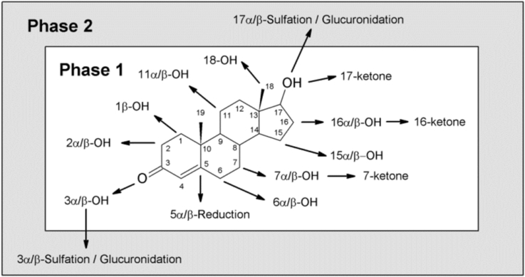
“Interestingly, phase 2 reactions can precede phase 1 reactions during androgen inactivation.”
§ Intracrine androgen biosynthesis, metabolism and action revisited > Directionality of biosynthetic steps, phase 1 metabolism and conjugation
plaindirect- Asserted: passes — The authors put it forward as true.
- Checkable: passes — It can be checked against evidence.
- Load-bearing: unclear — It is unclear whether the paper’s conclusions depend on it.
- Based: passes — The paper gives a reason why it holds.
In the paper
“The intracrine production of oestrogens from androgen precursors is an essential source of oestrogens in postmenopausal women (Simpson, 2003).”
§ Intracrine androgen biosynthesis, metabolism and action revisited > C19 steroid metabolism in peripheral target tissues – principles of intracrine androgen activation and inactivation > Enzymes involved in intracrine androgen metabolism
What the sources say
- Not checked yetcites Simpson, 2003 source
plaindirect- Asserted: passes — The authors put it forward as true.
- Checkable: passes — It can be checked against evidence.
- Load-bearing: unclear — It is unclear whether the paper’s conclusions depend on it.
- Based: passes — The paper gives a reason why it holds.
None of the sources cited for this claim have been checked yet.
In the paper
“Adipose tissue expresses isoforms of all enzymes required for the activation of androgens from circulating precursors (STS, HSD3B1, reductive 17βHSDs, namely HSD17B1, HSR17B3 and AKR1C3 with involvement in androgen activation, and SRD5A1) as well as their subsequent inactivation (reductive 3αHSDs (mainly AKR1C2), UGT2B15 and UGT2B17) (Blouin et al., 2009b, O'Reilly et al., 2014a, Tchernof et al.,”
§ Intracrine androgen biosynthesis, metabolism and action revisited > Intracrinology in metabolic target tissues > Adipose tissue
What the sources say
- Not checked yetcites Blouin et al., 2009b
- Not checked yetcites O'Reilly et al., 2014a
- Not checked yetcites Tchernof et al., 2015 source
plain- Asserted: passes — The authors put it forward as true.
- Checkable: passes — It can be checked against evidence.
- Load-bearing: passes — The paper’s conclusions depend on it.
- Based: passes — The paper gives a reason why it holds.
In the paper
“This prevents the conversion of 11OHA4 to 11KA4, which is a prerequisite for the subsequent conversion of 11KA4 to 11KT by AKR1C3 (O'Reilly et al., 2016).”
§ Intracrine androgen biosynthesis, metabolism and action revisited > Intracrinology in metabolic target tissues > Adipose tissue
What the sources say
- Not checked yetcites O'Reilly et al., 2016 source
plaindirect- Asserted: passes — The authors put it forward as true.
- Checkable: passes — It can be checked against evidence.
- Load-bearing: passes — The paper’s conclusions depend on it.
- Based: passes — The paper gives a reason why it holds.
The paper states this without a source or data behind it here, so there is nothing to check it against. The quote shows it is in the paper.
In the paper
“Unlike 5α-reduction, 5β-reduction inactivates classical AR signalling capability of an androgen.”
§ Intracrine androgen biosynthesis, metabolism and action revisited > C19 steroid metabolism in peripheral target tissues – principles of intracrine androgen activation and inactivation > Enzymes involved in intracrine androgen metabolism
plaindirect- Asserted: passes — The authors put it forward as true.
- Checkable: passes — It can be checked against evidence.
- Load-bearing: unclear — It is unclear whether the paper’s conclusions depend on it.
- Based: passes — The paper gives a reason why it holds.
In the paper
“As SULT2A1 expression is mainly limited to the liver, adrenal, colon and kidney (Riches et al., 2009), glucuronidation is the more important conjugation reaction in androgen target tissue.”
§ Intracrine androgen biosynthesis, metabolism and action revisited > C19 steroid metabolism in peripheral target tissues – principles of intracrine androgen activation and inactivation > Enzymes involved in intracrine androgen metabolism
What the sources say
- Not checked yetcites Riches et al., 2009 source
plaindirect- Asserted: passes — The authors put it forward as true.
- Checkable: passes — It can be checked against evidence.
- Load-bearing: unclear — It is unclear whether the paper’s conclusions depend on it.
- Based: passes — The paper gives a reason why it holds.
In the paper
“Androgen excess is recognised as the major determinant driving metabolic dysfunction observed in women with PCOS (Conway et al., 2014)”
§ Intracrine androgen biosynthesis, metabolism and action revisited > Intracrinology in metabolic target tissues > Adipose tissue
What the sources say
- Not checked yetcites Conway et al., 2014 source
plaindirect- Asserted: passes — The authors put it forward as true.
- Checkable: passes — It can be checked against evidence.
- Load-bearing: passes — The paper’s conclusions depend on it.
- Based: passes — The paper gives a reason why it holds.
In the paper
“Genetic variations of UGTs can change the glucuronidation efficiency for an androgen and alter its excretion (Piper et al., 2016).”
§ Intracrine androgen biosynthesis, metabolism and action revisited > Intracrinology in metabolic target tissues > Liver
What the sources say
- Not checked yetcites Piper et al., 2016 source
plaindirect- Asserted: passes — The authors put it forward as true.
- Checkable: passes — It can be checked against evidence.
- Load-bearing: unclear — It is unclear whether the paper’s conclusions depend on it.
- Based: passes — The paper gives a reason why it holds.
None of the sources cited for this claim have been checked yet.
In the paper
“androgens and the expression of the AR have demonstrated both positive and negative outcomes in breast cancer as reviewed in (Hickey et al., 2012, McNamara and Sasano, 2015a, McNamara and Sasano, 2016).”
§ Intracrine androgen biosynthesis, metabolism and action revisited > Intracrine androgen metabolism in reproductive target tissues > Breast and endometrium
What the sources say
- Not checked yetcites Hickey et al., 2012 source
- Not checked yetcites McNamara and Sasano, 2015a
- Not checked yetcites McNamara and Sasano, 2016
boostedcorrelational- Asserted: passes — The authors put it forward as true.
- Checkable: passes — It can be checked against evidence.
- Load-bearing: unclear — It is unclear whether the paper’s conclusions depend on it.
- Based: passes — The paper gives a reason why it holds.
In the paper
“Both androgens and oestrogens are synthesised from circulating androgen precursors in breast tissue and the homeostasis of these pathways determines health status (McNamara and Sasano, 2015b).”
§ Intracrine androgen biosynthesis, metabolism and action revisited > Intracrine androgen metabolism in reproductive target tissues > Breast and endometrium
What the sources say
- Not checked yetcites McNamara and Sasano, 2015b
plaindirect- Asserted: passes — The authors put it forward as true.
- Checkable: passes — It can be checked against evidence.
- Load-bearing: passes — The paper’s conclusions depend on it.
- Based: passes — The paper gives a reason why it holds.
None of the sources cited for this claim have been checked yet.
In the paper
“16α-hydroxy-DHEAS, 5-diol-sulfate, AST-sulfate, EpiAST-sulfate can be measured in nM concentrations in serum of both men and women (Labrie et al., 1997b, Sanchez-Guijo et al., 2015, Zang et al., 2017).”
§ Intracrine androgen biosynthesis, metabolism and action revisited > Intracrinology in metabolic target tissues > Liver
What the sources say
plain- Asserted: passes — The authors put it forward as true.
- Checkable: passes — It can be checked against evidence.
- Load-bearing: unclear — It is unclear whether the paper’s conclusions depend on it.
- Based: passes — The paper gives a reason why it holds.
In the paper
“Numerous studies have shown that androgen levels are higher within breast tissue than in circulation and are generally higher in benign tissue than cancerous tissue, demonstrating the relevance of intracrinology in breast and its dysregulation in cancer (Stanczyk et al., 2015).”
§ Intracrine androgen biosynthesis, metabolism and action revisited > Intracrine androgen metabolism in reproductive target tissues > Breast and endometrium
What the sources say
- Not checked yetcites Stanczyk et al., 2015 source
boostedcorrelational- Asserted: passes — The authors put it forward as true.
- Checkable: passes — It can be checked against evidence.
- Load-bearing: passes — The paper’s conclusions depend on it.
- Based: passes — The paper gives a reason why it holds.
None of the sources cited for this claim have been checked yet.
In the paper
“androgens have been shown to play a crucial role in the development of hepatocellular carcinoma (Kalra et al., 2008, Kanda et al., 2014).”
§ Intracrine androgen biosynthesis, metabolism and action revisited > Intracrinology in metabolic target tissues > Liver
What the sources say
- Not checked yetcites Kalra et al., 2008 source
- Not checked yetcites Kanda et al., 2014
boosteddirect- Asserted: passes — The authors put it forward as true.
- Checkable: passes — It can be checked against evidence.
- Load-bearing: unclear — It is unclear whether the paper’s conclusions depend on it.
- Based: passes — The paper gives a reason why it holds.
- Rests on an assumption: The liver expresses AR, making it a target tissue for classical androgen action.
In the paper
“While T production increases and stays elevated during decidualisation due to constantly increased AKR1C3 levels”
§ Intracrine androgen biosynthesis, metabolism and action revisited > Intracrine androgen metabolism in reproductive target tissues > Breast and endometrium
What the sources say
- Not checked yetcites Gibson et al., 2016 source
plaindirect- Asserted: passes — The authors put it forward as true.
- Checkable: passes — It can be checked against evidence.
- Load-bearing: unclear — It is unclear whether the paper’s conclusions depend on it.
- Based: passes — The paper gives a reason why it holds.
None of the sources cited for this claim have been checked yet.
In the paper
“The liver also shows high SULT expression and activity compared to other tissues, including SULT2A1 and SULT2B1 involved in androgen metabolism (Meloche and Falany, 2001, Riches et al., 2009).”
§ Intracrine androgen biosynthesis, metabolism and action revisited > Intracrinology in metabolic target tissues > Liver
What the sources say
- Not checked yetcites Meloche and Falany, 2001
- Not checked yetcites Riches et al., 2009 source
plaincorrelational- Asserted: passes — The authors put it forward as true.
- Checkable: passes — It can be checked against evidence.
- Load-bearing: unclear — It is unclear whether the paper’s conclusions depend on it.
- Based: passes — The paper gives a reason why it holds.
In the paper
“Both T and DHT have recently been shown to increase lipogenesis in human hepatocytes of female (but not male) donors (Nasiri et al., 2015)”
§ Intracrine androgen biosynthesis, metabolism and action revisited > Intracrinology in metabolic target tissues > Liver
What the sources say
- Not checked yetcites Nasiri et al., 2015 source
plaindirect- Asserted: passes — The authors put it forward as true.
- Checkable: passes — It can be checked against evidence.
- Load-bearing: unclear — It is unclear whether the paper’s conclusions depend on it.
- Based: passes — The paper gives a reason why it holds.
- Rests on an assumption: The liver expresses AR, making it a target tissue for classical androgen action.
None of the sources cited for this claim have been checked yet.
In the paper
“Importantly, women produce androgen metabolites in concentrations comparable to that of men (Labrie et al., 1997b, Trabert et al., 2016, Zang et al., 2017).”
§ Intracrine androgen biosynthesis, metabolism and action revisited > Intracrinology in metabolic target tissues > Liver
What the sources say
plain- Asserted: passes — The authors put it forward as true.
- Checkable: passes — It can be checked against evidence.
- Load-bearing: unclear — It is unclear whether the paper’s conclusions depend on it.
- Based: passes — The paper gives a reason why it holds.
In the paper
“It catalyses the interconversion of 7α- and 7β-OH-DHEA via the 7-keto intermediate (Muller et al., 2006).”
§ Intracrine androgen biosynthesis, metabolism and action revisited > Intracrinology in metabolic target tissues > Liver
What the sources say
- Not checked yetcites Muller et al., 2006 source
plaindirect- Asserted: passes — The authors put it forward as true.
- Checkable: passes — It can be checked against evidence.
- Load-bearing: unclear — It is unclear whether the paper’s conclusions depend on it.
- Based: passes — The paper gives a reason why it holds.
None of the sources cited for this claim have been checked yet.
In the paper
“Androgens and in particular the intraprostatic conversion of circulating T to DHT by SRD5A2 (Russell and Wilson, 1994) are required for normal prostate development and function (Andersson et al., 1991, Imperato-McGinley et al., 1974).”
§ Intracrine androgen biosynthesis, metabolism and action revisited > Intracrine androgen metabolism in reproductive target tissues > Prostate
What the sources say
plaindirect- Asserted: passes — The authors put it forward as true.
- Checkable: passes — It can be checked against evidence.
- Load-bearing: passes — The paper’s conclusions depend on it.
- Based: passes — The paper gives a reason why it holds.
In the paper
“time-dependent changes in SDR5A1 and AKR1C3 expression alter T and DHT secretion.”
§ Intracrine androgen biosynthesis, metabolism and action revisited > Intracrine androgen metabolism in reproductive target tissues > Breast and endometrium
What the sources say
- Not checked yetcites Gibson et al., 2016 source
plaindirect- Asserted: passes — The authors put it forward as true.
- Checkable: passes — It can be checked against evidence.
- Load-bearing: passes — The paper’s conclusions depend on it.
- Based: passes — The paper gives a reason why it holds.
In the paper
“While the contribution of de-novo intratumoral biosynthesis to local androgens has mostly been excluded (Hofland et al., 2010)”
§ Intracrine androgen biosynthesis, metabolism and action revisited > Intracrine androgen metabolism in reproductive target tissues > Prostate
What the sources say
- Not checked yetcites Hofland et al., 2010 source
plain- Asserted: passes — The authors put it forward as true.
- Checkable: passes — It can be checked against evidence.
- Load-bearing: unclear — It is unclear whether the paper’s conclusions depend on it.
- Based: passes — The paper gives a reason why it holds.
In the paper
“Selective inhibition of HSD3B1 has been shown to slow down proliferation of breast cancer cell line and might just represent an interesting drug target simultaneously tackling androgen and oestrogen synthesis (Thomas et al., 2011).”
§ Intracrine androgen biosynthesis, metabolism and action revisited > Intracrine androgen metabolism in reproductive target tissues > Breast and endometrium
What the sources say
- Not checked yetcites Thomas et al., 2011 source
boosteddirect- Asserted: passes — The authors put it forward as true.
- Checkable: passes — It can be checked against evidence.
- Load-bearing: unclear — It is unclear whether the paper’s conclusions depend on it.
- Based: passes — The paper gives a reason why it holds.
The paper states this without a source or data behind it here, so there is nothing to check it against. The quote shows it is in the paper.
In the paper
“The main androgen metabolites in circulation derived from hepatic and other peripheral metabolism are 3α-adiol-3-glucuronide, 3α-adiol-17-glucuronide, AST-glucuronide and ETIO-glucuronide with levels higher than for T.”
§ Intracrine androgen biosynthesis, metabolism and action revisited > Intracrinology in metabolic target tissues > Liver
plain- Asserted: passes — The authors put it forward as true.
- Checkable: passes — It can be checked against evidence.
- Load-bearing: unclear — It is unclear whether the paper’s conclusions depend on it.
- Based: passes — The paper gives a reason why it holds.
In the paper
“It should also be noted that menopausal status significantly influences the intracrinology of breast tissue (McNamara and Sasano, 2015b).”
§ Intracrine androgen biosynthesis, metabolism and action revisited > Intracrine androgen metabolism in reproductive target tissues > Breast and endometrium
What the sources say
- Not checked yetcites McNamara and Sasano, 2015b
plaindirect- Asserted: passes — The authors put it forward as true.
- Checkable: passes — It can be checked against evidence.
- Load-bearing: unclear — It is unclear whether the paper’s conclusions depend on it.
- Based: passes — The paper gives a reason why it holds.
In the paper
“Numerous studies have shown that androgen levels are higher within breast tissue than in circulation and are generally higher in benign tissue than cancerous tissue, demonstrating the relevance of intracrinology in breast and its dysregulation in cancer (Stanczyk et al., 2015).”
§ Intracrine androgen biosynthesis, metabolism and action revisited > Intracrine androgen metabolism in reproductive target tissues > Breast and endometrium
What the sources say
- Not checked yetcites Stanczyk et al., 2015 source
boostedcorrelational- Asserted: passes — The authors put it forward as true.
- Checkable: passes — It can be checked against evidence.
- Load-bearing: passes — The paper’s conclusions depend on it.
- Based: passes — The paper gives a reason why it holds.
In the paper
“DHT secretion initially increases, but then decreases with the progression of the decidualisation process following reductions in SRD5A1 levels (Gibson et al., 2016).”
§ Intracrine androgen biosynthesis, metabolism and action revisited > Intracrine androgen metabolism in reproductive target tissues > Breast and endometrium
What the sources say
- Not checked yetcites Gibson et al., 2016 source
plaindirect- Asserted: passes — The authors put it forward as true.
- Checkable: passes — It can be checked against evidence.
- Load-bearing: unclear — It is unclear whether the paper’s conclusions depend on it.
- Based: passes — The paper gives a reason why it holds.
In the paper
“Indeed, a recent study showed that the levels of these 11-oxygenated androgens were higher than those of the classical androgens in tissue from two patients with prostate cancer (du Toit et al., 2017).”
§ Intracrine androgen biosynthesis, metabolism and action revisited > Intracrine androgen metabolism in reproductive target tissues > Prostate
What the sources say
- Not checked yetcites du Toit et al., 2017 source
plainn = 2- Asserted: passes — The authors put it forward as true.
- Checkable: passes — It can be checked against evidence.
- Load-bearing: unclear — It is unclear whether the paper’s conclusions depend on it.
- Based: passes — The paper gives a reason why it holds.
None of the sources cited for this claim have been checked yet.
In the paper
“Genetic variations of UGTs significantly contribute to prostate cancer risk and progression and there is evidence for a down-regulation of UGTs in prostate cancer promoting intratumoral androgen accumulation (Barbier and Belanger, 2008, Gauthier-Landry et al., 2015).”
§ Intracrine androgen biosynthesis, metabolism and action revisited > Intracrine androgen metabolism in reproductive target tissues > Prostate
What the sources say
- Not checked yetcites Barbier and Belanger, 2008
- Not checked yetcites Gauthier-Landry et al., 2015 source
plaindirect- Asserted: passes — The authors put it forward as true.
- Checkable: passes — It can be checked against evidence.
- Load-bearing: unclear — It is unclear whether the paper’s conclusions depend on it.
- Based: passes — The paper gives a reason why it holds.
None of the sources cited for this claim have been checked yet.
In the paper
“Recent studies have also shown that prostate cancer cell lines are able to metabolise the adrenal androgen precursor 11OHA4 (Storbeck et al., 2013, Swart et al., 2013, Swart and Storbeck, 2015).”
§ Intracrine androgen biosynthesis, metabolism and action revisited > Intracrine androgen metabolism in reproductive target tissues > Prostate
What the sources say
plaindirect- Asserted: passes — The authors put it forward as true.
- Checkable: passes — It can be checked against evidence.
- Load-bearing: passes — The paper’s conclusions depend on it.
- Based: passes — The paper gives a reason why it holds.
None of the sources cited for this claim have been checked yet.
In the paper
“Circulating DHEA can also be used as a precursor to produce active androgens by 3βHSD, 5α-reductase and reductive 17βHSD activity (Nikolakis et al., 2016, Slominski et al., 2013).”
§ Intracrine androgen biosynthesis, metabolism and action revisited > Intracrine androgen metabolism in other tissues > Skin
What the sources say
plaindirect- Asserted: passes — The authors put it forward as true.
- Checkable: passes — It can be checked against evidence.
- Load-bearing: unclear — It is unclear whether the paper’s conclusions depend on it.
- Based: passes — The paper gives a reason why it holds.
In the paper
“Functional comparison of different epidermal cell types hint at differential functions for androgen biosynthesis and inactivation (Fritsch et al., 2001).”
§ Intracrine androgen biosynthesis, metabolism and action revisited > Intracrine androgen metabolism in other tissues > Skin
What the sources say
- Not checked yetcites Fritsch et al., 2001 source
hedgedcorrelational- Asserted: passes — The authors put it forward as true.
- Checkable: passes — It can be checked against evidence.
- Load-bearing: unclear — It is unclear whether the paper’s conclusions depend on it.
- Based: passes — The paper gives a reason why it holds.
In the paper
“Prostate cells also express CYP3A4 that can inactivate T by conversion to mainly 6βOH-T (with 2β-, 15α/β- and 11β-hydroxyl side product formation (Niwa et al., 2015))”
§ Intracrine androgen biosynthesis, metabolism and action revisited > Intracrine androgen metabolism in reproductive target tissues > Prostate
What the sources say
- Not checked yetcites Niwa et al., 2015 source
plaindirect- Asserted: passes — The authors put it forward as true.
- Checkable: passes — It can be checked against evidence.
- Load-bearing: unclear — It is unclear whether the paper’s conclusions depend on it.
- Based: passes — The paper gives a reason why it holds.
In the paper
“Significantly, both 11KT and 11KDHT have been shown to be able to induce androgen-regulated gene and protein expression as well as cell growth in androgen dependent prostate cancer cell lines”
§ Intracrine androgen biosynthesis, metabolism and action revisited > Intracrine androgen metabolism in reproductive target tissues > Prostate
What the sources say
- Not checked yetcites Pretorius et al., 2016 source
plaindirect- Asserted: passes — The authors put it forward as true.
- Checkable: passes — It can be checked against evidence.
- Load-bearing: passes — The paper’s conclusions depend on it.
- Based: passes — The paper gives a reason why it holds.
None of the sources cited for this claim have been checked yet.
In the paper
“Genetic variations of UGTs significantly contribute to prostate cancer risk and progression and there is evidence for a down-regulation of UGTs in prostate cancer promoting intratumoral androgen accumulation (Barbier and Belanger, 2008, Gauthier-Landry et al., 2015).”
§ Intracrine androgen biosynthesis, metabolism and action revisited > Intracrine androgen metabolism in reproductive target tissues > Prostate
What the sources say
- Not checked yetcites Barbier and Belanger, 2008
- Not checked yetcites Gauthier-Landry et al., 2015 source
plaindirect- Asserted: passes — The authors put it forward as true.
- Checkable: passes — It can be checked against evidence.
- Load-bearing: passes — The paper’s conclusions depend on it.
- Based: passes — The paper gives a reason why it holds.
In the paper
“In fact, studies with cultured sebocytes suggest that circulating DHEA may be the more important source of androgen precursors (Chen et al., 2010).”
§ Intracrine androgen biosynthesis, metabolism and action revisited > Intracrine androgen metabolism in other tissues > Skin
What the sources say
- Not checked yetcites Chen et al., 2010 source
hedgedconditional- Asserted: passes — The authors put it forward as true.
- Checkable: passes — It can be checked against evidence.
- Load-bearing: unclear — It is unclear whether the paper’s conclusions depend on it.
- Based: passes — The paper gives a reason why it holds.
None of the sources cited for this claim have been checked yet.
In the paper
“numerous studies have shown that CRPC is dependent on the intracrine conversion of circulating androgen precursors of adrenal origin to active androgens reviewed in (Capper et al., 2016, Luu-The et al., 2008, Pretorius et al., 2017, Sharifi and Auchus, 2012).”
§ Intracrine androgen biosynthesis, metabolism and action revisited > Intracrine androgen metabolism in reproductive target tissues > Prostate
What the sources say
boosteddirect- Asserted: passes — The authors put it forward as true.
- Checkable: passes — It can be checked against evidence.
- Load-bearing: passes — The paper’s conclusions depend on it.
- Based: passes — The paper gives a reason why it holds.
In the paper
“Acne-prone skin expresses higher levels of androgen generating enzymes than non-acne-prone skin (HSD17B3 and AKR1C3 converting A4 to T, STS and SRD5A1; summarized in (Nikolakis et al., 2016)).”
§ Intracrine androgen biosynthesis, metabolism and action revisited > Intracrine androgen metabolism in other tissues > Skin
What the sources say
- Not checked yetcites Nikolakis et al., 2016 source
plaincorrelational- Asserted: passes — The authors put it forward as true.
- Checkable: passes — It can be checked against evidence.
- Load-bearing: unclear — It is unclear whether the paper’s conclusions depend on it.
- Based: passes — The paper gives a reason why it holds.
- Rests on an assumption: Acne vulgaris and androgenic alopecia are associated with local androgen hyperproduction.
In the paper
“Several cellular components of the interfollicular epidermis and pilosebaceous unit have the capability of de-novo steroid biosynthesis supported by StAR activity (Anuka et al., 2013).”
§ Intracrine androgen biosynthesis, metabolism and action revisited > Intracrine androgen metabolism in other tissues > Skin
What the sources say
- Not checked yetcites Anuka et al., 2013 source
plaindirect- Asserted: passes — The authors put it forward as true.
- Checkable: passes — It can be checked against evidence.
- Load-bearing: unclear — It is unclear whether the paper’s conclusions depend on it.
- Based: passes — The paper gives a reason why it holds.
None of the sources cited for this claim have been checked yet.
In the paper
“The generation of DHT is essential for beard growth (Diamond et al., 1996, Messenger, 1993).”
§ Intracrine androgen biosynthesis, metabolism and action revisited > Intracrine androgen metabolism in other tissues > Skin
What the sources say
- Not checked yetcites Diamond et al., 1996
- Not checked yetcites Messenger, 1993 source
plaindirect- Asserted: passes — The authors put it forward as true.
- Checkable: passes — It can be checked against evidence.
- Load-bearing: unclear — It is unclear whether the paper’s conclusions depend on it.
- Based: passes — The paper gives a reason why it holds.
- Rests on an assumption: Local androgen availability in the skin is necessary to stimulate sebum secretion and hair growth.
In the paper
“Interestingly, the rate at which 11KT and 11KDHT are inactivated by prostate cancer cell lines has been shown to be significantly lower than for T and DHT, suggesting that these metabolites may remain active for longer than the classical androgens (Pretorius et al., 2016).”
§ Intracrine androgen biosynthesis, metabolism and action revisited > Intracrine androgen metabolism in reproductive target tissues > Prostate
What the sources say
- Not checked yetcites Pretorius et al., 2016 source
hedged- Asserted: passes — The authors put it forward as true.
- Checkable: passes — It can be checked against evidence.
- Load-bearing: passes — The paper’s conclusions depend on it.
- Based: passes — The paper gives a reason why it holds.
In the paper
“and a decrease of CYP3A4 expression is observed in prostate cancer (Fujimura et al., 2009).”
§ Intracrine androgen biosynthesis, metabolism and action revisited > Intracrine androgen metabolism in reproductive target tissues > Prostate
What the sources say
- Not checked yetcites Fujimura et al., 2009 source
plaincorrelational- Asserted: passes — The authors put it forward as true.
- Checkable: passes — It can be checked against evidence.
- Load-bearing: unclear — It is unclear whether the paper’s conclusions depend on it.
- Based: passes — The paper gives a reason why it holds.
In the paper
“In addition, the placenta possesses the enzymatic machinery capable of converting C19 steroids derived from the foetal adrenal and liver (DHEAS and 16αOH-DHEA, respectively) to oestrogens during their transplacental passage (Miller and Auchus, 2011).”
§ Intracrine androgen biosynthesis, metabolism and action revisited > Intracrine androgen metabolism in reproductive target tissues > Placenta
What the sources say
- Not checked yetcites Miller and Auchus, 2011 source
plaindirect- Asserted: passes — The authors put it forward as true.
- Checkable: passes — It can be checked against evidence.
- Load-bearing: passes — The paper’s conclusions depend on it.
- Based: passes — The paper gives a reason why it holds.
None of the sources cited for this claim have been checked yet.
In the paper
“The resulting steroids include glucocorticoids and androgens and these pathways were reviewed in (Labrie et al., 2000, Nikolakis et al., 2016, Slominski et al., 2013).”
§ Intracrine androgen biosynthesis, metabolism and action revisited > Intracrine androgen metabolism in other tissues > Skin
What the sources say
plaindirect- Asserted: passes — The authors put it forward as true.
- Checkable: passes — It can be checked against evidence.
- Load-bearing: unclear — It is unclear whether the paper’s conclusions depend on it.
- Based: passes — The paper gives a reason why it holds.
In the paper
“This mechanism protects the mother (and female foetus) from virilisation by the high concentrations of C19 steroid released into circulation by the foetal adrenal (Conte et al., 1994).”
§ Intracrine androgen biosynthesis, metabolism and action revisited > Intracrine androgen metabolism in reproductive target tissues > Placenta
What the sources say
- Not checked yetcites Conte et al., 1994
plaindirect- Asserted: passes — The authors put it forward as true.
- Checkable: passes — It can be checked against evidence.
- Load-bearing: passes — The paper’s conclusions depend on it.
- Based: passes — The paper gives a reason why it holds.
In the paper
“suggesting that these androgens may play a previously overlooked, but important role in the development and progression of CRPC (Pretorius et al., 2016).”
§ Intracrine androgen biosynthesis, metabolism and action revisited > Intracrine androgen metabolism in reproductive target tissues > Prostate
What the sources say
- Not checked yetcites Pretorius et al., 2016 source
hedgedconditional- Asserted: passes — The authors put it forward as true.
- Checkable: passes — It can be checked against evidence.
- Load-bearing: passes — The paper’s conclusions depend on it.
- Based: passes — The paper gives a reason why it holds.
A conclusion the paper draws from its own results — it cites no other source for it, so there is none to check. The quote shows it is in the paper.
In the paper
“C19 steroid conversion in the placenta thus has a protective systemic function rather than an intracrine function.”
§ Intracrine androgen biosynthesis, metabolism and action revisited > Intracrine androgen metabolism in reproductive target tissues > Placenta
plaindirect- Asserted: passes — The authors put it forward as true.
- Checkable: passes — It can be checked against evidence.
- Load-bearing: passes — The paper’s conclusions depend on it.
- Based: passes — The paper gives a reason why it holds.
- Rests on an assumption: 5α-reduced androgens cannot be aromatised.
None of the sources cited for this claim have been checked yet.
In the paper
“The flux through this pathway is due to the preference of SRD5A1, the dominant isoform expressed in CRPC, for A4 over T, coupled to the poor efficiency by which AKR1C3 converts A4 to T (Chang et al., 2011, Sharifi, 2012).”
§ Intracrine androgen biosynthesis, metabolism and action revisited > Intracrine androgen metabolism in reproductive target tissues > Prostate
What the sources say
- Not checked yetcites Chang et al., 2011 source
- Not checked yetcites Sharifi, 2012
plaindirect- Asserted: passes — The authors put it forward as true.
- Checkable: passes — It can be checked against evidence.
- Load-bearing: passes — The paper’s conclusions depend on it.
- Based: passes — The paper gives a reason why it holds.
In the paper
“Interestingly, the rate at which 11KT and 11KDHT are inactivated by prostate cancer cell lines has been shown to be significantly lower than for T and DHT, suggesting that these metabolites may remain active for longer than the classical androgens (Pretorius et al., 2016).”
§ Intracrine androgen biosynthesis, metabolism and action revisited > Intracrine androgen metabolism in reproductive target tissues > Prostate
What the sources say
- Not checked yetcites Pretorius et al., 2016 source
hedgedconditional- Asserted: passes — The authors put it forward as true.
- Checkable: passes — It can be checked against evidence.
- Load-bearing: passes — The paper’s conclusions depend on it.
- Based: passes — The paper gives a reason why it holds.
In the paper
“Significantly, it is the 11-keto C19 steroids that are more androgenic than their 11-hydroxy precursors (Storbeck et al., 2013)”
§ Intracrine androgen biosynthesis, metabolism and action revisited > Kidney androgen metabolism and renal excretion of androgens > The kidney as an androgen-metabolising organ
What the sources say
- Not checked yetcites Storbeck et al., 2013 source
boostedcorrelational- Asserted: passes — The authors put it forward as true.
- Checkable: passes — It can be checked against evidence.
- Load-bearing: passes — The paper’s conclusions depend on it.
- Based: passes — The paper gives a reason why it holds.
None of the sources cited for this claim have been checked yet.
In the paper
“The kidney has high levels of HSD11B2 activity which protects the mineralocorticoid receptor from activation by cortisol by inactivating it to cortisone (Edwards and Stewart, 1991, Funder et al., 1988).”
§ Intracrine androgen biosynthesis, metabolism and action revisited > Kidney androgen metabolism and renal excretion of androgens > The kidney as an androgen-metabolising organ
What the sources say
- Not checked yetcites Edwards and Stewart, 1991
- Not checked yetcites Funder et al., 1988 source
plaindirect- Asserted: passes — The authors put it forward as true.
- Checkable: passes — It can be checked against evidence.
- Load-bearing: passes — The paper’s conclusions depend on it.
- Based: passes — The paper gives a reason why it holds.
A conclusion the paper draws from its own results — it cites no other source for it, so there is none to check. The quote shows it is in the paper.
In the paper
“Elimination of EpiT thus follows the same pathways as T.”
§ Intracrine androgen biosynthesis, metabolism and action revisited > Kidney androgen metabolism and renal excretion of androgens > The kidney as an androgen-metabolising organ
plain- Asserted: passes — The authors put it forward as true.
- Checkable: passes — It can be checked against evidence.
- Load-bearing: unclear — It is unclear whether the paper’s conclusions depend on it.
- Based: passes — The paper gives a reason why it holds.
In the paper
“Surprisingly the kidney has been shown to be able to catalyse the biosynthesis of active androgens using PREG as precursor for DHEA production by CYP17A1 (Quinkler et al., 2003).”
§ Intracrine androgen biosynthesis, metabolism and action revisited > Kidney androgen metabolism and renal excretion of androgens > The kidney as an androgen-metabolising organ
What the sources say
- Not checked yetcites Quinkler et al., 2003 source
plaindirect- Asserted: passes — The authors put it forward as true.
- Checkable: passes — It can be checked against evidence.
- Load-bearing: passes — The paper’s conclusions depend on it.
- Based: passes — The paper gives a reason why it holds.
In the paper
“differences in the concentration of the individual 11-oxygenated steroids in the adrenal vein and inferior vena cava suggest that 11KA4 and 11KT may instead be formed in peripheral tissues (Turcu et al., 2016).”
§ Intracrine androgen biosynthesis, metabolism and action revisited > Kidney androgen metabolism and renal excretion of androgens > The kidney as an androgen-metabolising organ
What the sources say
- Not checked yetcites Turcu et al., 2016 source
hedgedconditional- Asserted: passes — The authors put it forward as true.
- Checkable: passes — It can be checked against evidence.
- Load-bearing: passes — The paper’s conclusions depend on it.
- Based: passes — The paper gives a reason why it holds.
In the paper
“Additionally, the expression of genes (AKR1D1 and AKR1C2) encoding enzymes contributing to phase 1 metabolism and inactivation of androgens have been demonstrated (Quinkler et al., 2003).”
§ Intracrine androgen biosynthesis, metabolism and action revisited > Kidney androgen metabolism and renal excretion of androgens > The kidney as an androgen-metabolising organ
What the sources say
- Not checked yetcites Quinkler et al., 2003 source
boosted- Asserted: passes — The authors put it forward as true.
- Checkable: passes — It can be checked against evidence.
- Load-bearing: unclear — It is unclear whether the paper’s conclusions depend on it.
- Based: passes — The paper gives a reason why it holds.
In the paper
“Indeed, a recent study showed that while 11OHA4 and 11OHT are of adrenal origin, differences in the concentration of the individual 11-oxygenated steroids in the adrenal vein and inferior vena cava suggest that 11KA4 and 11KT may instead be formed in peripheral tissues (Turcu et al., 2016).”
§ Intracrine androgen biosynthesis, metabolism and action revisited > Kidney androgen metabolism and renal excretion of androgens > The kidney as an androgen-metabolising organ
What the sources say
- Not checked yetcites Turcu et al., 2016 source
plaindirect- Asserted: passes — The authors put it forward as true.
- Checkable: passes — It can be checked against evidence.
- Load-bearing: passes — The paper’s conclusions depend on it.
- Based: passes — The paper gives a reason why it holds.
In the paper
“thus suggesting that A4 could be the direct precursor to EpiT.”
§ Intracrine androgen biosynthesis, metabolism and action revisited > Kidney androgen metabolism and renal excretion of androgens > The kidney as an androgen-metabolising organ
What the sources say
- Not checked yetcites Bellemare et al., 2005 source
hedgedconditional- Asserted: passes — The authors put it forward as true.
- Checkable: passes — It can be checked against evidence.
- Load-bearing: unclear — It is unclear whether the paper’s conclusions depend on it.
- Based: passes — The paper gives a reason why it holds.
None of the sources cited for this claim have been checked yet.
In the paper
“Excreted metabolites of EpiT are 5α- and 5β-androstane-3α,17α-diols (Piper et al., 2009, Shackleton et al., 1997b, Wilson and Lipsett, 1966).”
§ Intracrine androgen biosynthesis, metabolism and action revisited > Kidney androgen metabolism and renal excretion of androgens > The kidney as an androgen-metabolising organ
What the sources say
- Not checked yetcites Piper et al., 2009 source
- Not checked yetcites Shackleton et al., 1997b
- Not checked yetcites Wilson and Lipsett, 1966
plain- Asserted: passes — The authors put it forward as true.
- Checkable: passes — It can be checked against evidence.
- Load-bearing: unclear — It is unclear whether the paper’s conclusions depend on it.
- Based: passes — The paper gives a reason why it holds.
In the paper
“resulting in a significantly higher cortisone/cortisol ratio in saliva than in serum (Hawley and Keevil, 2016)”
§ Intracrine androgen biosynthesis, metabolism and action revisited > Intracrine androgen metabolism in other tissues > Salivary gland
What the sources say
- Not checked yetcites Hawley and Keevil, 2016
plaincorrelational- Asserted: passes — The authors put it forward as true.
- Checkable: passes — It can be checked against evidence.
- Load-bearing: unclear — It is unclear whether the paper’s conclusions depend on it.
- Based: passes — The paper gives a reason why it holds.
In the paper
“The main route of androgen excretion is the renal elimination of the conjugated metabolites.”
§ Intracrine androgen biosynthesis, metabolism and action revisited > Kidney androgen metabolism and renal excretion of androgens > Renal excretion and analytical considerations
What the sources say
- Not checked yetcites Buiarelli et al., 2004 source
plain- Asserted: passes — The authors put it forward as true.
- Checkable: passes — It can be checked against evidence.
- Load-bearing: unclear — It is unclear whether the paper’s conclusions depend on it.
- Based: passes — The paper gives a reason why it holds.
In the paper
“It should, however, be noted that the tissue used in this study was derived from tumour nephrectomies, thus bringing into question the relevance of these results to the healthy kidney.”
§ Intracrine androgen biosynthesis, metabolism and action revisited > Kidney androgen metabolism and renal excretion of androgens > The kidney as an androgen-metabolising organ
What the sources say
- Not checked yetcites Quinkler et al., 2003 source
hedged- Asserted: passes — The authors put it forward as true.
- Checkable: passes — It can be checked against evidence.
- Load-bearing: passes — The paper’s conclusions depend on it.
- Based: passes — The paper gives a reason why it holds.
None of the sources cited for this claim have been checked yet.
In the paper
“Oxidative HSD11B2 is present in the parotid gland (Hirasawa et al., 1997, Smith et al., 1996)”
§ Intracrine androgen biosynthesis, metabolism and action revisited > Intracrine androgen metabolism in other tissues > Salivary gland
What the sources say
- Not checked yetcites Hirasawa et al., 1997
- Not checked yetcites Smith et al., 1996
plain- Asserted: passes — The authors put it forward as true.
- Checkable: passes — It can be checked against evidence.
- Load-bearing: passes — The paper’s conclusions depend on it.
- Based: passes — The paper gives a reason why it holds.
In the paper
“This enzyme was specifically expressed in the mouse kidney (Bellemare et al., 2005).”
§ Intracrine androgen biosynthesis, metabolism and action revisited > Kidney androgen metabolism and renal excretion of androgens > The kidney as an androgen-metabolising organ
What the sources say
- Not checked yetcites Bellemare et al., 2005 source
plain- Asserted: passes — The authors put it forward as true.
- Checkable: passes — It can be checked against evidence.
- Load-bearing: unclear — It is unclear whether the paper’s conclusions depend on it.
- Based: passes — The paper gives a reason why it holds.
A conclusion the paper draws from its own results — it cites no other source for it, so there is none to check. The quote shows it is in the paper.
In the paper
“The kidney is therefore also likely a site of the conversion of 11-hydroxy C19 steroids to their corresponding 11-keto forms.”
§ Intracrine androgen biosynthesis, metabolism and action revisited > Kidney androgen metabolism and renal excretion of androgens > The kidney as an androgen-metabolising organ
hedgedconditional- Asserted: passes — The authors put it forward as true.
- Checkable: passes — It can be checked against evidence.
- Load-bearing: passes — The paper’s conclusions depend on it.
- Based: passes — The paper gives a reason why it holds.
A conclusion the paper draws from its own results — it cites no other source for it, so there is none to check. The quote shows it is in the paper.
In the paper
“Its dysregulation can impair local and systemic metabolic homeostasis and promote sex hormone-dependent cancer, but precise mechanisms remain to be elucidated.”
§ Intracrine androgen biosynthesis, metabolism and action revisited > Conclusions
plaindirect- Asserted: passes — The authors put it forward as true.
- Checkable: passes — It can be checked against evidence.
- Load-bearing: passes — The paper’s conclusions depend on it.
- Based: passes — The paper gives a reason why it holds.
None of the sources cited for this claim have been checked yet.
In the paper
“but to date only a limited number of methods for the simultaneous measurement of free, sulfated and glucuronidated androgens have been established, although significant progress has been made (Badoud et al., 2011, Doue et al., 2015, Galuska et al., 2013).”
§ Intracrine androgen biosynthesis, metabolism and action revisited > Kidney androgen metabolism and renal excretion of androgens > Renal excretion and analytical considerations
What the sources say
plain- Asserted: passes — The authors put it forward as true.
- Checkable: passes — It can be checked against evidence.
- Load-bearing: unclear — It is unclear whether the paper’s conclusions depend on it.
- Based: passes — The paper gives a reason why it holds.
A conclusion the paper draws from its own results — it cites no other source for it, so there is none to check. The quote shows it is in the paper.
In the paper
“Its dysregulation can impair local and systemic metabolic homeostasis and promote sex hormone-dependent cancer, but precise mechanisms remain to be elucidated.”
§ Intracrine androgen biosynthesis, metabolism and action revisited > Conclusions
plaindirect- Asserted: passes — The authors put it forward as true.
- Checkable: passes — It can be checked against evidence.
- Load-bearing: passes — The paper’s conclusions depend on it.
- Based: passes — The paper gives a reason why it holds.
None of the sources cited for this claim have been checked yet.
In the paper
“Additionally, androgen biosynthesis from C21 precursors can be reduced by CYP17A1 inhibition with abiraterone as successfully applied for the treatment of CRPC (Attard et al., 2008, de Bono et al., 2011, James et al., 2017).”
§ Intracrine androgen biosynthesis, metabolism and action revisited > Conclusions
What the sources say
plaindirect- Asserted: passes — The authors put it forward as true.
- Checkable: passes — It can be checked against evidence.
- Load-bearing: passes — The paper’s conclusions depend on it.
- Based: passes — The paper gives a reason why it holds.
None of the sources cited for this claim have been checked yet.
In the paper
“Besides its essential function for normal male sexual development (Auchus and Miller, 2012, Krone et al., 2007), androgen intracrinology plays a crucial role for physiology of peripheral tissues in both sexes.”
§ Intracrine androgen biosynthesis, metabolism and action revisited > Conclusions
What the sources say
- Not checked yetcites Auchus and Miller, 2012
- Not checked yetcites Krone et al., 2007 source
plaindirect- Asserted: passes — The authors put it forward as true.
- Checkable: passes — It can be checked against evidence.
- Load-bearing: passes — The paper’s conclusions depend on it.
- Based: passes — The paper gives a reason why it holds.
None of the sources cited for this claim have been checked yet.
In the paper
“The proof-of-concept has been established by aromatase inhibition in breast cancer and 5α-reductase inhibition in prostate hyperplasia (Chumsri et al., 2011, Lowe et al., 2003, McConnell et al., 1998).”
§ Intracrine androgen biosynthesis, metabolism and action revisited > Conclusions
What the sources say
boosteddirect- Asserted: passes — The authors put it forward as true.
- Checkable: passes — It can be checked against evidence.
- Load-bearing: passes — The paper’s conclusions depend on it.
- Based: passes — The paper gives a reason why it holds.
- It may state two separate claims as one. 85
- Probably not a substantial claim. 17
The cited sources support only part of this claim — read each source’s verdict below.
In the paper
“Both 11OHA4 and 11β-hydroxytestosterone (11OHT) may be converted to their 11-keto counterparts by the low levels of 11β-hydroxysteroid dehydrogenase type 2 (HSD11B2) present in the adrenal, though peripheral HSD11B2 may make a more significant contribution (Pretorius et al., 2017, Turcu et al., 2016).”
§ Intracrine androgen biosynthesis, metabolism and action revisited > De-novo androgen biosynthesis in the adrenal and gonads > Adrenal androgen biosynthesis
What the sources say
- Partly supportedcites Pretorius et al., 2017 sourceExperimental — accuracy not yet measured
Section 2 (p. 6) says low levels of adrenal 11βHSD2 convert 11OHA4 to 11KA4, and it describes 11OHT as a likely precursor to 11KT. It also suggests that 11KA4 and 11KT may form in peripheral tissues. The text does not specifically say that adrenal 11βHSD2 converts 11OHT to 11KT or that peripheral HSD11B2 makes the greater contribution. (LLM, conf=0.82)
- Partly supportedcites Turcu et al., 2016 sourceExperimental — accuracy not yet measured
The Results (‘Adrenal gland production of 11oxC19 steroids’; page unavailable) identify 11-ketoandrostenedione and 11-ketotestosterone as primarily peripheral metabolites of their 11β-hydroxylated precursors. This supports the proposed conversions and a greater peripheral contribution. Our copy does not identify HSD11B2 as the enzyme or establish that it is present at low levels in the adrenal. (LLM, conf=0.85)
hedgedconditional- Asserted: passes — The authors put it forward as true.
- Checkable: passes — It can be checked against evidence.
- Load-bearing: unclear — It is unclear whether the paper’s conclusions depend on it.
- Based: passes — The paper gives a reason why it holds.
It may state two separate claims as one.
- Partly supported
The sources with a verdict support this claim; the rest weren’t checked yet, have no full text we could read, or their verdicts aren’t rated yet.
In the paper
“The significant levels of 11KA4 measured in the serum of healthy premenopausal woman (O'Reilly et al., 2016) supports the involvement of peripheral tissue such as the kidney, especially considering that the adrenal expresses only low levels of HSD11B2 (Coulter et al., 1999).”
§ Intracrine androgen biosynthesis, metabolism and action revisited > Kidney androgen metabolism and renal excretion of androgens > The kidney as an androgen-metabolising organ
What the sources say
- Supportedcites O'Reilly et al., 2016 source
Table 1 reports serum 11KA4 of 2.7 nmol/L (IQR 2.0–3.9) in healthy female controls; the Discussion identifies this study’s controls as women aged 18–40. The O’Reilly citation is attached to that measurement. The inference about kidney involvement is the citing authors’ interpretation, and the statement about adrenal HSD11B2 carries a separate citation. (LLM, conf=0.92)
- Not checked yetcites Coulter et al., 1999 source
plaincorrelational- Asserted: passes — The authors put it forward as true.
- Checkable: passes — It can be checked against evidence.
- Load-bearing: passes — The paper’s conclusions depend on it.
- Based: passes — The paper gives a reason why it holds.
It may state two separate claims as one.
- Supported
None of the sources cited for this claim have been checked yet.
In the paper
“Free T concentrations usually range around 1–2% of total T and are also age-dependent in women (Haring et al., 2012) and men (Camacho et al., 2013, Damgaard-Olesen et al., 2016).”
§ Intracrine androgen biosynthesis, metabolism and action revisited > C19 steroids in circulation
What the sources say
plaincorrelational- Asserted: passes — The authors put it forward as true.
- Checkable: passes — It can be checked against evidence.
- Load-bearing: unclear — It is unclear whether the paper’s conclusions depend on it.
- Based: passes — The paper gives a reason why it holds.
It may state two separate claims as one.
In the paper
“Indeed, a recent study quantifying androgen metabolites and their sulfo- and glucoconjugates in serum, showed that T and DHT circulate predominantly in their unconjugated form, while their metabolites androsterone (AST, 5α-androstan-3α-ol-17-one), epiandrosterone (5α-androstan-3β-ol-17-one, EpiAST) and 3α-adiol (5α-androstan-3α,17β-diol) were mostly conjugated, each with specific preferences for”
§ Intracrine androgen biosynthesis, metabolism and action revisited > C19 steroids in circulation
What the sources say
- Not checked yetcites Zang et al., 2017 source
plain- Asserted: passes — The authors put it forward as true.
- Checkable: passes — It can be checked against evidence.
- Load-bearing: unclear — It is unclear whether the paper’s conclusions depend on it.
- Based: passes — The paper gives a reason why it holds.
It may state two separate claims as one.
In the paper
“They present with a decreased DHEA/DHEAS ratio, which increases to normal levels after puberty, and a slightly increased androgen activation rate as peripheral 5α-reductase activity seems to compensate for the loss of STS function (Idkowiak et al., 2016).”
§ Intracrine androgen biosynthesis, metabolism and action revisited > C19 steroid metabolism in peripheral target tissues – principles of intracrine androgen activation and inactivation > Cellular uptake and deconjugation
What the sources say
- Not checked yetcites Idkowiak et al., 2016 source
hedgeddirect- Asserted: passes — The authors put it forward as true.
- Checkable: passes — It can be checked against evidence.
- Load-bearing: passes — The paper’s conclusions depend on it.
- Based: passes — The paper gives a reason why it holds.
It may state two separate claims as one.
In the paper
“Circulating DHEA-fatty acids have been shown to account for ∼9% of total DHEA in serum (Wang et al., 2011).”
§ Intracrine androgen biosynthesis, metabolism and action revisited > C19 steroids in circulation
What the sources say
- Not checked yetcites Wang et al., 2011 source
plain- Asserted: passes — The authors put it forward as true.
- Checkable: passes — It can be checked against evidence.
- Load-bearing: fails — The paper’s conclusions may not depend on it.
- Based: passes — The paper gives a reason why it holds.
Probably not a substantial claim. The paper’s conclusions may not depend on it.
None of the sources cited for this claim have been checked yet.
In the paper
“However, while administration of DHEA yields significant increases in both DHEAS and active androgens (Arlt et al., 1998, Arlt et al., 1999b), the administration of DHEAS does not result in any increase of DHEA and downstream androgens in healthy adults (Hammer et al., 2005).”
§ Intracrine androgen biosynthesis, metabolism and action revisited > C19 steroid metabolism in peripheral target tissues – principles of intracrine androgen activation and inactivation > Cellular uptake and deconjugation
What the sources say
plaindirect- Asserted: passes — The authors put it forward as true.
- Checkable: passes — It can be checked against evidence.
- Load-bearing: passes — The paper’s conclusions depend on it.
- Based: passes — The paper gives a reason why it holds.
It may state two separate claims as one.
None of the sources cited for this claim have been checked yet.
In the paper
“Clinically this results in androgen excess manifesting with premature pubarche and a phenotype in women resembling polycystic ovary syndrome (PCOS), also observed in heterozygous carriers of major loss-of-function mutations (Noordam et al., 2009, Oostdijk et al., 2015).”
§ Intracrine androgen biosynthesis, metabolism and action revisited > De-novo androgen biosynthesis in the adrenal and gonads > Adrenal androgen biosynthesis
What the sources say
plaindirect- Asserted: passes — The authors put it forward as true.
- Checkable: passes — It can be checked against evidence.
- Load-bearing: passes — The paper’s conclusions depend on it.
- Based: passes — The paper gives a reason why it holds.
It may state two separate claims as one.
None of the sources cited for this claim have been checked yet.
In the paper
“Mutations of PAPS synthase type 2 (PAPSS2) lead to impaired PAPS synthesis and thus impaired sulfation, which results in enhanced conversion of DHEA to active androgens.”
§ Intracrine androgen biosynthesis, metabolism and action revisited > De-novo androgen biosynthesis in the adrenal and gonads > Adrenal androgen biosynthesis
What the sources say
plaindirect- Asserted: passes — The authors put it forward as true.
- Checkable: passes — It can be checked against evidence.
- Load-bearing: passes — The paper’s conclusions depend on it.
- Based: passes — The paper gives a reason why it holds.
It may state two separate claims as one.
A conclusion the paper draws from its own results — it cites no other source for it, so there is none to check. The quote shows it is in the paper.
In the paper
“It is only after adrenarche that high levels of C19 androgen precursors are available for peripheral activation and adrenarche thus represents the onset of significant intracrine androgen signalling in human life.”
§ Intracrine androgen biosynthesis, metabolism and action revisited > De-novo androgen biosynthesis in the adrenal and gonads > Adrenal androgen biosynthesis
plaindirect- Asserted: passes — The authors put it forward as true.
- Checkable: passes — It can be checked against evidence.
- Load-bearing: passes — The paper’s conclusions depend on it.
- Based: passes — The paper gives a reason why it holds.
It may state two separate claims as one.
In the paper
“Granulosa cells do not express CYP17A1 and their de-novo steroidogenic activity therefore stops at the stage of the C21 steroids progesterone (PROG) and pregnenolone (PREG) (Voutilainen et al., 1986).”
§ Intracrine androgen biosynthesis, metabolism and action revisited > De-novo androgen biosynthesis in the adrenal and gonads > Ovarian C19 steroid biosynthesis
What the sources say
- Not checked yetcites Voutilainen et al., 1986 source
plaindirect- Asserted: passes — The authors put it forward as true.
- Checkable: passes — It can be checked against evidence.
- Load-bearing: passes — The paper’s conclusions depend on it.
- Based: passes — The paper gives a reason why it holds.
It may state two separate claims as one.
A conclusion the paper draws from its own results — it cites no other source for it, so there is none to check. The quote shows it is in the paper.
In the paper
“The adrenal thus secretes only very low amounts of active androgens, but mainly androgen precursors that are activated in peripheral tissues.”
§ Intracrine androgen biosynthesis, metabolism and action revisited > De-novo androgen biosynthesis in the adrenal and gonads > Adrenal androgen biosynthesis
plaindirect- Asserted: passes — The authors put it forward as true.
- Checkable: passes — It can be checked against evidence.
- Load-bearing: passes — The paper’s conclusions depend on it.
- Based: passes — The paper gives a reason why it holds.
It may state two separate claims as one.
In the paper
“These precursors diffuse into the adjacent theca cells, which express CYP17A1 and HSD3B2 (but express only low levels of CYP11A1), and serve as substrates for the production of A4 (Patel et al., 2010).”
§ Intracrine androgen biosynthesis, metabolism and action revisited > De-novo androgen biosynthesis in the adrenal and gonads > Ovarian C19 steroid biosynthesis
What the sources say
- Not checked yetcites Patel et al., 2010 source
plaindirect- Asserted: passes — The authors put it forward as true.
- Checkable: passes — It can be checked against evidence.
- Load-bearing: passes — The paper’s conclusions depend on it.
- Based: passes — The paper gives a reason why it holds.
It may state two separate claims as one.
In the paper
“The presence of steroid 5α-reductase type 1 (SRD5A1), aldo-keto reductase 1C2 (AKR1C2), aldo-keto reductase 1C4 (AKR1C4) and 17β-hydroxysteroid dehydrogenase type 6 (HSD17B6) has also been demonstrated for ovarian theca cells and some of those are required for the functioning of the backdoor pathway, which produces DHT by-passing DHEA, A4 and T, (section 5.3), (Marti et al., 2017).”
§ Intracrine androgen biosynthesis, metabolism and action revisited > De-novo androgen biosynthesis in the adrenal and gonads > Ovarian C19 steroid biosynthesis
What the sources say
- Not checked yetcites Marti et al., 2017 source
boosteddirect- Asserted: passes — The authors put it forward as true.
- Checkable: passes — It can be checked against evidence.
- Load-bearing: unclear — It is unclear whether the paper’s conclusions depend on it.
- Based: passes — The paper gives a reason why it holds.
It may state two separate claims as one.
The paper states this without a source or data behind it here, so there is nothing to check it against. The quote shows it is in the paper.
In the paper
“Furthermore, the expression of HSD3B2 encoding 3β-hydroxysteroid dehydrogenase/Δ5−4-isomerase type 2 (HSD3B2) is relatively low in the zona reticularis, thereby ensuring the activity of the Δ5 pathway and the production of DHEA.”
§ Intracrine androgen biosynthesis, metabolism and action revisited > De-novo androgen biosynthesis in the adrenal and gonads > Adrenal androgen biosynthesis
plaindirect- Asserted: passes — The authors put it forward as true.
- Checkable: passes — It can be checked against evidence.
- Load-bearing: passes — The paper’s conclusions depend on it.
- Based: passes — The paper gives a reason why it holds.
It may state two separate claims as one.
None of the sources cited for this claim have been checked yet.
In the paper
“In the adrenal, DHEA can further be converted to A4 by HSD3B2 and A4 can be transformed to T by the low adrenal levels of aldo-keto-reductase family 1C3 (AKR1C3, also known as 17β-hydroxysteroid dehydrogensae type 5, HSD17B5), which has 17β-hydroxysteroid dehydrogenase (17βHSD) activity (Nakamura et al., 2009b, Rainey and Nakamura, 2008, Rege et al., 2013).”
§ Intracrine androgen biosynthesis, metabolism and action revisited > De-novo androgen biosynthesis in the adrenal and gonads > Adrenal androgen biosynthesis
What the sources say
- Not checked yetcites Nakamura et al., 2009b
- Not checked yetcites Rainey and Nakamura, 2008
- Not checked yetcites Rege et al., 2013 source
plaindirect- Asserted: passes — The authors put it forward as true.
- Checkable: passes — It can be checked against evidence.
- Load-bearing: unclear — It is unclear whether the paper’s conclusions depend on it.
- Based: passes — The paper gives a reason why it holds.
It may state two separate claims as one.
None of the sources cited for this claim have been checked yet.
In the paper
“It should be noted that adrenal C19 steroid production is initiated during adrenarche, a pre-pubertal event (approximately age 8 in girls and 9 in boys (Idkowiak et al., 2011)) unique to humans and higher primates (Abbott and Bird, 2009, Arlt et al., 2002, Auchus and Rainey, 2004, Miller, 2008).”
§ Intracrine androgen biosynthesis, metabolism and action revisited > De-novo androgen biosynthesis in the adrenal and gonads > Adrenal androgen biosynthesis
What the sources say
plaindirect- Asserted: passes — The authors put it forward as true.
- Checkable: passes — It can be checked against evidence.
- Load-bearing: unclear — It is unclear whether the paper’s conclusions depend on it.
- Based: passes — The paper gives a reason why it holds.
It may state two separate claims as one.
The paper states this without a source or data behind it here, so there is nothing to check it against. The quote shows it is in the paper.
In the paper
“AKR1C3 can also catalyse the conversion of some DHEA to 5-diol (androst-5-ene-3β,17β-diol), which can be sulfated by SULT2A1 or converted to T by HSD3B2.”
§ Intracrine androgen biosynthesis, metabolism and action revisited > De-novo androgen biosynthesis in the adrenal and gonads > Adrenal androgen biosynthesis
plaindirect- Asserted: passes — The authors put it forward as true.
- Checkable: passes — It can be checked against evidence.
- Load-bearing: unclear — It is unclear whether the paper’s conclusions depend on it.
- Based: passes — The paper gives a reason why it holds.
It may state two separate claims as one.
None of the sources cited for this claim have been checked yet.
In the paper
“Furthermore, while other rodents such as hamster (Cloutier et al., 1995) and guinea pig (Le Goascogne et al., 1991, Tremblay et al., 1995) do produce adrenal C19 steroids due to adrenal CYP17A1 expression, CYP17A1 substrate specificity in these species is significantly different from that of the human enzyme (Cloutier et al., 1997, Tremblay et al., 1995), thus limiting comparisons to human adrenal”
§ Intracrine androgen biosynthesis, metabolism and action revisited > De-novo androgen biosynthesis in the adrenal and gonads > Adrenal androgen biosynthesis
What the sources say
plaindirect- Asserted: passes — The authors put it forward as true.
- Checkable: passes — It can be checked against evidence.
- Load-bearing: passes — The paper’s conclusions depend on it.
- Based: passes — The paper gives a reason why it holds.
It may state two separate claims as one.
In the paper
“Therefore, unlike humans, mouse and rat adrenals do not produce significant levels of androgen precursors (van Weerden et al., 1992) which can serve as substrates for intracrine action and are therefore not valid models for studies of human androgen intracrinology.”
§ Intracrine androgen biosynthesis, metabolism and action revisited > De-novo androgen biosynthesis in the adrenal and gonads > Adrenal androgen biosynthesis
What the sources say
- Not checked yetcites van Weerden et al., 1992 source
plaindirect- Asserted: passes — The authors put it forward as true.
- Checkable: passes — It can be checked against evidence.
- Load-bearing: passes — The paper’s conclusions depend on it.
- Based: passes — The paper gives a reason why it holds.
It may state two separate claims as one.
In the paper
“17β-hydroxysteroid dehydrogenase type 1, HSD17B1, is limited to the granulosa cells, where it activates E1 to E2 (Nelson et al., 2001).”
§ Intracrine androgen biosynthesis, metabolism and action revisited > De-novo androgen biosynthesis in the adrenal and gonads > Ovarian C19 steroid biosynthesis
What the sources say
- Not checked yetcites Nelson et al., 2001 source
plaindirect- Asserted: passes — The authors put it forward as true.
- Checkable: passes — It can be checked against evidence.
- Load-bearing: unclear — It is unclear whether the paper’s conclusions depend on it.
- Based: passes — The paper gives a reason why it holds.
It may state two separate claims as one.
In the paper
“Ovarian steroidogenesis commences with puberty when the onset of hypothalamic-pituitary-gonadal signalling leads to an increase in follicle stimulating hormone and luteinizing hormone which in turn regulate steroidogenic activity (Herbison, 2016).”
§ Intracrine androgen biosynthesis, metabolism and action revisited > De-novo androgen biosynthesis in the adrenal and gonads > Ovarian C19 steroid biosynthesis
What the sources say
- Not checked yetcites Herbison, 2016 source
plaindirect- Asserted: passes — The authors put it forward as true.
- Checkable: passes — It can be checked against evidence.
- Load-bearing: fails — The paper’s conclusions may not depend on it.
- Based: passes — The paper gives a reason why it holds.
Probably not a substantial claim. The paper’s conclusions may not depend on it. It may state two separate claims as one.
The paper states this without a source or data behind it here, so there is nothing to check it against. The quote shows it is in the paper.
In the paper
“Due to the ample levels of cytochrome P450 steroid 11β-hydroxylase (CYP11B1), the adrenal gives rise to 11-oxygenated C19 steroids by converting A4 and T to their 11β-hydroxyl derivatives, with 11OHA4 being the major product due to the high abundance of A4.”
§ Intracrine androgen biosynthesis, metabolism and action revisited > De-novo androgen biosynthesis in the adrenal and gonads > Adrenal androgen biosynthesis
plaindirect- Asserted: passes — The authors put it forward as true.
- Checkable: passes — It can be checked against evidence.
- Load-bearing: unclear — It is unclear whether the paper’s conclusions depend on it.
- Based: passes — The paper gives a reason why it holds.
It may state two separate claims as one.
None of the sources cited for this claim have been checked yet.
In the paper
“Adrenarche is due to the development of the zona reticularis which is characterised by an increase in the expression of CYB5A (encoding cytochrome b5) and SULT2A1, and a decrease in the expression of HSD3B2, which in combination favours the biosynthesis of DHEA and DHEAS (Bird, 2012, Gell et al., 1996, Nakamura et al., 2009a).”
§ Intracrine androgen biosynthesis, metabolism and action revisited > De-novo androgen biosynthesis in the adrenal and gonads > Adrenal androgen biosynthesis
What the sources say
plaindirect- Asserted: passes — The authors put it forward as true.
- Checkable: passes — It can be checked against evidence.
- Load-bearing: passes — The paper’s conclusions depend on it.
- Based: passes — The paper gives a reason why it holds.
It may state two separate claims as one.
None of the sources cited for this claim have been checked yet.
In the paper
“A4 can either be secreted or converted to T by AKR1C3 in the theca cells (Nelson et al., 2001), but the majority of A4 diffuses back to the granulosa cells where it is converted to estrone (E1), estradiol (E2) and E1-sulfate (E1S) (Miller and Auchus, 2011).”
§ Intracrine androgen biosynthesis, metabolism and action revisited > De-novo androgen biosynthesis in the adrenal and gonads > Ovarian C19 steroid biosynthesis
What the sources say
plaindirect- Asserted: passes — The authors put it forward as true.
- Checkable: passes — It can be checked against evidence.
- Load-bearing: unclear — It is unclear whether the paper’s conclusions depend on it.
- Based: passes — The paper gives a reason why it holds.
It may state two separate claims as one.
The paper states this without a source or data behind it here, so there is nothing to check it against. The quote shows it is in the paper.
In the paper
“DHEA is converted to DHEAS by DHEA sulfotransferase (sulfotransferase 2A1, SULT2A1) and as such DHEAS is the major C19 steroid secreted by the adrenal, circulating in low micromolar concentrations.”
§ Intracrine androgen biosynthesis, metabolism and action revisited > De-novo androgen biosynthesis in the adrenal and gonads > Adrenal androgen biosynthesis
plaindirect- Asserted: passes — The authors put it forward as true.
- Checkable: passes — It can be checked against evidence.
- Load-bearing: unclear — It is unclear whether the paper’s conclusions depend on it.
- Based: passes — The paper gives a reason why it holds.
It may state two separate claims as one.
In the paper
“The concentration of these steroids that peripheral tissue is exposed to is determined by (1) the total concentration of the respective C19 steroids in circulation, (2) whether or not the specific steroid is bound to sex hormone binding globulin (SHBG) or albumin and (3) the availability of mechanisms for cellular influx and efflux, although this is only relevant for conjugated steroids which”
§ Intracrine androgen biosynthesis, metabolism and action revisited > C19 steroids in circulation
What the sources say
- Not checked yetcites Giorgi and Stein, 1981
plaindirect- Asserted: passes — The authors put it forward as true.
- Checkable: passes — It can be checked against evidence.
- Load-bearing: unclear — It is unclear whether the paper’s conclusions depend on it.
- Based: passes — The paper gives a reason why it holds.
It may state two separate claims as one.
None of the sources cited for this claim have been checked yet.
In the paper
“While ovarian steroidogenesis demonstrates a menstrual cycle-dependent profile in premenopausal women (Barbieri, 2014), the absence of ovarian follicles in the postmenopausal ovary significantly reduces the production of oestrogens (Labrie et al., 2011).”
§ Intracrine androgen biosynthesis, metabolism and action revisited > De-novo androgen biosynthesis in the adrenal and gonads > Ovarian C19 steroid biosynthesis
What the sources say
boosteddirect- Asserted: passes — The authors put it forward as true.
- Checkable: passes — It can be checked against evidence.
- Load-bearing: unclear — It is unclear whether the paper’s conclusions depend on it.
- Based: passes — The paper gives a reason why it holds.
It may state two separate claims as one.
The paper states this without a source or data behind it here, so there is nothing to check it against. The quote shows it is in the paper.
In the paper
“Post-natal testicular steroidogenesis is initiated by the onset of hypothalamic-pituitary-gonadal signalling during puberty.”
§ Intracrine androgen biosynthesis, metabolism and action revisited > De-novo androgen biosynthesis in the adrenal and gonads > Testicular C19 biosynthesis
plaindirect- Asserted: passes — The authors put it forward as true.
- Checkable: passes — It can be checked against evidence.
- Load-bearing: fails — The paper’s conclusions may not depend on it.
- Based: passes — The paper gives a reason why it holds.
Probably not a substantial claim. The paper’s conclusions may not depend on it.
None of the sources cited for this claim have been checked yet.
In the paper
“A decline of androgens in men starting in the third decade of life can be observed as aging results in a gradual development of testicular failure due to a decreased number of Leydig cells and response to hypothalamic-pituitary signalling (Beattie et al., 2015, Golan et al., 2015).”
§ Intracrine androgen biosynthesis, metabolism and action revisited > De-novo androgen biosynthesis in the adrenal and gonads > Testicular C19 biosynthesis
What the sources say
plaindirect- Asserted: passes — The authors put it forward as true.
- Checkable: passes — It can be checked against evidence.
- Load-bearing: unclear — It is unclear whether the paper’s conclusions depend on it.
- Based: passes — The paper gives a reason why it holds.
It may state two separate claims as one.
The paper states this without a source or data behind it here, so there is nothing to check it against. The quote shows it is in the paper.
In the paper
“It should be noted that all C19 steroid concentrations discussed above and indicated in Table 2 refer to the total concentrations of androgens in circulation and that this is not necessarily indicative of the steroid concentration available for cellular uptake.”
§ Intracrine androgen biosynthesis, metabolism and action revisited > C19 steroids in circulation
plain- Asserted: passes — The authors put it forward as true.
- Checkable: passes — It can be checked against evidence.
- Load-bearing: fails — The paper’s conclusions may not depend on it.
- Based: passes — The paper gives a reason why it holds.
Probably not a substantial claim. The paper’s conclusions may not depend on it. It may state two separate claims as one.
None of the sources cited for this claim have been checked yet.
In the paper
“Testicular androgen biosynthesis is carried out in the Leydig cells and, similar to the adrenal, follows the classical Δ5 pathway with only minor contribution of the Δ4 pathway (Fluck et al., 2003, Sherbet et al., 2003).”
§ Intracrine androgen biosynthesis, metabolism and action revisited > De-novo androgen biosynthesis in the adrenal and gonads > Testicular C19 biosynthesis
What the sources say
plaindirect- Asserted: passes — The authors put it forward as true.
- Checkable: passes — It can be checked against evidence.
- Load-bearing: unclear — It is unclear whether the paper’s conclusions depend on it.
- Based: passes — The paper gives a reason why it holds.
It may state two separate claims as one.
The paper states this without a source or data behind it here, so there is nothing to check it against. The quote shows it is in the paper.
In the paper
“HSD17B3 function is essential for testicular T generation from A4 and it is the only human HSD17B isoform with an established deficiency syndrome.”
§ Intracrine androgen biosynthesis, metabolism and action revisited > De-novo androgen biosynthesis in the adrenal and gonads > Testicular C19 biosynthesis
plaindirect- Asserted: passes — The authors put it forward as true.
- Checkable: passes — It can be checked against evidence.
- Load-bearing: unclear — It is unclear whether the paper’s conclusions depend on it.
- Based: passes — The paper gives a reason why it holds.
It may state two separate claims as one.
A conclusion the paper draws from its own results — it cites no other source for it, so there is none to check. The quote shows it is in the paper.
In the paper
“Although one could easily presume that most C19 steroids in circulation are produced by steroidogenic tissues, androgen metabolites released from peripheral tissue make a significant contribution to the circulating pool of C19 steroids and thus highlight the importance of local androgen activation and metabolism by intracrine mechanisms.”
§ Intracrine androgen biosynthesis, metabolism and action revisited > C19 steroids in circulation
plaindirect- Asserted: passes — The authors put it forward as true.
- Checkable: passes — It can be checked against evidence.
- Load-bearing: passes — The paper’s conclusions depend on it.
- Based: unclear — It is unclear why the paper says it holds.
It may state two separate claims as one.
In the paper
“However, the testes also express AKR1C3 and in cases of HSD17B3 deficiency the testes still produce low amounts of T via this enzyme (Werner et al., 2012).”
§ Intracrine androgen biosynthesis, metabolism and action revisited > De-novo androgen biosynthesis in the adrenal and gonads > Testicular C19 biosynthesis
What the sources say
- Not checked yetcites Werner et al., 2012 source
plaindirect- Asserted: passes — The authors put it forward as true.
- Checkable: passes — It can be checked against evidence.
- Load-bearing: unclear — It is unclear whether the paper’s conclusions depend on it.
- Based: passes — The paper gives a reason why it holds.
It may state two separate claims as one.
None of the sources cited for this claim have been checked yet.
In the paper
“Abnormal levels of SHBG and mutations altering the affinity for its ligands are associated with androgen excess and a PCOS phenotype, but have also been implicated in the pathogenesis of cancer and metabolic dysfunction (Hammond, 2016, Hogeveen et al., 2002).”
§ Intracrine androgen biosynthesis, metabolism and action revisited > C19 steroids in circulation
What the sources say
plaincorrelational- Asserted: passes — The authors put it forward as true.
- Checkable: passes — It can be checked against evidence.
- Load-bearing: unclear — It is unclear whether the paper’s conclusions depend on it.
- Based: passes — The paper gives a reason why it holds.
It may state two separate claims as one.
In the paper
“Although albumin binds all unconjugated steroids with low affinities (μmolar ranges), it makes a significant contribution to steroid binding due to its high abundance (Dunn et al., 1981).”
§ Intracrine androgen biosynthesis, metabolism and action revisited > C19 steroids in circulation
What the sources say
- Not checked yetcites Dunn et al., 1981 source
plaindirect- Asserted: passes — The authors put it forward as true.
- Checkable: passes — It can be checked against evidence.
- Load-bearing: unclear — It is unclear whether the paper’s conclusions depend on it.
- Based: passes — The paper gives a reason why it holds.
It may state two separate claims as one.
None of the sources cited for this claim have been checked yet.
In the paper
“Fetal testicular T is essential for the development of the male internal genitalia, while the local conversion of testicular T to DHT is essential for the development of external genitalia taking place between 8 and 12 weeks of gestation (Krone et al., 2007, Sobel et al., 2004).”
§ Intracrine androgen biosynthesis, metabolism and action revisited > De-novo androgen biosynthesis in the adrenal and gonads > Testicular C19 biosynthesis
What the sources say
plaindirect- Asserted: passes — The authors put it forward as true.
- Checkable: passes — It can be checked against evidence.
- Load-bearing: unclear — It is unclear whether the paper’s conclusions depend on it.
- Based: passes — The paper gives a reason why it holds.
It may state two separate claims as one.
None of the sources cited for this claim have been checked yet.
In the paper
“Systemic upregulation of 5α-reductase activity is observed in women with PCOS leading to enhanced glucocorticoid clearance and enhanced androgen activation in peripheral tissues (Fassnacht et al., 2003, Stewart et al., 1990, Vassiliadi et al., 2009)”
§ Intracrine androgen biosynthesis, metabolism and action revisited > C19 steroid metabolism in peripheral target tissues – principles of intracrine androgen activation and inactivation > Enzymes involved in intracrine androgen metabolism
What the sources say
plaindirect- Asserted: passes — The authors put it forward as true.
- Checkable: passes — It can be checked against evidence.
- Load-bearing: unclear — It is unclear whether the paper’s conclusions depend on it.
- Based: passes — The paper gives a reason why it holds.
It may state two separate claims as one.
In the paper
“mutations in human HSD3B1, which is almost exclusively expressed in peripheral tissues and placenta (Labrie et al., 1992, Simard et al., 2005), are not known, probably as they would prevent placental PROG production during pregnancy (Miller and Auchus, 2011).”
§ Intracrine androgen biosynthesis, metabolism and action revisited > C19 steroid metabolism in peripheral target tissues – principles of intracrine androgen activation and inactivation > Enzymes involved in intracrine androgen metabolism
What the sources say
- Not checked yetcites Miller and Auchus, 2011 source
hedgedconditional- Asserted: passes — The authors put it forward as true.
- Checkable: passes — It can be checked against evidence.
- Load-bearing: unclear — It is unclear whether the paper’s conclusions depend on it.
- Based: passes — The paper gives a reason why it holds.
It may state two separate claims as one.
None of the sources cited for this claim have been checked yet.
In the paper
“STS is a membrane-bound enzyme on the luminal side of the endoplasmic reticulum (Ghosh, 2007) and ubiquitously expressed in all human tissues (Reed et al., 2005).”
§ Intracrine androgen biosynthesis, metabolism and action revisited > C19 steroid metabolism in peripheral target tissues – principles of intracrine androgen activation and inactivation > Cellular uptake and deconjugation
What the sources say
plain- Asserted: passes — The authors put it forward as true.
- Checkable: passes — It can be checked against evidence.
- Load-bearing: unclear — It is unclear whether the paper’s conclusions depend on it.
- Based: passes — The paper gives a reason why it holds.
It may state two separate claims as one.
In the paper
“While unconjugated steroids can freely diffuse across the membrane due to their hydrophobic nature, steroid conjugates (sulfates and glucuronides) are hydrophilic and require active transport mechanisms by transmembrane proteins (Giorgi and Stein, 1981).”
§ Intracrine androgen biosynthesis, metabolism and action revisited > C19 steroid metabolism in peripheral target tissues – principles of intracrine androgen activation and inactivation > Cellular uptake and deconjugation
What the sources say
- Not checked yetcites Giorgi and Stein, 1981
plaindirect- Asserted: passes — The authors put it forward as true.
- Checkable: passes — It can be checked against evidence.
- Load-bearing: passes — The paper’s conclusions depend on it.
- Based: passes — The paper gives a reason why it holds.
It may state two separate claims as one.
None of the sources cited for this claim have been checked yet.
In the paper
“AKR1C3 is also overexpressed in prostate cancer, presumably promoting cancer progression by increasing intratumoral androgen levels (Adeniji et al., 2013, Fung et al., 2006).”
§ Intracrine androgen biosynthesis, metabolism and action revisited > C19 steroid metabolism in peripheral target tissues – principles of intracrine androgen activation and inactivation > Enzymes involved in intracrine androgen metabolism
What the sources say
hedgedconditional- Asserted: passes — The authors put it forward as true.
- Checkable: passes — It can be checked against evidence.
- Load-bearing: unclear — It is unclear whether the paper’s conclusions depend on it.
- Based: passes — The paper gives a reason why it holds.
It may state two separate claims as one.
In the paper
“However, STS activity is upregulated in several types of cancer (reviewed in (Mueller et al., 2015)) and has been proposed as drug target in hormone-dependent breast, prostate and endometrial cancers to prevent local oestrogen (including 5-diol, which has oestrogenic effects) and androgen formation from estrone sulfate, DHEAS and 5-diol-sufate.”
§ Intracrine androgen biosynthesis, metabolism and action revisited > C19 steroid metabolism in peripheral target tissues – principles of intracrine androgen activation and inactivation > Cellular uptake and deconjugation
What the sources say
- Not checked yetcites Mueller et al., 2015 source
plaindirect- Asserted: passes — The authors put it forward as true.
- Checkable: passes — It can be checked against evidence.
- Load-bearing: unclear — It is unclear whether the paper’s conclusions depend on it.
- Based: passes — The paper gives a reason why it holds.
It may state two separate claims as one.
None of the sources cited for this claim have been checked yet.
In the paper
“Organic anion-transporting polypeptides (OATPs) belong to the solute carrier organic anion (SLCO) transporter gene superfamily and are the primary transporters for the influx of conjugated steroids, while multi drug resistant (MDR) proteins belonging to the ATP-binding cassette (ABC) transporters are the primary transporters for the efflux of conjugated steroids (Deeley et al., 2006, Mueller et al”
§ Intracrine androgen biosynthesis, metabolism and action revisited > C19 steroid metabolism in peripheral target tissues – principles of intracrine androgen activation and inactivation > Cellular uptake and deconjugation
What the sources say
plaindirect- Asserted: passes — The authors put it forward as true.
- Checkable: passes — It can be checked against evidence.
- Load-bearing: passes — The paper’s conclusions depend on it.
- Based: passes — The paper gives a reason why it holds.
It may state two separate claims as one.
In the paper
“Once transported across the plasma membrane, a steroid sulfate ester is hydrolysed by steroid sulfatase (STS), with maintenance of stereo configuration yielding the respective hydroxysteroid that is subsequently accessible for enzymatic conversions or can exert biological functions (Hobkirk, 1993).”
§ Intracrine androgen biosynthesis, metabolism and action revisited > C19 steroid metabolism in peripheral target tissues – principles of intracrine androgen activation and inactivation > Cellular uptake and deconjugation
What the sources say
- Not checked yetcites Hobkirk, 1993 source
plaindirect- Asserted: passes — The authors put it forward as true.
- Checkable: passes — It can be checked against evidence.
- Load-bearing: passes — The paper’s conclusions depend on it.
- Based: passes — The paper gives a reason why it holds.
It may state two separate claims as one.
In the paper
“AKR1C3 (HSD17B5) has been suggested to make the major contribution to peripheral A4 activation by conversion to T (Miller and Auchus, 2011).”
§ Intracrine androgen biosynthesis, metabolism and action revisited > C19 steroid metabolism in peripheral target tissues – principles of intracrine androgen activation and inactivation > Enzymes involved in intracrine androgen metabolism
What the sources say
- Not checked yetcites Miller and Auchus, 2011 source
hedgedconditional- Asserted: passes — The authors put it forward as true.
- Checkable: passes — It can be checked against evidence.
- Load-bearing: fails — The paper’s conclusions may not depend on it.
- Based: passes — The paper gives a reason why it holds.
Probably not a substantial claim. The paper’s conclusions may not depend on it.
None of the sources cited for this claim have been checked yet.
In the paper
“It should again be noted that care should be taken when using animal models to study intracrinology as the enzyme isoforms vary greatly between species.”
§ Intracrine androgen biosynthesis, metabolism and action revisited > C19 steroid metabolism in peripheral target tissues – principles of intracrine androgen activation and inactivation > Enzymes involved in intracrine androgen metabolism
What the sources say
plaindirect- Asserted: passes — The authors put it forward as true.
- Checkable: passes — It can be checked against evidence.
- Load-bearing: unclear — It is unclear whether the paper’s conclusions depend on it.
- Based: passes — The paper gives a reason why it holds.
It may state two separate claims as one.
None of the sources cited for this claim have been checked yet.
In the paper
“It is mainly expressed in the liver (Charbonneau and The, 2001, Wu et al., 2009) and 5β-reduction therefore plays only a minor role in other tissues.”
§ Intracrine androgen biosynthesis, metabolism and action revisited > C19 steroid metabolism in peripheral target tissues – principles of intracrine androgen activation and inactivation > Enzymes involved in intracrine androgen metabolism
What the sources say
- Not checked yetcites Charbonneau and The, 2001
- Not checked yetcites Wu et al., 2009
plaindirect- Asserted: passes — The authors put it forward as true.
- Checkable: passes — It can be checked against evidence.
- Load-bearing: passes — The paper’s conclusions depend on it.
- Based: passes — The paper gives a reason why it holds.
It may state two separate claims as one.
In the paper
“5α-reduced metabolites are therefore conclusively more indicative for peripheral androgen metabolism than 5β-reduced metabolites, which are more likely to be of hepatic origin (Chen and Penning, 2014).”
§ Intracrine androgen biosynthesis, metabolism and action revisited > C19 steroid metabolism in peripheral target tissues – principles of intracrine androgen activation and inactivation > Enzymes involved in intracrine androgen metabolism
What the sources say
- Not checked yetcites Chen and Penning, 2014
boostedcorrelational- Asserted: passes — The authors put it forward as true.
- Checkable: passes — It can be checked against evidence.
- Load-bearing: unclear — It is unclear whether the paper’s conclusions depend on it.
- Based: passes — The paper gives a reason why it holds.
It may state two separate claims as one.
In the paper
“The activation of androgens within adipocytes has been shown to regulate proliferation and differentiation, insulin sensitivity, adipokine signalling and lipid metabolism (O'Reilly et al., 2014a).”
§ Intracrine androgen biosynthesis, metabolism and action revisited > Intracrinology in metabolic target tissues > Adipose tissue
What the sources say
- Not checked yetcites O'Reilly et al., 2014a
plaindirect- Asserted: passes — The authors put it forward as true.
- Checkable: passes — It can be checked against evidence.
- Load-bearing: passes — The paper’s conclusions depend on it.
- Based: passes — The paper gives a reason why it holds.
It may state two separate claims as one.
In the paper
“This metabolism prevents the over-activation of the AR as well as the release of significant amounts of the activated androgens back into circulation (Labrie, 1991).”
§ Intracrine androgen biosynthesis, metabolism and action revisited > C19 steroid metabolism in peripheral target tissues – principles of intracrine androgen activation and inactivation > Enzymes involved in intracrine androgen metabolism
What the sources say
- Not checked yetcites Labrie, 1991 source
plaindirect- Asserted: passes — The authors put it forward as true.
- Checkable: passes — It can be checked against evidence.
- Load-bearing: passes — The paper’s conclusions depend on it.
- Based: passes — The paper gives a reason why it holds.
It may state two separate claims as one.
The paper states this without a source or data behind it here, so there is nothing to check it against. The quote shows it is in the paper.
In the paper
“However, the classical pathway via T plays only a minor role in the generation of DHT from inactive C19 precursors.”
§ Intracrine androgen biosynthesis, metabolism and action revisited > Pathways of peripheral androgen activation > The classical androgen biosynthesis pathway
plaindirect- Asserted: passes — The authors put it forward as true.
- Checkable: passes — It can be checked against evidence.
- Load-bearing: fails — The paper’s conclusions may not depend on it.
- Based: fails — The paper may give no reason why it holds.
Probably not a substantial claim. The paper’s conclusions may not depend on it. The paper may give no reason why it holds.
In the paper
“This pathway is not present in the adrenal or gonads, but plays the major role for peripheral DHT generation from circulating precursors other than T (Luu-The and Labrie, 2010).”
§ Intracrine androgen biosynthesis, metabolism and action revisited > Pathways of peripheral androgen activation > The alternate 5α-dione pathway
What the sources say
- Not checked yetcites Luu-The and Labrie, 2010
plaindirect- Asserted: passes — The authors put it forward as true.
- Checkable: passes — It can be checked against evidence.
- Load-bearing: passes — The paper’s conclusions depend on it.
- Based: passes — The paper gives a reason why it holds.
It may state two separate claims as one.
In the paper
“As opposed to sulfation, glucuronidation is irreversible and ultimately inactivates the androgen and initiates its renal excretion by increasing polarity and water solubility of the molecule (Belanger et al., 2003).”
§ Intracrine androgen biosynthesis, metabolism and action revisited > C19 steroid metabolism in peripheral target tissues – principles of intracrine androgen activation and inactivation > Enzymes involved in intracrine androgen metabolism
What the sources say
- Not checked yetcites Belanger et al., 2003 source
plaindirect- Asserted: passes — The authors put it forward as true.
- Checkable: passes — It can be checked against evidence.
- Load-bearing: unclear — It is unclear whether the paper’s conclusions depend on it.
- Based: passes — The paper gives a reason why it holds.
It may state two separate claims as one.
None of the sources cited for this claim have been checked yet.
In the paper
“While 11OHT and 11OHDHT represent partial AR agonists, 11KT and 11KDHT are full agonists with the same AR activating potential as T and DHT, respectively (Pretorius et al., 2016, Storbeck et al., 2013).”
§ Intracrine androgen biosynthesis, metabolism and action revisited > Pathways of peripheral androgen activation > The 11-oxygenated androgen pathways
What the sources say
plain- Asserted: passes — The authors put it forward as true.
- Checkable: passes — It can be checked against evidence.
- Load-bearing: passes — The paper’s conclusions depend on it.
- Based: passes — The paper gives a reason why it holds.
It may state two separate claims as one.
None of the sources cited for this claim have been checked yet.
In the paper
“The production of T from A4 by AKR1C3 has been proposed to be the essential step of androgen activation in muscle (Lin et al., 1997, Longcope and Fineberg, 1985, Luu-The and Labrie, 2010), with the further conversion of T to DHT seeming to play only a minor role as there is no clarity regarding the expression of SRD5A isoforms (Luu-The and Labrie, 2010).”
§ Intracrine androgen biosynthesis, metabolism and action revisited > Intracrinology in metabolic target tissues > Skeletal muscle
What the sources say
- Not checked yetcites Lin et al., 1997
- Not checked yetcites Longcope and Fineberg, 1985
- Not checked yetcites Luu-The and Labrie, 2010
hedgeddirect- Asserted: passes — The authors put it forward as true.
- Checkable: passes — It can be checked against evidence.
- Load-bearing: fails — The paper’s conclusions may not depend on it.
- Based: passes — The paper gives a reason why it holds.
Probably not a substantial claim. The paper’s conclusions may not depend on it. It may state two separate claims as one.
None of the sources cited for this claim have been checked yet.
In the paper
“The liver has high levels of the 5β-reductase AKR1D1 and primarily produces 5β-reduced androgens (e.g. etiochonanolone, 5β-androstan-3α-ol-17-one, ETIO) in contrast to peripheral tissue which produce 5α-reduced species (Charbonneau and The, 2001, Chen and Penning, 2014, Penning, 2010).”
§ Intracrine androgen biosynthesis, metabolism and action revisited > Intracrinology in metabolic target tissues > Liver
What the sources say
- Not checked yetcites Charbonneau and The, 2001
- Not checked yetcites Chen and Penning, 2014
- Not checked yetcites Penning, 2010
plaindirect- Asserted: passes — The authors put it forward as true.
- Checkable: passes — It can be checked against evidence.
- Load-bearing: unclear — It is unclear whether the paper’s conclusions depend on it.
- Based: passes — The paper gives a reason why it holds.
It may state two separate claims as one.
A conclusion the paper draws from its own results — it cites no other source for it, so there is none to check. The quote shows it is in the paper.
In the paper
“This data supports the AKR1C3 catalysed conversion of A4 to T within adipose tissue and further suggests that not all T produced within adipocytes is inactivated, but that a portion of the T is also released in circulation.”
§ Intracrine androgen biosynthesis, metabolism and action revisited > Intracrinology in metabolic target tissues > Adipose tissue
hedgedcorrelational- Asserted: passes — The authors put it forward as true.
- Checkable: passes — It can be checked against evidence.
- Load-bearing: passes — The paper’s conclusions depend on it.
- Based: passes — The paper gives a reason why it holds.
It may state two separate claims as one.
None of the sources cited for this claim have been checked yet.
In the paper
“Mutations of AKR1D1 lead to severely reduced to completely absent urinary 5β-reduced steroid excretion and hepatic failure, as AKR1D1 is essentially involved in hepatic bile acid synthesis, but no other clinical manifestations (Gonzales et al., 2004, Lemonde et al., 2003, Palermo et al., 2008).”
§ Intracrine androgen biosynthesis, metabolism and action revisited > Intracrinology in metabolic target tissues > Liver
What the sources say
plaindirect- Asserted: passes — The authors put it forward as true.
- Checkable: passes — It can be checked against evidence.
- Load-bearing: unclear — It is unclear whether the paper’s conclusions depend on it.
- Based: passes — The paper gives a reason why it holds.
It may state two separate claims as one.
None of the sources cited for this claim have been checked yet.
In the paper
“CYP3A4 is the most abundant P450 in the liver (∼30%) (Shimada et al., 1994) and predominantly, though not exclusively, catalyses the 6β-hydroxylation of T and the 16α- and 7α-hydroxylation of DHEA (Niwa et al., 2015).”
§ Intracrine androgen biosynthesis, metabolism and action revisited > Intracrinology in metabolic target tissues > Liver
What the sources say
plaindirect- Asserted: passes — The authors put it forward as true.
- Checkable: passes — It can be checked against evidence.
- Load-bearing: fails — The paper’s conclusions may not depend on it.
- Based: passes — The paper gives a reason why it holds.
Probably not a substantial claim. The paper’s conclusions may not depend on it. It may state two separate claims as one.
None of the sources cited for this claim have been checked yet.
In the paper
“The formation of androgen linked di-glucuronides (second glucuronidation occurs on the first glucuronosyl moiety) is also possible (Murai et al., 2005, Murai et al., 2006).”
§ Intracrine androgen biosynthesis, metabolism and action revisited > Intracrinology in metabolic target tissues > Liver
What the sources say
hedged- Asserted: passes — The authors put it forward as true.
- Checkable: passes — It can be checked against evidence.
- Load-bearing: fails — The paper’s conclusions may not depend on it.
- Based: passes — The paper gives a reason why it holds.
Probably not a substantial claim. The paper’s conclusions may not depend on it.
In the paper
“While aromatase activity increases during decidualisation leading to an increased estrogen secretion (Gibson et al., 2013)”
§ Intracrine androgen biosynthesis, metabolism and action revisited > Intracrine androgen metabolism in reproductive target tissues > Breast and endometrium
What the sources say
- Not checked yetcites Gibson et al., 2013 source
plaindirect- Asserted: passes — The authors put it forward as true.
- Checkable: passes — It can be checked against evidence.
- Load-bearing: unclear — It is unclear whether the paper’s conclusions depend on it.
- Based: passes — The paper gives a reason why it holds.
It may state two separate claims as one.
None of the sources cited for this claim have been checked yet.
In the paper
“The formation of distinct di-sulfates is described for the synthetic androgen tibolone (Falany et al., 2004) and cholesterol (Cook et al., 2009).”
§ Intracrine androgen biosynthesis, metabolism and action revisited > Intracrinology in metabolic target tissues > Liver
What the sources say
plain- Asserted: passes — The authors put it forward as true.
- Checkable: passes — It can be checked against evidence.
- Load-bearing: fails — The paper’s conclusions may not depend on it.
- Based: passes — The paper gives a reason why it holds.
Probably not a substantial claim. The paper’s conclusions may not depend on it. It may state two separate claims as one.
None of the sources cited for this claim have been checked yet.
In the paper
“Breast cancer tissue has STS, 3βHSD, reductive 17βHSD, 5α-reductase and aromatase activities (Labrie et al., 2003, McNamara and Sasano, 2015b, Suzuki et al., 2005) as well as 3αHSD, UGT, and SULT activity (McNamara et al., 2013).”
§ Intracrine androgen biosynthesis, metabolism and action revisited > Intracrine androgen metabolism in reproductive target tissues > Breast and endometrium
What the sources say
plain- Asserted: passes — The authors put it forward as true.
- Checkable: passes — It can be checked against evidence.
- Load-bearing: passes — The paper’s conclusions depend on it.
- Based: passes — The paper gives a reason why it holds.
It may state two separate claims as one.
In the paper
“Interestingly, the concentration of 17-keto metabolites in circulation is higher than that of the 17β-hydroxy metabolites with AST-glucuronide and -sulfate being the most abundant (see (Kalogera et al., 2013) for a review of LC-MS/MS based studies of circulating androgen and their glucuronides).”
§ Intracrine androgen biosynthesis, metabolism and action revisited > Intracrinology in metabolic target tissues > Liver
What the sources say
- Not checked yetcites Kalogera et al., 2013 source
plain- Asserted: passes — The authors put it forward as true.
- Checkable: passes — It can be checked against evidence.
- Load-bearing: unclear — It is unclear whether the paper’s conclusions depend on it.
- Based: passes — The paper gives a reason why it holds.
It may state two separate claims as one.
In the paper
“11KA4 is then preferentially converted to 11KT by AKR1C3 and 11KT can then be 5α-reduced to yield 11KDHT (Pretorius et al., 2017) (Fig. 6).”
§ Intracrine androgen biosynthesis, metabolism and action revisited > Intracrine androgen metabolism in reproductive target tissues > Prostate
What the sources say
- Not checked yetcites Pretorius et al., 2017 source
plaindirect- Asserted: passes — The authors put it forward as true.
- Checkable: passes — It can be checked against evidence.
- Load-bearing: passes — The paper’s conclusions depend on it.
- Based: passes — The paper gives a reason why it holds.
It may state two separate claims as one.
In the paper
“Because of the ability of prostate cancer cells to convert 3α-adiol back to DHT its glucuronidation is of particular importance for androgen inactivation and the regulation of cancer progression (Chouinard et al., 2007).”
§ Intracrine androgen biosynthesis, metabolism and action revisited > Intracrine androgen metabolism in reproductive target tissues > Prostate
What the sources say
- Not checked yetcites Chouinard et al., 2007 source
plaindirect- Asserted: passes — The authors put it forward as true.
- Checkable: passes — It can be checked against evidence.
- Load-bearing: passes — The paper’s conclusions depend on it.
- Based: passes — The paper gives a reason why it holds.
It may state two separate claims as one.
None of the sources cited for this claim have been checked yet.
In the paper
“The placenta expresses CYP11A1 and catalyses StAR-independent (Sugawara et al., 1995) de-novo steroidogenesis probably facilitated by MLN64 with StAR-like activity (Bose et al., 2000).”
§ Intracrine androgen biosynthesis, metabolism and action revisited > Intracrine androgen metabolism in reproductive target tissues > Placenta
What the sources say
hedgedconditional- Asserted: passes — The authors put it forward as true.
- Checkable: passes — It can be checked against evidence.
- Load-bearing: unclear — It is unclear whether the paper’s conclusions depend on it.
- Based: passes — The paper gives a reason why it holds.
It may state two separate claims as one.
None of the sources cited for this claim have been checked yet.
In the paper
“The presence of SRD5A1 allows for the local production of DHT (Luu-The et al., 1994), with the 5α-dione pathway bypassing T being the preferred route (Samson et al., 2010, Sugimoto et al., 1995) (Fig. 6).”
§ Intracrine androgen biosynthesis, metabolism and action revisited > Intracrine androgen metabolism in other tissues > Skin
What the sources say
plaindirect- Asserted: passes — The authors put it forward as true.
- Checkable: passes — It can be checked against evidence.
- Load-bearing: unclear — It is unclear whether the paper’s conclusions depend on it.
- Based: passes — The paper gives a reason why it holds.
It may state two separate claims as one.
In the paper
“and 5α-reductase inhibitors are an established treatment for male pattern alopecia (Kaufman and Dawber, 1999).”
§ Intracrine androgen biosynthesis, metabolism and action revisited > Intracrine androgen metabolism in other tissues > Skin
What the sources say
- Not checked yetcites Kaufman and Dawber, 1999
boosteddirect- Asserted: passes — The authors put it forward as true.
- Checkable: passes — It can be checked against evidence.
- Load-bearing: fails — The paper’s conclusions may not depend on it.
- Based: passes — The paper gives a reason why it holds.
Probably not a substantial claim. The paper’s conclusions may not depend on it.
In the paper
“However, CYP17A1 activity is sufficient for the production of T, E1 and E2, as has been demonstrated for trophoblasts (Escobar et al., 2011).”
§ Intracrine androgen biosynthesis, metabolism and action revisited > Intracrine androgen metabolism in reproductive target tissues > Placenta
What the sources say
- Not checked yetcites Escobar et al., 2011 source
boosteddirect- Asserted: passes — The authors put it forward as true.
- Checkable: passes — It can be checked against evidence.
- Load-bearing: unclear — It is unclear whether the paper’s conclusions depend on it.
- Based: passes — The paper gives a reason why it holds.
It may state two separate claims as one.
In the paper
“a reductive 17αHSD (product of AKR1C21) has been isolated from the mouse and has been shown to convert 17-keto steroids to their 17α-hydroxyl, thus suggesting that A4 could be the direct precursor to EpiT.”
§ Intracrine androgen biosynthesis, metabolism and action revisited > Kidney androgen metabolism and renal excretion of androgens > The kidney as an androgen-metabolising organ
What the sources say
- Not checked yetcites Bellemare et al., 2005 source
plaindirect- Asserted: passes — The authors put it forward as true.
- Checkable: passes — It can be checked against evidence.
- Load-bearing: passes — The paper’s conclusions depend on it.
- Based: passes — The paper gives a reason why it holds.
It may state two separate claims as one.
In the paper
“UGTs and SULTs are expressed and may contribute to phase 2 metabolism (Slominski et al., 2013).”
§ Intracrine androgen biosynthesis, metabolism and action revisited > Intracrine androgen metabolism in other tissues > Skin
What the sources say
- Not checked yetcites Slominski et al., 2013 source
hedgedconditional- Asserted: passes — The authors put it forward as true.
- Checkable: passes — It can be checked against evidence.
- Load-bearing: fails — The paper’s conclusions may not depend on it.
- Based: passes — The paper gives a reason why it holds.
Probably not a substantial claim. The paper’s conclusions may not depend on it. It may state two separate claims as one.
None of the sources cited for this claim have been checked yet.
In the paper
“Renal excretion of conjugates takes advantage of (1) the hydrophilic water-soluble nature of steroid conjugates and (2) the fact that their transport across the cell membrane requires active transport mechanisms which allow for the concentration of the molecules on one side of the membrane (Pritchard and Miller, 1996, VanWert et al., 2010).”
§ Intracrine androgen biosynthesis, metabolism and action revisited > Kidney androgen metabolism and renal excretion of androgens > Renal excretion and analytical considerations
What the sources say
- Not checked yetcites Pritchard and Miller, 1996
- Not checked yetcites VanWert et al., 2010 source
plaindirect- Asserted: passes — The authors put it forward as true.
- Checkable: passes — It can be checked against evidence.
- Load-bearing: unclear — It is unclear whether the paper’s conclusions depend on it.
- Based: passes — The paper gives a reason why it holds.
It may state two separate claims as one.
In the paper
“Steroid measurements in saliva can be superior to that of serum as they rely on a non-invasive, cheap collection technique and are representative of the bioavailable, non-protein bound, levels of the steroid that can passively diffuse from circulation into saliva in the salivary glands (Vining et al., 1983).”
§ Intracrine androgen biosynthesis, metabolism and action revisited > Intracrine androgen metabolism in other tissues > Salivary gland
What the sources say
- Not checked yetcites Vining et al., 1983 source
plaindirect- Asserted: passes — The authors put it forward as true.
- Checkable: passes — It can be checked against evidence.
- Load-bearing: passes — The paper’s conclusions depend on it.
- Based: passes — The paper gives a reason why it holds.
It may state two separate claims as one.
In the paper
“DHEA could be further metabolised to T, via A4 or 5α-dione, and the resulting T could be 5α-reduced to DHT.”
§ Intracrine androgen biosynthesis, metabolism and action revisited > Kidney androgen metabolism and renal excretion of androgens > The kidney as an androgen-metabolising organ
What the sources say
- Not checked yetcites Quinkler et al., 2003 source
hedgedconditional- Asserted: passes — The authors put it forward as true.
- Checkable: passes — It can be checked against evidence.
- Load-bearing: unclear — It is unclear whether the paper’s conclusions depend on it.
- Based: passes — The paper gives a reason why it holds.
It may state two separate claims as one.
A conclusion the paper draws from its own results — it cites no other source for it, so there is none to check. The quote shows it is in the paper.
In the paper
“and therefore while the kidney inactivates glucocorticoids it may play a vital role in activating 11-oxygenated androgens.”
§ Intracrine androgen biosynthesis, metabolism and action revisited > Kidney androgen metabolism and renal excretion of androgens > The kidney as an androgen-metabolising organ
hedgedconditional- Asserted: passes — The authors put it forward as true.
- Checkable: passes — It can be checked against evidence.
- Load-bearing: passes — The paper’s conclusions depend on it.
- Based: passes — The paper gives a reason why it holds.
It may state two separate claims as one.
None of the sources cited for this claim have been checked yet.
In the paper
“In the skin, A4 and T can be aromatised to oestrogens, which induce beneficial effect on keratinocyte proliferation, production of extracellular matrix components and wound healing (Kanda and Watanabe, 2004a, Kanda and Watanabe, 2004b, Raine-Fenning et al., 2003).”
§ Intracrine androgen biosynthesis, metabolism and action revisited > Intracrine androgen metabolism in other tissues > Skin
What the sources say
- Not checked yetcites Kanda and Watanabe, 2004a
- Not checked yetcites Kanda and Watanabe, 2004b
- Not checked yetcites Raine-Fenning et al., 2003 source
plaindirect- Asserted: passes — The authors put it forward as true.
- Checkable: passes — It can be checked against evidence.
- Load-bearing: unclear — It is unclear whether the paper’s conclusions depend on it.
- Based: passes — The paper gives a reason why it holds.
It may state two separate claims as one.
In the paper
“Sulfo- and glucoconjugates are excreted in different ratios for every androgen, while the excretion of free forms is negligible (Buiarelli et al., 2004).”
§ Intracrine androgen biosynthesis, metabolism and action revisited > Kidney androgen metabolism and renal excretion of androgens > Renal excretion and analytical considerations
What the sources say
- Not checked yetcites Buiarelli et al., 2004 source
plain- Asserted: passes — The authors put it forward as true.
- Checkable: passes — It can be checked against evidence.
- Load-bearing: unclear — It is unclear whether the paper’s conclusions depend on it.
- Based: passes — The paper gives a reason why it holds.
It may state two separate claims as one.
- Rests on an assumption: Steroid sulfates are generally excreted at a lower rate than their glucuronidated counterparts as STS catalyses their desulfation.
None of the sources cited for this claim have been checked yet.
In the paper
“The ratio of urinary T and EpiT-glucuronide is used as an indicator of exogenous T administration in doping analysis (Mareck et al., 2010, Shackleton et al., 1997a, Shackleton et al., 1997b, Wada, 2015).”
§ Intracrine androgen biosynthesis, metabolism and action revisited > Kidney androgen metabolism and renal excretion of androgens > The kidney as an androgen-metabolising organ
What the sources say
- Not checked yetcites Mareck et al., 2010 source
- Not checked yetcites Shackleton et al., 1997a
- Not checked yetcites Shackleton et al., 1997b
plaincorrelational- Asserted: passes — The authors put it forward as true.
- Checkable: passes — It can be checked against evidence.
- Load-bearing: fails — The paper’s conclusions may not depend on it.
- Based: passes — The paper gives a reason why it holds.
Probably not a substantial claim. The paper’s conclusions may not depend on it.
In the paper
“EpiT circulates with low nM concentrations and EpiT/T ratios are between 0.1 for women and up to 1 in men as measured in serum of healthy subjects by immunoassay (Havlikova et al., 2002).”
§ Intracrine androgen biosynthesis, metabolism and action revisited > Kidney androgen metabolism and renal excretion of androgens > The kidney as an androgen-metabolising organ
What the sources say
- Not checked yetcites Havlikova et al., 2002 source
plain- Asserted: passes — The authors put it forward as true.
- Checkable: passes — It can be checked against evidence.
- Load-bearing: unclear — It is unclear whether the paper’s conclusions depend on it.
- Based: passes — The paper gives a reason why it holds.
It may state two separate claims as one.
In the paper
“Organic anion transporters are highly expressed in renal epithelia (Emami Riedmaier et al., 2012).”
§ Intracrine androgen biosynthesis, metabolism and action revisited > Kidney androgen metabolism and renal excretion of androgens > Renal excretion and analytical considerations
What the sources say
- Not checked yetcites Emami Riedmaier et al., 2012 source
plain- Asserted: passes — The authors put it forward as true.
- Checkable: passes — It can be checked against evidence.
- Load-bearing: fails — The paper’s conclusions may not depend on it.
- Based: passes — The paper gives a reason why it holds.
Probably not a substantial claim. The paper’s conclusions may not depend on it.
In the paper
“A range of liquid chromatography tandem mass spectrometry assays for salivary cortisol/cortisone and salivary androgens have thus been developed and implemented in routine clinical laboratories (Keevil, 2016).”
§ Intracrine androgen biosynthesis, metabolism and action revisited > Intracrine androgen metabolism in other tissues > Salivary gland
What the sources say
- Not checked yetcites Keevil, 2016 source
plain- Asserted: passes — The authors put it forward as true.
- Checkable: passes — It can be checked against evidence.
- Load-bearing: fails — The paper’s conclusions may not depend on it.
- Based: passes — The paper gives a reason why it holds.
Probably not a substantial claim. The paper’s conclusions may not depend on it.
In the paper
“In addition, DHEA can be 7α-hydroxylated by CYP7B1, which is expressed in the skin (Hennebert et al., 2007), thereby preventing its metabolism to active androgens.”
§ Intracrine androgen biosynthesis, metabolism and action revisited > Intracrine androgen metabolism in other tissues > Skin
What the sources say
- Not checked yetcites Hennebert et al., 2007 source
plaindirect- Asserted: passes — The authors put it forward as true.
- Checkable: passes — It can be checked against evidence.
- Load-bearing: unclear — It is unclear whether the paper’s conclusions depend on it.
- Based: passes — The paper gives a reason why it holds.
It may state two separate claims as one.
In the paper
“Salivary glands express STS, SULT2B1, HSD3B1, AKR1C3, SRD5A1 and CYP19A1 and the activity of the encoded enzymes allows for the conversion of circulating DHEAS and DHEA to DHT and oestrogens (Spaan et al., 2009).”
§ Intracrine androgen biosynthesis, metabolism and action revisited > Intracrine androgen metabolism in other tissues > Salivary gland
What the sources say
- Not checked yetcites Spaan et al., 2009 source
plaindirect- Asserted: passes — The authors put it forward as true.
- Checkable: passes — It can be checked against evidence.
- Load-bearing: passes — The paper’s conclusions depend on it.
- Based: passes — The paper gives a reason why it holds.
It may state two separate claims as one.
None of the sources cited for this claim have been checked yet.
In the paper
“While urinary 11-oxygenated ETIO is predominantly a product of glucocorticoid metabolism, urinary 11-oxygenated AST results mainly from 11-oxygenated androgens (Jones et al., 2016, Shackleton et al., 2008).”
§ Intracrine androgen biosynthesis, metabolism and action revisited > Kidney androgen metabolism and renal excretion of androgens > Renal excretion and analytical considerations
What the sources say
plaindirect- Asserted: passes — The authors put it forward as true.
- Checkable: passes — It can be checked against evidence.
- Load-bearing: unclear — It is unclear whether the paper’s conclusions depend on it.
- Based: passes — The paper gives a reason why it holds.
It may state two separate claims as one.
None of the sources cited for this claim have been checked yet.
In the paper
“Urinary 11-oxygenated AST and 11-oxygenated ETIO originate from androgen and glucocorticoid metabolism, however in different ratios.”
§ Intracrine androgen biosynthesis, metabolism and action revisited > Kidney androgen metabolism and renal excretion of androgens > Renal excretion and analytical considerations
What the sources say
plaindirect- Asserted: passes — The authors put it forward as true.
- Checkable: passes — It can be checked against evidence.
- Load-bearing: unclear — It is unclear whether the paper’s conclusions depend on it.
- Based: passes — The paper gives a reason why it holds.
It may state two separate claims as one.
In the paper
“Besides the classical conjugates, T metabolites conjugated with cysteine have been detected in urine and plasma (Fabregat et al., 2013).”
§ Intracrine androgen biosynthesis, metabolism and action revisited > Kidney androgen metabolism and renal excretion of androgens > Renal excretion and analytical considerations
What the sources say
- Not checked yetcites Fabregat et al., 2013 source
plain- Asserted: passes — The authors put it forward as true.
- Checkable: passes — It can be checked against evidence.
- Load-bearing: fails — The paper’s conclusions may not depend on it.
- Based: passes — The paper gives a reason why it holds.
Probably not a substantial claim. The paper’s conclusions may not depend on it.
A conclusion the paper draws from its own results — it cites no other source for it, so there is none to check. The quote shows it is in the paper.
In the paper
“Also, the reverse co-regulation of androgen and glucocorticoid action (SRD5A enzymes and HSD11B2 activate androgens, but inactivate glucocorticoids) is a striking phenomenon”
§ Intracrine androgen biosynthesis, metabolism and action revisited > Conclusions
plaindirect- Asserted: passes — The authors put it forward as true.
- Checkable: passes — It can be checked against evidence.
- Load-bearing: passes — The paper’s conclusions depend on it.
- Based: fails — The paper may give no reason why it holds.
Probably not a substantial claim. The paper may give no reason why it holds. It may state two separate claims as one.
In the paper
“As intracrine androgen activation takes place in the cell and may start with the uptake of an androgen precursor and end with the efflux of an inactive androgen metabolite, circulating levels of active androgens are not a valid reflection of total body androgen action (O'Reilly et al., 2014b).”
§ Intracrine androgen biosynthesis, metabolism and action revisited > Kidney androgen metabolism and renal excretion of androgens > Renal excretion and analytical considerations
What the sources say
- Not checked yetcites O'Reilly et al., 2014b
plain- Asserted: passes — The authors put it forward as true.
- Checkable: passes — It can be checked against evidence.
- Load-bearing: passes — The paper’s conclusions depend on it.
- Based: passes — The paper gives a reason why it holds.
It may state two separate claims as one.
None of the sources cited for this claim have been checked yet.
In the paper
“AST and ETIO are measured as representatives of active androgens, while DHEA and 16αOH-DHEA are measured as representatives of direct androgen precursors (Arlt et al., 2011, Krone et al., 2010).”
§ Intracrine androgen biosynthesis, metabolism and action revisited > Kidney androgen metabolism and renal excretion of androgens > Renal excretion and analytical considerations
What the sources say
plain- Asserted: passes — The authors put it forward as true.
- Checkable: passes — It can be checked against evidence.
- Load-bearing: unclear — It is unclear whether the paper’s conclusions depend on it.
- Based: passes — The paper gives a reason why it holds.
It may state two separate claims as one.
In the paper
“The urinary steroid metabolome represents the total of all steroid biosynthetic and metabolising pathways and has proven to be a specific and sensitive biomarker for the diagnosis of steroidogenic disease (Arlt et al., 2011).”
§ Intracrine androgen biosynthesis, metabolism and action revisited > Kidney androgen metabolism and renal excretion of androgens > Renal excretion and analytical considerations
What the sources say
- Not checked yetcites Arlt et al., 2011 source
boostedcorrelational- Asserted: passes — The authors put it forward as true.
- Checkable: passes — It can be checked against evidence.
- Load-bearing: unclear — It is unclear whether the paper’s conclusions depend on it.
- Based: passes — The paper gives a reason why it holds.
It may state two separate claims as one.
In the paper
“De-conjugation by β-glucuronidase is time-consuming and may lead to imprecision due to variability in hydrolysis efficiency (Trabert et al., 2016)”
§ Intracrine androgen biosynthesis, metabolism and action revisited > Kidney androgen metabolism and renal excretion of androgens > Renal excretion and analytical considerations
What the sources say
- Not checked yetcites Trabert et al., 2016 source
hedgedconditional- Asserted: passes — The authors put it forward as true.
- Checkable: passes — It can be checked against evidence.
- Load-bearing: unclear — It is unclear whether the paper’s conclusions depend on it.
- Based: passes — The paper gives a reason why it holds.
It may state two separate claims as one.
None of the sources cited for this claim have been checked yet.
In the paper
“As previously mentioned, 5β-reduction (→ ETIO) is indicative of hepatic metabolism (Chen and Penning, 2014), while 5α-reduction (→ AST) takes place in most peripheral tissues (Russell and Wilson, 1994).”
§ Intracrine androgen biosynthesis, metabolism and action revisited > Kidney androgen metabolism and renal excretion of androgens > Renal excretion and analytical considerations
What the sources say
- Not checked yetcites Chen and Penning, 2014
- Not checked yetcites Russell and Wilson, 1994 source
hedgeddirect- Asserted: passes — The authors put it forward as true.
- Checkable: passes — It can be checked against evidence.
- Load-bearing: unclear — It is unclear whether the paper’s conclusions depend on it.
- Based: passes — The paper gives a reason why it holds.
It may state two separate claims as one.
The paper relies on this without support.
Androgen signalling is dependent on androgen receptor activation, mostly by testosterone and 5α-dihydrotestosterone.
“Androgen signalling is dependent on androgen receptor activation, mostly by testosterone and 5α-dihydrotestosterone.”
The paper relies on this without support.
Intracrine pathways play the major role for peripheral androgen action.
“It should be noted that while intracrine pathways play the major role for peripheral androgen action, active androgens such as T are produced by the testes and the adrenal, albeit only low levels, and these do not require activation in target tissue.”
The paper relies on this without support.
The liver expresses AR, making it a target tissue for classical androgen action.
“Although the liver is traditionally considered only in terms of phase 1 and phase 2 metabolism, the liver also expresses AR turning it into a target tissue for classical androgen action.”
The paper relies on this without support.
De-novo steroidogenesis in the placenta yields mainly PREG and PROG due to low levels of CYP17A1.
“De-novo steroidogenesis in the placenta yields mainly PREG and PROG due to low levels of CYP17A1.”
The paper relies on this without support.
5α-reduced androgens cannot be aromatised.
“However, 5α-reduced androgens cannot be aromatised and therefore can transfer from fetus to mother unhindered by the placenta, which is observed e.g. in aromatase deficiency and PORD.”
The paper relies on this without support.
Local androgen availability in the skin is necessary to stimulate sebum secretion and hair growth.
“Local androgen availability in the skin is necessary to stimulate sebum secretion and hair growth.”
The paper relies on this without support.
Acne vulgaris and androgenic alopecia are associated with local androgen hyperproduction.
“Acne vulgaris and androgenic alopecia are associated with local androgen hyperproduction.”
The paper relies on this without support.
Steroid sulfates are generally excreted at a lower rate than their glucuronidated counterparts as STS catalyses their desulfation.
“Steroid sulfates are generally excreted at a lower rate than their glucuronidated counterparts as STS catalyse their desulfation.”
The paper relies on this without support.
Significant differences in adrenal development, C19 steroidogenesis and subsequent metabolism exist between humans and common animal models, i.e. rodent and non-human primates.
“significant differences in adrenal development, C19 steroidogenesis and subsequent metabolism between humans and common animal models, i.e. rodent and non-human primates, must be taken into account.”
The paper relies on this without support.
Less is known about androgen action in non-classical target tissues, such as the liver and skeletal muscle, than in reproductive tissues.
“While androgen metabolism in reproductive tissues is well studied, less is known to date about androgen action in non-classical target tissues, such as the liver and skeletal muscle, which are major compartments responsible for metabolic regulation and metabolic health.”
Claims other papers make about this one
Each is checked against this paper’s own text: does it say what the citing paper says it does?
In the citing paper
“A4 can serve as an additional substrate for AKR1C3, yielding testosterone [45].”
From Human steroid biosynthesis, metabolism and excretion are differentially reflected by serum and urine steroid metabolomes: a comprehensive review (2019) · cites this paper as [45]
In the citing paper
“11KA4 in turn serves as a substrate for AKR1C3 expressed in peripheral tissues such as adipose tissue, yielding 11KT, which binds and activates the human androgen receptor with an affinity and potency similar to that of testosterone [45,[70], [71], [72]].”
From Human steroid biosynthesis, metabolism and excretion are differentially reflected by serum and urine steroid metabolomes: a comprehensive review (2019) · cites this paper as [45]
In the citing paper
“This process of peripheral cell specific activation and inactivation is known as pre- and post-receptor steroid metabolism or steroid ” intracrinology” [45,[59], [60], [61]].”
From Human steroid biosynthesis, metabolism and excretion are differentially reflected by serum and urine steroid metabolomes: a comprehensive review (2019) · cites this paper as [45]
In the citing paper
“It should be highlighted that with the exception of testosterone produced by the testes, the vast majority of C19 steroids produced by the adrenal and ovaries are inactive androgen precursors.”
From Human steroid biosynthesis, metabolism and excretion are differentially reflected by serum and urine steroid metabolomes: a comprehensive review (2019) · cites this paper as [45]
In the citing paper
“A4 can also be 5α-reduced to 5α-androstanedione prior to conversion to DHT by AKR1C3, thereby bypassing testosterone (Fig. 3) [45,55].”
From Human steroid biosynthesis, metabolism and excretion are differentially reflected by serum and urine steroid metabolomes: a comprehensive review (2019) · cites this paper as [45]
In the citing paper
“Subsequent 5α-reduction of testosterone yields the more potent androgen 5α-dihydrotestosterone (DHT), with this step therefore serving as a target-specific amplification of the androgen signal, a pre-receptor activation of testosterone to DHT [45].”
From Human steroid biosynthesis, metabolism and excretion are differentially reflected by serum and urine steroid metabolomes: a comprehensive review (2019) · cites this paper as [45]
In the citing paper
“It is important to note the that circulating androgen metabolome consist of both active androgens and androgen precursors, with both of these contributing to androgen action in target tissues [45].”
From Human steroid biosynthesis, metabolism and excretion are differentially reflected by serum and urine steroid metabolomes: a comprehensive review (2019) · cites this paper as [45]
In the citing paper
“Although albumin binds all unconjugated steroids with low affinities (micromolar ranges), it makes a significant contribution to steroid binding due to its high abundance [45,287].”
From Human steroid biosynthesis, metabolism and excretion are differentially reflected by serum and urine steroid metabolomes: a comprehensive review (2019) · cites this paper as [45]
Self-check
Percentages, sums and reported differences re-computed from the paper’s own numbers.
Each contradiction between two passages listed here was confirmed by an independent check.
Rated minor: it would not touch a result — a value no result rests on, a p-value that stays on the same side of the significance level, or a conflict that is clerical, resolved elsewhere in the paper, or outside its own findings.
In the paper
§ Intracrine androgen biosynthesis, metabolism and action revisited > C19 steroid metabolism in peripheral target tissues – principles of intracrine androgen activation and inactivation > Enzymes involved in intracrine androgen metabolism
“All active androgens (T, DHT, 11KT and 11KDHT) share the 3-keto-Δ4 and 17β-hydroxyl motifs”
§ Intracrine androgen biosynthesis, metabolism and action revisited > C19 steroid metabolism in peripheral target tissues – principles of intracrine androgen activation and inactivation > Enzymes involved in intracrine androgen metabolism
“Enzymes with 5α-reductase activity can reduce the Δ4-double bond which, in the presence of the 3-keto and 17β-hydroxyl, leads to maximum androgen potential as is observed for DHT and 11KDHT (Pretorius et al., 2016).”
Rated minor: it would not touch a result — a value no result rests on, a p-value that stays on the same side of the significance level, or a conflict that is clerical, resolved elsewhere in the paper, or outside its own findings.
In the paper
§ Intracrine androgen biosynthesis, metabolism and action revisited > Intracrinology in metabolic target tissues > Liver
“HSD11B1, which oxidises 11-keto-steroids to their 11β-hydroxyl form.”
§ Intracrine androgen biosynthesis, metabolism and action revisited > Introduction
“| HSD11B1 | 11β-hydroxysteroid dehydrogenase type 1, HSD11B1 | (predominantly) reductive 11βHSD |”
Citations
Cites
The metabolic phenotype in obesity: Fat mass, body fat distribution, and adipose tissue function
Goossens, G. H. (2017) · Obesity Facts · doi:10.1159/000471488 · Cited by 8,481
Abiraterone and Increased Survival in Metastatic Prostate Cancer
de Bono, J. S., Logothetis, C. J., Molina, A., Fizazi, K., North, S., Chu, L., Chi, K. N., Jones, R. J., Goodman, O. B., Saad, F., et al. (2011) · New England Journal of Medicine · doi:10.1056/nejmoa1014618 · Cited by 4,407
Zanger, U. M., & Schwab, M. (2013) · Pharmacology & Therapeutics · doi:10.1016/j.pharmthera.2012.12.007 · Cited by 4,169 · cited 1×
“…of different substrates, limited selectivity, multiple substrate binding sites positively or negatively influencing the active site, influence of the allosteric modulator cytochrome b5 and competition for their electron transfer protein cytochrome P450 reductase) (Zanger and Schwab, 2013).”
A Critical Evaluation of Simple Methods for the Estimation of Free Testosterone in Serum
Vermeulen, A., Verdonck, L., & Kaufman, J. M. (1999) · The Journal of Clinical Endocrinology & Metabolism · doi:10.1210/jcem.84.10.6079 · Cited by 3,824 · cited 1×
“Several indirect (mathematical) and direct (experimental) approaches can be used (Vermeulen et al., 1999).”
The Diagnosis of Polycystic Ovary Syndrome during Adolescence
Witchel, S. F., Oberfield, S., Rosenfield, R. L., Codner, E., Bonny, A., Ibáñez, L., Pena, A., Horikawa, R., Gomez-Lobo, V., Joel, D., et al. (2015) · Hormone Research in Paediatrics · doi:10.1159/000375530 · Cited by 3,113
Shimada, T., Yamazaki, H., Mimura, M., Inui, Y., & Guengerich, F. P. (1994) · The Journal of Pharmacology and Experimental Therapeutics · doi:10.1016/s0022-3565(25)22379-5 · Cited by 2,785 · cited 2×
“CYP3A4 is the most abundant P450 in the liver (~30%) (Shimada et al., 1994) and”
The Molecular Biology, Biochemistry, and Physiology of Human Steroidogenesis and Its Disorders
Miller, W. L., & Auchus, R. J. (2011) · Endocrine Reviews · doi:10.1210/er.2010-0013 · Cited by 2,458 · cited 9×
“In humans, the backdoor pathway is relevant in pathological conditions when 17OHPROG accumulates, such as in congenital adrenal hyperplasia due to cytochrome P450 oxidoreductase deficiency (PORD) or 21-hydroxylase deficiency (Arlt et al., 2004; Homma et al., 2006; Kamrath et al., 2012; Krone et al., 2012; Miller and A…”
InfluentialAbiraterone for prostate cancer not previously treated with hormone therapy
James, N. D., de Bono, J. S., Spears, M. R., Clarke, N. W., Mason, M. D., Dearnaley, D. P., Ritchie, A. W. S., Amos, C. L., Gilson, C., Jones, R. J., et al. (2017) · New England Journal of Medicine · doi:10.1056/nejmoa1702900 · Cited by 1,801
Mineralocorticoid action: target tissue specificity is enzyme, not receptor, mediated
Funder, J. W., Pearce, P. T., Smith, R., & Smith, A. I. (1988) · Science · doi:10.1126/science.2845584 · Cited by 1,628 · cited 1×
“The kidney has high levels of HSD11B2 activity which protects the mineralocorticoid receptor from activation by cortisol by inactivating it to cortisone (Edwards and Stewart, 1991; Funder et al., 1988).”
BioGPS: an extensible and customizable portal for querying and organizing gene annotation resources
Wu, C., Orozco, C., Boyer, J., Leglise, M., Goodale, J., Batalov, S., Hodge, C. L., Haase, J., Janes, J. O., Huss, J. W., et al. (2009) · Genome biology · doi:10.1186/gb-2009-10-11-r130 · Cited by 1,392 · cited 1×
“It is mainly expressed in the liver (Charbonneau and The, 2001; Wu et al., 2009) and 5breduction therefore plays only a minor role in other tissues.”
Research works printed but not linked: 1
- Mendonca, 1999, 17Beta-hydroxysteroid dehydrogenase 3 deficiency in women, J. Clin. Endocrinol. Metab., 84, 802
Cited by
Sorted by citations
Schiffer, L., Barnard, L., Baranowski, E. S., Gilligan, L. C., Taylor, A. E., Arlt, W., Shackleton, C. H. L., & Storbeck, K.-H. (2019) · The Journal of Steroid Biochemistry and Molecular Biology · doi:10.1016/j.jsbmb.2019.105439 · Cited by 450
Self-citationThe gut microbiota is a major regulator of androgen metabolism in intestinal contents
Colldén, H., Landin, A., Wallenius, V., Elebring, E., Fändriks, L., Nilsson, M. E., Ryberg, H., Poutanen, M., Sjögren, K., Vandenput, L., et al. (2019) · American Journal of Physiology-Endocrinology and Metabolism · doi:10.1152/ajpendo.00338.2019 · Cited by 257
Anabolic–androgenic steroids: How do they work and what are the risks?
Bond, P., Smit, D. L., & de Ronde, W. (2022) · Frontiers in Endocrinology · doi:10.3389/fendo.2022.1059473 · Cited by 235
“AAS are predominantly bioinactivated in the liver, but also in the kidneys and various other androgen-sensitive tissues (25).”
BackgroundThe use of isotretinoin for acne – an update on optimal dosing, surveillance, and adverse effects
Bagatin, E., & Costa, C. S. (2020) · Expert Review of Clinical Pharmacology · doi:10.1080/17512433.2020.1796637 · Cited by 180
“production does not interfere in the serum levels and may explain the presence of acne in preadolescence and after menopause [59].”
BackgroundSex, Gender, and Sex Hormones in Pulmonary Hypertension and Right Ventricular Failure
Hester, J., Ventetuolo, C. E., & Lahm, T. (2019) · Comprehensive physiology · doi:10.1002/cphy.c190011 · Cited by 183
“Local production of steroids by a target cell has been termed “intracrinology” (207, 360).”
Metadata
- Authors
- Schiffer, L., Arlt, W., & Storbeck, K.-H.
- Affiliations
- University of Birmingham (GB)
- Stellenbosch University (ZA)
- Corresponding author
- Arlt, Wiebke
- Venue
- Molecular and Cellular Endocrinology
- Publisher
- Elsevier BV
- Published
- 2018-04
- Volume
- 465
- Pages
- 4-26
- ISSN
- 0303-7207 (ISSN-L)
- PMC
- PMC6565845
- OpenAlex
- W2752706152
- Semantic Scholar
- 9d8d894b81455769f4a5eb82ce3b04e7d865f8a1
- Cited by
- 217
- References
- 349
- Citations by year
- 2026: 22 · 2025: 24 · 2024: 30 · 2023: 33 · 2022: 27 · 2021: 29 · 2020: 24 · 2019: 19 · 2018: 9
- FWCI
- 2.07
- Type
- journal article
- Language
- en
- Open access
- hybrid
- doi.org
- Locations
- Molecular and Cellular Endocrinology (publishedVersion, cc-by)
- PubMed Central (publishedVersion, cc-by)
- PubMed (publishedVersion)
- University of Birmingham Research Portal (University of Birmingham) (submittedVersion, cc-by-nc-nd)
- Spiral (Imperial College London) (submittedVersion, cc-by)
- Funders
- Wellcome Trust (092283)
- Academy of Medical Science
- National Research Foundation of South Africa (98886)
- Topics
- Health Sciences › Medicine › Endocrinology, Diabetes and Metabolism › Hormonal and reproductive studies
- Health Sciences › Medicine › Endocrinology, Diabetes and Metabolism › Adrenal Hormones and Disorders
- Health Sciences › Medicine › Endocrinology, Diabetes and Metabolism › Sexual Differentiation and Disorders
- Keywords
- androgen production, androgen metabolism, androgen receptor, androgen transport, liver, kidney, renal excretion, testosterone, dihydrotestosterone
- MeSH terms
- Androgens, Animals, Humans, Organ Specificity, Reproduction, Steroids, Biosynthetic Pathways
About these checks
Registry, retraction, Cites and statistics results are computed, not judged, with one exception: a printed reference that no index lists is read once by a model before it counts as a citation error. Claims are extracted by a model and kept only when their quote appears verbatim in the text; each cited source is then checked, with a model’s help, for whether it says what the claim relies on. Those verdicts can be wrong — read the claim’s quote and the cited source before relying on one.
A language model drew the claims from the paper on 2026-10-10, under version 3 of the claim definition (model deepseek-v4.1-flash, prompt extract-v3).
A second model labelled each claim on 2026-10-10 (model jev-1.13-20260917, definition revision gold-guideline-v0.2).
Cited sources are checked with a language model’s help (gpt-6-sol).
Statistics and arithmetic are recomputed, not judged.
Confidence bands are shown only for fields calibrated on our annotated test set. None is calibrated yet, so no band is shown.
What counts as a claim ›How we measure confidence ›How we check this ›
Are you an author, or do you see a problem with this page? Request a review
