

The DOI matches its Crossref record; 228 of the 228 references it cites were found in a registry or an index; 18 of the 193 sources its claims cite were checked for support.
Tumor-on-a-chip: from bioinspired design to biomedical application
Open accessAbstract
Cancer is one of the leading causes of human death, despite enormous efforts to explore cancer biology and develop anticancer therapies. The main challenges in cancer research are establishing an efficient tumor microenvironment in vitro and exploring efficient means for screening anticancer drugs to reveal the nature of cancer and develop treatments. The tumor microenvironment possesses human-specific biophysical and biochemical factors that are difficult to recapitulate in conventional in vitro planar cell models and in vivo animal models. Therefore, model limitations have hindered the translation of basic research findings to clinical applications. In this review, we introduce the recent progress in tumor-on-a-chip devices for cancer biology research, medicine assessment, and biomedical applications in detail. The emerging tumor-on-a-chip platforms integrating 3D cell culture, microfluidic technology, and tissue engineering have successfully mimicked the pivotal structural and functional characteristics of the in vivo tumor microenvironment. The recent advances in tumor-on-a-chip platforms for cancer biology studies and biomedical applications are detailed and analyzed in this review. This review should be valuable for further understanding the mechanisms of the tumor evolution process, screening anticancer drugs, and developing cancer therapies, and it addresses the challenges and potential opportunities in predicting drug screening and cancer treatment.
Overview
AI snapshot
Written from the paper’s full text.
TL;DR
This review surveys tumor-on-a-chip platforms that combine microfluidics, 3D cell culture, and tissue engineering to better mimic the human tumor microenvironment for cancer biology and drug screening. It covers fabrication methods, materials, organ-specific tumor chip types, and applications, while outlining challenges for clinical and industrial translation.
Key findings
- Conventional preclinical models have predictive limits: over 80% of anticancer drug candidates fail clinical screening due to weak efficacy or adverse events, and concordance between animal models and clinical trials averages barely 8%.
- Tumor-on-a-chip systems can recreate key TME features such as 3D architecture, biochemical gradients, dynamic flow, cell–cell and cell–matrix interactions, and vascular structures; examples include lung, liver, brain, colorectal, breast, and pancreatic tumor chips.
- Applications include modeling organ-level physiology, tumor morphology and drug response, cancer invasion/metastasis, and drug screening; multiorgan chips have been used to assess efficacy and off-target toxicity of anticancer therapeutics.
- The review reports cancer burden figures: an estimated 18.1 million new cancer cases and 9.6 million cancer-related deaths in 2018; by 2040, worldwide cancer cases are projected to increase by 60%, with ~81% in developing countries. It also notes metastasis is responsible for more than 90% of cancer-related deaths and breast cancer bone metastases occur at ~70% frequency.
Methods
- Narrative review of tumor-on-a-chip platforms, covering microfabrication techniques (photolithography, soft lithography, replica molding, microcontact printing, bioprinting), materials (PDMS, PMMA, gelatin/GelMA, bacterial cellulose paper, BME/Matrigel), and advanced models including organoids, spheroids, vascularized systems, and multiorgan chips.
- The paper synthesizes prior studies across tumor types—lung, liver, brain, colorectal, breast, and pancreatic—and applications in cancer biology, drug screening, and metastasis modeling.
- No primary experimental data, systematic search strategy, or meta-analysis is reported; the work is a review.
Limitations
- The authors state that tumor chips still struggle to simulate complex signal regulation, especially endocrine and immune signals from nonadjacent organs.
- The authors state challenges in industrial manufacture and data standardization, including the need for user-friendly systems and standardized data across laboratories.
- The authors state that new biocompatible materials are needed because PDMS can adsorb hydrophobic compounds such as drugs and proteins, potentially reducing effective drug concentrations and causing experimental errors. As evident from the text, this is a review without original experimental validation or systematic review methodology.
Model-written summary. Check it against the paper before relying on it.
Integrity signals
Registry
Cites
Statistics
Figures
Verification
Claims the paper makes
98 Not checked yet · 30 Own interpretation · 12 No basis given · 8 Supported · 2 Supported so far · 1 Partly supported · 1 Mixed support · 1 Own result · accuracy not yet measured
The sources with a verdict support this claim; the rest weren’t checked yet, have no full text we could read, or their verdicts aren’t rated yet.
In the paper
“One major reason for these issues is that the results of animal experiments cannot directly verify the toxicity and side effects of drugs in humans due to species differences12,13.”
§ Tumor-on-a-chip: from bioinspired design to biomedical application > Introduction
What the sources say
- Not checked yetcites [12] Sertkaya, A., Wong, H.-H., Jessup, A. & Beleche, T. Key cost drivers of pharmaceutical clinical trials in the United States. Clin. Trials 13, 117–126 (2016). source
- Supportedcites [13] Hachey, S. J. & Hughes, C. C. W. Applications of tumor chip technology. Lab Chip 18, 2893–2912 (2018). source
The Animal Models section says species-specific differences impede translation of animal-study results to humans. The Integrated Tumor-Organ-Chip Systems section adds that drug toxicity and side effects can remain undetected until clinical trials or later, supporting the claim that animal results cannot directly establish human safety. (LLM, conf=0.88)
plaindirect- Asserted: passes — The authors put it forward as true.
- Checkable: passes — It can be checked against evidence.
- Load-bearing: passes — The paper’s conclusions depend on it.
- Based: passes — The paper gives a reason why it holds.
Every source the paper cites for this claim was checked and supports it.
In the paper
“Tuncer et al. explored drug inhibitory effects (IC50) in monolayer (2D) and spheroid (3D) cultures, indicating that monolayer cell cultures may provide misleading results, since the produced IC50 values were almost identical for several cases in which spheroid cultures resulted in significantly distinct IC50 average values25.”
§ Tumor-on-a-chip: from bioinspired design to biomedical application > Introduction
What the sources say
- Supportedcites [25] Berrouet, C., Dorilas, N., Rejniak, K. A. & Tuncer, N. Comparison of drug inhibitory effects (IC50) in monolayer and spheroid cultures. Bull Math Biol 82, 68 (2020). source
The Discussion states that the authors’ simulations found nearly identical IC50 values in monolayer cultures for several cases where spheroid cultures yielded significantly distinct values. It explicitly concludes that monolayer cultures may therefore provide misleading results. The finding comes from computational simulations, as the paper makes clear. (LLM, conf=0.94)
hedgedconditional- Asserted: passes — The authors put it forward as true.
- Checkable: passes — It can be checked against evidence.
- Load-bearing: passes — The paper’s conclusions depend on it.
- Based: passes — The paper gives a reason why it holds.
- Supported
The cited sources support only part of this claim — read each source’s verdict below.
In the paper
“Alternatively, animal models, as the gold standard in cancer biology, can imitate the TME to provide essential information regarding tumor growth and tumor response to drug compounds in vivo for the study of the pharmacokinetics and mechanism of action after drug treatment26–30.”
§ Tumor-on-a-chip: from bioinspired design to biomedical application > Introduction
What the sources say
- Partly supportedcites [26] NDong, C. et al. Tumor cell targeting by iron oxide nanoparticles is dominated by different factors in vitro versus in vivo. PLoS ONE 10, e0115636 (2015). sourceExperimental — accuracy not yet measured
The Methods (BT-474 tumor model; page not provided) describe a mouse tumor model, and the Results (In vivo tumor targeting of IONP; page not provided) report nanoparticle distribution in tumor-bearing mice. This supports the use of an animal model to study in vivo tumor targeting, but the paper does not establish that animal models are the gold standard or report tumor growth, response to drug treatment, or a drug’s mechanism of action. (LLM, conf=0.85)
- Partly supportedcites [27] Petryk, A. A., Giustini, A. J., Gottesman, R. E., Kaufman, P. A. & Hoopes, P. J. Magnetic nanoparticle hyperthermia enhancement of cisplatin chemotherapy cancer treatment. Int. J. Hyperth. 29, 845–851 (2013). sourceExperimental — accuracy not yet measured
The Materials and methods (Model; page unavailable) describe a mouse tumour model, and the Results (page unavailable) measure tumour growth and response to cisplatin treatment in vivo. The paper does not establish that animal models are the gold standard or imitate the tumour microenvironment, and this experiment does not study drug pharmacokinetics or investigate its mechanism of action after treatment. (LLM, conf=0.86)
- Not checked yetcites [28] Chakrabarti, R. & Kang, Y. Mouse Models of Cancer 367–380 (Springer, 2015). source
- Partly supportedcites [29] Cekanova, M. & Rathore, K. Animal models and therapeutic molecular targets of cancer: utility and limitations. Drug Des., Dev. Ther. 8, 1911 (2014). sourceExperimental — accuracy not yet measured
The Introduction (page not available) says in vivo animal models can recapitulate cancer development and response to therapy, supporting their use to study tumor growth and drug response. The Rodent xenograft cancer model section (page not available) cautions that xenografts lack stroma–tumor interactions, so the claim that animal models imitate the tumor microenvironment needs qualification. The retrieved passages do not establish their use specifically for pharmacokinetics or post-treatment mechanism-of-action studies. (LLM, conf=0.86)
- Supportedcites [30] Day, C.-P., Merlino, G. & Van Dyke, T. Preclinical mouse cancer models: a maze of opportunities and challenges. Cell 163, 39–53 (2015). source
The Mouse Models Designed after Patient Cancers section says that some mouse models preserve tumor-microenvironment characteristics and allow evaluation of tumor growth, drug delivery, and therapeutic response. Experimental Considerations says that drug pharmacokinetics and target modification (pharmacodynamics) should be measured in tumor-bearing mice. The paper qualifies these capabilities by model type, consistent with the claim that animal models can provide this information. (LLM, conf=0.87)
plaindirect- Asserted: passes — The authors put it forward as true.
- Checkable: passes — It can be checked against evidence.
- Load-bearing: passes — The paper’s conclusions depend on it.
- Based: passes — The paper gives a reason why it holds.
- Partly supported
The sources with a verdict support this claim; the rest weren’t checked yet, have no full text we could read, or their verdicts aren’t rated yet.
In the paper
“Seymour and Ghert et al. demonstrated that obtaining concordance between animal models and clinical trials remains challenging, with an average rate of concordant results that barely reaches 8%21,39,40.”
§ Tumor-on-a-chip: from bioinspired design to biomedical application > Introduction
What the sources say
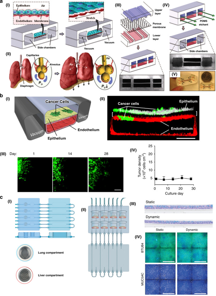
- Supportedcites [21] Hoarau-Véchot, J., Rafii, A., Touboul, C. & Pasquier, J. Halfway between 2D and animal models: are 3D cultures the ideal tool to study cancer-microenvironment interactions? Int. J. Mol. Sci. 19, 181 (2018). source
The Introduction states that concordance between animal models and clinical trials remains challenging and that the average rate of concordant results barely reaches 8%. It cites works by Seymour and Ghert, listed as references 10 and 11. (LLM, conf=0.95)
- Not checked yetcites [39] Voskoglou-Nomikos, T., Pater, J. L. & Seymour, L. Clinical predictive value of the in vitro cell line, human xenograft, and mouse allograft preclinical cancer models. Clin. Cancer Res. 9, 4227–4239 (2003).
- Could not verifycites [40] Katt, M. E., Placone, A. L., Wong, A. D., Xu, Z. S. & Searson, P. C. In vitro tumor models: advantages, disadvantages, variables, and selecting the right platform. Front. Bioeng. Biotechnol. 4, 12 (2016). sourceExperimental — accuracy not yet measured
The Introduction discusses limitations of animal tumor models, but does not compare their results with clinical trials. Searches for concordance, clinical trials, and an 8% rate found no passage reporting the claimed figure; the clinical-trials passage in “Embedded Ex Vivo Tumor Sections” concerns an in vitro drug-sensitivity test instead. (LLM, conf=0.74)
boosted- Asserted: passes — The authors put it forward as true.
- Checkable: passes — It can be checked against evidence.
- Load-bearing: passes — The paper’s conclusions depend on it.
- Based: passes — The paper gives a reason why it holds.
- Supported
Every source the paper cites for this claim was checked and supports it.
In the paper
“This platform can evaluate cell viability by observing the content of adenosine triphosphate (ATP) and the number of apoptotic necrotic cells.”
§ Tumor-on-a-chip: from bioinspired design to biomedical application > Different types of tumor-on-a-chip > Lung tumor chip
What the sources say
- Supportedcites [79] Bovard, D. et al. A lung/liver-on-a-chip platform for acute and chronic toxicity studies. Lab a Chip 18, 3814–3829 (2018). source
The ‘Measurement of cell viability’ section explicitly says the authors evaluated viability of the lung and liver tissues using ATP content and the number of apoptotic and necrotic cells. This directly supports the attributed claim. (LLM, conf=0.95)
plain- Asserted: passes — The authors put it forward as true.
- Checkable: passes — It can be checked against evidence.
- Load-bearing: unclear — It is unclear whether the paper’s conclusions depend on it.
- Based: passes — The paper gives a reason why it holds.
- Supported
Every source the paper cites for this claim was checked and supports it.
In the paper
“To solve this problem, researchers developed multiorgan-on-a-chip systems to evaluate the efficacy and off-target toxicity of anticancer therapeutics.”
§ Tumor-on-a-chip: from bioinspired design to biomedical application > Manufacture of tumor-on-a-chip > Advanced tumor-on-a-chip
What the sources say
- Supportedcites [102] Sun, W. et al. Organ-on-a-chip for cancer and immune organs modeling. Adv. Healthc. Mater. 8, e1801363 (2019). source
The Cancer modeling > Cancer-on-a-chip section (page not supplied) describes a multiorgan platform used to monitor both the anticancer efficacy of doxorubicin and its liver metabolism-mediated cardiac toxicity. This supports the claim that such systems were developed to evaluate therapeutic efficacy and off-target toxicity. (LLM, conf=0.94)
- Supportedcites [184] Cong, Y. et al. Drug toxicity evaluation based on organ-on-a-chip technology: a review. Micromachines 11, 381 (2020). source
Section 8.2 describes a multi-organ chip with tumor, liver, and healthy-tissue cells designed to assess drug efficacy and cytotoxicity in different organ-specific cells. It also describes a liver–tumor–bone marrow device used to test the cytotoxicity of anticancer drugs, supporting the off-target toxicity aspect. (LLM, conf=0.91)
plaindirect- Asserted: passes — The authors put it forward as true.
- Checkable: passes — It can be checked against evidence.
- Load-bearing: passes — The paper’s conclusions depend on it.
- Based: passes — The paper gives a reason why it holds.
- Supported
Every source the paper cites for this claim was checked and supports it.
In the paper
“Researchers have found that when liver spheroids are present, AFB1 toxicity in the lung-on-a-chip is reduced, demonstrating that liver detoxification can reduce AFB1 toxicity.”
§ Tumor-on-a-chip: from bioinspired design to biomedical application > Different types of tumor-on-a-chip > Lung tumor chip
What the sources say
- Supportedcites [79] Bovard, D. et al. A lung/liver-on-a-chip platform for acute and chronic toxicity studies. Lab a Chip 18, 3814–3829 (2018). source
The Introduction states that AFB1 toxicity was reduced in the presence of HepaRG™ liver spheroids. In “Assessment of AFB1 toxicity using the lung/liver-on-a-chip,” lung tissues cocultured with the spheroids showed delayed cytotoxicity, which the authors suggest resulted from conversion of AFB1 to a less toxic metabolite. (LLM, conf=0.94)
boosteddirect- Asserted: passes — The authors put it forward as true.
- Checkable: passes — It can be checked against evidence.
- Load-bearing: passes — The paper’s conclusions depend on it.
- Based: passes — The paper gives a reason why it holds.
- Supported
Every source the paper cites for this claim was checked and supports it.
In the paper
“They developed a multisite metastasis-on-a-chip platform to assess the metastatic preference of cancer cells117.”
§ Tumor-on-a-chip: from bioinspired design to biomedical application > Application of tumor modeling > Modeling cancer invasion and metastasis
What the sources say
- Supportedcites [117] Aleman, J. & Skardal, A. A multi-site metastasis-on-a-chip microphysiological system for assessing metastatic preference of cancer cells. Biotechnol. Bioeng. 116, 936–944, https://doi.org/10.1002/bit.26871 (2019). source
The Results and Discussion section describes expanding the metastasis-on-a-chip system to four downstream sites and tracking cancer cells from a primary organoid to potential metastatic sites. It explicitly says the platform can distinguish which tissue constructs metastatic cells prefer to home to and colonize. (LLM, conf=0.94)
plain- Asserted: passes — The authors put it forward as true.
- Checkable: passes — It can be checked against evidence.
- Load-bearing: unclear — It is unclear whether the paper’s conclusions depend on it.
- Based: passes — The paper gives a reason why it holds.
- Supported
Every source the paper cites for this claim was checked and supports it.
In the paper
“Compared with those in the monolayer model, the cells in the spheroid-based 3D model exhibit higher drug resistance during drug testing, and the 3D model is closer to the chemical environment of native tissues215.”
§ Tumor-on-a-chip: from bioinspired design to biomedical application > Different types of tumor-on-a-chip > Pancreatic tumor chip
What the sources say
- Supportedcites [215] Wen, Z. et al. A spheroid-based 3-D culture model for pancreatic cancer drug testing, using the acid phosphatase assay. Braz. J. Med. Biol. Res. 46, 634–642 (2013). source
The Introduction says 3-D cultures more closely mimic tumor-tissue microenvironments than 2-D cultures, including concentration gradients of signaling molecules and therapeutic agents. The Abstract also reports that the pancreatic cancer spheroids had much higher drug resistance than monolayers. (LLM, conf=0.93)
plaincorrelational- Asserted: passes — The authors put it forward as true.
- Checkable: passes — It can be checked against evidence.
- Load-bearing: passes — The paper’s conclusions depend on it.
- Based: passes — The paper gives a reason why it holds.
- Supported
None of the sources cited for this claim have been checked yet.
In the paper
“2D cell monolayers and three-dimensional cell culture models have been widely employed as initial screening models to provide a cost-efficient and simplified method to elucidate the mechanism of cancer biology or identify the efficacy and safety of a drug candidate14–16; however, they are unable to recapitulate complex biochemical and biophysical factors of the tumor microenvironment (TME) in vivo”
§ Tumor-on-a-chip: from bioinspired design to biomedical application > Introduction
What the sources say
- Not checked yetcites [17] Antoine, E. E., Vlachos, P. P., Rylander, M. N. & Zeugolis, D. Tunable collagen I hydrogels for engineered physiological tissue micro-environments. PLoS ONE 10, e0122500 (2015). source
- Not checked yetcites [18] Asghar, W. et al. Engineering cancer microenvironments for in vitro 3-D tumor models. Mater. Today 18, 539–553 (2015). source
- Not checked yetcites [19] Kapałczyńska, M. et al. 2D and 3D cell cultures–a comparison of different types of cancer cell cultures. Arch. Med. Sci. 14, 910 (2018).
- Not checked yetcites [20] Fang, Y. & Eglen, R. M. Three-dimensional cell cultures in drug discovery and development. SLAS Discov. 22, 456–472 (2017). source
- Not checked yetcites [21] Hoarau-Véchot, J., Rafii, A., Touboul, C. & Pasquier, J. Halfway between 2D and animal models: are 3D cultures the ideal tool to study cancer-microenvironment interactions? Int. J. Mol. Sci. 19, 181 (2018). source
- Not checked yetcites [22] Jabbarzadegan, M. et al. Application of arteether-loaded polyurethane nanomicelles to induce immune response in breast cancer model. Artif. Cells Nanomed. Biotechnol. 45, 808–816 (2017). source
- Not checked yetcites [23] Chung, M., Ahn, J., Son, K., Kim, S. & Jeon, N. L. Biomimetic model of tumor microenvironment on microfluidic platform. Adv. Healthc. Mater. 6, 1700196 (2017). source
- Not checked yetcites [24] Stock, K. et al. Capturing tumor complexity in vitro: Comparative analysis of 2D and 3D tumor models for drug discovery. Sci. Rep. 6, 1–15 (2016). source
plaindirect- Asserted: passes — The authors put it forward as true.
- Checkable: passes — It can be checked against evidence.
- Load-bearing: passes — The paper’s conclusions depend on it.
- Based: passes — The paper gives a reason why it holds.
None of the sources cited for this claim have been checked yet.
In the paper
“Furthermore, microfluidic systems can link different organs-on-a-chip to potentially simulate the complexity of multiorgan drug metabolism and pharmacokinetics9,47,50,68–71.”
§ Tumor-on-a-chip: from bioinspired design to biomedical application > Introduction
What the sources say
- Not checked yetcites [9] Satoh, T. et al. A multi-throughput multi-organ-on-a-chip system on a plate formatted pneumatic pressure-driven medium circulation platform. Lab a Chip 18, 115–125 (2018). source
- Not checked yetcites [47] Zhang, Y. S., Zhang, Y.-N. & Zhang, W. Cancer-on-a-chip systems at the frontier of nanomedicine. Drug Discov. Today 22, 1392–1399 (2017). source
- Not checked yetcites [50] Ronaldson-Bouchard, K. & Vunjak-Novakovic, G. Organs-on-a-chip: a fast track for engineered human tissues in drug development. Cell Stem Cell 22, 310–324 (2018). source
- Not checked yetcites [68] Wang, Y. I. et al. Self-contained, low-cost Body-on-a-Chip systems for drug development. Exp. Biol. Med. 242, 1701–1713 (2017). source
- Not checked yetcites [69] Tsamandouras, N. et al. Integrated gut and liver microphysiological systems for quantitative in vitro pharmacokinetic studies. AAPS J. 19, 1499–1512 (2017). source
- Not checked yetcites [70] Skardal, A. et al. Multi-tissue interactions in an integrated three-tissue organ-on-a-chip platform. Sci. Rep. 7, 8837 (2017). source
- Not checked yetcites [71] Edington, C. D. et al. Interconnected microphysiological systems for quantitative biology and pharmacology studies. Sci. Rep. 8, 4530 (2018). source
hedgedconditional- Asserted: passes — The authors put it forward as true.
- Checkable: passes — It can be checked against evidence.
- Load-bearing: unclear — It is unclear whether the paper’s conclusions depend on it.
- Based: passes — The paper gives a reason why it holds.
None of the sources cited for this claim have been checked yet.
In the paper
“Species-specific differences between animals and humans cause differences in drug efficacy and toxicity tests in physiology and cell biology31–37.”
§ Tumor-on-a-chip: from bioinspired design to biomedical application > Introduction
What the sources say
- Not checked yetcites [31] Engelman, R. & Kerr, W. G. Autoimmunity 433–441 (Springer, 2012). source
- Not checked yetcites [32] Kang, T. H. & Kim, H. J. Farewell to animal testing: innovations on human intestinal microphysiological systems. Micromachines 7, 107 (2016). source
- Not checked yetcites [33] Hintze, K. J. et al. Broad scope method for creating humanized animal models for animal health and disease research through antibiotic treatment and human fecal transfer. Gut Microbes 5, 183–191 (2014). source
- Not checked yetcites [34] Rongvaux, A. et al. Development and function of human innate immune cells in a humanized mouse model. Nat. Biotechnol. 32, 364–372 (2014). source
- Not checked yetcites [35] Seok, J. et al. Genomic responses in mouse models poorly mimic human inflammatory diseases. Proc. Natl Acad. Sci. USA 110, 3507–3512 (2013). source
- Not checked yetcites [36] Rubin, E. H. & Gilliland, D. G. Drug development and clinical trials—the path to an approved cancer drug. Nat. Rev. Clin. Oncol. 9, 215 (2012). source
- Not checked yetcites [37] Zhou, Z. et al. The use of functionalized silk fibroin films as a platform for optical diffraction‐based sensing applications. Adv. Mater. 29, 1605471 (2017). source
plaindirect- Asserted: passes — The authors put it forward as true.
- Checkable: passes — It can be checked against evidence.
- Load-bearing: passes — The paper’s conclusions depend on it.
- Based: passes — The paper gives a reason why it holds.
A conclusion the paper draws from its own results — it cites no other source for it, so there is none to check. The quote shows it is in the paper.
In the paper
“3D models are able to better reflect the real tumor setting with a more natural response to different soluble factors present in the TME.”
§ Tumor-on-a-chip: from bioinspired design to biomedical application > Introduction
plaindirect- Asserted: passes — The authors put it forward as true.
- Checkable: passes — It can be checked against evidence.
- Load-bearing: passes — The paper’s conclusions depend on it.
- Based: passes — The paper gives a reason why it holds.
None of the sources cited for this claim have been checked yet.
In the paper
“However, the success rate of anticancer drugs in clinical trials is very low, with over 80% of drug candidates failing during clinical screening due to weak efficacy or adverse events4–7.”
§ Tumor-on-a-chip: from bioinspired design to biomedical application > Introduction
What the sources say
- Not checked yetcites [4] Smietana, K., Siatkowski, M. & Moller, M. Trends in clinical success rates. Nat. Rev. Drug Discov. 15, 379–380 (2016). source
- Not checked yetcites [5] Heylman, C., Sobrino, A., Shirure, V. S., Hughes, C. C. & George, S. C. A strategy for integrating essential three-dimensional microphysiological systems of human organs for realistic anticancer drug screening. Exp. Biol. Med. 239, 1240–1254 (2014). source
- Not checked yetcites [6] Arrowsmith, J. & Miller, P. Trial watch: Phase II and Phase III attrition rates 2011-2012. Nat. Rev. Drug Discov. 12, 569 (2013). source
- Not checked yetcites [7] Hay, M., Thomas, D. W., Craighead, J. L., Economides, C. & Rosenthal, J. Clinical development success rates for investigational drugs. Nat. Biotechnol. 32, 40–51 (2014). source
plaindirect- Asserted: passes — The authors put it forward as true.
- Checkable: passes — It can be checked against evidence.
- Load-bearing: passes — The paper’s conclusions depend on it.
- Based: passes — The paper gives a reason why it holds.
A conclusion the paper draws from its own results — it cites no other source for it, so there is none to check. The quote shows it is in the paper.
In the paper
“These differences strongly suggest that the current disease and drug screening preclinical models possess certain predictive defects13.”
§ Tumor-on-a-chip: from bioinspired design to biomedical application > Introduction
hedged- Asserted: passes — The authors put it forward as true.
- Checkable: passes — It can be checked against evidence.
- Load-bearing: passes — The paper’s conclusions depend on it.
- Based: passes — The paper gives a reason why it holds.
None of the sources cited for this claim have been checked yet.
In the paper
“Through precise microfluidic designs, microfluidic devices can reconstruct physiological dynamic characteristics in tissues, such as physiological flow, shear stress, nutrient delivery, and drug action, in a controllable environment, providing a platform for cell culture, microenvironment creation, and organ simulation or in vitro assessment of organ tissue60–67.”
§ Tumor-on-a-chip: from bioinspired design to biomedical application > Introduction
What the sources say
- Not checked yetcites [60] Orcheston-Findlay, L., Hashemi, A., Garrill, A. & Nock, V. A microfluidic gradient generator to simulate the oxygen microenvironment in cancer cell culture. Microelectron. Eng. 195, 107–113 (2018). source
- Not checked yetcites [61] Ma, Y.-H. V., Middleton, K., You, L. & Sun, Y. A review of microfluidic approaches for investigating cancer extravasation during metastasis. Microsyst. Nanoeng. 4, 1–13 (2018). source
- Not checked yetcites [62] Wan, L. et al. Mimicking embedded vasculature structure for 3D cancer on a chip approaches through micromilling. Sci. Rep. 7, 1–8 (2017). source
- Not checked yetcites [63] Chen, M. B. et al. On-chip human microvasculature assay for visualization and quantification of tumor cell extravasation dynamics. Nat. Protoc. 12, 865–880 (2017). source
- Not checked yetcites [64] Fabre, K. M., Livingston, C. & Tagle, D. A. Organs-on-chips (microphysiological systems): tools to expedite efficacy and toxicity testing in human tissue. Exp. Biol. Med. 239, 1073–1077 (2014). source
- Not checked yetcites [65] Polacheck, W. J., German, A. E., Mammoto, A., Ingber, D. E. & Kamm, R. D. Mechanotransduction of fluid stresses governs 3D cell migration. Proc. Natl Acad. Sci. USA 111, 2447–2452 (2014). source
- Not checked yetcites [66] Shirure, V. S., Lezia, A., Tao, A., Alonzo, L. F. & George, S. C. Low levels of physiological interstitial flow eliminate morphogen gradients and guide angiogenesis. Angiogenesis 20, 493–504 (2017). source
- Not checked yetcites [67] Rothbauer, M., Zirath, H. & Ertl, P. Recent advances in microfluidic technologies for cell-to-cell interaction studies. Lab Chip 18, 249–270 (2018). source
plaindirect- Asserted: passes — The authors put it forward as true.
- Checkable: passes — It can be checked against evidence.
- Load-bearing: passes — The paper’s conclusions depend on it.
- Based: passes — The paper gives a reason why it holds.
In the paper
“Despite this, PDMS has a disadvantage, namely, its hydrophobicity, which limits the application of PDMS in chemical screening because PDMS may bind or absorb hydrophobic molecules163.”
§ Tumor-on-a-chip: from bioinspired design to biomedical application > Manufacture of tumor-on-a-chip > Materials for tumor-on-a-chip
What the sources say
- Not checked yetcites [163] Auner, A. W., Tasneem, K. M., Markov, D. A., McCawley, L. J. & Hutson, M. S. Chemical-PDMS binding kinetics and implications for bioavailability in microfluidic devices. Lab a Chip 19, 864–874 (2019). source
hedgedconditional- Asserted: passes — The authors put it forward as true.
- Checkable: passes — It can be checked against evidence.
- Load-bearing: passes — The paper’s conclusions depend on it.
- Based: passes — The paper gives a reason why it holds.
In the paper
“In an in vitro metastasis model, migrated tumor cells must be further exposed to varying microenvironments and oxygen gradients to simulate the process of in vivo intravasation126.”
§ Tumor-on-a-chip: from bioinspired design to biomedical application > Manufacture of tumor-on-a-chip
What the sources say
- Not checked yetcites [126] Ehsan, S. M., Welch-Reardon, K. M., Waterman, M. L., Hughes, C. C. & George, S. C. A three-dimensional in vitro model of tumor cell intravasation. Integr. Biol. 6, 603–610 (2014). source
plaindirect- Asserted: passes — The authors put it forward as true.
- Checkable: passes — It can be checked against evidence.
- Load-bearing: passes — The paper’s conclusions depend on it.
- Based: passes — The paper gives a reason why it holds.
In the paper
“The vasculature of tumors is very different from the blood vessels that supply healthy tissues, especially in terms of heterogeneity, permeability, multidirectional blood flow, and unordered distribution throughout the tumor160.”
§ Tumor-on-a-chip: from bioinspired design to biomedical application > Manufacture of tumor-on-a-chip > Principle and fabrication of tumor-on-a-chip
What the sources say
- Not checked yetcites [160] Nagy, J. A., Chang, S. H., Dvorak, A. M. & Dvorak, H. F. Why are tumour blood vessels abnormal and why is it important to know? Br. J. Cancer 100, 865–869 (2009). source
plain- Asserted: passes — The authors put it forward as true.
- Checkable: passes — It can be checked against evidence.
- Load-bearing: passes — The paper’s conclusions depend on it.
- Based: passes — The paper gives a reason why it holds.
None of the sources cited for this claim have been checked yet.
In the paper
“In addition, bioprinting technology is able to create a biomimetic microenvironment for the heterogeneous distribution of biologically relevant proteins and growth factors, which are important to control tumor cell signaling, proliferation, and migration156,157.”
§ Tumor-on-a-chip: from bioinspired design to biomedical application > Manufacture of tumor-on-a-chip > Principle and fabrication of tumor-on-a-chip
What the sources say
- Not checked yetcites [156] Choudhury, D., Anand, S. & Naing, M. W. The Arrival of Commercial Bioprinters - Towards 3D Bioprinting Revolution! Int. J. Bioprint. 4, https://doi.org/10.18063/ijb.v4i2.139 (2018). source
- Not checked yetcites [157] Keenan, T. M. & Folch, A. Biomolecular gradients in cell culture systems. Lab a Chip 8, 34–57 (2008). source
plaindirect- Asserted: passes — The authors put it forward as true.
- Checkable: passes — It can be checked against evidence.
- Load-bearing: passes — The paper’s conclusions depend on it.
- Based: passes — The paper gives a reason why it holds.
In the paper
“It has been a major challenge to realize ECM-like heterogeneous composition in microfluidic channels of tumor-on-a-chip152.”
§ Tumor-on-a-chip: from bioinspired design to biomedical application > Manufacture of tumor-on-a-chip > Principle and fabrication of tumor-on-a-chip
What the sources say
- Not checked yetcites [152] Colosi, C. et al. Microfluidic bioprinting of heterogeneous 3D tissue constructs using low-viscosity bioink. Adv. Mater. 28, 677–684 (2016). source
plain- Asserted: passes — The authors put it forward as true.
- Checkable: passes — It can be checked against evidence.
- Load-bearing: passes — The paper’s conclusions depend on it.
- Based: passes — The paper gives a reason why it holds.
None of the sources cited for this claim have been checked yet.
In the paper
“Various studies have demonstrated that complex components can significantly affect the growth and metastasis of cancer via mechanical and biochemical factors113–116.”
§ Tumor-on-a-chip: from bioinspired design to biomedical application > Introduction
What the sources say
- Not checked yetcites [113] Swartz, M. A. et al. Tumor microenvironment complexity: emerging roles in cancer therapy. Cancer Res. 72, 2473–2480 (2012). source
- Not checked yetcites [114] Wang, M. et al. Role of tumor microenvironment in tumorigenesis. J. Cancer 8, 761–773 (2017). source
- Not checked yetcites [115] Tavora, B. et al. Endothelial-cell FAK targeting sensitizes tumours to DNA-damaging therapy. Nature 514, 112–116 (2014). source
- Not checked yetcites [116] Romero-López, M. et al. Recapitulating the human tumor microenvironment: colon tumor-derived extracellular matrix promotes angiogenesis and tumor cell growth. Biomaterials 116, 118–129 (2017). source
boosteddirect- Asserted: passes — The authors put it forward as true.
- Checkable: passes — It can be checked against evidence.
- Load-bearing: passes — The paper’s conclusions depend on it.
- Based: passes — The paper gives a reason why it holds.
None of the sources cited for this claim have been checked yet.
In the paper
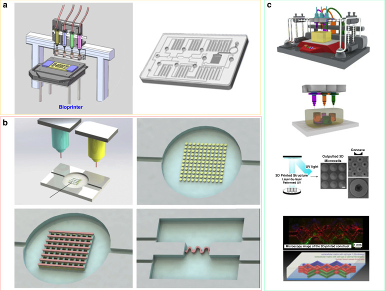
“Various biofunctional materials and cell types can thus be printed simultaneously onto a substrate of cell-compatible biomaterials to build 3D complex constructs with good spatial resolution and reproducibility (Fig. 3a)130,148–151.”
§ Tumor-on-a-chip: from bioinspired design to biomedical application > Manufacture of tumor-on-a-chip > Principle and fabrication of tumor-on-a-chip
What the sources say
- Not checked yetcites [130] Yang, Q., Lian, Q. & Xu, F. Perspective: fabrication of integrated organ-on-a-chip via bioprinting. Biomicrofluidics 11, 031301 (2017). source
- Not checked yetcites [148] Moroni, L. et al. Biofabrication strategies for 3D in vitro models and regenerative medicine. Nat. Rev. Mater. 3, 21–37 (2018). source
- Not checked yetcites [149] Costantini, M. et al. 3D bioprinting of BM-MSCs-loaded ECM biomimetic hydrogels for in vitro neocartilage formation. Biofabrication 8, 035002 (2016). source
- Not checked yetcites [150] Kang, H. W. et al. A 3D bioprinting system to produce human-scale tissue constructs with structural integrity. Nat. Biotechnol. 34, 312–319 (2016). source
- Not checked yetcites [151] Kolesky, D. B., Homan, K. A., Skylar-Scott, M. A. & Lewis, J. A. Three-dimensional bioprinting of thick vascularized tissues. Proc. Natl Acad. Sci. USA 113, 3179–3184 (2016). source
plaindirect- Asserted: passes — The authors put it forward as true.
- Checkable: passes — It can be checked against evidence.
- Load-bearing: passes — The paper’s conclusions depend on it.
- Based: passes — The paper gives a reason why it holds.
None of the sources cited for this claim have been checked yet.
In the paper
“Based on bioprinting technology, bioinks can create cell aggregates comprising different types of cells, including cancer-associated fibroblasts, immune cells and endothelial cells, to form vascular networks153–155.”
§ Tumor-on-a-chip: from bioinspired design to biomedical application > Manufacture of tumor-on-a-chip > Principle and fabrication of tumor-on-a-chip
What the sources say
- Not checked yetcites [153] Dababneh, A. B. & Ozbolat, I. T. Bioprinting technology: a current state-of-the-art review. J. Manuf. Sci. Eng. 136, https://doi.org/10.1115/1.4028512 (2014). source
- Not checked yetcites [154] Junttila, M. R. & de Sauvage, F. J. Influence of tumour micro-environment heterogeneity on therapeutic response. Nature 501, 346–354 (2013). source
- Not checked yetcites [155] Xu, F. et al. A three-dimensional in vitro ovarian cancer coculture model using a high-throughput cell patterning platform. Biotechnol. J. 6, 204–212 (2011). source
plaindirect- Asserted: passes — The authors put it forward as true.
- Checkable: passes — It can be checked against evidence.
- Load-bearing: unclear — It is unclear whether the paper’s conclusions depend on it.
- Based: passes — The paper gives a reason why it holds.
In the paper
“To solve this problem, Kang’s group developed a protocol using poly(methyl methacrylate) (PMMA) substrates chemically and robustly bound to porous orbitally etched polyethylene terephthalate (PETE) membranes to construct a microfluidic device that is impermeable to small lipophilic molecules164.”
§ Tumor-on-a-chip: from bioinspired design to biomedical application > Manufacture of tumor-on-a-chip > Materials for tumor-on-a-chip
What the sources say
- Not checked yetcites [164] Nguyen, T. et al. Robust chemical bonding of PMMA microfluidic devices to porous PETE membranes for reliable cytotoxicity testing of drugs. Lab Chip 19, 3706–3713 (2019). source
plaindirect- Asserted: passes — The authors put it forward as true.
- Checkable: passes — It can be checked against evidence.
- Load-bearing: passes — The paper’s conclusions depend on it.
- Based: passes — The paper gives a reason why it holds.
The paper states this without a source or data behind it here, so there is nothing to check it against. The quote shows it is in the paper.
In the paper
“First, it has the ability to mimic the heterogeneous microenvironment and complex 3D microstructures of the tumor.”
§ Tumor-on-a-chip: from bioinspired design to biomedical application > Manufacture of tumor-on-a-chip > Principle and fabrication of tumor-on-a-chip
plaindirect- Asserted: passes — The authors put it forward as true.
- Checkable: passes — It can be checked against evidence.
- Load-bearing: passes — The paper’s conclusions depend on it.
- Based: passes — The paper gives a reason why it holds.
In the paper
“Successful vascularization has always been a major challenge in the production of functional tissues for tumor in vitro models161.”
§ Tumor-on-a-chip: from bioinspired design to biomedical application > Manufacture of tumor-on-a-chip > Principle and fabrication of tumor-on-a-chip
What the sources say
- Not checked yetcites [161] Rouwkema, J. & Khademhosseini, A. Vascularization and angiogenesis in tissue engineering: beyond creating static networks. Trends Biotechnol. 34, 733–745 (2016). source
plain- Asserted: passes — The authors put it forward as true.
- Checkable: passes — It can be checked against evidence.
- Load-bearing: passes — The paper’s conclusions depend on it.
- Based: passes — The paper gives a reason why it holds.
- Rests on an assumption: Vascularization is very important to maintain tissue activities and can be used to separate different tissue compartments.
In the paper
“Moreover, these methods are capable of fabricating only the microfluidic chip itself, while the other elements (i.e., microtissues, stimulus-loading components, and results-readout sensors) require additional processes130.”
§ Tumor-on-a-chip: from bioinspired design to biomedical application > Manufacture of tumor-on-a-chip > Principle and fabrication of tumor-on-a-chip
What the sources say
- Not checked yetcites [130] Yang, Q., Lian, Q. & Xu, F. Perspective: fabrication of integrated organ-on-a-chip via bioprinting. Biomicrofluidics 11, 031301 (2017). source
plaindirect- Asserted: passes — The authors put it forward as true.
- Checkable: passes — It can be checked against evidence.
- Load-bearing: unclear — It is unclear whether the paper’s conclusions depend on it.
- Based: passes — The paper gives a reason why it holds.
None of the sources cited for this claim have been checked yet.
In the paper
“In recent years, bioprinting technology based on layer-by-layer printing has been used for tumor-on-a-chip fabrication (Fig. 3)132,145–147.”
§ Tumor-on-a-chip: from bioinspired design to biomedical application > Manufacture of tumor-on-a-chip > Principle and fabrication of tumor-on-a-chip
What the sources say
- Not checked yetcites [132] Lee, H. & Cho, D.-W. One-step fabrication of an organ-on-a-chip with spatial heterogeneity using a 3D bioprinting technology. Lab a Chip 16, 2618–2625 (2016). source
- Not checked yetcites [145] Wang, Y. et al. 3D bioprinting of breast cancer models for drug resistance study. ACS Biomater. Sci. Eng. 4, 4401–4411 (2018). source
- Not checked yetcites [146] Knowlton, S., Onal, S., Yu, C. H., Zhao, J. J. & Tasoglu, S. Bioprinting for cancer research. Trends Biotechnol. 33, 504–513 (2015). source
- Not checked yetcites [147] Matai, I., Kaur, G., Seyedsalehi, A., McClinton, A. & Laurencin, C. T. Progress in 3D bioprinting technology for tissue/organ regenerative engineering. Biomaterials 226, 119536 (2020). source
plaindirect- Asserted: passes — The authors put it forward as true.
- Checkable: passes — It can be checked against evidence.
- Load-bearing: unclear — It is unclear whether the paper’s conclusions depend on it.
- Based: passes — The paper gives a reason why it holds.
None of the sources cited for this claim have been checked yet.
In the paper
“It can be used to mimic in vivo tumor microenvironments because it has the ability to provide metabolism, proliferation, and species concentration gradients similar to those found in vivo175,176.”
§ Tumor-on-a-chip: from bioinspired design to biomedical application > Manufacture of tumor-on-a-chip > Advanced tumor-on-a-chip
What the sources say
- Not checked yetcites [175] Sant, S. & Johnston, P. A. The production of 3D tumor spheroids for cancer drug discovery. Drug Discov. Today 23, 27–36 (2017). source
- Not checked yetcites [176] Nunes, A. S., Barros, A. S., Costa, E. C., Moreira, A. F. & Correia, I. J. 3D tumor spheroids as in vitro models to mimic in vivo human solid tumors resistance to therapeutic drugs. Biotechnol. Bioeng. 116, 206–226 (2019). source
plaindirect- Asserted: passes — The authors put it forward as true.
- Checkable: passes — It can be checked against evidence.
- Load-bearing: passes — The paper’s conclusions depend on it.
- Based: passes — The paper gives a reason why it holds.
A conclusion the paper draws from its own results — it cites no other source for it, so there is none to check. The quote shows it is in the paper.
In the paper
“Hence, the liver needs to be cocultured in a microscale cell culture simulation system (μCCA) to study the pharmaceutical aspects of anticancer drugs.”
§ Tumor-on-a-chip: from bioinspired design to biomedical application > Different types of tumor-on-a-chip > Liver tumor chip
plaindirect- Asserted: passes — The authors put it forward as true.
- Checkable: passes — It can be checked against evidence.
- Load-bearing: passes — The paper’s conclusions depend on it.
- Based: passes — The paper gives a reason why it holds.
In the paper
“The experimental results showed the importance of drug detection in three-dimensional tumor spheroid models.”
§ Tumor-on-a-chip: from bioinspired design to biomedical application > Manufacture of tumor-on-a-chip > Advanced tumor-on-a-chip
What the sources say
- Not checked yetcites [178] Patra, B., Peng, C.-C., Liao, W.-H., Lee, C.-H. & Tung, Y.-C. Drug testing and flow cytometry analysis on a large number of uniform sized tumor spheroids using a microfluidic device. Sci. Rep. 6, 21061 (2016). source
plain- Asserted: passes — The authors put it forward as true.
- Checkable: passes — It can be checked against evidence.
- Load-bearing: passes — The paper’s conclusions depend on it.
- Based: passes — The paper gives a reason why it holds.
In the paper
“It was found that the PVN is a niche for BTSCs, and the microvascular diameter can be used as a pathway for tumor metastasis.”
§ Tumor-on-a-chip: from bioinspired design to biomedical application > Different types of tumor-on-a-chip > Brain tumor chip
What the sources say
- Not checked yetcites [196] Xiao, Y. et al. Ex vivo dynamics of human glioblastoma cells in a microvasculature‐on‐a‐chip system correlates with tumor heterogeneity and subtypes. Adv. Sci. 6, 1801531 (2019). source
plaindirect- Asserted: passes — The authors put it forward as true.
- Checkable: passes — It can be checked against evidence.
- Load-bearing: passes — The paper’s conclusions depend on it.
- Based: passes — The paper gives a reason why it holds.
A conclusion the paper draws from its own results — it cites no other source for it, so there is none to check. The quote shows it is in the paper.
In the paper
“Adverse reactions caused by drug metabolism increase the role of hepatocytes in organ-on-a-chip systems.”
§ Tumor-on-a-chip: from bioinspired design to biomedical application > Different types of tumor-on-a-chip > Liver tumor chip
plaindirect- Asserted: passes — The authors put it forward as true.
- Checkable: passes — It can be checked against evidence.
- Load-bearing: unclear — It is unclear whether the paper’s conclusions depend on it.
- Based: passes — The paper gives a reason why it holds.
In the paper
“Through this platform, the toxicity of AFB1 to liver–lung crosstalk could be evaluated.”
§ Tumor-on-a-chip: from bioinspired design to biomedical application > Different types of tumor-on-a-chip > Lung tumor chip
What the sources say
- Not checked yetcites [79] Bovard, D. et al. A lung/liver-on-a-chip platform for acute and chronic toxicity studies. Lab a Chip 18, 3814–3829 (2018). source
hedgeddirect- Asserted: passes — The authors put it forward as true.
- Checkable: passes — It can be checked against evidence.
- Load-bearing: passes — The paper’s conclusions depend on it.
- Based: passes — The paper gives a reason why it holds.
In the paper
“Researchers have found that when liver spheroids are present, AFB1 toxicity in the lung-on-a-chip is reduced, demonstrating that liver detoxification can reduce AFB1 toxicity.”
§ Tumor-on-a-chip: from bioinspired design to biomedical application > Different types of tumor-on-a-chip > Lung tumor chip
What the sources say
- Not checked yetcites [79] Bovard, D. et al. A lung/liver-on-a-chip platform for acute and chronic toxicity studies. Lab a Chip 18, 3814–3829 (2018). source
boosteddirect- Asserted: passes — The authors put it forward as true.
- Checkable: passes — It can be checked against evidence.
- Load-bearing: passes — The paper’s conclusions depend on it.
- Based: passes — The paper gives a reason why it holds.
A conclusion the paper draws from its own results — it cites no other source for it, so there is none to check. The quote shows it is in the paper.
In the paper
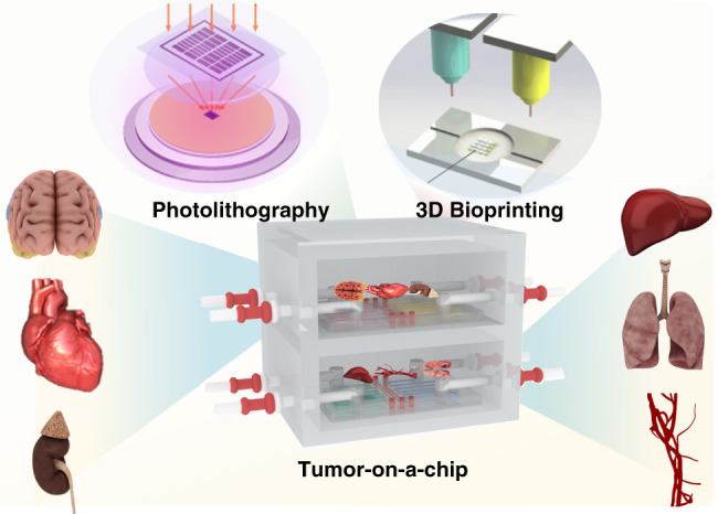
“By combining advanced microfabrication techniques, such as photolithography, soft lithography and bioprinting, with microfluidic and tissue engineering techniques, tumor chips can ideally reproduce specific key aspects of the tumor microenvironment, such as biochemical gradients and niche factors, dynamic cell–cell and cell–matrix interactions, and complex tissue structures comprised of tumor and”
§ Tumor-on-a-chip: from bioinspired design to biomedical application > Different types of tumor-on-a-chip
plaindirect- Asserted: passes — The authors put it forward as true.
- Checkable: passes — It can be checked against evidence.
- Load-bearing: passes — The paper’s conclusions depend on it.
- Based: unclear — It is unclear why the paper says it holds.
None of the sources cited for this claim have been checked yet.
In the paper
“This model can simulate multiple physiological functions of the lung.”
§ Tumor-on-a-chip: from bioinspired design to biomedical application > Different types of tumor-on-a-chip > Lung tumor chip
What the sources say
- Not checked yetcites [79] Bovard, D. et al. A lung/liver-on-a-chip platform for acute and chronic toxicity studies. Lab a Chip 18, 3814–3829 (2018). source
- Not checked yetcites [98] Hassell, B. A. et al. Human organ chip models recapitulate orthotopic lung cancer growth, therapeutic responses, and tumor dormancy in vitro. Cell Rep. 21, 508–516 (2017). source
plaindirect- Asserted: passes — The authors put it forward as true.
- Checkable: passes — It can be checked against evidence.
- Load-bearing: passes — The paper’s conclusions depend on it.
- Based: passes — The paper gives a reason why it holds.
In the paper
“Moreover, through lung tumor chips, researchers also found that coculture of tumor cells and alveolar epithelial cells can increase cell–cell interactions, but endothelial cells can inhibit this effect98.”
§ Tumor-on-a-chip: from bioinspired design to biomedical application > Different types of tumor-on-a-chip > Lung tumor chip
What the sources say
- Not checked yetcites [98] Hassell, B. A. et al. Human organ chip models recapitulate orthotopic lung cancer growth, therapeutic responses, and tumor dormancy in vitro. Cell Rep. 21, 508–516 (2017). source
plaindirect- Asserted: passes — The authors put it forward as true.
- Checkable: passes — It can be checked against evidence.
- Load-bearing: unclear — It is unclear whether the paper’s conclusions depend on it.
- Based: passes — The paper gives a reason why it holds.
The paper states this without a source or data behind it here, so there is nothing to check it against. The quote shows it is in the paper.
In the paper
“Microfluidic chips can create precise concentration gradients that are continuously modulated through the side channels of the surrounding vascular simulation system and therefore can assess the efficiency of drug delivery.”
§ Tumor-on-a-chip: from bioinspired design to biomedical application > Different types of tumor-on-a-chip > Colorectal-tumor chip
plaindirect- Asserted: passes — The authors put it forward as true.
- Checkable: passes — It can be checked against evidence.
- Load-bearing: passes — The paper’s conclusions depend on it.
- Based: passes — The paper gives a reason why it holds.
In the paper
“Researchers have used organ-on-a-chip technology to construct a microvascular system with selective barrier function similar to that in the body to simulate the microstructure and microphysiological functions of human organs to better describe organ functions and characteristics179.”
§ Tumor-on-a-chip: from bioinspired design to biomedical application > Manufacture of tumor-on-a-chip > Advanced tumor-on-a-chip
What the sources say
- Not checked yetcites [179] Wang, X., Sun, Q. & Pei, J. Microfluidic-based 3D engineered microvascular networks and their applications in vascularized microtumor models. Micromachines 9, https://doi.org/10.3390/mi9100493 (2018). source
plaindirect- Asserted: passes — The authors put it forward as true.
- Checkable: passes — It can be checked against evidence.
- Load-bearing: unclear — It is unclear whether the paper’s conclusions depend on it.
- Based: passes — The paper gives a reason why it holds.
In the paper
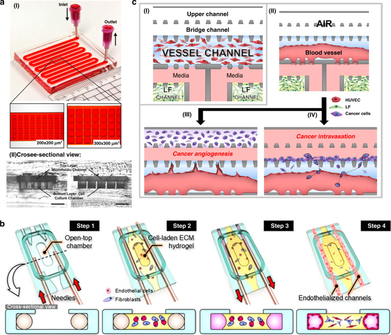
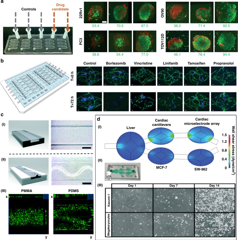
“Patra et al. used a microfluidic device to perform drug detection and flow cytometry analysis on a large number of uniformly sized tumor spheroids (Fig. 4a)178.”
§ Tumor-on-a-chip: from bioinspired design to biomedical application > Manufacture of tumor-on-a-chip > Advanced tumor-on-a-chip
What the sources say
- Not checked yetcites [178] Patra, B., Peng, C.-C., Liao, W.-H., Lee, C.-H. & Tung, Y.-C. Drug testing and flow cytometry analysis on a large number of uniform sized tumor spheroids using a microfluidic device. Sci. Rep. 6, 21061 (2016). source
plain- Asserted: passes — The authors put it forward as true.
- Checkable: passes — It can be checked against evidence.
- Load-bearing: unclear — It is unclear whether the paper’s conclusions depend on it.
- Based: passes — The paper gives a reason why it holds.
None of the sources cited for this claim have been checked yet.
In the paper
“Therefore, researchers propose that 3D-cultured breast tumor chips can be used to predict antitumor drug responses211,212.”
§ Tumor-on-a-chip: from bioinspired design to biomedical application > Different types of tumor-on-a-chip > Breast tumor chip
What the sources say
- Not checked yetcites [211] Lanz, H. L. et al. Therapy response testing of breast cancer in a 3D high-throughput perfused microfluidic platform. BMC Cancer 17, 709 (2017). source
- Not checked yetcites [212] Boyle, P. Triple-negative breast cancer: epidemiological considerations and recommendations. Ann. Oncol. 23, vi7–vi12 (2012). source
plaindirect- Asserted: passes — The authors put it forward as true.
- Checkable: passes — It can be checked against evidence.
- Load-bearing: passes — The paper’s conclusions depend on it.
- Based: passes — The paper gives a reason why it holds.
In the paper
“They injected mepitastatin and irinotecan into the cells, and the results showed that this model can be used as a glioma chip model for drug screening and release tests.”
§ Tumor-on-a-chip: from bioinspired design to biomedical application > Different types of tumor-on-a-chip > Brain tumor chip
What the sources say
- Not checked yetcites [197] Fan, Y., Nguyen, D. T., Akay, Y., Xu, F. & Akay, M. Engineering a brain cancer chip for high-throughput drug screening. Sci. Rep. 6, 25062 (2016). source
plaindirect- Asserted: passes — The authors put it forward as true.
- Checkable: passes — It can be checked against evidence.
- Load-bearing: passes — The paper’s conclusions depend on it.
- Based: passes — The paper gives a reason why it holds.
In the paper
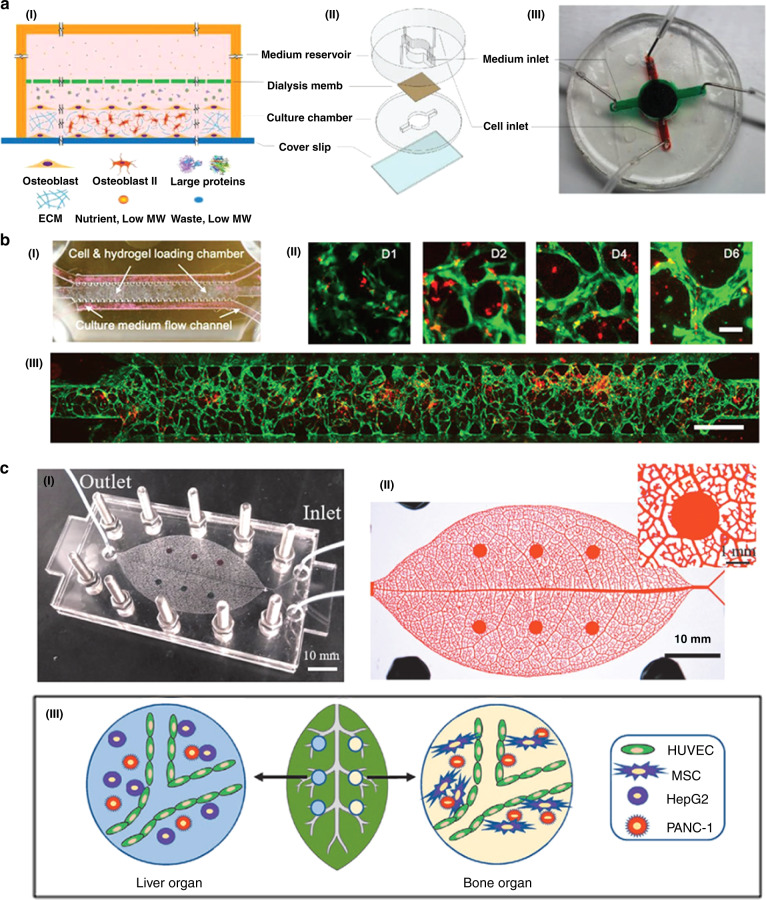
“There is a report of a 3D bone chip with mineralized collagen bone tissue growing naturally (Fig. 6a)213.”
§ Tumor-on-a-chip: from bioinspired design to biomedical application > Different types of tumor-on-a-chip > Breast tumor chip
What the sources say
- Not checked yetcites [213] Hao, S. et al. A spontaneous 3D bone-on-a-chip for bone metastasis study of breast cancer cells. Small 14, e1702787 (2018). source
plain- Asserted: passes — The authors put it forward as true.
- Checkable: passes — It can be checked against evidence.
- Load-bearing: passes — The paper’s conclusions depend on it.
- Based: passes — The paper gives a reason why it holds.
In the paper
“Based on spheroids, Sung’s group proposed a 3D microfluidic model of breast cancer invasion using surface tension pumping to achieve the sequential loading of cells at different time points177.”
§ Tumor-on-a-chip: from bioinspired design to biomedical application > Manufacture of tumor-on-a-chip > Advanced tumor-on-a-chip
What the sources say
- Not checked yetcites [177] Sung, K. E. et al. Transition to invasion in breast cancer: a microfluidic in vitro model enables examination of spatial and temporal effects. Integr. Biol. 3, 439–450 (2011). source
plaindirect- Asserted: passes — The authors put it forward as true.
- Checkable: passes — It can be checked against evidence.
- Load-bearing: unclear — It is unclear whether the paper’s conclusions depend on it.
- Based: passes — The paper gives a reason why it holds.
In the paper
“As a result, researchers believe that tumor cell growth fills the alveolar space and reduces lung breathing movement, which results in positive feedback and promotes tumor cell growth and invasion.”
§ Tumor-on-a-chip: from bioinspired design to biomedical application > Different types of tumor-on-a-chip > Lung tumor chip
What the sources say
- Not checked yetcites [98] Hassell, B. A. et al. Human organ chip models recapitulate orthotopic lung cancer growth, therapeutic responses, and tumor dormancy in vitro. Cell Rep. 21, 508–516 (2017). source
hedgeddirect- Asserted: passes — The authors put it forward as true.
- Checkable: passes — It can be checked against evidence.
- Load-bearing: passes — The paper’s conclusions depend on it.
- Based: passes — The paper gives a reason why it holds.
In the paper
“Based on this chip, a bone metastasis model of breast tumor cells was established.”
§ Tumor-on-a-chip: from bioinspired design to biomedical application > Different types of tumor-on-a-chip > Breast tumor chip
What the sources say
- Not checked yetcites [213] Hao, S. et al. A spontaneous 3D bone-on-a-chip for bone metastasis study of breast cancer cells. Small 14, e1702787 (2018). source
plaindirect- Asserted: passes — The authors put it forward as true.
- Checkable: passes — It can be checked against evidence.
- Load-bearing: passes — The paper’s conclusions depend on it.
- Based: passes — The paper gives a reason why it holds.
In the paper
“It was found that the PVN is a niche for BTSCs, and the microvascular diameter can be used as a pathway for tumor metastasis.”
§ Tumor-on-a-chip: from bioinspired design to biomedical application > Different types of tumor-on-a-chip > Brain tumor chip
What the sources say
- Not checked yetcites [196] Xiao, Y. et al. Ex vivo dynamics of human glioblastoma cells in a microvasculature‐on‐a‐chip system correlates with tumor heterogeneity and subtypes. Adv. Sci. 6, 1801531 (2019). source
plaincorrelational- Asserted: passes — The authors put it forward as true.
- Checkable: passes — It can be checked against evidence.
- Load-bearing: passes — The paper’s conclusions depend on it.
- Based: passes — The paper gives a reason why it holds.
The paper states this without a source or data behind it here, so there is nothing to check it against. The quote shows it is in the paper.
In the paper
“therefore, the microfluidic technology of organ chips is also used to study the formation of tumor blood vessels.”
§ Tumor-on-a-chip: from bioinspired design to biomedical application > Manufacture of tumor-on-a-chip > Advanced tumor-on-a-chip
plain- Asserted: passes — The authors put it forward as true.
- Checkable: passes — It can be checked against evidence.
- Load-bearing: passes — The paper’s conclusions depend on it.
- Based: passes — The paper gives a reason why it holds.
- Rests on an assumption: Tumor blood vessels have a great effect on the occurrence and development of tumors.
The paper states this without a source or data behind it here, so there is nothing to check it against. The quote shows it is in the paper.
In the paper
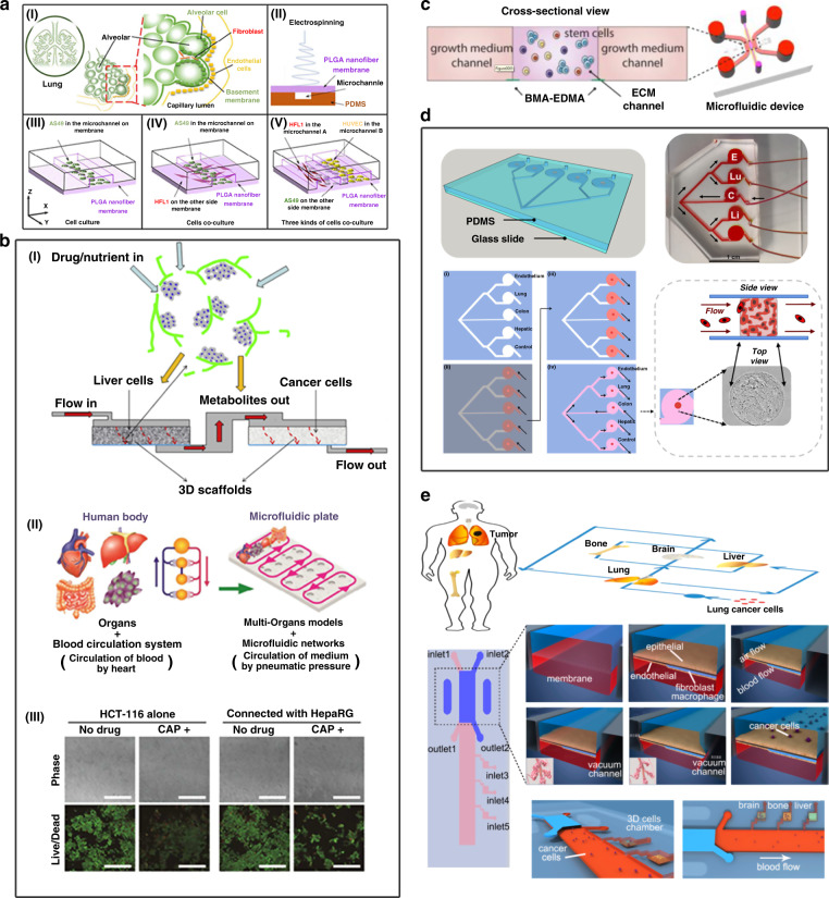
“It has been verified that nanomedicine can be effectively delivered in a gradient diffusion manner.”
§ Tumor-on-a-chip: from bioinspired design to biomedical application > Different types of tumor-on-a-chip > Colorectal-tumor chip
boosteddirect- Asserted: passes — The authors put it forward as true.
- Checkable: passes — It can be checked against evidence.
- Load-bearing: passes — The paper’s conclusions depend on it.
- Based: passes — The paper gives a reason why it holds.
A conclusion the paper draws from its own results — it cites no other source for it, so there is none to check. The quote shows it is in the paper.
In the paper
“By reproducing the dynamics of multiorgan interactions, multiorgan-on-a-chip systems can be used to study various dynamics of diseases and drug activities in mechanistic detail.”
§ Tumor-on-a-chip: from bioinspired design to biomedical application > Manufacture of tumor-on-a-chip > Advanced tumor-on-a-chip
plaindirect- Asserted: passes — The authors put it forward as true.
- Checkable: passes — It can be checked against evidence.
- Load-bearing: passes — The paper’s conclusions depend on it.
- Based: passes — The paper gives a reason why it holds.
In the paper
“The microvascularized colorectal-tumor-on-a-chip model was developed to investigate sprouting formation and drug screening applications.”
§ Tumor-on-a-chip: from bioinspired design to biomedical application > Different types of tumor-on-a-chip > Colorectal-tumor chip
What the sources say
- Not checked yetcites [210] Carvalho, M. R. et al. Colorectal tumor-on-a-chip system: a 3D tool for precision onco-nanomedicine. Sci. Adv. 5, eaaw1317 (2019). source
plain- Asserted: passes — The authors put it forward as true.
- Checkable: passes — It can be checked against evidence.
- Load-bearing: passes — The paper’s conclusions depend on it.
- Based: passes — The paper gives a reason why it holds.
In the paper
“Moreover, through lung tumor chips, researchers also found that coculture of tumor cells and alveolar epithelial cells can increase cell–cell interactions, but endothelial cells can inhibit this effect98.”
§ Tumor-on-a-chip: from bioinspired design to biomedical application > Different types of tumor-on-a-chip > Lung tumor chip
What the sources say
- Not checked yetcites [98] Hassell, B. A. et al. Human organ chip models recapitulate orthotopic lung cancer growth, therapeutic responses, and tumor dormancy in vitro. Cell Rep. 21, 508–516 (2017). source
plaindirect- Asserted: passes — The authors put it forward as true.
- Checkable: passes — It can be checked against evidence.
- Load-bearing: unclear — It is unclear whether the paper’s conclusions depend on it.
- Based: passes — The paper gives a reason why it holds.
In the paper
“They observed significant changes in cell morphology and death rate upon increasing colchicine concentration or treatment time.”
§ Tumor-on-a-chip: from bioinspired design to biomedical application > Different types of tumor-on-a-chip > Brain tumor chip
What the sources say
- Not checked yetcites [198] Liu, W. et al. Assay of glioma cell responses to an anticancer drug in a cell-based microfluidic device. Microfluid. Nanofluid. 9, 717–725 (2010). source
plaindirect- Asserted: passes — The authors put it forward as true.
- Checkable: passes — It can be checked against evidence.
- Load-bearing: passes — The paper’s conclusions depend on it.
- Based: passes — The paper gives a reason why it holds.
None of the sources cited for this claim have been checked yet.
In the paper
“The researchers compared the effects of several antitumor drugs, such as paclitaxel, olaparib, and cisplatin, in 2D- and 3D-cultured breast tumor cells and found that the drug response was weakened in 3D-cultured tumor cells.”
§ Tumor-on-a-chip: from bioinspired design to biomedical application > Different types of tumor-on-a-chip > Breast tumor chip
What the sources say
- Not checked yetcites [211] Lanz, H. L. et al. Therapy response testing of breast cancer in a 3D high-throughput perfused microfluidic platform. BMC Cancer 17, 709 (2017). source
- Not checked yetcites [212] Boyle, P. Triple-negative breast cancer: epidemiological considerations and recommendations. Ann. Oncol. 23, vi7–vi12 (2012). source
plaindirect- Asserted: passes — The authors put it forward as true.
- Checkable: passes — It can be checked against evidence.
- Load-bearing: passes — The paper’s conclusions depend on it.
- Based: passes — The paper gives a reason why it holds.
In the paper
“In this model, the proliferation of HCT-116 cancer cells was inhibited by capecitabine (CAP) and its metabolite 5-fluorouracil (5-fu).”
§ Tumor-on-a-chip: from bioinspired design to biomedical application > Application of tumor modeling > Modeling tumor morphology and drug response with high fidelity
What the sources say
- Not checked yetcites [9] Satoh, T. et al. A multi-throughput multi-organ-on-a-chip system on a plate formatted pneumatic pressure-driven medium circulation platform. Lab a Chip 18, 115–125 (2018). source
plaindirect- Asserted: passes — The authors put it forward as true.
- Checkable: passes — It can be checked against evidence.
- Load-bearing: unclear — It is unclear whether the paper’s conclusions depend on it.
- Based: passes — The paper gives a reason why it holds.
In the paper
“Compared with the traditional polydimethylsiloxane (PDMS) chip model, the organ chip using PMMA has more reliable cytotoxicity results, which has important research applications in drug screening.”
§ Tumor-on-a-chip: from bioinspired design to biomedical application > Application of tumor modeling > Tumor models for drug screening
What the sources say
- Not checked yetcites [164] Nguyen, T. et al. Robust chemical bonding of PMMA microfluidic devices to porous PETE membranes for reliable cytotoxicity testing of drugs. Lab Chip 19, 3706–3713 (2019). source
plaindirect- Asserted: passes — The authors put it forward as true.
- Checkable: passes — It can be checked against evidence.
- Load-bearing: passes — The paper’s conclusions depend on it.
- Based: passes — The paper gives a reason why it holds.
The paper states this without a source or data behind it here, so there is nothing to check it against. The quote shows it is in the paper.
In the paper
“This in vitro 3D layer-by-layer model could be integrated with the blood vascular system to build blood capillary models, which are used for assessing the efficacies of different drugs against pancreatic cancer cells, such as the hematogenous metastases of MIA PaCa-2 pancreatic cancer cells and the lymphogenous metastases of BxPC3 pancreatic cancer cells.”
§ Tumor-on-a-chip: from bioinspired design to biomedical application > Different types of tumor-on-a-chip > Pancreatic tumor chip
hedgeddirect- Asserted: passes — The authors put it forward as true.
- Checkable: passes — It can be checked against evidence.
- Load-bearing: passes — The paper’s conclusions depend on it.
- Based: passes — The paper gives a reason why it holds.
In the paper
“Ma et al. established a two-chamber (3D-mPTC) tissue model based on three-dimensional microscale perfusion in early research, which was combined with the cytotoxicity of liver metabolism anticancer drugs for detection, and realized the most basic organ chip model (Fig. 7b)219.”
§ Tumor-on-a-chip: from bioinspired design to biomedical application > Application of tumor modeling > Modeling tumor morphology and drug response with high fidelity
What the sources say
- Not checked yetcites [219] Ma, L. et al. Towards personalized medicine with a three-dimensional micro-scale perfusion-based two-chamber tissue model system. Biomaterials 33, 4353–4361 (2012). source
plain- Asserted: passes — The authors put it forward as true.
- Checkable: passes — It can be checked against evidence.
- Load-bearing: unclear — It is unclear whether the paper’s conclusions depend on it.
- Based: passes — The paper gives a reason why it holds.
In the paper
“This vascularized MSC microenvironment provided a suitable platform for pancreatic cancer cell growth.”
§ Tumor-on-a-chip: from bioinspired design to biomedical application > Different types of tumor-on-a-chip > Pancreatic tumor chip
What the sources say
- Not checked yetcites [217] Mao, M. et al. Human-on-leaf-chip: a biomimetic vascular system integrated with chamber-specific organs. Small 16, 2000546 (2020). source
plaindirect- Asserted: passes — The authors put it forward as true.
- Checkable: passes — It can be checked against evidence.
- Load-bearing: passes — The paper’s conclusions depend on it.
- Based: passes — The paper gives a reason why it holds.
In the paper
“Gervais et al. made use of a simple microfluidic platform that can reliably trap samples to incubate eight different types of microdissected tissues (MDTs) in a low-shear stress environment.”
§ Tumor-on-a-chip: from bioinspired design to biomedical application > Application of tumor modeling > Tumor models for drug screening
What the sources say
- Not checked yetcites [121] Astolfi, M. et al. Micro-dissected tumor tissues on chip: an ex vivo method for drug testing and personalized therapy. Lab a Chip 16, 312–325 (2016). source
plain- Asserted: passes — The authors put it forward as true.
- Checkable: passes — It can be checked against evidence.
- Load-bearing: unclear — It is unclear whether the paper’s conclusions depend on it.
- Based: passes — The paper gives a reason why it holds.
In the paper
“Hickman et al. took advantage of a pumpless 4-organ system (liver, cardiac, neuronal, and muscle) to evaluate the human response to five different drugs for 14 days.”
§ Tumor-on-a-chip: from bioinspired design to biomedical application > Application of tumor modeling > Tumor models for drug screening
What the sources say
- Not checked yetcites [229] Oleaga, C. et al. Multi-organ toxicity demonstration in a functional human in vitro system composed of four organs. Sci. Rep. 6, 20030 (2016). source
plain- Asserted: passes — The authors put it forward as true.
- Checkable: passes — It can be checked against evidence.
- Load-bearing: unclear — It is unclear whether the paper’s conclusions depend on it.
- Based: passes — The paper gives a reason why it holds.
In the paper
“Through this platform, the researchers analyzed MDT viability by confocal microscopy and flow cytometry, providing information on chemosensitivity testing and drug response (Fig. 8a)121.”
§ Tumor-on-a-chip: from bioinspired design to biomedical application > Application of tumor modeling > Tumor models for drug screening
What the sources say
- Not checked yetcites [121] Astolfi, M. et al. Micro-dissected tumor tissues on chip: an ex vivo method for drug testing and personalized therapy. Lab a Chip 16, 312–325 (2016). source
plaindirect- Asserted: passes — The authors put it forward as true.
- Checkable: passes — It can be checked against evidence.
- Load-bearing: unclear — It is unclear whether the paper’s conclusions depend on it.
- Based: passes — The paper gives a reason why it holds.
In the paper
“Skardal’s group demonstrated the utility of a 2-organoid metastasis-on-a-chip (MOC) platform224. By using microfluidics to provide circulating flow through the organoid system, tumor cells grow in the primary focus and enter the circulation, with the result that CRC cells of the colon organoids diffuse into the circulation, and then the transferred cells settle on the downstream liver organoids.”
§ Tumor-on-a-chip: from bioinspired design to biomedical application > Application of tumor modeling > Modeling cancer invasion and metastasis
What the sources say
- Not checked yetcites [224] Skardal, A., Devarasetty, M., Forsythe, S., Atala, A. & Soker, S. A reductionist metastasis‐on‐a‐chip platform for in vitro tumor progression modeling and drug screening. Biotechnol. Bioeng. 113, 2020–2032 (2016). source
boosteddirect- Asserted: passes — The authors put it forward as true.
- Checkable: passes — It can be checked against evidence.
- Load-bearing: passes — The paper’s conclusions depend on it.
- Based: passes — The paper gives a reason why it holds.
In the paper
“Compared with those in the monolayer model, the cells in the spheroid-based 3D model exhibit higher drug resistance during drug testing, and the 3D model is closer to the chemical environment of native tissues215.”
§ Tumor-on-a-chip: from bioinspired design to biomedical application > Different types of tumor-on-a-chip > Pancreatic tumor chip
What the sources say
- Not checked yetcites [215] Wen, Z. et al. A spheroid-based 3-D culture model for pancreatic cancer drug testing, using the acid phosphatase assay. Braz. J. Med. Biol. Res. 46, 634–642 (2013). source
plaincorrelational- Asserted: passes — The authors put it forward as true.
- Checkable: passes — It can be checked against evidence.
- Load-bearing: passes — The paper’s conclusions depend on it.
- Based: passes — The paper gives a reason why it holds.
In the paper
“Since pancreatic cancers are invasive solid tumors with hypovascular structures and extensive fibrosis214, developing pancreatic tumor-on-a-chip systems for in vitro experiments is essential for enhancing pancreatic drug screening efficiency before animal model testing and clinical trials.”
§ Tumor-on-a-chip: from bioinspired design to biomedical application > Different types of tumor-on-a-chip > Pancreatic tumor chip
What the sources say
- Not checked yetcites [214] Jiang, H. et al. Targeting focal adhesion kinase renders pancreatic cancers responsive to checkpoint immunotherapy. Nat. Med. 22, 851–860 (2016). source
plaindirect- Asserted: passes — The authors put it forward as true.
- Checkable: passes — It can be checked against evidence.
- Load-bearing: passes — The paper’s conclusions depend on it.
- Based: passes — The paper gives a reason why it holds.
In the paper
“This technology provided a novel tool to improve the predictive ability of preclinical efficacy/toxicity studies229.”
§ Tumor-on-a-chip: from bioinspired design to biomedical application > Application of tumor modeling > Tumor models for drug screening
What the sources say
- Not checked yetcites [229] Oleaga, C. et al. Multi-organ toxicity demonstration in a functional human in vitro system composed of four organs. Sci. Rep. 6, 20030 (2016). source
plaindirect- Asserted: passes — The authors put it forward as true.
- Checkable: passes — It can be checked against evidence.
- Load-bearing: passes — The paper’s conclusions depend on it.
- Based: passes — The paper gives a reason why it holds.
In the paper
“Wang’s group took advantage of another multiorgan-on-a-chip platform to study lung cancer metastasis (Fig. 7e)225. The system consists of four organs: one upstream lung and three downstream parallel brain, bone, and liver organs. This model simulated the metastasis of lung cancer to the brain, bone, and liver.”
§ Tumor-on-a-chip: from bioinspired design to biomedical application > Application of tumor modeling > Modeling cancer invasion and metastasis
What the sources say
- Not checked yetcites [225] Xu, Z. et al. Design and construction of a multi-organ microfluidic chip mimicking the in vivo microenvironment of lung cancer metastasis. ACS Appl. Mater. Interfaces 8, 25840–25847 (2016). source
plain- Asserted: passes — The authors put it forward as true.
- Checkable: passes — It can be checked against evidence.
- Load-bearing: unclear — It is unclear whether the paper’s conclusions depend on it.
- Based: passes — The paper gives a reason why it holds.
In the paper
“PMMA organ chips have been successfully demonstrated to show more reliable cytotoxicity results for vincristine using human lung adenocarcinoma cells.”
§ Tumor-on-a-chip: from bioinspired design to biomedical application > Application of tumor modeling > Tumor models for drug screening
What the sources say
- Not checked yetcites [164] Nguyen, T. et al. Robust chemical bonding of PMMA microfluidic devices to porous PETE membranes for reliable cytotoxicity testing of drugs. Lab Chip 19, 3706–3713 (2019). source
boosteddirect- Asserted: passes — The authors put it forward as true.
- Checkable: passes — It can be checked against evidence.
- Load-bearing: passes — The paper’s conclusions depend on it.
- Based: passes — The paper gives a reason why it holds.
A result the paper reports from its own data — it cites no other source for it, so there is none to check. The quote shows it is in the paper.
In the paper
“In this system, under the circulating flow of liquid, it is found through fluorescence imaging tracking that HCT-116 CRC cells preferentially enter the liver and lung structures, which are the corresponding organs with the most CRC metastasis in human patients.”
§ Tumor-on-a-chip: from bioinspired design to biomedical application > Application of tumor modeling > Modeling cancer invasion and metastasis
plaincorrelational- Asserted: passes — The authors put it forward as true.
- Checkable: passes — It can be checked against evidence.
- Load-bearing: passes — The paper’s conclusions depend on it.
- Based: passes — The paper gives a reason why it holds.
The paper states this without a source or data behind it here, so there is nothing to check it against. The quote shows it is in the paper.
In the paper
“Therefore, although the culture of a single cell type can mimic some aspects of the tissue microenvironment on a chip, it is usually not enough to produce organ-like functions.”
§ Tumor-on-a-chip: from bioinspired design to biomedical application > Application of tumor modeling > Modeling organ-level physiology and disease
plain- Asserted: passes — The authors put it forward as true.
- Checkable: passes — It can be checked against evidence.
- Load-bearing: passes — The paper’s conclusions depend on it.
- Based: passes — The paper gives a reason why it holds.
- Rests on an assumption: The tissue itself is composed of different cell types.
In the paper
“Toh et al. described a microfluidic tumor-on-a-chip cell migration model incorporating a microfluidic system to understand the progress of cell intravasation226.”
§ Tumor-on-a-chip: from bioinspired design to biomedical application > Application of tumor modeling > Modeling cancer invasion and metastasis
What the sources say
- Not checked yetcites [226] Toh, Y.-C., Raja, A., Yu, H. & Van Noort, D. A 3D microfluidic model to recapitulate cancer cell migration and invasion. Bioengineering 5, 29 (2018). source
plain- Asserted: passes — The authors put it forward as true.
- Checkable: passes — It can be checked against evidence.
- Load-bearing: unclear — It is unclear whether the paper’s conclusions depend on it.
- Based: passes — The paper gives a reason why it holds.
A conclusion the paper draws from its own results — it cites no other source for it, so there is none to check. The quote shows it is in the paper.
In the paper
“By building a better predictive model, organ chip model technology significantly reduces research and development costs between preclinical and human trials.”
§ Tumor-on-a-chip: from bioinspired design to biomedical application > Application of tumor modeling > Tumor models for drug screening
plaindirect- Asserted: passes — The authors put it forward as true.
- Checkable: passes — It can be checked against evidence.
- Load-bearing: passes — The paper’s conclusions depend on it.
- Based: unclear — It is unclear why the paper says it holds.
None of the sources cited for this claim have been checked yet.
In the paper
“Compared with blood vessels in the healthy liver microenvironment, breast cancer cells cultured in the microenvironment of this model have higher vascular porosity and tumor microenvironment permeability.”
§ Tumor-on-a-chip: from bioinspired design to biomedical application > Application of tumor modeling > Modeling tumor morphology and drug response with high fidelity
What the sources say
- Not checked yetcites [9] Satoh, T. et al. A multi-throughput multi-organ-on-a-chip system on a plate formatted pneumatic pressure-driven medium circulation platform. Lab a Chip 18, 115–125 (2018). source
- Not checked yetcites [220] Imura, Y., Sato, K. & Yoshimura, E. Micro total bioassay system for ingested substances: assessment of intestinal absorption, hepatic metabolism, and bioactivity. Anal. Chem. 82, 9983–9988 (2010). source
plaincorrelational- Asserted: passes — The authors put it forward as true.
- Checkable: passes — It can be checked against evidence.
- Load-bearing: passes — The paper’s conclusions depend on it.
- Based: passes — The paper gives a reason why it holds.
In the paper
“Later, Satoh et al. established a two-organ system model composed of liver and tumor9.”
§ Tumor-on-a-chip: from bioinspired design to biomedical application > Application of tumor modeling > Modeling tumor morphology and drug response with high fidelity
What the sources say
- Not checked yetcites [9] Satoh, T. et al. A multi-throughput multi-organ-on-a-chip system on a plate formatted pneumatic pressure-driven medium circulation platform. Lab a Chip 18, 115–125 (2018). source
plain- Asserted: passes — The authors put it forward as true.
- Checkable: passes — It can be checked against evidence.
- Load-bearing: unclear — It is unclear whether the paper’s conclusions depend on it.
- Based: passes — The paper gives a reason why it holds.
In the paper
“This model provides a new in vitro model for studying bone metastases from breast tumors213.”
§ Tumor-on-a-chip: from bioinspired design to biomedical application > Different types of tumor-on-a-chip > Breast tumor chip
What the sources say
- Not checked yetcites [213] Hao, S. et al. A spontaneous 3D bone-on-a-chip for bone metastasis study of breast cancer cells. Small 14, e1702787 (2018). source
plain- Asserted: passes — The authors put it forward as true.
- Checkable: passes — It can be checked against evidence.
- Load-bearing: passes — The paper’s conclusions depend on it.
- Based: passes — The paper gives a reason why it holds.
A conclusion the paper draws from its own results — it cites no other source for it, so there is none to check. The quote shows it is in the paper.
In the paper
“The development of tumor-on-a-chip technology has a great ability to replicate the tumor microenvironment in vitro.”
§ Tumor-on-a-chip: from bioinspired design to biomedical application > Application of tumor modeling > Modeling organ-level physiology and disease
boosteddirect- Asserted: passes — The authors put it forward as true.
- Checkable: passes — It can be checked against evidence.
- Load-bearing: passes — The paper’s conclusions depend on it.
- Based: passes — The paper gives a reason why it holds.
In the paper
“Diclofenac and imatinib have cytostatic effects on bone marrow.”
§ Tumor-on-a-chip: from bioinspired design to biomedical application > Application of tumor modeling > Tumor models for drug screening
What the sources say
- Not checked yetcites [230] McAleer, C. W. et al. Multi-organ system for the evaluation of efficacy and off-target toxicity of anticancer therapeutics. Sci. Transl. Med. 11, https://doi.org/10.1126/scitranslmed.aav1386 (2019). source
plaindirect- Asserted: passes — The authors put it forward as true.
- Checkable: passes — It can be checked against evidence.
- Load-bearing: unclear — It is unclear whether the paper’s conclusions depend on it.
- Based: passes — The paper gives a reason why it holds.
In the paper
“This 3D platform is suitable for the efficacy/toxicity screening of various tissues under more complex conditions than physiological environments.”
§ Tumor-on-a-chip: from bioinspired design to biomedical application > Application of tumor modeling > Tumor models for drug screening
What the sources say
- Not checked yetcites [86] Phan, D. T. et al. A vascularized and perfused organ-on-a-chip platform for large-scale drug screening applications. Lab a Chip 17, 511–520 (2017). source
plain- Asserted: passes — The authors put it forward as true.
- Checkable: passes — It can be checked against evidence.
- Load-bearing: passes — The paper’s conclusions depend on it.
- Based: passes — The paper gives a reason why it holds.
In the paper
“Subsequently, Hickman’s group found that antileukemia drugs can be tested by coculturing primary human hepatocytes and human bone marrow cell cultures of two types of cancer in a chip model (Fig. 8d)230.”
§ Tumor-on-a-chip: from bioinspired design to biomedical application > Application of tumor modeling > Tumor models for drug screening
What the sources say
- Not checked yetcites [230] McAleer, C. W. et al. Multi-organ system for the evaluation of efficacy and off-target toxicity of anticancer therapeutics. Sci. Transl. Med. 11, https://doi.org/10.1126/scitranslmed.aav1386 (2019). source
plaindirect- Asserted: passes — The authors put it forward as true.
- Checkable: passes — It can be checked against evidence.
- Load-bearing: unclear — It is unclear whether the paper’s conclusions depend on it.
- Based: passes — The paper gives a reason why it holds.
In the paper
“In 2014, Vidi et al. developed a breast-on-a-chip platform consisting of a breast luminal epithelium monolayer on a semicircular acrylic support to mimic cancer mammary ducts218.”
§ Tumor-on-a-chip: from bioinspired design to biomedical application > Application of tumor modeling > Modeling tumor morphology and drug response with high fidelity
What the sources say
- Not checked yetcites [218] Vidi, P.-A. et al. Disease-on-a-chip: mimicry of tumor growth in mammary ducts. Lab a Chip 14, 172–177 (2014). source
plain- Asserted: passes — The authors put it forward as true.
- Checkable: passes — It can be checked against evidence.
- Load-bearing: unclear — It is unclear whether the paper’s conclusions depend on it.
- Based: passes — The paper gives a reason why it holds.
A conclusion the paper draws from its own results — it cites no other source for it, so there is none to check. The quote shows it is in the paper.
In the paper
“However, due to the complexity of the physiological structure and microenvironment in vivo, tumor chips still face many challenges before widespread integration of these platforms into practical pharmaceutical industrial and clinical applications:”
§ Tumor-on-a-chip: from bioinspired design to biomedical application > Conclusions and future perspectives
plaindirect- Asserted: passes — The authors put it forward as true.
- Checkable: passes — It can be checked against evidence.
- Load-bearing: passes — The paper’s conclusions depend on it.
- Based: passes — The paper gives a reason why it holds.
- It may state two separate claims as one. 56
- Probably not a substantial claim. 24
Some cited sources support this claim and some don’t — read each source’s verdict below.
In the paper
“Crucially, these organ chip microdevices are able to recapitulate organ-level responses to toxins79,96,97, drugs86,98, radiation99, cigarette smoke100, pathogens96, and normal microorganisms96, as well as the dependence on flow-circulating immune cells and organs to recruit specific in vitro inflammatory responses101, which are very important for preclinical drug screening and disease modeling.”
§ Tumor-on-a-chip: from bioinspired design to biomedical application > Introduction
What the sources say
- Not checked yetcites [79] Bovard, D. et al. A lung/liver-on-a-chip platform for acute and chronic toxicity studies. Lab a Chip 18, 3814–3829 (2018). source
- Not checked yetcites [96] Kim, H. J., Li, H., Collins, J. J. & Ingber, D. E. Contributions of microbiome and mechanical deformation to intestinal bacterial overgrowth and inflammation in a human gut-on-a-chip. Proc. Natl Acad. Sci. USA 113, E7–E15 (2016).
- Not checked yetcites [97] Ozkan, A., Ghousifam, N., Hoopes, P. J., Yankeelov, T. E. & Rylander, M. N. In vitro vascularized liver and tumor tissue microenvironments on a chip for dynamic determination of nanoparticle transport and toxicity. Biotechnol. Bioeng. 116, 1201–1219 (2019). source
- Not checked yetcites [98] Hassell, B. A. et al. Human organ chip models recapitulate orthotopic lung cancer growth, therapeutic responses, and tumor dormancy in vitro. Cell Rep. 21, 508–516 (2017). source
- Supportedcites [99] Jalili-Firoozinezhad, S. et al. Modeling radiation injury-induced cell death and countermeasure drug responses in a human gut-on-a-chip. Cell Death Dis. 9, 223 (2018). source
Split into 2 atoms — 2 confirmed, 0 partially supported, 0 disputed, 0 misattributed, 0 unverifiable. Expand the claim to see per-atom verdicts.
- Not checked yetcites [100] Benam, K. H. et al. Matched-comparative modeling of normal and diseased human airway responses using a microengineered breathing lung chip. Cell Syst. 3, 456–466.e454 (2016). source
- Disputedcites [101] Huh, D. et al. A human disease model of drug toxicity-induced pulmonary edema in a lung-on-a-chip microdevice. Sci. Transl. Med. 4, 159ra147 (2012). sourceExperimental — accuracy not yet measured
Split into 2 atoms — 0 confirmed, 1 partially supported, 1 disputed, 0 misattributed, 0 unverifiable. Expand the claim to see per-atom verdicts.
Checked by a reasoning model — may be wrong.
plaindirect- Asserted: passes — The authors put it forward as true.
- Checkable: passes — It can be checked against evidence.
- Load-bearing: passes — The paper’s conclusions depend on it.
- Based: passes — The paper gives a reason why it holds.
It may state two separate claims as one.
Every source the paper cites for this claim was checked and supports it.
In the paper
“In addition, the cell culture format (two-dimensional monolayer versus three-dimensional spheroid) and spheroid size play key roles in drug responses and show the advantages of connecting these two technologies in drug screening applications.”
§ Tumor-on-a-chip: from bioinspired design to biomedical application > Manufacture of tumor-on-a-chip > Advanced tumor-on-a-chip
What the sources say
- Supportedcites [178] Patra, B., Peng, C.-C., Liao, W.-H., Lee, C.-H. & Tung, Y.-C. Drug testing and flow cytometry analysis on a large number of uniform sized tumor spheroids using a microfluidic device. Sci. Rep. 6, 21061 (2016). source
The Abstract states that the experimental results suggest culture format (2D monolayer versus 3D spheroid) and spheroid size play critical roles in drug responses. It also says that bridging flow cytometry and microfluidics offers advantages for pharmaceutical drug screening. The experiments used HepG2 tumor spheroids. (LLM, conf=0.95)
plaincorrelational- Asserted: passes — The authors put it forward as true.
- Checkable: passes — It can be checked against evidence.
- Load-bearing: passes — The paper’s conclusions depend on it.
- Based: passes — The paper gives a reason why it holds.
It may state two separate claims as one.
- Supported
Every source the paper cites for this claim was checked and supports it.
In the paper
“Earlier, Huh et al.131 used a bionic microsystem to reconstruct the alveolar–capillary interface of the key function of the human lung. This model can produce a complex, integrated organ-level response to bacteria and inflammatory cytokines entering the alveolar space.”
§ Tumor-on-a-chip: from bioinspired design to biomedical application > Application of tumor modeling > Modeling organ-level physiology and disease
What the sources say
- Supportedcites [131] Huh, D. et al. Reconstituting organ-level lung functions on a chip. Science 328, 1662–1668 (2010). source
The Abstract describes a biomimetic microsystem that reconstitutes the human lung’s alveolar-capillary interface and reproduces integrated organ-level responses to bacteria and inflammatory cytokines introduced into the alveolar space. The ‘Recapitulation of Whole Organ Responses’ section also describes the bacterial and cytokine experiments. (LLM, conf=0.95)
plaindirect- Asserted: passes — The authors put it forward as true.
- Checkable: passes — It can be checked against evidence.
- Load-bearing: unclear — It is unclear whether the paper’s conclusions depend on it.
- Based: passes — The paper gives a reason why it holds.
It may state two separate claims as one.
- Supported
None of the sources cited for this claim have been checked yet.
In the paper
“In addition, animal studies are hindered by animal expenses, low‐throughput drug optimization and ethical controversy31–35,38.”
§ Tumor-on-a-chip: from bioinspired design to biomedical application > Introduction
What the sources say
- Not checked yetcites [31] Engelman, R. & Kerr, W. G. Autoimmunity 433–441 (Springer, 2012). source
- Not checked yetcites [32] Kang, T. H. & Kim, H. J. Farewell to animal testing: innovations on human intestinal microphysiological systems. Micromachines 7, 107 (2016). source
- Not checked yetcites [33] Hintze, K. J. et al. Broad scope method for creating humanized animal models for animal health and disease research through antibiotic treatment and human fecal transfer. Gut Microbes 5, 183–191 (2014). source
- Not checked yetcites [34] Rongvaux, A. et al. Development and function of human innate immune cells in a humanized mouse model. Nat. Biotechnol. 32, 364–372 (2014). source
- Not checked yetcites [35] Seok, J. et al. Genomic responses in mouse models poorly mimic human inflammatory diseases. Proc. Natl Acad. Sci. USA 110, 3507–3512 (2013). source
- Not checked yetcites [38] Mak, I. W., Evaniew, N. & Ghert, M. Lost in translation: animal models and clinical trials in cancer treatment. Am. J. Transl. Res. 6, 114–118 (2014).
plaindirect- Asserted: passes — The authors put it forward as true.
- Checkable: passes — It can be checked against evidence.
- Load-bearing: passes — The paper’s conclusions depend on it.
- Based: passes — The paper gives a reason why it holds.
It may state two separate claims as one.
None of the sources cited for this claim have been checked yet.
In the paper
“Organ-on-a-chip devices possess good performance and improved recapitulation of the native microenvironment, and they allow high‐throughput tests with decreased cost, ethical advantages and enhanced reproducibility47,55–57.”
§ Tumor-on-a-chip: from bioinspired design to biomedical application > Introduction
What the sources say
- Not checked yetcites [47] Zhang, Y. S., Zhang, Y.-N. & Zhang, W. Cancer-on-a-chip systems at the frontier of nanomedicine. Drug Discov. Today 22, 1392–1399 (2017). source
- Not checked yetcites [55] Chan, C. Y. et al. Accelerating drug discovery via organs-on-chips. Lab a Chip 13, 4697–4710 (2013). source
- Not checked yetcites [56] Park, S. E., Georgescu, A. & Huh, D. Organoids-on-a-chip. Science 364, 960–965 (2019). source
- Not checked yetcites [57] Zhang, B., Korolj, A., Lai, B. F. L. & Radisic, M. Advances in organ-on-a-chip engineering. Nat. Rev. Mater. 3, 257–278 (2018). source
plaindirect- Asserted: passes — The authors put it forward as true.
- Checkable: passes — It can be checked against evidence.
- Load-bearing: unclear — It is unclear whether the paper’s conclusions depend on it.
- Based: passes — The paper gives a reason why it holds.
It may state two separate claims as one.
A conclusion the paper draws from its own results — it cites no other source for it, so there is none to check. The quote shows it is in the paper.
In the paper
“The reason is that in these experiments, cells are grown in a monolayer, all well exposed to the drug, while in vivo tumors expand as three-dimensional multicellular masses, where inner cells have limited drug exposure.”
§ Tumor-on-a-chip: from bioinspired design to biomedical application > Introduction
plaindirect- Asserted: passes — The authors put it forward as true.
- Checkable: passes — It can be checked against evidence.
- Load-bearing: passes — The paper’s conclusions depend on it.
- Based: passes — The paper gives a reason why it holds.
It may state two separate claims as one.
None of the sources cited for this claim have been checked yet.
In the paper
“Tumors possess a complicated microenvironment with a dense extracellular matrix (ECM), irregular vessels, assorted stromal, immune and inflammatory cells, cancer stem cells, and limited perfusion of nutrients109–112.”
§ Tumor-on-a-chip: from bioinspired design to biomedical application > Introduction
What the sources say
- Not checked yetcites [109] Kim, J. Y. et al. 3D spherical microtissues and microfluidic technology for multi-tissue experiments and analysis. J. Biotechnol. 205, 24–35 (2015). source
- Not checked yetcites [110] Portillo-Lara, R. & Annabi, N. Microengineered cancer-on-a-chip platforms to study the metastatic microenvironment. Lab a Chip 16, 4063–4081 (2016). source
- Not checked yetcites [111] Maley, C. C. et al. Classifying the evolutionary and ecological features of neoplasms. Nat. Rev. Cancer 17, 605–619 (2017). source
- Not checked yetcites [112] Fetah, K. L. et al. Cancer modeling-on-a-chip with future artificial intelligence integration. Small 15, 1901985 (2019). source
plain- Asserted: passes — The authors put it forward as true.
- Checkable: passes — It can be checked against evidence.
- Load-bearing: passes — The paper’s conclusions depend on it.
- Based: passes — The paper gives a reason why it holds.
It may state two separate claims as one.
In the paper
“Currently, many types of healthy and diseased tissues and organs, including microvascular obstructions, cystic fibrosis models, heart72,73, kidney74–76, liver77–79, lung79,80, pancreas, brain81–86, skin87–89, eye90,91, intestine92, and neuropsychiatric disorder models93–95, have been constructed on microfluidic chips.”
§ Tumor-on-a-chip: from bioinspired design to biomedical application > Introduction
What the sources say
- Not checked yetcites 72–95
plain- Asserted: passes — The authors put it forward as true.
- Checkable: passes — It can be checked against evidence.
- Load-bearing: fails — The paper’s conclusions may not depend on it.
- Based: passes — The paper gives a reason why it holds.
Probably not a substantial claim. The paper’s conclusions may not depend on it.
In the paper
“The results of drug testing demonstrated that human lung adenocarcinoma cells cultured in the PMMA microfluidic device give more reliable results regarding the cytotoxicity of vincristine than human lung adenocarcinoma cells cultured in the polydimethylsiloxane (PDMS) device. This is because the small molecules cannot permeate the PMMA material.”
§ Tumor-on-a-chip: from bioinspired design to biomedical application > Manufacture of tumor-on-a-chip > Materials for tumor-on-a-chip
What the sources say
- Not checked yetcites [164] Nguyen, T. et al. Robust chemical bonding of PMMA microfluidic devices to porous PETE membranes for reliable cytotoxicity testing of drugs. Lab Chip 19, 3706–3713 (2019). source
boosteddirect- Asserted: passes — The authors put it forward as true.
- Checkable: passes — It can be checked against evidence.
- Load-bearing: passes — The paper’s conclusions depend on it.
- Based: passes — The paper gives a reason why it holds.
It may state two separate claims as one.
In the paper
“Organoids derived from these stem cells can be integrated into tumor chips, and because of the combination of the two technologies, organ-specific structures and gene expression signatures in vivo could be mimicked better than in either model alone172.”
§ Tumor-on-a-chip: from bioinspired design to biomedical application > Manufacture of tumor-on-a-chip > Advanced tumor-on-a-chip
What the sources say
- Not checked yetcites [172] Kasendra, M. et al. Development of a primary human small intestine-on-a-chip using biopsy-derived organoids. Sci. Rep. 8, 2871 (2018). source
hedgeddirect- Asserted: passes — The authors put it forward as true.
- Checkable: passes — It can be checked against evidence.
- Load-bearing: unclear — It is unclear whether the paper’s conclusions depend on it.
- Based: passes — The paper gives a reason why it holds.
It may state two separate claims as one.
In the paper
“BME/Matrigel can also enable 3D tumor culture and coculture of other cells, and researchers can adjust the microenvironment to be suitable for tumors by supplementing various factors on BME/Matrigel, which can better inform the mechanism of tumor growth169.”
§ Tumor-on-a-chip: from bioinspired design to biomedical application > Manufacture of tumor-on-a-chip > Materials for tumor-on-a-chip
What the sources say
- Not checked yetcites [169] Benton, G., Arnaoutova, I., George, J., Kleinman, H. K. & Koblinski, J. Matrigel: From discovery and ECM mimicry to assays and models for cancer research. Adv. Drug Deliv. Rev. 79-80, 3–18 (2014). source
plaindirect- Asserted: passes — The authors put it forward as true.
- Checkable: passes — It can be checked against evidence.
- Load-bearing: unclear — It is unclear whether the paper’s conclusions depend on it.
- Based: passes — The paper gives a reason why it holds.
It may state two separate claims as one.
- Rests on an assumption: BME/Matrigel is a biomaterial with a composition similar to that of the early conservative basement membrane, which promotes the organization of different tissue types and microtissues of different species.
- Rests on an assumption: Under similar conditions, tumor cells have high proliferation and are sometimes aggressive in BME/Matrigel, which can be used to evaluate the malignancy of tumors and explore tumor treatment methods.
A conclusion the paper draws from its own results — it cites no other source for it, so there is none to check. The quote shows it is in the paper.
In the paper
“Hence, bioprinting technology has the potential to build miniaturized, multiorgan bionic pathophysiological models, accelerate the pace of research, and promote the application of technology in the medical field.”
§ Tumor-on-a-chip: from bioinspired design to biomedical application > Manufacture of tumor-on-a-chip > Principle and fabrication of tumor-on-a-chip
hedgedconditional- Asserted: passes — The authors put it forward as true.
- Checkable: fails — It may be too vague to check against evidence.
- Load-bearing: fails — The paper’s conclusions may not depend on it.
- Based: passes — The paper gives a reason why it holds.
Probably not a substantial claim. It may be too vague to check against evidence. The paper’s conclusions may not depend on it. It may state two separate claims as one.
A conclusion the paper draws from its own results — it cites no other source for it, so there is none to check. The quote shows it is in the paper.
In the paper
“These tumor chips can simulate the 3D microstructure and microphysiological functions of in vivo human organs/tissues, providing a better understanding of the mechanisms of tumor growth and metastasis.”
§ Tumor-on-a-chip: from bioinspired design to biomedical application > Introduction
plaindirect- Asserted: passes — The authors put it forward as true.
- Checkable: passes — It can be checked against evidence.
- Load-bearing: passes — The paper’s conclusions depend on it.
- Based: fails — The paper may give no reason why it holds.
Probably not a substantial claim. The paper may give no reason why it holds. It may state two separate claims as one.
The paper states this without a source or data behind it here, so there is nothing to check it against. The quote shows it is in the paper.
In the paper
“It is worth mentioning that soft lithography fabrication technology has the ability to change the chemistry of the substrate surface, making it a promising low-cost method for manufacturing microfluidic chips.”
§ Tumor-on-a-chip: from bioinspired design to biomedical application > Manufacture of tumor-on-a-chip > Principle and fabrication of tumor-on-a-chip
plaindirect- Asserted: passes — The authors put it forward as true.
- Checkable: passes — It can be checked against evidence.
- Load-bearing: fails — The paper’s conclusions may not depend on it.
- Based: fails — The paper may give no reason why it holds.
Probably not a substantial claim. The paper’s conclusions may not depend on it. The paper may give no reason why it holds. It may state two separate claims as one.
A conclusion the paper draws from its own results — it cites no other source for it, so there is none to check. The quote shows it is in the paper.
In the paper
“In contrast to the tumor growth microenvironment in vivo, tumor models in vitro are usually under atmospheric oxygen conditions, which are not representative of the conditions in vivo. Therefore, the results of these studies can be misleading.”
§ Tumor-on-a-chip: from bioinspired design to biomedical application > Manufacture of tumor-on-a-chip
plaindirect- Asserted: passes — The authors put it forward as true.
- Checkable: passes — It can be checked against evidence.
- Load-bearing: passes — The paper’s conclusions depend on it.
- Based: passes — The paper gives a reason why it holds.
It may state two separate claims as one.
- Rests on an assumption: Oxygen tension is a key regulator of cellular behavior.
In the paper
“Although many tumor chips have been successfully manufactured by photolithography or related manufacturing processes involving photolithography, they still have some shortcomings, such as the need for multiple masks and a multistep photolithography process, which makes the fabrication process time-consuming and costly.”
§ Tumor-on-a-chip: from bioinspired design to biomedical application > Manufacture of tumor-on-a-chip > Principle and fabrication of tumor-on-a-chip
What the sources say
- Not checked yetcites [130] Yang, Q., Lian, Q. & Xu, F. Perspective: fabrication of integrated organ-on-a-chip via bioprinting. Biomicrofluidics 11, 031301 (2017). source
plaindirect- Asserted: passes — The authors put it forward as true.
- Checkable: passes — It can be checked against evidence.
- Load-bearing: unclear — It is unclear whether the paper’s conclusions depend on it.
- Based: passes — The paper gives a reason why it holds.
It may state two separate claims as one.
In the paper
“Gelatin is a collagen-derived natural biopolymer. Due to its cellular response characteristics and the ability to deliver a wide range of biomolecules, it is widely used in drug transport models and tissue engineering165.”
§ Tumor-on-a-chip: from bioinspired design to biomedical application > Manufacture of tumor-on-a-chip > Materials for tumor-on-a-chip
What the sources say
- Not checked yetcites [165] Hoch, E., Hirth, T., Tovar, G. E. M. & Borchers, K. Chemical tailoring of gelatin to adjust its chemical and physical properties for functional bioprinting. J. Mater. Chem. B 1, 5675–5685 (2013). source
plaindirect- Asserted: passes — The authors put it forward as true.
- Checkable: passes — It can be checked against evidence.
- Load-bearing: fails — The paper’s conclusions may not depend on it.
- Based: passes — The paper gives a reason why it holds.
Probably not a substantial claim. The paper’s conclusions may not depend on it. It may state two separate claims as one.
- Rests on an assumption: Gelatin can promote cell growth and polymerize with proteins, growth factor nucleotides, polysaccharides and other polyionic complexes, and has broad prospects for drug transport models due to controlled sustained and/or targeted release of bioactive molecules.
A conclusion the paper draws from its own results — it cites no other source for it, so there is none to check. The quote shows it is in the paper.
In the paper
“Multiorgan-on-a-chip systems that include the interactions of cell cocultures have shown great potential in screening anticancer drugs and cancer therapies.”
§ Tumor-on-a-chip: from bioinspired design to biomedical application > Introduction
boostedcorrelational- Asserted: passes — The authors put it forward as true.
- Checkable: passes — It can be checked against evidence.
- Load-bearing: fails — The paper’s conclusions may not depend on it.
- Based: fails — The paper may give no reason why it holds.
Probably not a substantial claim. The paper’s conclusions may not depend on it. The paper may give no reason why it holds.
None of the sources cited for this claim have been checked yet.
In the paper
“Chips based on microfluidics can be designed to realize oxygen gradients with high spatial and temporal resolution, which can be used to simulate the physiological effects of oxygen on tumor progression and metastasis127,128.”
§ Tumor-on-a-chip: from bioinspired design to biomedical application > Manufacture of tumor-on-a-chip
What the sources say
- Not checked yetcites [127] Chen, Y.-A. et al. Generation of oxygen gradients in microfluidic devices for cell culture using spatially confined chemical reactions. Lab a Chip 11, 3626–3633 (2011). source
- Not checked yetcites [128] Brennan, M. D., Rexius-Hall, M. L., Elgass, L. J. & Eddington, D. T. Oxygen control with microfluidics. Lab a Chip 14, 4305–4318 (2014). source
plaindirect- Asserted: passes — The authors put it forward as true.
- Checkable: passes — It can be checked against evidence.
- Load-bearing: passes — The paper’s conclusions depend on it.
- Based: passes — The paper gives a reason why it holds.
It may state two separate claims as one.
In the paper
“A bioprinted blood vessel network can imitate these abnormalities, which could be further used to test and compare the behavior of healthy and abnormal blood vessels under different conditions and treatments162.”
§ Tumor-on-a-chip: from bioinspired design to biomedical application > Manufacture of tumor-on-a-chip > Principle and fabrication of tumor-on-a-chip
What the sources say
- Not checked yetcites [162] Knowlton, S. et al. Advancing cancer research using bioprinting for tumor-on-a-chip platforms. Int. J. Bioprint. 2, 3–8 (2016). source
hedgedconditional- Asserted: passes — The authors put it forward as true.
- Checkable: passes — It can be checked against evidence.
- Load-bearing: passes — The paper’s conclusions depend on it.
- Based: passes — The paper gives a reason why it holds.
It may state two separate claims as one.
- Rests on an assumption: Vascularization is very important to maintain tissue activities and can be used to separate different tissue compartments.
None of the sources cited for this claim have been checked yet.
In the paper
“Researchers have used this paper equipment to make various microchannels and have successfully established 3D paper vascularized breast tumor models, resulting in the implementation of new materials for simple, low-cost models167,168.”
§ Tumor-on-a-chip: from bioinspired design to biomedical application > Manufacture of tumor-on-a-chip > Materials for tumor-on-a-chip
What the sources say
- Not checked yetcites [167] Cheng, F. et al. Generation of cost-effective paper-based tissue models through matrix-assisted sacrificial 3D printing. Nano Lett. 19, 3603–3611 (2019). source
- Not checked yetcites [168] Gao, Q., He, Y., Fu, J. Z., Liu, A. & Ma, L. Coaxial nozzle-assisted 3D bioprinting with built-in microchannels for nutrients delivery. Biomaterials 61, 203–215 (2015). source
plaindirect- Asserted: passes — The authors put it forward as true.
- Checkable: passes — It can be checked against evidence.
- Load-bearing: unclear — It is unclear whether the paper’s conclusions depend on it.
- Based: passes — The paper gives a reason why it holds.
It may state two separate claims as one.
- Rests on an assumption: Bacterial cellulose has good biocompatibility and cost effects, and bacterial cellulose nanofibers can be processed and dried to form a stable paper device for building models.
In the paper
“Tumor-on-a-chip can be used to simulate tumor growth and expansion, angiogenesis, and progression from early to advanced lesions involving epithelial–mesenchymal transition (EMT), tumor cell invasion and metastasis129.”
§ Tumor-on-a-chip: from bioinspired design to biomedical application > Manufacture of tumor-on-a-chip > Principle and fabrication of tumor-on-a-chip
What the sources say
- Not checked yetcites [129] Sontheimer-Phelps, A., Hassell, B. A. & Ingber, D. E. Modelling cancer in microfluidic human organs-on-chips. Nat. Rev. Cancer 19, 65–81 (2019). source
plaindirect- Asserted: passes — The authors put it forward as true.
- Checkable: passes — It can be checked against evidence.
- Load-bearing: passes — The paper’s conclusions depend on it.
- Based: passes — The paper gives a reason why it holds.
It may state two separate claims as one.
In the paper
“In addition, photocrosslinked gelatin methacrylate (GelMA) hydrogels have been used in corneal tissue engineering, peripheral nerve regeneration and cartilage structure preparation166.”
§ Tumor-on-a-chip: from bioinspired design to biomedical application > Manufacture of tumor-on-a-chip > Materials for tumor-on-a-chip
What the sources say
- Not checked yetcites [166] Echave, M. C. et al. Recent advances in gelatin-based therapeutics. Expert Opin. Biol. Ther. 19, 773–779 (2019). source
plain- Asserted: passes — The authors put it forward as true.
- Checkable: passes — It can be checked against evidence.
- Load-bearing: fails — The paper’s conclusions may not depend on it.
- Based: passes — The paper gives a reason why it holds.
Probably not a substantial claim. The paper’s conclusions may not depend on it.
None of the sources cited for this claim have been checked yet.
In the paper
“A 3D coculture model of tumor spheroids has been developed to study different in vitro cancer types, such as lung and salivary gland cancers, as well as invasion and extravasation, as cell migration leads to infiltration and extravasation events during metastasis (Fig. 4)67,174.”
§ Tumor-on-a-chip: from bioinspired design to biomedical application > Manufacture of tumor-on-a-chip > Advanced tumor-on-a-chip
What the sources say
- Not checked yetcites [67] Rothbauer, M., Zirath, H. & Ertl, P. Recent advances in microfluidic technologies for cell-to-cell interaction studies. Lab Chip 18, 249–270 (2018). source
- Not checked yetcites [174] Swaminathan, S., Hamid, Q., Sun, W. & Clyne, A. M. Bioprinting of 3D breast epithelial spheroids for human cancer models. Biofabrication 11, 025003 (2019). source
plaindirect- Asserted: passes — The authors put it forward as true.
- Checkable: passes — It can be checked against evidence.
- Load-bearing: passes — The paper’s conclusions depend on it.
- Based: passes — The paper gives a reason why it holds.
It may state two separate claims as one.
None of the sources cited for this claim have been checked yet.
In the paper
“Ideally, tumor-on-a-chip platforms have the ability to recreate many cardinal TME features and show great promise as a novel technology for studying the mechanisms of tumor evolution, screening anticancer drugs and cancer therapies, and enabling precision medicine14,112,117–122.”
§ Tumor-on-a-chip: from bioinspired design to biomedical application > Introduction
What the sources say
- Not checked yetcites [14] Tsai, H.-F., Trubelja, A., Shen, A. Q. & Bao, G. Tumour-on-a-chip: microfluidic models of tumour morphology, growth and microenvironment. J. R. Soc. Interface 14, 20170137 (2017). source
- Not checked yetcites [112] Fetah, K. L. et al. Cancer modeling-on-a-chip with future artificial intelligence integration. Small 15, 1901985 (2019). source
- Not checked yetcites [117] Aleman, J. & Skardal, A. A multi-site metastasis-on-a-chip microphysiological system for assessing metastatic preference of cancer cells. Biotechnol. Bioeng. 116, 936–944, https://doi.org/10.1002/bit.26871 (2019). source
- Not checked yetcites [118] Han, B., Qu, C., Park, K., Konieczny, S. F. & Korc, M. Recapitulation of complex transport and action of drugs at the tumor microenvironment using tumor-microenvironment-on-chip. Cancer Lett. 380, 319–329 (2016). source
- Not checked yetcites [119] Kumar, V. & Varghese, S. Ex vivo tumor-on-a-chip platforms to study intercellular interactions within the tumor microenvironment. Adv. Healthc. Mater. 8, e1801198 (2019). source
- Not checked yetcites [120] Kennedy, R. et al. A patient tumour-on-a-chip system for personalised investigation of radiotherapy based treatment regimens. Sci. Rep. 9, 6327 (2019). source
- Not checked yetcites [121] Astolfi, M. et al. Micro-dissected tumor tissues on chip: an ex vivo method for drug testing and personalized therapy. Lab a Chip 16, 312–325 (2016). source
- Not checked yetcites [122] Shirure, V. S. et al. Tumor-on-a-chip platform to investigate progression and drug sensitivity in cell lines and patient-derived organoids. Lab a Chip 18, 3687–3702 (2018). source
plain- Asserted: passes — The authors put it forward as true.
- Checkable: passes — It can be checked against evidence.
- Load-bearing: passes — The paper’s conclusions depend on it.
- Based: passes — The paper gives a reason why it holds.
It may state two separate claims as one.
In the paper
“The microvascular system plays an important role in the circulatory system, in which blood flows: the microvascular system provides nutrition and removes waste products from the body179.”
§ Tumor-on-a-chip: from bioinspired design to biomedical application > Manufacture of tumor-on-a-chip > Advanced tumor-on-a-chip
What the sources say
- Not checked yetcites [179] Wang, X., Sun, Q. & Pei, J. Microfluidic-based 3D engineered microvascular networks and their applications in vascularized microtumor models. Micromachines 9, https://doi.org/10.3390/mi9100493 (2018). source
plaindirect- Asserted: passes — The authors put it forward as true.
- Checkable: passes — It can be checked against evidence.
- Load-bearing: fails — The paper’s conclusions may not depend on it.
- Based: passes — The paper gives a reason why it holds.
Probably not a substantial claim. The paper’s conclusions may not depend on it. It may state two separate claims as one.
The paper states this without a source or data behind it here, so there is nothing to check it against. The quote shows it is in the paper.
In the paper
“A variety of human-derived in vitro tumor-on-a-chip models have been developed for studying various characteristics associated with tumor progression, such as growth, angiogenesis, metastasis, and drug response.”
§ Tumor-on-a-chip: from bioinspired design to biomedical application > Different types of tumor-on-a-chip
plaincorrelational- Asserted: passes — The authors put it forward as true.
- Checkable: passes — It can be checked against evidence.
- Load-bearing: fails — The paper’s conclusions may not depend on it.
- Based: passes — The paper gives a reason why it holds.
Probably not a substantial claim. The paper’s conclusions may not depend on it.
In the paper
“As a result, researchers believe that tumor cell growth fills the alveolar space and reduces lung breathing movement, which results in positive feedback and promotes tumor cell growth and invasion.”
§ Tumor-on-a-chip: from bioinspired design to biomedical application > Different types of tumor-on-a-chip > Lung tumor chip
What the sources say
- Not checked yetcites [98] Hassell, B. A. et al. Human organ chip models recapitulate orthotopic lung cancer growth, therapeutic responses, and tumor dormancy in vitro. Cell Rep. 21, 508–516 (2017). source
hedgeddirect- Asserted: passes — The authors put it forward as true.
- Checkable: passes — It can be checked against evidence.
- Load-bearing: passes — The paper’s conclusions depend on it.
- Based: passes — The paper gives a reason why it holds.
It may state two separate claims as one.
A conclusion the paper draws from its own results — it cites no other source for it, so there is none to check. The quote shows it is in the paper.
In the paper
“Briefly, using μCCA systems as a coculture model with liver cells can be the best candidate to research drug resistance and metastasis of cancers.”
§ Tumor-on-a-chip: from bioinspired design to biomedical application > Different types of tumor-on-a-chip > Liver tumor chip
plain- Asserted: passes — The authors put it forward as true.
- Checkable: fails — It may be too vague to check against evidence.
- Load-bearing: fails — The paper’s conclusions may not depend on it.
- Based: fails — The paper may give no reason why it holds.
Probably not a substantial claim. It may be too vague to check against evidence. The paper’s conclusions may not depend on it. The paper may give no reason why it holds.
In the paper
“It allows cells to be cultured in a small amount of liquid and is currently used for glioma research195.”
§ Tumor-on-a-chip: from bioinspired design to biomedical application > Different types of tumor-on-a-chip > Brain tumor chip
What the sources say
- Not checked yetcites [195] Saliba, J. et al. Development of microplatforms to mimic the in vivo architecture of CNS and PNS physiology and their diseases. Genes 9, https://doi.org/10.3390/genes9060285 (2018). source
plain- Asserted: passes — The authors put it forward as true.
- Checkable: passes — It can be checked against evidence.
- Load-bearing: unclear — It is unclear whether the paper’s conclusions depend on it.
- Based: passes — The paper gives a reason why it holds.
It may state two separate claims as one.
The paper states this without a source or data behind it here, so there is nothing to check it against. The quote shows it is in the paper.
In the paper
“Since the tumor-on-a-chip system is usually composed of cells from a single organ, its recombination ability is limited.”
§ Tumor-on-a-chip: from bioinspired design to biomedical application > Manufacture of tumor-on-a-chip > Advanced tumor-on-a-chip
plaindirect- Asserted: passes — The authors put it forward as true.
- Checkable: passes — It can be checked against evidence.
- Load-bearing: passes — The paper’s conclusions depend on it.
- Based: passes — The paper gives a reason why it holds.
It may state two separate claims as one.
A conclusion the paper draws from its own results — it cites no other source for it, so there is none to check. The quote shows it is in the paper.
In the paper
“The liver/lung-on-a-chip platform can evaluate the compound toxicity of drugs and provide new ideas for future drug development.”
§ Tumor-on-a-chip: from bioinspired design to biomedical application > Different types of tumor-on-a-chip > Lung tumor chip
plaindirect- Asserted: passes — The authors put it forward as true.
- Checkable: passes — It can be checked against evidence.
- Load-bearing: passes — The paper’s conclusions depend on it.
- Based: passes — The paper gives a reason why it holds.
It may state two separate claims as one.
In the paper
“This model can be used to describe the dynamics and heterogeneity of tumor cells in vivo and represents a new way to study specific tumors (Fig. 6b)196.”
§ Tumor-on-a-chip: from bioinspired design to biomedical application > Different types of tumor-on-a-chip > Brain tumor chip
What the sources say
- Not checked yetcites [196] Xiao, Y. et al. Ex vivo dynamics of human glioblastoma cells in a microvasculature‐on‐a‐chip system correlates with tumor heterogeneity and subtypes. Adv. Sci. 6, 1801531 (2019). source
plain- Asserted: passes — The authors put it forward as true.
- Checkable: passes — It can be checked against evidence.
- Load-bearing: passes — The paper’s conclusions depend on it.
- Based: passes — The paper gives a reason why it holds.
It may state two separate claims as one.
In the paper
“The blood vascular system can mimic the human colorectal-tumor microenvironment, which reconstructs the physiological function of microvascular tissue209.”
§ Tumor-on-a-chip: from bioinspired design to biomedical application > Different types of tumor-on-a-chip > Colorectal-tumor chip
What the sources say
- Not checked yetcites [209] Carvalho, M. R., Reis, R. L. & Oliveira, J. M. Dendrimer nanoparticles for colorectal cancer applications. J. Mater. Chem. B 8, 1128–1138 (2020). source
plaindirect- Asserted: passes — The authors put it forward as true.
- Checkable: passes — It can be checked against evidence.
- Load-bearing: passes — The paper’s conclusions depend on it.
- Based: passes — The paper gives a reason why it holds.
It may state two separate claims as one.
In the paper
“They can predict the direct toxicity of the drug itself or the indirect toxicity to the liver or other organs caused by the liver metabolites of the drug.”
§ Tumor-on-a-chip: from bioinspired design to biomedical application > Different types of tumor-on-a-chip > Liver tumor chip
What the sources say
- Not checked yetcites [188] Kashaninejad, N. et al. Organ-tumor-on-a-chip for chemosensitivity assay: a critical review. Micromachines 7, 130 (2016). source
plaindirect- Asserted: passes — The authors put it forward as true.
- Checkable: passes — It can be checked against evidence.
- Load-bearing: passes — The paper’s conclusions depend on it.
- Based: passes — The paper gives a reason why it holds.
It may state two separate claims as one.
In the paper
“CRC cells, labeled in red, and HCoMECs, in yellow, respond to the incoming VEGF growth factor.”
§ Tumor-on-a-chip: from bioinspired design to biomedical application > Different types of tumor-on-a-chip > Colorectal-tumor chip
What the sources say
- Not checked yetcites [210] Carvalho, M. R. et al. Colorectal tumor-on-a-chip system: a 3D tool for precision onco-nanomedicine. Sci. Adv. 5, eaaw1317 (2019). source
plaindirect- Asserted: passes — The authors put it forward as true.
- Checkable: passes — It can be checked against evidence.
- Load-bearing: passes — The paper’s conclusions depend on it.
- Based: passes — The paper gives a reason why it holds.
It may state two separate claims as one.
In the paper
“Through the platform, researchers recapitulated orthotopic lung cancer growth, therapeutic responses, and tumor dormancy in vitro (Fig. 5b)98.”
§ Tumor-on-a-chip: from bioinspired design to biomedical application > Different types of tumor-on-a-chip > Lung tumor chip
What the sources say
- Not checked yetcites [98] Hassell, B. A. et al. Human organ chip models recapitulate orthotopic lung cancer growth, therapeutic responses, and tumor dormancy in vitro. Cell Rep. 21, 508–516 (2017). source
plaindirect- Asserted: passes — The authors put it forward as true.
- Checkable: passes — It can be checked against evidence.
- Load-bearing: passes — The paper’s conclusions depend on it.
- Based: passes — The paper gives a reason why it holds.
It may state two separate claims as one.
The paper states this without a source or data behind it here, so there is nothing to check it against. The quote shows it is in the paper.
In the paper
“In addition, the trend of the target gene can be used to analyze the gene expression level.”
§ Tumor-on-a-chip: from bioinspired design to biomedical application > Different types of tumor-on-a-chip > Colorectal-tumor chip
plaincorrelational- Asserted: passes — The authors put it forward as true.
- Checkable: passes — It can be checked against evidence.
- Load-bearing: fails — The paper’s conclusions may not depend on it.
- Based: fails — The paper may give no reason why it holds.
Probably not a substantial claim. The paper’s conclusions may not depend on it. The paper may give no reason why it holds.
The paper states this without a source or data behind it here, so there is nothing to check it against. The quote shows it is in the paper.
In the paper
“Endothelial cells lining in the microvasculature play a key gatekeeper role in avoiding colorectal cancer.”
§ Tumor-on-a-chip: from bioinspired design to biomedical application > Different types of tumor-on-a-chip > Colorectal-tumor chip
plaindirect- Asserted: passes — The authors put it forward as true.
- Checkable: passes — It can be checked against evidence.
- Load-bearing: passes — The paper’s conclusions depend on it.
- Based: fails — The paper may give no reason why it holds.
Probably not a substantial claim. The paper may give no reason why it holds.
In the paper
“Employing this device, the researchers proved the relevance of tumor invasion and cell migration assays in breast cancer biology, showing good consistency in tumor invasion studies in in vivo xenograft models.”
§ Tumor-on-a-chip: from bioinspired design to biomedical application > Manufacture of tumor-on-a-chip > Advanced tumor-on-a-chip
What the sources say
- Not checked yetcites [177] Sung, K. E. et al. Transition to invasion in breast cancer: a microfluidic in vitro model enables examination of spatial and temporal effects. Integr. Biol. 3, 439–450 (2011). source
boosteddirect- Asserted: passes — The authors put it forward as true.
- Checkable: passes — It can be checked against evidence.
- Load-bearing: passes — The paper’s conclusions depend on it.
- Based: passes — The paper gives a reason why it holds.
It may state two separate claims as one.
In the paper
“This research will be helpful in developing glioma-related anticancer drugs and for developing glial cell-based biosensors for glioma detection198.”
§ Tumor-on-a-chip: from bioinspired design to biomedical application > Different types of tumor-on-a-chip > Brain tumor chip
What the sources say
- Not checked yetcites [198] Liu, W. et al. Assay of glioma cell responses to an anticancer drug in a cell-based microfluidic device. Microfluid. Nanofluid. 9, 717–725 (2010). source
plaindirect- Asserted: passes — The authors put it forward as true.
- Checkable: passes — It can be checked against evidence.
- Load-bearing: passes — The paper’s conclusions depend on it.
- Based: passes — The paper gives a reason why it holds.
It may state two separate claims as one.
A conclusion the paper draws from its own results — it cites no other source for it, so there is none to check. The quote shows it is in the paper.
In the paper
“The liver tumor chip could provide a platform for liver cancer treatment.”
§ Tumor-on-a-chip: from bioinspired design to biomedical application > Different types of tumor-on-a-chip > Liver tumor chip
hedgedconditional- Asserted: passes — The authors put it forward as true.
- Checkable: passes — It can be checked against evidence.
- Load-bearing: fails — The paper’s conclusions may not depend on it.
- Based: fails — The paper may give no reason why it holds.
Probably not a substantial claim. The paper’s conclusions may not depend on it. The paper may give no reason why it holds.
None of the sources cited for this claim have been checked yet.
In the paper
“There is a large difference in morphology between 2D and 3D cultures of breast tumor cells. In 2D culture, nonmalignant cells are morphologically similar to malignant cells. However, in 3D culture, nonmalignant breast tissue cells of are polar and arranged in a tube, and the malignant cells form spherical tissue.”
§ Tumor-on-a-chip: from bioinspired design to biomedical application > Different types of tumor-on-a-chip > Breast tumor chip
What the sources say
- Not checked yetcites [211] Lanz, H. L. et al. Therapy response testing of breast cancer in a 3D high-throughput perfused microfluidic platform. BMC Cancer 17, 709 (2017). source
- Not checked yetcites [212] Boyle, P. Triple-negative breast cancer: epidemiological considerations and recommendations. Ann. Oncol. 23, vi7–vi12 (2012). source
plaincorrelational- Asserted: passes — The authors put it forward as true.
- Checkable: passes — It can be checked against evidence.
- Load-bearing: passes — The paper’s conclusions depend on it.
- Based: passes — The paper gives a reason why it holds.
It may state two separate claims as one.
In the paper
“The 3 μm PLGA nanofiber membrane has good controlled porosity, molecular permeability and biocompatibility and can be used to simulate the alveolar respiratory membrane.”
§ Tumor-on-a-chip: from bioinspired design to biomedical application > Application of tumor modeling > Modeling organ-level physiology and disease
What the sources say
- Not checked yetcites [80] Yang, X. et al. Nanofiber membrane supported lung-on-a-chip microdevice for anti-cancer drug testing. Lab a Chip 18, 486–495 (2018). source
plain- Asserted: passes — The authors put it forward as true.
- Checkable: passes — It can be checked against evidence.
- Load-bearing: passes — The paper’s conclusions depend on it.
- Based: passes — The paper gives a reason why it holds.
It may state two separate claims as one.
A conclusion the paper draws from its own results — it cites no other source for it, so there is none to check. The quote shows it is in the paper.
In the paper
“Improved tumor modeling has great potential for studying the basic mechanisms of organ physiology and disease.”
§ Tumor-on-a-chip: from bioinspired design to biomedical application > Application of tumor modeling > Modeling organ-level physiology and disease
plainconditional- Asserted: passes — The authors put it forward as true.
- Checkable: fails — It may be too vague to check against evidence.
- Load-bearing: passes — The paper’s conclusions depend on it.
- Based: passes — The paper gives a reason why it holds.
Probably not a substantial claim. It may be too vague to check against evidence.
A conclusion the paper draws from its own results — it cites no other source for it, so there is none to check. The quote shows it is in the paper.
In the paper
“It has a potential role in the personalized treatment of lung tumors, other clinical treatments and tissue engineering.”
§ Tumor-on-a-chip: from bioinspired design to biomedical application > Application of tumor modeling > Modeling organ-level physiology and disease
hedged- Asserted: passes — The authors put it forward as true.
- Checkable: passes — It can be checked against evidence.
- Load-bearing: passes — The paper’s conclusions depend on it.
- Based: passes — The paper gives a reason why it holds.
Probably not a substantial claim.
In the paper
“Nguyen et al. used (3-glycidyloxypropyl) trimethoxysilane to bond polymethyl methacrylate (PMMA) polyethylene terephthalate (PETE) orbital etching film (GLYMO), which can be used in microfluidic devices that are not permeable to small molecules and can enable reliable cytotoxicity tests, such as those for anticancer drugs (Fig. 8c)164.”
§ Tumor-on-a-chip: from bioinspired design to biomedical application > Application of tumor modeling > Tumor models for drug screening
What the sources say
- Not checked yetcites [164] Nguyen, T. et al. Robust chemical bonding of PMMA microfluidic devices to porous PETE membranes for reliable cytotoxicity testing of drugs. Lab Chip 19, 3706–3713 (2019). source
plaindirect- Asserted: passes — The authors put it forward as true.
- Checkable: passes — It can be checked against evidence.
- Load-bearing: passes — The paper’s conclusions depend on it.
- Based: passes — The paper gives a reason why it holds.
It may state two separate claims as one.
In the paper
“This model was one of the first in vitro models to simulate tumor cell metastasis, reproducing metastasis from a three-dimensional primary tissue to a three-dimensional target tissue.”
§ Tumor-on-a-chip: from bioinspired design to biomedical application > Application of tumor modeling > Modeling cancer invasion and metastasis
What the sources say
- Not checked yetcites [224] Skardal, A., Devarasetty, M., Forsythe, S., Atala, A. & Soker, S. A reductionist metastasis‐on‐a‐chip platform for in vitro tumor progression modeling and drug screening. Biotechnol. Bioeng. 113, 2020–2032 (2016). source
plaindirect- Asserted: passes — The authors put it forward as true.
- Checkable: passes — It can be checked against evidence.
- Load-bearing: unclear — It is unclear whether the paper’s conclusions depend on it.
- Based: passes — The paper gives a reason why it holds.
It may state two separate claims as one.
A conclusion the paper draws from its own results — it cites no other source for it, so there is none to check. The quote shows it is in the paper.
In the paper
“Compared with conventional in vitro planar cell models and in vivo animal models, tumor-on-a-chip systems can mimic the complexity of in vivo tumor masses, which are considered to be reliable for the development of effective anticancer therapies.”
§ Tumor-on-a-chip: from bioinspired design to biomedical application > Conclusions and future perspectives
plaindirect- Asserted: passes — The authors put it forward as true.
- Checkable: passes — It can be checked against evidence.
- Load-bearing: passes — The paper’s conclusions depend on it.
- Based: fails — The paper may give no reason why it holds.
Probably not a substantial claim. The paper may give no reason why it holds.
- Rests on an assumption: Tumor-on-a-chip systems recapitulate in vivo tumors to improve anticancer drug screening strategies.
In the paper
“Anti-imatinib does not affect liver vitality, and diclofenac reduces liver vitality by 30%.”
§ Tumor-on-a-chip: from bioinspired design to biomedical application > Application of tumor modeling > Tumor models for drug screening
What the sources say
- Not checked yetcites [230] McAleer, C. W. et al. Multi-organ system for the evaluation of efficacy and off-target toxicity of anticancer therapeutics. Sci. Transl. Med. 11, https://doi.org/10.1126/scitranslmed.aav1386 (2019). source
plaindirect- Asserted: passes — The authors put it forward as true.
- Checkable: passes — It can be checked against evidence.
- Load-bearing: unclear — It is unclear whether the paper’s conclusions depend on it.
- Based: passes — The paper gives a reason why it holds.
It may state two separate claims as one.
In the paper
“The multiorgan chip system can be used not only for drug screening but also for predicting preclinical target efficacy, metabolic conversion rate, and target off-target toxicity228.”
§ Tumor-on-a-chip: from bioinspired design to biomedical application > Application of tumor modeling > Tumor models for drug screening
What the sources say
- Not checked yetcites [228] Abaci, H. E. & Shuler, M. L. Human-on-a-chip design strategies and principles for physiologically based pharmacokinetics/pharmacodynamics modeling. Integr. Biol. 7, 383–391 (2015). source
plaindirect- Asserted: passes — The authors put it forward as true.
- Checkable: passes — It can be checked against evidence.
- Load-bearing: passes — The paper’s conclusions depend on it.
- Based: passes — The paper gives a reason why it holds.
It may state two separate claims as one.
In the paper
“The bonding strength between the two substrates is sufficient for culture exchange, and even at a gauge pressure higher than 135 kPa, the fluid can still pass through the device without leakage.”
§ Tumor-on-a-chip: from bioinspired design to biomedical application > Application of tumor modeling > Tumor models for drug screening
What the sources say
- Not checked yetcites [164] Nguyen, T. et al. Robust chemical bonding of PMMA microfluidic devices to porous PETE membranes for reliable cytotoxicity testing of drugs. Lab Chip 19, 3706–3713 (2019). source
plain- Asserted: passes — The authors put it forward as true.
- Checkable: passes — It can be checked against evidence.
- Load-bearing: passes — The paper’s conclusions depend on it.
- Based: passes — The paper gives a reason why it holds.
It may state two separate claims as one.
In the paper
“This platform integrates a 3D microenvironment that plays a key role in the invasiveness of cancer cells, creating an attractive model for antimigration and anti-invasive cancer drug testing that can be multiplexed for high-throughput analysis.”
§ Tumor-on-a-chip: from bioinspired design to biomedical application > Application of tumor modeling > Modeling cancer invasion and metastasis
What the sources say
- Not checked yetcites [226] Toh, Y.-C., Raja, A., Yu, H. & Van Noort, D. A 3D microfluidic model to recapitulate cancer cell migration and invasion. Bioengineering 5, 29 (2018). source
plaindirect- Asserted: passes — The authors put it forward as true.
- Checkable: passes — It can be checked against evidence.
- Load-bearing: passes — The paper’s conclusions depend on it.
- Based: passes — The paper gives a reason why it holds.
It may state two separate claims as one.
A conclusion the paper draws from its own results — it cites no other source for it, so there is none to check. The quote shows it is in the paper.
In the paper
“A large number of tumor-on-a-chip platforms have been designed and established, simulating tumors in the lung, liver, breast, and brain.”
§ Tumor-on-a-chip: from bioinspired design to biomedical application > Conclusions and future perspectives
plain- Asserted: passes — The authors put it forward as true.
- Checkable: passes — It can be checked against evidence.
- Load-bearing: fails — The paper’s conclusions may not depend on it.
- Based: fails — The paper may give no reason why it holds.
Probably not a substantial claim. The paper’s conclusions may not depend on it. The paper may give no reason why it holds.
A conclusion the paper draws from its own results — it cites no other source for it, so there is none to check. The quote shows it is in the paper.
In the paper
“They are mainly applied to screen anticancer drugs and perform fundamental research on cancer metastasis to understand the biological basis of cancer.”
§ Tumor-on-a-chip: from bioinspired design to biomedical application > Conclusions and future perspectives
plain- Asserted: passes — The authors put it forward as true.
- Checkable: passes — It can be checked against evidence.
- Load-bearing: passes — The paper’s conclusions depend on it.
- Based: passes — The paper gives a reason why it holds.
It may state two separate claims as one.
In the paper
“Phan et al. created arrayed vascularized microtumors (VMTs) and used them for blind-hole screening (Fig. 8b)86.”
§ Tumor-on-a-chip: from bioinspired design to biomedical application > Application of tumor modeling > Tumor models for drug screening
What the sources say
- Not checked yetcites [86] Phan, D. T. et al. A vascularized and perfused organ-on-a-chip platform for large-scale drug screening applications. Lab a Chip 17, 511–520 (2017). source
plain- Asserted: passes — The authors put it forward as true.
- Checkable: passes — It can be checked against evidence.
- Load-bearing: unclear — It is unclear whether the paper’s conclusions depend on it.
- Based: passes — The paper gives a reason why it holds.
It may state two separate claims as one.
In the paper
“Tamoxifen reduces the activity of breast cancer cells only after producing metabolites but does not affect vulvar cancer cells.”
§ Tumor-on-a-chip: from bioinspired design to biomedical application > Application of tumor modeling > Tumor models for drug screening
What the sources say
- Not checked yetcites [230] McAleer, C. W. et al. Multi-organ system for the evaluation of efficacy and off-target toxicity of anticancer therapeutics. Sci. Transl. Med. 11, https://doi.org/10.1126/scitranslmed.aav1386 (2019). source
plaindirect- Asserted: passes — The authors put it forward as true.
- Checkable: passes — It can be checked against evidence.
- Load-bearing: unclear — It is unclear whether the paper’s conclusions depend on it.
- Based: passes — The paper gives a reason why it holds.
It may state two separate claims as one.
A conclusion the paper draws from its own results — it cites no other source for it, so there is none to check. The quote shows it is in the paper.
In the paper
“Tumor-on-a-chip platforms can mimic the main in vivo TME features, exhibiting great promise as more realistic and accurate models for studying the metastasis, distribution and mechanisms of tumor growth, as well as drug toxicity and therapeutic efficacy.”
§ Tumor-on-a-chip: from bioinspired design to biomedical application > Conclusions and future perspectives
plain- Asserted: passes — The authors put it forward as true.
- Checkable: passes — It can be checked against evidence.
- Load-bearing: passes — The paper’s conclusions depend on it.
- Based: passes — The paper gives a reason why it holds.
It may state two separate claims as one.
- Rests on an assumption: Tumor-on-a-chip systems recapitulate in vivo tumors to improve anticancer drug screening strategies.
The paper states this without a source or data behind it here, so there is nothing to check it against. The quote shows it is in the paper.
In the paper
“The key step in replicating the functions at the organ level is to combine two or more different tissue types.”
§ Tumor-on-a-chip: from bioinspired design to biomedical application > Application of tumor modeling > Modeling organ-level physiology and disease
plaindirect- Asserted: passes — The authors put it forward as true.
- Checkable: passes — It can be checked against evidence.
- Load-bearing: passes — The paper’s conclusions depend on it.
- Based: fails — The paper may give no reason why it holds.
Probably not a substantial claim. The paper may give no reason why it holds.
- Rests on an assumption: An organ is a hierarchical structure composed of two or more different tissues.
In the paper
“The combined use of tamoxifen and verapamil will produce nontargeted cardiac effects, which are manifested in reduced contractility, decreased beating frequency, and slower conduction speed but do not affect viability.”
§ Tumor-on-a-chip: from bioinspired design to biomedical application > Application of tumor modeling > Tumor models for drug screening
What the sources say
- Not checked yetcites [230] McAleer, C. W. et al. Multi-organ system for the evaluation of efficacy and off-target toxicity of anticancer therapeutics. Sci. Transl. Med. 11, https://doi.org/10.1126/scitranslmed.aav1386 (2019). source
hedgeddirect- Asserted: passes — The authors put it forward as true.
- Checkable: passes — It can be checked against evidence.
- Load-bearing: unclear — It is unclear whether the paper’s conclusions depend on it.
- Based: passes — The paper gives a reason why it holds.
It may state two separate claims as one.
In the paper
“They can analyze small compound libraries, including FDA-approved compounds, and successfully identify antitumor drugs.”
§ Tumor-on-a-chip: from bioinspired design to biomedical application > Application of tumor modeling > Tumor models for drug screening
What the sources say
- Not checked yetcites [86] Phan, D. T. et al. A vascularized and perfused organ-on-a-chip platform for large-scale drug screening applications. Lab a Chip 17, 511–520 (2017). source
plaindirect- Asserted: passes — The authors put it forward as true.
- Checkable: passes — It can be checked against evidence.
- Load-bearing: passes — The paper’s conclusions depend on it.
- Based: passes — The paper gives a reason why it holds.
It may state two separate claims as one.
A conclusion the paper draws from its own results — it cites no other source for it, so there is none to check. The quote shows it is in the paper.
In the paper
“These models show that cell-based in vitro culture systems can be used to evaluate the target efficacy and target toxicity of the parent drug and its metabolites and can improve the efficiency of drug evaluation in preclinical studies.”
§ Tumor-on-a-chip: from bioinspired design to biomedical application > Application of tumor modeling > Tumor models for drug screening
plaindirect- Asserted: passes — The authors put it forward as true.
- Checkable: passes — It can be checked against evidence.
- Load-bearing: passes — The paper’s conclusions depend on it.
- Based: passes — The paper gives a reason why it holds.
It may state two separate claims as one.
In the paper
“Compared with tumor cells cultured in the traditional planar monoculture method, tumor cells grown in channels have different morphologies and display different sensitivities to anticancer drugs.”
§ Tumor-on-a-chip: from bioinspired design to biomedical application > Application of tumor modeling > Modeling tumor morphology and drug response with high fidelity
What the sources say
- Not checked yetcites [218] Vidi, P.-A. et al. Disease-on-a-chip: mimicry of tumor growth in mammary ducts. Lab a Chip 14, 172–177 (2014). source
plaincorrelational- Asserted: passes — The authors put it forward as true.
- Checkable: passes — It can be checked against evidence.
- Load-bearing: passes — The paper’s conclusions depend on it.
- Based: passes — The paper gives a reason why it holds.
It may state two separate claims as one.
In the paper
“Nishiguchi et al. investigated a pancreatic cancer model with a blood capillary structure that is able to replicate cellular morphologies and reflect the death of endothelial cells during the metastasis process216.”
§ Tumor-on-a-chip: from bioinspired design to biomedical application > Different types of tumor-on-a-chip > Pancreatic tumor chip
What the sources say
- Not checked yetcites [216] Nishiguchi, A. et al. In vitro 3D blood/lymph-vascularized human stromal tissues for preclinical assays of cancer metastasis. Biomaterials 179, 144–155 (2018). source
plaincorrelational- Asserted: passes — The authors put it forward as true.
- Checkable: passes — It can be checked against evidence.
- Load-bearing: unclear — It is unclear whether the paper’s conclusions depend on it.
- Based: passes — The paper gives a reason why it holds.
It may state two separate claims as one.
A conclusion the paper draws from its own results — it cites no other source for it, so there is none to check. The quote shows it is in the paper.
In the paper
“Although tumor-on-a-chip platforms can mimic the in vivo TME and even establish a body-on-a-chip system through connections with organ-on-a-chip devices, it is difficult to simulate the various complex signal regulation responses of other nonadjacent organs in the human body to cancer, especially signals from the endocrine system or the immune system.”
§ Tumor-on-a-chip: from bioinspired design to biomedical application > Conclusions and future perspectives
plain- Asserted: passes — The authors put it forward as true.
- Checkable: passes — It can be checked against evidence.
- Load-bearing: passes — The paper’s conclusions depend on it.
- Based: fails — The paper may give no reason why it holds.
Probably not a substantial claim. The paper may give no reason why it holds. It may state two separate claims as one.
A conclusion the paper draws from its own results — it cites no other source for it, so there is none to check. The quote shows it is in the paper.
In the paper
“In conclusion, although tumor-on-a-chip platforms still face many challenges, they are promising platforms to facilitate the development of cancer therapies.”
§ Tumor-on-a-chip: from bioinspired design to biomedical application > Conclusions and future perspectives
plain- Asserted: passes — The authors put it forward as true.
- Checkable: fails — It may be too vague to check against evidence.
- Load-bearing: passes — The paper’s conclusions depend on it.
- Based: passes — The paper gives a reason why it holds.
Probably not a substantial claim. It may be too vague to check against evidence. It may state two separate claims as one.
A conclusion the paper draws from its own results — it cites no other source for it, so there is none to check. The quote shows it is in the paper.
In the paper
“A variety of candidate materials have been developed, but these devices need to be optically transparent, easily moldable, mechanically adjustable, nonreactive and economical, limiting the available candidates.”
§ Tumor-on-a-chip: from bioinspired design to biomedical application > Conclusions and future perspectives
plaindirect- Asserted: passes — The authors put it forward as true.
- Checkable: passes — It can be checked against evidence.
- Load-bearing: passes — The paper’s conclusions depend on it.
- Based: fails — The paper may give no reason why it holds.
Probably not a substantial claim. The paper may give no reason why it holds. It may state two separate claims as one.
A conclusion the paper draws from its own results — it cites no other source for it, so there is none to check. The quote shows it is in the paper.
In the paper
“As an alternative to conventional preclinical models, tumor-on-a-chip platforms have the potential to improve many fields of basic research and drug development.”
§ Tumor-on-a-chip: from bioinspired design to biomedical application > Conclusions and future perspectives
hedgedconditional- Asserted: passes — The authors put it forward as true.
- Checkable: fails — It may be too vague to check against evidence.
- Load-bearing: passes — The paper’s conclusions depend on it.
- Based: fails — The paper may give no reason why it holds.
Probably not a substantial claim. It may be too vague to check against evidence. The paper may give no reason why it holds.
A conclusion the paper draws from its own results — it cites no other source for it, so there is none to check. The quote shows it is in the paper.
In the paper
“Tumor-on-a-chip systems are based on esoteric microfabrication technologies, and there is a great need to explore how to rationally design microfluidic devices for precise control of the physicochemical properties of the chips.”
§ Tumor-on-a-chip: from bioinspired design to biomedical application > Conclusions and future perspectives
plain- Asserted: passes — The authors put it forward as true.
- Checkable: fails — It may be too vague to check against evidence.
- Load-bearing: passes — The paper’s conclusions depend on it.
- Based: passes — The paper gives a reason why it holds.
Probably not a substantial claim. It may be too vague to check against evidence. It may state two separate claims as one.
A conclusion the paper draws from its own results — it cites no other source for it, so there is none to check. The quote shows it is in the paper.
In the paper
“Moreover, not all researchers or users are proficient in microfabrication facilities and the related expertise; therefore, it is important to develop user-friendly on-chip systems and simultaneously standardize the data from different laboratories so that researchers who are not experts can immediately use these emerging models for research and obtain meaningful data for clinical translation.”
§ Tumor-on-a-chip: from bioinspired design to biomedical application > Conclusions and future perspectives
plaindirect- Asserted: passes — The authors put it forward as true.
- Checkable: fails — It may be too vague to check against evidence.
- Load-bearing: passes — The paper’s conclusions depend on it.
- Based: passes — The paper gives a reason why it holds.
Probably not a substantial claim. It may be too vague to check against evidence. It may state two separate claims as one.
The paper relies on this without support.
Oxygen tension is a key regulator of cellular behavior.
“To create biomimetic 3D tumor models that better encapsulate pathologic processes, several important factors in the tumor microenvironment must be considered, including oxygen tension, which is a key regulator of cellular behavior.”
The paper relies on this without support.
Vascularization is very important to maintain tissue activities and can be used to separate different tissue compartments.
“Vascularization is very important to maintain tissue activities and can be used to separate different tissue compartments.”
The paper relies on this without support.
The most widely used material for fabricating tumor-on-a-chip devices is polydimethylsiloxane (PDMS).
“The most widely used material for fabricating tumor-on-a-chip devices is polydimethylsiloxane (PDMS).”
The paper relies on this without support.
PDMS has advantages including optical transparency, breathability, biocompatibility, and flexibility, enabling continuous microscopic observation of tissue constructs for real-time evaluation of cell behavior and response to treatment.
“The advantages of PDMS include optical transparency, breathability, biocompatibility, and flexibility, enabling the continuous microscopic observation of tissue constructs for real-time evaluation of cell behavior and response to treatment.”
The paper relies on this without support.
In contrast to glass or plastic substrates, devices based on PDMS provide cells with a mechanical environment that is closer to the physical characteristics of soft tissues, possessing porosity and lower rigidity.
“In contrast to glass or plastic substrates, devices based on PDMS provide cells with a mechanical environment that is closer to the physical characteristics of soft tissues, possessing porosity and lower rigidity.”
The paper relies on this without support.
Gelatin can promote cell growth and polymerize with proteins, growth factor nucleotides, polysaccharides and other polyionic complexes, and has broad prospects for drug transport models due to controlled sustained and/or targeted release of bioactive molecules.
“Gelatin cannot only promote cell growth but also polymerize with proteins, growth factor nucleotides, polysaccharides and other polyionic complexes. This material has broad prospects for drug transport models due to the controlled sustained and/or targeted release of bioactive molecules.”
The paper relies on this without support.
Bacterial cellulose has good biocompatibility and cost effects, and bacterial cellulose nanofibers can be processed and dried to form a stable paper device for building models.
“Bacterial cellulose has the comprehensive advantages of cellulose at the nanoscale and is derived from natural sources, which have good biocompatibility and cost effects. The bacterial cellulose nanofibers are processed and dried to form a stable paper device for building models.”
The paper relies on this without support.
BME/Matrigel is a biomaterial with a composition similar to that of the early conservative basement membrane, which promotes the organization of different tissue types and microtissues of different species.
“Basement membrane extract (BME/Matrigel) is a biomaterial with a composition similar to that of the early conservative basement membrane, which promotes the organization of different tissue types and microtissues of different species.”
The paper relies on this without support.
Under similar conditions, tumor cells have high proliferation and are sometimes aggressive in BME/Matrigel, which can be used to evaluate the malignancy of tumors and explore tumor treatment methods.
“Under similar conditions, tumor cells have high proliferation and are sometimes aggressive in BME/Matrigel, which can be used to evaluate the malignancy of tumors and explore tumor treatment methods.”
The paper relies on this without support.
Tumor blood vessels have a great effect on the occurrence and development of tumors.
“Tumor blood vessels have a great effect on the occurrence and development of tumors; therefore, the microfluidic technology of organ chips is also used to study the formation of tumor blood vessels.”
The paper relies on this without support.
Lung cancer is one of the most common cancers and has a high fatality rate.
“Lung cancer is one of the most common cancers and has a high fatality rate.”
The paper relies on this without support.
Colorectal cancer is the second leading cause of cancer-related death due to the high incidence of cancer metastasis and the low cure rate of chemotherapy.
“Colorectal cancer (CRC) is the second leading cause of cancer-related death due to the high incidence of cancer metastasis and the low cure rate of chemotherapy.”
The paper relies on this without support.
In breast cancer, the frequency of bone metastases is approximately 70%.
“In breast cancer, the frequency of bone metastases is ~70%.”
The paper relies on this without support.
An organ is a hierarchical structure composed of two or more different tissues.
“An organ is a hierarchical structure composed of two or more different tissues.”
The paper relies on this without support.
The tissue itself is composed of different cell types.
“In addition, the tissue itself is composed of different cell types.”
The paper relies on this without support.
Tumor-on-a-chip systems recapitulate in vivo tumors to improve anticancer drug screening strategies.
“It is fundamental that tumor-on-a-chip systems recapitulate in vivo tumors to improve anticancer drug screening strategies.”
The paper relies on this without support.
Complex 3D organs are not well represented by monolayer cell models, the standard format for many drug screens.
“Complex 3D organs are not well represented by monolayer cell models, the standard format for many drug screens.”
The paper relies on this without support.
PDMS is the most widely used material to fabricate tumor-on-a-chip devices.
“Polydimethylsiloxane (PDMS) is the most widely used material to fabricate tumor-on-a-chip devices; however, PDMS is known to easily adsorb hydrophobic compounds, such as drugs and proteins, which may reduce effective drug concentrations and activity and can cause experimental errors, limiting its application.”
The paper relies on this without support.
PDMS easily adsorbs hydrophobic compounds, such as drugs and proteins, which may reduce effective drug concentrations and activity and can cause experimental errors.
“Polydimethylsiloxane (PDMS) is the most widely used material to fabricate tumor-on-a-chip devices; however, PDMS is known to easily adsorb hydrophobic compounds, such as drugs and proteins, which may reduce effective drug concentrations and activity and can cause experimental errors, limiting its application.”
The paper relies on this without support.
Most tumor-on-a-chip systems mimic tumors only in situ.
“Currently, most tumor-on-a-chip systems mimic tumors only in situ; thus, the mechanism of tumor cell metastasis is still unclear, particularly the reasons for the initial activation of tumor cell growth and metastasis (such as specific signaling pathways) and the role of the microenvironment in regulating this phenomenon.”
The paper relies on this without support.
The mechanism of tumor cell metastasis is still unclear, particularly the reasons for the initial activation of tumor cell growth and metastasis and the role of the microenvironment in regulating this phenomenon.
“Currently, most tumor-on-a-chip systems mimic tumors only in situ; thus, the mechanism of tumor cell metastasis is still unclear, particularly the reasons for the initial activation of tumor cell growth and metastasis (such as specific signaling pathways) and the role of the microenvironment in regulating this phenomenon.”
Claims other papers make about this one
Each is checked against this paper’s own text: does it say what the citing paper says it does?
The check wasn’t confident either way, so it gives no verdict — never disputed.
In the citing paper
“Depending on the presence and spatial distribution of these cytotoxic T cells, one can distinguish two immunophenotypes, namely, immunologically ‘hot’ and ‘cold’ tumors (Liu et al., 2021; Wang et al., 2023).”
From Microphysiological systems as models for immunologically ‘cold’ tumors (2024) · cites this paper as Liu et al., 2021
Experimental — accuracy not yet measuredThe check wasn’t confident either way, so it gives no verdict — never disputed.
In the citing paper
“Examples of immunologically ‘cold’ tumors include ovarian, prostate, pancreatic, glioma and some renal cell cancers, while ‘hot’ tumors are typically present in the liver, lung or bladder (Liu et al., 2021; Huang et al., 2022; Wang et al., 2023).”
From Microphysiological systems as models for immunologically ‘cold’ tumors (2024) · cites this paper as Liu et al., 2021
Experimental — accuracy not yet measuredThe check wasn’t confident either way, so it gives no verdict — never disputed.
In the citing paper
“In fact, in immune excluded ‘cold’ tumors, cytotoxic T cells accumulate around the tumor but fail to efficiently infiltrate, whereas in immune-desert ‘cold’ tumors, tumor-specific CD8+ T cells are completely absent from the tumor and its periphery (Liu et al., 2021).”
From Microphysiological systems as models for immunologically ‘cold’ tumors (2024) · cites this paper as Liu et al., 2021
Experimental — accuracy not yet measuredThe check wasn’t confident either way, so it gives no verdict — never disputed.
In the citing paper
“Additionally, immunologically ‘cold’ tumors are characterized by the presence of immunosuppressive cell populations such as tumor-associated macrophages (TAMs), myeloid-derived suppressor cells (MDSCs) and regulatory T cells (Tregs) (Hegde et al., 2016; Liu et al., 2021; Guha et al., 2022).”
From Microphysiological systems as models for immunologically ‘cold’ tumors (2024) · cites this paper as Liu et al., 2021
Experimental — accuracy not yet measuredThe check wasn’t confident either way, so it gives no verdict — never disputed.
In the citing paper
“While immunologically ‘hot’ tumors tend to be more responsive to ICI treatment, immunologically ‘cold’ tumors rarely respond to ICI monotherapy due to the absence or exhaustion of TILs (Liu et al. (2021); Herbst et al. (2014).”
From Microphysiological systems as models for immunologically ‘cold’ tumors (2024) · cites this paper as Liu et al., 2021
Experimental — accuracy not yet measured
Self-check
Percentages, sums and reported differences re-computed from the paper’s own numbers.
Each contradiction between two passages listed here was confirmed by an independent check.
Rated minor: it would not touch a result — a value no result rests on, a p-value that stays on the same side of the significance level, or a conflict that is clerical, resolved elsewhere in the paper, or outside its own findings.
In the paper
§ Tumor-on-a-chip: from bioinspired design to biomedical application > Different types of tumor-on-a-chip
“Brain tumor chip | 197 | Glioblastoma cells (U87) | Pitavastatin Irinotecan”
§ Tumor-on-a-chip: from bioinspired design to biomedical application > Different types of tumor-on-a-chip > Brain tumor chip
“They injected mepitastatin and irinotecan into the cells”
Rated minor: it would not touch a result — a value no result rests on, a p-value that stays on the same side of the significance level, or a conflict that is clerical, resolved elsewhere in the paper, or outside its own findings.
In the paper
§ Tumor-on-a-chip: from bioinspired design to biomedical application > References
“159. Knowlton S Advancing cancer research using bioprinting for tumor-on-a-chip platforms Int. J. Bioprint. 2016 2 111 116”
§ Tumor-on-a-chip: from bioinspired design to biomedical application > References
“162. Knowlton, S. et al. Advancing cancer research using bioprinting for tumor-on-a-chip platforms. Int. J. Bioprint.2, 3–8 (2016).”
Rated minor: it would not touch a result — a value no result rests on, a p-value that stays on the same side of the significance level, or a conflict that is clerical, resolved elsewhere in the paper, or outside its own findings.
In the paper
§ Tumor-on-a-chip: from bioinspired design to biomedical application > Manufacture of tumor-on-a-chip > Advanced tumor-on-a-chip
“Adapted with permission182. Copyright 2016, Springer Nature.”
§ Tumor-on-a-chip: from bioinspired design to biomedical application > References
“182. Paek J Microphysiological engineering of self-assembled and perfusable microvascular beds for the production of vascularized three-dimensional human microtissues ACS Nano 2019 13 7627 7643”
Rated minor: it would not touch a result — a value no result rests on, a p-value that stays on the same side of the significance level, or a conflict that is clerical, resolved elsewhere in the paper, or outside its own findings.
In the paper
§ Tumor-on-a-chip: from bioinspired design to biomedical application > Manufacture of tumor-on-a-chip > Advanced tumor-on-a-chip
“Adapted with permission183. Copyright 2016, Springer Nature.”
§ Tumor-on-a-chip: from bioinspired design to biomedical application > References
“183. Lee H Park W Ryu H Jeon NL A microfluidic platform for quantitative analysis of cancer angiogenesis and intravasation Biomicrofluidics 2014 8 054102 054102”
Citations
Cites
Bray, F., Ferlay, J., Soerjomataram, I., Siegel, R. L., Torre, L. A., & Jemal, A. (2018) · CA: A Cancer Journal for Clinicians · doi:10.3322/caac.21492 · Cited by 84,109
Ror2 signaling regulates Golgi structure and transport through IFT20 for tumor invasiveness
Nishita, M., Park, S.-Y., Nishio, T., Kamizaki, K., Wang, Z., Tamada, K., Takumi, T., Hashimoto, R., Otani, H., Pazour, G. J., et al. (2017) · Scientific Reports · doi:10.1038/s41598-016-0028-x · Cited by 15,947
Reconstituting organ-level lung functions on a chip
Huh, D., Matthews, B. D., Mammoto, A., Montoya-Zavala, M., Hsin, H. Y., & Ingber, D. E. (2010) · Science · doi:10.1126/science.1188302 · Cited by 4,250
Bhatia, S. N., & Ingber, D. E. (2014) · Nature Biotechnology · doi:10.1038/nbt.2989 · Cited by 3,305
Genomic responses in mouse models poorly mimic human inflammatory diseases
Seok, J., Warren, H. S., Cuenca, A. G., Mindrinos, M. N., Baker, H. V., Xu, W., Richards, D. R., McDonald-Smith, G. P., Gao, H., Hennessy, L., et al. (2013) · Proceedings of the National Academy of Sciences · doi:10.1073/pnas.1222878110 · Cited by 3,005
The present and future role of microfluidics in biomedical research
Sackmann, E. K., Fulton, A. L., & Beebe, D. J. (2014) · Nature · doi:10.1038/nature13118 · Cited by 2,995
Influence of tumour micro-environment heterogeneity on therapeutic response
Junttila, M. R., & de Sauvage, F. J. (2013) · Nature · doi:10.1038/nature12626 · Cited by 2,694
A 3D bioprinting system to produce human-scale tissue constructs with structural integrity
Kang, H.-W., Lee, S. J., Ko, I. K., Kengla, C., Yoo, J. J., & Atala, A. (2016) · Nature Biotechnology · doi:10.1038/nbt.3413 · Cited by 2,622
Clinical development success rates for investigational drugs
Hay, M., Thomas, D. W., Craighead, J. L., Economides, C., & Rosenthal, J. (2014) · Nature Biotechnology · doi:10.1038/nbt.2786 · Cited by 2,536
Ferlay, J., Colombet, M., Soerjomataram, I., Dyba, T., Randi, G., Bettio, M., Gavin, A., Visser, O. J., & Bray, F. (2018) · European Journal of Cancer · doi:10.1016/j.ejca.2018.07.005 · Cited by 2,479
Research works printed but not linked: 7
- Katt, M. E., Placone, A. L., Wong, A. D., Xu, Z. S. & Searson, P. C. In vitro tumor models: advantages, disadvantages, variables, and selecting the right platform. Front. Bioeng. Biotechnol. 4, 12 (2016). · DOI · Paper page
- Rothbauer, M., Zirath, H. & Ertl, P. Recent advances in microfluidic technologies for cell-to-cell interaction studies. Lab Chip 18, 249–270 (2018). · DOI · Paper page
- Hachey, S. J. & Hughes, C. C. Applications of tumor chip technology. Lab a Chip 18, 2893–2912 (2018). · DOI · Paper page
- Kumar, V. & Varghese, S. Ex vivo tumor-on-a-chip platforms to study intercellular interactions within the tumor microenvironment. Adv. Healthc. Mater. 8, 1801198 (2019). · DOI
- Knowlton, S. et al. Advancing cancer research using bioprinting for tumor-on-a-chip platforms. Int. J. Bioprint. 2, 3–8 (2016). · DOI
- Kashaninejad, N. et al. Organ-tumor-on-a-chip for chemosensitivity assay: a critical review. Micromachines 7, 130 (2016). · DOI · Paper page
- Fan, Y., Nguyen, D. T., Akay, Y., Xu, F. & Akay, M. Engineering a brain cancer chip for high-throughput drug screening. Sci. Rep. 6, 25062 (2016). · DOI · Paper page
Cited by
Sorted by citations
Shen, P., Jia, Y., Zhou, W., Zheng, W., Wu, Y., Qu, S., Du, S., Wang, S., Shi, H., Sun, J., et al. (2023) · Acta Pharmaceutica Sinica B · doi:10.1016/j.apsb.2023.04.010 · Cited by 42
Microfluidic-based human prostate-cancer-on-chip
Jiang, L., Khawaja, H., Tahsin, S., Clarkson, T. A., Miranti, C. K., & Zohar, Y. (2024) · Frontiers in Bioengineering and Biotechnology · doi:10.3389/fbioe.2024.1302223 · Cited by 26
Microphysiological systems as models for immunologically ‘cold’ tumors
Gaebler, D., Hachey, S. J., & Hughes, C. C. W. (2024) · Frontiers in Cell and Developmental Biology · doi:10.3389/fcell.2024.1389012 · Cited by 14
Metadata
- Authors
- Liu, X., Fang, J., Huang, S., Wu, X., Xie, X., Wang, J., Liu, F., Zhang, M., Peng, Z., & Hu, N.
- Affiliations
- Sun Yat-sen University (CN)
- Corresponding author
- Hu, Ning
- ORCID
- Venue
- Microsystems & Nanoengineering
- Published
- 2021-06-21
- Volume
- 7
- Issue
- 1
- Pages
- 50-50
- ISSN
- 2055-7434 (ISSN-L)
- PMC
- PMC8433302
- OpenAlex
- W3176320563
- Semantic Scholar
- 2f5d9685d30e06242dbb6e60ba18bcba059f9a94
- Cited by
- 246
- References
- 228
- Citations by year
- 2026: 46 · 2025: 53 · 2024: 57 · 2023: 41 · 2022: 44 · 2021: 5
- FWCI
- 3.11
- Type
- journal article
- Language
- en
- Open access
- gold
- doi.org
- Locations
- Microsystems & Nanoengineering (publishedVersion, cc-by)
- Europe PMC (PubMed Central) (publishedVersion, other-oa)
- PubMed Central (publishedVersion, cc-by)
- PubMed (publishedVersion)
- DOAJ (DOAJ: Directory of Open Access Journals) (submittedVersion, cc-by-sa)
- Funders
- the 100 Talents Program of Sun Yat-sen University
- GuangDong Basic and Applied Basic Research Foundation of China
- Open Fund of State Key Laboratory of Biocontrol at Sun Yat-sen University
- National Natural Science Foundation of China (81770608)
- Topics
- Physical Sciences › Engineering › Biomedical Engineering › 3D Printing in Biomedical Research
- Physical Sciences › Engineering › Biomedical Engineering › Microfluidic and Capillary Electrophoresis Applications
- Health Sciences › Medicine › Oncology › Cancer Cells and Metastasis
- Keywords
- tumor-on-a-chip, tumor microenvironment, 3D cell culture, microfluidics, tissue engineering, anticancer drug screening, cancer therapy, in vitro tumor model, biomimetic design, drug repurposing
About these checks
Registry, retraction, Cites and statistics results are computed, not judged, with one exception: a printed reference that no index lists is read once by a model before it counts as a citation error. Claims are extracted by a model and kept only when their quote appears verbatim in the text; each cited source is then checked, with a model’s help, for whether it says what the claim relies on. Those verdicts can be wrong — read the claim’s quote and the cited source before relying on one.
A language model drew the claims from the paper on 2026-10-07, under version 3 of the claim definition (model deepseek-v4.1-flash, prompt extract-v3).
A second model labelled each claim on 2026-10-07 (model jev-1.13-20260917, definition revision gold-guideline-v0.2).
Cited sources are checked with a language model’s help (gpt-6-sol).
Statistics and arithmetic are recomputed, not judged.
Confidence bands are shown only for fields calibrated on our annotated test set. None is calibrated yet, so no band is shown.
What counts as a claim ›How we measure confidence ›How we check this ›
Are you an author, or do you see a problem with this page? Request a review
