

The DOI matches its Crossref record; 422 of the 422 references it cites were found in a registry or an index; 25 of the 299 sources its claims cite were checked for support.
Advances in COVID-19 mRNA vaccine development
Open accessAbstract
To date, the coronavirus disease 2019 (COVID-19) caused by severe acute respiratory syndrome coronavirus 2 (SARS-CoV-2) has determined 399,600,607 cases and 5,757,562 deaths worldwide. COVID-19 is a serious threat to human health globally. The World Health Organization (WHO) has declared COVID-19 pandemic a major public health emergency. Vaccination is the most effective and economical intervention for controlling the spread of epidemics, and consequently saving lives and protecting the health of the population. Various techniques have been employed in the development of COVID-19 vaccines. Among these, the COVID-19 messenger RNA (mRNA) vaccine has been drawing increasing attention owing to its great application prospects and advantages, which include short development cycle, easy industrialization, simple production process, flexibility to respond to new variants, and the capacity to induce better immune response. This review summarizes current knowledge on the structural characteristics, antigen design strategies, delivery systems, industrialization potential, quality control, latest clinical trials and real-world data of COVID-19 mRNA vaccines as well as mRNA technology. Current challenges and future directions in the development of preventive mRNA vaccines for major infectious diseases are also discussed.
Overview
AI snapshot
Written from the paper’s full text.
TL;DR
This narrative review summarizes the structural design, antigen engineering, delivery systems, clinical trial results, and manufacturing/quality-control landscape of COVID-19 mRNA vaccines as of early 2022. It argues that mRNA platforms offer short development cycles, easy industrialization, and flexibility against new variants, while flagging ultra-cold-chain storage and long-term durability as open problems.
Key findings
- As of February 8, 2022, WHO listed 337 COVID-19 vaccine candidates in development, of which 47 were mRNA vaccines and 23 had entered clinical trials.
- BNT162b2 (30 μg, two doses 21 days apart) showed 95% efficacy against COVID-19 in individuals aged 16+, with efficacy of 91.3% over six months; a booster trial in over 5,000 people showed 95.6% protection. Real-world Qatar data showed effectiveness of 89.5% against Alpha, 75.0% against Beta, and 51.9% against Delta.
- mRNA-1273 (100 μg, two doses 28 days apart) showed 94.1% efficacy in a phase III trial of 30,420 participants; real-world effectiveness was 100% against Alpha, 96.4% against Beta, and 73.1% against Delta, and 95.2% against Delta vs 62.5% against Omicron after three doses.
- CureVac's CVnCoV (12 μg, unmodified nucleosides) showed only 48.2% overall efficacy and its rolling review was terminated by EMA; saRNA vaccines require far lower doses (0.1–10 μg) than mRNA-1273 (100 μg) or BNT162b2 (30 μg), but clinical seroconversion rates were incomplete (e.g., 8–61% for Imperial College London's LNP-nCoVsaRNA; ARCT-021 did not reach 100% seroconversion).
- The mRNA vaccine production cycle from IVT to mRNA-LNP complex is approximately 10 days, with product potentially marketable within 40 days; storage requirements differ (mRNA-1273 six months at −20 °C; BNT162b2 six months at −60 to −80 °C; CVnCoV three months at −60 °C).
Methods
- Design: narrative review of the literature on COVID-19 mRNA vaccine technology, covering mRNA types (non-replicating, saRNA, taRNA, circRNA), antigen design, delivery systems, clinical trials, production, and quality control.
- Data: no primary data collection; the review synthesizes published preclinical studies, clinical trial registrations and results, real-world effectiveness data, and regulatory actions, with tables compiling UTR designs, antigen design strategies, and clinical trial candidates.
- Analysis: descriptive synthesis; no meta-analysis or statistical pooling is reported.
Limitations
- The paper does not state explicit limitations of its own review.
- Evident from the text: this is a narrative (non-systematic) review with no stated search strategy, inclusion criteria, or risk-of-bias assessment, so selection of cited trials and real-world studies may be selective.
- Evident from the text: clinical trial participant numbers and locations are noted as subject to change ("Clinical trials are regularly updated, therefore locations and the number of participants of clinical trials reported above are subjected to change"), and the review's data cut off in early 2022, predating later variant and durability data.
Model-written summary. Check it against the paper before relying on it.
Integrity signals
Registry
Cites
Statistics
Figures
Verification
Claims the paper makes
142 Not checked yet · 26 No basis given · 20 Own interpretation · 9 Partly supported · 2 Supported · accuracy not yet measured
The cited sources support only part of this claim — read each source’s verdict below.
In the paper
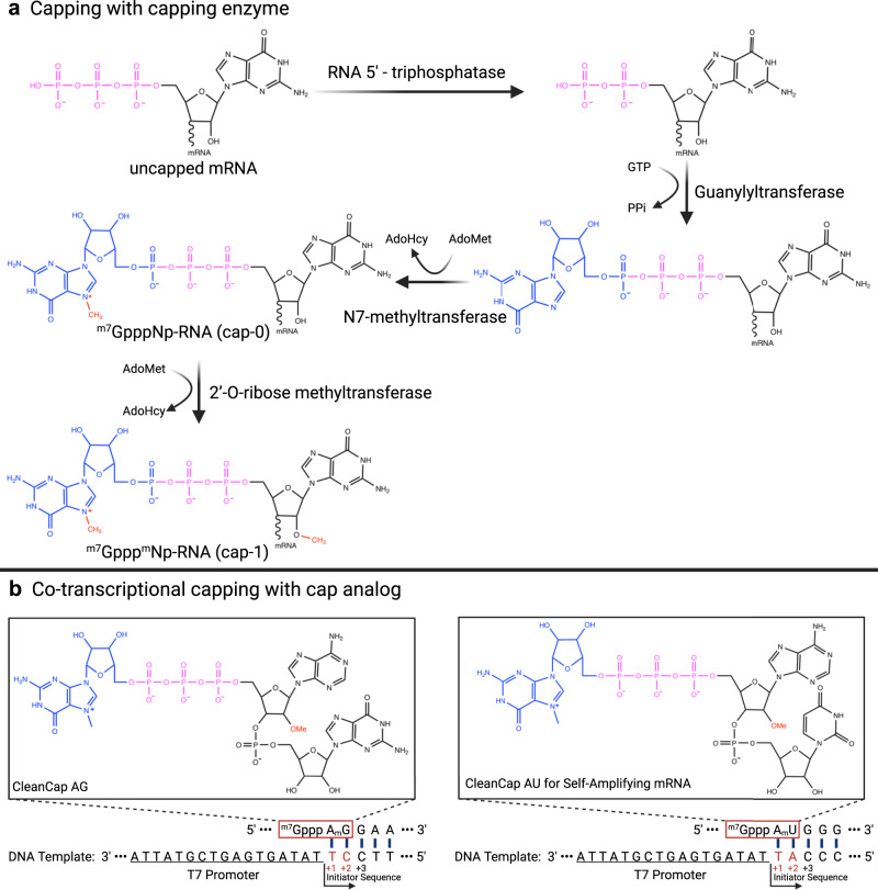
“Currently, COVID-19 mRNA vaccines BNT162b1 and BNT162b2 developed by BioNTech employ TriLink Cap1 cap analog (m27,3′-O)Gppp(m2′-O)ApG for co-transcriptional capping.27,130”
§ Advances in COVID-19 mRNA vaccine development > Different Types of COVID-19 mRNA Vaccines > Conventional non-replicating mRNA
What the sources say
- Partly supportedcites [27] Vogel, A. et al. A prefusion SARS-CoV-2 spike RNA vaccine is highly immunogenic and prevents lung infection in non-human primates. BioRxiv, https://doi.org/10.1101/2020.09.08.280818 (2020). sourceExperimental — accuracy not yet measured
The “In vitro transcription and purification of RNA” section states that the BNT162b2 RNA was transcribed in the presence of the specified TriLink cap1 analogue, supporting co-transcriptional capping for BNT162b2. The paper says BNT162b1 uses the same RNA platform, but does not explicitly establish that it uses this particular cap analogue. (LLM, conf=0.84)
- Not checked yetcites [130] Sahin, U., Muik, A., Derhovanessian, E., Vogler, I. & Türeci, Z. Publisher Correction: COVID-19 vaccine BNT162b1 elicits human antibody and TH1 T cell responses. Nature, 590, E17 (2021). source
plaindirect- Asserted: passes — The authors put it forward as true.
- Checkable: passes — It can be checked against evidence.
- Load-bearing: unclear — It is unclear whether the paper’s conclusions depend on it.
- Based: passes — The paper gives a reason why it holds.
- Partly supported
Every source the paper cites for this claim was checked and supports it.
In the paper
“In addition, circRNA has recently been developed for COVID-19 mRNA vaccines due to its natural high stability.112”
§ Advances in COVID-19 mRNA vaccine development > Different Types of COVID-19 mRNA Vaccines
What the sources say
- Supportedcites [112] Qu, L. et al. Circular RNA vaccines against SARS-CoV-2 and emerging variants. BioRxiv, https://doi.org/10.1101/2021.03.16.435594 (2021). source
The Introduction says the authors attempted to develop circRNA vaccines against SARS-CoV-2 and its variants given circRNA’s inherent stability. The Results report that their circRNA vaccine was more stable than the mRNA comparators. (LLM, conf=0.93)
plaindirect- Asserted: passes — The authors put it forward as true.
- Checkable: passes — It can be checked against evidence.
- Load-bearing: unclear — It is unclear whether the paper’s conclusions depend on it.
- Based: passes — The paper gives a reason why it holds.
- Supported
The cited sources support only part of this claim — read each source’s verdict below.
In the paper
“To improve the safety, efficacy, and stability of mRNA vaccines, researchers usually performed codon optimization172,239–243 on the antigen-coding sequence to enhance translation efficiency.”
§ Advances in COVID-19 mRNA vaccine development > Antigen design for COVID-19 mRNA vaccines
What the sources say
- Partly supportedcites [172] Mauger, D. M., Cabral, B. J., Presnyak, V., Su, S. V. & Mcfadyen, I. J. mRNA structure regulates protein expression through changes in functional half-life. Proc. Natl Acad. Sci. USA 116, 201908052 (2019). sourceExperimental — accuracy not yet measured
The Results section, “Codon Usage and mRNA Structure Synergize to Determine Ribosome Loading and mRNA Half-Life,” reports that increasing codon optimality increased median protein expression in the tested eGFP mRNAs. It does not establish that researchers usually optimize antigen-coding sequences in mRNA vaccines, or that the observed increase in protein output reflects improved translation efficiency specifically. (LLM, conf=0.83)
- Partly supportedcites [239] Hanson, G. & Coller, J. Codon optimality, bias and usage in translation and mRNA decay. Nat. Rev. Mol. Cell Biol. 19, 20–30 (2018). sourceExperimental — accuracy not yet measured
The Translation efficiency section says codon optimization can substantially affect gene expression, and the Codons affect translation elongation section reports faster translation of optimized mRNAs. The paper does not establish that researchers usually optimize antigen-coding sequences in mRNA vaccines. (LLM, conf=0.86)
- Partly supportedcites [240] Presnyak, V. et al. Codon optimality is a major determinant of mRNA stability. Cell 160, 1111–1124 (2015). sourceExperimental — accuracy not yet measured
The Results section (‘Optimal codon content influences translational efficiency’; page not provided) reports that an optimized yeast HIS3 coding sequence produced more protein per mRNA than a non-optimal version. This supports the rationale for codon optimization, but the paper does not establish that researchers usually optimize antigen-coding sequences in mRNA vaccines. (LLM, conf=0.85)
- Partly supportedcites [241] Buhr, F. et al. Synonymous codons direct cotranslational folding toward different protein conformations. Mol. Cell 61, 341–351 (2016). sourceExperimental — accuracy not yet measured
The Results section describes codon substitution as a common way to optimize heterologous gene expression and discusses its effects on translation efficiency. It studies bovine γ-B crystallin expressed in E. coli, however, and does not establish that researchers usually optimize antigen-coding sequences for mRNA vaccines. (LLM, conf=0.84)
- Not checked yetcites [242] Yang, Y., Ma, X. & Huo, Y.-X. Application of codon optimization strategy in heterologous protein expression. Chin. J. Biotechnol. 35, 2227–2237 (2019).
- Partly supportedcites [243] Kudla, G., Murray, A. W., Tollervey, D. & Plotkin, J. B. Coding-sequence determinants of gene expression in Escherichia coli. Science 324, 255–258 (2009). sourceExperimental — accuracy not yet measured
The paper’s abstract says codon bias influences global translation efficiency in E. coli. It studies GFP constructs, however, not antigen-coding sequences or mRNA vaccines, and does not establish that researchers usually perform codon optimization for that purpose. It also reports that codon bias did not correlate with expression of the individual GFP genes. (LLM, conf=0.83)
plaindirect- Asserted: passes — The authors put it forward as true.
- Checkable: passes — It can be checked against evidence.
- Load-bearing: unclear — It is unclear whether the paper’s conclusions depend on it.
- Based: passes — The paper gives a reason why it holds.
- Partly supported
The cited sources support only part of this claim — read each source’s verdict below.
In the paper
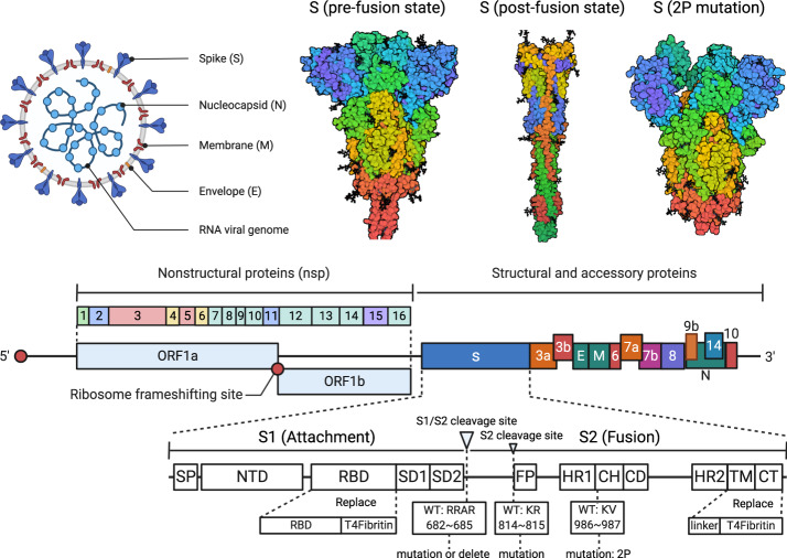
“The trimeric S protein on the surface of SARS-CoV-2 plays a key role in mediating host cell invasion.”
§ Advances in COVID-19 mRNA vaccine development > Antigen design for COVID-19 mRNA vaccines
What the sources say
- Supportedcites [228] Yao, H. et al. Molecular architecture of the SARS-CoV-2 virus. Cell 183, 730–738. e713 (2020). source
The Introduction describes S as trimeric and says its binding to the host-cell receptor ACE2 is essential for membrane fusion. The Results and Discussion section, “The Molecular Landscape of the SARS-CoV-2 Virus,” places the spikes on the viral envelope. Together, these passages support the claim that surface S mediates host-cell invasion. (LLM, conf=0.94)
- Partly supportedcites [229] Lu, R. et al. Genomic characterisation and epidemiology of 2019 novel coronavirus: implications for virus origins and receptor binding. Lancet 395, 565–574 (2020). sourceExperimental — accuracy not yet measured
The Results (page not provided) state that the envelope spike (S) protein mediates receptor binding and membrane fusion, supporting its role in host-cell invasion. Our copy does not describe the S protein as trimeric, so that structural detail is not supported by this source. (LLM, conf=0.86)
- Supportedcites [230] Hsieh, C.-L. et al. Structure-based design of prefusion-stabilized SARS-CoV-2 spikes. Science 369, 1501–1505 (2020). source
The paper states that spike (S) glycoproteins decorate coronavirus virions, bind host-cell receptors, and mediate cell entry through membrane fusion (Abstract). Its Fig. 1 caption describes the SARS-CoV-2 spike as trimeric. Together, these passages support the claim. (LLM, conf=0.94)
plaindirect- Asserted: passes — The authors put it forward as true.
- Checkable: passes — It can be checked against evidence.
- Load-bearing: passes — The paper’s conclusions depend on it.
- Based: passes — The paper gives a reason why it holds.
- Supported
The cited sources support only part of this claim — read each source’s verdict below.
In the paper
“BioNTech and Pfizer collaborated to develop five COVID-19 mRNA vaccine candidates at the beginning of the pandemic, which were based on nucleoside-modified mRNA (BNT162b2, BNT162b1, BNT162b3), non-modified mRNA (BNT162a1) and self-amplifying mRNA (BNT162c2).”
§ Advances in COVID-19 mRNA vaccine development > Progress in clinical research on mRNA Vaccines
What the sources say
- Not checked yetcites [130] Sahin, U., Muik, A., Derhovanessian, E., Vogler, I. & Türeci, Z. Publisher Correction: COVID-19 vaccine BNT162b1 elicits human antibody and TH1 T cell responses. Nature, 590, E17 (2021). source
- Partly supportedcites [376] Mulligan, M. J. et al. Phase 1/2 study to describe the safety and immunogenicity of a COVID-19 RNA Vaccine Candidate (BNT162b1) in adults 18 to 55 years of age: interim report. medRxiv, https://doi.org/10.1101/2020.06.30.20142570 (2020). sourceExperimental — accuracy not yet measured
Split into 2 atoms — 0 confirmed, 2 partially supported, 0 disputed, 0 misattributed, 0 unverifiable. Expand the claim to see per-atom verdicts.
- Partly supportedcites [377] Li, J. et al. Safety and immunogenicity of the SARS-CoV-2 BNT162b1 mRNA vaccine in younger and older Chinese adults: a randomized, placebo-controlled, double-blind phase 1 study. Nat. Med. 27, 1062–1070 (2021). sourceExperimental — accuracy not yet measured
Split into 2 atoms — 0 confirmed, 2 partially supported, 0 disputed, 0 misattributed, 0 unverifiable. Expand the claim to see per-atom verdicts.
plain- Asserted: passes — The authors put it forward as true.
- Checkable: passes — It can be checked against evidence.
- Load-bearing: unclear — It is unclear whether the paper’s conclusions depend on it.
- Based: passes — The paper gives a reason why it holds.
The cited sources support only part of this claim — read each source’s verdict below.
In the paper
“In a phase III clinical trial involving 30,420 people, participants aged 18 or older were given two doses of 100 μg of mRNA-1273 with a 28-day interval, and vaccine efficacy was 94.1% (Table 5)”
§ Advances in COVID-19 mRNA vaccine development > Progress in clinical research on mRNA Vaccines
What the sources say
- Supportedcites [410] Baden, L. R. et al. Efficacy and safety of the mRNA-1273 SARS-CoV-2 vaccine. N. Engl. J. Med. 384, 403–416 (2021). source
The Trial Population section (p. 5) reports 30,420 randomized participants; Participants, Randomization, and Data Blinding (p. 2) specifies eligibility at age 18 or older. Methods (p. 1) describes two 100 μg mRNA-1273 injections 28 days apart, and Efficacy (p. 9) reports 94.1% efficacy against symptomatic infection compared with placebo. (LLM, conf=0.94)
- Partly supportedcites [412] Ali, K. et al. Evaluation of mRNA-1273 SARS-CoV-2 vaccine in adolescents. N. Engl. J. Med. 385, 2241–2251 (2021). sourceExperimental — accuracy not yet measured
The introductory passage (Abstract > Conclusions; page unavailable) describes a phase 3 trial in adults aged 18 or older and says mRNA-1273 was highly effective. But our copy does not give that adult trial’s enrollment or 94.1% efficacy figure. The two 100 μg doses 28 days apart are specified for this paper’s adolescent trial (Abstract > Methods), not explicitly for the adult trial. (LLM, conf=0.87)
- Not checked yetcites [413] Teo, S. P. Review of COVID-19 mRNA Vaccines: BNT162b2 and mRNA-1273. J. Pharm. Pract., 08971900211009650, https://doi.org/10.1177/08971900211009650 (2021). source
- Partly supportedcites [414] Chu, L. et al. Immune memory response after a booster injection of mRNA-1273 for severe acute respiratory Syndrome Coronavirus-2 (SARS-CoV-2). medRxiv, https://doi.org/10.1101/2021.09.29.21264089 (2021). sourceExperimental — accuracy not yet measured
The Main section reports 94.1% vaccine efficacy in the phase 3 COVE study and identifies authorization for adults aged 18 or older. Methods > Study outcomes describes the COVE regimen as two 100-µg doses 28 days apart. Our copy does not give the claimed phase 3 enrollment of 30,420 people; its own study was a separate phase 2 booster trial. (LLM, conf=0.87)
plaindirect- Asserted: passes — The authors put it forward as true.
- Checkable: passes — It can be checked against evidence.
- Load-bearing: passes — The paper’s conclusions depend on it.
- Based: passes — The paper gives a reason why it holds.
- Supported
None of the sources cited for this claim have been checked yet.
In the paper
“Till now, all of these candidates have entered clinical trials (Table 5).130,376,377”
§ Advances in COVID-19 mRNA vaccine development > Progress in clinical research on mRNA Vaccines
What the sources say
- Not checked yetcites [130] Sahin, U., Muik, A., Derhovanessian, E., Vogler, I. & Türeci, Z. Publisher Correction: COVID-19 vaccine BNT162b1 elicits human antibody and TH1 T cell responses. Nature, 590, E17 (2021). source
- Not checked yetcites [376] Mulligan, M. J. et al. Phase 1/2 study to describe the safety and immunogenicity of a COVID-19 RNA Vaccine Candidate (BNT162b1) in adults 18 to 55 years of age: interim report. medRxiv, https://doi.org/10.1101/2020.06.30.20142570 (2020). source
- Not checked yetcites [377] Li, J. et al. Safety and immunogenicity of the SARS-CoV-2 BNT162b1 mRNA vaccine in younger and older Chinese adults: a randomized, placebo-controlled, double-blind phase 1 study. Nat. Med. 27, 1062–1070 (2021). source
plain- Asserted: passes — The authors put it forward as true.
- Checkable: passes — It can be checked against evidence.
- Load-bearing: unclear — It is unclear whether the paper’s conclusions depend on it.
- Based: passes — The paper gives a reason why it holds.
None of the sources cited for this claim have been checked yet.
In the paper
“Currently, five mRNA vaccines for influenza virus encoding the HA antigen have entered clinical trials: mRNA-1851, mRNA-1440, and mRNA-1010368,369 from Moderna; MRT-5400 and MRT-5401 co-developed by Sanofi and Translate Bio.370”
§ Advances in COVID-19 mRNA vaccine development > Progress in clinical research on mRNA Vaccines
What the sources say
- Not checked yetcites [368] A seasonal flu investigational vaccine clinical trial for healthy adults, https://trials.modernatx.com/study/?id=mRNA-1010-P101 (2021).
- Not checked yetcites [369] Abbasi, J. Moderna’s mRNA vaccine for seasonal flu enters clinical trials. JAMA 326, 1365–1365 (2021).
- Not checked yetcites [370] Sanofi and Translate Bio Initiate Phase 1 Clinical Trial of mRNA Influenza Vaccine, https://translatebio.gcs-web.com/news-releases/news-release-details/sanofi-and-translate-bio-initiate-phase-1-clinical-trial-mrna (2021).
plain- Asserted: passes — The authors put it forward as true.
- Checkable: passes — It can be checked against evidence.
- Load-bearing: unclear — It is unclear whether the paper’s conclusions depend on it.
- Based: passes — The paper gives a reason why it holds.
None of the sources cited for this claim have been checked yet.
In the paper
“Several mRNA vaccine candidates for viral agents other than SARS-CoV-2 have entered clinical trials (Table 4), including CMV,359 influenza virus,288,360 rabies virus,86,88 Zika virus,90,361–364 RSV,365,366 human metapneumovirus (hMPV).367”
§ Advances in COVID-19 mRNA vaccine development > Progress in clinical research on mRNA Vaccines
plain- Asserted: passes — The authors put it forward as true.
- Checkable: passes — It can be checked against evidence.
- Load-bearing: unclear — It is unclear whether the paper’s conclusions depend on it.
- Based: passes — The paper gives a reason why it holds.
In the paper
“Non-replicating mRNA vaccines deliver exclusively genetic information coding for the target antigen, thus containing the 5′-cap, 5′ untranslated region (UTR), 3′ UTR, and 3′-poly(A) tail regions.108”
§ Advances in COVID-19 mRNA vaccine development > Different Types of COVID-19 mRNA Vaccines
What the sources say
- Not checked yetcites [108] Pardi, N., Hogan, M. J., Porter, F. W. & Weissman, D. mRNA vaccines—a new era in vaccinology. Nat. Rev. Drug Discov. 17, 261–279 (2018). source
plain- Asserted: passes — The authors put it forward as true.
- Checkable: passes — It can be checked against evidence.
- Load-bearing: unclear — It is unclear whether the paper’s conclusions depend on it.
- Based: passes — The paper gives a reason why it holds.
- It may state two separate claims as one. 84
- Probably not a substantial claim. 33
- The quote only partly states this claim. 1
The cited sources support only part of this claim — read each source’s verdict below.
In the paper
“Nucleoside-modified mRNA can reduce the activation of TLRs, retinoic acid-inducible gene I (RIG-I), protein kinase R (PKR), and 2′-5′-oligoadenylate synthetase (OAS). Additionally, nucleoside modification increases translation activity and resistance against RNase L-mediated degradation.54,81,173–176”
§ Advances in COVID-19 mRNA vaccine development > Different Types of COVID-19 mRNA Vaccines > Conventional non-replicating mRNA
What the sources say
- Not checked yetcites [54] Hornung, V. et al. 5′-Triphosphate RNA is the ligand for RIG-I. Science 314, 994–997 (2006). source
- Not checked yetcites [81] Karikó, K., Buckstein, M., Ni, H. & Weissman, D. Suppression of RNA recognition by Toll-like receptors: the impact of nucleoside modification and the evolutionary origin of RNA. Immunity 23, 165–175 (2005). source
- Not checked yetcites [173] Andries, O. et al. N1-methylpseudouridine-incorporated mRNA outperforms pseudouridine-incorporated mRNA by providing enhanced protein expression and reduced immunogenicity in mammalian cell lines and mice. J. Control. Rel. 217, 337–344 (2015). source
- Partly supportedcites [174] Anderson, B. R. et al. Incorporation of pseudouridine into mRNA enhances translation by diminishing PKR activation. Nucleic Acids Res 38, 5884–5892 (2010). sourceExperimental — accuracy not yet measured
Split into 2 atoms — 0 confirmed, 2 partially supported, 0 disputed, 0 misattributed, 0 unverifiable. Expand the claim to see per-atom verdicts.
- Supportedcites [175] Anderson, B. R. et al. Nucleoside modifications in RNA limit activation of 2′-5′-oligoadenylate synthetase and increase resistance to cleavage by RNase L. Nucleic Acids Res 39, 9329–9338 (2011). source
Split into 2 atoms — 2 confirmed, 0 partially supported, 0 disputed, 0 misattributed, 0 unverifiable. Expand the claim to see per-atom verdicts.
- Supportedcites [176] Svitkin, Y. V. et al. N1-methyl-pseudouridine in mRNA enhances translation through eIF2α-dependent and independent mechanisms by increasing ribosome density. Nucleic Acids Res 45, 6023–6036 (2017). source
Split into 2 atoms — 2 confirmed, 0 partially supported, 0 disputed, 0 misattributed, 0 unverifiable. Expand the claim to see per-atom verdicts.
plaindirect- Asserted: passes — The authors put it forward as true.
- Checkable: passes — It can be checked against evidence.
- Load-bearing: passes — The paper’s conclusions depend on it.
- Based: passes — The paper gives a reason why it holds.
It may state two separate claims as one.
The cited sources support only part of this claim — read each source’s verdict below.
In the paper
“Codon preference varies extensively in different organisms,249 therefore adjusting the balance between codon usage frequency and host tRNA availability can significantly improve translation efficiency and in vivo expression of the target antigen.250,251”
§ Advances in COVID-19 mRNA vaccine development > Antigen design for COVID-19 mRNA vaccines
What the sources say
- Supportedcites [249] Sharp, P. M. & Li, W. H. The codon Adaptation Index–a measure of directional synonymous codon usage bias, and its potential applications. Nucl. Acids Res 15, 1281–1295 (1987). source
Citation 249 is attached to the first clause only. The Introduction states that synonymous codon usage is partly species- or taxon-specific and describes comparisons between organisms; the later claim about tRNA availability and antigen expression is attached to citations 250 and 251, not this source. (LLM, conf=0.93)
- Partly supportedcites [250] Wu, Q. et al. Translation affects mRNA stability in a codon-dependent manner in human cells. elife 8, e45396 (2019). sourceExperimental — accuracy not yet measured
The Discussion reports a relationship between codon optimality and tRNA abundance, and the Results report higher fluorescence from an optimal-codon reporter than a non-optimal one in human cells (pages not provided). The paper does not show that balancing host tRNA availability against codon usage improves translation efficiency or in vivo expression of a target antigen. Its cross-species comparison suggests some differences in codon regulation, but also reports substantial similarity between humans and zebrafish, rather than extensive variation across organisms. (LLM, conf=0.83)
- Not checked yetcites [251] Gustafsson, C., Govindarajan, S. & Minshull, J. Codon bias and heterologous protein expression. Trends Biotechnol. 22, 346–353 (2004). source
plaindirect- Asserted: passes — The authors put it forward as true.
- Checkable: passes — It can be checked against evidence.
- Load-bearing: passes — The paper’s conclusions depend on it.
- Based: passes — The paper gives a reason why it holds.
It may state two separate claims as one.
- Supported
Every source the paper cites for this claim was checked and supports it.
In the paper
“indicated that the incidence of anaphylaxis after BNT162b2 vaccination in Japan was higher, which points towards considering race-related adverse effects401 whose underlying causes are still unknown.”
§ Advances in COVID-19 mRNA vaccine development > Progress in clinical research on mRNA Vaccines
What the sources say
- Supportedcites [401] Hashimoto, T. et al. High anaphylaxis rates following vaccination with the Pfizer BNT162b2 mRNA vaccine against COVID-19 in Japanese healthcare workers: a secondary analysis of initial post-approval safety data. J. Travel Med. 28, taab090 (2021). source
The discussion compares estimated anaphylaxis rates after BNT162b2 vaccination in Japan with a lower US rate, while noting that the reasons for the difference are unknown. The conclusion says incidence may be higher in certain populations, such as the Japanese, and calls for further safety studies that account for ethnicity and race. The cited claim reflects those findings and the recommendation, not an established racial cause. (LLM, conf=0.88)
hedgedcorrelational- Asserted: passes — The authors put it forward as true.
- Checkable: passes — It can be checked against evidence.
- Load-bearing: unclear — It is unclear whether the paper’s conclusions depend on it.
- Based: passes — The paper gives a reason why it holds.
It may state two separate claims as one.
- Supported
The cited sources support only part of this claim — read each source’s verdict below.
In the paper
“BioNTech/Pfizer proceeded with BNT162b2 in global phase II/III clinical trials on July 27, 2020, which received EUA by the US government on December 11, 2020379,380 as well as the European Union conditional marketing authorization (CMA) on December 21, 2020.381,382”
§ Advances in COVID-19 mRNA vaccine development > Progress in clinical research on mRNA Vaccines
What the sources say
- Partly supportedcites [379] Lamb, Y. N. BNT162b2 mRNA COVID-19 Vaccine: First Approval. Drugs 81, 495-501, https://doi.org/10.1007/s40265-021-01480-7 sourceExperimental — accuracy not yet measured
The Scientific Summary (Protective Efficacy; page not supplied) describes the multinational phase II/III trial but does not date its start to July 27, 2020. The Introduction confirms EU conditional marketing authorization on December 21, and its References list a US FDA emergency-use-authorization announcement dated December 11, 2020. (LLM, conf=0.86)
- Not checked yetcites [380] FDA takes key action in fight against COVID-19 by issuing emergency use authorization for first COVID-19 Vaccine, https://www.fda.gov/news-events/press-announcements/fda-takes-key-action-fight-against-covid-19-issuing-emergency-use-authorization-first-covid-19 (2020).
- Not checked yetcites [381] Pfizer and BioNTech receive authorization in the European Union for COVID-19 vaccine, https://www.pfizer.com/news/press-release/press-release-detail/pfizer-and-biontech-receive-authorization-european-union (2020).
- Supportedcites [382] Cavaleri, M., Enzmann, H., Straus, S. & Cooke, E. The European Medicines Agency’s EU conditional marketing authorisations for COVID-19 vaccines. Lancet. (2021). source
The cited paper states that the European Commission granted conditional marketing authorisation to BNT162b2 on Dec 21, 2020 (opening section; page not provided). The US EUA and trial-start dates are outside the EU-authorisation clause supported by this citation. (LLM, conf=0.95)
plain- Asserted: passes — The authors put it forward as true.
- Checkable: passes — It can be checked against evidence.
- Load-bearing: unclear — It is unclear whether the paper’s conclusions depend on it.
- Based: passes — The paper gives a reason why it holds.
It may state two separate claims as one.
- Partly supported
The cited sources support only part of this claim — read each source’s verdict below.
In the paper
“Phase I clinical data showed that CVnCoV has acceptable tolerance and high immunogenicity.29,427”
§ Advances in COVID-19 mRNA vaccine development > Progress in clinical research on mRNA Vaccines
What the sources say
- Supportedcites [29] Kremsner, P. G. et al. Safety and immunogenicity of an mRNA-lipid nanoparticle vaccine candidate against SARS-CoV-2: A phase 1 randomized clinical trial. Wien. Klin. Wochenschr. 133, 931–941 (2021). source
The Discussion reports an acceptable reactogenicity profile in the phase 1 trial. The Abstract Results report strong immune responses at the 12 μg dose, including 100% seroconversion for S-protein or RBD antibodies and responses comparable to convalescent sera. The immunogenicity finding is strongest for that dose, rather than all doses equally. (LLM, conf=0.91)
- Could not verifycites [427] Gebre, M. et al. Optimization of non-coding regions improves protective efficacy of an mRNA SARS-CoV-2 vaccine in nonhuman primates. BioRxiv, https://doi.org/10.1101/2021.08.13.456316 (2021). sourceExperimental — accuracy not yet measured
The introduction reports human Phase 2b/3 efficacy results, not Phase I tolerability or immunogenicity. The Vaccine Immunogenicity section reports responses in cynomolgus macaques, so it cannot establish what Phase I clinical data showed. Searches for Phase I tolerability and immunogenicity found no passage addressing the claim. (LLM, conf=0.75)
plain- Asserted: passes — The authors put it forward as true.
- Checkable: passes — It can be checked against evidence.
- Load-bearing: unclear — It is unclear whether the paper’s conclusions depend on it.
- Based: passes — The paper gives a reason why it holds.
It may state two separate claims as one.
- Supported
In the paper
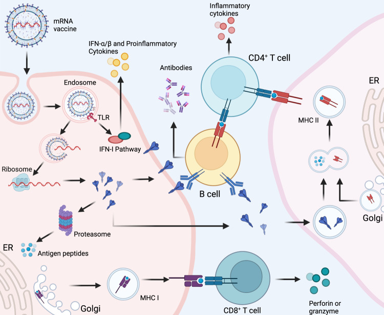
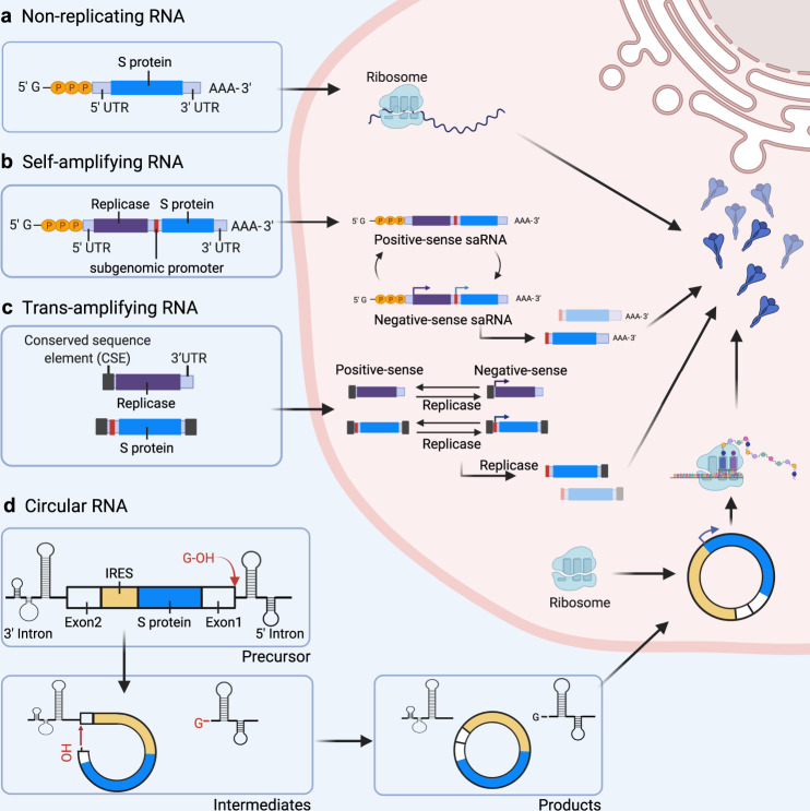
“mRNA vaccines can be categorized as non-replicating mRNA, self-amplifying mRNA (saRNA) and circular RNA (circRNA) based on their genetic characteristics (Fig. 3).107”
§ Advances in COVID-19 mRNA vaccine development > Different Types of COVID-19 mRNA Vaccines
What the sources say
- Not checked yetcites [107] Iavarone, C., O’hagan, D. T., Yu, D., Delahaye, N. F. & Ulmer, J. B. Mechanism of action of mRNA-based vaccines. Expert Rev. Vaccines 16, 871–881 (2017). source
plain- Asserted: passes — The authors put it forward as true.
- Checkable: passes — It can be checked against evidence.
- Load-bearing: fails — The paper’s conclusions may not depend on it.
- Based: passes — The paper gives a reason why it holds.
Probably not a substantial claim. The paper’s conclusions may not depend on it.
A conclusion the paper draws from its own results — it cites no other source for it, so there is none to check. The quote shows it is in the paper.
In the paper
“Among these, the COVID-19 messenger RNA (mRNA) vaccine has been drawing increasing attention owing to its great application prospects and advantages, which include short development cycle, easy industrialization, simple production process, flexibility to respond to new variants, and the capacity to induce better immune response.”
§ Advances in COVID-19 mRNA vaccine development > Abstract
plaindirect- Asserted: passes — The authors put it forward as true.
- Checkable: passes — It can be checked against evidence.
- Load-bearing: passes — The paper’s conclusions depend on it.
- Based: fails — The paper may give no reason why it holds.
Probably not a substantial claim. The paper may give no reason why it holds. It may state two separate claims as one.
In the paper
“Studies have shown that when poly(A) tail size increases to 120 bp, the expression level of the corresponding protein increases accordingly. However, when poly(A) tail size is greater than 120 bp, the expression level of the corresponding protein did not increase.152”
§ Advances in COVID-19 mRNA vaccine development > Different Types of COVID-19 mRNA Vaccines > Conventional non-replicating mRNA
What the sources say
- Not checked yetcites [152] Kormann, M. S. et al. Expression of therapeutic proteins after deliveryof chemically modified mRNA in mice. Nat. Biotechnol. 29, 154–157 (2011). source
plaindirect- Asserted: passes — The authors put it forward as true.
- Checkable: passes — It can be checked against evidence.
- Load-bearing: unclear — It is unclear whether the paper’s conclusions depend on it.
- Based: passes — The paper gives a reason why it holds.
It may state two separate claims as one.
In the paper
“Since the cap1 structure has only been described to date in eukaryotic mRNAs, it can be used as a signature of self-RNA, thus reducing the activation of pattern recognition receptor (PRR) and consequently improving translation efficiency of mRNA in vivo.119”
§ Advances in COVID-19 mRNA vaccine development > Different Types of COVID-19 mRNA Vaccines > Conventional non-replicating mRNA
What the sources say
- Not checked yetcites [119] Hyde, J. L. & Diamond, M. S. Innate immune restriction and antagonism of viral RNA lacking 2׳-O methylation. Virology 479, 66–74 (2015). source
plaindirect- Asserted: passes — The authors put it forward as true.
- Checkable: passes — It can be checked against evidence.
- Load-bearing: passes — The paper’s conclusions depend on it.
- Based: passes — The paper gives a reason why it holds.
It may state two separate claims as one.
In the paper
“The 5′-cap structure prevents mRNA from degradation by exonucleases, thereby maintaining mRNA stability and enabling translation initiation.115”
§ Advances in COVID-19 mRNA vaccine development > Different Types of COVID-19 mRNA Vaccines > Conventional non-replicating mRNA
What the sources say
- Not checked yetcites [115] Kwon, H. et al. Emergence of synthetic mRNA: in vitro synthesis of mRNA and its applications in regenerative medicine. Biomaterials 156, 172–193 (2018). source
plaindirect- Asserted: passes — The authors put it forward as true.
- Checkable: passes — It can be checked against evidence.
- Load-bearing: unclear — It is unclear whether the paper’s conclusions depend on it.
- Based: passes — The paper gives a reason why it holds.
It may state two separate claims as one.
The paper states this without a source or data behind it here, so there is nothing to check it against. The quote shows it is in the paper.
In the paper
“At present, the cap1 structure is most commonly applied for capping mRNA vaccines.”
§ Advances in COVID-19 mRNA vaccine development > Different Types of COVID-19 mRNA Vaccines > Conventional non-replicating mRNA
plain- Asserted: passes — The authors put it forward as true.
- Checkable: passes — It can be checked against evidence.
- Load-bearing: fails — The paper’s conclusions may not depend on it.
- Based: passes — The paper gives a reason why it holds.
Probably not a substantial claim. The paper’s conclusions may not depend on it.
In the paper
“Co-transcriptional capping is the most commonly used method in IVT for mRNA,115”
§ Advances in COVID-19 mRNA vaccine development > Different Types of COVID-19 mRNA Vaccines > Conventional non-replicating mRNA
What the sources say
- Not checked yetcites [115] Kwon, H. et al. Emergence of synthetic mRNA: in vitro synthesis of mRNA and its applications in regenerative medicine. Biomaterials 156, 172–193 (2018). source
plain- Asserted: passes — The authors put it forward as true.
- Checkable: passes — It can be checked against evidence.
- Load-bearing: fails — The paper’s conclusions may not depend on it.
- Based: passes — The paper gives a reason why it holds.
Probably not a substantial claim. The paper’s conclusions may not depend on it.
The paper states this without a source or data behind it here, so there is nothing to check it against. The quote shows it is in the paper.
In the paper
“The advantages of non-replicating mRNA vaccines include the use of an RNA molecule of simple structure and shorter length. An optimized or modified mRNA can have greatly enhanced biological activity.”
§ Advances in COVID-19 mRNA vaccine development > Different Types of COVID-19 mRNA Vaccines
plaindirect- Asserted: passes — The authors put it forward as true.
- Checkable: passes — It can be checked against evidence.
- Load-bearing: unclear — It is unclear whether the paper’s conclusions depend on it.
- Based: passes — The paper gives a reason why it holds.
It may state two separate claims as one.
In the paper
“The poly(A) tail plays an important role in maintaining mRNA stability and translation efficiency.116”
§ Advances in COVID-19 mRNA vaccine development > Different Types of COVID-19 mRNA Vaccines > Conventional non-replicating mRNA
What the sources say
- Not checked yetcites [116] Gallie, D. R. The cap and poly (A) tail function synergistically to regulate mRNA translational efficiency. Genes Dev. 5, 2108–2116 (1991). source
plaindirect- Asserted: passes — The authors put it forward as true.
- Checkable: passes — It can be checked against evidence.
- Load-bearing: fails — The paper’s conclusions may not depend on it.
- Based: passes — The paper gives a reason why it holds.
Probably not a substantial claim. The paper’s conclusions may not depend on it.
None of the sources cited for this claim have been checked yet.
In the paper
“At present, the 3′ UTR is mainly derived from hemoglobin subunit α (HBA) and subunit β (HBB) genes,140 but it can also be derived from albumin (ALB), heat-shock protein 70 (Hsp70), tyrosine hydroxylase (TH), and collagen alpha 1 (COL1A1) genes.141–143”
§ Advances in COVID-19 mRNA vaccine development > Different Types of COVID-19 mRNA Vaccines > Conventional non-replicating mRNA
What the sources say
- Not checked yetcites [140] Wadhwa, A., Aljabbari, A., Lokras, A., Foged, C. & Thakur, A. Opportunities and challenges in the delivery of mRNA-based vaccines. Pharmaceutics 12, 102 (2020). source
- Not checked yetcites [141] Zhang, B. Vaccines and compositions based on S antigen protein of SARS-CoV-2. CN112480217A (2021).
- Not checked yetcites [142] Karikó, K., Muramatsu, H., Keller, J. M. & Weissman, D. Increased erythropoiesis in mice injected with submicrogram quantities of pseudouridine-containing mRNA encoding erythropoietin. Mol. Ther. 20, 948–953 (2012). source
- Not checked yetcites [143] Sepac, A. et al. Comparison of cardiomyogenic potential among human ESC and iPSC lines. Cell Transpl. 21, 2523–2530 (2012). source
plain- Asserted: passes — The authors put it forward as true.
- Checkable: passes — It can be checked against evidence.
- Load-bearing: unclear — It is unclear whether the paper’s conclusions depend on it.
- Based: passes — The paper gives a reason why it holds.
It may state two separate claims as one.
None of the sources cited for this claim have been checked yet.
In the paper
“In mammalian cells, actively translated mRNAs generally contain 100–250 adenosine residues.117,150”
§ Advances in COVID-19 mRNA vaccine development > Different Types of COVID-19 mRNA Vaccines > Conventional non-replicating mRNA
What the sources say
- Not checked yetcites [117] To, K. K. & Cho, W. C. An overview of rational design of mRNA-based therapeutics and vaccines. Expert Opin. Drug Discov. 16, 1307–1317 (2021). source
- Not checked yetcites [150] Tang, T. T. & Passmore, L. A. Recognition of poly (A) RNA through its intrinsic helical structure. Cold Spring Harb. Symp. Quant. Biol. 84, 21–30 (2019). source
plain- Asserted: passes — The authors put it forward as true.
- Checkable: passes — It can be checked against evidence.
- Load-bearing: fails — The paper’s conclusions may not depend on it.
- Based: passes — The paper gives a reason why it holds.
Probably not a substantial claim. The paper’s conclusions may not depend on it.
None of the sources cited for this claim have been checked yet.
In the paper
“The 5′ UTR is mainly involved in translation of its downstream ORF sequence.132,133 The Kozak sequence is generally added after the 5′ UTR sequence to improve translation efficiency.134”
§ Advances in COVID-19 mRNA vaccine development > Different Types of COVID-19 mRNA Vaccines > Conventional non-replicating mRNA
What the sources say
- Not checked yetcites [132] Balzer, Le,S., Onsager, I., Lorentzen, J. A. & Lale, R. Dual UTR-A novel 5′ untranslated region design for synthetic biology applications. Synth. Biol. 5, ysaa006 (2020). source
- Not checked yetcites [133] Leppek, K., Das, R. & Barna, M. Functional 5′ UTR mRNA structures in eukaryotic translation regulation and how to find them. Nat. Rev. Mol. Cell Biol. 19, 158–174 (2018). source
- Not checked yetcites [134] Nance, K. D. & Meier, J. L. Modifications in an emergency: the role of N1-Methylpseudouridine in COVID-19 vaccines. ACS Cent. Sci. 7, 748–756 (2021). source
plaindirect- Asserted: passes — The authors put it forward as true.
- Checkable: passes — It can be checked against evidence.
- Load-bearing: unclear — It is unclear whether the paper’s conclusions depend on it.
- Based: passes — The paper gives a reason why it holds.
It may state two separate claims as one.
In the paper
“The vaccinia capping enzyme (VCE) integrates the enzymatic activity of RTPase, GTase, and G-N7 MTase, which can be capped to generate a cap0 structure, and then 2′-O-MTase can be used to generate a methylated cap1 structure, thus reaching a capping efficiency of 100%.52”
§ Advances in COVID-19 mRNA vaccine development > Different Types of COVID-19 mRNA Vaccines > Conventional non-replicating mRNA
What the sources say
- Not checked yetcites [52] Linares-Fernández, S., Lacroix, C., Exposito, J. Y. & Verrier, B. Tailoring mRNA vaccine to balance innate/adaptive immune response. Trends Mol. Med. 26, 311–323 (2019). source
plaindirect- Asserted: passes — The authors put it forward as true.
- Checkable: passes — It can be checked against evidence.
- Load-bearing: unclear — It is unclear whether the paper’s conclusions depend on it.
- Based: passes — The paper gives a reason why it holds.
It may state two separate claims as one.
In the paper
“Of note, it has been reported that the COVID-19 vaccine mRNA-1273 developed by Moderna employs the capping enzyme described above.28”
§ Advances in COVID-19 mRNA vaccine development > Different Types of COVID-19 mRNA Vaccines > Conventional non-replicating mRNA
What the sources say
- Not checked yetcites [28] Corbett, K. S. et al. SARS-CoV-2 mRNA vaccine design enabled by prototype pathogen preparedness. Nature 586, 567–571 (2020). source
plain- Asserted: passes — The authors put it forward as true.
- Checkable: passes — It can be checked against evidence.
- Load-bearing: fails — The paper’s conclusions may not depend on it.
- Based: passes — The paper gives a reason why it holds.
Probably not a substantial claim. The paper’s conclusions may not depend on it.
None of the sources cited for this claim have been checked yet.
In the paper
“A non-replicating mRNA contains an open reading frame (ORF) encoding the gene coding for the target antigen flanked by 5′ and 3′ UTR. The 5′ end contains a 7-methylguanosine cap structure (5′-cap, m7G), whereas the 3′ end contains a poly(A) tail structure.”
§ Advances in COVID-19 mRNA vaccine development > Different Types of COVID-19 mRNA Vaccines > Conventional non-replicating mRNA
What the sources say
- Not checked yetcites [113] Schlake, T., Thess, A., Fotin-Mleczek, M. & Kallen, K.-J. Developing mRNA-vaccine technologies. RNA Biol. 9, 1319–1330 (2012). source
- Not checked yetcites [114] Youn, H. & Chung, J. K. Modified mRNA as an alternative to plasmid DNA (pDNA) for transcript replacement and vaccination therapy. Expert Opin. Biol. Ther. 15, 1–12 (2015). source
plain- Asserted: passes — The authors put it forward as true.
- Checkable: passes — It can be checked against evidence.
- Load-bearing: unclear — It is unclear whether the paper’s conclusions depend on it.
- Based: passes — The paper gives a reason why it holds.
It may state two separate claims as one.
None of the sources cited for this claim have been checked yet.
In the paper
“The poly(A) tail can also bind to multiple poly(A)-binding proteins (PABPs) while working synergistically with 5′ m7G cap sequences to regulate translational efficiency.146,147”
§ Advances in COVID-19 mRNA vaccine development > Different Types of COVID-19 mRNA Vaccines > Conventional non-replicating mRNA
What the sources say
- Not checked yetcites [146] Goss, D. J. & Kleiman, F. E. Poly (A) binding proteins: are they all created equal? Wiley Interdiscip. Rev.: RNA 4, 167–179 (2013). source
- Not checked yetcites [147] Wang, Z. & Kiledjian, M. The poly (A)-binding protein and an mRNA stability protein jointly regulate an endoribonuclease activity. Mol. Cell. Biol. 20, 6334–6341 (2000). source
plaindirect- Asserted: passes — The authors put it forward as true.
- Checkable: passes — It can be checked against evidence.
- Load-bearing: fails — The paper’s conclusions may not depend on it.
- Based: passes — The paper gives a reason why it holds.
Probably not a substantial claim. The paper’s conclusions may not depend on it. It may state two separate claims as one.
In the paper
“The current new generation of cap analogs is the CleanCap® cap analogs developed by TriLink BioTechnologies (San Diego, CA, USA),126 which can be co-transcribed with the target mRNA to obtain the cap1 structure, thus solving the issues of low efficiency and high enzyme costs of traditional capping methods.”
§ Advances in COVID-19 mRNA vaccine development > Different Types of COVID-19 mRNA Vaccines > Conventional non-replicating mRNA
What the sources say
- Not checked yetcites [126] Henderson, J. M. et al. Cap 1 messenger RNA synthesis with co-transcriptional CleanCap® analog by in vitro transcription. Curr. Protoc. 1, e39 (2021). source
plaindirect- Asserted: passes — The authors put it forward as true.
- Checkable: passes — It can be checked against evidence.
- Load-bearing: unclear — It is unclear whether the paper’s conclusions depend on it.
- Based: passes — The paper gives a reason why it holds.
It may state two separate claims as one.
The paper states this without a source or data behind it here, so there is nothing to check it against. The quote shows it is in the paper.
In the paper
“Lastly, saRNA vaccines are currently in preclinical and clinical trials.”
§ Advances in COVID-19 mRNA vaccine development > Different Types of COVID-19 mRNA Vaccines
plain- Asserted: passes — The authors put it forward as true.
- Checkable: passes — It can be checked against evidence.
- Load-bearing: fails — The paper’s conclusions may not depend on it.
- Based: passes — The paper gives a reason why it holds.
Probably not a substantial claim. The paper’s conclusions may not depend on it.
None of the sources cited for this claim have been checked yet.
In the paper
“Studies have shown that adenylate-uridylate-rich elements are involved in mRNA degradation. Degradation rate and translation life cycle can be adjusted by replacing adenylate-uridylate-rich sequences found in the 3′ UTR.137–139”
§ Advances in COVID-19 mRNA vaccine development > Different Types of COVID-19 mRNA Vaccines > Conventional non-replicating mRNA
What the sources say
- Not checked yetcites [137] Mayr, C. What are 3′ UTRs doing?. Cold Spring Harb. Perspect. Biol. 11, a034728 (2018). source
- Not checked yetcites [138] Fotin-Mleczek, M. et al. Messenger RNA-based vaccines with dual activity induce balanced TLR-7 dependent adaptive immune responses and provide antitumor activity. J. Immunother. 34, 1–15 (2011). source
- Not checked yetcites [139] Eberhardt, W., Doller, A., Akool, E.-S. & Pfeilschifter, J. Modulation of mRNA stability as a novel therapeutic approach. Pharmacol. Ther. 114, 56–73 (2007). source
plaindirect- Asserted: passes — The authors put it forward as true.
- Checkable: passes — It can be checked against evidence.
- Load-bearing: unclear — It is unclear whether the paper’s conclusions depend on it.
- Based: passes — The paper gives a reason why it holds.
It may state two separate claims as one.
None of the sources cited for this claim have been checked yet.
In the paper
“Conversely, the function of the 3′ UTR is to maintain mRNA stability.135,136”
§ Advances in COVID-19 mRNA vaccine development > Different Types of COVID-19 mRNA Vaccines > Conventional non-replicating mRNA
What the sources say
- Not checked yetcites [135] Mayr, C. Regulation by 3′-untranslated regions. Annu. Rev. Genet. 51, 171–194 (2017). source
- Not checked yetcites [136] Matoulkova, E., Michalova, E., Vojtesek, B. & Hrstka, R. The role of the 3’untranslated region in post-transcriptional regulation of protein expression in mammalian cells. RNA Biol. 9, 563–576 (2012). source
plaindirect- Asserted: passes — The authors put it forward as true.
- Checkable: passes — It can be checked against evidence.
- Load-bearing: fails — The paper’s conclusions may not depend on it.
- Based: passes — The paper gives a reason why it holds.
Probably not a substantial claim. The paper’s conclusions may not depend on it.
In the paper
“mRNA contains 5′ and 3′ UTRs, whose functions are related, respectively, to regulating translation and maintaining mRNA stability.131”
§ Advances in COVID-19 mRNA vaccine development > Different Types of COVID-19 mRNA Vaccines > Conventional non-replicating mRNA
What the sources say
- Not checked yetcites [131] Zarghampoor, F., Azarpira, N., Khatami, S. R., Behzad-Behbahani, A. & Foroughmand, A. M. Improved translation efficiency of therapeutic mRNA. Gene 707, 231–238 (2019). source
plaincorrelational- Asserted: passes — The authors put it forward as true.
- Checkable: passes — It can be checked against evidence.
- Load-bearing: fails — The paper’s conclusions may not depend on it.
- Based: passes — The paper gives a reason why it holds.
Probably not a substantial claim. The paper’s conclusions may not depend on it. It may state two separate claims as one.
None of the sources cited for this claim have been checked yet.
In the paper
“The use of CleanCap® cap analogs reduces the probability of reverse capped, uncapped, and cap0 intermediates, and the co-transcribed mRNA only possesses a cap1 structure, therefore capping rate can be 90% or higher.52,126”
§ Advances in COVID-19 mRNA vaccine development > Different Types of COVID-19 mRNA Vaccines > Conventional non-replicating mRNA
What the sources say
- Not checked yetcites [52] Linares-Fernández, S., Lacroix, C., Exposito, J. Y. & Verrier, B. Tailoring mRNA vaccine to balance innate/adaptive immune response. Trends Mol. Med. 26, 311–323 (2019). source
- Not checked yetcites [126] Henderson, J. M. et al. Cap 1 messenger RNA synthesis with co-transcriptional CleanCap® analog by in vitro transcription. Curr. Protoc. 1, e39 (2021). source
plaindirect- Asserted: passes — The authors put it forward as true.
- Checkable: passes — It can be checked against evidence.
- Load-bearing: unclear — It is unclear whether the paper’s conclusions depend on it.
- Based: passes — The paper gives a reason why it holds.
It may state two separate claims as one.
In the paper
“however, an mRNA of cap 0 is likely to be recognized as exogenous RNA by the host, which could stimulate the innate immune response of the host and ultimately trigger inflammatory responses.117”
§ Advances in COVID-19 mRNA vaccine development > Different Types of COVID-19 mRNA Vaccines > Conventional non-replicating mRNA
What the sources say
- Not checked yetcites [117] To, K. K. & Cho, W. C. An overview of rational design of mRNA-based therapeutics and vaccines. Expert Opin. Drug Discov. 16, 1307–1317 (2021). source
hedgedconditional- Asserted: passes — The authors put it forward as true.
- Checkable: passes — It can be checked against evidence.
- Load-bearing: passes — The paper’s conclusions depend on it.
- Based: passes — The paper gives a reason why it holds.
It may state two separate claims as one.
None of the sources cited for this claim have been checked yet.
In the paper
“A vaccine platform (RNActive®) was designed by CureVac186 combining the use of co-delivered RNA and protamine (a polycationic peptide) complex as adjuvant, which has been shown to effectively trigger innate immune responses and enhance vaccine immunogenicity.186–189”
§ Advances in COVID-19 mRNA vaccine development > Different Types of COVID-19 mRNA Vaccines > Conventional non-replicating mRNA
What the sources say
- Not checked yetcites [186] Rauch, S., Lutz, J., Kowalczyk, A., Schlake, T. & Heidenreich, R. RNActive® technology: generation and testing of stable and immunogenic mRNA vaccines. Methods Mol. Biol. 1499, 89–107 (2017). source
- Not checked yetcites [187] Edwards, D. K. et al. Adjuvant effects of a sequence-engineered mRNA vaccine: translational profiling demonstrates similar human and murine innate response. J. Transl. Med 15, 1–18 (2017). source
- Not checked yetcites [188] Kallen, K.-J. et al. A novel, disruptive vaccination technology: self-adjuvanted RNActive® vaccines. Hum. Vaccine Immunother. 9, 2263–2276 (2013). source
- Not checked yetcites [189] Kowalczyk, A. et al. Self-adjuvanted mRNA vaccines induce local innate immune responses that lead to a potent and boostable adaptive immunity. Vaccine 34, 3882–3893 (2016). source
boosteddirect- Asserted: passes — The authors put it forward as true.
- Checkable: passes — It can be checked against evidence.
- Load-bearing: unclear — It is unclear whether the paper’s conclusions depend on it.
- Based: passes — The paper gives a reason why it holds.
It may state two separate claims as one.
A conclusion the paper draws from its own results — it cites no other source for it, so there is none to check. The quote shows it is in the paper.
In the paper
“In addition, considering that the length of nsP1–4 sequence is approximately 7 kb, the full length of a saRNA sequence is usually above 9 kb, which might hinder cloning construction.”
§ Advances in COVID-19 mRNA vaccine development > Different Types of COVID-19 mRNA Vaccines > saRNA
hedgedconditional- Asserted: passes — The authors put it forward as true.
- Checkable: passes — It can be checked against evidence.
- Load-bearing: passes — The paper’s conclusions depend on it.
- Based: passes — The paper gives a reason why it holds.
It may state two separate claims as one.
None of the sources cited for this claim have been checked yet.
In the paper
“Recently, two different saRNA vaccines developed independently by Arcturus Therapeutics and Imperial College London have shown favorable immune responses against SARS-CoV-2, and have entered clinical trials.211–213”
§ Advances in COVID-19 mRNA vaccine development > Different Types of COVID-19 mRNA Vaccines > saRNA
What the sources say
- Not checked yetcites [211] McKay, P. F. et al. Self-amplifying RNA SARS-CoV-2 lipid nanoparticle vaccine candidate induces high neutralizing antibody titers in mice. Nat. Commun. 11, 1–7 (2020). source
- Not checked yetcites [212] Pollock, K. M. et al. Safety and Immunogenicity of a Self-Amplifying RNA Vaccine Against COVID-19: COVAC1, a Phase I, Dose-Ranging Trial. EClinicalMedicine, https://doi.org/10.1016/j.eclinm.2021.101262 (2022). source
- Not checked yetcites [213] de Alwis, R. et al. A single dose of self-transcribing and replicating RNA-based SARS-CoV-2 vaccine produces protective adaptive immunity in mice. Mol. Ther. 29, 1970–1983 (2021). source
plaindirect- Asserted: passes — The authors put it forward as true.
- Checkable: passes — It can be checked against evidence.
- Load-bearing: unclear — It is unclear whether the paper’s conclusions depend on it.
- Based: passes — The paper gives a reason why it holds.
It may state two separate claims as one.
None of the sources cited for this claim have been checked yet.
In the paper
“To avoid this, Karikó and colleagues found that adding modified nucleosides during IVT of mRNA can significantly reduce the host inflammatory response without affecting protein expression.81,159”
§ Advances in COVID-19 mRNA vaccine development > Different Types of COVID-19 mRNA Vaccines > Conventional non-replicating mRNA
What the sources say
- Not checked yetcites [81] Karikó, K., Buckstein, M., Ni, H. & Weissman, D. Suppression of RNA recognition by Toll-like receptors: the impact of nucleoside modification and the evolutionary origin of RNA. Immunity 23, 165–175 (2005). source
- Not checked yetcites [159] Karikó, K. et al. Incorporation of pseudouridine into mRNA yields superior nonimmunogenic vector with increased translational capacity and biological stability. Mol. Ther. 16, 1833–1840 (2008). source
plaindirect- Asserted: passes — The authors put it forward as true.
- Checkable: passes — It can be checked against evidence.
- Load-bearing: passes — The paper’s conclusions depend on it.
- Based: passes — The paper gives a reason why it holds.
It may state two separate claims as one.
In the paper
“Current studies have shown that the segmented poly(A) tail extends mRNA half-life and improves translation efficiency compared to the long-chain poly(A) tail.154”
§ Advances in COVID-19 mRNA vaccine development > Different Types of COVID-19 mRNA Vaccines > Conventional non-replicating mRNA
What the sources say
- Not checked yetcites [154] Trepotec, Z., Geiger, J., Plank, C., Aneja, M. K. & Rudolph, C. Segmented poly(A) tails significantly reduce recombination of plasmid DNA without affecting mRNA translation efficiency or half-life. RNA 25, 507–518 (2019). source
plaindirect- Asserted: passes — The authors put it forward as true.
- Checkable: passes — It can be checked against evidence.
- Load-bearing: unclear — It is unclear whether the paper’s conclusions depend on it.
- Based: passes — The paper gives a reason why it holds.
It may state two separate claims as one.
None of the sources cited for this claim have been checked yet.
In the paper
“saRNA vaccines have nonetheless certain shortcomings, including the risk of excessive activation of the inflammatory response, and the production of viral nsP produced by the alphavirus replicon that may interfere with normal signal transduction in target host cells.214,215”
§ Advances in COVID-19 mRNA vaccine development > Different Types of COVID-19 mRNA Vaccines > saRNA
What the sources say
- Not checked yetcites [214] Maruggi, G., Zhang, C., Li, J., Ulmer, J. B. & Yu, D. mRNA as a transformative technology for vaccine development to control infectious diseases. Mol. Ther. 27, 757–772 (2019). source
- Not checked yetcites [215] Jensen, S. & Thomsen, A. R. Sensing of RNA viruses: a review of innate immune receptors involved in recognizing RNA virus invasion. J. Virol. 86, 2900–2910 (2012). source
hedgedconditional- Asserted: passes — The authors put it forward as true.
- Checkable: passes — It can be checked against evidence.
- Load-bearing: unclear — It is unclear whether the paper’s conclusions depend on it.
- Based: passes — The paper gives a reason why it holds.
It may state two separate claims as one.
None of the sources cited for this claim have been checked yet.
In the paper
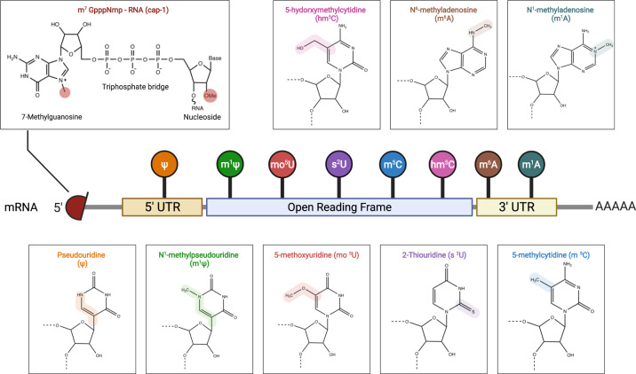
“Studies have found that replacing original nucleosides with m6A and s2U inhibits activation of TLR3, whereas activation of TLR7 and TLR8 is blocked when using ψ, 5-methyluridine (m5U), m6A, and s2U, thereby inhibiting the innate immune responses and improving protein translation efficiency.81,159,171”
§ Advances in COVID-19 mRNA vaccine development > Different Types of COVID-19 mRNA Vaccines > Conventional non-replicating mRNA
What the sources say
- Not checked yetcites [81] Karikó, K., Buckstein, M., Ni, H. & Weissman, D. Suppression of RNA recognition by Toll-like receptors: the impact of nucleoside modification and the evolutionary origin of RNA. Immunity 23, 165–175 (2005). source
- Not checked yetcites [159] Karikó, K. et al. Incorporation of pseudouridine into mRNA yields superior nonimmunogenic vector with increased translational capacity and biological stability. Mol. Ther. 16, 1833–1840 (2008). source
- Not checked yetcites [171] Yan, K. et al. Toll-like receptor 3 and RIG-I-like receptor activation induces innate antiviral responses in mouse ovarian granulosa cells. Mol. Cell. Endocrinol. 372, 73–85 (2013). source
plaindirect- Asserted: passes — The authors put it forward as true.
- Checkable: passes — It can be checked against evidence.
- Load-bearing: passes — The paper’s conclusions depend on it.
- Based: passes — The paper gives a reason why it holds.
It may state two separate claims as one.
In the paper
“Kormann and colleagues152 replaced 25% of mRNA cytosine with m5C and 25% of uridine with s2U, which improved mRNA stability and increased protein translation in mice.”
§ Advances in COVID-19 mRNA vaccine development > Different Types of COVID-19 mRNA Vaccines > Conventional non-replicating mRNA
What the sources say
- Not checked yetcites [152] Kormann, M. S. et al. Expression of therapeutic proteins after deliveryof chemically modified mRNA in mice. Nat. Biotechnol. 29, 154–157 (2011). source
plaindirect- Asserted: passes — The authors put it forward as true.
- Checkable: passes — It can be checked against evidence.
- Load-bearing: unclear — It is unclear whether the paper’s conclusions depend on it.
- Based: passes — The paper gives a reason why it holds.
It may state two separate claims as one.
The paper states this without a source or data behind it here, so there is nothing to check it against. The quote shows it is in the paper.
In the paper
“Both Moderna and BioNTech use pseudouridine modifications to ensure mRNA stability in their COVID-19 vaccine formulation.”
§ Advances in COVID-19 mRNA vaccine development > Different Types of COVID-19 mRNA Vaccines > Conventional non-replicating mRNA
plaindirect- Asserted: passes — The authors put it forward as true.
- Checkable: passes — It can be checked against evidence.
- Load-bearing: fails — The paper’s conclusions may not depend on it.
- Based: fails — The paper may give no reason why it holds.
Probably not a substantial claim. The paper’s conclusions may not depend on it. The paper may give no reason why it holds.
None of the sources cited for this claim have been checked yet.
In the paper
“dsRNA can activate PRR such as TLR3,52,156 whereas ssRNA activates TLR7 and TLR8 to produce IFN-I, thus inducing inflammation in the host and interrupting mRNA translation.157,158”
§ Advances in COVID-19 mRNA vaccine development > Different Types of COVID-19 mRNA Vaccines > Conventional non-replicating mRNA
What the sources say
- Not checked yetcites [52] Linares-Fernández, S., Lacroix, C., Exposito, J. Y. & Verrier, B. Tailoring mRNA vaccine to balance innate/adaptive immune response. Trends Mol. Med. 26, 311–323 (2019). source
- Not checked yetcites [156] Kariko, K., Muramatsu, H., Ludwig, J. & Weissman, D. Generating the optimal mRNA for therapy: HPLC purification eliminates immune activation and improves translation of nucleoside-modified, protein-encoding mRNA. Nucleic Acids Res 39, e142–e142 (2011). source
- Not checked yetcites [157] Vaidyanathan, S. et al. Uridine depletion and chemical modification increase Cas9 mRNA activity and reduce immunogenicity without HPLC purification. Mol. Ther. Nucleic Acids 12, 530–542 (2018). source
- Not checked yetcites [158] Isaacs, A., Cox, R. & Rotem, Z. Foreign nucleic acids as the stimulus to make interferon. Lancet, 282, 113–116 (1963). source
plaindirect- Asserted: passes — The authors put it forward as true.
- Checkable: passes — It can be checked against evidence.
- Load-bearing: passes — The paper’s conclusions depend on it.
- Based: passes — The paper gives a reason why it holds.
It may state two separate claims as one.
In the paper
“Li and colleagues developed an in vitro evolution strategy, and six mutations (namely A1979G, G3936C, A4311G, A4758G, G4796T, G4944A) were identified in the nsP2 and nsP3 of the VEEV replicon, which were shown to promote expression of subgenomic RNA in cells.210”
§ Advances in COVID-19 mRNA vaccine development > Different Types of COVID-19 mRNA Vaccines > saRNA
What the sources say
- Not checked yetcites [210] Li, Y. et al. In vitro evolution of enhanced RNA replicons for immunotherapy. Sci. Rep. 9, 1–10 (2019). source
plaindirect- Asserted: passes — The authors put it forward as true.
- Checkable: passes — It can be checked against evidence.
- Load-bearing: unclear — It is unclear whether the paper’s conclusions depend on it.
- Based: passes — The paper gives a reason why it holds.
It may state two separate claims as one.
None of the sources cited for this claim have been checked yet.
In the paper
“Currently, nucleoside-modified mRNA vaccines employ 100% chemically modified nucleosides replacing natural nucleosides, and m1ψ is often used to replace uridine during IVT to improve the safety and stability of mRNA vaccines.52,134,172”
§ Advances in COVID-19 mRNA vaccine development > Different Types of COVID-19 mRNA Vaccines > Conventional non-replicating mRNA
What the sources say
- Not checked yetcites [52] Linares-Fernández, S., Lacroix, C., Exposito, J. Y. & Verrier, B. Tailoring mRNA vaccine to balance innate/adaptive immune response. Trends Mol. Med. 26, 311–323 (2019). source
- Not checked yetcites [134] Nance, K. D. & Meier, J. L. Modifications in an emergency: the role of N1-Methylpseudouridine in COVID-19 vaccines. ACS Cent. Sci. 7, 748–756 (2021). source
- Not checked yetcites [172] Mauger, D. M., Cabral, B. J., Presnyak, V., Su, S. V. & Mcfadyen, I. J. mRNA structure regulates protein expression through changes in functional half-life. Proc. Natl Acad. Sci. USA 116, 201908052 (2019). source
plaindirect- Asserted: passes — The authors put it forward as true.
- Checkable: passes — It can be checked against evidence.
- Load-bearing: unclear — It is unclear whether the paper’s conclusions depend on it.
- Based: passes — The paper gives a reason why it holds.
It may state two separate claims as one.
In the paper
“The idea of using in vitro synthesized saRNA as a preventive vaccine was first proposed by Zhou and colleagues in 1994, using a modified Semliki Forest virus (SFV) replicon to express the nucleoprotein (NP) of the influenza virus.197”
§ Advances in COVID-19 mRNA vaccine development > Different Types of COVID-19 mRNA Vaccines > saRNA
What the sources say
- Not checked yetcites [197] Zhou, X. et al. Self-replicating Semliki Forest virus RNA as recombinant vaccine. Vaccine 12, 1510–1514 (1994). source
plain- Asserted: passes — The authors put it forward as true.
- Checkable: passes — It can be checked against evidence.
- Load-bearing: fails — The paper’s conclusions may not depend on it.
- Based: passes — The paper gives a reason why it holds.
Probably not a substantial claim. The paper’s conclusions may not depend on it.
In the paper
“Decades later, Geall and colleagues were the first to use LNP to encapsulate a saRNA chimera composed of the Venezuelan equine encephalitis virus (VEEV) and Sindbis virus (SINV) replicons into a vaccine which was used to immunize mice, and immunogenicity was significantly improved compared with unencapsulated group.199”
§ Advances in COVID-19 mRNA vaccine development > Different Types of COVID-19 mRNA Vaccines > saRNA
What the sources say
- Not checked yetcites [199] Geall, A. J. et al. Nonviral delivery of self-amplifying RNA vaccines. Proc. Natl Acad. Sci. USA 109, 14604–14609 (2012). source
plaindirect- Asserted: passes — The authors put it forward as true.
- Checkable: passes — It can be checked against evidence.
- Load-bearing: unclear — It is unclear whether the paper’s conclusions depend on it.
- Based: passes — The paper gives a reason why it holds.
It may state two separate claims as one.
A conclusion the paper draws from its own results — it cites no other source for it, so there is none to check. The quote shows it is in the paper.
In the paper
“With its highly stable properties without nucleotide modification, circRNA can potentially become a novel platform for vaccine and drug development.”
§ Advances in COVID-19 mRNA vaccine development > Different Types of COVID-19 mRNA Vaccines > CircRNA
hedgedconditional- Asserted: passes — The authors put it forward as true.
- Checkable: passes — It can be checked against evidence.
- Load-bearing: fails — The paper’s conclusions may not depend on it.
- Based: fails — The paper may give no reason why it holds.
Probably not a substantial claim. The paper’s conclusions may not depend on it. The paper may give no reason why it holds.
In the paper
“Studies have found that the S protein prefusion conformation induces better immunogenicity and can be therefore considered an ideal target antigen.237”
§ Advances in COVID-19 mRNA vaccine development > Antigen design for COVID-19 mRNA vaccines
What the sources say
- Not checked yetcites [237] Yadav, T. et al. Recombinant vaccines for COVID-19. Hum. Vaccin Immunother. 16, 2905–2912 (2020). source
plaindirect- Asserted: passes — The authors put it forward as true.
- Checkable: passes — It can be checked against evidence.
- Load-bearing: passes — The paper’s conclusions depend on it.
- Based: passes — The paper gives a reason why it holds.
It may state two separate claims as one.
A conclusion the paper draws from its own results — it cites no other source for it, so there is none to check. The quote shows it is in the paper.
In the paper
“In summary, although considered a byproduct of the mRNA splicing process, circRNA has now emerged as an important new class of non-coding RNAs.”
§ Advances in COVID-19 mRNA vaccine development > Different Types of COVID-19 mRNA Vaccines > CircRNA
plain- Asserted: passes — The authors put it forward as true.
- Checkable: passes — It can be checked against evidence.
- Load-bearing: fails — The paper’s conclusions may not depend on it.
- Based: passes — The paper gives a reason why it holds.
Probably not a substantial claim. The paper’s conclusions may not depend on it.
The paper states this without a source or data behind it here, so there is nothing to check it against. The quote shows it is in the paper.
In the paper
“The S1 subunit comprises the signal peptide (SP), RBD, N-terminal domain (NTD), C-terminal domain 1 (CTD1), and C-terminal domain 2 (CTD2), and primarily interacts with the cellular angiotensin-converting enzyme 2 (ACE2) receptor via RBD.”
§ Advances in COVID-19 mRNA vaccine development > Antigen design for COVID-19 mRNA vaccines
plaindirect- Asserted: passes — The authors put it forward as true.
- Checkable: passes — It can be checked against evidence.
- Load-bearing: unclear — It is unclear whether the paper’s conclusions depend on it.
- Based: passes — The paper gives a reason why it holds.
It may state two separate claims as one.
In the paper
“the results revealed that circRNA prepared with sequences of Delta strains resulted in broad-spectrum protection and production of neutralizing antibodies against both Delta and Omicron.”
§ Advances in COVID-19 mRNA vaccine development > Different Types of COVID-19 mRNA Vaccines > CircRNA
What the sources say
- Not checked yetcites [227] Qu, L. et al. Circular RNA Vaccines against SARS-CoV-2 and Emerging Variants. BioRxiv, https://doi.org/10.1101/2021.03.16.435594 (2022). source
plaindirect- Asserted: passes — The authors put it forward as true.
- Checkable: passes — It can be checked against evidence.
- Load-bearing: passes — The paper’s conclusions depend on it.
- Based: passes — The paper gives a reason why it holds.
It may state two separate claims as one.
None of the sources cited for this claim have been checked yet.
In the paper
“The S2 subunit comprises the fusion peptide (FP), double heptad repeat (HR), central helix region (CH), connector domain (CD), transmembrane domain (TMD), and cytoplasmic tail (CT), and the S2 subunit is responsible for mediating the fusion between the virus and the host cell membrane233–236”
§ Advances in COVID-19 mRNA vaccine development > Antigen design for COVID-19 mRNA vaccines
What the sources say
- Not checked yetcites [233] Wang, Q. et al. Structural and functional basis of SARS-CoV-2 entry by using human ACE2. Cell 181, 894–904. e899 (2020). source
- Not checked yetcites [234] Shang, J. et al. Structural basis of receptor recognition by SARS-CoV-2. Nature 581, 221–224 (2020). source
- Not checked yetcites [235] Singh, S. P., Pritam, M., Pandey, B. & Yadav, T. P. Microstructure, pathophysiology and potential therapeutics of COVID-19: A comprehensive review. J. Med. Virol. 93, 275–299 (2020). source
- Not checked yetcites [236] Cai, Y. et al. Distinct conformational states of SARS-CoV-2 spike protein. Science 369, 1586–1592 (2020). source
plaindirect- Asserted: passes — The authors put it forward as true.
- Checkable: passes — It can be checked against evidence.
- Load-bearing: unclear — It is unclear whether the paper’s conclusions depend on it.
- Based: passes — The paper gives a reason why it holds.
It may state two separate claims as one.
In the paper
“However, vaccines produced with circRNA prepared based on Omicron sequences provided a narrower protection, and produced neutralizing antibodies could protect only against Omicron but not against Delta.”
§ Advances in COVID-19 mRNA vaccine development > Different Types of COVID-19 mRNA Vaccines > CircRNA
What the sources say
- Not checked yetcites [227] Qu, L. et al. Circular RNA Vaccines against SARS-CoV-2 and Emerging Variants. BioRxiv, https://doi.org/10.1101/2021.03.16.435594 (2022). source
plaindirect- Asserted: passes — The authors put it forward as true.
- Checkable: passes — It can be checked against evidence.
- Load-bearing: passes — The paper’s conclusions depend on it.
- Based: passes — The paper gives a reason why it holds.
It may state two separate claims as one.
In the paper
“Most neutralizing antibodies are molded based on epitopes of S protein in prefusion conformation, which is covered once the S protein acquires the postfusion conformation, thus reducing the production of neutralizing antibodies.238”
§ Advances in COVID-19 mRNA vaccine development > Antigen design for COVID-19 mRNA vaccines
What the sources say
- Not checked yetcites [238] Walls, A. C. et al. Structure, function and antigenicity of the SARS-CoV-2 spike glycoprotein. Cell 181 (2020). source
plaindirect- Asserted: passes — The authors put it forward as true.
- Checkable: passes — It can be checked against evidence.
- Load-bearing: passes — The paper’s conclusions depend on it.
- Based: passes — The paper gives a reason why it holds.
It may state two separate claims as one.
None of the sources cited for this claim have been checked yet.
In the paper
“CircRNA is a highly stable single-stranded RNA with a covalently closed loop structure (Fig. 3), including a large category of non-coding RNAs generated by backsplicing in eukaryotic cells.218–220”
§ Advances in COVID-19 mRNA vaccine development > Different Types of COVID-19 mRNA Vaccines > CircRNA
What the sources say
- Not checked yetcites [218] Zhang, X.-O. et al. Complementary sequence-mediated exon circularization. Cell 159, 134–147 (2014). source
- Not checked yetcites [219] Kristensen, L. S. et al. The biogenesis, biology and characterization of circular RNAs. Nat. Rev. Genet. 20, 675–691 (2019). source
- Not checked yetcites [220] Chen, L.-L. The biogenesis and emerging roles of circular RNAs. Nat. Rev. Mol. Cell Biol. 17, 205–211 (2016). source
plain- Asserted: passes — The authors put it forward as true.
- Checkable: passes — It can be checked against evidence.
- Load-bearing: passes — The paper’s conclusions depend on it.
- Based: passes — The paper gives a reason why it holds.
Probably not a substantial claim.
None of the sources cited for this claim have been checked yet.
In the paper
“The S1/S2 cleavage site strategy employs direct deletion of the sequence Q677TNSPRRARYSV687 in wild-type SARS-CoV-2 protein S to Q677TILRYSV683238 or mutation of amino acids (RRAR to GGSG)141 that ultimately prevent the S protein from cleavage in the host cell, thus maintaining its structural stability and inducing stronger immune responses.”
§ Advances in COVID-19 mRNA vaccine development > Antigen design for COVID-19 mRNA vaccines
What the sources say
- Not checked yetcites [238] Walls, A. C. et al. Structure, function and antigenicity of the SARS-CoV-2 spike glycoprotein. Cell 181 (2020). source
- Not checked yetcites [141] Zhang, B. Vaccines and compositions based on S antigen protein of SARS-CoV-2. CN112480217A (2021).
plaindirect- Asserted: passes — The authors put it forward as true.
- Checkable: passes — It can be checked against evidence.
- Load-bearing: passes — The paper’s conclusions depend on it.
- Based: passes — The paper gives a reason why it holds.
It may state two separate claims as one.
In the paper
“After immunizing mice with the obtained vaccine, long-lasting neutralizing antibodies and Th1-biased cellular immune responses were produced.”
§ Advances in COVID-19 mRNA vaccine development > Different Types of COVID-19 mRNA Vaccines > CircRNA
What the sources say
- Not checked yetcites [112] Qu, L. et al. Circular RNA vaccines against SARS-CoV-2 and emerging variants. BioRxiv, https://doi.org/10.1101/2021.03.16.435594 (2021). source
plaindirect- Asserted: passes — The authors put it forward as true.
- Checkable: passes — It can be checked against evidence.
- Load-bearing: passes — The paper’s conclusions depend on it.
- Based: passes — The paper gives a reason why it holds.
It may state two separate claims as one.
The paper states this without a source or data behind it here, so there is nothing to check it against. The quote shows it is in the paper.
In the paper
“Although the optimized chemical structure of PEI has higher gene transfection efficiency, it still induces strong cytotoxicity due to the high cationic charge density.”
§ Advances in COVID-19 mRNA vaccine development > Delivery systems > Other delivery systems
plaindirect- Asserted: passes — The authors put it forward as true.
- Checkable: passes — It can be checked against evidence.
- Load-bearing: unclear — It is unclear whether the paper’s conclusions depend on it.
- Based: passes — The paper gives a reason why it holds.
It may state two separate claims as one.
In the paper
“The mRNA vaccine BNT111 developed by BioNTech’s FixVac platform to treat advanced melanoma has entered phase II clinical trials and was assigned the FDA Fast Track designation on November 19, 2021.349”
§ Advances in COVID-19 mRNA vaccine development > Progress in clinical research on mRNA Vaccines
What the sources say
- Not checked yetcites [349] BioNTech Receives FDA Fast Track Designation for its FixVac Candidate BNT111 in Advanced Melanoma, https://investors.biontech.de/news-releases/news-release-details/biontech-receives-fda-fast-track-designation-its-fixvac (2021).
plain- Asserted: passes — The authors put it forward as true.
- Checkable: passes — It can be checked against evidence.
- Load-bearing: unclear — It is unclear whether the paper’s conclusions depend on it.
- Based: passes — The paper gives a reason why it holds.
It may state two separate claims as one.
None of the sources cited for this claim have been checked yet.
In the paper
“Helper phospholipids are amphiphilic lipids that support the lipid bilayer structure, help promote fusion with endosomal membranes, and determine the specificity of target organs.293,294”
§ Advances in COVID-19 mRNA vaccine development > Delivery systems > LNPs
What the sources say
- Not checked yetcites [293] Cheng, X. & Lee, R. J. The role of helper lipids in lipid nanoparticles (LNPs) designed for oligonucleotide delivery. Adv. Drug Deliv. Rev. 99, 129–137 (2016). source
- Not checked yetcites [294] Cheng, Q. et al. Selective organ targeting (SORT) nanoparticles for tissue-specific mRNA delivery and CRISPR–Cas gene editing. Nat. Nanotechnol. 15, 313–320 (2020). source
plaindirect- Asserted: passes — The authors put it forward as true.
- Checkable: passes — It can be checked against evidence.
- Load-bearing: fails — The paper’s conclusions may not depend on it.
- Based: passes — The paper gives a reason why it holds.
Probably not a substantial claim. The paper’s conclusions may not depend on it. It may state two separate claims as one.
The paper states this without a source or data behind it here, so there is nothing to check it against. The quote shows it is in the paper.
In the paper
“LNPs is a nano-scale vesicle which simulates the lipid structure of the cell membrane and can encapsulate mRNA in its cavity, being considered the most investigated mRNA vaccine delivery system.”
§ Advances in COVID-19 mRNA vaccine development > Delivery systems > LNPs
plain- Asserted: passes — The authors put it forward as true.
- Checkable: passes — It can be checked against evidence.
- Load-bearing: fails — The paper’s conclusions may not depend on it.
- Based: passes — The paper gives a reason why it holds.
Probably not a substantial claim. The paper’s conclusions may not depend on it. It may state two separate claims as one.
None of the sources cited for this claim have been checked yet.
In the paper
“Moderna’s mRNA personalized cancer vaccine mRNA-4157 is comprised of 34 unique neoantigen genes (encoded by tumor-specific mutated genes) combined in a single mRNA vaccine; this vaccine was proven safe and tolerable in combination with pembrolizumab in phase I clinical trials.”
§ Advances in COVID-19 mRNA vaccine development > Progress in clinical research on mRNA Vaccines
What the sources say
- Not checked yetcites [355] Moderna Announces Clinical Updates on Personalized Cancer Vaccine Program, https://investors.modernatx.com/news-releases/news-release-details/moderna-announces-clinical-updates-personalized-cancer-vaccine (2020).
- Not checked yetcites [356] Burris, H. A. et al. A phase I multicenter study to assess the safety, tolerability, and immunogenicity of mRNA-4157 alone in patients with resected solid tumors and in combination with pembrolizumab in patients with unresectable solid tumors. J. Clin. Oncol. 37, 2523 (2019). source
- Not checked yetcites [357] Bauman, J. et al. 798 Safety, tolerability, and immunogenicity of mRNA-4157 in combination with pembrolizumab in subjects with unresectable solid tumors (KEYNOTE-603): an update. J. Immunother. Cancer 8, A846–A846 (2020).
- Not checked yetcites [358] Burris, H. A. III et al. A phase 1, open-label, multicenter study to assess the safety, tolerability, and immunogenicity of mRNA-4157 alone in subjects with resected solid tumors and in combination with pembrolizumab in subjects with unresectable solid tumors (Keynote-603). J. Clin. Oncol. 37, 2523–2523 (2019). source
plaindirectn = 10- Asserted: passes — The authors put it forward as true.
- Checkable: passes — It can be checked against evidence.
- Load-bearing: unclear — It is unclear whether the paper’s conclusions depend on it.
- Based: passes — The paper gives a reason why it holds.
It may state two separate claims as one.
In the paper
“Yang and colleagues330 applied similar LPP technology to encapsulate mRNA in two steps using ionizable lipid, DOPE and PEG-lipid to generate COVID-19 mRNA vaccine with a core-shell structure, which showed significant protection in mice and non-human primates.”
§ Advances in COVID-19 mRNA vaccine development > Delivery systems > Other delivery systems
What the sources say
- Not checked yetcites [330] Yang, R. et al. A core-shell structured COVID-19 mRNA vaccine with favorable biodistribution pattern and promising immunity. Signal Transduct. Target. Ther. 6, 1–10 (2021). source
plaindirect- Asserted: passes — The authors put it forward as true.
- Checkable: passes — It can be checked against evidence.
- Load-bearing: unclear — It is unclear whether the paper’s conclusions depend on it.
- Based: passes — The paper gives a reason why it holds.
It may state two separate claims as one.
In the paper
“However, it was later found that the di-linoleic alkyl tail in DLin-MC3-DMA is prone to degradation, and repeated booster doses can potentially lead to cumulative toxicity.289”
§ Advances in COVID-19 mRNA vaccine development > Delivery systems > LNPs
What the sources say
- Not checked yetcites [289] Buschmann, M. D. et al. Nanomaterial delivery systems for mRNA vaccines. Vaccines 9, 65 (2021). source
hedgedconditional- Asserted: passes — The authors put it forward as true.
- Checkable: passes — It can be checked against evidence.
- Load-bearing: passes — The paper’s conclusions depend on it.
- Based: passes — The paper gives a reason why it holds.
It may state two separate claims as one.
None of the sources cited for this claim have been checked yet.
In the paper
“Based on these observations, Moderna has developed an ionizable lipid (namely Lipid H, SM-102), whose tail adopts larger branches which in turn increases potency, and whose introduced ester bonds increase biodegradability (Fig. 7).290,291”
§ Advances in COVID-19 mRNA vaccine development > Delivery systems > LNPs
What the sources say
- Not checked yetcites [290] Sabnis, S. et al. A novel amino lipid series for mRNA delivery: improved endosomal escape and sustained pharmacology and safety in non-human primates. Mol. Ther. 26, 1509–1519 (2018). source
- Not checked yetcites [291] Hassett, K. J. et al. Optimization of lipid nanoparticles for intramuscular administration of mRNA vaccines. Mol. Ther. Nucleic Acids 15, 1–11 (2019). source
plaindirect- Asserted: passes — The authors put it forward as true.
- Checkable: passes — It can be checked against evidence.
- Load-bearing: unclear — It is unclear whether the paper’s conclusions depend on it.
- Based: passes — The paper gives a reason why it holds.
It may state two separate claims as one.
In the paper
“Ionizable lipids are neutral at physiological pH, thus making them safer and more stable for use as delivery systems in vaccines.285”
§ Advances in COVID-19 mRNA vaccine development > Delivery systems > LNPs
What the sources say
- Not checked yetcites [285] Cullis, P. R. & Hope, M. J. Lipid nanoparticle systems for enabling gene therapies. Mol. Ther. 25, 1467–1475 (2017). source
plaindirect- Asserted: passes — The authors put it forward as true.
- Checkable: passes — It can be checked against evidence.
- Load-bearing: passes — The paper’s conclusions depend on it.
- Based: passes — The paper gives a reason why it holds.
It may state two separate claims as one.
None of the sources cited for this claim have been checked yet.
In the paper
“Previous studies have demonstrated that the use of BNT111 alone or in combination with PD-1 antibody can activate tumor antigen-specific CD4+ and CD8+ T cells and elicit durable immune responses.345,350–352”
§ Advances in COVID-19 mRNA vaccine development > Progress in clinical research on mRNA Vaccines
What the sources say
- Not checked yetcites [345] Sahin, U. et al. An RNA vaccine drives immunity in checkpoint-inhibitor-treated melanoma. Nature 585, 107–112 (2020). source
- Not checked yetcites [350] Hou, X., Zaks, T., Langer, R. & Dong, Y. Lipid nanoparticles for mRNA delivery. Nat. Rev. Mater. 66, 1078–1094 (2021). source
- Not checked yetcites [351] Tay, B. Q. et al. Evolution of cancer vaccines—challenges, achievements, and future directions. Vaccines 9, 535 (2021). source
- Not checked yetcites [352] Jin, M.-Z. & Wang, X.-P. Immunogenic cell death-based cancer vaccines. Front. Immunol. 12, 697964 (2021). source
boosteddirect- Asserted: passes — The authors put it forward as true.
- Checkable: passes — It can be checked against evidence.
- Load-bearing: unclear — It is unclear whether the paper’s conclusions depend on it.
- Based: passes — The paper gives a reason why it holds.
It may state two separate claims as one.
None of the sources cited for this claim have been checked yet.
In the paper
“Finally, the overall response rate (ORR) for the treatment of 10 cases of HPV-negative head and neck squamous cell carcinoma (HPV-HNSCC) with the mRNA-4157 vaccine was 50%, of which 2 cases achieved complete remission (CR).355–358”
§ Advances in COVID-19 mRNA vaccine development > Progress in clinical research on mRNA Vaccines
What the sources say
- Not checked yetcites [355] Moderna Announces Clinical Updates on Personalized Cancer Vaccine Program, https://investors.modernatx.com/news-releases/news-release-details/moderna-announces-clinical-updates-personalized-cancer-vaccine (2020).
- Not checked yetcites [356] Burris, H. A. et al. A phase I multicenter study to assess the safety, tolerability, and immunogenicity of mRNA-4157 alone in patients with resected solid tumors and in combination with pembrolizumab in patients with unresectable solid tumors. J. Clin. Oncol. 37, 2523 (2019). source
- Not checked yetcites [357] Bauman, J. et al. 798 Safety, tolerability, and immunogenicity of mRNA-4157 in combination with pembrolizumab in subjects with unresectable solid tumors (KEYNOTE-603): an update. J. Immunother. Cancer 8, A846–A846 (2020).
- Not checked yetcites [358] Burris, H. A. III et al. A phase 1, open-label, multicenter study to assess the safety, tolerability, and immunogenicity of mRNA-4157 alone in subjects with resected solid tumors and in combination with pembrolizumab in subjects with unresectable solid tumors (Keynote-603). J. Clin. Oncol. 37, 2523–2523 (2019). source
plaindirectn = 10- Asserted: passes — The authors put it forward as true.
- Checkable: passes — It can be checked against evidence.
- Load-bearing: passes — The paper’s conclusions depend on it.
- Based: passes — The paper gives a reason why it holds.
It may state two separate claims as one.
None of the sources cited for this claim have been checked yet.
In the paper
“LPPs have higher stability, low cytotoxicity, cell delivery and endosomal escape efficiency.327–329”
§ Advances in COVID-19 mRNA vaccine development > Delivery systems > Other delivery systems
What the sources say
- Not checked yetcites [327] Rezaee, M., Oskuee, R. K., Nassirli, H. & Malaekeh-Nikouei, B. Progress in the development of lipopolyplexes as efficient non-viral gene delivery systems. J. Control. Rel. 236, 1–14 (2016). source
- Not checked yetcites [328] Guevara, M. L., Jilesen, Z., Stojdl, D. & Persano, S. Codelivery of mRNA with α-Galactosylceramide using a new lipopolyplex formulation induces a strong antitumor response upon intravenous administration. ACS Omega 4, 13015–13026 (2019). source
- Not checked yetcites [329] Kaczmarek, J. C. et al. Polymer–lipid nanoparticles for systemic delivery of mRNA to the lungs. Angew. Chem. Int Ed. Engl. 128, 14012–14016 (2016). source
plaincorrelational- Asserted: passes — The authors put it forward as true.
- Checkable: passes — It can be checked against evidence.
- Load-bearing: passes — The paper’s conclusions depend on it.
- Based: passes — The paper gives a reason why it holds.
It may state two separate claims as one.
None of the sources cited for this claim have been checked yet.
In the paper
“mRNA vaccines have shown good efficacy in the treatment of acute myelocytic leukemia (AML),339–341 non-small cell lung cancer (NSCLC),342,343 and melanoma344–348 (Table 3).”
§ Advances in COVID-19 mRNA vaccine development > Progress in clinical research on mRNA Vaccines
What the sources say
- Not checked yetcites [339] Van Tendeloo, V. F. et al. Induction of complete and molecular remissions in acute myeloid leukemia by Wilms’ tumor 1 antigen-targeted dendritic cell vaccination. Proc. Natl Acad. Sci. USA 107, 13824–13829 (2010). source
- Not checked yetcites [340] Anguille, S. et al. Dendritic cell vaccination as postremission treatment to prevent or delay relapse in acute myeloid leukemia. Blood 130, 1713–1721 (2017). source
- Not checked yetcites [341] Khoury, H. J. et al. Immune responses and long-term disease recurrence status after telomerase-based dendritic cell immunotherapy in patients with acute myeloid leukemia. Cancer 123, 3061–3072 (2017). source
- Not checked yetcites [342] Sebastian, M. et al. Messenger RNA vaccination and B-cell responses in NSCLC patients. J. Clin. Oncol. 30, 2573–2573 (2012). source
- Not checked yetcites [343] Sebastian, M. et al. A phase I/IIa study of the mRNA-based cancer immunotherapy CV9201 in patients with stage IIIB/IV non-small cell lung cancer. Cancer Immunol. Immunother. 68, 799–812 (2019). source
plaindirect- Asserted: passes — The authors put it forward as true.
- Checkable: passes — It can be checked against evidence.
- Load-bearing: unclear — It is unclear whether the paper’s conclusions depend on it.
- Based: passes — The paper gives a reason why it holds.
It may state two separate claims as one.
The paper states this without a source or data behind it here, so there is nothing to check it against. The quote shows it is in the paper.
In the paper
“LNPs are composed of four components: ionizable lipids, helper phospholipids, cholesterol, and PEGylated lipids, among which, ionizable lipids are considered the key components.”
§ Advances in COVID-19 mRNA vaccine development > Delivery systems > LNPs
plain- Asserted: passes — The authors put it forward as true.
- Checkable: passes — It can be checked against evidence.
- Load-bearing: fails — The paper’s conclusions may not depend on it.
- Based: passes — The paper gives a reason why it holds.
Probably not a substantial claim. The paper’s conclusions may not depend on it. It may state two separate claims as one.
None of the sources cited for this claim have been checked yet.
In the paper
“Specific immune responses against at least one antigen were detected in 63% of patients after treatment, and the frequency of activated IgD+ CD38hi B cells increased by more than two-fold in 60% of evaluated patients.343,353,354”
§ Advances in COVID-19 mRNA vaccine development > Progress in clinical research on mRNA Vaccines
What the sources say
- Not checked yetcites [343] Sebastian, M. et al. A phase I/IIa study of the mRNA-based cancer immunotherapy CV9201 in patients with stage IIIB/IV non-small cell lung cancer. Cancer Immunol. Immunother. 68, 799–812 (2019). source
- Not checked yetcites [353] Tang, X. et al. Therapeutic prospects of mRNA-based gene therapy for glioblastoma. Front. Oncol. 9, 1208 (2019). source
- Not checked yetcites [354] Jarzebska, N. T., Mellett, M., Frei, J., Kuendig, T. M. & Pascolo, S. Protamine for RNA transfection: from heparin antagonist to RNA delivery. Pharmaceutics 13, 877 (2021). source
plaindirect- Asserted: passes — The authors put it forward as true.
- Checkable: passes — It can be checked against evidence.
- Load-bearing: unclear — It is unclear whether the paper’s conclusions depend on it.
- Based: passes — The paper gives a reason why it holds.
It may state two separate claims as one.
In the paper
“In LNPs formulations, cholesterol increases stability by filling gaps among LNPs molecules and aiding fusion with endosomal membranes, thereby promoting uptake of the vaccine complex.302”
§ Advances in COVID-19 mRNA vaccine development > Delivery systems > LNPs
What the sources say
- Not checked yetcites [302] Yang, S.-T., Kreutzberger, A. J., Lee, J., Kiessling, V. & Tamm, L. K. The role of cholesterol in membrane fusion. Chem. Phys. Lipids 199, 136–143 (2016). source
plaindirect- Asserted: passes — The authors put it forward as true.
- Checkable: passes — It can be checked against evidence.
- Load-bearing: unclear — It is unclear whether the paper’s conclusions depend on it.
- Based: passes — The paper gives a reason why it holds.
It may state two separate claims as one.
In the paper
“Moreover, this LPP-based mRNA vaccine exhibited intrinsic adjuvant activity, which stimulates dendritic cells (DCs) to secrete cytokines and inhibit tumor growth by activating the TLR7/8 signaling pathway, resulting in a significant antitumor activity.”
§ Advances in COVID-19 mRNA vaccine development > Delivery systems > Other delivery systems
What the sources say
- Not checked yetcites [326] Persano, S. et al. Lipopolyplex potentiates anti-tumor immunity of mRNA-based vaccination. Biomaterials 125, 81–89 (2017). source
plaindirect- Asserted: passes — The authors put it forward as true.
- Checkable: passes — It can be checked against evidence.
- Load-bearing: passes — The paper’s conclusions depend on it.
- Based: passes — The paper gives a reason why it holds.
It may state two separate claims as one.
None of the sources cited for this claim have been checked yet.
In the paper
“The efficacy of the vaccine at preventing COVID-19 infection in individuals aged 16 was 95%, and over the six subsequent months, vaccine efficacy was 91.3%.385–387”
§ Advances in COVID-19 mRNA vaccine development > Progress in clinical research on mRNA Vaccines
What the sources say
- Not checked yetcites [385] Polack, F. P. et al. Safety and efficacy of the BNT162b2 mRNA Covid-19 vaccine. N. Engl. J. Med. 383, 2603–2615 (2020). source
- Not checked yetcites [386] Thomas, S. J. et al. Safety and Efficacy of the BNT162b2 mRNA Covid-19 Vaccine through 6 Months. N. Engl. J. Med., NEJMoa2110345, https://doi.org/10.1056/NEJMoa2110345 (2021). source
- Not checked yetcites [387] Tober-Lau, P. et al. Long-term immunogenicity of BNT162b2 vaccination in the elderly and in younger health care workers. medRxiv, https://doi.org/10.1101/2021.08.26.21262468 (2021). source
plaindirect- Asserted: passes — The authors put it forward as true.
- Checkable: passes — It can be checked against evidence.
- Load-bearing: unclear — It is unclear whether the paper’s conclusions depend on it.
- Based: passes — The paper gives a reason why it holds.
It may state two separate claims as one.
None of the sources cited for this claim have been checked yet.
In the paper
“On September 22, 2021, the FDA approved the BNT162b2 vaccine for administration in high-risk populations as a booster,395 then expanded eligibility on November 19, 2021 as a single booster of both BNT162b2 and mRNA-1273 to individuals aged 18 and older after completion of the primary vaccination scheme with any FDA-authorized or previously approved COVID-19 vaccine.396”
§ Advances in COVID-19 mRNA vaccine development > Progress in clinical research on mRNA Vaccines
What the sources say
- Not checked yetcites [395] FDA. FDA Authorizes Booster Dose of Pfizer-BioNTech COVID-19 Vaccine for Certain Populations, https://www.fda.gov/news-events/press-announcements/fda-authorizes-booster-dose-pfizer-biontech-covid-19-vaccine-certain-populations (2021).
- Not checked yetcites [396] FDA. Coronavirus (COVID-19) Update: FDA Expands Eligibility for COVID-19 Vaccine Boosters, https://www.fda.gov/news-events/press-announcements/coronavirus-covid-19-update-fda-expands-eligibility-covid-19-vaccine-boosters (2021).
plain- Asserted: passes — The authors put it forward as true.
- Checkable: passes — It can be checked against evidence.
- Load-bearing: unclear — It is unclear whether the paper’s conclusions depend on it.
- Based: passes — The paper gives a reason why it holds.
It may state two separate claims as one.
None of the sources cited for this claim have been checked yet.
In the paper
“The BNT162b2 mRNA vaccine was then added to the WHO Emergency Use Listing (EUL) on December 31, 2020,383 and approved on May 10 and May 28, 2021 by the US government and the European Union, respectively, for administration in adolescents aged 12 to 15.384”
§ Advances in COVID-19 mRNA vaccine development > Progress in clinical research on mRNA Vaccines
What the sources say
- Not checked yetcites [383] World Health Organisation. WHO issues its first emergency use validation for a COVID-19 vaccine and emphasizes need for equitable global access, https://www.who.int/news/item/31-12-2020-who-issues-its-first-emergency-use-validation-for-a-covid-19-vaccine-and-emphasizes-need-for-equitable-global-access (2020).
- Not checked yetcites [384] Coronavirus (COVID-19) Update: FDA Authorizes Pfizer-BioNTech COVID-19 Vaccine for Emergency Use in Adolescents in Another Important Action in Fight Against Pandemic, https://www.fda.gov/news-events/press-announcements/coronavirus-covid-19-update-fda-authorizes-pfizer-biontech-covid-19-vaccine-emergency-use (2021).
plain- Asserted: passes — The authors put it forward as true.
- Checkable: passes — It can be checked against evidence.
- Load-bearing: unclear — It is unclear whether the paper’s conclusions depend on it.
- Based: passes — The paper gives a reason why it holds.
It may state two separate claims as one.
In the paper
“Real-world data from Qatar indicated that the BNT162b2 vaccine had only 51.9% effectiveness against the Delta variant, which was significantly lower compared to 75.0% and 89.5% effectiveness conferred by the vaccine against Beta and Alpha variants, respectively.102”
§ Advances in COVID-19 mRNA vaccine development > Progress in clinical research on mRNA Vaccines
What the sources say
- Not checked yetcites [102] Tang, P. et al. BNT162b2 and mRNA-1273 COVID-19 vaccine effectiveness against the SARS-CoV-2 Delta variant in Qatar. Nat. Med. 27, 2136–2143 (2021). source
hedgedcorrelational- Asserted: passes — The authors put it forward as true.
- Checkable: passes — It can be checked against evidence.
- Load-bearing: unclear — It is unclear whether the paper’s conclusions depend on it.
- Based: passes — The paper gives a reason why it holds.
It may state two separate claims as one.
None of the sources cited for this claim have been checked yet.
In the paper
“Studies have shown that the protective effect of BNT162b2 against SARS-CoV-2 infection peaks after the second dose and then quickly declines, with the humoral immune response sharply decreasing.388–391”
§ Advances in COVID-19 mRNA vaccine development > Progress in clinical research on mRNA Vaccines
What the sources say
- Not checked yetcites [388] Chemaitelly, H. et al. Waning of BNT162b2 vaccine protection against SARS-CoV-2 infection in Qatar. N. Engl. J. Med. https://doi.org/10.1056/NEJMoa2114114 (2021). source
- Not checked yetcites [389] Levin, E. G. et al. Waning Immune humoral response to BNT162b2 Covid-19 vaccine over 6 months. N. Engl. J. Med. 385, e84 (2021). source
- Not checked yetcites [390] Moustsen-Helms, I. R. et al. Vaccine effectiveness after 1st and 2nd dose of the BNT162b2 mRNA Covid-19 Vaccine in long-term care facility residents and healthcare workers–a Danish cohort study. MedRxiv, https://doi.org/10.1101/2021.03.08.21252200 (2021). source
- Not checked yetcites [391] Levine-Tiefenbrun, M. et al. Viral loads of Delta-variant SARS-CoV2 breakthrough infections following vaccination and booster with the BNT162b2 vaccine. medRxiv, https://doi.org/10.1101/2021.08.29.21262798 (2021). source
plaindirect- Asserted: passes — The authors put it forward as true.
- Checkable: passes — It can be checked against evidence.
- Load-bearing: unclear — It is unclear whether the paper’s conclusions depend on it.
- Based: passes — The paper gives a reason why it holds.
It may state two separate claims as one.
None of the sources cited for this claim have been checked yet.
In the paper
“The FDA granted BNT162b2 emergency approval for use in children and adolescents aged 5 to 11 on October 29, 2021.103,106”
§ Advances in COVID-19 mRNA vaccine development > Progress in clinical research on mRNA Vaccines
What the sources say
- Not checked yetcites [103] Walter, E. B. et al. Evaluation of the BNT162b2 Covid-19 vaccine in children 5 to 11 years of age. N. Engl. J. Med 386, 35–46 (2021). source
- Not checked yetcites [106] FDA Authorizes Pfizer-BioNTech COVID-19 Vaccine for Emergency Use in Children 5 through 11 Years of Age, https://www.fda.gov/news-events/press-announcements/fda-authorizes-pfizer-biontech-covid-19-vaccine-emergency-use-children-5-through-11-years-age (2021). source
plain- Asserted: passes — The authors put it forward as true.
- Checkable: passes — It can be checked against evidence.
- Load-bearing: fails — The paper’s conclusions may not depend on it.
- Based: passes — The paper gives a reason why it holds.
Probably not a substantial claim. The paper’s conclusions may not depend on it.
In the paper
“The US FDA officially authorized BNT162b2 for vaccination of individuals aged 16 and older on August 23, 2021.105”
§ Advances in COVID-19 mRNA vaccine development > Progress in clinical research on mRNA Vaccines
What the sources say
- Not checked yetcites [105] FDA Approves First COVID-19 Vaccine | FDA, https://www.fda.gov/news-events/press-announcements/fda-approves-first-covid-19-vaccine (2021).
plain- Asserted: passes — The authors put it forward as true.
- Checkable: passes — It can be checked against evidence.
- Load-bearing: fails — The paper’s conclusions may not depend on it.
- Based: passes — The paper gives a reason why it holds.
Probably not a substantial claim. The paper’s conclusions may not depend on it.
In the paper
“Current clinical trials showed that the mRNA-1440 vaccine against H10N8 and the mRNA-1851 vaccine against H7N9 influenza viruses were well tolerated and elicited robust humoral immune responses when tested separately (Table 4).360”
§ Advances in COVID-19 mRNA vaccine development > Progress in clinical research on mRNA Vaccines
What the sources say
- Not checked yetcites [360] Feldman, R. A. et al. mRNA vaccines against H10N8 and H7N9 influenza viruses of pandemic potential are immunogenic and well tolerated in healthy adults in phase 1 randomized clinical trials. Vaccine 37, 3326–3334 (2019). source
plaindirect- Asserted: passes — The authors put it forward as true.
- Checkable: passes — It can be checked against evidence.
- Load-bearing: unclear — It is unclear whether the paper’s conclusions depend on it.
- Based: passes — The paper gives a reason why it holds.
It may state two separate claims as one.
None of the sources cited for this claim have been checked yet.
In the paper
“The neutralization effect of the booster in each immunization group against SARS-CoV-2 variants Beta, Gamma and Delta reached a level comparable to that observed against the wild-type D614G strain.”
§ Advances in COVID-19 mRNA vaccine development > Progress in clinical research on mRNA Vaccines
What the sources say
- Not checked yetcites [415] Choi, A. et al. Safety and immunogenicity of SARS-CoV-2 variant mRNA vaccine boosters in healthy adults: an interim analysis. Nat. Med., https://doi.org/10.1038/s41591-021-01527-y (2021). source
- Not checked yetcites [416] Wu, K. et al. Preliminary analysis of safety and immunogenicity of a SARS-CoV-2 variant vaccine booster. Medrxiv, https://doi.org/10.1101/2021.05.05.21256716 (2021). source
plaindirect- Asserted: passes — The authors put it forward as true.
- Checkable: passes — It can be checked against evidence.
- Load-bearing: passes — The paper’s conclusions depend on it.
- Based: passes — The paper gives a reason why it holds.
The quote only partly states this claim.
None of the sources cited for this claim have been checked yet.
In the paper
“ARCoV is reportedly safe and well-tolerated at an amount of 15 μg, which induced high titers of neutralizing antibodies titers (approximately two-fold higher that those of patients which had recovered from COVID-19 infection).439”
§ Advances in COVID-19 mRNA vaccine development > Progress in clinical research on mRNA Vaccines
What the sources say
- Not checked yetcites [438] A Phase III Clinical Study of a SARS-CoV-2 Messenger Ribonucleic Acid (mRNA) vaccine candidate against COVID-19 in population aged 18 years and above, https://clinicaltrials.gov/ct2/show/NCT04847102 (2021).
- Not checked yetcites [439] Chen, G.-L. et al. Safety and immunogenicity of the SARS-CoV-2 ARCoV mRNA vaccine in Chinese adults: a randomised, double-blind, placebo-controlled, phase 1 trial. Lancet Microbe., https://doi.org/10.1016/s2666-5247(21)00280-9 (2022). source
hedgeddirect- Asserted: passes — The authors put it forward as true.
- Checkable: passes — It can be checked against evidence.
- Load-bearing: unclear — It is unclear whether the paper’s conclusions depend on it.
- Based: passes — The paper gives a reason why it holds.
It may state two separate claims as one.
None of the sources cited for this claim have been checked yet.
In the paper
“Results of preclinical trials showed that MRT5500 induced a Th1-biased immune response in mice and non-human primates and prevented Th2-bias response which can induce vaccine-related enhanced respiratory disease (VAERD).433–435”
§ Advances in COVID-19 mRNA vaccine development > Progress in clinical research on mRNA Vaccines
What the sources say
- Not checked yetcites [433] Graham, B. S. Rapid COVID-19 vaccine development. Science 368, 945–946 (2020). source
- Not checked yetcites [434] Kalnin, K. V. et al. Immunogenicity and efficacy of mRNA COVID-19 vaccine MRT5500 in preclinical animal models. npj Vaccines 6, 61 (2021). source
- Not checked yetcites [435] Kalnin, K. V. et al. Immunogenicity of novel mRNA COVID-19 vaccine MRT5500 in mice and non-human primates. BioRxiv, https://doi.org/10.1101/2020.10.14.337535 (2020). source
plaindirect- Asserted: passes — The authors put it forward as true.
- Checkable: passes — It can be checked against evidence.
- Load-bearing: unclear — It is unclear whether the paper’s conclusions depend on it.
- Based: passes — The paper gives a reason why it holds.
It may state two separate claims as one.
None of the sources cited for this claim have been checked yet.
In the paper
“Real-world data showed that effectiveness of mRNA-1273 against Alpha and Beta variants was 100% and 96.4%, respectively,417,418 whereas effectiveness against Delta was slightly lower (73.1%).102”
§ Advances in COVID-19 mRNA vaccine development > Progress in clinical research on mRNA Vaccines
What the sources say
- Not checked yetcites [417] Chemaitelly, H. et al. mRNA-1273 COVID-19 vaccine effectiveness against the B.1.1.7 and B.1.351 variants and severe COVID-19 disease in Qatar. Nat. Med. 27, 1614–1621 (2021). source
- Not checked yetcites [418] Günl, F. et al. Shooting at a moving target—effectiveness and emerging challenges for SARS-CoV-2 vaccine development. Vaccines 9, 1052 (2021). source
- Not checked yetcites [102] Tang, P. et al. BNT162b2 and mRNA-1273 COVID-19 vaccine effectiveness against the SARS-CoV-2 Delta variant in Qatar. Nat. Med. 27, 2136–2143 (2021). source
plaindirect- Asserted: passes — The authors put it forward as true.
- Checkable: passes — It can be checked against evidence.
- Load-bearing: unclear — It is unclear whether the paper’s conclusions depend on it.
- Based: passes — The paper gives a reason why it holds.
It may state two separate claims as one.
None of the sources cited for this claim have been checked yet.
In the paper
“No serious safety concerns have been identified so far, and most adverse events were mild or moderate”
§ Advances in COVID-19 mRNA vaccine development > Progress in clinical research on mRNA Vaccines
What the sources say
- Not checked yetcites [410] Baden, L. R. et al. Efficacy and safety of the mRNA-1273 SARS-CoV-2 vaccine. N. Engl. J. Med. 384, 403–416 (2021). source
- Not checked yetcites [411] Moderna announces TeenCOVE study of its COVID-19 vaccine in adolescents meets primary endpoint and plans to submit data to regulators in early June, https://investors.modernatx.com/news-releases/news-release-details/moderna-announces-teencove-study-its-covid-19-vaccine (2021).
plain- Asserted: passes — The authors put it forward as true.
- Checkable: passes — It can be checked against evidence.
- Load-bearing: unclear — It is unclear whether the paper’s conclusions depend on it.
- Based: passes — The paper gives a reason why it holds.
It may state two separate claims as one.
In the paper
“administration of three doses of mRNA-1273 provided a high and durable protection against Delta infection (95.2%) but lower protection against Omicron (62.5%).”
§ Advances in COVID-19 mRNA vaccine development > Progress in clinical research on mRNA Vaccines
What the sources say
- Not checked yetcites [421] Tseng, H. F. et al. Effectiveness of mRNA-1273 against SARS-CoV-2 omicron and delta variants. medRxiv, 2022.2001.2007.22268919, https://doi.org/10.1101/2022.01.07.22268919 (2022). source
plaindirect- Asserted: passes — The authors put it forward as true.
- Checkable: passes — It can be checked against evidence.
- Load-bearing: passes — The paper’s conclusions depend on it.
- Based: passes — The paper gives a reason why it holds.
It may state two separate claims as one.
In the paper
“In previously preclinical trials, CVnCoV was shown to induce high levels of neutralizing antibodies in rodents and non-human primates, outbalancing immune responses mediated by CD4+ and CD8+ T cells as well as showing better efficacy against SARS-CoV-2 D614G variant.426”
§ Advances in COVID-19 mRNA vaccine development > Progress in clinical research on mRNA Vaccines
What the sources say
- Not checked yetcites [426] Rauch, S. et al. mRNA-based SARS-CoV-2 vaccine candidate CVnCoV induces high levels of virus-neutralising antibodies and mediates protection in rodents. npj Vaccines 6, 57 (2021). source
plaindirect- Asserted: passes — The authors put it forward as true.
- Checkable: passes — It can be checked against evidence.
- Load-bearing: unclear — It is unclear whether the paper’s conclusions depend on it.
- Based: passes — The paper gives a reason why it holds.
It may state two separate claims as one.
In the paper
“In phase II clinical trials, 228 COVID-19 cases were reported, from which 204 have been sampled for whole-genome sequencing, which showed that only 3% of the patients had been infected by wild-type SARS-CoV-2, whereas 14 variants have been identified in the remaining patients.425”
§ Advances in COVID-19 mRNA vaccine development > Progress in clinical research on mRNA Vaccines
What the sources say
- Not checked yetcites [425] Kremsner, P. G. et al. Efficacy and safety of the CVnCoV SARS-CoV-2 mRNA vaccine candidate in ten countries in Europe and Latin America (HERALD): a randomised, observer-blinded, placebo-controlled, phase 2b/3 trial. Lancet Infect. Dis., https://doi.org/10.1016/s1473-3099(21)00677-0 (2021). source
plainn = 228- Asserted: passes — The authors put it forward as true.
- Checkable: passes — It can be checked against evidence.
- Load-bearing: passes — The paper’s conclusions depend on it.
- Based: passes — The paper gives a reason why it holds.
It may state two separate claims as one.
In the paper
“Preliminary results from phase IIb/III clinical trials showed that the overall efficacy of the vaccine was 48.2% (Table 5), which failed to meet prespecified success criteria.425”
§ Advances in COVID-19 mRNA vaccine development > Progress in clinical research on mRNA Vaccines
What the sources say
- Not checked yetcites [425] Kremsner, P. G. et al. Efficacy and safety of the CVnCoV SARS-CoV-2 mRNA vaccine candidate in ten countries in Europe and Latin America (HERALD): a randomised, observer-blinded, placebo-controlled, phase 2b/3 trial. Lancet Infect. Dis., https://doi.org/10.1016/s1473-3099(21)00677-0 (2021). source
plainn = 228- Asserted: passes — The authors put it forward as true.
- Checkable: passes — It can be checked against evidence.
- Load-bearing: passes — The paper’s conclusions depend on it.
- Based: passes — The paper gives a reason why it holds.
It may state two separate claims as one.
The paper states this without a source or data behind it here, so there is nothing to check it against. The quote shows it is in the paper.
In the paper
“Abnormal transcription products can activate the host innate immune response, thereby causing inflammation and reducing the translation efficiency of delivered mRNA.”
§ Advances in COVID-19 mRNA vaccine development > Production and quality control of mRNA vaccines > Production of mRNA vaccines
plaindirect- Asserted: passes — The authors put it forward as true.
- Checkable: passes — It can be checked against evidence.
- Load-bearing: passes — The paper’s conclusions depend on it.
- Based: passes — The paper gives a reason why it holds.
It may state two separate claims as one.
A conclusion the paper draws from its own results — it cites no other source for it, so there is none to check. The quote shows it is in the paper.
In the paper
“After over 30 years of research, mRNA vaccines have become a promising technology platform for vaccine development.”
§ Advances in COVID-19 mRNA vaccine development > Conclusion and future perspectives
plaindirect- Asserted: passes — The authors put it forward as true.
- Checkable: passes — It can be checked against evidence.
- Load-bearing: passes — The paper’s conclusions depend on it.
- Based: fails — The paper may give no reason why it holds.
Probably not a substantial claim. The paper may give no reason why it holds.
The paper states this without a source or data behind it here, so there is nothing to check it against. The quote shows it is in the paper.
In the paper
“Purified mRNA is not suitable for clinical use unless it is protected by a delivery formulation.”
§ Advances in COVID-19 mRNA vaccine development > Production and quality control of mRNA vaccines > Production of mRNA vaccines
plaindirect- Asserted: passes — The authors put it forward as true.
- Checkable: passes — It can be checked against evidence.
- Load-bearing: passes — The paper’s conclusions depend on it.
- Based: fails — The paper may give no reason why it holds.
Probably not a substantial claim. The paper may give no reason why it holds.
A conclusion the paper draws from its own results — it cites no other source for it, so there is none to check. The quote shows it is in the paper.
In the paper
“Compared with the traditional production methods for inactivated, subunit, or viral vector vaccines, mRNA vaccine technology avoids complicated and time-consuming production steps, while also reducing the risk of contamination from cell sources.”
§ Advances in COVID-19 mRNA vaccine development > Production and quality control of mRNA vaccines > Production of mRNA vaccines
plaindirect- Asserted: passes — The authors put it forward as true.
- Checkable: passes — It can be checked against evidence.
- Load-bearing: passes — The paper’s conclusions depend on it.
- Based: passes — The paper gives a reason why it holds.
It may state two separate claims as one.
None of the sources cited for this claim have been checked yet.
In the paper
“the results of phase I/II clinical trials of the saRNA vaccine ARCT-021 developed by Arcturus Therapeutics indicated the production of robust anti-spike specific antibodies when 5.0–7.5 μg was administered per dose group, but it also failed to reach 100% seroconversion rate.441,442”
§ Advances in COVID-19 mRNA vaccine development > Progress in clinical research on mRNA Vaccines
What the sources say
- Not checked yetcites [441] Arcturus therapeutics announces positive interim ARCT-021 (LUNAR-COV19) Phase 1/2 study results for both single shot and prime-boost regimens, and up to $220 million in additional financial commitments from Singapore | Arcturus Therapeutics, Inc, https://ir.arcturusrx.com/news-releases/news-release-details/arcturus-therapeutics-announces-positive-interim-arct-021-lunar (2020).
- Not checked yetcites [442] Low, J. G. et al. A phase 1/2 randomized, double-blinded, placebo controlled ascending dose trial to assess the safety, tolerability and immunogenicity of ARCT-021 in healthy adults. medRxiv, https://doi.org/10.1101/2021.07.01.21259831 (2021). source
hedgeddirect- Asserted: passes — The authors put it forward as true.
- Checkable: passes — It can be checked against evidence.
- Load-bearing: unclear — It is unclear whether the paper’s conclusions depend on it.
- Based: passes — The paper gives a reason why it holds.
It may state two separate claims as one.
The paper states this without a source or data behind it here, so there is nothing to check it against. The quote shows it is in the paper.
In the paper
“Studies showed that mRNA is highly susceptible to degradation by the ubiquitous RNases, therefore ultra-low temperature storage is recommended to limit RNase activity whilst ensuring mRNA stability effectively.”
§ Advances in COVID-19 mRNA vaccine development > Production and quality control of mRNA vaccines > Quality control of mRNA vaccines
plaindirect- Asserted: passes — The authors put it forward as true.
- Checkable: passes — It can be checked against evidence.
- Load-bearing: passes — The paper’s conclusions depend on it.
- Based: passes — The paper gives a reason why it holds.
It may state two separate claims as one.
The paper states this without a source or data behind it here, so there is nothing to check it against. The quote shows it is in the paper.
In the paper
“Proper mRNA capping plays a critical role in mRNA transcription and translation efficiency, as well as in reducing inflammation response.”
§ Advances in COVID-19 mRNA vaccine development > Production and quality control of mRNA vaccines > Quality control of mRNA vaccines
plaindirect- Asserted: passes — The authors put it forward as true.
- Checkable: passes — It can be checked against evidence.
- Load-bearing: passes — The paper’s conclusions depend on it.
- Based: passes — The paper gives a reason why it holds.
It may state two separate claims as one.
The paper states this without a source or data behind it here, so there is nothing to check it against. The quote shows it is in the paper.
In the paper
“Most advanced mRNA vaccines employ LNPs as delivery systems.”
§ Advances in COVID-19 mRNA vaccine development > Production and quality control of mRNA vaccines > Production of mRNA vaccines
plain- Asserted: passes — The authors put it forward as true.
- Checkable: passes — It can be checked against evidence.
- Load-bearing: fails — The paper’s conclusions may not depend on it.
- Based: fails — The paper may give no reason why it holds.
Probably not a substantial claim. The paper’s conclusions may not depend on it. The paper may give no reason why it holds.
A conclusion the paper draws from its own results — it cites no other source for it, so there is none to check. The quote shows it is in the paper.
In the paper
“We believe that the safety and protection conferred by current mRNA vaccines are satisfactory, but their long-term protection will only be determined after additional clinical studies are performed.”
§ Advances in COVID-19 mRNA vaccine development > Conclusion and future perspectives
hedged- Asserted: passes — The authors put it forward as true.
- Checkable: passes — It can be checked against evidence.
- Load-bearing: passes — The paper’s conclusions depend on it.
- Based: passes — The paper gives a reason why it holds.
It may state two separate claims as one.
The paper states this without a source or data behind it here, so there is nothing to check it against. The quote shows it is in the paper.
In the paper
“Prior to the emergence of COVID-19, mRNA technology was mostly used for developing novel cancer therapeutic drugs showing promising results.”
§ Advances in COVID-19 mRNA vaccine development > Conclusion and future perspectives
plain- Asserted: passes — The authors put it forward as true.
- Checkable: passes — It can be checked against evidence.
- Load-bearing: fails — The paper’s conclusions may not depend on it.
- Based: fails — The paper may give no reason why it holds.
Probably not a substantial claim. The paper’s conclusions may not depend on it. The paper may give no reason why it holds.
In the paper
“As a result, determining mRNA capping efficiency is critical for quality control of mRNA vaccines, which is usually determined by liquid chromatography and mass spectrometry (LC-MS).467”
§ Advances in COVID-19 mRNA vaccine development > Production and quality control of mRNA vaccines > Quality control of mRNA vaccines
What the sources say
- Not checked yetcites [467] Beverly, M., Dell, A., Parmar, P. & Houghton, L. Label-free analysis of mRNA capping efficiency using RNase H probes and LC-MS. Anal. Bioanal. Chem. 408, 5021–5030 (2016). source
plain- Asserted: passes — The authors put it forward as true.
- Checkable: passes — It can be checked against evidence.
- Load-bearing: unclear — It is unclear whether the paper’s conclusions depend on it.
- Based: passes — The paper gives a reason why it holds.
It may state two separate claims as one.
A conclusion the paper draws from its own results — it cites no other source for it, so there is none to check. The quote shows it is in the paper.
In the paper
“At the moment, COVID-19 mRNA vaccines are playing a key role in limiting the spread of the current pandemic.”
§ Advances in COVID-19 mRNA vaccine development > Conclusion and future perspectives
plaindirect- Asserted: passes — The authors put it forward as true.
- Checkable: passes — It can be checked against evidence.
- Load-bearing: passes — The paper’s conclusions depend on it.
- Based: fails — The paper may give no reason why it holds.
Probably not a substantial claim. The paper may give no reason why it holds.
The paper states this without a source or data behind it here, so there is nothing to check it against. The quote shows it is in the paper.
In the paper
“Moreover, delivery of mRNA has evolved from its naked form to LNP-based delivery.”
§ Advances in COVID-19 mRNA vaccine development > Conclusion and future perspectives
plain- Asserted: passes — The authors put it forward as true.
- Checkable: passes — It can be checked against evidence.
- Load-bearing: fails — The paper’s conclusions may not depend on it.
- Based: fails — The paper may give no reason why it holds.
Probably not a substantial claim. The paper’s conclusions may not depend on it. The paper may give no reason why it holds.
A conclusion the paper draws from its own results — it cites no other source for it, so there is none to check. The quote shows it is in the paper.
In the paper
“mRNA vaccines, unlike traditional vaccines, may enable adjustment of antigen design and even allow combining sequences from several variants to respond to new mutations in the virus genome.”
§ Advances in COVID-19 mRNA vaccine development > Conclusion and future perspectives
hedgedconditional- Asserted: passes — The authors put it forward as true.
- Checkable: passes — It can be checked against evidence.
- Load-bearing: passes — The paper’s conclusions depend on it.
- Based: fails — The paper may give no reason why it holds.
Probably not a substantial claim. The paper may give no reason why it holds. It may state two separate claims as one.
The paper states this without a source or data behind it here, so there is nothing to check it against. The quote shows it is in the paper.
In the paper
“Safety, efficacy, and quality control of vaccine production are determined by measuring critical process parameters (CPPs) and intermediate critical quality attributes (IQAs).”
§ Advances in COVID-19 mRNA vaccine development > Production and quality control of mRNA vaccines > Quality control of mRNA vaccines
plaindirect- Asserted: passes — The authors put it forward as true.
- Checkable: passes — It can be checked against evidence.
- Load-bearing: fails — The paper’s conclusions may not depend on it.
- Based: passes — The paper gives a reason why it holds.
Probably not a substantial claim. The paper’s conclusions may not depend on it.
None of the sources cited for this claim have been checked yet.
In the paper
“A more recent storage approach involves employing freeze-drying to mRNA vaccine conservation, which was shown to effectively reduce storage requirements, being thus considered a promising technique to improve mRNA vaccine stability.472,473”
§ Advances in COVID-19 mRNA vaccine development > Production and quality control of mRNA vaccines > Quality control of mRNA vaccines
What the sources say
- Not checked yetcites [472] Stitz, L. et al. A thermostable messenger RNA based vaccine against rabies. PLoS Negl. Trop. Dis. 11, e0006108 (2017). source
- Not checked yetcites [473] Studies to address new formulations, https://www.pfizer.com/science/coronavirus/vaccine/studies-address-new-formulations (2022).
boosteddirect- Asserted: passes — The authors put it forward as true.
- Checkable: passes — It can be checked against evidence.
- Load-bearing: unclear — It is unclear whether the paper’s conclusions depend on it.
- Based: passes — The paper gives a reason why it holds.
It may state two separate claims as one.
In the paper
“the saRNA vaccine LNP-nCoVsaRNA developed by the Imperial College London showed seroconversion rate from 8% to 61% as determined by ELISA when 0.1–10.0 μg was administered per dose group, demonstrated inferior immunogenicity in humans compared to that observed in mice.212”
§ Advances in COVID-19 mRNA vaccine development > Progress in clinical research on mRNA Vaccines
What the sources say
- Not checked yetcites [212] Pollock, K. M. et al. Safety and Immunogenicity of a Self-Amplifying RNA Vaccine Against COVID-19: COVAC1, a Phase I, Dose-Ranging Trial. EClinicalMedicine, https://doi.org/10.1016/j.eclinm.2021.101262 (2022). source
boostedcorrelational- Asserted: passes — The authors put it forward as true.
- Checkable: passes — It can be checked against evidence.
- Load-bearing: passes — The paper’s conclusions depend on it.
- Based: passes — The paper gives a reason why it holds.
It may state two separate claims as one.
The paper states this without a source or data behind it here, so there is nothing to check it against. The quote shows it is in the paper.
In the paper
“The COVID-19 pandemic has fostered the growth of mRNA vaccine platforms as a means to prevent and treat several infectious diseases, and a new generation of vaccines has progressively reached the public and gained increasing attention.”
§ Advances in COVID-19 mRNA vaccine development > Conclusion and future perspectives
plaindirect- Asserted: passes — The authors put it forward as true.
- Checkable: passes — It can be checked against evidence.
- Load-bearing: passes — The paper’s conclusions depend on it.
- Based: passes — The paper gives a reason why it holds.
Probably not a substantial claim. It may state two separate claims as one.
A conclusion the paper draws from its own results — it cites no other source for it, so there is none to check. The quote shows it is in the paper.
In the paper
“Production of mRNA vaccines does not require culturing cells or viruses as in traditional vaccine production technology, relying instead on in vitro synthesis technology. Therefore, the production cycle is shorter and easy to scale up, hence offering the possibility of quick industrialization of vaccine production.”
§ Advances in COVID-19 mRNA vaccine development > Production and quality control of mRNA vaccines > Production of mRNA vaccines
plaindirect- Asserted: passes — The authors put it forward as true.
- Checkable: passes — It can be checked against evidence.
- Load-bearing: passes — The paper’s conclusions depend on it.
- Based: passes — The paper gives a reason why it holds.
It may state two separate claims as one.
- Rests on an assumption: The entire production cycle for an mRNA vaccine from IVT of mRNA to preparation of mRNA-LNP complexes might last approximately 10 days, and including qualification and release, the product can be available on the market within 40 days.
In the paper
“For instance, the mRNA-1273 vaccine from Moderna maintains its effectiveness for six months at −20 °C, whereas the effectiveness of the BNT162b2 vaccine from BioNTech is maintained for six months when stored at −60 to −80 °C. Conversely, the CVnCoV vaccine from CureVac can be stored at −60 °C for three months.”
§ Advances in COVID-19 mRNA vaccine development > Production and quality control of mRNA vaccines > Quality control of mRNA vaccines
What the sources say
- Not checked yetcites [469] Uddin, M. N. & Roni, M. A. Challenges of storage and stability of mRNA-Based COVID-19 vaccines. Vaccines 9, https://doi.org/10.3390/vaccines9091033 (2021). source
plain- Asserted: passes — The authors put it forward as true.
- Checkable: passes — It can be checked against evidence.
- Load-bearing: passes — The paper’s conclusions depend on it.
- Based: passes — The paper gives a reason why it holds.
It may state two separate claims as one.
A conclusion the paper draws from its own results — it cites no other source for it, so there is none to check. The quote shows it is in the paper.
In the paper
“We believe that the safety and protection conferred by current mRNA vaccines are satisfactory, but their long-term protection will only be determined after additional clinical studies are performed.”
§ Advances in COVID-19 mRNA vaccine development > Conclusion and future perspectives
plain- Asserted: passes — The authors put it forward as true.
- Checkable: passes — It can be checked against evidence.
- Load-bearing: passes — The paper’s conclusions depend on it.
- Based: fails — The paper may give no reason why it holds.
Probably not a substantial claim. The paper may give no reason why it holds.
A conclusion the paper draws from its own results — it cites no other source for it, so there is none to check. The quote shows it is in the paper.
In the paper
“However, the requirement for storage at ultra-low temperature conditions might represent a challenge in transportation and storage of mRNA vaccines.”
§ Advances in COVID-19 mRNA vaccine development > Conclusion and future perspectives
hedgedconditional- Asserted: passes — The authors put it forward as true.
- Checkable: passes — It can be checked against evidence.
- Load-bearing: passes — The paper’s conclusions depend on it.
- Based: fails — The paper may give no reason why it holds.
Probably not a substantial claim. The paper may give no reason why it holds.
The paper states this without a source or data behind it here, so there is nothing to check it against. The quote shows it is in the paper.
In the paper
“Due to its advantages, such as a quick development cycle, no requirement for cell culture, and high immunogenicity, an mRNA vaccine has become the world’s first COVID-19 vaccine authorized by the FDA.”
§ Advances in COVID-19 mRNA vaccine development > Conclusion and future perspectives
plaindirect- Asserted: passes — The authors put it forward as true.
- Checkable: passes — It can be checked against evidence.
- Load-bearing: passes — The paper’s conclusions depend on it.
- Based: fails — The paper may give no reason why it holds.
Probably not a substantial claim. The paper may give no reason why it holds.
The paper relies on this without support.
Vaccination is the most effective and economical intervention for controlling the spread of epidemics and saving lives.
“Vaccination is the most effective and economical intervention for controlling the spread of epidemics, and consequently saving lives and protecting the health of the population.”
The paper relies on this without support.
Currently, no specific drug has been identified for COVID-19 prevention or treatment.
“Currently, no specific drug has been identified for COVID-19 prevention or treatment, and vaccination is the most economical and effective intervention to limit the spread of SARS-CoV-2.”
The paper relies on this without support.
mRNA is a negatively charged and unstable molecule.
“Since mRNA is a negatively charged and unstable molecule, it is generally encapsulated in a delivery vehicle in order to enter the target cell.”
The paper relies on this without support.
Among 37 people who presented anaphylaxis after BNT162b2 vaccination, 57% had a history of allergy and four had a history of cosmetic allergy.
“This is related to the fact 57% of the 37 people who presented anaphylaxis had a history of allergy, and four had a history of cosmetic allergy, suggesting a potential role for PEG in inducing anaphylaxis.”
The paper relies on this without support.
The entire production cycle for an mRNA vaccine from IVT of mRNA to preparation of mRNA-LNP complexes might last approximately 10 days, and including qualification and release, the product can be available on the market within 40 days.
“From IVT of mRNA to preparation of mRNA-LNP complexes, the entire production cycle for an mRNA vaccine might last approximately 10 days. Considering also the time required for qualification and release, the product can be available on the market within 40 days.”
The paper relies on this without support.
mRNA vaccine technology is broadly compatible with any mRNA sequences and virtually all vaccines based on proteins can be produced using this technology.
“As a technology platform, mRNA vaccine technology is broadly compatible with any mRNA sequences and virtually all vaccines based on proteins can be produced using this technology.”
The paper relies on this without support.
mRNA is highly susceptible to degradation by ubiquitous RNases.
“Studies showed that mRNA is highly susceptible to degradation by the ubiquitous RNases, therefore ultra-low temperature storage is recommended to limit RNase activity whilst ensuring mRNA stability effectively.”
Claims other papers make about this one
Each is checked against this paper’s own text: does it say what the citing paper says it does?
The source backs part of the claim, not all of it — or backs it for a different population, condition or comparison.
In the citing paper
“Messenger RNA (mRNA) has emerged as a promising therapeutic molecule with numerous clinical applications in treating central nervous system disorders, tumors, COVID-19, and other diseases.”
From Exosome for mRNA delivery: Strategies and therapeutic applications (2024) · cites this paper as [3]
Experimental — accuracy not yet measured
Self-check
Percentages, sums and reported differences re-computed from the paper’s own numbers.
Each contradiction between two passages listed here was confirmed by an independent check.
Rated major: it would touch a result — a value a result rests on, reported and recomputed p-values on opposite sides of the significance level, two passages that conflict with no sign which is right, or a cited work retracted before this paper was published.
In the paper
§ Advances in COVID-19 mRNA vaccine development > Different Types of COVID-19 mRNA Vaccines > Conventional non-replicating mRNA
“Among unmodified-nucleoside COVID-19 vaccines currently in clinical trials are included CVnCoV developed by CureVac (clinical trial terminated), ARCoV developed by Abogen,185”
§ Advances in COVID-19 mRNA vaccine development > Antigen design for COVID-19 mRNA vaccines
“| Abogen/ARCoV | RBD | + | NI | NI | NA | 185 |”
Rated minor: it would not touch a result — a value no result rests on, a p-value that stays on the same side of the significance level, or a conflict that is clerical, resolved elsewhere in the paper, or outside its own findings.
In the paper
§ Advances in COVID-19 mRNA vaccine development > Progress in clinical research on mRNA Vaccines
“95.7% efficacy against B.1.35 in Qatar417”
§ Advances in COVID-19 mRNA vaccine development > Progress in clinical research on mRNA Vaccines
“Real-world data showed that effectiveness of mRNA-1273 against Alpha and Beta variants was 100% and 96.4%, respectively,417,418”
Citations
Cites
Hoffmann, M., Kleine-Weber, H., Schroeder, S., Krüger, N., Herrler, T., Erichsen, S., Schiergens, T. S., Herrler, G., Wu, N.-H., Nitsche, A., et al. (2020) · Cell · doi:10.1016/j.cell.2020.02.052 · Cited by 20,286
Wang, D., Hu, B., Hu, C., Zhu, F., Liu, X., Zhang, J., Wang, B., Xiang, H., Cheng, Z., Xiong, Y., et al. (2020) · JAMA · doi:10.1001/jama.2020.1585 · Cited by 20,148
Self-citationLa certeza de lo impredecible: Cultura Educación y Sociedad en tiempos de COVID19
Martínez González, M. (2020) · DSPACE System - Metalibrary (University of the Coast) · Cited by 18,463
Martínez–González, M. B. (2020) · DSPACE System - Metalibrary (University of the Coast) · Cited by 18,292
Ror2 signaling regulates Golgi structure and transport through IFT20 for tumor invasiveness
Nishita, M., Park, S.-Y., Nishio, T., Kamizaki, K., Wang, Z., Tamada, K., Takumi, T., Hashimoto, R., Otani, H., Pazour, G. J., et al. (2017) · Scientific Reports · doi:10.1038/s41598-016-0028-x · Cited by 15,947
Safety and Efficacy of the BNT162b2 mRNA Covid-19 Vaccine
Polack, F. P., Thomas, S. J., Kitchin, N., Absalon, J., Gurtman, A., Lockhart, S., Perez, J. L., Pérez Marc, G., Moreira, E. D., Zerbini, C., et al. (2020) · New England Journal of Medicine · doi:10.1056/nejmoa2034577 · Cited by 14,592
Lu, R., Zhao, X., Li, J., Niu, P., Yang, B., Wu, H., Wang, W., Song, H., Huang, B., Zhu, N., et al. (2020) · The Lancet · doi:10.1016/s0140-6736(20)30251-8 · Cited by 11,907
Efficacy and Safety of the mRNA-1273 SARS-CoV-2 Vaccine
Baden, L. R., El Sahly, H. M., Essink, B., Kotloff, K., Frey, S., Novak, R., Diemert, D., Spector, S. A., Rouphael, N., Creech, C. B., et al. (2021) · New England Journal of Medicine · doi:10.1056/nejmoa2035389 · Cited by 10,119 · cited 1×
“No serious safety concerns have been identified so far, and most adverse events were mild or moderate; the most common adverse effect was pain at the vaccine injection site on both shots, whereas headache, fatigue, myalgia and chills were adverted after the second shot.(410,411) In a phase III clinical trial involving…”
BackgroundCryo-EM structure of the 2019-nCoV spike in the prefusion conformation
Wrapp, D., Wang, N., Corbett, K. S., Goldsmith, J. A., Hsieh, C.-L., Abiona, O., Graham, B. S., & McLellan, J. S. (2020) · Science · doi:10.1126/science.abb2507 · Cited by 8,897
Structure, function and antigenicity of the SARS-CoV-2 spike glycoprotein
Walls, A. C., Park, Y.-J., Tortorici, M. A., Wall, A., McGuire, A. T., & Veesler, D. (2020) · doi:10.1101/2020.02.19.956581 · Cited by 8,586
Research works printed but not linked: 9
- Qu, L. et al. Circular RNA Vaccines against SARS-CoV-2 and Emerging Variants. BioRxiv, https://doi.org/10.1101/2021.03.16.435594 (2022). · DOI · Paper page
- Zhang, H. et al. Lineardesign: Efficient algorithms for optimized mrna sequence design. arXiv preprint arXiv:2004.10177, https://arxiv.org/abs/2004.10177 (2021). · arXiv
- Walsh, E. E. et al. Safety and Immunogenicity of Two RNA-Based Covid-19 Vaccine Candidates. N. Engl. J. Med 383, 2439–2450 (2020). · DOI · Paper page
- Burris, H. A. III et al. A phase 1, open-label, multicenter study to assess the safety, tolerability, and immunogenicity of mRNA-4157 alone in subjects with resected solid tumors and in combination with pembrolizumab in subjects with unresectable solid tumors (Keynote-603). J. Clin. Oncol. 37, 2523–2523 (2019). · DOI · Paper page
- Mulligan, M. J. et al. Phase I/II study of COVID-19 RNA vaccine BNT162b1 in adults. Nature 586, 589–593 (2020). · DOI · Paper page
- Pfizer and BioNTech receive authorization in the European Union for COVID-19 vaccine, https://www.pfizer.com/news/press-release/press-release-detail/pfizer-and-biontech-receive-authorization-european-union (2020).
- Pfizer. Pfizer and BioNTech announce Phase 3 trial data showing high efficacy of a booster dose of their COVID-19 vaccine, https://www.pfizer.com/news/press-release/press-release-detail/pfizer-and-biontech-announce-phase-3-trial-data-showing (2021).
- Sanofi to focus its COVID-19 development efforts on the recombinant vaccine candidate, https://www.sanofi.com/en/media-room/press-releases/2021/2021-09-28-18-44-47-2304800 (2021).
- Sanofi announces positive Phase 1/2 study interim results for its first mRNA-based vaccine candidate, https://www.sanofi.com/en/media-room/press-releases/2021/2021-09-28-08-00-00-2304069 (2021).
Cited by
Sorted by citations
Items 201–300 of 631
Parveen, A., & Elkordy, A. (2024) · Pathogens · doi:10.3390/pathogens13060500 · Cited by 13
Unveiling the Threat of Disease X: Preparing for the Next Global Pandemic
Zhao, M., Lei, L., Jiang, Y., Tian, Y., Huang, Y., & Yang, M. (2025) · Journal of Medical Virology · doi:10.1002/jmv.70227 · Cited by 13
A self-amplifying mRNA vaccine expressing PRV gD induces robust immunity against virulent mutants
Ling, T., Zhang, X., Huan-huan, L., Ya-Ting, Z., Ya-mei, L., RongLi, G., DAMIANI, A. M., Sai-Sai, C., Chuan-jian, Z., Rui, D., et al. (2025) · npj Vaccines · doi:10.1038/s41541-025-01251-5 · Cited by 13
Gupta, C., Yadav, H., Dhanwani, I., & Gulati, S. (2025) · ACS Applied Nano Materials · doi:10.1021/acsanm.5c03503 · Cited by 13
Effective Synthesis of High-Integrity mRNA Using In Vitro Transcription
He, W., Zhang, X., Zou, Y., Li, J., Wang, C., He, Y.-C., Jin, Q., & Ye, J. (2024) · Molecules · doi:10.3390/molecules29112461 · Cited by 13
Zhang, Y., Chen, C., Su, M., Wang, J., Li, C., & Yang, X. (2024) · Nano Letters · doi:10.1021/acs.nanolett.3c04507 · Cited by 12
Zhang, Y., Zhuang, X., Hu, Y., Chen, J., Hao, K., Tang, Z., Tian, M., Tian, H., Jin, N., & Chen, X. (2024) · Biomacromolecules · doi:10.1021/acs.biomac.4c00365 · Cited by 12
Leyden, M. C., Oviedo, F., Saxena, S., Kumar, R., Le, N., & Reineke, T. M. (2024) · Bioconjugate Chemistry · doi:10.1021/acs.bioconjchem.4c00028 · Cited by 12
Current mRNA-based vaccine strategies for glioma treatment
Mao, M., Yang, W., & Zhang, X. (2024) · Critical Reviews in Oncology/Hematology · doi:10.1016/j.critrevonc.2024.104459 · Cited by 12
Schnetzinger, F., Clénet, D., Gilbert, P., Guzzi, A., Paludi, M., Weusten, J. J. A. M., & Hesselink, R. (2024) · Vaccines · doi:10.3390/vaccines12091000 · Cited by 12
Ototake, M., Inagaki, M., Kimura, S., Onda, K., Tada, M., Kawaguchi, D., Murase, H., Fukuchi, K., Gao, Y., Kokubo, K., et al. (2024) · Nucleic Acids Research · doi:10.1093/nar/gkae847 · Cited by 12
mRNA Vaccines Against COVID-19 as Trailblazers for Other Human Infectious Diseases
Brandi, R., Paganelli, A., D’Amelio, R., Giuliani, P., Lista, F. R., Salemi, S., & Paganelli, R. (2024) · Vaccines · doi:10.3390/vaccines12121418 · Cited by 13
mRNA folding algorithms for structure and codon optimization
Ward, M., Richardson, M. O. G., & Metkar, M. (2025) · Briefings in Bioinformatics · doi:10.1093/bib/bbaf386 · Cited by 12
Raptis, C., Berger, C. T., Ciurea, A., Andrey, D. O., Polysopoulos, C., Lescuyer, P., Maletic, T., Riek, M., Scherer, A., von Loga, I., et al. (2022) · Frontiers in Immunology · doi:10.3389/fimmu.2022.1016927 · Cited by 12
Disparities in COVID-19 clinical studies from high-income and low-and middle-income countries
Sha, Y., Ren, J., Jie, X., Li, P., Luo, J., Yin, J., Luo, L., Li, Z., & Luo, G. (2023) · International Journal of Infectious Diseases · doi:10.1016/j.ijid.2023.04.393 · Cited by 11
Cao, H., Li, H., Luan, N., Zhang, H., Lin, K., Hu, J., Song, J., & Liu, C. (2024) · Vaccine · doi:10.1016/j.vaccine.2024.01.057 · Cited by 11
Wimalawansa, S. J. (2024) · Heliyon · doi:10.1016/j.heliyon.2024.e34691 · Cited by 11
Zhang, W., Wang, L., Chu, L., Ma, X., Gao, W., Wu, Y., Qiao, Y., Wang, X., Zhao, L., Hu, H., et al. (2025) · Frontiers in Immunology · doi:10.3389/fimmu.2025.1562865 · Cited by 11
Promising Cancer Vaccine for Glioblastoma Therapy: A Focus on mRNA Vaccine
Barati, S., Ghoflchi, S., Hosseinzadeh, P., Pouramini, S., Hosseini, H., & Jalili‐Nik, M. (2025) · Cancer Medicine · doi:10.1002/cam4.71187 · Cited by 11
Oh, A., Lee, S., Park, H.-J., Yoon, S., Ha, D., Lee, J., Lee, S., Roh, G., Cho, Y., Lee, S., et al. (2025) · npj Vaccines · doi:10.1038/s41541-025-01287-7 · Cited by 11
Morajkar, R. V., Kumar, A. S., Kunkalekar, R. K., & Vernekar, A. A. (2022) · Biosafety and Health · doi:10.1016/j.bsheal.2022.06.001 · Cited by 11
The crossroad of nanovesicles and oral delivery of insulin
Fadaei, M. R., Mohammadi, M., Fadaei, M. S., & Jaafari, M. R. (2023) · Expert Opinion on Drug Delivery · doi:10.1080/17425247.2023.2266992 · Cited by 11
Maher, S., assaly, N. M. E., Aly, D. M., Atta, S. A., Fteah, A. M., Badawi, H., Zahran, M. Y., & Kamel, M. M. (2024) · Virology Journal · doi:10.1186/s12985-024-02546-0 · Cited by 11
COVID-19: vaccination, therapeutics and a review of the science and public health
Shah, S. K., Bhandari, K., Shah, A., & Chaurasiya, G. (2024) · Annals of Medicine and Surgery · doi:10.1097/ms9.0000000000002374 · Cited by 11
Enhancing spleen-targeted mRNA delivery with branched biodegradable tails in lipid nanoparticles
Ren, Y., Zeng, L., Tang, Y., Liao, J. P., Jiang, M., Cao, X., Fan, H., & Chen, J. (2024) · Journal of Materials Chemistry B · doi:10.1039/d4tb00960f · Cited by 10
A convenient analytic method for gel quantification using ImageJ paired with Python or R
Tomlinson, C., Rajasekaran, A., Brochu‐Gaudreau, K., Dubois, C. M., Farmilo, A. J., Gris, P., Khatiz, A., Matthews, A., Piltonen, M., Amrani, A., et al. (2024) · PLoS ONE · doi:10.1371/journal.pone.0308297 · Cited by 10
mRNA-based cancer vaccines: A new frontier in personalized immunotherapy
Garg, P., Salgia, R., & Singhal, S. S. (2026) · Biochimica et Biophysica Acta (BBA) - Reviews on Cancer · doi:10.1016/j.bbcan.2026.189577 · Cited by 10
Ikrar, T., Muchsin, W., & Sophian, A. (2026) · Journal of Controlled Release · doi:10.1016/j.jconrel.2026.115019 · Cited by 10
Development of DNA Vaccine Candidate against SARS-CoV-2
Wang, X., Rcheulishvili, N., Cai, J., Liu, C., Xie, F., Hu, X., Yang, N., Hou, M., Papukashvili, D., He, Y., et al. (2022) · Viruses · doi:10.3390/v14051049 · Cited by 10
Nowill, A. E., Caruso, M., & de Campos-Lima, P. O. (2023) · Frontiers in Immunology · doi:10.3389/fimmu.2023.1133225 · Cited by 10
mRNA vaccines against infectious diseases and future direction
Aleem, M. T., Munir, F., Shakoor, A., & Gao, F. (2024) · International Immunopharmacology · doi:10.1016/j.intimp.2024.112320 · Cited by 10
Zhou, H., Leng, P., Wang, Y., Yang, K., Li, C., Ojcius, D. M., Wang, P., & Jiang, S. (2024) · 中国科学通报:英文版 · doi:10.1016/j.scib.2024.02.041 · Cited by 10
Long-Lasting Enhanced Cytokine Responses Following SARS-CoV-2 BNT162b2 mRNA Vaccination
Cabău, G., Badii, M. O., Mirea, A.-M., Gaal, O. I., van Emst, L., Popp, R. A., Crișan, T. O., & Joosten, L. A. B. (2024) · Vaccines · doi:10.3390/vaccines12070736 · Cited by 10
Strategies for developing self-assembled nanoparticle vaccines against SARS-CoV-2 infection
Yang, K., Zeng, Y., Wu, X., Li, J., & Guo, J. (2024) · Frontiers in Immunology · doi:10.3389/fimmu.2024.1392898 · Cited by 10
Self-citationKey considerations for a prostate cancer mRNA vaccine
Lin, G., Elkashif, A., Saha, C., Coulter, J. A., Dunne, N., & McCarthy, H. O. (2025) · Critical Reviews in Oncology/Hematology · doi:10.1016/j.critrevonc.2025.104643 · Cited by 10
Against Clostridioides difficile Infection: An Update on Vaccine Development
Wang, J., Ma, Q., & Tian, S. (2025) · Toxins · doi:10.3390/toxins17050222 · Cited by 9
Yoo, Y. J., Kim, S., Wickramasinghe, A., Kim, J., Song, J., Kim, Y., Gil, J., Noh, Y., Lee, M., Oh, S., et al. (2025) · Cellular and Molecular Immunology · doi:10.1038/s41423-025-01306-6 · Cited by 9
Zhang, Y., Li, J., Wang, Z., Kuang, Y., Li, S., & Wang, X. (2025) · Animal Diseases · doi:10.1186/s44149-025-00186-7 · Cited by 9
Yang, L., Li, S., Hou, C., Zhang, Y., Cheng, L., Wang, Z., He, W., & Zhang, W. (2025) · ACS Nano · doi:10.1021/acsnano.5c00694 · Cited by 9
Poly(beta-amino esters): applications in immunology
Bayraktutan, H., Kopiasz, R. J., Elsherbeny, A., Gurnani, P., & Alexander, C. (2025) · Chemical Science · doi:10.1039/d5sc05951h · Cited by 9
mRNA vaccine developed for sequential selective organ-to-cell targeting of glioma
Lin, S., Li, Y., Huang, S., Peng, J., Liu, C., Hu, Z., Deng, W., & Deng, G. (2025) · Nature Communications · doi:10.1038/s41467-025-67331-1 · Cited by 9
Self-citationLee, Y.-Z., Zhang, Y.-N., Newby, M. L., Ward, G., Gomes, K. B., Auclair, S., DesRoberts, C., Allen, J. D., Ward, A. B., Stanfield, R. L., et al. (2025) · Nature Communications · doi:10.1038/s41467-025-66367-7 · Cited by 9
The evolution of lipid nanoparticles: Paving the way for next-generation nucleic acid medicines
Lin, X., Xiang, H., Wu, J., Liao, R., Li, Y., Xu, B., Xu, Y., Shen, Y., Li, Q., & Tian, Y. (2026) · Asian Journal of Pharmaceutical Sciences · doi:10.1016/j.ajps.2026.101121 · Cited by 7
Self-citationResolving altered base-pairing of RNA modifications with DNA nanoswitches
Todkari, I. A., Chandrasekaran, A. R., Punnoose, J. A., Mao, S., Haruehanroengra, P., Beckles, C. A., Sheng, J., & Halvorsen, K. (2023) · Nucleic Acids Research · doi:10.1093/nar/gkad802 · Cited by 9
Membrane-coated protein nanoparticles for mRNA delivery
Cai, X., Ullah, A., Qian, R., Cui, J., Wu, L. H., & Shen, S. (2024) · Journal of Drug Delivery Science and Technology · doi:10.1016/j.jddst.2024.105427 · Cited by 9
Giri-Rachman, E. A., Kurnianti, A. M. F., Rizarullah, R., Setyadi, A. H., Artarini, A., Tan, M. I., Riani, C., Natalia, D., Aditama, R., & Nugrahapraja, H. (2025) · Journal of Genetic Engineering and Biotechnology · doi:10.1016/j.jgeb.2025.100524 · Cited by 9
Kensinger, A. H., Makowski, J. A., Pellegrene, K. A., Imperatore, J. A., Cunningham, C. L., Frye, C. J., Lackey, P. E., Mihailescu, M., & Evanseck, J. D. (2022) · ACS Physical Chemistry Au · doi:10.1021/acsphyschemau.2c00032 · Cited by 9
Autochthonous Peruvian Natural Plants as Potential SARS-CoV-2 Mpro Main Protease Inhibitors
Peralta-Moreno, M. N., Anton-Muñoz, V., Ortega-Alarcón, D., Jiménez-Alesanco, A., Vega, S., Abián, O., Velázquez‐Campoy, A., Thomson, T. M., Granadino‐Roldán, J. M., Machicado, C., et al. (2023) · Pharmaceuticals · doi:10.3390/ph16040585 · Cited by 9
Effective Synthesis of mRNA during In Vitro Transcription with Fewer Impurities Produced
He, W., Geng, Q., Ji, G., Li, J., Wang, D., He, Y.-C., Jin, Q., & Ye, J. (2024) · Molecules · doi:10.3390/molecules29194713 · Cited by 8
BioNTech/Pfizer (BNT162b2) COVID-19 mRNA vaccine: manufacturing, immunogenicity, efficacy and safety
Alkhalil, E., Jarad, B., Saleh, T. A., Aldarwish, W., & Joujeh, D. (2025) · Prospects in Pharmaceutical Sciences · doi:10.56782/pps.292 · Cited by 8
The Future of mRNA Vaccines: Potential Beyond COVID-19
Saxena, S., Mandrah, V., Tariq, W., Das, P., Sambhav, K., & Devi, S. H. (2025) · Cureus · doi:10.7759/cureus.84529 · Cited by 8
Wang, J., Wu, K. W., Liu, Y., Wang, S., Li, Z., Zhang, D., Liu, R., Ren, Y., Shao, S., & Liu, Q. (2025) · npj Vaccines · doi:10.1038/s41541-025-01222-w · Cited by 8
Kannan, D., Rajasingh, S., Madasamy, P., & Rajasingh, J. (2025) · European Journal of Pharmacology · doi:10.1016/j.ejphar.2025.178179 · Cited by 8
Iwai, H., Kimura, Y., Honma, M., Nakamoto, K., Hashimoto, A., Motosawa, K., Atago, T., Asano, K., Hashiya, F., Abe, N., et al. (2025) · Nature Communications · doi:10.1038/s41467-025-65788-8 · Cited by 8
Kim, J. G., Xu, J., Lee, D., Song, J., Lee, S., Hong, M., Zhu, J., Zhu, A. T., Patwari, K., Shim, E.-H., et al. (2026) · ACS Nano · doi:10.1021/acsnano.5c19036 · Cited by 8
Expanding global access to mRNA vaccines
Eshaghi, B., Langer, R., & Jaklenec, A. (2026) · Nature Reviews Bioengineering · doi:10.1038/s44222-026-00424-8 · Cited by 8
Current Progress in Vaccines against Merkel Cell Carcinoma: A Narrative Review and Update
Gambichler, T., Schrama, D., Käpynen, R., Weyer-Fahlbusch, S. S., Becker, J. C., Susok, L., Kreppel, F., & Rached, N. A. (2024) · Vaccines · doi:10.3390/vaccines12050533 · Cited by 8
Development of Glycan-masked SARS-CoV-2 RBD vaccines against SARS-related coronaviruses
Liang, Z., Li, C., Gong, X., Ye, G., Jiang, Y., Shi, H., Hussain, A., Zhao, M., Li, M., Tian, Y., et al. (2024) · PLoS Pathogens · doi:10.1371/journal.ppat.1012599 · Cited by 8
Self-citationA comprehensive investigation of Glycoprotein-based nucleic acid vaccines for Hantaan Virus
Zhang, J., Zhang, J., Wang, Y., Sun, Y., Wang, Y., Wang, Y., Yang, D., Qiao, X., Liu, X., Ding, J., et al. (2024) · npj Vaccines · doi:10.1038/s41541-024-00991-0 · Cited by 8
Self-citationJang, E., Lee, Y., Ko, E., Jeong, M., Chang, J., & Lee, H. (2025) · Journal of Pharmaceutical Investigation · doi:10.1007/s40005-024-00719-1 · Cited by 8
Thornhill-Wadolowski, E., Ruter, D. L., Yan, F., Gajera, M., Kurt, E., Samanta, L., Leigh, K., Zhu, J., Guo, Z., Wang, Z., et al. (2025) · Vaccines · doi:10.3390/vaccines13060628 · Cited by 8
Zhang, L., Sun, X., Wang, R., & Ma, F. (2023) · Frontiers in Public Health · doi:10.3389/fpubh.2023.1151999 · Cited by 8
den Hartog, Y., Malahe, S. R. K., Rietdijk, W. J. R., Dieterich, M., Gommers, L., Geers, D., Bogers, S., van Baarle, D., Diavatopoulos, D. A., Messchendorp, A. L., et al. (2023) · npj Vaccines · doi:10.1038/s41541-023-00664-4 · Cited by 8
Design principles and applications of synthetic self‐replicating RNAs
Wagner, A., & Mutschler, H. (2023) · Wiley Interdisciplinary Reviews - RNA · doi:10.1002/wrna.1803 · Cited by 8
IgA nephropathy after COVID-19 vaccination and analysis of reported cases
Mima, A., & Lee, S. (2023) · Heliyon · doi:10.1016/j.heliyon.2023.e17206 · Cited by 8
Poultry Vaccine Technology Platforms
Bublot, M. (2023) · Avian Diseases · doi:10.1637/aviandiseases-d-23-99987 · Cited by 8
Son, S., Park, M., Kim, J. K., & Lee, K. (2024) · Advanced Science · doi:10.1002/advs.202307541 · Cited by 8
Good Manufacturing Practices and Upscaling of mRNA Vaccine Production
Stamoula, Ε., Vavilis, T., Dardalas, I., & Papazisis, G. (2024) · Wiley eBooks · doi:10.1002/9783527838394.ch6 · Cited by 7
mRNA vaccines for prostate cancer: A novel promising immunotherapy
Tang, Y., Yi, X., & Ai, J. (2025) · Biochimica et Biophysica Acta (BBA) - Reviews on Cancer · doi:10.1016/j.bbcan.2025.189333 · Cited by 6
Yakovlev, V. A., Litvinova, V. R., Борисовна, Р. Н., Borgoyakova, M. B., Tigeeva, E. V., Starostina, E. V., Ivanova, K. I., Gudymo, A. S., Danilchenko, N. V., Perfilyeva, O. N., et al. (2025) · Vaccines · doi:10.3390/vaccines13080883 · Cited by 7
Harnessing RNA molecules for therapy: advances in design, delivery, and clinical development
Zou, J., He, X., Li, Z., Song, B., Xing, X., Yu, J., & He, W. (2025) · Targetome · doi:10.48130/targetome-0025-0003 · Cited by 7
Nanomaterial-enabled RNA therapeutics: bridging delivery barriers to clinical translation
Pandey, K., Jha, S., Chaliha, L. B., Gosai, H. B., & Patel, R. (2026) · Frontiers in Nanotechnology · doi:10.3389/fnano.2026.1860221 · Cited by 7
Singh, C. D., Alom, K. M., Kannan, D. K., Singh, T. S., Samantaray, S., Kumara, G. S. R., & Seo, Y. J. (2023) · Bioorganic Chemistry · doi:10.1016/j.bioorg.2023.106897 · Cited by 7
An Overview of SARS-CoV-2 Etiopathogenesis and Recent Developments in COVID-19 Vaccines
Mathew, D. S., Pandya, T., Pandya, H., Vaghela, Y., & Subbian, S. (2023) · Biomolecules · doi:10.3390/biom13111565 · Cited by 7
Fan, X., Zhang, Y., Guo, R., Yue, K., Smagghe, G. J., Lu, Y., & Wang, L. (2024) · Cellular & Molecular Biology Letters · doi:10.1186/s11658-024-00564-y · Cited by 7
Unleashing the potential of mRNA : Overcoming delivery challenges with nanoparticles
Chen, Q., Huo, K.-G., Ji, S., Pang, S., Sun, T., Niu, Y., Jiang, Z., Zhang, P., Han, S., & Li, J. (2024) · Bioengineering & Translational Medicine · doi:10.1002/btm2.10713 · Cited by 8
Yang, W.-S., Kim, D.-K., Kang, S., Lai, C., Cha, I., Chang, P., & Jung, J. U. (2024) · mBio · doi:10.1128/mbio.02913-24 · Cited by 7
Kisakov, D. N., Карпенко, Л. И., Kisakova, L. A., Sharabrin, S. V., Borgoyakova, M. B., Starostina, E. V., Taranov, O. S., Ivleva, E. K., Pyankov, O. V., Zaykovskaya, A. V., et al. (2025) · Vaccines · doi:10.3390/vaccines13010065 · Cited by 7
A novel in-vitro expression assay by LC/MS/MS enables multi-antigen mRNA vaccine characterization
Wang, H. L., Kajbaf, K., Gau, B. C., Dawdy, A., Edwards, R., Bare, B., Raymundo, G., Iturrizaga, J., Ernsky, C., Walker, M. S., et al. (2025) · Scientific Reports · doi:10.1038/s41598-025-94616-8 · Cited by 7
Zhou, C., Liu, Z., Peng, Z.-R., Luo, X.-Y., Zhang, X., Zhang, J. G., Zhang, L., Chen, W., & Yang, Y. (2025) · Gut Microbes · doi:10.1080/19490976.2025.2484386 · Cited by 7
Zhang, J., He, Q., Yan, X., Liu, J., Bai, Y., An, C., Cui, B., Gao, F., Mao, Q., Wang, J., et al. (2022) · MedComm · doi:10.1002/mco2.188 · Cited by 7
Self-citationRecent Advances in mRNA Vaccine Development
Aga, A. M., Kelel, M., & Gemeda, M. T. (2023) · Open Access Journal of Microbiology & Biotechnology · doi:10.23880/oajmb-16000275 · Cited by 7
Huang, H., Hwang, J., Anilkumar, S., & Kiick, K. L. (2024) · Biomacromolecules · doi:10.1021/acs.biomac.3c01361 · Cited by 7
Integrated modeling of protein and RNA
Liu, H., & Zhao, Y. (2024) · Briefings in Bioinformatics · doi:10.1093/bib/bbae139 · Cited by 7
mRNA vaccine technology for infectious diseases and beyond
Hao, P., Xiao, L., Li, X., & Zhong, W. (2024) · Science China Life Sciences · doi:10.1007/s11427-024-2639-3 · Cited by 7
Anders, A. G., & Ruotolo, B. T. (2024) · Journal of the American Chemical Society · doi:10.1021/jacs.4c11066 · Cited by 7
Vosoughi, P., Naghib, S. M., Rahmanian, M., & Mozafari, M. R. (2025) · Current Topics in Medicinal Chemistry · doi:10.2174/0115680266325847241121034100 · Cited by 7
Woo, S., Park, P., An, T., Fatima, M., Moon, Y.-E., Lee, S., Youn, H., & Hong, K. (2025) · Human Vaccines & Immunotherapeutics · doi:10.1080/21645515.2025.2457187 · Cited by 7
Li, X., Xu, Y., Li, X., Liu, W., Yao, D., Chen, W., Yu, H., He, L., Lu, S., Jiang, C., et al. (2022) · Frontiers in Immunology · doi:10.3389/fimmu.2022.978977 · Cited by 7
The Burden of Omicron Variant in Pakistan: An Updated Review
Khan, S., Khan, S., Haider, F., Malik, J., Khan, F., Khan, I., Ullah, I., Zia, M. A., & Kousar, S. (2022) · COVID · doi:10.3390/covid2100105 · Cited by 7
Mesenchymal stem cell engineering by ARCA analog-capped mRNA
Andrzejewska, A., Grzela, R., Stankiewicz-Drogoń, A., Rogujski, P., Nagaraj, S., Darżynkiewicz, E., Łukomska, B., & Janowski, M. (2023) · Molecular Therapy — Nucleic Acids · doi:10.1016/j.omtn.2023.07.006 · Cited by 7
Detection and Quantification of 5moU RNA Modification from Direct RNA Sequencing Data
Li, J., Sun, F., He, K., Zhang, L., Meng, J., Huang, D., & Zhang, Y. (2024) · Current Genomics · doi:10.2174/0113892029288843240402042529 · Cited by 6
Strategies of rational and structure-driven vaccine design for Arenaviruses
Peter, A. S., Hoffmann, D. S., Klier, J., Lange, C. M., Moeller, J., Most, V., Wüst, C. K., Beining, M., Güleşen, S., Junker, H., et al. (2024) · Infection Genetics and Evolution · doi:10.1016/j.meegid.2024.105626 · Cited by 6
COVID-19 vaccines: current and future challenges
Mohammadi, D., Ghasemi, M., Manouchehrian, N., Zafarmand, M., Akbari, M., & Boroumand, A. B. (2024) · Frontiers in Pharmacology · doi:10.3389/fphar.2024.1434181 · Cited by 6
Diagnostic and Therapeutic Advances of RNAs in Precision Medicine of Gastrointestinal Tumors
Liu, R., Zhou, J., Chen, X., Zhang, J., Chen, Q., Liu, X., & Yao, K. (2024) · Biomedicines · doi:10.3390/biomedicines13010047 · Cited by 6
Self-citationNaito, T. (2025) · Human Vaccines & Immunotherapeutics · doi:10.1080/21645515.2025.2530291 · Cited by 6
An engineered poly(A) tail attenuates gut ischemia/reperfusion-induced acute lung injury
Murao, A., Jha, A., Aziz, M., & Wang, P. (2024) · Surgery · doi:10.1016/j.surg.2024.01.002 · Cited by 6
Optimizing Nucleic Acid Delivery Systems through Barcode Technology
Park, S., Kim, M., & Lee, J. W. (2024) · ACS Synthetic Biology · doi:10.1021/acssynbio.3c00602 · Cited by 6
Variation in structural motifs within SARS-related coronavirus spike proteins
Hills, F. R., Eruera, A.-R., Hodgkinson‐Bean, J., Jorge, F., Easingwood, R. A., Brown, S. H. J., Bouwer, J. C., Li, Y., Burga, L. N., & Bostina, M. (2024) · PLoS Pathogens · doi:10.1371/journal.ppat.1012158 · Cited by 6
Self-citationAlmanzar, G., Koosha, K., Vogt, T., Stein, A., Ziegler, L., Asam, C., Weps, M., Schwägerl, V., Richter, L. M., Hepp, N., et al. (2024) · Journal of Medical Virology · doi:10.1002/jmv.29739 · Cited by 6
Metadata
- Authors
- Fang, E., Liu, X., Li, M., Zhang, Z., Song, L., Zhu, B., Wu, X., Liu, J., Zhao, D., & Li, Y.
- Affiliations
- National Institutes for Food and Drug Control (CN)
- Texas A&M University (US)
- Corresponding author
- Li, Yuhua
- Venue
- Signal Transduction and Targeted Therapy
- Published
- 2022-03-23
- Volume
- 7
- Issue
- 1
- Pages
- 94-94
- ISSN
- 2059-3635 (ISSN-L)
- PMC
- PMC8940982
- OpenAlex
- W4220852564
- Cited by
- 601
- References
- 415
- Citations by year
- 2026: 81 · 2025: 158 · 2024: 186 · 2023: 134 · 2022: 42
- FWCI
- 20.09
- Type
- journal article
- Language
- en
- Open access
- gold
- doi.org
- Locations
- Signal Transduction and Targeted Therapy (publishedVersion, cc-by)
- PubMed Central (publishedVersion, cc-by)
- PubMed (publishedVersion)
- DOAJ (DOAJ: Directory of Open Access Journals) (submittedVersion, cc-by-sa)
- Topics
- Health Sciences › Medicine › Infectious Diseases › SARS-CoV-2 and COVID-19 Research
- Life Sciences › Biochemistry, Genetics and Molecular Biology › Molecular Biology › RNA Interference and Gene Delivery
- Health Sciences › Medicine › Infectious Diseases › SARS-CoV-2 detection and testing
- Keywords
- COVID-19 pandemic, SARS-CoV-2, mRNA vaccines, vaccine development, antigen design, mRNA delivery systems, vaccine quality control, clinical trials, real-world data, mRNA technology, prophylactic vaccines
- MeSH terms
- COVID-19, SARS-CoV-2, COVID-19 Vaccines, mRNA Vaccines, Humans, RNA, Messenger, Vaccines, Synthetic, Pandemics
About these checks
Registry, retraction, Cites and statistics results are computed, not judged, with one exception: a printed reference that no index lists is read once by a model before it counts as a citation error. Claims are extracted by a model and kept only when their quote appears verbatim in the text; each cited source is then checked, with a model’s help, for whether it says what the claim relies on. Those verdicts can be wrong — read the claim’s quote and the cited source before relying on one.
A language model drew the claims from the paper on 2026-10-10, under version 3 of the claim definition (model deepseek-v4.1-flash, prompt extract-v3).
A second model labelled each claim on 2026-10-10 (model jev-1.13-20260917, definition revision gold-guideline-v0.2).
Cited sources are checked with a language model’s help (gpt-6-sol).
Statistics and arithmetic are recomputed, not judged.
Confidence bands are shown only for fields calibrated on our annotated test set. None is calibrated yet, so no band is shown.
What counts as a claim ›How we measure confidence ›How we check this ›
Are you an author, or do you see a problem with this page? Request a review
