

The DOI matches its Crossref record; 297 of the 297 references it cites were found in a registry or an index; none of the 212 sources its claims cite is checked for support yet.
The role of microglia in retinal neurodegeneration: Alzheimer’s disease, parkinson, and glaucoma.
Open accessAbstract
Microglia, the immunocompetent cells of the central nervous system (CNS), act as neuropathology sensors and are neuroprotective under physiological conditions. Microglia react to injury and degeneration with immune-phenotypic and morphological changes, proliferation, migration, and inflammatory cytokine production. An uncontrolled microglial response secondary to sustained CNS damage can put neuronal survival at risk due to excessive inflammation. A neuroinflammatory response is considered among the etiological factors of the major aged-related neurodegenerative diseases of the CNS, and microglial cells are key players in these neurodegenerative lesions. The retina is an extension of the brain and therefore the inflammatory response in the brain can occur in the retina. The brain and retina are affected in several neurodegenerative diseases, including Alzheimer's disease (AD), Parkinson's disease (PD), and glaucoma. AD is an age-related neurodegeneration of the CNS characterized by neuronal and synaptic loss in the cerebral cortex, resulting in cognitive deficit and dementia. The extracellular deposits of beta-amyloid (Aβ) and intraneuronal accumulations of hyperphosphorylated tau protein (pTau) are the hallmarks of this disease. These deposits are also found in the retina and optic nerve. PD is a neurodegenerative locomotor disorder with the progressive loss of dopaminergic neurons in the substantia nigra. This is accompanied by Lewy body inclusion composed of α-synuclein (α-syn) aggregates. PD also involves retinal dopaminergic cell degeneration. Glaucoma is a multifactorial neurodegenerative disease of the optic nerve, characterized by retinal ganglion cell loss. In this pathology, deposition of Aβ, synuclein, and pTau has also been detected in retina. These neurodegenerative diseases share a common pathogenic mechanism, the neuroinflammation, in which microglia play an important role. Microglial activation has been reported in AD, PD, and glaucoma in relation to protein aggregates and degenerated neurons. The activated microglia can release pro-inflammatory cytokines which can aggravate and propagate neuroinflammation, thereby degenerating neurons and impairing brain as well as retinal function. The aim of the present review is to describe the contribution in retina to microglial-mediated neuroinflammation in AD, PD, and glaucomatous neurodegeneration.
Overview
AI snapshot
Written from the paper’s full text.
TL;DR
This review argues that microglial-mediated neuroinflammation is a shared pathogenic mechanism in Alzheimer’s disease, Parkinson’s disease, and glaucoma, and that retinal microglial changes may mirror brain pathology. It proposes that modulating microglial phenotype, rather than simply blocking activation, could be a therapeutic strategy and that retinal imaging may aid early diagnosis and monitoring.
Key findings
- AD, PD, and glaucoma share chronic, age-related neurodegeneration with protein aggregates, glial activation, and neuroinflammation; microglial activation has been reported in all three, including in retina.
- Microglia can adopt neurotoxic M1-like or neuroprotective M2-like phenotypes; the review concludes that shifting M1 toward M2 may be more effective than completely blocking microglial activation.
- In AD models, retinal Aβ plaques appeared in APPswe/PS1ΔE9 mice as early as 2.5 months, preceding brain detection; in PD patients, macular inner retinal layer thinning of 15–20% has been reported.
- In glaucoma, microglial activation is described as one of the first events, preceding RGC loss; minocycline or high-dose irradiation reduced microglial activation and RGC death in experimental models.
Methods
- Narrative review of literature on microglia and retinal neurodegeneration in AD, PD, and glaucoma.
- Synthesizes human postmortem/clinical findings and animal models, including transgenic AD mice, MPTP/rotenone PD models, and experimental/DBA/2J glaucoma models.
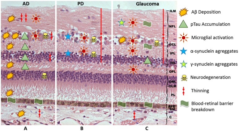
- Includes a summary table (Table 1) of retinal changes and a schematic figure (Figure 1) of hypothetical neuroinflammatory events; no systematic search or meta-analysis is described.
Limitations
- The paper states that the role of microglia in glaucoma pathophysiology is poorly understood and that better knowledge is needed.
- The paper notes contradictory evidence: Williams et al. found no evidence of Tau, Aβ, TDP-43, ubiquitin, or α-syn deposits in any part of the eyeball in AD patients.
- Evident from the text: this is a narrative review without stated systematic search, quality appraisal, or primary data, and it relies heavily on animal models and postmortem human tissue.
Model-written summary. Check it against the paper before relying on it.
Integrity signals
Registry
Cites
Statistics
Figures
Verification
Claims the paper makes
155 Not checked yet · 39 No basis given · 21 Own interpretation · 1 Sources not found
The paper states this without a source or data behind it here, so there is nothing to check it against. The quote shows it is in the paper.
In the paper
“In this pathology, deposition of Aβ, synuclein, and pTau has also been detected in retina.”
§ The Role of Microglia in Retinal Neurodegeneration: Alzheimer's Disease, Parkinson, and Glaucoma > Abstract
plaincorrelational- Asserted: passes — The authors put it forward as true.
- Checkable: passes — It can be checked against evidence.
- Load-bearing: unclear — It is unclear whether the paper’s conclusions depend on it.
- Based: unclear — It is unclear why the paper says it holds.
The paper states this without a source or data behind it here, so there is nothing to check it against. The quote shows it is in the paper.
In the paper
“Microglial activation has been reported in AD, PD, and glaucoma in relation to protein aggregates and degenerated neurons.”
§ The Role of Microglia in Retinal Neurodegeneration: Alzheimer's Disease, Parkinson, and Glaucoma > Abstract
plaincorrelational- Asserted: passes — The authors put it forward as true.
- Checkable: passes — It can be checked against evidence.
- Load-bearing: passes — The paper’s conclusions depend on it.
- Based: unclear — It is unclear why the paper says it holds.
None of the sources cited for this claim have been checked yet.
In the paper
“In addition, there is a dendritic atrophy of the lateral geniculate nucleus, the site to which the RGC axons project (Gupta et al., 2007; Park and Ou, 2013).”
§ The Role of Microglia in Retinal Neurodegeneration: Alzheimer's Disease, Parkinson, and Glaucoma > Introduction
What the sources say
- Not checked yetcites Gupta et al., 2007 source
- Not checked yetcites Park and Ou, 2013
plain- Asserted: passes — The authors put it forward as true.
- Checkable: passes — It can be checked against evidence.
- Load-bearing: unclear — It is unclear whether the paper’s conclusions depend on it.
- Based: passes — The paper gives a reason why it holds.
The paper states this without a source or data behind it here, so there is nothing to check it against. The quote shows it is in the paper.
In the paper
“A neuroinflammatory response is considered among the etiological factors of the major aged-related neurodegenerative diseases of the CNS, and microglial cells are key players in these neurodegenerative lesions.”
§ The Role of Microglia in Retinal Neurodegeneration: Alzheimer's Disease, Parkinson, and Glaucoma > Abstract
plaincorrelational- Asserted: passes — The authors put it forward as true.
- Checkable: passes — It can be checked against evidence.
- Load-bearing: passes — The paper’s conclusions depend on it.
- Based: passes — The paper gives a reason why it holds.
A conclusion the paper draws from its own results — it cites no other source for it, so there is none to check. The quote shows it is in the paper.
In the paper
“An uncontrolled microglial response secondary to sustained CNS damage can put neuronal survival at risk due to excessive inflammation.”
§ The Role of Microglia in Retinal Neurodegeneration: Alzheimer's Disease, Parkinson, and Glaucoma > Abstract
plaindirect- Asserted: passes — The authors put it forward as true.
- Checkable: passes — It can be checked against evidence.
- Load-bearing: passes — The paper’s conclusions depend on it.
- Based: passes — The paper gives a reason why it holds.
In the paper
“Glaucoma, a neurodegenerative disease of the optic nerve, is characterized by death of retinal ganglion cells (RGCs) (de Hoz et al., 2016).”
§ The Role of Microglia in Retinal Neurodegeneration: Alzheimer's Disease, Parkinson, and Glaucoma > Introduction
What the sources say
- Not checked yetcites de Hoz et al., 2016 source
plain- Asserted: passes — The authors put it forward as true.
- Checkable: passes — It can be checked against evidence.
- Load-bearing: unclear — It is unclear whether the paper’s conclusions depend on it.
- Based: passes — The paper gives a reason why it holds.
In the paper
“These deposits can induce neuronal death by apoptosis (Garcia-Ospina et al., 2003).”
§ The Role of Microglia in Retinal Neurodegeneration: Alzheimer's Disease, Parkinson, and Glaucoma > Introduction
What the sources say
- Not checked yetcites Garcia-Ospina et al., 2003
plaindirect- Asserted: passes — The authors put it forward as true.
- Checkable: passes — It can be checked against evidence.
- Load-bearing: unclear — It is unclear whether the paper’s conclusions depend on it.
- Based: passes — The paper gives a reason why it holds.
None of the sources cited for this claim have been checked yet.
In the paper
“These alterations can lead to secondary damage such as, excitotoxicity, neurotrophin deprivation, oxidative damage, mitochondrial dysfunction, and eventually RGC death (Nickells, 1999; Gallego et al., 2012).”
§ The Role of Microglia in Retinal Neurodegeneration: Alzheimer's Disease, Parkinson, and Glaucoma > Introduction
What the sources say
plaindirect- Asserted: passes — The authors put it forward as true.
- Checkable: passes — It can be checked against evidence.
- Load-bearing: unclear — It is unclear whether the paper’s conclusions depend on it.
- Based: passes — The paper gives a reason why it holds.
In the paper
“In neurodegenerative diseases, neuroinflammation constitutes a fundamental process in which microglial cells play a key role (Glass et al., 2010).”
§ The Role of Microglia in Retinal Neurodegeneration: Alzheimer's Disease, Parkinson, and Glaucoma > Microglial activation
What the sources say
- Not checked yetcites Glass et al., 2010 source
plaincorrelational- Asserted: passes — The authors put it forward as true.
- Checkable: passes — It can be checked against evidence.
- Load-bearing: passes — The paper’s conclusions depend on it.
- Based: passes — The paper gives a reason why it holds.
None of the sources cited for this claim have been checked yet.
In the paper
“Recently, it has been reported that microglia release extracellular microvesicles (Evs) by exocytosis.”
§ The Role of Microglia in Retinal Neurodegeneration: Alzheimer's Disease, Parkinson, and Glaucoma > Microglial activation
plaindirect- Asserted: passes — The authors put it forward as true.
- Checkable: passes — It can be checked against evidence.
- Load-bearing: unclear — It is unclear whether the paper’s conclusions depend on it.
- Based: passes — The paper gives a reason why it holds.
- Probably not a substantial claim. 48
- It may state two separate claims as one. 94
The paper states this without a source or data behind it here, so there is nothing to check it against. The quote shows it is in the paper.
In the paper
“The brain and retina are affected in several neurodegenerative diseases, including Alzheimer's disease (AD), Parkinson's disease (PD), and glaucoma.”
§ The Role of Microglia in Retinal Neurodegeneration: Alzheimer's Disease, Parkinson, and Glaucoma > Abstract
plaincorrelational- Asserted: passes — The authors put it forward as true.
- Checkable: passes — It can be checked against evidence.
- Load-bearing: passes — The paper’s conclusions depend on it.
- Based: passes — The paper gives a reason why it holds.
Probably not a substantial claim.
None of the sources cited for this claim have been checked yet.
In the paper
“they have similar mechanisms of cell injury and deposition of protein aggregates in specific anatomical areas (Wostyn et al., 2010; Kaarniranta et al., 2011; Ghiso et al., 2013)”
§ The Role of Microglia in Retinal Neurodegeneration: Alzheimer's Disease, Parkinson, and Glaucoma > Introduction
What the sources say
plaincorrelational- Asserted: passes — The authors put it forward as true.
- Checkable: passes — It can be checked against evidence.
- Load-bearing: passes — The paper’s conclusions depend on it.
- Based: passes — The paper gives a reason why it holds.
It may state two separate claims as one.
A conclusion the paper draws from its own results — it cites no other source for it, so there is none to check. The quote shows it is in the paper.
In the paper
“These neurodegenerative diseases share a common pathogenic mechanism, the neuroinflammation, in which microglia play an important role.”
§ The Role of Microglia in Retinal Neurodegeneration: Alzheimer's Disease, Parkinson, and Glaucoma > Abstract
plaincorrelational- Asserted: passes — The authors put it forward as true.
- Checkable: passes — It can be checked against evidence.
- Load-bearing: passes — The paper’s conclusions depend on it.
- Based: passes — The paper gives a reason why it holds.
It may state two separate claims as one.
In the paper
“In addition, AD is frequently associated with vascular dysfunctions and inflammation (Dudvarski Stankovic et al., 2016).”
§ The Role of Microglia in Retinal Neurodegeneration: Alzheimer's Disease, Parkinson, and Glaucoma > Introduction
What the sources say
- Not checked yetcites Dudvarski Stankovic et al., 2016 source
plaincorrelational- Asserted: passes — The authors put it forward as true.
- Checkable: passes — It can be checked against evidence.
- Load-bearing: fails — The paper’s conclusions may not depend on it.
- Based: passes — The paper gives a reason why it holds.
Probably not a substantial claim. The paper’s conclusions may not depend on it. It may state two separate claims as one.
The paper states this without a source or data behind it here, so there is nothing to check it against. The quote shows it is in the paper.
In the paper
“The loss of neurons is accompanied by abnormal intracytoplasmic filamentous aggregates called Lewy bodies.”
§ The Role of Microglia in Retinal Neurodegeneration: Alzheimer's Disease, Parkinson, and Glaucoma > Introduction
plaincorrelational- Asserted: passes — The authors put it forward as true.
- Checkable: passes — It can be checked against evidence.
- Load-bearing: fails — The paper’s conclusions may not depend on it.
- Based: passes — The paper gives a reason why it holds.
Probably not a substantial claim. The paper’s conclusions may not depend on it.
In the paper
“These aggregates (deposited in somas and axons) are constituted by α-syn, parkin, phosphorylated neurofilament and components of the protheosomic-ubiquitin pathway (Orr et al., 2002).”
§ The Role of Microglia in Retinal Neurodegeneration: Alzheimer's Disease, Parkinson, and Glaucoma > Introduction
What the sources say
- Not checked yetcites Orr et al., 2002 source
plain- Asserted: passes — The authors put it forward as true.
- Checkable: passes — It can be checked against evidence.
- Load-bearing: fails — The paper’s conclusions may not depend on it.
- Based: passes — The paper gives a reason why it holds.
Probably not a substantial claim. The paper’s conclusions may not depend on it.
None of the sources cited for this claim have been checked yet.
In the paper
“they are slow and chronic neurodegenerative disorders with a strong age-related incidence”
§ The Role of Microglia in Retinal Neurodegeneration: Alzheimer's Disease, Parkinson, and Glaucoma > Introduction
plaincorrelational- Asserted: passes — The authors put it forward as true.
- Checkable: passes — It can be checked against evidence.
- Load-bearing: unclear — It is unclear whether the paper’s conclusions depend on it.
- Based: passes — The paper gives a reason why it holds.
It may state two separate claims as one.
The paper states this without a source or data behind it here, so there is nothing to check it against. The quote shows it is in the paper.
In the paper
“Microglia, the immunocompetent cells of the central nervous system (CNS), act as neuropathology sensors and are neuroprotective under physiological conditions.”
§ The Role of Microglia in Retinal Neurodegeneration: Alzheimer's Disease, Parkinson, and Glaucoma > Abstract
plaindirect- Asserted: passes — The authors put it forward as true.
- Checkable: passes — It can be checked against evidence.
- Load-bearing: passes — The paper’s conclusions depend on it.
- Based: fails — The paper may give no reason why it holds.
Probably not a substantial claim. The paper may give no reason why it holds. It may state two separate claims as one.
In the paper
“The major pathology related to AD is the extracellular deposit of β-amyloid (Aβ) in the form of parenchymal plaques and cerebral amyloid angiopathy co-existing with intraneuronal accumulations of hyperphosphorylated tau (pTau) (neurofribillary tangles) (Ghiso et al., 2013).”
§ The Role of Microglia in Retinal Neurodegeneration: Alzheimer's Disease, Parkinson, and Glaucoma > Introduction
What the sources say
- Not checked yetcites Ghiso et al., 2013 source
plain- Asserted: passes — The authors put it forward as true.
- Checkable: passes — It can be checked against evidence.
- Load-bearing: fails — The paper’s conclusions may not depend on it.
- Based: passes — The paper gives a reason why it holds.
Probably not a substantial claim. The paper’s conclusions may not depend on it. It may state two separate claims as one.
None of the sources cited for this claim have been checked yet.
In the paper
“AD has been related to genetic mutations, among them in the gene encoding the Aβ precursor protein peptide, mutations in the presenilins genes (Calabrese et al., 2001) and the presence of the APOE ε4 allele (Martínez-Lazcano et al., 2010).”
§ The Role of Microglia in Retinal Neurodegeneration: Alzheimer's Disease, Parkinson, and Glaucoma > Introduction
What the sources say
- Not checked yetcites Calabrese et al., 2001 source
- Not checked yetcites Martínez-Lazcano et al., 2010
plaincorrelational- Asserted: passes — The authors put it forward as true.
- Checkable: passes — It can be checked against evidence.
- Load-bearing: fails — The paper’s conclusions may not depend on it.
- Based: passes — The paper gives a reason why it holds.
Probably not a substantial claim. The paper’s conclusions may not depend on it. It may state two separate claims as one.
None of the sources cited for this claim have been checked yet.
In the paper
“Genetic factors include mutations in α-syn (Olanow and Tatton, 1999; Pérez and Arancibia, 2007), while mitochondrial dysfunction and oxidative stress may also act by causing the accumulation of misfolded proteins (Dauer and Przedborski, 2003).”
§ The Role of Microglia in Retinal Neurodegeneration: Alzheimer's Disease, Parkinson, and Glaucoma > Introduction
What the sources say
- Not checked yetcites Olanow and Tatton, 1999
- Not checked yetcites Pérez and Arancibia, 2007
- Not checked yetcites Dauer and Przedborski, 2003 source
hedgedconditional- Asserted: passes — The authors put it forward as true.
- Checkable: passes — It can be checked against evidence.
- Load-bearing: fails — The paper’s conclusions may not depend on it.
- Based: passes — The paper gives a reason why it holds.
Probably not a substantial claim. The paper’s conclusions may not depend on it. It may state two separate claims as one.
In the paper
“Neurodegeneration could be due to exposure to dopaminergic neurotoxins e.g., herbicides (MPTP), insecticides (Rotenone), and metals (Hernández-Montiel, 2006).”
§ The Role of Microglia in Retinal Neurodegeneration: Alzheimer's Disease, Parkinson, and Glaucoma > Introduction
What the sources say
- Not checked yetcites Hernández-Montiel, 2006
hedgedconditional- Asserted: passes — The authors put it forward as true.
- Checkable: passes — It can be checked against evidence.
- Load-bearing: fails — The paper’s conclusions may not depend on it.
- Based: passes — The paper gives a reason why it holds.
Probably not a substantial claim. The paper’s conclusions may not depend on it.
In the paper
“The clinical symptoms of PD are mainly motor problems, including bradykinesia, rigidity, tremors, and postural instability. In addition, PD presents non-motor symptoms including disorganized speech and altered moods (Fakhoury, 2016).”
§ The Role of Microglia in Retinal Neurodegeneration: Alzheimer's Disease, Parkinson, and Glaucoma > Introduction
What the sources say
- Not checked yetcites Fakhoury, 2016 source
plain- Asserted: passes — The authors put it forward as true.
- Checkable: passes — It can be checked against evidence.
- Load-bearing: fails — The paper’s conclusions may not depend on it.
- Based: passes — The paper gives a reason why it holds.
Probably not a substantial claim. The paper’s conclusions may not depend on it. It may state two separate claims as one.
The paper states this without a source or data behind it here, so there is nothing to check it against. The quote shows it is in the paper.
In the paper
“AD is an age-related neurodegeneration of the CNS characterized by neuronal and synaptic loss in the cerebral cortex, resulting in cognitive deficit and dementia.”
§ The Role of Microglia in Retinal Neurodegeneration: Alzheimer's Disease, Parkinson, and Glaucoma > Abstract
plaindirect- Asserted: passes — The authors put it forward as true.
- Checkable: passes — It can be checked against evidence.
- Load-bearing: fails — The paper’s conclusions may not depend on it.
- Based: fails — The paper may give no reason why it holds.
Probably not a substantial claim. The paper’s conclusions may not depend on it. The paper may give no reason why it holds.
None of the sources cited for this claim have been checked yet.
In the paper
“Recently, neurodegenerative lesions have been detected in the intracranial optic nerve, the lateral geniculate nucleus, and the visual cortex (Gupta et al., 2006, 2007), suggesting that this pathology could be grouped as a neurodegenerative disease (Yucel et al., 2003).”
§ The Role of Microglia in Retinal Neurodegeneration: Alzheimer's Disease, Parkinson, and Glaucoma > Introduction
What the sources say
- Not matchedcites Gupta et al., 2006, 2007
Couldn’t be matched to a paper in Crossref, so it can’t be checked.
- Not checked yetcites Yucel et al., 2003 source
hedgedcorrelational- Asserted: passes — The authors put it forward as true.
- Checkable: passes — It can be checked against evidence.
- Load-bearing: passes — The paper’s conclusions depend on it.
- Based: passes — The paper gives a reason why it holds.
It may state two separate claims as one.
- Not matched
The paper states this without a source or data behind it here, so there is nothing to check it against. The quote shows it is in the paper.
In the paper
“The main etiological factors proposed for PD are aging, environmental toxins, and genetic factors.”
§ The Role of Microglia in Retinal Neurodegeneration: Alzheimer's Disease, Parkinson, and Glaucoma > Introduction
plaindirect- Asserted: passes — The authors put it forward as true.
- Checkable: passes — It can be checked against evidence.
- Load-bearing: fails — The paper’s conclusions may not depend on it.
- Based: passes — The paper gives a reason why it holds.
Probably not a substantial claim. The paper’s conclusions may not depend on it.
In the paper
“AD involves progressive memory loss and dementia (Sharma and Lipincott, 2017)”
§ The Role of Microglia in Retinal Neurodegeneration: Alzheimer's Disease, Parkinson, and Glaucoma > Introduction
What the sources say
- Not checked yetcites Sharma and Lipincott, 2017
plain- Asserted: passes — The authors put it forward as true.
- Checkable: passes — It can be checked against evidence.
- Load-bearing: fails — The paper’s conclusions may not depend on it.
- Based: passes — The paper gives a reason why it holds.
Probably not a substantial claim. The paper’s conclusions may not depend on it. It may state two separate claims as one.
The paper states this without a source or data behind it here, so there is nothing to check it against. The quote shows it is in the paper.
In the paper
“PD is a neurodegenerative locomotor disorder with the progressive loss of dopaminergic neurons in the substantia nigra.”
§ The Role of Microglia in Retinal Neurodegeneration: Alzheimer's Disease, Parkinson, and Glaucoma > Abstract
plain- Asserted: passes — The authors put it forward as true.
- Checkable: passes — It can be checked against evidence.
- Load-bearing: fails — The paper’s conclusions may not depend on it.
- Based: fails — The paper may give no reason why it holds.
Probably not a substantial claim. The paper’s conclusions may not depend on it. The paper may give no reason why it holds. It may state two separate claims as one.
The paper states this without a source or data behind it here, so there is nothing to check it against. The quote shows it is in the paper.
In the paper
“Glaucoma is a multifactorial neurodegenerative disease of the optic nerve, characterized by retinal ganglion cell loss.”
§ The Role of Microglia in Retinal Neurodegeneration: Alzheimer's Disease, Parkinson, and Glaucoma > Abstract
plain- Asserted: passes — The authors put it forward as true.
- Checkable: passes — It can be checked against evidence.
- Load-bearing: fails — The paper’s conclusions may not depend on it.
- Based: fails — The paper may give no reason why it holds.
Probably not a substantial claim. The paper’s conclusions may not depend on it. The paper may give no reason why it holds. It may state two separate claims as one.
A conclusion the paper draws from its own results — it cites no other source for it, so there is none to check. The quote shows it is in the paper.
In the paper
“The activated microglia can release pro-inflammatory cytokines which can aggravate and propagate neuroinflammation, thereby degenerating neurons and impairing brain as well as retinal function.”
§ The Role of Microglia in Retinal Neurodegeneration: Alzheimer's Disease, Parkinson, and Glaucoma > Abstract
plaindirect- Asserted: passes — The authors put it forward as true.
- Checkable: passes — It can be checked against evidence.
- Load-bearing: passes — The paper’s conclusions depend on it.
- Based: unclear — It is unclear why the paper says it holds.
It may state two separate claims as one.
In the paper
“while the PD is a chronic and progressive movement disorder (Orr et al., 2002).”
§ The Role of Microglia in Retinal Neurodegeneration: Alzheimer's Disease, Parkinson, and Glaucoma > Introduction
What the sources say
- Not checked yetcites Orr et al., 2002 source
plain- Asserted: passes — The authors put it forward as true.
- Checkable: passes — It can be checked against evidence.
- Load-bearing: fails — The paper’s conclusions may not depend on it.
- Based: passes — The paper gives a reason why it holds.
Probably not a substantial claim. The paper’s conclusions may not depend on it.
The paper states this without a source or data behind it here, so there is nothing to check it against. The quote shows it is in the paper.
In the paper
“The extracellular deposits of beta-amyloid (Aβ) and intraneuronal accumulations of hyperphosphorylated tau protein (pTau) are the hallmarks of this disease.”
§ The Role of Microglia in Retinal Neurodegeneration: Alzheimer's Disease, Parkinson, and Glaucoma > Abstract
plain- Asserted: passes — The authors put it forward as true.
- Checkable: passes — It can be checked against evidence.
- Load-bearing: fails — The paper’s conclusions may not depend on it.
- Based: fails — The paper may give no reason why it holds.
Probably not a substantial claim. The paper’s conclusions may not depend on it. The paper may give no reason why it holds.
In the paper
“Alzheimer's Disease (AD) and Parkinson's Disease (PD) are the most common neurodegenerative disorders (de Lau and Breteler, 2006).”
§ The Role of Microglia in Retinal Neurodegeneration: Alzheimer's Disease, Parkinson, and Glaucoma > Introduction
What the sources say
- Not checked yetcites de Lau and Breteler, 2006 source
plain- Asserted: passes — The authors put it forward as true.
- Checkable: passes — It can be checked against evidence.
- Load-bearing: fails — The paper’s conclusions may not depend on it.
- Based: passes — The paper gives a reason why it holds.
Probably not a substantial claim. The paper’s conclusions may not depend on it.
In the paper
“Glaucoma is an age-related multifactorial neurodegenerative disease of the optic nerve, with an irreversible decrease in RGCs, causing a visual-field loss and cupping of the optic nerve head (Quigley et al., 1988).”
§ The Role of Microglia in Retinal Neurodegeneration: Alzheimer's Disease, Parkinson, and Glaucoma > Introduction
What the sources say
- Not checked yetcites Quigley et al., 1988 source
plaindirect- Asserted: passes — The authors put it forward as true.
- Checkable: passes — It can be checked against evidence.
- Load-bearing: fails — The paper’s conclusions may not depend on it.
- Based: passes — The paper gives a reason why it holds.
Probably not a substantial claim. The paper’s conclusions may not depend on it. It may state two separate claims as one.
The paper states this without a source or data behind it here, so there is nothing to check it against. The quote shows it is in the paper.
In the paper
“A neuroinflammatory response is considered among the etiological factors of the major aged-related neurodegenerative diseases of the CNS, and microglial cells are key players in these neurodegenerative lesions.”
§ The Role of Microglia in Retinal Neurodegeneration: Alzheimer's Disease, Parkinson, and Glaucoma > Abstract
plaincorrelational- Asserted: passes — The authors put it forward as true.
- Checkable: passes — It can be checked against evidence.
- Load-bearing: passes — The paper’s conclusions depend on it.
- Based: fails — The paper may give no reason why it holds.
Probably not a substantial claim. The paper may give no reason why it holds.
The paper states this without a source or data behind it here, so there is nothing to check it against. The quote shows it is in the paper.
In the paper
“This is accompanied by Lewy body inclusion composed of α-synuclein (α-syn) aggregates.”
§ The Role of Microglia in Retinal Neurodegeneration: Alzheimer's Disease, Parkinson, and Glaucoma > Abstract
plaincorrelational- Asserted: passes — The authors put it forward as true.
- Checkable: passes — It can be checked against evidence.
- Load-bearing: fails — The paper’s conclusions may not depend on it.
- Based: passes — The paper gives a reason why it holds.
Probably not a substantial claim. The paper’s conclusions may not depend on it.
In the paper
“However, it is now known to have a multifactorial origin and it seems to result from a complex interaction of multiple environmental and genetic factors (Wostyn et al., 2010).”
§ The Role of Microglia in Retinal Neurodegeneration: Alzheimer's Disease, Parkinson, and Glaucoma > Introduction
What the sources say
- Not checked yetcites Wostyn et al., 2010 source
hedgedconditional- Asserted: passes — The authors put it forward as true.
- Checkable: passes — It can be checked against evidence.
- Load-bearing: fails — The paper’s conclusions may not depend on it.
- Based: passes — The paper gives a reason why it holds.
Probably not a substantial claim. The paper’s conclusions may not depend on it. It may state two separate claims as one.
The paper states this without a source or data behind it here, so there is nothing to check it against. The quote shows it is in the paper.
In the paper
“These deposits are also found in the retina and optic nerve.”
§ The Role of Microglia in Retinal Neurodegeneration: Alzheimer's Disease, Parkinson, and Glaucoma > Abstract
plain- Asserted: passes — The authors put it forward as true.
- Checkable: passes — It can be checked against evidence.
- Load-bearing: passes — The paper’s conclusions depend on it.
- Based: fails — The paper may give no reason why it holds.
Probably not a substantial claim. The paper may give no reason why it holds.
The paper states this without a source or data behind it here, so there is nothing to check it against. The quote shows it is in the paper.
In the paper
“In glaucoma, increased intraocular pressure (IOP), vascular dysregulation, and the immune system activation can trigger several changes in retina and optic nerve including: disrupted axonal transport and neurofilament accumulation, microvascular abnormalities, extracellular matrix remodeling, and glial cell activation.”
§ The Role of Microglia in Retinal Neurodegeneration: Alzheimer's Disease, Parkinson, and Glaucoma > Introduction
plaindirect- Asserted: passes — The authors put it forward as true.
- Checkable: passes — It can be checked against evidence.
- Load-bearing: unclear — It is unclear whether the paper’s conclusions depend on it.
- Based: passes — The paper gives a reason why it holds.
It may state two separate claims as one.
The paper states this without a source or data behind it here, so there is nothing to check it against. The quote shows it is in the paper.
In the paper
“Microglia react to injury and degeneration with immune-phenotypic and morphological changes, proliferation, migration, and inflammatory cytokine production.”
§ The Role of Microglia in Retinal Neurodegeneration: Alzheimer's Disease, Parkinson, and Glaucoma > Abstract
plaindirect- Asserted: passes — The authors put it forward as true.
- Checkable: passes — It can be checked against evidence.
- Load-bearing: passes — The paper’s conclusions depend on it.
- Based: fails — The paper may give no reason why it holds.
Probably not a substantial claim. The paper may give no reason why it holds. It may state two separate claims as one.
In the paper
“PD is characterized by the progressive loss of dopaminergic neurons in the substantia nigra pars compacta and the nerve terminals in the striatum (Dauer and Przedborski, 2003).”
§ The Role of Microglia in Retinal Neurodegeneration: Alzheimer's Disease, Parkinson, and Glaucoma > Introduction
What the sources say
- Not checked yetcites Dauer and Przedborski, 2003 source
plain- Asserted: passes — The authors put it forward as true.
- Checkable: passes — It can be checked against evidence.
- Load-bearing: fails — The paper’s conclusions may not depend on it.
- Based: passes — The paper gives a reason why it holds.
Probably not a substantial claim. The paper’s conclusions may not depend on it.
The paper states this without a source or data behind it here, so there is nothing to check it against. The quote shows it is in the paper.
In the paper
“PD also involves retinal dopaminergic cell degeneration.”
§ The Role of Microglia in Retinal Neurodegeneration: Alzheimer's Disease, Parkinson, and Glaucoma > Abstract
plaincorrelational- Asserted: passes — The authors put it forward as true.
- Checkable: passes — It can be checked against evidence.
- Load-bearing: passes — The paper’s conclusions depend on it.
- Based: fails — The paper may give no reason why it holds.
Probably not a substantial claim. The paper may give no reason why it holds.
A conclusion the paper draws from its own results — it cites no other source for it, so there is none to check. The quote shows it is in the paper.
In the paper
“Excessive microglia activation might prompt the release of cytotoxic factors, causing neuronal damage, which could accelerate the progression of some CNS diseases.”
§ The Role of Microglia in Retinal Neurodegeneration: Alzheimer's Disease, Parkinson, and Glaucoma > Microglial activation
hedgedconditional- Asserted: passes — The authors put it forward as true.
- Checkable: passes — It can be checked against evidence.
- Load-bearing: unclear — It is unclear whether the paper’s conclusions depend on it.
- Based: passes — The paper gives a reason why it holds.
It may state two separate claims as one.
In the paper
“The second microglial phenotype, M2-like, secrete anti-inflammatory mediators and neurotrophic factors, thus inducing a supportive microenvironment for neurons (Kettenmann et al., 2011).”
§ The Role of Microglia in Retinal Neurodegeneration: Alzheimer's Disease, Parkinson, and Glaucoma > Microglial activation
What the sources say
- Not checked yetcites Kettenmann et al., 2011 source
plaindirect- Asserted: passes — The authors put it forward as true.
- Checkable: passes — It can be checked against evidence.
- Load-bearing: unclear — It is unclear whether the paper’s conclusions depend on it.
- Based: passes — The paper gives a reason why it holds.
It may state two separate claims as one.
None of the sources cited for this claim have been checked yet.
In the paper
“Changes in OCT measurements have been used to study the course of particular neurologic diseases such AD (Garcia-Martin et al., 2014; Maldonado et al., 2015; Salobrar-Garcia et al., 2015, 2016a,b), PD (Yu et al., 2014; Stemplewitz et al., 2015; Boeke et al., 2016; Satue et al., 2017), and glaucoma (Leung, 2016; Fallon et al., 2017), suggesting that the data compiled may be useful as a biomarker in”
§ The Role of Microglia in Retinal Neurodegeneration: Alzheimer's Disease, Parkinson, and Glaucoma > Introduction
What the sources say
hedgedcorrelational- Asserted: passes — The authors put it forward as true.
- Checkable: passes — It can be checked against evidence.
- Load-bearing: passes — The paper’s conclusions depend on it.
- Based: passes — The paper gives a reason why it holds.
It may state two separate claims as one.
In the paper
“The fractalkine expressed by neurons can induce adenosine release from microglia. This adenosine (via adenosine A1 receptor) can activate survival pathways in neurons sensitive to excitotoxicity challenge (Lauro et al., 2008, 2010).”
§ The Role of Microglia in Retinal Neurodegeneration: Alzheimer's Disease, Parkinson, and Glaucoma > Microglial activation
What the sources say
- Not matchedcites Lauro et al., 2008, 2010
Couldn’t be matched to a paper in Crossref, so it can’t be checked.
plaindirect- Asserted: passes — The authors put it forward as true.
- Checkable: passes — It can be checked against evidence.
- Load-bearing: unclear — It is unclear whether the paper’s conclusions depend on it.
- Based: passes — The paper gives a reason why it holds.
It may state two separate claims as one.
- Not matched
The paper states this without a source or data behind it here, so there is nothing to check it against. The quote shows it is in the paper.
In the paper
“Microglia constitute the first line of immune defense in CNS.”
§ The Role of Microglia in Retinal Neurodegeneration: Alzheimer's Disease, Parkinson, and Glaucoma > Microglial activation
plain- Asserted: passes — The authors put it forward as true.
- Checkable: passes — It can be checked against evidence.
- Load-bearing: fails — The paper’s conclusions may not depend on it.
- Based: passes — The paper gives a reason why it holds.
Probably not a substantial claim. The paper’s conclusions may not depend on it.
In the paper
“Thus, it has been described that the retina is a “window to the brain,” and the manifestation of disease in the brain is the same as in the retina (MacCormick et al., 2015).”
§ The Role of Microglia in Retinal Neurodegeneration: Alzheimer's Disease, Parkinson, and Glaucoma > Introduction
What the sources say
- Not checked yetcites MacCormick et al., 2015 source
plaincorrelational- Asserted: passes — The authors put it forward as true.
- Checkable: passes — It can be checked against evidence.
- Load-bearing: passes — The paper’s conclusions depend on it.
- Based: passes — The paper gives a reason why it holds.
It may state two separate claims as one.
None of the sources cited for this claim have been checked yet.
In the paper
“These microvesicles are involved in all immune activities and can be protective or detrimental, affecting some pathologies of the CNS.”
§ The Role of Microglia in Retinal Neurodegeneration: Alzheimer's Disease, Parkinson, and Glaucoma > Microglial activation
plaindirect- Asserted: passes — The authors put it forward as true.
- Checkable: passes — It can be checked against evidence.
- Load-bearing: fails — The paper’s conclusions may not depend on it.
- Based: passes — The paper gives a reason why it holds.
Probably not a substantial claim. The paper’s conclusions may not depend on it. It may state two separate claims as one.
None of the sources cited for this claim have been checked yet.
In the paper
“However, in certain circumstances, the neuroinflammation can help stimulate myelin repair or remove toxic aggregated proteins and cell debris from CNS (Ding et al., 2004; Simard et al., 2006; Glezer et al., 2007).”
§ The Role of Microglia in Retinal Neurodegeneration: Alzheimer's Disease, Parkinson, and Glaucoma > Microglial activation
What the sources say
plainconditional- Asserted: passes — The authors put it forward as true.
- Checkable: passes — It can be checked against evidence.
- Load-bearing: unclear — It is unclear whether the paper’s conclusions depend on it.
- Based: passes — The paper gives a reason why it holds.
It may state two separate claims as one.
A conclusion the paper draws from its own results — it cites no other source for it, so there is none to check. The quote shows it is in the paper.
In the paper
“Although the exact mechanism bringing about this neuronal death remains unknown, these neurodegenerative disorders seem to have pathogenic mechanisms in common.”
§ The Role of Microglia in Retinal Neurodegeneration: Alzheimer's Disease, Parkinson, and Glaucoma > Introduction
hedgedcorrelational- Asserted: passes — The authors put it forward as true.
- Checkable: passes — It can be checked against evidence.
- Load-bearing: passes — The paper’s conclusions depend on it.
- Based: passes — The paper gives a reason why it holds.
It may state two separate claims as one.
In the paper
“These cells survey the CNS in order to detect homeostasis alteration and they respond accordingly, combining a defensive service with neuroprotective functions (Verkhratsky and Butt, 2013).”
§ The Role of Microglia in Retinal Neurodegeneration: Alzheimer's Disease, Parkinson, and Glaucoma > Microglial activation
What the sources say
- Not checked yetcites Verkhratsky and Butt, 2013
plaindirect- Asserted: passes — The authors put it forward as true.
- Checkable: passes — It can be checked against evidence.
- Load-bearing: fails — The paper’s conclusions may not depend on it.
- Based: passes — The paper gives a reason why it holds.
Probably not a substantial claim. The paper’s conclusions may not depend on it. It may state two separate claims as one.
In the paper
“The change of microglia between M1 and M2 phenotypes is a dynamic process and microglial activation can switch from M2 to M1 phenotype during the course of disease (Cherry et al., 2014).”
§ The Role of Microglia in Retinal Neurodegeneration: Alzheimer's Disease, Parkinson, and Glaucoma > Microglial activation
What the sources say
- Not checked yetcites Cherry et al., 2014 source
plaindirect- Asserted: passes — The authors put it forward as true.
- Checkable: passes — It can be checked against evidence.
- Load-bearing: unclear — It is unclear whether the paper’s conclusions depend on it.
- Based: passes — The paper gives a reason why it holds.
It may state two separate claims as one.
In the paper
“The morphology of activated microglia includes a retraction of processes, enlargement of the soma, and increased expression of myeloid cell markers (Ransohoff and Cardona, 2010).”
§ The Role of Microglia in Retinal Neurodegeneration: Alzheimer's Disease, Parkinson, and Glaucoma > Microglial activation
What the sources say
- Not checked yetcites Ransohoff and Cardona, 2010 source
plaincorrelational- Asserted: passes — The authors put it forward as true.
- Checkable: passes — It can be checked against evidence.
- Load-bearing: unclear — It is unclear whether the paper’s conclusions depend on it.
- Based: passes — The paper gives a reason why it holds.
It may state two separate claims as one.
None of the sources cited for this claim have been checked yet.
In the paper
“After this acute M1 activation, microglial cells can suffer an uncontrolled activation leading to a state of chronic inflammation. In this state, microglia release neurotoxic inflammatory factors (TNF-α, IL-1α, IL-1β, IL-6, NO, hydrogen peroxide, superoxide anion, chemokines, and glutamate), which lead to neuronal death (Block et al., 2007; Lull and Block, 2010; Burguillos et al., 2011; Kettenmann”
§ The Role of Microglia in Retinal Neurodegeneration: Alzheimer's Disease, Parkinson, and Glaucoma > Microglial activation
What the sources say
plaindirect- Asserted: passes — The authors put it forward as true.
- Checkable: passes — It can be checked against evidence.
- Load-bearing: passes — The paper’s conclusions depend on it.
- Based: passes — The paper gives a reason why it holds.
It may state two separate claims as one.
None of the sources cited for this claim have been checked yet.
In the paper
“M1 microglial cells present amoeboid morphology as well as high phagocytic capacity and motility (Varnum and Ikezu, 2012; Gonzalez et al., 2014; Jones and Bouvier, 2014).”
§ The Role of Microglia in Retinal Neurodegeneration: Alzheimer's Disease, Parkinson, and Glaucoma > Microglial activation
What the sources say
- Not checked yetcites Varnum and Ikezu, 2012
- Not checked yetcites Gonzalez et al., 2014 source
- Not checked yetcites Jones and Bouvier, 2014
plaincorrelational- Asserted: passes — The authors put it forward as true.
- Checkable: passes — It can be checked against evidence.
- Load-bearing: unclear — It is unclear whether the paper’s conclusions depend on it.
- Based: passes — The paper gives a reason why it holds.
It may state two separate claims as one.
The paper states this without a source or data behind it here, so there is nothing to check it against. The quote shows it is in the paper.
In the paper
“The neuroinflammatory process occurs not only in the brain, but also in the retina, which is a projection of the CNS.”
§ The Role of Microglia in Retinal Neurodegeneration: Alzheimer's Disease, Parkinson, and Glaucoma > Introduction
plain- Asserted: passes — The authors put it forward as true.
- Checkable: passes — It can be checked against evidence.
- Load-bearing: passes — The paper’s conclusions depend on it.
- Based: passes — The paper gives a reason why it holds.
It may state two separate claims as one.
The paper states this without a source or data behind it here, so there is nothing to check it against. The quote shows it is in the paper.
In the paper
“EVs have a heterogeneous molecular composition, including receptors, integrins and cytokines, bioactive lipids, miRNA, mRNA, DNA, and organelles, being similar to their parental cells.”
§ The Role of Microglia in Retinal Neurodegeneration: Alzheimer's Disease, Parkinson, and Glaucoma > Microglial activation
plain- Asserted: passes — The authors put it forward as true.
- Checkable: passes — It can be checked against evidence.
- Load-bearing: fails — The paper’s conclusions may not depend on it.
- Based: passes — The paper gives a reason why it holds.
Probably not a substantial claim. The paper’s conclusions may not depend on it. It may state two separate claims as one.
In the paper
“M2 microglia are the major effector cells with the potential to dampen pro-inflammatory immune responses and promote the expression of repair genes (Tang and Le, 2016).”
§ The Role of Microglia in Retinal Neurodegeneration: Alzheimer's Disease, Parkinson, and Glaucoma > Microglial activation
What the sources say
- Not checked yetcites Tang and Le, 2016 source
plainconditional- Asserted: passes — The authors put it forward as true.
- Checkable: passes — It can be checked against evidence.
- Load-bearing: unclear — It is unclear whether the paper’s conclusions depend on it.
- Based: passes — The paper gives a reason why it holds.
It may state two separate claims as one.
In the paper
“Microglial cells are CNS resident immune cells which have sensor and effector functions as well as phagocytic capacity (Streit et al., 2005).”
§ The Role of Microglia in Retinal Neurodegeneration: Alzheimer's Disease, Parkinson, and Glaucoma > Microglial activation
What the sources say
- Not checked yetcites Streit et al., 2005 source
plain- Asserted: passes — The authors put it forward as true.
- Checkable: passes — It can be checked against evidence.
- Load-bearing: fails — The paper’s conclusions may not depend on it.
- Based: passes — The paper gives a reason why it holds.
Probably not a substantial claim. The paper’s conclusions may not depend on it. It may state two separate claims as one.
None of the sources cited for this claim have been checked yet.
In the paper
“The retina and brain are associated over a range of neurological and neurovascular conditions of varying etiologies, because the retina and brain are similar, and respond similarly to disease.”
§ The Role of Microglia in Retinal Neurodegeneration: Alzheimer's Disease, Parkinson, and Glaucoma > Introduction
plaincorrelational- Asserted: passes — The authors put it forward as true.
- Checkable: passes — It can be checked against evidence.
- Load-bearing: passes — The paper’s conclusions depend on it.
- Based: passes — The paper gives a reason why it holds.
It may state two separate claims as one.
The paper states this without a source or data behind it here, so there is nothing to check it against. The quote shows it is in the paper.
In the paper
“Microglial cells may undergo two different kinds of activation in response to infections or injuries. The first is a neurotoxic phenotype called M1-like.”
§ The Role of Microglia in Retinal Neurodegeneration: Alzheimer's Disease, Parkinson, and Glaucoma > Microglial activation
hedgedconditional- Asserted: passes — The authors put it forward as true.
- Checkable: passes — It can be checked against evidence.
- Load-bearing: fails — The paper’s conclusions may not depend on it.
- Based: passes — The paper gives a reason why it holds.
Probably not a substantial claim. The paper’s conclusions may not depend on it.
In the paper
“After injury these cells become activated and in this state change their morphology, proliferate, migrate to the damage sites, modify the expression of enzymes and receptors, and release a variety of inflammatory factors, such as NO, tumor necrosis factor (TNF-α), interleukin (IL-6) among others (Magni et al., 2012).”
§ The Role of Microglia in Retinal Neurodegeneration: Alzheimer's Disease, Parkinson, and Glaucoma > Microglial activation
What the sources say
- Not checked yetcites Magni et al., 2012 source
plaindirect- Asserted: passes — The authors put it forward as true.
- Checkable: passes — It can be checked against evidence.
- Load-bearing: unclear — It is unclear whether the paper’s conclusions depend on it.
- Based: passes — The paper gives a reason why it holds.
It may state two separate claims as one.
None of the sources cited for this claim have been checked yet.
In the paper
“These cells can release anti-inflammatory cytokines including IL-4, IL-13, IL-10, TGF-β and neurotrophic factors, such as insulin-like growth factor 1 (IGF-1) to assist inflammation resolution and promote neuron survival (Suh et al., 2013; Tang and Le, 2016).”
§ The Role of Microglia in Retinal Neurodegeneration: Alzheimer's Disease, Parkinson, and Glaucoma > Microglial activation
What the sources say
plaindirect- Asserted: passes — The authors put it forward as true.
- Checkable: passes — It can be checked against evidence.
- Load-bearing: unclear — It is unclear whether the paper’s conclusions depend on it.
- Based: passes — The paper gives a reason why it holds.
It may state two separate claims as one.
The paper states this without a source or data behind it here, so there is nothing to check it against. The quote shows it is in the paper.
In the paper
“Neuroinflammation, the inflammation of central nervous system (CNS), is essential to protect the tissue. However, uncontrolled and prolonged neuroinflammation is potentially harmful and can cause cellular damage.”
§ The Role of Microglia in Retinal Neurodegeneration: Alzheimer's Disease, Parkinson, and Glaucoma > Introduction
plainconditional- Asserted: passes — The authors put it forward as true.
- Checkable: passes — It can be checked against evidence.
- Load-bearing: passes — The paper’s conclusions depend on it.
- Based: passes — The paper gives a reason why it holds.
It may state two separate claims as one.
In the paper
“Fractalkine, involved in indirect neuroprotection, is released by neurons and the receptor is expressed by microglia, their interactions constituting a neuron-microglial signaling system (Ransohoff and El Khoury, 2015).”
§ The Role of Microglia in Retinal Neurodegeneration: Alzheimer's Disease, Parkinson, and Glaucoma > Microglial activation
What the sources say
- Not checked yetcites Ransohoff and El Khoury, 2015 source
plaincorrelational- Asserted: passes — The authors put it forward as true.
- Checkable: passes — It can be checked against evidence.
- Load-bearing: unclear — It is unclear whether the paper’s conclusions depend on it.
- Based: passes — The paper gives a reason why it holds.
It may state two separate claims as one.
In the paper
“Microglia express CD11b/c, D45low, and the chemokine fractalkine receptor (CX3CR1) (Dudvarski Stankovic et al., 2016).”
§ The Role of Microglia in Retinal Neurodegeneration: Alzheimer's Disease, Parkinson, and Glaucoma > Microglial activation
What the sources say
- Not checked yetcites Dudvarski Stankovic et al., 2016 source
plain- Asserted: passes — The authors put it forward as true.
- Checkable: passes — It can be checked against evidence.
- Load-bearing: fails — The paper’s conclusions may not depend on it.
- Based: passes — The paper gives a reason why it holds.
Probably not a substantial claim. The paper’s conclusions may not depend on it. It may state two separate claims as one.
In the paper
“The microglial EVs representing a “liquid biopsy” of their parental cells, and could provide information on the functional phenotype (protective or damaging) of microglial cells over the course of neurodegeneration (Nigro et al., 2016).”
§ The Role of Microglia in Retinal Neurodegeneration: Alzheimer's Disease, Parkinson, and Glaucoma > Microglial activation
What the sources say
- Not checked yetcites Nigro et al., 2016 source
hedgedcorrelational- Asserted: passes — The authors put it forward as true.
- Checkable: passes — It can be checked against evidence.
- Load-bearing: unclear — It is unclear whether the paper’s conclusions depend on it.
- Based: passes — The paper gives a reason why it holds.
It may state two separate claims as one.
The paper states this without a source or data behind it here, so there is nothing to check it against. The quote shows it is in the paper.
In the paper
“After injury, astrocytes produce cytokines and chemokines (CCL2, CXCL1, CXCL10, GM-CSF, and IL-6), which activate microglia and recruit peripheral immune cells to the CNS.”
§ The Role of Microglia in Retinal Neurodegeneration: Alzheimer's Disease, Parkinson, and Glaucoma > Microglial activation
plaindirect- Asserted: passes — The authors put it forward as true.
- Checkable: passes — It can be checked against evidence.
- Load-bearing: unclear — It is unclear whether the paper’s conclusions depend on it.
- Based: passes — The paper gives a reason why it holds.
It may state two separate claims as one.
In the paper
“Microglia and astrocytes are the main innate immune effector cells in the CNS (da Fonseca et al., 2014).”
§ The Role of Microglia in Retinal Neurodegeneration: Alzheimer's Disease, Parkinson, and Glaucoma > Microglial activation
What the sources say
- Not checked yetcites da Fonseca et al., 2014 source
plain- Asserted: passes — The authors put it forward as true.
- Checkable: passes — It can be checked against evidence.
- Load-bearing: fails — The paper’s conclusions may not depend on it.
- Based: passes — The paper gives a reason why it holds.
Probably not a substantial claim. The paper’s conclusions may not depend on it.
In the paper
“death occurs in one or more populations of neurons (RGCs in glaucoma, hippocampal and cortical neurons in AD and nigrostriatal dopaminergic neurons in PD) (Mattson, 2000).”
§ The Role of Microglia in Retinal Neurodegeneration: Alzheimer's Disease, Parkinson, and Glaucoma > Introduction
What the sources say
- Not checked yetcites Mattson, 2000 source
plain- Asserted: passes — The authors put it forward as true.
- Checkable: passes — It can be checked against evidence.
- Load-bearing: passes — The paper’s conclusions depend on it.
- Based: passes — The paper gives a reason why it holds.
It may state two separate claims as one.
In the paper
“In their state of high activation, microglial cells acquire an amoeboid morphology and act like macrophages, engulfing debris (Brown and Neher, 2014).”
§ The Role of Microglia in Retinal Neurodegeneration: Alzheimer's Disease, Parkinson, and Glaucoma > Microglial activation
What the sources say
- Not checked yetcites Brown and Neher, 2014 source
plaindirect- Asserted: passes — The authors put it forward as true.
- Checkable: passes — It can be checked against evidence.
- Load-bearing: unclear — It is unclear whether the paper’s conclusions depend on it.
- Based: passes — The paper gives a reason why it holds.
It may state two separate claims as one.
A conclusion the paper draws from its own results — it cites no other source for it, so there is none to check. The quote shows it is in the paper.
In the paper
“All this could generate oxidative stress, which leads to neuronal death.”
§ The Role of Microglia in Retinal Neurodegeneration: Alzheimer's Disease, Parkinson, and Glaucoma > Microglial activation
hedgeddirect- Asserted: passes — The authors put it forward as true.
- Checkable: passes — It can be checked against evidence.
- Load-bearing: unclear — It is unclear whether the paper’s conclusions depend on it.
- Based: passes — The paper gives a reason why it holds.
It may state two separate claims as one.
The paper states this without a source or data behind it here, so there is nothing to check it against. The quote shows it is in the paper.
In the paper
“Microglia, together with endothelial cells, pericytes, and astrocytes, form the functional blood-brain barrier (BBB) that selectively separates the brain parenchyma from blood circulation.”
§ The Role of Microglia in Retinal Neurodegeneration: Alzheimer's Disease, Parkinson, and Glaucoma > Microglial activation
plaindirect- Asserted: passes — The authors put it forward as true.
- Checkable: passes — It can be checked against evidence.
- Load-bearing: fails — The paper’s conclusions may not depend on it.
- Based: fails — The paper may give no reason why it holds.
Probably not a substantial claim. The paper’s conclusions may not depend on it. The paper may give no reason why it holds.
In the paper
“Their assembly and activation regulates activation of caspase-1, which catalyzes the cleavage and activation of proinflammatory cytokines of the IL-1β family, promoting the secretion of these, now biologically active, cytokines. These cytokines could induce neuronal degeneration (Gold and El Khoury, 2015).”
§ The Role of Microglia in Retinal Neurodegeneration: Alzheimer's Disease, Parkinson, and Glaucoma > AD and microglia
What the sources say
- Not checked yetcites Gold and El Khoury, 2015 source
hedgeddirect- Asserted: passes — The authors put it forward as true.
- Checkable: passes — It can be checked against evidence.
- Load-bearing: passes — The paper’s conclusions depend on it.
- Based: passes — The paper gives a reason why it holds.
It may state two separate claims as one.
In the paper
“The activated microglia can induce the dysfunction of the BBB, being correlated with the disruption of the BBB in the neurodegenerative diseases (Dudvarski Stankovic et al., 2016).”
§ The Role of Microglia in Retinal Neurodegeneration: Alzheimer's Disease, Parkinson, and Glaucoma > Microglial activation
What the sources say
- Not checked yetcites Dudvarski Stankovic et al., 2016 source
plaindirect- Asserted: passes — The authors put it forward as true.
- Checkable: passes — It can be checked against evidence.
- Load-bearing: unclear — It is unclear whether the paper’s conclusions depend on it.
- Based: passes — The paper gives a reason why it holds.
It may state two separate claims as one.
In the paper
“This pathway plays a critical role in the initiation and regulation of innate immune responses and adaptive immunity (Yan et al., 2016).”
§ The Role of Microglia in Retinal Neurodegeneration: Alzheimer's Disease, Parkinson, and Glaucoma > Microglial activation
What the sources say
- Not checked yetcites Yan et al., 2016 source
plaindirect- Asserted: passes — The authors put it forward as true.
- Checkable: passes — It can be checked against evidence.
- Load-bearing: fails — The paper’s conclusions may not depend on it.
- Based: passes — The paper gives a reason why it holds.
Probably not a substantial claim. The paper’s conclusions may not depend on it. It may state two separate claims as one.
In the paper
“This model postulates that the initial deposition of Aβ triggers mechanisms that progressively lead to Tau pathology, synaptic dysfunction, inflammation, neuronal loss and finally to dementia (De Strooper and Karran, 2016).”
§ The Role of Microglia in Retinal Neurodegeneration: Alzheimer's Disease, Parkinson, and Glaucoma > AD and microglia
What the sources say
- Not checked yetcites De Strooper and Karran, 2016 source
plaindirect- Asserted: passes — The authors put it forward as true.
- Checkable: passes — It can be checked against evidence.
- Load-bearing: fails — The paper’s conclusions may not depend on it.
- Based: passes — The paper gives a reason why it holds.
Probably not a substantial claim. The paper’s conclusions may not depend on it.
In the paper
“As mentioned above, for phagocytosis to occur the expression of specific receptors on the microglial surface is necessary. The principal receptors are the TLRs, which have high affinity for pathogens, and TREM2, which recognizes apoptotic cellular substances (Hsieh et al., 2009).”
§ The Role of Microglia in Retinal Neurodegeneration: Alzheimer's Disease, Parkinson, and Glaucoma > Microglial activation
What the sources say
- Not checked yetcites Hsieh et al., 2009 source
plaindirect- Asserted: passes — The authors put it forward as true.
- Checkable: passes — It can be checked against evidence.
- Load-bearing: unclear — It is unclear whether the paper’s conclusions depend on it.
- Based: passes — The paper gives a reason why it holds.
It may state two separate claims as one.
None of the sources cited for this claim have been checked yet.
In the paper
“The complement also participates in the physiological process, termed synaptic pruning. The synapses and axons have to be labeled by complement components C1q and C3 before being phagocytosed, which prompts their selective recognition by microglial cells (Paolicelli et al., 2011; Linnartz et al., 2012; Schafer et al., 2012).”
§ The Role of Microglia in Retinal Neurodegeneration: Alzheimer's Disease, Parkinson, and Glaucoma > Microglial activation
What the sources say
plaindirect- Asserted: passes — The authors put it forward as true.
- Checkable: passes — It can be checked against evidence.
- Load-bearing: unclear — It is unclear whether the paper’s conclusions depend on it.
- Based: passes — The paper gives a reason why it holds.
It may state two separate claims as one.
In the paper
“This pathway constitutes a pattern-recognition system by which microglial cells respond to foreign antigens and inflammation in the CNS (Hanisch and Kettenmann, 2007).”
§ The Role of Microglia in Retinal Neurodegeneration: Alzheimer's Disease, Parkinson, and Glaucoma > Microglial activation
What the sources say
- Not checked yetcites Hanisch and Kettenmann, 2007 source
plaindirect- Asserted: passes — The authors put it forward as true.
- Checkable: passes — It can be checked against evidence.
- Load-bearing: fails — The paper’s conclusions may not depend on it.
- Based: passes — The paper gives a reason why it holds.
Probably not a substantial claim. The paper’s conclusions may not depend on it.
In the paper
“The RAGE is an important cell-surface receptor for Aβ in the endothelial cells, neurons, and microglia, and increased expression in these cell types has been demonstrated in AD (Yan et al., 1996).”
§ The Role of Microglia in Retinal Neurodegeneration: Alzheimer's Disease, Parkinson, and Glaucoma > AD and microglia
What the sources say
- Not checked yetcites Yan et al., 1996 source
boostedcorrelational- Asserted: passes — The authors put it forward as true.
- Checkable: passes — It can be checked against evidence.
- Load-bearing: unclear — It is unclear whether the paper’s conclusions depend on it.
- Based: passes — The paper gives a reason why it holds.
It may state two separate claims as one.
In the paper
“Activated microglia can induce A1 astrocytes by secreting IL-1α, TNF, and C1q, and this type of astrocyte contribute to the death of neurons in the neurodegenerative disorders and could be analog to the M1-like phenotype microglia (Liddelow et al., 2017).”
§ The Role of Microglia in Retinal Neurodegeneration: Alzheimer's Disease, Parkinson, and Glaucoma > Microglial activation
What the sources say
- Not checked yetcites Liddelow et al., 2017 source
hedgeddirect- Asserted: passes — The authors put it forward as true.
- Checkable: passes — It can be checked against evidence.
- Load-bearing: passes — The paper’s conclusions depend on it.
- Based: passes — The paper gives a reason why it holds.
It may state two separate claims as one.
In the paper
“This inflammation not only occurs in response to Aβ deposition, but is also capable of generating, via feedback mechanisms, more Aβ while weakening the mechanisms responsible for its elimination (Parpura et al., 2012).”
§ The Role of Microglia in Retinal Neurodegeneration: Alzheimer's Disease, Parkinson, and Glaucoma > AD and microglia
What the sources say
- Not checked yetcites Parpura et al., 2012 source
plaindirect- Asserted: passes — The authors put it forward as true.
- Checkable: passes — It can be checked against evidence.
- Load-bearing: unclear — It is unclear whether the paper’s conclusions depend on it.
- Based: passes — The paper gives a reason why it holds.
It may state two separate claims as one.
In the paper
“The primary pathogenic process in AD is the accumulation of Aβ protein. This protein aggregates into extracellular amyloid plaques, which are the hallmark of this pathology (Southam et al., 2016).”
§ The Role of Microglia in Retinal Neurodegeneration: Alzheimer's Disease, Parkinson, and Glaucoma > AD and microglia
What the sources say
- Not checked yetcites Southam et al., 2016 source
plaindirect- Asserted: passes — The authors put it forward as true.
- Checkable: passes — It can be checked against evidence.
- Load-bearing: unclear — It is unclear whether the paper’s conclusions depend on it.
- Based: passes — The paper gives a reason why it holds.
It may state two separate claims as one.
In the paper
“This receptor is a critical regulator of microglia and macrophage phenotype and is involved in neurodegenerative diseases (Andreasson et al., 2016).”
§ The Role of Microglia in Retinal Neurodegeneration: Alzheimer's Disease, Parkinson, and Glaucoma > Microglial activation
What the sources say
- Not checked yetcites Andreasson et al., 2016 source
plaindirect- Asserted: passes — The authors put it forward as true.
- Checkable: passes — It can be checked against evidence.
- Load-bearing: unclear — It is unclear whether the paper’s conclusions depend on it.
- Based: passes — The paper gives a reason why it holds.
It may state two separate claims as one.
In the paper
“The interaction of Aβ with RAGE causes oxidative stress in neurons, enhances inflammatory responses in microglia, and is involved in reversed transport of Aβ across the BBB in endothelial cells (Deane et al., 2012).”
§ The Role of Microglia in Retinal Neurodegeneration: Alzheimer's Disease, Parkinson, and Glaucoma > AD and microglia
What the sources say
- Not checked yetcites Deane et al., 2012 source
plaindirect- Asserted: passes — The authors put it forward as true.
- Checkable: passes — It can be checked against evidence.
- Load-bearing: unclear — It is unclear whether the paper’s conclusions depend on it.
- Based: passes — The paper gives a reason why it holds.
It may state two separate claims as one.
In the paper
“This pathway, induced in the microglial cells by anti-inflammatory cytokines, is modulated by CD33 and is down-regulated by agonists of TLR2, 4, and 9, as well as by inflammatory stimuli such as lipopolysaccharides and RNA interference. An overexpression of TREM2 promotes phagocytosis and reduces the pro-inflammatory response (Han et al., 2017).”
§ The Role of Microglia in Retinal Neurodegeneration: Alzheimer's Disease, Parkinson, and Glaucoma > Microglial activation
What the sources say
- Not checked yetcites Han et al., 2017 source
plaindirect- Asserted: passes — The authors put it forward as true.
- Checkable: passes — It can be checked against evidence.
- Load-bearing: unclear — It is unclear whether the paper’s conclusions depend on it.
- Based: passes — The paper gives a reason why it holds.
It may state two separate claims as one.
None of the sources cited for this claim have been checked yet.
In the paper
“The signaling pathway TREM2 regulates apoptosis, the immune response, and phagocytic activity.”
§ The Role of Microglia in Retinal Neurodegeneration: Alzheimer's Disease, Parkinson, and Glaucoma > Microglial activation
plaindirect- Asserted: passes — The authors put it forward as true.
- Checkable: passes — It can be checked against evidence.
- Load-bearing: unclear — It is unclear whether the paper’s conclusions depend on it.
- Based: passes — The paper gives a reason why it holds.
It may state two separate claims as one.
The paper states this without a source or data behind it here, so there is nothing to check it against. The quote shows it is in the paper.
In the paper
“These activated cells can release both pro- and anti-inflammatory mediators, leading to a state of chronic inflammation in the tissue.”
§ The Role of Microglia in Retinal Neurodegeneration: Alzheimer's Disease, Parkinson, and Glaucoma > AD and microglia
plaindirect- Asserted: passes — The authors put it forward as true.
- Checkable: passes — It can be checked against evidence.
- Load-bearing: passes — The paper’s conclusions depend on it.
- Based: passes — The paper gives a reason why it holds.
It may state two separate claims as one.
In the paper
“Although microglia are the main agents responsible for phagocytosis of cell debris in the CNS, the complement system can play a primordial role in removing damaged and apoptotic cells (Fakhoury, 2016).”
§ The Role of Microglia in Retinal Neurodegeneration: Alzheimer's Disease, Parkinson, and Glaucoma > Microglial activation
What the sources say
- Not checked yetcites Fakhoury, 2016 source
plaindirect- Asserted: passes — The authors put it forward as true.
- Checkable: passes — It can be checked against evidence.
- Load-bearing: unclear — It is unclear whether the paper’s conclusions depend on it.
- Based: passes — The paper gives a reason why it holds.
It may state two separate claims as one.
None of the sources cited for this claim have been checked yet.
In the paper
“In addition, NLRP3 inflammasome activation reduces phagocytosis of Aβ by microglial cells, thus increasing the Aβ depositions and contributing to the pathogenesis of AD (Garlanda et al., 2013; Gold and El Khoury, 2015).”
§ The Role of Microglia in Retinal Neurodegeneration: Alzheimer's Disease, Parkinson, and Glaucoma > AD and microglia
What the sources say
plaindirect- Asserted: passes — The authors put it forward as true.
- Checkable: passes — It can be checked against evidence.
- Load-bearing: passes — The paper’s conclusions depend on it.
- Based: passes — The paper gives a reason why it holds.
It may state two separate claims as one.
None of the sources cited for this claim have been checked yet.
In the paper
“After acute inflammatory damage, the brain glial cells respond to repair the tissue. If the stimulus persists, it produces an inflammatory chronic state that leads to neuronal dysfunction, injury, and loss (Streit et al., 2004; Calsolaro and Edison, 2016).”
§ The Role of Microglia in Retinal Neurodegeneration: Alzheimer's Disease, Parkinson, and Glaucoma > AD and microglia
What the sources say
plaindirect- Asserted: passes — The authors put it forward as true.
- Checkable: passes — It can be checked against evidence.
- Load-bearing: passes — The paper’s conclusions depend on it.
- Based: passes — The paper gives a reason why it holds.
It may state two separate claims as one.
The paper states this without a source or data behind it here, so there is nothing to check it against. The quote shows it is in the paper.
In the paper
“After damage, microglia transform into active phagocytes. These cells migrate to the damaged area and adopt an amoeboid morphology, releasing both pro- and anti-inflammatory molecules. They also have the capacity to remove apoptotic cells and debris.”
§ The Role of Microglia in Retinal Neurodegeneration: Alzheimer's Disease, Parkinson, and Glaucoma > Microglial activation
plaindirect- Asserted: passes — The authors put it forward as true.
- Checkable: passes — It can be checked against evidence.
- Load-bearing: unclear — It is unclear whether the paper’s conclusions depend on it.
- Based: passes — The paper gives a reason why it holds.
It may state two separate claims as one.
In the paper
“Microglial cells can activate the complement by local secretion of the complement component from both the classical and the alternative pathway and also express C3 and C5 (Luo et al., 2011).”
§ The Role of Microglia in Retinal Neurodegeneration: Alzheimer's Disease, Parkinson, and Glaucoma > Microglial activation
What the sources say
- Not checked yetcites Luo et al., 2011
plaindirect- Asserted: passes — The authors put it forward as true.
- Checkable: passes — It can be checked against evidence.
- Load-bearing: unclear — It is unclear whether the paper’s conclusions depend on it.
- Based: passes — The paper gives a reason why it holds.
It may state two separate claims as one.
In the paper
“In addition, activated microglia are capable of upregulating CD11c, MHC I, and MHC II to act as antigen-presenting cells, which activate T cells. This activation would in turn damage the nervous system (Xu et al., 2016).”
§ The Role of Microglia in Retinal Neurodegeneration: Alzheimer's Disease, Parkinson, and Glaucoma > Microglial activation
What the sources say
- Not checked yetcites Xu et al., 2016
hedgeddirect- Asserted: passes — The authors put it forward as true.
- Checkable: passes — It can be checked against evidence.
- Load-bearing: unclear — It is unclear whether the paper’s conclusions depend on it.
- Based: passes — The paper gives a reason why it holds.
It may state two separate claims as one.
In the paper
“Although the same JAK/STAT components are used, the gene expression in response to a specific cytokine, depending on the cell type (van Boxel-Dezaire et al., 2006).”
§ The Role of Microglia in Retinal Neurodegeneration: Alzheimer's Disease, Parkinson, and Glaucoma > Microglial activation
What the sources say
- Not checked yetcites van Boxel-Dezaire et al., 2006 source
plaincorrelational- Asserted: passes — The authors put it forward as true.
- Checkable: passes — It can be checked against evidence.
- Load-bearing: fails — The paper’s conclusions may not depend on it.
- Based: passes — The paper gives a reason why it holds.
Probably not a substantial claim. The paper’s conclusions may not depend on it.
In the paper
“Cytokines bind to receptors in the microglia and activate the JAK/STAT signaling pathway (Yan et al., 2016).”
§ The Role of Microglia in Retinal Neurodegeneration: Alzheimer's Disease, Parkinson, and Glaucoma > Microglial activation
What the sources say
- Not checked yetcites Yan et al., 2016 source
plaindirect- Asserted: passes — The authors put it forward as true.
- Checkable: passes — It can be checked against evidence.
- Load-bearing: unclear — It is unclear whether the paper’s conclusions depend on it.
- Based: passes — The paper gives a reason why it holds.
It may state two separate claims as one.
The paper states this without a source or data behind it here, so there is nothing to check it against. The quote shows it is in the paper.
In the paper
“Microglial cells are important for the normal functioning of neurons in the CNS. They provide trophic support to neurons and regulate synapses.”
§ The Role of Microglia in Retinal Neurodegeneration: Alzheimer's Disease, Parkinson, and Glaucoma > AD and microglia
plaindirect- Asserted: passes — The authors put it forward as true.
- Checkable: passes — It can be checked against evidence.
- Load-bearing: passes — The paper’s conclusions depend on it.
- Based: passes — The paper gives a reason why it holds.
It may state two separate claims as one.
None of the sources cited for this claim have been checked yet.
In the paper
“Both inflammation and reduced microglial phagocytic capacity in AD can contribute to the decline in synaptic plasticity observed in this pathology (Ritzel et al., 2015; Udeochu et al., 2016).”
§ The Role of Microglia in Retinal Neurodegeneration: Alzheimer's Disease, Parkinson, and Glaucoma > AD and microglia
What the sources say
plainconditional- Asserted: passes — The authors put it forward as true.
- Checkable: passes — It can be checked against evidence.
- Load-bearing: passes — The paper’s conclusions depend on it.
- Based: passes — The paper gives a reason why it holds.
It may state two separate claims as one.
In the paper
“The synapse loss in the hippocampus and association cortices is an early hallmark of AD and strongly correlates with cognitive impairment (Hong et al., 2016).”
§ The Role of Microglia in Retinal Neurodegeneration: Alzheimer's Disease, Parkinson, and Glaucoma > AD and microglia
What the sources say
- Not checked yetcites Hong et al., 2016 source
boostedcorrelational- Asserted: passes — The authors put it forward as true.
- Checkable: passes — It can be checked against evidence.
- Load-bearing: unclear — It is unclear whether the paper’s conclusions depend on it.
- Based: passes — The paper gives a reason why it holds.
It may state two separate claims as one.
A conclusion the paper draws from its own results — it cites no other source for it, so there is none to check. The quote shows it is in the paper.
In the paper
“Presumably, microglial proliferation around plaques could serve as a line of defense to limit the deposition of amyloid.”
§ The Role of Microglia in Retinal Neurodegeneration: Alzheimer's Disease, Parkinson, and Glaucoma > AD and microglia
hedgedconditional- Asserted: passes — The authors put it forward as true.
- Checkable: passes — It can be checked against evidence.
- Load-bearing: fails — The paper’s conclusions may not depend on it.
- Based: fails — The paper may give no reason why it holds.
Probably not a substantial claim. The paper’s conclusions may not depend on it. The paper may give no reason why it holds.
In the paper
“It has been suggested that mitochondrial malfunction leads to reduced energy metabolism and induces neuroinflammation via NO and ROS production, which ultimately entails neurodegeneration (Vivekanantham et al., 2015).”
§ The Role of Microglia in Retinal Neurodegeneration: Alzheimer's Disease, Parkinson, and Glaucoma > Parkinson and microglia
What the sources say
- Not checked yetcites Vivekanantham et al., 2015 source
hedgeddirect- Asserted: passes — The authors put it forward as true.
- Checkable: passes — It can be checked against evidence.
- Load-bearing: fails — The paper’s conclusions may not depend on it.
- Based: passes — The paper gives a reason why it holds.
Probably not a substantial claim. The paper’s conclusions may not depend on it. It may state two separate claims as one.
A conclusion the paper draws from its own results — it cites no other source for it, so there is none to check. The quote shows it is in the paper.
In the paper
“During development, microglia are involved in synapse elimination and these mechanisms may be aberrantly reactivated in the aged brain, contributing to the synapse loss in AD.”
§ The Role of Microglia in Retinal Neurodegeneration: Alzheimer's Disease, Parkinson, and Glaucoma > AD and microglia
hedgedconditional- Asserted: passes — The authors put it forward as true.
- Checkable: passes — It can be checked against evidence.
- Load-bearing: passes — The paper’s conclusions depend on it.
- Based: passes — The paper gives a reason why it holds.
It may state two separate claims as one.
None of the sources cited for this claim have been checked yet.
In the paper
“The activation of the fractalkine receptor (CX3CR1) in microglial cells, increases synaptic strength. However, deficiency in this receptor results in a reduced hippocampal synaptic plasticity (Rogers et al., 2011; Clark et al., 2015).”
§ The Role of Microglia in Retinal Neurodegeneration: Alzheimer's Disease, Parkinson, and Glaucoma > AD and microglia
What the sources say
plaindirect- Asserted: passes — The authors put it forward as true.
- Checkable: passes — It can be checked against evidence.
- Load-bearing: unclear — It is unclear whether the paper’s conclusions depend on it.
- Based: passes — The paper gives a reason why it holds.
It may state two separate claims as one.
In the paper
“TREM2 inhibits pro-inflammatory cytokine production, facilitates phagocytosis and promotes cell survival. Thus, TREM2 dysfunction could induce the loss of the homeostasis in the tissue (Painter et al., 2015).”
§ The Role of Microglia in Retinal Neurodegeneration: Alzheimer's Disease, Parkinson, and Glaucoma > AD and microglia
What the sources say
- Not checked yetcites Painter et al., 2015 source
hedgeddirect- Asserted: passes — The authors put it forward as true.
- Checkable: passes — It can be checked against evidence.
- Load-bearing: unclear — It is unclear whether the paper’s conclusions depend on it.
- Based: passes — The paper gives a reason why it holds.
It may state two separate claims as one.
In the paper
“In AD patients, astrocytes in the entorhinal cortex accumulate Aβ, this accumulation being positively correlated with the extent of AD (Nagele et al., 2003).”
§ The Role of Microglia in Retinal Neurodegeneration: Alzheimer's Disease, Parkinson, and Glaucoma > AD and microglia
What the sources say
- Not checked yetcites Nagele et al., 2003 source
plaincorrelational- Asserted: passes — The authors put it forward as true.
- Checkable: passes — It can be checked against evidence.
- Load-bearing: unclear — It is unclear whether the paper’s conclusions depend on it.
- Based: passes — The paper gives a reason why it holds.
It may state two separate claims as one.
None of the sources cited for this claim have been checked yet.
In the paper
“In rat PD models, both deficiency CX3CL1 or CX3CR1 as well as the dysfunction of CD200-CD200R signaling have been shown to increase microglial activation and the degeneration of DA neurons (Wang et al., 2011; Zhang et al., 2011).”
§ The Role of Microglia in Retinal Neurodegeneration: Alzheimer's Disease, Parkinson, and Glaucoma > Parkinson and microglia
What the sources say
boosteddirect- Asserted: passes — The authors put it forward as true.
- Checkable: passes — It can be checked against evidence.
- Load-bearing: passes — The paper’s conclusions depend on it.
- Based: passes — The paper gives a reason why it holds.
It may state two separate claims as one.
In the paper
“Moreover, astrocytes can also induce microglia to perform Aβ phagocytosis by regulating the release of the apo E and the ATP-binding cassette (ABCA) protein. Studies in vitro have demonstrated that microglial phagocytosis of Aβ is more effective in the presence of supernatants derived from astrocytes (Terwel et al., 2011).”
§ The Role of Microglia in Retinal Neurodegeneration: Alzheimer's Disease, Parkinson, and Glaucoma > AD and microglia
What the sources say
- Not checked yetcites Terwel et al., 2011 source
boosteddirect- Asserted: passes — The authors put it forward as true.
- Checkable: passes — It can be checked against evidence.
- Load-bearing: unclear — It is unclear whether the paper’s conclusions depend on it.
- Based: passes — The paper gives a reason why it holds.
It may state two separate claims as one.
In the paper
“The release of brain-derived neurotrophic factor (BDNF) by microglial cells induces synaptic pruning. However, the depletion of this factor in the microglia results in learning and memory impairment (Parkhurst et al., 2013).”
§ The Role of Microglia in Retinal Neurodegeneration: Alzheimer's Disease, Parkinson, and Glaucoma > AD and microglia
What the sources say
- Not checked yetcites Parkhurst et al., 2013 source
plaindirect- Asserted: passes — The authors put it forward as true.
- Checkable: passes — It can be checked against evidence.
- Load-bearing: unclear — It is unclear whether the paper’s conclusions depend on it.
- Based: passes — The paper gives a reason why it holds.
It may state two separate claims as one.
In the paper
“Activated microglia can shed MVs in response to several signals, including cytokines. These MVs contain bioactive molecules (i.e., IL-1β, proteases, and MHC-II) which modulate the activity of neuronal and non-neuronal cells (Antonucci et al., 2012).”
§ The Role of Microglia in Retinal Neurodegeneration: Alzheimer's Disease, Parkinson, and Glaucoma > AD and microglia
What the sources say
- Not checked yetcites Antonucci et al., 2012 source
plaindirect- Asserted: passes — The authors put it forward as true.
- Checkable: passes — It can be checked against evidence.
- Load-bearing: unclear — It is unclear whether the paper’s conclusions depend on it.
- Based: passes — The paper gives a reason why it holds.
It may state two separate claims as one.
In the paper
“In addition, microglial-specific genetic alterations may be related to this microglial dysfunction. The expression of beclin 1, a protein associated with autophagy pathway, is reduced in the brain of patients with AD, leading to disruption in phagocytosis and retromer-mediated recycling of the phagocytic receptors CD36 and TREM2 in microglia (Mosher and Wyss-Coray, 2014).”
§ The Role of Microglia in Retinal Neurodegeneration: Alzheimer's Disease, Parkinson, and Glaucoma > AD and microglia
What the sources say
- Not checked yetcites Mosher and Wyss-Coray, 2014 source
hedgeddirect- Asserted: passes — The authors put it forward as true.
- Checkable: passes — It can be checked against evidence.
- Load-bearing: unclear — It is unclear whether the paper’s conclusions depend on it.
- Based: passes — The paper gives a reason why it holds.
It may state two separate claims as one.
In the paper
“In addition, aged microglia show reduced phagocytic capacity. This process could be due partly to a reduction in the ability of microglia to recognize phagocytic targets (Udeochu et al., 2016).”
§ The Role of Microglia in Retinal Neurodegeneration: Alzheimer's Disease, Parkinson, and Glaucoma > AD and microglia
What the sources say
- Not checked yetcites Udeochu et al., 2016 source
hedgedconditional- Asserted: passes — The authors put it forward as true.
- Checkable: passes — It can be checked against evidence.
- Load-bearing: passes — The paper’s conclusions depend on it.
- Based: passes — The paper gives a reason why it holds.
It may state two separate claims as one.
In the paper
“In AD brains reactive microglia has been found colocalized with amyloid plaques. In addition, the reactive astrocytes accumulate around senile plaques next to the activated microglia (Heneka et al., 2015).”
§ The Role of Microglia in Retinal Neurodegeneration: Alzheimer's Disease, Parkinson, and Glaucoma > AD and microglia
What the sources say
- Not checked yetcites Heneka et al., 2015 source
plaincorrelational- Asserted: passes — The authors put it forward as true.
- Checkable: passes — It can be checked against evidence.
- Load-bearing: unclear — It is unclear whether the paper’s conclusions depend on it.
- Based: passes — The paper gives a reason why it holds.
It may state two separate claims as one.
In the paper
“In the healthy adult brain, these complement components are downregulated. However, in aging brains, C1q and C3 are highly upregulated and are deposited on synapses, particularly in the hippocampus, the most vulnerable region in the synapse loss in AD (Bialas and Stevens, 2013).”
§ The Role of Microglia in Retinal Neurodegeneration: Alzheimer's Disease, Parkinson, and Glaucoma > AD and microglia
What the sources say
- Not checked yetcites Bialas and Stevens, 2013 source
plaincorrelational- Asserted: passes — The authors put it forward as true.
- Checkable: passes — It can be checked against evidence.
- Load-bearing: passes — The paper’s conclusions depend on it.
- Based: passes — The paper gives a reason why it holds.
It may state two separate claims as one.
In the paper
“In addition to Aβ plaques, pTau was observed from OPL to GCL in the retina of AD patients. Also, pTau has been found from the ONL to GCL in the transgenic mouse (Liu et al., 2009).”
§ The Role of Microglia in Retinal Neurodegeneration: Alzheimer's Disease, Parkinson, and Glaucoma > Neurodegenerative diseases and the eye > Alzheimer's disease
What the sources say
- Not checked yetcites Liu et al., 2009 source
plain- Asserted: passes — The authors put it forward as true.
- Checkable: passes — It can be checked against evidence.
- Load-bearing: unclear — It is unclear whether the paper’s conclusions depend on it.
- Based: passes — The paper gives a reason why it holds.
It may state two separate claims as one.
None of the sources cited for this claim have been checked yet.
In the paper
“Aβ deposition was observed from the outer nuclear layer (ONL) to nerve fiber layer (NFL), being more abundant in the superior region of the retina where greater neuronal degeneration has been detected (Hardy and Selkoe, 2002; Selkoe, 2004, 2008; Alexandrov et al., 2011; Ratnayaka et al., 2015; Hart et al., 2016; Table 1).”
§ The Role of Microglia in Retinal Neurodegeneration: Alzheimer's Disease, Parkinson, and Glaucoma > Neurodegenerative diseases and the eye > Alzheimer's disease
What the sources say
plaincorrelational- Asserted: passes — The authors put it forward as true.
- Checkable: passes — It can be checked against evidence.
- Load-bearing: unclear — It is unclear whether the paper’s conclusions depend on it.
- Based: passes — The paper gives a reason why it holds.
It may state two separate claims as one.
In the paper
“This alloform presents higher cellular toxicity, more aggregation capacity, and a more direct relation with AD pathology (Qiu et al., 2015).”
§ The Role of Microglia in Retinal Neurodegeneration: Alzheimer's Disease, Parkinson, and Glaucoma > Neurodegenerative diseases and the eye > Alzheimer's disease
What the sources say
- Not checked yetcites Qiu et al., 2015 source
plaincorrelational- Asserted: passes — The authors put it forward as true.
- Checkable: passes — It can be checked against evidence.
- Load-bearing: unclear — It is unclear whether the paper’s conclusions depend on it.
- Based: passes — The paper gives a reason why it holds.
It may state two separate claims as one.
None of the sources cited for this claim have been checked yet.
In the paper
“Among them, there is a reduction in the number of optic nerve head axons and a decrease in the thickness of the peripapillary and macular retinal nerve fiber layer (RNFL) (Tsai et al., 1991; Hedges et al., 1996; Danesh-Meyer et al., 2006; Iseri et al., 2006; Paquet et al., 2007; Garcia-Martin et al., 2014; Salobrar-Garcia et al., 2015; Salobrar-García et al., 2016c) (Table 1).”
§ The Role of Microglia in Retinal Neurodegeneration: Alzheimer's Disease, Parkinson, and Glaucoma > Neurodegenerative diseases and the eye > Alzheimer's disease
What the sources say
plaincorrelational- Asserted: passes — The authors put it forward as true.
- Checkable: passes — It can be checked against evidence.
- Load-bearing: unclear — It is unclear whether the paper’s conclusions depend on it.
- Based: passes — The paper gives a reason why it holds.
It may state two separate claims as one.
In the paper
“Neuroinflammation is produced by the set of integrated responses of all the CNS immune cells including microglia, astrocytes and infiltrating T-lymphocytes (Le et al., 2016).”
§ The Role of Microglia in Retinal Neurodegeneration: Alzheimer's Disease, Parkinson, and Glaucoma > Parkinson and microglia
What the sources say
- Not checked yetcites Le et al., 2016
plaindirect- Asserted: passes — The authors put it forward as true.
- Checkable: passes — It can be checked against evidence.
- Load-bearing: fails — The paper’s conclusions may not depend on it.
- Based: passes — The paper gives a reason why it holds.
Probably not a substantial claim. The paper’s conclusions may not depend on it.
In the paper
“By contrast, quinolinic acid, released by microglia, can activate the NMDA receptor-signaling pathway, leading to excitotoxicity and increasing the inflammatory response.”
§ The Role of Microglia in Retinal Neurodegeneration: Alzheimer's Disease, Parkinson, and Glaucoma > Parkinson and microglia
What the sources say
- Not checked yetcites Lim C. K. et al., 2017 source
plaindirect- Asserted: passes — The authors put it forward as true.
- Checkable: passes — It can be checked against evidence.
- Load-bearing: passes — The paper’s conclusions depend on it.
- Based: passes — The paper gives a reason why it holds.
It may state two separate claims as one.
In the paper
“In addition, in PD patient's pKr-2 expression is significantly increased and co-localized in activated microglial in the substantia nigra, leading to disruption of the nigrostriatal DA projection.”
§ The Role of Microglia in Retinal Neurodegeneration: Alzheimer's Disease, Parkinson, and Glaucoma > Parkinson and microglia
What the sources say
- Not checked yetcites Leem et al., 2016 source
plaindirect- Asserted: passes — The authors put it forward as true.
- Checkable: passes — It can be checked against evidence.
- Load-bearing: passes — The paper’s conclusions depend on it.
- Based: passes — The paper gives a reason why it holds.
It may state two separate claims as one.
In the paper
“The high cytosolic concentrations of free DA can produce oxidative stress and can interact with α-syn, promoting the neurodegenerative process (Mosharov et al., 2006).”
§ The Role of Microglia in Retinal Neurodegeneration: Alzheimer's Disease, Parkinson, and Glaucoma > Parkinson and microglia
What the sources say
- Not checked yetcites Mosharov et al., 2006 source
plaindirect- Asserted: passes — The authors put it forward as true.
- Checkable: passes — It can be checked against evidence.
- Load-bearing: unclear — It is unclear whether the paper’s conclusions depend on it.
- Based: passes — The paper gives a reason why it holds.
It may state two separate claims as one.
None of the sources cited for this claim have been checked yet.
In the paper
“Gliosis in the PD is an atypical activation where astrogliosis is largely absent while the microglia is highly activated by the disease.”
§ The Role of Microglia in Retinal Neurodegeneration: Alzheimer's Disease, Parkinson, and Glaucoma > Parkinson and microglia
What the sources say
plaindirect- Asserted: passes — The authors put it forward as true.
- Checkable: passes — It can be checked against evidence.
- Load-bearing: passes — The paper’s conclusions depend on it.
- Based: passes — The paper gives a reason why it holds.
It may state two separate claims as one.
In the paper
“Moreover, serum alteration of the populations of T-cell repertoires and of interleukin-2 receptors has been detected (Yang et al., 2001a).”
§ The Role of Microglia in Retinal Neurodegeneration: Alzheimer's Disease, Parkinson, and Glaucoma > Neurodegenerative diseases and the eye > Glaucoma
What the sources say
- Not checked yetcites Yang et al., 2001a
plaincorrelational- Asserted: passes — The authors put it forward as true.
- Checkable: passes — It can be checked against evidence.
- Load-bearing: unclear — It is unclear whether the paper’s conclusions depend on it.
- Based: passes — The paper gives a reason why it holds.
It may state two separate claims as one.
In the paper
“CFH functions as a cofactor in the inactivation of C3b in the alternative complement pathway, and thus low CFH levels result in complement activation, triggering inflammation in the retina and brain (Alexandrov et al., 2011).”
§ The Role of Microglia in Retinal Neurodegeneration: Alzheimer's Disease, Parkinson, and Glaucoma > Neurodegenerative diseases and the eye > Alzheimer's disease
What the sources say
- Not checked yetcites Alexandrov et al., 2011 source
plaindirect- Asserted: passes — The authors put it forward as true.
- Checkable: passes — It can be checked against evidence.
- Load-bearing: unclear — It is unclear whether the paper’s conclusions depend on it.
- Based: passes — The paper gives a reason why it holds.
It may state two separate claims as one.
A conclusion the paper draws from its own results — it cites no other source for it, so there is none to check. The quote shows it is in the paper.
In the paper
“The follow up of neuroinflammatory processes in the retinal tissue may be useful for the early diagnosis and monitoring of neurodegenerative diseases.”
§ The Role of Microglia in Retinal Neurodegeneration: Alzheimer's Disease, Parkinson, and Glaucoma > Conclusion
hedgedconditional- Asserted: passes — The authors put it forward as true.
- Checkable: fails — It may be too vague to check against evidence.
- Load-bearing: passes — The paper’s conclusions depend on it.
- Based: passes — The paper gives a reason why it holds.
Probably not a substantial claim. It may be too vague to check against evidence.
A conclusion the paper draws from its own results — it cites no other source for it, so there is none to check. The quote shows it is in the paper.
In the paper
“The differential activation of microglia (M1 or M2 phenotypes) can produce a neurotoxic or neuroprotective environment, and could constitute a key in neuroinflammation regulation.”
§ The Role of Microglia in Retinal Neurodegeneration: Alzheimer's Disease, Parkinson, and Glaucoma > Conclusion
plaindirect- Asserted: passes — The authors put it forward as true.
- Checkable: passes — It can be checked against evidence.
- Load-bearing: passes — The paper’s conclusions depend on it.
- Based: fails — The paper may give no reason why it holds.
Probably not a substantial claim. The paper may give no reason why it holds.
A conclusion the paper draws from its own results — it cites no other source for it, so there is none to check. The quote shows it is in the paper.
In the paper
“In the regulation of microglial activation, several cell types including, neurons, astrocytes, and T-cells are involved.”
§ The Role of Microglia in Retinal Neurodegeneration: Alzheimer's Disease, Parkinson, and Glaucoma > Conclusion
plaindirect- Asserted: passes — The authors put it forward as true.
- Checkable: passes — It can be checked against evidence.
- Load-bearing: fails — The paper’s conclusions may not depend on it.
- Based: fails — The paper may give no reason why it holds.
Probably not a substantial claim. The paper’s conclusions may not depend on it. The paper may give no reason why it holds.
In the paper
“As mentioned above, PD is a motor disorder associated with degeneration of dopaminergic neurons in the substantia nigra (Inzelberg et al., 2004).”
§ The Role of Microglia in Retinal Neurodegeneration: Alzheimer's Disease, Parkinson, and Glaucoma > Neurodegenerative diseases and the eye > Parkinson's disease
What the sources say
- Not checked yetcites Inzelberg et al., 2004 source
plaincorrelational- Asserted: passes — The authors put it forward as true.
- Checkable: passes — It can be checked against evidence.
- Load-bearing: fails — The paper’s conclusions may not depend on it.
- Based: passes — The paper gives a reason why it holds.
Probably not a substantial claim. The paper’s conclusions may not depend on it.
A conclusion the paper draws from its own results — it cites no other source for it, so there is none to check. The quote shows it is in the paper.
In the paper
“In the search for a new strategy to control neuroinflammation, it might be more effective to change the M1 phenotype to the M2 phenotype than to block microglial activation completely.”
§ The Role of Microglia in Retinal Neurodegeneration: Alzheimer's Disease, Parkinson, and Glaucoma > Conclusion
hedgedconditional- Asserted: passes — The authors put it forward as true.
- Checkable: fails — It may be too vague to check against evidence.
- Load-bearing: passes — The paper’s conclusions depend on it.
- Based: fails — The paper may give no reason why it holds.
Probably not a substantial claim. It may be too vague to check against evidence. The paper may give no reason why it holds.
None of the sources cited for this claim have been checked yet.
In the paper
“Moreover, abnormalities in visual function have been reported (Bodis-Wollner, 1990; Nowacka et al., 2014) in PD patients and correlated with changes in retinal tissue (La Morgia et al., 2013; Yu et al., 2014) (Table 1).”
§ The Role of Microglia in Retinal Neurodegeneration: Alzheimer's Disease, Parkinson, and Glaucoma > Neurodegenerative diseases and the eye > Parkinson's disease
What the sources say
plaincorrelational- Asserted: passes — The authors put it forward as true.
- Checkable: passes — It can be checked against evidence.
- Load-bearing: unclear — It is unclear whether the paper’s conclusions depend on it.
- Based: passes — The paper gives a reason why it holds.
It may state two separate claims as one.
A conclusion the paper draws from its own results — it cites no other source for it, so there is none to check. The quote shows it is in the paper.
In the paper
“When the neuroinflammatory process is triggered by protein aggregates (Aß, α-syn, pTau etc.), peripheral immune cells infiltrate CNS and prompt more activation on resident microglia, favoring neuroinflammatory processes.”
§ The Role of Microglia in Retinal Neurodegeneration: Alzheimer's Disease, Parkinson, and Glaucoma > Conclusion
plaindirect- Asserted: passes — The authors put it forward as true.
- Checkable: passes — It can be checked against evidence.
- Load-bearing: fails — The paper’s conclusions may not depend on it.
- Based: fails — The paper may give no reason why it holds.
Probably not a substantial claim. The paper’s conclusions may not depend on it. The paper may give no reason why it holds.
The paper relies on this without support.
LRRK2 is the most commonly mutated gene in both idiopathic and familial PD.
“LRRK2 is the most commonly mutated gene in both idiopathic and familial PD.”
The paper relies on this without support.
Persistent microglial activation exerts harmful effects that result in dopaminergic neuron death.
“Persistent microglial activation is known to exert harmful effects that result in dopaminergic neuron death.”
The paper relies on this without support.
Dopaminergic neurons are especially sensitive to factors that can induce cell damage and eventually cell death.
“In addition, dopaminergic neurons seem to be especially sensitive to several factors that can induce cell damage and eventually cell death.”
The paper relies on this without support.
Astrocytes secrete glutathione and transport it to neurons in response to neural excitatory stimuli.
“Astrocytes are responsible for secreting glutathione and transporting to neurons in response to neural excitatory stimuli.”
The paper relies on this without support.
α-syn aggregates are related to neurodegenerative disorders, including PD.
“However, α-syn aggregates are related to neurodegenerative disorders, including PD.”
The paper relies on this without support.
Under physiological conditions, some microglial cells express very low levels of MHC-II, although pro-inflammatory cytokines can upregulate MHC-II expression by microglial cells.
“Under physiological conditions, some microglial cells express very low levels of MHC-II, although certain pro-inflammatory cytokines (e.g., TNF-α or IFN-γ) can upregulate MHC-II expression by microglial cells.”
The paper relies on this without support.
For retinal homeostasis, the level of complement proteins should be low.
“For retinal homeostasis, the level of complement proteins should be low.”
The paper relies on this without support.
The retina is a projection of the CNS.
“because the retina is a projection of the CNS.”
Claims other papers make about this one
Each is checked against this paper’s own text: does it say what the citing paper says it does?
The source backs part of the claim, not all of it — or backs it for a different population, condition or comparison.
In the citing paper
“In the early stage of AD, microglia migrate to Aβ plagues,45, 46”
From Acoustofluidic assembly of 3D neurospheroids to model Alzheimer's disease (2020) · cites this paper as [46]
Experimental — accuracy not yet measuredThe source backs part of the claim, not all of it — or backs it for a different population, condition or comparison.
In the citing paper
“forming a protective barrier to protect brain tissue from the neurotoxicity of Aβ plaques”
From Acoustofluidic assembly of 3D neurospheroids to model Alzheimer's disease (2020) · cites this paper as [46]
Experimental — accuracy not yet measuredIn the citing paper
“Although microglia are also neuroprotective under physiological conditions (Ramirez et al., 2017), it has been proposed that dysregulated overactive microglia, those clearing oxysterol debris by way of example, may contribute to the prolonged inflammation and chronic disease progression (Gao and Hong, 2008).”
From Microtubule-associated protein 1 light chain 3B, (LC3B) is necessary to maintain lipid-mediated homeostasis in the retinal pigment epithelium (2018) · cites this paper as Ramirez et al., 2017
Self-check
Percentages, sums and reported differences re-computed from the paper’s own numbers.
Each contradiction between two passages listed here was confirmed by an independent check.
Rated minor: it would not touch a result — a value no result rests on, a p-value that stays on the same side of the significance level, or a conflict that is clerical, resolved elsewhere in the paper, or outside its own findings.
In the paper
§ The Role of Microglia in Retinal Neurodegeneration: Alzheimer's Disease, Parkinson, and Glaucoma > Neurodegenerative diseases and the eye > Parkinson's disease
“Also in the retina of mice treated with rotenone (pesticide that elicits DA neuron degenerations), an experimental model of PD, a correlation between functional and structural alterations were located in the retina, specifically in the photoreceptors and their synaptic connections with second-order neurons (Esteve-Rudd et al., 2011).”
§ The Role of Microglia in Retinal Neurodegeneration: Alzheimer's Disease, Parkinson, and Glaucoma > References
“Esteve-Rudd J. Fernandez-Sanchez L. Lax P. De Juan E. Martin-Nieto J. Cuenca N. (2011). Rotenone induces degeneration of photoreceptors and impairs the dopaminergic system in the rat retina.”
Citations
Cites
1 retracted
Bialas, A. R., & Stevens, B. (2013) · Nature Neuroscience · doi:10.1038/nn.3560 · Cited by 519
RetractedThe Amyloid Hypothesis of Alzheimer’s Disease: Progress and Problems on the Road to Therapeutics
Hardy, J., & Selkoe, D. J. (2002) · Science · doi:10.1126/science.1072994 · Cited by 13,864
Staging of brain pathology related to sporadic Parkinson’s disease
Braak, H., Tredici, K. D., Rüb, U., de Vos, R. A. I., Jansen Steur, E. N. H., & Braak, E. (2003) · Neurobiology of Aging · doi:10.1016/s0197-4580(02)00065-9 · Cited by 10,777
Amyloid oligomers exacerbate tau pathology in a mouse model of tauopathy
Selenica, M.-L. B., Brownlow, M., Jimenez, J. P., Lee, D. C., Pena, G., Dickey, C. A., Gordon, M. N., & Morgan, D. (2013) · Neurodegenerative Diseases · doi:10.1159/000337230 · Cited by 10,359
Polazzi, E., Mengoni, I., Caprini, M., Peña‐Altamira, E., Kurtys, E., & Monti, B. (2012) · Neurosignals · doi:10.1159/000337115 · Cited by 9,023
Neurotoxic reactive astrocytes are induced by activated microglia
Liddelow, S. A., Guttenplan, K. A., Clarke, L. E., Bennett, F. C., Bohlen, C. J., Schirmer, L., Bennett, M. L., Münch, A. E., Chung, W.-S., Peterson, T. C., et al. (2017) · Nature · doi:10.1038/nature21029 · Cited by 7,971
The number of people with glaucoma worldwide in 2010 and 2020
Quigley, H. A. (2006) · British Journal of Ophthalmology · doi:10.1136/bjo.2005.081224 · Cited by 7,417
Neuroinflammation in Alzheimer's disease
Heneka, M. T., Carson, M. J., Khoury, J. E., Landreth, G. E., Brosseron, F., Feinstein, D. L., Jacobs, A. H., Wyss-Coray, T., Vitorica, J., Ransohoff, R. M., et al. (2015) · The Lancet Neurology · doi:10.1016/s1474-4422(15)70016-5 · Cited by 6,226
Dauer, W., & Przedborski, S. (2003) · Neuron · doi:10.1016/s0896-6273(03)00568-3 · Cited by 5,469
Astrocytes: Biology and pathology
Sofroniew, M. V., & Vinters, H. V. (2010) · Acta Neuropathologica · doi:10.1007/s00401-009-0619-8 · Cited by 5,196
Microglia: a sensor for pathological events in the CNS
Kreutzberg, G. W. (1996) · Trends in Neurosciences · doi:10.1016/0166-2236(96)10049-7 · Cited by 4,396
Synaptic Pruning by Microglia Is Necessary for Normal Brain Development
Paolicelli, R. C., Bolasco, G., Pagani, F., Maggi, L., Scianni, M., Panzanelli, P., Giustetto, M., Ferreira, T. A., Guiducci, E., Dumas, L., et al. (2011) · Science · doi:10.1126/science.1202529 · Cited by 4,244
Correlative Memory Deficits, Aβ Elevation, and Amyloid Plaques in Transgenic Mice
Hsiao, K., Chapman, P., Nilsen, S., Eckman, C., Harigaya, Y., Younkin, S., Yang, F., & Cole, G. (1996) · Science · doi:10.1126/science.274.5284.99 · Cited by 4,210
Triple-Transgenic Model of Alzheimer’s Disease with Plaques and Tangles
Oddo, S., Caccamo, A., Shepherd, J. D., Murphy, M. P., Golde, T. E., Kayed, R., Metherate, R., Mattson, M. P., Akbari, Y., & LaFerla, F. M. (2003) · Neuron · doi:10.1016/s0896-6273(03)00434-3 · Cited by 4,126
Microglia-mediated neurotoxicity: uncovering the molecular mechanisms
Block, M. L., Zecca, L., & Hong, J.-S. (2007) · Nature Reviews Neuroscience · doi:10.1038/nrn2038 · Cited by 4,110
Epidemiology of Parkinson's disease
de Lau, L. M., & Breteler, M. M. (2006) · The Lancet Neurology · doi:10.1016/s1474-4422(06)70471-9 · Cited by 4,110
Microglia Sculpt Postnatal Neural Circuits in an Activity and Complement-Dependent Manner
Schafer, D. P., Lehrman, E. K., Kautzman, A. G., Koyama, R., Mardinly, A. R., Yamasaki, R., Ransohoff, R. M., Greenberg, M. E., Barres, B. A., & Stevens, B. (2012) · Neuron · doi:10.1016/j.neuron.2012.03.026 · Cited by 4,089 · cited 2×
“The synapses and axons have to be labeled by complement components C1q and C3 before being phagocytosed, which prompts their selective recognition by microglial cells (Paolicelli et al., 2011; Linnartz et al., 2012; Schafer et al., 2012).”
BackgroundMicroglia: Active sensor and versatile effector cells in the normal and pathologic brain
Hanisch, U.-K., & Kettenmann, H. (2007) · Nature Neuroscience · doi:10.1038/nn1997 · Cited by 3,676
Mechanisms underlying inflammation in neurodegeneration
Glass, C. K., Saijo, K., Winner, B., Marchetto, M. C., & Gage, F. H. (2010) · Cell · doi:10.1016/j.cell.2010.02.016 · Cited by 3,580
Kettenmann, H., Hanisch, U.-K., Noda, M., & Verkhratsky, A. (2011) · Physiological Reviews · doi:10.1152/physrev.00011.2010 · Cited by 3,547
Oakley, H., Cole, S. L., Logan, S., Maus, E., Shao, P., Craft, J., Guillozet-Bongaarts, A., Ohno, M., Disterhoft, J., Van Eldik, L., et al. (2006) · The Journal of Neuroscience · doi:10.1523/jneurosci.1202-06.2006 · Cited by 3,530 · cited 3×
“2000; Lukiw et al., 2001; Hardy and Selkoe, 2002; Oddo et al., 2003; Selkoe, 2004, 2008; Kumar-Singh et al., 2005; Oakley et al., 2006; Philipson et al., 2010; Alexandrov et al., 2011; Koronyo-Hamaoui et al., 2011; Kayabasi et al., 2014; Qiu et al., 2015; Ratnayaka et al., 2015; Hart et al.,…”
BackgroundUttara, B., Singh, A., Zamboni, P., & Mahajan, R. (2009) · Current Neuropharmacology · doi:10.2174/157015909787602823 · Cited by 3,378
NLRP3 is activated in Alzheimer’s disease and contributes to pathology in APP/PS1 mice
Heneka, M. T., Kummer, M. P., Stutz, A., Delekate, A., Schwartz, S., Vieira-Saecker, A., Griep, A., Axt, D., Remus, A., Tzeng, T.-C., et al. (2013) · Nature · doi:10.1038/nature11729 · Cited by 2,898
Microglia promote learning-dependent synapse formation through brain-derived neurotrophic factor
Parkhurst, C. N., Yang, G., Ninan, I., Savas, J. N., Yates, J. R., Lafaille, J. J., Hempstead, B. L., Littman, D. R., & Gan, W.-B. (2013) · Cell · doi:10.1016/j.cell.2013.11.030 · Cited by 2,599
Variant of TREM2 Associated with the Risk of Alzheimer's Disease
Jonsson, T., Stefansson, H., Steinberg, S., Jonsdottir, I., Jonsson, P. V., Snaedal, J., Bjornsson, S., Huttenlocher, J., Levey, A. I., Lah, J. J., et al. (2013) · New England Journal of Medicine · doi:10.1056/nejmoa1211103 · Cited by 2,558
Differential Roles of M1 and M2 Microglia in Neurodegenerative Diseases
Tang, Y., & Le, W. (2016) · Molecular Neurobiology · doi:10.1007/s12035-014-9070-5 · Cited by 2,190
RAGE and amyloid-β peptide neurotoxicity in Alzheimer's disease
Yan, S. D., Chen, X., Fu, J., Chen, M., Zhu, H., Roher, A., Slattery, T., Zhao, L., Nagashima, M., Morser, J., et al. (1996) · Nature · doi:10.1038/382685a0 · Cited by 2,012
The Cellular Phase of Alzheimer’s Disease
De Strooper, B., & Karran, E. (2016) · Cell · doi:10.1016/j.cell.2015.12.056 · Cited by 1,990
The interleukin-1 family: back to the future
Garlanda, C., Dinarello, C. A., & Mantovani, A. (2013) · Immunity · doi:10.1016/j.immuni.2013.11.010 · Cited by 1,945 · cited 1×
“In addition, NLRP3 inflammasome activation reduces phagocytosis of Aβ by microglial cells, thus increasing the Aβ depositions and contributing to the pathogenesis of AD (Garlanda et al., 2013; Gold and El Khoury, 2015).”
BackgroundNeuroinflammation and M2 Microglia: The Good, the Bad, and the Inflamed
Cherry, J. D., Olschowka, J. A., & O’Banion, M. K. (2014) · Journal of Neuroinflammation · doi:10.1186/1742-2094-11-98 · Cited by 1,748
Neuroinflammation in Alzheimer's disease: current evidence and future directions
Calsolaro, V., & Edison, P. (2016) · Alzheimer's & Dementia · doi:10.1016/j.jalz.2016.02.010 · Cited by 1,726
Apoptosis in neurodegenerative disorders
Mattson, M. P. (2000) · Nature Reviews Molecular Cell Biology · doi:10.1038/35040009 · Cited by 1,605
Giasson, B. I., Duda, J. E., Murray, I. V. J., Chen, Q., Souza, J. M., Hurtig, H. I., Ischiropoulos, H., Trojanowski, J. Q., & Lee, V. M. ‐. (2000) · Science · doi:10.1126/science.290.5493.985 · Cited by 1,567
Conway, K. A., Harper, J. D., & Lansbury, P. T. (1998) · Nature Medicine · doi:10.1038/3311 · Cited by 1,520
Stewart, C. R., Stuart, L. M., Wilkinson, K., van Gils, J. M., Deng, J., Halle, A., Rayner, K. J., Boyer, L., Zhong, R., Frazier, W. A., et al. (2010) · Nature Immunology · doi:10.1038/ni.1836 · Cited by 1,513
Anderson, J. P., Walker, D., Goldstein, J. M., de Laat, R., Banducci, K., Caccavello, R. J., Barbour, R., Huang, J., Kling, K., Lee, M., et al. (2006) · Journal of Biological Chemistry · doi:10.1074/jbc.m600933200 · Cited by 1,452
ETIOLOGY AND PATHOGENESIS OF PARKINSON'S DISEASE
Olanow, C. W., & Tatton, W. G. (1999) · Annual Review of Neuroscience · doi:10.1146/annurev.neuro.22.1.123 · Cited by 1,409
Holcomb, L., Gordon, M. N., McGowan, E., Yu, X., Benkovic, S., Jantzen, P., Wright, K., Saad, I., Mueller, R., Morgan, D., et al. (1998) · Nature Medicine · doi:10.1038/nm0198-097 · Cited by 1,386
MACROPHAGE RECEPTORS AND IMMUNE RECOGNITION
Taylor, P. R., Martínez‐Pomares, L., Stacey, M. A., Lin, H.-H., Brown, G. D., & Gordon, S. (2005) · Annual Review of Immunology · doi:10.1146/annurev.immunol.23.021704.115816 · Cited by 1,366
Astrocytes are active players in cerebral innate immunity
Farina, C., Aloisi, F., & Meinl, E. (2007) · Trends in Immunology · doi:10.1016/j.it.2007.01.005 · Cited by 1,304
Wyss-Coray (2006) · doi:10.1038/nm1484 · Cited by 1,284
Simard, A. R., Soulet, D., Gowing, G., Julien, J.-P., & Rivest, S. (2006) · Neuron · doi:10.1016/j.neuron.2006.01.022 · Cited by 1,265
Streit, W. J., Walter, S. A., & Pennell, N. A. (1999) · Progress in Neurobiology · doi:10.1016/s0301-0082(98)00069-0 · Cited by 1,096
In-vivo measurement of activated microglia in dementia
Cagnin, A., Brooks, D. J., Kennedy, A. M., Gunn, R. N., Myers, R., Turkheimer, F. E., Jones, T., & Banati, R. B. (2001) · The Lancet · doi:10.1016/s0140-6736(01)05625-2 · Cited by 1,054
Microglial Activation and Chronic Neurodegeneration
Lull, M. E., & Block, M. L. (2010) · Neurotherapeutics · doi:10.1016/j.nurt.2010.05.014 · Cited by 1,047 · cited 2×
“In this state, microglia release neurotoxic inflammatory factors (TNF-α, IL-1α, IL-1β, IL-6, NO, hydrogen peroxide, superoxide anion, chemokines, and glutamate), which lead to neuronal death (Block et al., 2007; Lull and Block, 2010; Burguillos et al., 2011; Kettenmann et al., 2011; Gordon et al., 2012).”
BackgroundMicroglia and neuroinflammation: a pathological perspective
Streit (2004) · Journal of Neuroinflammation · doi:10.1186/1742-2094-1-14 · Cited by 1,039
Soluble oligomers of the amyloid β-protein impair synaptic plasticity and behavior
Selkoe, D. J. (2008) · Behavioural Brain Research · doi:10.1016/j.bbr.2008.02.016 · Cited by 1,038
Sheedy, F. J., Grebe, A., Rayner, K. J., Kalantari, P., Ramkhelawon, B., Carpenter, S. B., Becker, C. E., Ediriweera, H. N., Mullick, A. E., Golenbock, D. T., et al. (2013) · Nature Immunology · doi:10.1038/ni.2639 · Cited by 935
Neuroinflammation in Parkinson's disease and its potential as therapeutic target
Wang, Q., Liu, Y., & Zhou, J. (2015) · Translational Neurodegeneration · doi:10.1186/s40035-015-0042-0 · Cited by 884
Cell biology of protein misfolding: the examples of Alzheimer's and Parkinson's diseases
Selkoe, D. J. (2004) · Nature Cell Biology · doi:10.1038/ncb1104-1054 · Cited by 863
Microglial Phagocytosis of Live Neurons
Brown, G. C., & Neher, J. J. (2014) · Nature Reviews Neuroscience · doi:10.1038/nrn3710 · Cited by 853
Hamza, T. H., Zabetian, C. P., Tenesa, A., Laederach, A., Montimurro, J. S., Yearout, D., Kay, D. M., Doheny, K. F., Paschall, J. E., Pugh, E., et al. (2010) · Nature Genetics · doi:10.1038/ng.642 · Cited by 808
The myeloid cells of the central nervous system parenchyma
Ransohoff, R. M., & Cardona, A. E. (2010) · Nature · doi:10.1038/nature09615 · Cited by 804
UDP acting at P2Y6 receptors is a mediator of microglial phagocytosis
Koizumi, S., Shigemoto-Mogami, Y., Nasu-Tada, K., Shinozaki, Y., Ohsawa, K., Tsuda, M., Joshi, B. V., Jacobson, K. A., Kohsaka, S., & Inoue, K. (2007) · Nature · doi:10.1038/nature05704 · Cited by 802
Microglia: biology and pathology
Graeber, M. B., & Streit, W. J. (2009) · Acta Neuropathologica · doi:10.1007/s00401-009-0622-0 · Cited by 736
TLR signaling tailors innate immune responses in human microglia and astrocytes
Jack, C. S., Arbour, N., Manusow, J., Montgrain, V., Blain, M., McCrea, E., Shapiro, A., & Antel, J. P. (2005) · The Journal of Immunology · doi:10.4049/jimmunol.175.7.4320 · Cited by 734 · cited 1×
“Concretely, TLR4 and TLR2 are associated with both neuro-inflammation and clearance of protein aggregates in neurodegenerative disorders (Jack et al., 2005).”
BackgroundToll‐like receptor 4 is required for α‐synuclein dependent activation of microglia and astroglia
Fellner, L., Irschick, R., Schanda, K., Reindl, M., Klimaschewski, L., Poewe, W., Wenning, G. K., & Stefanova, N. (2013) · Glia · doi:10.1002/glia.22437 · Cited by 697
Phagocytosis of Microglia in the Central Nervous System Diseases
Fu, R., Shen, Q., Xu, P., Luo, J. J., & Tang, Y. (2014) · Molecular Neurobiology · doi:10.1007/s12035-013-8620-6 · Cited by 687
Koronyo-Hamaoui, M., Koronyo, Y., Ljubimov, A. V., Miller, C. A., Ko, M. K., Black, K. L., Schwartz, M., & Farkas, D. L. (2011) · NeuroImage · doi:10.1016/j.neuroimage.2010.06.020 · Cited by 672
CX3CR1 deficiency leads to impairment of hippocampal cognitive function and synaptic plasticity
Rogers, J. T., Morganti, J. M., Bachstetter, A. D., Hudson, C. E., Peters, M. M., Grimmig, B. A., Weeber, E. J., Bickford, P. C., & Gemma, C. (2011) · The Journal of Neuroscience · doi:10.1523/jneurosci.3667-11.2011 · Cited by 624
Deane, R., Singh, I., Sagare, A. P., Bell, R. D., Ross, N. T., LaRue, B., Love, R., Perry, S., Paquette, N., Deane, R. J., et al. (2012) · Journal of Clinical Investigation · doi:10.1172/jci58642 · Cited by 619
Microglial dysfunction in brain aging and Alzheimer's disease
Mosher, K. I., & Wyss-Coray, T. (2014) · Biochemical Pharmacology · doi:10.1016/j.bcp.2014.01.008 · Cited by 618
Caspase signalling controls microglia activation and neurotoxicity
Burguillos, M. A., Deierborg, T., Kavanagh, E., Persson, A., Hajji, N., Garcia-Quintanilla, A., Cano, J., Brundin, P., Englund, E., Venero, J. L., et al. (2011) · Nature · doi:10.1038/nature09788 · Cited by 585
The impact of microglial activation on blood-brain barrier in brain diseases
da Fonseca, A. C. C., Matias, D., Garcia, C., Amaral, R., Geraldo, L. H., Freitas, C., & Lima, F. R. S. (2014) · Frontiers in Cellular Neuroscience · doi:10.3389/fncel.2014.00362 · Cited by 572
Retinal microglia: Just bystander or target for therapy?
Karlstetter, M., Scholz, R., Rutar, M., Wong, W. T., Provis, J. M., & Langmann, T. (2014) · Progress in Retinal and Eye Research · doi:10.1016/j.preteyeres.2014.11.004 · Cited by 570
Chronic human glaucoma causing selectively greater loss of large optic nerve fibers
Quigley, H. A., Dunkelberger, G. R., & Green, W. R. (1988) · Ophthalmology · doi:10.1016/s0161-6420(88)33176-3 · Cited by 563
Glial cells in (patho)physiology
Parpura, V., Heneka, M. T., Montana, V., Oliet, S. H. R., Schousboe, A., Haydon, P. G., Stout, R. F., Spray, D. C., Reichenbach, A., Pannicke, T., et al. (2012) · Journal of Neurochemistry · doi:10.1111/j.1471-4159.2012.07664.x · Cited by 561
Neurovascular dysfunction and neurodegeneration in dementia and Alzheimer's disease
Nelson, A. R., Sweeney, M. D., Sagare, A. P., & Zlokovic, B. V. (2016) · Biochimica et Biophysica Acta (BBA) - Molecular Basis of Disease · doi:10.1016/j.bbadis.2015.12.016 · Cited by 559
New insights on the role of microglia in synaptic pruning in health and disease
Hong, S., Dissing-Olesen, L., & Stevens, B. (2016) · Current Opinion in Neurobiology · doi:10.1016/j.conb.2015.12.004 · Cited by 557
Mogi, M., Harada, M., Narabayashi, H., Inagaki, H., Minami, M., & Nagatsu, T. (1996) · Neuroscience Letters · doi:10.1016/0304-3940(96)12706-3 · Cited by 545
Complex modulation of cell type-specific signaling in response to type I interferons
van Boxel-Dezaire, A. H. H., Rani, M. R. S., & Stark, G. R. (2006) · Immunity · doi:10.1016/j.immuni.2006.08.014 · Cited by 530
Microglia Biology in Health and Disease
Garden, G. A., & Möller, T. (2006) · Journal of Neuroimmune Pharmacology · doi:10.1007/s11481-006-9015-5 · Cited by 511
Microglia activation in retinal degeneration
Langmann, T. (2007) · Journal of Leukocyte Biology · doi:10.1189/jlb.0207114 · Cited by 496
A role for TREM2 ligands in the phagocytosis of apoptotic neuronal cells by microglia
Hsieh, C. L., Koike, M., Spusta, S. C., Niemi, E. C., Yenari, M., Nakamura, M. C., & Seaman, W. E. (2009) · Journal of Neurochemistry · doi:10.1111/j.1471-4159.2009.06042.x · Cited by 475
Cosenza‐Nashat, M., Zhao, M. ‐., Suh, H. ‐., Morgan, J., Natividad, R., Morgello, S., & Lee, S. C. (2009) · Neuropathology and Applied Neurobiology · doi:10.1111/j.1365-2990.2008.01006.x · Cited by 459 · cited 1×
“Thus, it has been proposed that neuroimaging of TSPO using PET is a good marker of neuroinflammation (Cosenza-Nashat et al., 2009; Pasqualetti et al., 2015).”
BackgroundGupta, N. (2006) · British Journal of Ophthalmology · doi:10.1136/bjo.2005.086769 · Cited by 434
Nakazawa, T., Nakazawa, C., Matsubara, A., Noda, K., Hisatomi, T., She, H., Michaud, N. A., Hafezi‐Moghadam, A., Miller, J. W., & Benowitz, L. I. (2006) · Journal of Neuroscience · doi:10.1523/jneurosci.2801-06.2006 · Cited by 432
Astrocytes accumulate Aβ42 and give rise to astrocytic amyloid plaques in Alzheimer disease brains
Nagele, R. G., D’Andrea, M. R., Lee, H., Venkataraman, V., & Wang, H.-Y. (2003) · Brain Research · doi:10.1016/s0006-8993(03)02361-8 · Cited by 416
Varnum, M. M., & Ikezu, T. (2012) · Archivum Immunologiae et Therapiae Experimentalis · doi:10.1007/s00005-012-0181-2 · Cited by 412
Michelucci, A., Heurtaux, T., Grandbarbe, L., Morga, E., & Heuschling, P. (2009) · Journal of Neuroimmunology · doi:10.1016/j.jneuroim.2009.02.003 · Cited by 411
Harms, A. S., Cao, S., Rowse, A. L., Thome, A. D., Li, X., Mangieri, L. R., Cron, R. Q., Shacka, J. J., Raman, C., & Standaert, D. G. (2013) · Journal of Neuroscience · doi:10.1523/jneurosci.5610-12.2013 · Cited by 406
Oxidative stress and mitochondrial dysfunction in glaucoma
Chrysostomou, V., Rezania, F., Trounce, I. A., & Crowston, J. G. (2013) · Current Opinion in Pharmacology · doi:10.1016/j.coph.2012.09.008 · Cited by 403
İşeri, P., Altıntaş, Ö., Tokay, T., & Yüksel, N. (2006) · Journal of Neuro-Ophthalmology · doi:10.1097/01.wno.0000204645.56873.26 · Cited by 402
Cuenca, N., Fernández-Sánchez, L., Campello, L., Maneu, V., De la Villa, P., Lax, P., & Pinilla, I. (2014) · Progress in Retinal and Eye Research · doi:10.1016/j.preteyeres.2014.07.001 · Cited by 401
TNF-alpha and TNF-alpha receptor-1 in the retina of normal and glaucomatous eyes.
Tezel, G., Li, L. Y., Patil, R. V., & Wax, M. B. (2001) · PubMed · Cited by 398
Yücel, Y. (2003) · Progress in Retinal and Eye Research · doi:10.1016/s1350-9462(03)00026-0 · Cited by 384
Tofaris, G. K., Razzaq, A., Ghetti, B. F., Lilley, K. S., & Spillantini, M. G. (2003) · Journal of Biological Chemistry · doi:10.1074/jbc.m308041200 · Cited by 375
Neuroprotective role of the innate immune system by microglia
Glezer, I., Simard, A. R., & Rivest, S. (2007) · Neuroscience · doi:10.1016/j.neuroscience.2007.02.055 · Cited by 371
Neuron–glia crosstalk in health and disease: fractalkine and CX 3 CR1 take centre stage
Sheridan, G. K., & Murphy, K. J. (2013) · Open Biology · doi:10.1098/rsob.130181 · Cited by 358
Abnormal retinal thickness in patients with mild cognitive impairment and Alzheimer's disease
Paquet, C., Boissonnot, M., Roger, F., Dighiero, P. L., Gil, R., & Hugon, J. (2007) · Neuroscience Letters · doi:10.1016/j.neulet.2007.02.090 · Cited by 353
González, H., Elgueta, D., Montoya, A., & Pacheco, R. (2014) · Journal of Neuroimmunology · doi:10.1016/j.jneuroim.2014.07.012 · Cited by 352
Early microglia activation in a mouse model of chronic glaucoma
Bosco, A., Steele, M. R., & Vetter, M. L. (2010) · The Journal of Comparative Neurology · doi:10.1002/cne.22516 · Cited by 350
Activated microglia in the human glaucomatous optic nerve head
Yuan, L., & Neufeld, A. H. (2001) · Journal of Neuroscience Research · doi:10.1002/jnr.1104 · Cited by 348
Retinal nerve fiber layer thinning in Parkinson disease
Inzelberg, R., Ramirez, J. A., Nisipeanu, P., & Ophir, A. (2004) · Vision Research · doi:10.1016/j.visres.2004.06.009 · Cited by 343 · cited 3×
“…2007; Kesler et al., 2011; Garcia-Martin et al., 2014; Maldonado et al., 2015; Salobrar-Garcia et al., 2015, 2016a,b; Salobrar-García et al., 2016c Inzelberg et al., 2004; Yu et al., 2014; Stemplewitz et al., 2015; Boeke et al., 2016; Satue et al., 2017 Leung, 2016; Fallon et al., 2017 Inner…”
BackgroundNitric Oxide Synthase in the Human Glaucomatous Optic Nerve Head
Neufeld, A. H. (1997) · Archives of Ophthalmology · doi:10.1001/archopht.1997.01100150499009 · Cited by 342
Microglia-Mediated Inflammation and Neurodegenerative Disease
Xu, L., Dan, H., & Bai, Y. (2015) · Molecular Neurobiology · doi:10.1007/s12035-015-9593-4 · Cited by 340
Nitration of Tyrosine 10 Critically Enhances Amyloid β Aggregation and Plaque Formation
Kummer, M. P., Hermes, M., Delekarte, A., Hammerschmidt, T., Kumar, S., Terwel, D., Walter, J., Pape, H.-C., König, S., Roeber, S., et al. (2011) · Neuron · doi:10.1016/j.neuron.2011.07.001 · Cited by 336
Antonucci, F., Turola, E., Riganti, L., Caleo, M., Gabrielli, M., Perrotta, C., Novellino, L., Clementi, E., Giussani, P., Viani, P., et al. (2012) · The EMBO Journal · doi:10.1038/emboj.2011.489 · Cited by 328
Function of microglia and macrophages in secondary damage after spinal cord injury
Ren, Y., Zhou, X., & He, X. (2014) · Neural Regeneration Research · doi:10.4103/1673-5374.143423 · Cited by 328
The Role of Innate and Adaptive Immunity in Parkinson's Disease
Kannarkat, G. T., Boss, J. M., & Tansey, M. G. (2013) · Journal of Parkinson’s Disease · doi:10.3233/jpd-130250 · Cited by 324
Colony-stimulating factors as promoters of ameboid microglia
Giulian, D., & Ingeman, J. (1988) · Journal of Neuroscience · doi:10.1523/jneurosci.08-12-04707.1988 · Cited by 323
Hatfield, C. F. (2004) · Brain · doi:10.1093/brain/awh129 · Cited by 323
Brown, G. C., & Vilalta, A. (2015) · Brain Research · doi:10.1016/j.brainres.2015.08.031 · Cited by 322
Bosco, A., Inman, D. M., Steele, M. R., Wu, G., Soto, I., Marsh‐Armstrong, N., Hubbard, W. C., Calkins, D. J., Horner, P. J., & Vetter, M. L. (2008) · Investigative Ophthalmology & Visual Science · doi:10.1167/iovs.07-1337 · Cited by 322
Yan, Z., Gibson, S. A., Buckley, J. A., Qin, H., & Benveniste, E. N. (2018) · Clinical Immunology · doi:10.1016/j.clim.2016.09.014 · Cited by 318
An inflammatory review of Parkinson's disease
Orr, C. F., Rowe, D. B., & Halliday, G. M. (2002) · Progress in Neurobiology · doi:10.1016/s0301-0082(02)00127-2 · Cited by 309
Retinal thickness in patients with mild cognitive impairment and Alzheimer's disease
Kesler, A., Vakhapova, V., Korczyn, A. D., Naftaliev, E., & Neudorfer, M. (2011) · Clinical Neurology and Neurosurgery · doi:10.1016/j.clineuro.2011.02.014 · Cited by 305
Schlamp, C. L., Li, Y., Dietz, J. A., Janssen, K. T., & Nickells, R. W. (2006) · BMC Neuroscience · doi:10.1186/1471-2202-7-66 · Cited by 303
Amyloid-β Deposits Lead to Retinal Degeneration in a Mouse Model of Alzheimer Disease
Ning, A., Cui, J., To, E., Ashe, K. H., & Matsubara, J. (2008) · Investigative Opthalmology & Visual Science · doi:10.1167/iovs.08-1849 · Cited by 303
Microglia in Health and Disease
Ransohoff, R. M., & Khoury, J. E. (2016) · Cold Spring Harbor Perspectives in Biology · doi:10.1101/cshperspect.a020560 · Cited by 303
Involvement of P2X 4 and P2Y 12 receptors in ATP‐induced microglial chemotaxis
Ohsawa, K., Irino, Y., Nakamura, Y., Akazawa, C., Inoue, K., & Kohsaka, S. (2007) · Glia · doi:10.1002/glia.20489 · Cited by 301
Microglia–blood vessel interactions: a double-edged sword in brain pathologies
Dudvarski Stankovic, N., Teodorczyk, M., Ploen, R., Zipp, F., & Schmidt, M. H. H. (2016) · Acta Neuropathologica · doi:10.1007/s00401-015-1524-y · Cited by 301
Caspase activation and amyloid precursor protein cleavage in rat ocular hypertension.
McKinnon, S. J., Lehman, D. M., Kerrigan, L. A., Merges, C. A., Pease, M. E., Kerrigan, D., Ransom, N. L., Tahzib, N. G., Reitsamer, H. A., Levkovitch-Verbin, H., et al. (2002) · PubMed · Cited by 296
Bodis-Wollner, I. (1990) · Trends in Neurosciences · doi:10.1016/0166-2236(90)90113-o · Cited by 296
Inducible nitric oxide synthase in tangle-bearing neurons of patients with Alzheimer's disease.
Yoram Vodovotz, B., Lucia, M. S., Flanders, K. C., Chesler, L., Xie, Q. W., Smith, T. W., Weidner, J. R., Mumford, R. A., Webber, R., Nathan, C., et al. (1996) · The Journal of Experimental Medicine · doi:10.1084/jem.184.4.1425 · Cited by 292
Glial Physiology and Pathophysiology
Verkhratsky, A., & Butt, A. M. (2013) · Wiley eBooks · doi:10.1002/9781118402061 · Cited by 274
Neurobiology of retinal dopamine in relation to degenerative states of the tissue
Djamgoz, M. B. A., Hankins, M. W., Hirano, J., & Archer, S. N. (1997) · Vision Research · doi:10.1016/s0042-6989(97)00129-6 · Cited by 274
Inner Retinal Layer Thinning in Parkinson Disease
Hajee, M. E. (2009) · Archives of Ophthalmology · doi:10.1001/archophthalmol.2009.106 · Cited by 269
Ocular indicators of Alzheimer’s: exploring disease in the retina
Hart, N. J., Koronyo, Y., Black, K. L., & Koronyo‐Hamaoui, M. (2016) · Acta Neuropathologica · doi:10.1007/s00401-016-1613-6 · Cited by 264
Calabrese, V., Scapagnini, G., Giuffrida Stella, A. M., Bates, T. E., & Clark, J. B. (2001) · Neurochemical Research · doi:10.1023/a:1010955807739 · Cited by 263
Role of microglia in the central nervous system's immune response
Streit, W. J., Conde, J. R., Fendrick, S. E., Flanary, B. E., & Mariani, C. L. (2005) · Neurological Research · doi:10.1179/016164105x49463a · Cited by 258
Appel, S. H., Beers, D. R., & Henkel, J. S. (2010) · Trends in Immunology · doi:10.1016/j.it.2009.09.003 · Cited by 256
Doorn, K. J., Moors, T., Drukarch, B., van de Berg, W. D., Lucassen, P. J., & van Dam, A.-M. (2014) · Acta Neuropathologica Communications · doi:10.1186/s40478-014-0090-1 · Cited by 254
Neuroinflammation in glaucoma: A new opportunity
Williams, P. A., Marsh-Armstrong, N., Howell, G. R., Bosco, A., Danias, J., Simon, J., Di Polo, A., Kuehn, M. H., Przedborski, S., Raff, M., et al. (2017) · Experimental Eye Research · doi:10.1016/j.exer.2017.02.014 · Cited by 253
Johnson, E. C., Jia, L., Cepurna, W. O., Doser, T. A., & Morrison, J. C. (2007) · Investigative Ophthalmology & Visual Science · doi:10.1167/iovs.06-1282 · Cited by 249
Age-related macular degeneration (AMD): Alzheimer's disease in the eye?
Kaarniranta, K., Salminen, A., Haapasalo, A., Soininen, H., & Hiltunen, M. (2011) · Journal of Alzheimer s Disease · doi:10.3233/jad-2011-101908 · Cited by 248
Microglia in the Optic Nerve Head and the Region of Parapapillary Chorioretinal Atrophy in Glaucoma
Neufeld, A. H. (1999) · Archives of Ophthalmology · doi:10.1001/archopht.117.8.1050 · Cited by 248
Enhanced tau aggregation in the presence of Amyloid β
Bennett, R. E., DeVos, S. L., Dujardin, S., Corjuc, B., Gor, R., Gonzalez, J., Roe, A. D., Frosch, M. P., Pitstick, R., Carlson, G. A., et al. (2017) · The American Journal of Pathology · doi:10.1016/j.ajpath.2017.03.011 · Cited by 245
Perez, S. E., Lumayag, S., Kovacs, B., Mufson, E. J., & Xu, S. (2009) · Investigative Ophthalmology & Visual Science · doi:10.1167/iovs.08-2384 · Cited by 236
Microglia in the aging brain: relevance to neurodegeneration
Luo, X.-G., Ding, J.-Q., & Chen, S.-D. (2010) · Molecular Neurodegeneration · doi:10.1186/1750-1326-5-12 · Cited by 236 · cited 2×
“…Farina et al., 2007; Inman and Horner, 2007; Johnson et al., 2007; Hammam et al., 2008; Tezel, 2009; Ebneter et al., 2010; Graeber and Streit, 2010; Luo et al., 2010; Bosco et al., 2011, 2012; Kettenmann et al., 2011; London et al., 2011; Bosco et al., 2012; Gallego et al., 2012; Varnum and…”
BackgroundHowell, G. R., Soto, I., Zhu, X., Ryan, M. M., Macalinao, D. G., Sousa, G. L., Caddle, L. B., MacNicoll, K. H., Barbay, J. M., Porciatti, V., et al. (2012) · Journal of Clinical Investigation · doi:10.1172/jci61135 · Cited by 233
Apoptosis of Retinal Ganglion Cells in Glaucoma
Nickells, R. W. (1999) · Survey of Ophthalmology · doi:10.1016/s0039-6257(99)00029-6 · Cited by 232
Contribution of Microglia‐Mediated Neuroinflammation to Retinal Degenerative Diseases
Madeira, M. H., Boia, R., Santos, P. F., Ambrósio, A. F., & Santiago, A. R. (2015) · Mediators of Inflammation · doi:10.1155/2015/673090 · Cited by 232
Glaucoma ocular Alzheimer s disease
McKinnon, S. J. (2003) · Frontiers in bioscience · doi:10.2741/1172 · Cited by 229
Takeuchi, A., Irizarry, M. C., Duff, K., Saido, T. C., Hsiao Ashe, K., Hasegawa, M., Mann, D. M. A., Hyman, B. T., & Iwatsubo, T. (2000) · The American Journal of Pathology · doi:10.1016/s0002-9440(10)64544-0 · Cited by 229
Suh, H.-S., Zhao, M.-L., Derico, L., Choi, N., & Lee, S. C. (2013) · Journal of Neuroinflammation · doi:10.1186/1742-2094-10-37 · Cited by 226
Gallego, B. I., Salazar, J. J., de Hoz, R., Rojas, B., Ramírez, A. I., Salinas-Navarro, M., Ortín-Martínez, A., Valiente-Soriano, F. J., Avilés-Trigueros, M., Villegas-Perez, M. P., et al. (2012) · Journal of Neuroinflammation · doi:10.1186/1742-2094-9-92 · Cited by 224
Self-citationThe Eye As a Biomarker for Alzheimer's Disease
Lim, J. K. H., Li, Q.-X., He, Z., Vingrys, A. J., Wong, V. H. Y., Currier, N. V., Mullen, J., Bui, B. V., & Nguyen, C. T. O. (2016) · Frontiers in Neuroscience · doi:10.3389/fnins.2016.00536 · Cited by 221
London, A., Itskovich, E., Benhar, I., Kalchenko, V., Mack, M., Jung, S., & Schwartz, M. (2011) · The Journal of Experimental Medicine · doi:10.1084/jem.20101202 · Cited by 217
Alzheimer’s disease as an inflammatory disease
Bolós, M., Perea, J. R., & Avila, J. (2017) · BioMolecular Concepts · doi:10.1515/bmc-2016-0029 · Cited by 216
Naskar, R., Wissing, M., & Thanos, S. (2002) · PubMed · Cited by 209
Zecca, L., Wilms, H., Geick, S., Claasen, J.-H., Brandenburg, L.-O., Holzknecht, C., Panizza, M. L., Zucca, F. A., Deuschl, G., Sievers, J., et al. (2008) · Acta Neuropathologica · doi:10.1007/s00401-008-0361-7 · Cited by 205
Reactive nonproliferative gliosis predominates in a chronic mouse model of glaucoma
Inman, D. M., & Horner, P. J. (2007) · Glia · doi:10.1002/glia.20516 · Cited by 205
Complement Component 1Q (C1Q) Upregulation in Retina of Murine, Primate, and Human Glaucomatous Eyes
Stasi, K., Nagel, D., Yang, X., Wang, R., Ren, L., Podos, S. M., Mittag, T. W., & Danias, J. (2006) · Investigative Ophthalmology & Visual Science · doi:10.1167/iovs.05-0830 · Cited by 201
Retinal Nerve Fiber Layer Thickness Changes in Parkinson Disease: A Meta-Analysis
Yu, J.-G., Feng, Y.-F., Xiang, Y., Huang, J.-H., Savini, G., Parisi, V., Yang, W.-J., & Fu, X.-A. (2014) · PLoS ONE · doi:10.1371/journal.pone.0085718 · Cited by 200
Ebneter, A., Casson, R. J., Wood, J. P. M., & Chidlow, G. (2010) · Investigative Ophthalmology & Visual Science · doi:10.1167/iovs.10-5284 · Cited by 198
Microglia in the healthy and degenerating retina: Insights from novel mouse models
Karlstetter, M., Ebert, S., & Langmann, T. (2010) · Immunobiology · doi:10.1016/j.imbio.2010.05.010 · Cited by 197 · cited 1×
“An overexpression of these latter inflammatory mediators can contribute to retinal degeneration (Langmann, 2007; Karlstetter et al., 2010).”
BackgroundMicroarray analysis of retinal gene expression in the DBA/2J model of glaucoma
Steele, M. R., Inman, D. M., Calkins, D. J., Horner, P. J., & Vetter, M. L. (2006) · Investigative Opthalmology & Visual Science · doi:10.1167/iovs.05-0865 · Cited by 196
α‐synuclein in the inner retina in parkinson disease
Bodis‐Wollner, I., Kozlowski, P. B., Glazman, S., & Miri, S. (2014) · Annals of Neurology · doi:10.1002/ana.24182 · Cited by 192
Terwel, D., Steffensen, K. R., Verghese, P. B., Kummer, M. P., Gustafsson, J.-Å., Holtzman, D. M., & Heneka, M. T. (2011) · The Journal of Neuroscience · doi:10.1523/jneurosci.6546-10.2011 · Cited by 190
Rojas, B., Gallego, B., Ramirez, A. I., Salazar, J. J., de Hoz, R., Valiente‐Soriano, F. J., Avilés‐Trigueros, M., Villegas‐Pérez, M. P., Vidal‐Sanz, M., Triviño, A., et al. (2014) · Journal of Neuroinflammation · doi:10.1186/1742-2094-11-133 · Cited by 188
Self-citationβ-amyloid, microglia, and the inflammasome in Alzheimer’s disease
Gold, M., & El Khoury, J. (2015) · Seminars in Immunopathology · doi:10.1007/s00281-015-0518-0 · Cited by 187
Retinal Macroglial Responses in Health and Disease
de Hoz, R., Rojas, B., Ramírez, A. I., Salazar, J. J., Gallego, B. I., Triviño, A., & Ramírez, J. M. (2016) · BioMed Research International · doi:10.1155/2016/2954721 · Cited by 187
Self-citationLiu, B., Rasool, S., Yang, Z., Glabe, C. G., Schreiber, S. S., Ge, J., & Tan, Z. (2009) · The American Journal of Pathology · doi:10.2353/ajpath.2009.090159 · Cited by 182
A β 42 and A β 40: similarities and differences
Qiu, T., Liu, Q., Chen, Y., Zhao, Y., & Li, Y. (2015) · Journal of Peptide Science · doi:10.1002/psc.2789 · Cited by 180
Kumar‐Singh, S., Pirici, D. N., McGowan, E. M., Serneels, S., Ceuterick, C., Hardy, J., Duff, K., Dickson, D. W., & Broeckhoven, C. V. (2005) · American Journal Of Pathology · doi:10.1016/s0002-9440(10)62995-1 · Cited by 180
Astrocyte-Secreted Matricellular Proteins in CNS Remodelling during Development and Disease
Jones, E. V., & Bouvier, D. S. (2014) · Neural Plasticity · doi:10.1155/2014/321209 · Cited by 173
Selective activation of microglia facilitates synaptic strength
Clark, A. K., Gruber-Schoffnegger, D., Drdla-Schutting, R., Gerhold, K. J., Malcangio, M., & Sandkühler, J. (2015) · The Journal of Neuroscience · doi:10.1523/jneurosci.2061-14.2015 · Cited by 171
Neuroinflammation in Parkinson's disease: role in neurodegeneration and tissue repair
Vivekanantham, S., Shah, S., Dewji, R., Dewji, A., Khatri, C., & Ologunde, R. (2015) · International Journal of Neuroscience · doi:10.3109/00207454.2014.982795 · Cited by 171
Mitochondrial dysfunction in glaucoma and emerging bioenergetic therapies
Lee, S., Bergen, N. J. V., Kong, G. Y. X., Chrysostomou, V., Waugh, H. S., O’Neill, E. C., Crowston, J. G., & Trounce, I. A. (2010) · Experimental Eye Research · doi:10.1016/j.exer.2010.07.015 · Cited by 170
TREM2 in CNS homeostasis and neurodegenerative disease
Painter, M. M., Atagi, Y., Liu, C.-C., Rademakers, R., Xu, H., Fryer, J. D., & Bu, G. (2015) · Molecular Neurodegeneration · doi:10.1186/s13024-015-0040-9 · Cited by 169
α-Synuclein Overexpression Increases Cytosolic Catecholamine Concentration
Mosharov, E. V., Staal, R. G. W., Bové, J., Prou, D., Hananiya, A., Markov, D., Poulsen, N., Larsen, K. E., Moore, C. M. H., Troyer, M. D., et al. (2006) · The Journal of Neuroscience · doi:10.1523/jneurosci.0519-06.2006 · Cited by 165
Reduction of optic nerve fibers in patients with Alzheimer disease identified by laser imaging
Danesh-Meyer, H. V., Birch, H., Ku, J. Y.-F., Carroll, S., & Gamble, G. (2006) · Neurology · doi:10.1212/01.wnl.0000244490.07925.8b · Cited by 164
The Role of Glia, Mitochondria, and the Immune System in Glaucoma
Tezel, G. (2009) · Investigative Ophthalmology & Visual Science · doi:10.1167/iovs.08-2717 · Cited by 164
Microglia, the first line of defence in brain pathologies.
Kreutzberg, G. W. (1995) · PubMed · Cited by 164
Optic nerve head and nerve fiber layer in Alzheimer's disease
Tsai, C. S. (1991) · Archives of Ophthalmology · doi:10.1001/archopht.1991.01080020045040 · Cited by 163
Dementia of the eye: the role of amyloid beta in retinal degeneration
Ratnayaka, J. A., Serpell, L. C., & Lotery, A. J. (2015) · Eye · doi:10.1038/eye.2015.100 · Cited by 160
Involvement of the kynurenine pathway in the pathogenesis of Parkinson's disease
Lim, C. K., Fernández-Gomez, F. J., Braidy, N., Estrada, C., Costa, C., Costa, S., Bessede, A., Fernandez-Villalba, E., Zinger, A., Herrero, M. T., et al. (2017) · Progress in Neurobiology · doi:10.1016/j.pneurobio.2015.12.009 · Cited by 159
Wu, L., Vadakkan, K. I., & Zhuo, M. (2007) · Glia · doi:10.1002/glia.20500 · Cited by 156
Zhang, Q.-S., Heng, Y., Yuan, Y.-H., & Chen, N.-H. (2017) · Toxicology Letters · doi:10.1016/j.toxlet.2016.11.002 · Cited by 156
Zhang, S., Wang, X.-J., Tian, L.-P., Pan, J., Lu, G.-Q., Zhang, Y.-J., Ding, J.-Q., & Chen, S.-D. (2011) · Journal of Neuroinflammation · doi:10.1186/1742-2094-8-154 · Cited by 154 · cited 1×
“In rat PD models, both deficiency CX3CL1 or CX3CR1 as well as the dysfunction of CD200CD200R signaling have been shown to increase microglial activation and the degeneration of DA neurons (Wang et al., 2011; Zhang et al., 2011).”
BackgroundOcular changes in TgF344-AD rat model of Alzheimer's disease
Tsai, Y., Lu, B., Ljubimov, A. V., Girman, S., Ross-Cisneros, F. N., Sadun, A. A., Svendsen, C. N., Cohen, R. M., & Wang, S. (2014) · Investigative Opthalmology & Visual Science · doi:10.1167/iovs.13-12888 · Cited by 153
Evidence of Inflammatory System Involvement in Parkinson’s Disease
Chao, Y., Wong, S. C., & Tan, E. K. (2014) · BioMed Research International · doi:10.1155/2014/308654 · Cited by 152
Phosphorylated α-synuclein-immunoreactive retinal neuronal elements in Parkinson's disease subjects
Beach, T. G., Carew, J., Serrano, G. E., Adler, C. H., Shill, H. A., Sue, L. I., Sabbagh, M. N., Akiyama, H., & Cuenca, N. (2014) · Neuroscience Letters · doi:10.1016/j.neulet.2014.04.027 · Cited by 146
Oral Microbiome Link to Neurodegeneration in Glaucoma
Astafurov, K. V., Elhawy, E., Ren, L., Dong, C. Q., Igboin, C. O., Hyman, L., Griffen, A. L., Mittag, T. W., & Danias, J. (2014) · PLoS ONE · doi:10.1371/journal.pone.0104416 · Cited by 146
Glia in the pathogenesis of neurodegenerative diseases
Verkhratsky, A., Parpura, V., Pekna, M., Pekny, M., & Sofroniew, M. (2014) · Biochemical Society Transactions · doi:10.1042/bst20140107 · Cited by 141
Ding, Y. M., Jaumotte, J. D., Signore, A. P., & Zigmond, M. J. (2004) · Journal of Neurochemistry · doi:10.1111/j.1471-4159.2004.02415.x · Cited by 137
Retinal tau pathology in human glaucomas
Gupta, N., Fong, J., Ang, L. C., & Yücel, Y. H. (2008) · Canadian Journal of Ophthalmology · doi:10.3129/i07-185 · Cited by 135
Early axonopathy preceding neurofibrillary tangles in mutant tau transgenic mice
Leroy, K., Bretteville, A., Schindowski, K., Gilissen, E., Authelet, M., De Decker, R., Yilmaz, Z., Buée, L., & Brion, J.-P. (2007) · The American Journal of Pathology · doi:10.2353/ajpath.2007.070345 · Cited by 135
Age- and location-related changes in microglial function
Ritzel, R. M., Patel, A. R., Pan, S., Crapser, J., Hammond, M., Jellison, E., & McCullough, L. D. (2015) · Neurobiology of Aging · doi:10.1016/j.neurobiolaging.2015.02.016 · Cited by 134
Clinical and Ocular Histopathological Findings in a Patient With Normal-Pressure Glaucoma
Wax, M. B. (1998) · Archives of Ophthalmology · doi:10.1001/archopht.116.8.993 · Cited by 134
Early Reduction of Microglia Activation by Irradiation in a Model of Chronic Glaucoma
Bosco, A., Crish, S. D., Steele, M. R., Romero, C. O., Inman, D. M., Horner, P. J., Calkins, D. J., & Vetter, M. L. (2012) · PLoS ONE · doi:10.1371/journal.pone.0043602 · Cited by 134
Linnartz, B., Kopatz, J., Tenner, A. J., & Neumann, H. (2012) · The Journal of Neuroscience · doi:10.1523/jneurosci.3830-11.2012 · Cited by 134
Gasparini, L., Anthony Crowther, R., Martin, K. R., Berg, N., Coleman, M., Goedert, M., & Spillantini, M. G. (2011) · Neurobiology of Aging · doi:10.1016/j.neurobiolaging.2009.03.002 · Cited by 133
The Role of Neuroinflammation in Dementias
Pasqualetti, G., Brooks, D. J., & Edison, P. (2015) · Current Neurology and Neuroscience Reports · doi:10.1007/s11910-015-0531-7 · Cited by 133 · cited 1×
“Thus, it has been proposed that neuroimaging of TSPO using PET is a good marker of neuroinflammation (Cosenza-Nashat et al., 2009; Pasqualetti et al., 2015).”
BackgroundThe complex relationships between microglia, alpha-synuclein, and LRRK2 in Parkinson's disease
Schapansky, J., Nardozzi, J. D., & LaVoie, M. J. (2015) · Neuroscience · doi:10.1016/j.neuroscience.2014.09.049 · Cited by 132
Gramlich, O. W., Gehrke-Beck, S., Hohenstein-Blaul, N. V. T. U., Boehm, N., Ziegler, A., Vetter, J. M., Pfeiffer, N., & Grus, F. H. (2013) · PLoS ONE · doi:10.1371/journal.pone.0057557 · Cited by 132
Retinal amyloid peptides and complement factor H in transgenic models of Alzheimer's disease
Alexandrov, P. N., Pogue, A., Bhattacharjee, S., & Lukiw, W. J. (2011) · Neuroreport · doi:10.1097/wnr.0b013e3283497334 · Cited by 128
Krasodomska, K., Lubiński, W., Potemkowski, A., & Honczarenko, K. (2010) · Documenta Ophthalmologica · doi:10.1007/s10633-010-9238-x · Cited by 127
Targeting innate immunity for neurodegenerative disorders of the central nervous system
Andreasson, K. I., Bachstetter, A. D., Colonna, M., Ginhoux, F., Holmes, C., Lamb, B., Landreth, G., Lee, D. C., Low, D., Lynch, M. A., et al. (2016) · Journal of Neurochemistry · doi:10.1111/jnc.13667 · Cited by 126
Immune regulation toward immunomodulation for neuroprotection in glaucoma
Tezel, G. (2012) · Current Opinion in Pharmacology · doi:10.1016/j.coph.2012.09.013 · Cited by 123
Protective Microglia and Their Regulation in Parkinson’s Disease
Le, W., Wu, J., & Tang, Y. (2016) · Frontiers in Molecular Neuroscience · doi:10.3389/fnmol.2016.00089 · Cited by 123
Gupta, N., Ly, T., Zhang, Q., Kaufman, P. L., Weinreb, R. N., & Yücel, Y. H. (2007) · Experimental Eye Research · doi:10.1016/j.exer.2006.09.013 · Cited by 122
Ophthalmological features of Parkinson disease
Safranow, K. (2014) · Medical Science Monitor · doi:10.12659/msm.890861 · Cited by 119 · cited 1×
“Moreover, abnormalities in visual function have been reported (Bodis-Wollner, 1990; Nowacka et al., 2014) in PD patients and correlated with changes in retinal tissue (La Morgia et al., 2013; Yu et al., 2014) (Table 1).”
BackgroundCao, S., Standaert, D. G., & Harms, A. S. (2012) · Journal of Neuroinflammation · doi:10.1186/1742-2094-9-259 · Cited by 115
T cell subsets and sIL-2R/IL-2 levels in patients with glaucoma
Yang, J., Patil, R. V., Yu, H., Gordon, M. O., & Wax, M. B. (2001) · American Journal of Ophthalmology · doi:10.1016/s0002-9394(00)00862-x · Cited by 114
Add Alzheimer's disease to the list of autoimmune diseases
D’Andrea, M. R. (2005) · Medical Hypotheses · doi:10.1016/j.mehy.2004.08.024 · Cited by 113
Serum autoantibodies to heat shock proteins in glaucoma patients from Japan and the United States
Wax, M. (2001) · Ophthalmology · doi:10.1016/s0161-6420(00)00525-x · Cited by 112
Lauro, C., Di Angelantonio, S., Cipriani, R., Sobrero, F., Antonilli, L., Brusadin, V., Ragozzino, D., & Limatola, C. (2008) · The Journal of Immunology · doi:10.4049/jimmunol.180.11.7590 · Cited by 110
Loss of temporal retinal nerve fibers in Parkinson disease: a mitochondrial pattern?
La Morgia, C., Barboni, P., Rizzo, G., Carbonelli, M., Savini, G., Scaglione, C., Capellari, S., Bonazza, S., Giannoccaro, M. P., Calandra‐Buonaura, G., et al. (2013) · European Journal of Neurology · doi:10.1111/j.1468-1331.2012.03701.x · Cited by 110 · cited 1×
“Moreover, abnormalities in visual function have been reported (Bodis-Wollner, 1990; Nowacka et al., 2014) in PD patients and correlated with changes in retinal tissue (La Morgia et al., 2013; Yu et al., 2014) (Table 1).”
Background
Research works printed but not linked: 5
- Garcia-Ospina, 2003, Neuronal DNA damage correlates with a positive detection of c-Jun, nuclear factor kB, p53 and Par-4 transcription factors in Alzheimer's disease, Rev. Neurol, 36, 1004
- Guimaraes, 2009, Mechanisms of secondary degeneration in the central nervous system during acute neural disorders and white matter damage, Rev. Neurol., 48, 304
- Martínez-Lazcano, 2010, Radicales libres y estrés oxidativo en las enfermedades neurodegenerativas, Mensaje Bioquim, 34, 43
- Park, 2013, Neurodegeneration in Glaucoma and Alzheimer's disease, 59
- Salobrar-García, cular manifestations of dementia: the eye and its role in the diagnosis and monitoring, Chapter 15
Cited by
Sorted by citations
Microglia Phenotypes in Aging and Neurodegenerative Diseases
Wendimu, M. Y., & Hooks, S. B. (2022) · Cells · doi:10.3390/cells11132091 · Cited by 336
Lee, K. S., Lin, S., Copland, D. A., Dick, A. D., & Liu, J. (2021) · Journal of Neuroinflammation · doi:10.1186/s12974-021-02088-0 · Cited by 150
Acoustofluidic assembly of 3D neurospheroids to model Alzheimer's disease
Cai, H., Ao, Z., Hu, L., Moon, Y., Wu, Z., Lu, H.-C., Kim, J., & Guo, F. (2020) · The Analyst · doi:10.1039/d0an01373k · Cited by 70
Wong, K. A., & Benowitz, L. I. (2022) · International Journal of Molecular Sciences · doi:10.3390/ijms231710179 · Cited by 66
Retinal and brain microglia in multiple sclerosis and neurodegeneration
Choi, S., Guo, L., & Cordeiro, M. F. (2021) · Cells · doi:10.3390/cells10061507 · Cited by 63
Metadata
- Authors
- Ramirez, A. I., de Hoz, R., Salobrar-Garcia, E., Salazar, J. J., Rojas, B., Ajoy, D., López-Cuenca, I., Rojas, P., Triviño, A., & Ramírez, J. M.
- Affiliations
- Universidad Complutense de Madrid (ES)
- Venue
- Frontiers in Aging Neuroscience
- Publisher
- Frontiers Media SA
- Published
- 2017-07-06
- Volume
- 9
- Pages
- 214-214
- ISSN
- 1663-4365 (ISSN-L)
- PMC
- PMC5498525
- OpenAlex
- W2728593064
- Semantic Scholar
- d19f3f3a860a90577db4955cf34ab6d6c58a68eb
- Cited by
- 466
- References
- 297
- Citations by year
- 2026: 25 · 2025: 41 · 2024: 50 · 2023: 51 · 2022: 66 · 2021: 78 · 2020: 59 · 2019: 47 · 2018: 38 · 2017: 11
- FWCI
- 4.09
- Type
- journal article
- Language
- en
- Open access
- gold
- doi.org
- Licence
- cc-by
- Locations
- Frontiers in Aging Neuroscience (publishedVersion, cc-by)
- Europe PMC (PubMed Central) (publishedVersion, cc-by)
- PubMed Central (publishedVersion, cc-by)
- PubMed (publishedVersion)
- Library Open Repository (Universidad Complutense Madrid) (submittedVersion)
- LA Referencia (Red Federada de Repositorios Institucionales de Publicaciones Científicas) (submittedVersion, other-oa)
- DOAJ (DOAJ: Directory of Open Access Journals) (submittedVersion, cc-by-sa)
- Funders
- Instituto de Salud Carlos III (RD16/0008/0005)
- Ministerio de Economía y Competitividad (SAF-2014-53779-R)
- Ministerio de Educación, Cultura y Deporte (FPU13/01910)
- Topics
- Life Sciences › Neuroscience › Neurology › Neuroinflammation and Neurodegeneration Mechanisms
- Health Sciences › Medicine › Ophthalmology › Glaucoma and retinal disorders
- Health Sciences › Medicine › Ophthalmology › Retinal Diseases and Treatments
- Keywords
- microglia, retinal neurodegeneration, neuroinflammation, Alzheimer's disease, Parkinson's disease, glaucoma, amyloid-beta, tau protein, alpha-synuclein, Lewy bodies, retinal ganglion cell death, dopaminergic neurons, optic nerve, inflammatory cytokines, microglial activation
About these checks
Registry, retraction, Cites and statistics results are computed, not judged, with one exception: a printed reference that no index lists is read once by a model before it counts as a citation error. Claims are extracted by a model and kept only when their quote appears verbatim in the text; each cited source is then checked, with a model’s help, for whether it says what the claim relies on. Those verdicts can be wrong — read the claim’s quote and the cited source before relying on one.
A language model drew the claims from the paper on 2026-10-09, under version 3 of the claim definition (model deepseek-v4.1-flash, prompt extract-v3).
A second model labelled each claim on 2026-10-09 (model jev-1.13-20260917, definition revision gold-guideline-v0.2).
Statistics and arithmetic are recomputed, not judged.
Confidence bands are shown only for fields calibrated on our annotated test set. None is calibrated yet, so no band is shown.
What counts as a claim ›How we measure confidence ›How we check this ›
Are you an author, or do you see a problem with this page? Request a review
