

The DOI matches its Crossref record; 514 of the 515 references it cites were found in a registry or an index; 240 of the 528 sources its claims cite were checked for support.
Revisiting the Pathogenesis of X-Linked Adrenoleukodystrophy
Open accessAbstract
Background: X-ALD is a white matter (WM) disease caused by mutations in the ABCD1 gene encoding the transporter of very-long-chain fatty acids (VLCFAs) into peroxisomes. Strikingly, the same ABCD1 mutation causes either devastating brain inflammatory demyelination during childhood or, more often, progressive spinal cord axonopathy starting in middle-aged adults. The accumulation of undegraded VLCFA in glial cell membranes and myelin has long been thought to be the central mechanism of X-ALD. Methods: This review discusses studies in mouse and drosophila models that have modified our views of X-ALD pathogenesis. Results: In the Abcd1 knockout (KO) mouse that mimics the spinal cord disease, the late manifestations of axonopathy are rapidly reversed by ABCD1 gene transfer into spinal cord oligodendrocytes (OLs). In a peroxin-5 KO mouse model, the selective impairment of peroxisomal biogenesis in OLs achieves an almost perfect phenocopy of cerebral ALD. A drosophila knockout model revealed that VLCFA accumulation in glial myelinating cells causes the production of a toxic lipid able to poison axons and activate inflammatory cells. Other mouse models showed the critical role of OLs in providing energy substrates to axons. In addition, studies on microglial changing substates have improved our understanding of neuroinflammation. Conclusions: Animal models supporting a primary role of OLs and axonal pathology and a secondary role of microglia allow us to revisit of X-ALD mechanisms. Beyond ABCD1 mutations, pathogenesis depends on unidentified contributors, such as genetic background, cell-specific epigenomics, potential environmental triggers, and stochasticity of crosstalk between multiple cell types among billions of glial cells and neurons.
Overview
AI snapshot
Written from the paper’s full text.
TL;DR
This review revisits X-linked adrenoleukodystrophy pathogenesis, arguing that oligodendrocyte dysfunction and axonal pathology are primary while microglial neuroinflammation is secondary, based largely on mouse, Drosophila, and human postmortem/imaging evidence. It concludes that ABCD1 mutations are necessary but not sufficient for cerebral ALD, with unidentified genetic, epigenetic, environmental, and stochastic factors determining phenotype.
Key findings
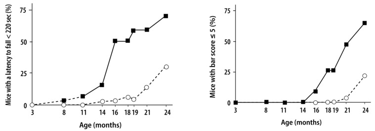
- In 40 Abcd1−/− mice followed to 24 months, motor and coordination deficits appeared in ~25% at 18 months, ~70% had severe motor manifestations at 24 months, and 33% still showed no clinical pathology; no brain lesions, significant demyelination, or neuroinflammation were detected.
- OL-targeted ABCD1 gene transfer reversed late AMN-like axonopathy: after intracisternal AAV9-MAG-hABCD1 at 18 months, treated mice had restored near-normal motor performances within 1–3 months, with hABCD1 expression at 24 months in 22% of cervical spinal cord white matter OLs and 22% of astrocytes.
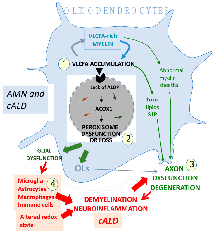
- A CNP-Pex5 KO mouse with OL-selective peroxisome impairment produced an almost perfect phenocopy of cerebral ALD, including VLCFA accumulation, axonal loss, lower plasmalogens, early microglial increase, demyelination, and inflammation; Drosophila showed VLCFA accumulation in glial myelinating cells generates toxic S1P that poisons axons and activates inflammatory cells.
- Clinical/epidemiological data cited include annual incidence ~1/10,000 by neonatal screening, silent adrenal insufficiency in 80% of boys/adolescents with cALD and 70% of AMN patients, slowly progressive AMN symptoms in ~65% of heterozygous women by age 60, and 172 “likely pathogenic” plus 825 “pathogenic” ABCD1 variants in the registry.
Methods
- Narrative review; the abstract states it discusses studies in mouse and Drosophila models that have modified views of X-ALD pathogenesis.
- Evidence synthesized includes human postmortem CNS specimens, MRI/DTI/MRS/PET imaging, Abcd1 KO and other mouse models, Drosophila, zebrafish, C. elegans, patient fibroblasts, iPSCs, multi-omics, and gene therapy studies.
- No new primary data or systematic search/meta-analysis is described; the main analysis is an interpretive synthesis of published findings.
Limitations
- Paper-stated: the review “will raise more hypotheses than certainties”; primary and secondary mechanisms are intermingled, and the spatiotemporal trajectory leading to cALD versus AMN is currently impossible to discern.
- Paper-stated: animal models cannot faithfully replicate human mechanisms and disease course, notably neuroinflammatory responses; postmortem human CNS studies are limited to advanced cALD/AMN stages and have rarely been performed since the late 1970s; iPSC models often resemble a fetal state, reprogramming alters epigenetic marks, and X-ALD iPSC studies rely on only one or two patient lines.
- Evident from the text: as a narrative review, it does not systematically appraise all evidence or quantify certainty; many mechanistic claims rest on small animal or human samples and cross-species extrapolation.
Model-written summary. Check it against the paper before relying on it.
Integrity signals
Registry
Cites
Statistics
Figures
Verification
Claims the paper makes
97 No full text of the sources · 82 Partly supported · 60 Own interpretation · 54 Supported · 39 No basis given · 31 Not checked yet · 15 Supported so far · 7 Couldn’t confirm · 6 Own result · 2 Not supported · 1 Mixed support · accuracy not yet measured
At least one cited source doesn’t support this claim or points to a different paper, and none confirmed it — see the verdicts below.
In the paper
“When microglia fail to degrade VLCFA-enriched myelin fragments and undergo apoptosis, their contents may be released into surrounding tissue and trigger an inflammatory reaction [178].”
§ Revisiting the Pathogenesis of X-Linked Adrenoleukodystrophy > 6. Main Characteristics of Neural Cell Populations in Health and VLCFA-Related Diseases > 6.3. Microglia, Macrophages, Immune Cells
What the sources say
- Disputedcites [178] Gong, 2017, Microglial dysfunction as a key pathological change in adrenomyeloneuropathy, Ann. Neurol. sourceExperimental — accuracy not yet measured
The Results (page unavailable) report increased microglial phagocytosis and neuronal injury in AMN, explicitly in the absence of overt inflammation. The Discussion (page unavailable) describes microglial apoptosis only in connection with excess free VLCFA, not failure to degrade VLCFA-enriched myelin fragments. The paper does not describe apoptotic microglia releasing contents that trigger inflammation; its reported mechanism points in the opposite direction. (LLM, conf=0.82)
Checked by a reasoning model — may be wrong.
hedgedconditional- Asserted: passes — The authors put it forward as true.
- Checkable: passes — It can be checked against evidence.
- Load-bearing: passes — The paper’s conclusions depend on it.
- Based: passes — The paper gives a reason why it holds.
- Disputed
At least one cited source doesn’t support this claim or points to a different paper, and none confirmed it — see the verdicts below.
In the paper
“Reactive astrocytes can upregulate VLCFA-rich phosphatidylcholines, which drive the death of neurons and OLs, probably via lipoparticle secretion [415].”
§ Revisiting the Pathogenesis of X-Linked Adrenoleukodystrophy > 6. Main Characteristics of Neural Cell Populations in Health and VLCFA-Related Diseases > 6.4. Astrocytes
What the sources say
- Disputedcites [415] Ioannou, 2019, Neuron-astrocyte metabolic coupling protects against activity-induced fatty acid toxicity, Cell sourceExperimental — accuracy not yet measured
The Discussion (p. 11) describes the opposite direction of transfer: hyperactive neurons release toxic fatty acids in lipoprotein particles, which astrocytes take up and detoxify to protect neurons. The copy does not describe reactive astrocytes producing VLCFA-rich phosphatidylcholines or causing oligodendrocyte death. (LLM, conf=0.88)
Checked by a reasoning model — may be wrong.
hedgeddirect- Asserted: passes — The authors put it forward as true.
- Checkable: passes — It can be checked against evidence.
- Load-bearing: unclear — It is unclear whether the paper’s conclusions depend on it.
- Based: passes — The paper gives a reason why it holds.
- Disputed
Every source the paper cites for this claim was checked and supports it.
In the paper
“However, in light of the recent estimation of overall X-ALD incidence [2], it is likely that cALD is less frequent.”
§ Revisiting the Pathogenesis of X-Linked Adrenoleukodystrophy > 2. Clinical Manifestations > 2.1. cALD
What the sources say
- Supportedcites [2] Moser, A.B., Jones, R.O., Hubbard, W.C., Tortorelli, S., Orsini, J.J., Caggana, M., Vogel, B.H., and Raymond, G.V. (2016). Newborn screening for X-linked adrenoleukodystrophy. Int. J. Neonatal Screen., 2. source
The citation is attached to the estimate of overall X-ALD incidence, not to the citing authors’ inference that cALD is less frequent. The Introduction reports an estimated minimum birth incidence of about one in 17,000; the Discussion says New York screening found a similar rate. (LLM, conf=0.88)
hedged- Asserted: passes — The authors put it forward as true.
- Checkable: passes — It can be checked against evidence.
- Load-bearing: unclear — It is unclear whether the paper’s conclusions depend on it.
- Based: passes — The paper gives a reason why it holds.
- Supported
Every source the paper cites for this claim was checked and supports it.
In the paper
“In 1990, a single patient showed that allogeneic transplantation of hematopoietic stem cells (HSCs) derived from bone marrow could induce remission of childhood cALD [24], opening the way for dozens of patients to receive the only treatment available for this disease [25] and”
§ Revisiting the Pathogenesis of X-Linked Adrenoleukodystrophy > 1. Introduction
What the sources say
- Supportedcites [24] Aubourg, 1990, Reversal of early neurologic and neuroradiologic manifestations of X-linked adrenoleukodystrophy by bone marrow transplantation, N. Engl. J. Med. source
The Bone Marrow Transplantation section (p. 3) describes a bone marrow infusion from the eight-year-old patient's twin brother. The opening section (p. 1) reports that his neurologic symptoms and MRI brain lesions disappeared within 18 months, supporting the claim of possible remission in this single case of early childhood cerebral adrenoleukodystrophy. (LLM, conf=0.88)
hedgeddirectSingle case- Asserted: passes — The authors put it forward as true.
- Checkable: passes — It can be checked against evidence.
- Load-bearing: unclear — It is unclear whether the paper’s conclusions depend on it.
- Based: passes — The paper gives a reason why it holds.
- Supported
None of the sources cited for this claim have been checked yet.
In the paper
“Electron microscopy reveals characteristic cytoplasmic inclusions (“spicules”) in OLs, perivascular macrophages, and/or microglia but not in astrocytes.”
§ Revisiting the Pathogenesis of X-Linked Adrenoleukodystrophy > 3. X-ALD Patients’ CNS Lesions in Postmortem Studies and MR Images > 3.1. Brain Lesions in Patients with cALD or AMN
What the sources say
- Not checked — no full text of the sourcecites [17] Schaumburg, 1975, Adrenoleukodystrophy: A clinical and pathological study of 17 cases, Arch. Neurol. source
- Not checked — no full text of the sourcecites [20] Budka, 1976, Spastic paraplegia associated with Addison’s disease: Adult variant of adreno-leukodystrophy, J. Neurol. source
- Not checked — no full text of the sourcecites [22] Igarashi, 1976, Fatty acid abnormality in adrenoleukodystrophy, J. Neurochem. source
- Not checked — no full text of the sourcecites [74] Schaumburg, 1974, Adreno-leukodystrophy (sex-linked Schilder disease). Ultrastructural demonstration of specific cytoplasmic inclusions in the central nervous system, Arch. Neurol. source
- Not checked yetcites [75] Johnson, 1976, Histochemical characteristics of the striated inclusions of adrenoleukodystrophy, J. Histochem. Cytochem. source
Checked before — its earlier verdict is held back until a new check runs.
- Not checked — no full text of the sourcecites [76] Powers, 1973, The adrenal cortex in adreno leukodystrophy, Arch. Pathol. Lab. Med.
- Not checked — no full text of the sourcecites [77] Powers, 2005, Adreno-leukodystrophy: A personal historical note, Acta Neuropathol. source
- Not checked — no full text of the sourcecites [78] Menkes, 1977, Adrenoleukodystrophy: Accumulation of cholesterol esters with very long chain fatty acids, Neurology source
- Not checked yetcites [79] Gong, 2024, Role of Basal Forebrain Neurons in Adrenomyeloneuropathy in Mice and Humans, Ann. Neurol. source
Checked before — its earlier verdict is held back until a new check runs.
- Could not verifycites [80] Espinosa, 2012, Functional genomic analysis unravels a metabolic-inflammatory interplay in adrenoleukodystrophy, Hum. Mol. Genet. sourceExperimental — accuracy not yet measured
The Introduction discusses inflammatory pathology in cerebral ALD, and Materials and Methods (Human brain samples) describes staining brain sections, but neither reports electron microscopy or cytoplasmic spicules. Searches for “spicules” and for electron-microscopy inclusions in the named cell types found no passage addressing the claim; page numbers were unavailable in our copy. (LLM, conf=0.76)
plain- Asserted: passes — The authors put it forward as true.
- Checkable: passes — It can be checked against evidence.
- Load-bearing: unclear — It is unclear whether the paper’s conclusions depend on it.
- Based: passes — The paper gives a reason why it holds.
The cited sources support only part of this claim — read each source’s verdict below.
In the paper
“In some patients, the pyramidal tract signals may become as intense as in cALD [31].”
§ Revisiting the Pathogenesis of X-Linked Adrenoleukodystrophy > 4. Imaging of CNS Lesions
What the sources say
- Partly supportedcites [31] Engelen, 2012, X-linked adrenoleukodystrophy (X-ALD): Clinical presentation and guidelines for diagnosis, follow-up and management, Orphanet J. Rare Dis. sourceExperimental — accuracy not yet measured
The Adrenomyeloneuropathy (AMN) section says pyramidal-tract MRI signals can become more intense, and that this is considered a manifestation of cerebral ALD when the abnormality also extends beyond the internal capsules. It does not make a direct intensity comparison with cerebral ALD, and the claim omits the extension criterion. (LLM, conf=0.82)
hedged- Asserted: passes — The authors put it forward as true.
- Checkable: passes — It can be checked against evidence.
- Load-bearing: unclear — It is unclear whether the paper’s conclusions depend on it.
- Based: passes — The paper gives a reason why it holds.
- Partly supported
The sources with a verdict support this claim; the rest weren’t checked yet, have no full text we could read, or their verdicts aren’t rated yet.
In the paper
“Magnetization transfer or diffusion tensor-based imaging sequences can detect abnormalities in spinal cord tracts [65,97,98].”
§ Revisiting the Pathogenesis of X-Linked Adrenoleukodystrophy > 4. Imaging of CNS Lesions
What the sources say
- Not checked — no full text of the sourcecites [65] Dubey, 2005, Diffusion tensor–based imaging reveals occult abnormalities in adrenomyeloneuropathy, Ann. Neurol. Off. J. Am. Neurol. Assoc. Child Neurol. Soc. source
- Supportedcites [97] Smith, 2009, Quantitative magnetization transfer characteristics of the human cervical spinal cord in vivo: Application to adrenomyeloneuropathy, Magn. Reson. Med. Off. J. Int. Soc. Magn. Reson. Med. source
The Introduction identifies AMN as an adult form of X-ALD. The Discussion reports significant magnetization-transfer differences in the cervical spinal cord’s dorsal columns between affected participants and controls, supporting the magnetization-transfer alternative in the citing sentence. (LLM, conf=0.92)
- Not checked — no full text of the sourcecites [98] Fatemi, 2005, Magnetization transfer MRI demonstrates spinal cord abnormalities in adrenomyeloneuropathy, Neurology source
plaincorrelational- Asserted: passes — The authors put it forward as true.
- Checkable: passes — It can be checked against evidence.
- Load-bearing: unclear — It is unclear whether the paper’s conclusions depend on it.
- Based: passes — The paper gives a reason why it holds.
Every source the paper cites for this claim was checked and supports it.
In the paper
“In patients with “pure” AMN, brain MRI remains normal or shows subtle abnormalities, such as moderately increased signal intensities on FLAIR and T2 sequences in the pyramidal tracts in the internal capsules, brainstem, and/or pons [31].”
§ Revisiting the Pathogenesis of X-Linked Adrenoleukodystrophy > 4. Imaging of CNS Lesions
What the sources say
- Supportedcites [31] Engelen, 2012, X-linked adrenoleukodystrophy (X-ALD): Clinical presentation and guidelines for diagnosis, follow-up and management, Orphanet J. Rare Dis. source
The Adrenomyeloneuropathy (AMN) section states that brain MRI is normal or may show subtle, moderately increased signal in the pyramidal tracts of the brainstem, pons, and internal capsules on FLAIR and T2 sequences. It distinguishes these findings from manifestations of cerebral ALD, consistent with the claim’s reference to “pure” AMN. (LLM, conf=0.93)
plain- Asserted: passes — The authors put it forward as true.
- Checkable: passes — It can be checked against evidence.
- Load-bearing: unclear — It is unclear whether the paper’s conclusions depend on it.
- Based: passes — The paper gives a reason why it holds.
- Supported
Every source the paper cites for this claim was checked and supports it.
In the paper
“Those brain lesions may enter an active phase of neuroinflammation with gadolinium enhancement [31].”
§ Revisiting the Pathogenesis of X-Linked Adrenoleukodystrophy > 4. Imaging of CNS Lesions
What the sources say
- Supportedcites [31] Engelen, 2012, X-linked adrenoleukodystrophy (X-ALD): Clinical presentation and guidelines for diagnosis, follow-up and management, Orphanet J. Rare Dis. source
In “Adrenomyeloneuropathy (AMN),” the paper states that cerebral demyelinating lesions in AMN patients can enter an active neuroinflammatory phase with gadolinium enhancement. Its Figure 3 description also documents enhancement appearing on follow-up MRI in a patient with AMN. (LLM, conf=0.94)
hedgedconditional- Asserted: passes — The authors put it forward as true.
- Checkable: passes — It can be checked against evidence.
- Load-bearing: unclear — It is unclear whether the paper’s conclusions depend on it.
- Based: passes — The paper gives a reason why it holds.
- Supported
The cited sources support only part of this claim — read each source’s verdict below.
In the paper
“In humans, two other ATP-synthase ABC transporters can transport VLCFAcyl-CoA into peroxisomes: adrenoleukodystrophy-related protein (ALDR), coded by ABCD2, and 70 kDa peroxisomal membrane protein (PMP70), coded by ABCD3 [146].”
§ Revisiting the Pathogenesis of X-Linked Adrenoleukodystrophy > 5. The ABCD1 Gene and Its Expression > 5.3. ALDP, the ABCD1-Encoded Protein
What the sources say
- Partly supportedcites [146] Wiesinger, 2013, Impaired very long-chain acyl-CoA β-oxidation in human X-linked adrenoleukodystrophy fibroblasts is a direct consequence of ABCD1 transporter dysfunction, J. Biol. Chem. sourceExperimental — accuracy not yet measured
The paper identifies ALDRP as ABCD2 and PMP70 as ABCD3, both peroxisomal ABC transporters (introductory passage; page unavailable). The Discussion reports that ABCD3 contributes to residual VLCFA degradation in human fibroblasts, but says it remains unresolved whether ABCD3 uses VLCFAs directly; ABCD2 is virtually absent in those cells. Thus the paper does not establish the claim that both transport VLCFA–CoA into peroxisomes. (LLM, conf=0.88)
plaindirect- Asserted: passes — The authors put it forward as true.
- Checkable: passes — It can be checked against evidence.
- Load-bearing: unclear — It is unclear whether the paper’s conclusions depend on it.
- Based: passes — The paper gives a reason why it holds.
- Partly supported
- Probably not a substantial claim. 69
- It may state two separate claims as one. 129
- The quote only partly states this claim. 3
Some cited sources support this claim and some don’t — read each source’s verdict below.
In the paper
“Based on neonatal screening in the USA since 2013, the annual incidence was found to be ~1/10,000 [2,7], much higher than previously thought [15].”
§ Revisiting the Pathogenesis of X-Linked Adrenoleukodystrophy > 1. Introduction
What the sources say
- Disputedcites [2] Moser, A.B., Jones, R.O., Hubbard, W.C., Tortorelli, S., Orsini, J.J., Caggana, M., Vogel, B.H., and Raymond, G.V. (2016). Newborn screening for X-linked adrenoleukodystrophy. Int. J. Neonatal Screen., 2. sourceExperimental — accuracy not yet measured
The New York screening section says screening began on 30 December 2013, but the Discussion reports a birth incidence of one in 14,700—not approximately one in 10,000. It also describes that rate as close to the earlier estimate of one in 17,000. In the citing sentence, the separate claim that incidence was 'much higher than previously thought' is attached to [15], not to this source. (LLM, conf=0.88)
Checked by a reasoning model — may be wrong.
- Supportedcites [7] Priestley, J.R.C., Adang, L.A., Drewes Williams, S., Lichter-Konecki, U., Menello, C., Engelhardt, N.M., DiPerna, J.C., DiBoscio, B., Ahrens-Nicklas, R.C., and Edmondson, A.C. (2022). Newborn Screening for X-Linked Adrenoleukodystrophy: Review of Data and Outcomes in Pennsylvania. Int. J. Neonatal Screen., 8. source
The Results (§3.4) estimate U.S. X-ALD incidence at about 1 in 10,500 live births from newborn-screening data across seven states. The Discussion says screening has taken place since 2013 and that the aggregate estimate is higher than the previous estimate of 1 in 17,000 births. The claim’s ~1 in 10,000 is a reasonable approximation. (LLM, conf=0.93)
- Not checked yetcites [15] Wiesinger, 2015, The genetic landscape of X-linked adrenoleukodystrophy: Inheritance, mutations, modifier genes, and diagnosis, Appl. Clin. Genet.
plain- Asserted: passes — The authors put it forward as true.
- Checkable: passes — It can be checked against evidence.
- Load-bearing: fails — The paper’s conclusions may not depend on it.
- Based: passes — The paper gives a reason why it holds.
Probably not a substantial claim. The paper’s conclusions may not depend on it. It may state two separate claims as one.
- Disputed
The cited sources support only part of this claim — read each source’s verdict below.
In the paper
“Loss of sensation in the legs is followed by a spastic gait with positive Babinski reflex, which progresses inexorably, associated with ataxia, leg pain, impotence, and voiding abnormalities [49,50,51,52].”
§ Revisiting the Pathogenesis of X-Linked Adrenoleukodystrophy > 2. Clinical Manifestations > 2.2. AMN
What the sources say
- Not checked — no full text of the sourcecites [49] Powers, 2000, Adrenomyeloneuropathy: A neuropathologic review featuring its noninflammatory myelopathy, J. Neuropathol. Exp. Neurol. source
- Partly supportedcites [50] Corre, 2021, Beyond gait and balance: Urinary and bowel dysfunction in X-linked adrenoleukodystrophy, Orphanet J. Rare Dis. sourceExperimental — accuracy not yet measured
The Background and Discussion (page not provided) describe AMN as involving sensory impairment, a slowly progressive spastic gait disorder, sensory ataxia, and bladder dysfunction. They do not establish that leg sensory loss precedes gait changes or report a positive Babinski reflex, leg pain, or impotence. The Results (Cross-sectional symptom prevalence and age of onset; page not provided) instead report that urinary or bowel symptoms preceded other myelopathy symptoms in 57.8% of symptomatic patients. (LLM, conf=0.88)
- Partly supportedcites [51] Hofereiter, 2015, Bladder and bowel dysfunction is common in both men and women with mutation of the ABCD1 gene for X-linked adrenoleukodystrophy, JIMD Rep. sourceExperimental — accuracy not yet measured
The Introduction describes AMN as slowly progressive spastic paraparesis with sensory disturbances, and the Discussion reports that lower urinary tract symptoms are common. Neither section establishes that loss of sensation precedes a spastic gait or documents a positive Babinski reflex, ataxia, leg pain, or impotence. Page numbers are unavailable in our copy. (LLM, conf=0.88)
- Not checked — no full text of the sourcecites [52] Silveri, 2004, Voiding dysfunction in X-linked adrenoleukodystrophy: Symptom score and urodynamic findings, J. Urol. source
plaincorrelational- Asserted: passes — The authors put it forward as true.
- Checkable: passes — It can be checked against evidence.
- Load-bearing: unclear — It is unclear whether the paper’s conclusions depend on it.
- Based: passes — The paper gives a reason why it holds.
It may state two separate claims as one.
The cited sources support only part of this claim — read each source’s verdict below.
In the paper
“Strikingly, the same ABCD1 mutation causes either devastating brain inflammatory demyelination during childhood or, more often, progressive spinal cord axonopathy starting in middle-aged adults.”
§ Revisiting the Pathogenesis of X-Linked Adrenoleukodystrophy > Abstract
What the sources say
- Not checked yetcites [30] Powers, 1985, Adreno-leukodystrophy (adreno-testiculo-leukomyelo-neuropathic-complex), Clin. Neuropathol.
- Partly supportedcites [31] Engelen, 2012, X-linked adrenoleukodystrophy (X-ALD): Clinical presentation and guidelines for diagnosis, follow-up and management, Orphanet J. Rare Dis. sourceExperimental — accuracy not yet measured
The ‘Evolution over time’ section says identical ABCD1 mutations can produce markedly different neurological phenotypes. The ‘Cerebral ALD’ and ‘Adrenomyeloneuropathy (AMN)’ sections describe childhood inflammatory brain demyelination and a usually slowly progressive spinal-cord axonopathy, respectively. However, the paper says AMN usually develops in the third or fourth decade, not specifically in middle age; it also notes that AMN and cerebral demyelination can occur in the same person rather than being strictly either-or outcomes. (LLM, conf=0.86)
plaindirect- Asserted: passes — The authors put it forward as true.
- Checkable: passes — It can be checked against evidence.
- Load-bearing: passes — The paper’s conclusions depend on it.
- Based: passes — The paper gives a reason why it holds.
It may state two separate claims as one.
The cited sources support only part of this claim — read each source’s verdict below.
In the paper
“Anecdotical cases of head trauma precipitating cALD have been reported with inflammatory demyelination beginning at the site of the contusion [43,44,45,46].”
§ Revisiting the Pathogenesis of X-Linked Adrenoleukodystrophy > 2. Clinical Manifestations > 2.1. cALD
What the sources say
- Not checked — no full text of the sourcecites [43] Bouquet, 2015, Dramatic worsening of adult-onset X-linked adrenoleukodystrophy after head trauma, Neurology source
- Not checked — no full text of the sourcecites [44] Weller, 1992, Very-late-onset adrenoleukodystrophy: Possible precipitation of demyelination by cerebral contusion, Neurology source
- Not checked — no full text of the sourcecites [45] Raymond, 2010, Head trauma can initiate the onset of adreno-leukodystrophy, J. Neurol. Sci. source
- Partly supportedcites [46] Budhram, 2017, Activation of cerebral X-linked adrenoleukodystrophy after head trauma, Can. J. Neurol. Sci. sourceExperimental — accuracy not yet measured
The case description reports cerebral X-ALD developing after head trauma, with new frontal-predominant white matter abnormalities in the region of traumatic microbleeds. The discussion says abnormalities may be most prominent at the site of injury, but does not establish that inflammatory demyelination began at a contusion site; it describes inflammation as a possible mechanism. (LLM, conf=0.85)
plaindirect- Asserted: passes — The authors put it forward as true.
- Checkable: passes — It can be checked against evidence.
- Load-bearing: fails — The paper’s conclusions may not depend on it.
- Based: passes — The paper gives a reason why it holds.
Probably not a substantial claim. The paper’s conclusions may not depend on it.
The cited sources support only part of this claim — read each source’s verdict below.
In the paper
“Although ALD is an X-linked disease, slowly progressive AMN symptoms occur in ~65% of heterozygous women by the age of 60 years [31,47,59,60] due to the inactivation of the non-mutated X chromosome and the mosaic expression of X-linked genes in neural cells [60,61,62,63].”
§ Revisiting the Pathogenesis of X-Linked Adrenoleukodystrophy > 2. Clinical Manifestations > 2.2. AMN
What the sources say
- Partly supportedcites [31] Engelen, 2012, X-linked adrenoleukodystrophy (X-ALD): Clinical presentation and guidelines for diagnosis, follow-up and management, Orphanet J. Rare Dis. sourceExperimental — accuracy not yet measured
The “Women with X-ALD” section (page unavailable) estimates that AMN in heterozygous women is likely to approach 65% by age 60. It describes skewed X-inactivation in neuronal cells as a possible contributor, not an established cause, and says a recent study did not confirm its association with symptoms. It does not state that mosaic expression of X-linked genes in neural cells causes the symptoms. (LLM, conf=0.90)
- Not checked yetcites [47] Scriver, C.R., Beaudet, A.L., Sly, W.S., and Valle, D. (2001). X-linked adrenoleukodystrophy. The Metabolic and Molecular Bases of Inherited Disease, McGraw-Hill.
- Not checked — no full text of the sourcecites [59] Moser, 1984, Adrenoleukodystrophy: Clinical and biochemical manifestations in carriers, Neurology source
- Partly supportedcites [60] Huffnagel, 2019, Disease progression in women with X-linked adrenoleukodystrophy is slow, Orphanet J. Rare Dis. sourceExperimental — accuracy not yet measured
The Background (page not provided) says that more than 80% of women with ALD eventually develop progressive spinal cord disease, and the Results > Progression rates section describes progression as very slow. The paper does not give the claimed ~65% prevalence by age 60 or attribute symptoms to inactivation of the non-mutated X chromosome and mosaic expression in neural cells. (LLM, conf=0.88)
- Not checked yetcites [61] Naidu, S., Washington, C., Thirumalai, S., Smith, K., Moser, H., and Watkins, P. (1997). X-Chromosome Inactivation in Symptomatic Heterozygotes of X-Linked Adrenoleukodystrophy, Lippincott-Raven Publ.
- Not checked — no full text of the sourcecites [62] Maier, 2002, Symptoms in carriers of adrenoleukodystrophy relate to skewed X inactivation, Ann. Neurol. Off. J. Am. Neurol. Assoc. Child Neurol. Soc. source
- Partly supportedcites [63] Salsano, 2012, Preferential expression of mutant ABCD1 allele is common in adrenoleukodystrophy female carriers but unrelated to clinical symptoms, Orphanet J. Rare Dis. sourceExperimental — accuracy not yet measured
The Background (page unavailable) describes progressive AMN-like symptoms in female X-ALD carriers and explains that X-inactivation makes females mosaic for X-linked gene expression. But it estimates symptoms in about 20% of carriers, not 65% by age 60. The Discussion (page unavailable) reports no significant association between symptoms and X-inactivation patterns in blood; it says X-inactivation in carriers’ brains was not investigated, so it does not establish the claimed cause in neural cells. (LLM, conf=0.88)
plaindirect- Asserted: passes — The authors put it forward as true.
- Checkable: passes — It can be checked against evidence.
- Load-bearing: unclear — It is unclear whether the paper’s conclusions depend on it.
- Based: passes — The paper gives a reason why it holds.
It may state two separate claims as one.
- Partly supported
The cited sources that could be checked couldn’t confirm or dispute this claim.
In the paper
“Spicules are straight and curved striated structures made of degenerated myelin [17,20,22,74,75,76,77,78,79,80], which are also observed in 15% of Schwann cells surrounding myelinated axons in the adrenals [19,81].”
§ Revisiting the Pathogenesis of X-Linked Adrenoleukodystrophy > 3. X-ALD Patients’ CNS Lesions in Postmortem Studies and MR Images > 3.1. Brain Lesions in Patients with cALD or AMN
What the sources say
- Not checked — no full text of the sourcecites [19] Powell, 1975, Adrenoleukodystrophy: Electron microscopic findings, Arch. Neurol. source
- Could not verifycites [81] Budka, 2023, A historical look using virtual microscopy: The first case report of adrenomyeloneuropathy (AMN), Free Neuropathol. sourceExperimental — accuracy not yet measured
The citation appears attached to the claim about 15% of Schwann cells surrounding myelinated axons in the adrenals. The Fig. 4 Adrenal section describes occasional cytoplasmic striations in cortical cells, but our copy does not mention Schwann cells or the 15% figure; searches for those terms found no supporting passage. (LLM, conf=0.76)
plain- Asserted: passes — The authors put it forward as true.
- Checkable: passes — It can be checked against evidence.
- Load-bearing: unclear — It is unclear whether the paper’s conclusions depend on it.
- Based: passes — The paper gives a reason why it holds.
It may state two separate claims as one.
The cited sources support only part of this claim — read each source’s verdict below.
In the paper
“During the follow-up of a patient’s brother with elevated VLCFAs or a child found positive at neonatal screening, MRI is able to detect early demyelinating lesions generally localized in the splenium of the corpus callosum (a region with a very high density of OLs), which progress to involve the adjacent parieto-occipital white matter [14,31,90,91,92].”
§ Revisiting the Pathogenesis of X-Linked Adrenoleukodystrophy > 4. Imaging of CNS Lesions
What the sources say
- Partly supportedcites [14] Bonaventura, E., Alberti, L., Lucchi, S., Cappelletti, L., Fazzone, S., Cattaneo, E., Bellini, M., Izzo, G., Parazzini, C., and Bosetti, A. (2023). Newborn screening for X-linked adrenoleukodystrophy in Italy: Diagnostic algorithm and disease monitoring. Front. Neurol., 13. sourceExperimental — accuracy not yet measured
The Neuroradiological assessment section says MRI can detect early cALD lesions, most often in the genu or splenium of the corpus callosum. It does not describe progression into adjacent parieto-occipital white matter, and it notes that some early lesions may arrest rather than progress. Page numbers are unavailable in our copy. (LLM, conf=0.88)
- Supportedcites [31] Engelen, 2012, X-linked adrenoleukodystrophy (X-ALD): Clinical presentation and guidelines for diagnosis, follow-up and management, Orphanet J. Rare Dis. source
The “Cerebral ALD” section states that in 80% of patients the initial lesion is in the splenium of the corpus callosum and progresses into adjacent parieto-occipital white matter. The “Clinical management” section recommends MRI screening to detect early abnormalities, including limited changes in the splenium. Page numbers are not provided in our copy. (LLM, conf=0.94)
- Partly supportedcites [90] Mallack, 2021, MRI surveillance of boys with X-linked adrenoleukodystrophy identified by newborn screening: Meta-analysis and consensus guidelines, J. Inherit. Metab. Dis. sourceExperimental — accuracy not yet measured
The Introduction says lesions most often begin in the splenium of the corpus callosum and spread into adjacent white matter, and the Results describe early lesions detectable on MRI. Our copy does not specify that the adjacent white matter is parieto-occipital or support the parenthetical claim about oligodendrocyte density. No page numbers were provided for these passages. (LLM, conf=0.85)
- Not checked yetcites [91] Liberato, 2019, MRI brain lesions in asymptomatic boys with X-linked adrenoleukodystrophy, Neurology source
Checked before — its earlier verdict is held back until a new check runs.
- Partly supportedcites [92] Kemp, 2001, ABCD1 mutations and the X-linked adrenoleukodystrophy mutation database: Role in diagnosis and clinical correlations, Hum. Mutat. sourceExperimental — accuracy not yet measured
The Diagnosis section (page unavailable) says MRI shows parieto-occipital demyelination in approximately 80% of patients with the childhood cerebral phenotype. It does not describe lesions beginning in the splenium and progressing into adjacent white matter, or establish that pattern for X-ALD generally. (LLM, conf=0.85)
plaincorrelational- Asserted: passes — The authors put it forward as true.
- Checkable: passes — It can be checked against evidence.
- Load-bearing: unclear — It is unclear whether the paper’s conclusions depend on it.
- Based: passes — The paper gives a reason why it holds.
It may state two separate claims as one.
- Partly supported
Every source the paper cites for this claim was checked and supports it.
In the paper
“While writing this review, we updated the previous counts [100] and found a total of 172 variants annotated as “likely pathogenic” and 825 as “pathogenic” in the ABCD1 registry (https://adrenoleukodystrophy.info, accessed on 15 May 2025).”
§ Revisiting the Pathogenesis of X-Linked Adrenoleukodystrophy > 5. The ABCD1 Gene and Its Expression > 5.1. The X-Linked ABCD1 Gene
What the sources say
- Supportedcites [100] Mallack, E.J., Gao, K., Engelen, M., and Kemp, S. (2022). Structure and function of the ABCD1 variant database: 20 years, 940 pathogenic variants, and 3400 cases of adrenoleukodystrophy. Cells, 11. source
Section 2.4 reports the ABCD1 database’s previous count: 948 unique (likely) pathogenic variants as of December 2021. In the citing sentence, [100] is attached to “the previous counts”; the authors say they themselves updated those counts and found the May 2025 figures. The 172 and 825 figures are therefore not attributed to this source. (LLM, conf=0.88)
plain- Asserted: passes — The authors put it forward as true.
- Checkable: passes — It can be checked against evidence.
- Load-bearing: fails — The paper’s conclusions may not depend on it.
- Based: passes — The paper gives a reason why it holds.
Probably not a substantial claim. The paper’s conclusions may not depend on it. It may state two separate claims as one.
- Supported
The sources with a verdict support this claim; the rest weren’t checked yet, have no full text we could read, or their verdicts aren’t rated yet.
In the paper
“At more advanced stages, typical confluent and symmetrical areas of T2 hyperintensity reflect demyelination in the splenium of the corpus callosum, parieto-occipital or frontal white matter, or pyramidal tracts within the internal capsules, pons, and brainstem.”
§ Revisiting the Pathogenesis of X-Linked Adrenoleukodystrophy > 4. Imaging of CNS Lesions
What the sources say
- Supportedcites [31] Engelen, 2012, X-linked adrenoleukodystrophy (X-ALD): Clinical presentation and guidelines for diagnosis, follow-up and management, Orphanet J. Rare Dis. source
The Cerebral ALD section describes demyelinating lesions that progress from the corpus callosum into parieto-occipital or frontal white matter, as well as lesions involving the pyramidal tracts. It reports increased T2 signal in the listed regions. The Differential diagnosis section also describes confluent white-matter changes with increased T2 signal. (LLM, conf=0.90)
- Not checked — no full text of the sourcecites [93] Pasco, 1991, Contribution of MRI to the diagnosis of cerebral lesions of adrenoleukodystrophy, Pediatr. Radiol. source
- Not checked — no full text of the sourcecites [94] Loes, 2003, Analysis of MRI patterns aids prediction of progression in X-linked adrenoleukodystrophy, Neurology source
plaindirect- Asserted: passes — The authors put it forward as true.
- Checkable: passes — It can be checked against evidence.
- Load-bearing: unclear — It is unclear whether the paper’s conclusions depend on it.
- Based: passes — The paper gives a reason why it holds.
It may state two separate claims as one.
- Supported
The sources with a verdict support this claim; the rest weren’t checked yet, have no full text we could read, or their verdicts aren’t rated yet.
In the paper
“Gadolinium extravasation, a hallmark of neuroinflammation and blood–brain barrier (BBB) breach, can occur early when demyelinating lesions are still restricted to the corpus callosum or pyramidal tracts but is often seen later, once the demyelinating lesions have extended [31,93,94].”
§ Revisiting the Pathogenesis of X-Linked Adrenoleukodystrophy > 4. Imaging of CNS Lesions
What the sources say
- Supportedcites [31] Engelen, 2012, X-linked adrenoleukodystrophy (X-ALD): Clinical presentation and guidelines for diagnosis, follow-up and management, Orphanet J. Rare Dis. source
Split into 2 atoms — 2 confirmed, 0 partially supported, 0 disputed, 0 misattributed, 0 unverifiable. Expand the claim to see per-atom verdicts.
- Not checked — no full text of the sourcecites [93] Pasco, 1991, Contribution of MRI to the diagnosis of cerebral lesions of adrenoleukodystrophy, Pediatr. Radiol. source
- Not checked — no full text of the sourcecites [94] Loes, 2003, Analysis of MRI patterns aids prediction of progression in X-linked adrenoleukodystrophy, Neurology source
plaincorrelational- Asserted: passes — The authors put it forward as true.
- Checkable: passes — It can be checked against evidence.
- Load-bearing: unclear — It is unclear whether the paper’s conclusions depend on it.
- Based: passes — The paper gives a reason why it holds.
It may state two separate claims as one.
- Supported
The sources with a verdict support this claim; the rest weren’t checked yet, have no full text we could read, or their verdicts aren’t rated yet.
In the paper
“Consistently, the residual peroxisomal β-oxidation of VLCFA is ~20% of normal in X-ALD fibroblasts [150,151] and can be restored by ABCD2 or ABCD3 overexpression [152,153].”
§ Revisiting the Pathogenesis of X-Linked Adrenoleukodystrophy > 5. The ABCD1 Gene and Its Expression > 5.3. ALDP, the ABCD1-Encoded Protein
What the sources say
- Not checked — no full text of the sourcecites [150] McGuinness, 2001, Evaluation of pharmacological induction of fatty acid β-oxidation in X-linked Adrenoleukodystrophy, Mol. Genet. Metab. source
- Supportedcites [151] Kemp, 2004, Method for measurement of peroxisomal very-long-chain fatty acid β-oxidation in human skin fibroblasts using stable-isotope-labeled tetracosanoic acid, Clin. Chem. source
This source is cited for the residual β-oxidation figure, not for the separate overexpression claim, which has its own citations. Table 1 reports that X-ALD fibroblasts formed D₃-C16:0 at 15% of the control amount; the paper identifies this measure as an indicator of peroxisomal β-oxidation activity. That supports the citing sentence’s approximate figure of 20%. (LLM, conf=0.88)
- Not checked — no full text of the sourcecites [152] Netik, 1999, Adrenoleukodystrophy-related protein can compensate functionally for adrenoleukodystrophy protein deficiency (X-ALD): Implications for therapy, Hum. Mol. Genet. source
- Not checked — no full text of the sourcecites [153] Kemp, 1998, Gene redundancy and pharmacological gene therapy: Implications for X-linked adrenoleukodystrophy, Nat. Med. source
plaindirect- Asserted: passes — The authors put it forward as true.
- Checkable: passes — It can be checked against evidence.
- Load-bearing: unclear — It is unclear whether the paper’s conclusions depend on it.
- Based: passes — The paper gives a reason why it holds.
It may state two separate claims as one.
The cited sources support only part of this claim — read each source’s verdict below.
In the paper
“Abcd1−/− mice, despite being congenic littermates living in the same cage, show variable motor deficits at different ages, thereby supporting the importance of stochasticity for determining phenotypes [136].”
§ Revisiting the Pathogenesis of X-Linked Adrenoleukodystrophy > 5. The ABCD1 Gene and Its Expression > 5.2. Individual Variability of Disease Phenotypes
What the sources say
- Partly supportedcites [136] Singh, 2010, Pathomechanisms underlying X-adrenoleukodystrophy: A three-hit hypothesis, Brain Pathol. sourceExperimental — accuracy not yet measured
The Introduction (page unavailable) reports motor and locomotor deficits in ABCD1-null mice at around 20 months of age. It also suggests stochastic factors may affect outcomes in people with X-ALD, but does not report variable ages of motor deficits among congenic, same-cage mouse littermates or draw that inference from mice. (LLM, conf=0.86)
plaindirect- Asserted: passes — The authors put it forward as true.
- Checkable: passes — It can be checked against evidence.
- Load-bearing: passes — The paper’s conclusions depend on it.
- Based: passes — The paper gives a reason why it holds.
It may state two separate claims as one.
- Partly supported
The sources with a verdict support this claim; the rest weren’t checked yet, have no full text we could read, or their verdicts aren’t rated yet.
In the paper
“Transport involves cooperative binding of two VLCFAcyl-CoAs, ATP binding, and ALDP homodimerization [147], followed by outward-facing ALDP conformational change allowing for the release of VLCFAcylCoA into the peroxisome matrix [148], where VLCFAcyl-CoA undergoes peroxisomal β-oxidation [149].”
§ Revisiting the Pathogenesis of X-Linked Adrenoleukodystrophy > 5. The ABCD1 Gene and Its Expression > 5.3. ALDP, the ABCD1-Encoded Protein
What the sources say
- Not checked yetcites [147] Kemp, 2011, Mammalian peroxisomal ABC transporters: From endogenous substrates to pathology and clinical significance, Br. J. Pharmacol. source
Checked before — its earlier verdict is held back until a new check runs.
- Supportedcites [148] Chen, 2022, Structural basis of substrate recognition and translocation by human very long-chain fatty acid transporter ABCD1, Nat. Commun. source
Split into 2 atoms — 2 confirmed, 0 partially supported, 0 disputed, 0 misattributed, 0 unverifiable. Expand the claim to see per-atom verdicts.
- Not checked yetcites [149] Wanders, 2023, The physiological functions of human peroxisomes, Physiol. Rev. source
Checked before — its earlier verdict is held back until a new check runs.
plaindirect- Asserted: passes — The authors put it forward as true.
- Checkable: passes — It can be checked against evidence.
- Load-bearing: unclear — It is unclear whether the paper’s conclusions depend on it.
- Based: passes — The paper gives a reason why it holds.
It may state two separate claims as one.
- Not checked yet
The cited sources support only part of this claim — read each source’s verdict below.
In the paper
“Unless they are relatively common and have a major phenotypic effect, modifier-gene variants (likely to exist) may only be identified if agnostic whole-genome analysis studies are able to recruit and compare thousands of well-defined patients with cALD with thousands of patients with “pure” AMN [117,118].”
§ Revisiting the Pathogenesis of X-Linked Adrenoleukodystrophy > 5. The ABCD1 Gene and Its Expression > 5.2. Individual Variability of Disease Phenotypes
What the sources say
- Not checked — no full text of the sourcecites [117] Semmler, 2009, Genetic variants of methionine metabolism and X-ALD phenotype generation: Results of a new study sample, J. Neurol. source
- Partly supportedcites [118] Barbier, M., Sabbagh, A., Kasper, E., Asheuer, M., Ahouansou, O., Pribill, I., Forss-Petter, S., Vidaud, M., Berger, J., and Aubourg, P. (2012). CD1 gene polymorphisms and phenotypic variability in X-linked adrenoleukodystrophy. PLoS ONE, 7. sourceExperimental — accuracy not yet measured
The Discussion (page unavailable) says genetic variants may contribute to X-ALD phenotypic differences and that the candidate-gene strategy has major limitations. The Methods describe comparing well-defined childhood cerebral ALD and “pure” AMN groups. The paper does not say that identifying modifier variants requires agnostic whole-genome analysis or thousands of patients in each group. (LLM, conf=0.85)
hedgedconditional- Asserted: passes — The authors put it forward as true.
- Checkable: passes — It can be checked against evidence.
- Load-bearing: unclear — It is unclear whether the paper’s conclusions depend on it.
- Based: passes — The paper gives a reason why it holds.
It may state two separate claims as one.
The cited sources support only part of this claim — read each source’s verdict below.
In the paper
“Influenza infection may enter the brain through several routes [122,123] or send deleterious signals to the brain via sensory neurons [124] and trigger neuroinflammation or associated chronic alterations in the CNS [125].”
§ Revisiting the Pathogenesis of X-Linked Adrenoleukodystrophy > 5. The ABCD1 Gene and Its Expression > 5.2. Individual Variability of Disease Phenotypes
What the sources say
- Supportedcites [122] Kitamura, 2024, Viral entry and translation in brain endothelia provoke influenza-associated encephalopathy, Acta Neuropathol. source
The citation is attached to the claim that influenza may enter the brain through several routes, not to the later claims about sensory neurons or chronic CNS changes. The Discussion (page unavailable) says that routes of CNS invasion have long been debated, discusses hematogenous spread, and reports viral antigen in the brains of both intravenously and intranasally inoculated mice. (LLM, conf=0.86)
- Partly supportedcites [123] Constant, O., Maarifi, G., Blanchet, F.P., Van de Perre, P., Simonin, Y., and Salinas, S. (2022). Role of Dendritic Cells in Viral Brain Infections. Front. Immunol., 13. sourceExperimental — accuracy not yet measured
The Discussion (page unavailable) says viral brain infection can lead to neuroinflammation and that infection-related inflammation can have chronic CNS consequences. It does not establish these outcomes specifically for influenza; the only influenza mention found is in a reference title. The routes and sensory-neuron clauses are assigned to other citations, not this source. (LLM, conf=0.86)
- Supportedcites [124] Bin, 2023, An airway-to-brain sensory pathway mediates influenza-induced sickness, Nature source
Citation [124] is attached to the claim that influenza sends signals to the brain via sensory neurons, not to the separate claims about viral entry or chronic CNS changes. The Abstract and Discussion describe influenza-induced prostaglandin signals detected by peripheral sensory neurons and conveyed to the brain, where they mediate sickness responses. (LLM, conf=0.90)
- Supportedcites [125] Hosseini, 2018, Long-term neuroinflammation induced by influenza A virus infection and the impact on hippocampal neuron morphology and function, J. Neurosci. source
Citation [125] is attached to the claim that influenza infection may trigger neuroinflammation and chronic CNS alterations, not to the preceding claims about routes of entry or sensory neurons. The Discussion reports that neuroinflammation following influenza infection produced long-lasting changes in hippocampal neurons, synapses, and cognitive function in adult animals. (LLM, conf=0.91)
hedgedconditional- Asserted: passes — The authors put it forward as true.
- Checkable: passes — It can be checked against evidence.
- Load-bearing: fails — The paper’s conclusions may not depend on it.
- Based: passes — The paper gives a reason why it holds.
Probably not a substantial claim. The paper’s conclusions may not depend on it. It may state two separate claims as one.
- Supported
The sources with a verdict support this claim; the rest weren’t checked yet, have no full text we could read, or their verdicts aren’t rated yet.
In the paper
“Indeed, while COVID-19 is very rarely neuroinvasive [127], its impact on the brain might be immune-mediated [126] and cause glial neuroinflammation [128,129,130].”
§ Revisiting the Pathogenesis of X-Linked Adrenoleukodystrophy > 5. The ABCD1 Gene and Its Expression > 5.2. Individual Variability of Disease Phenotypes
What the sources say
- Not checked — no full text of the sourcecites [127] Meinhardt, 2024, The neurobiology of SARS-CoV-2 infection, Nat. Rev. Neurosci. source
- Supportedcites [126] Wood, 2025, Posthospitalization COVID-19 cognitive deficits at 1 year are global and associated with elevated brain injury markers and gray matter volume reduction, Nat. Med. source
The citation to this paper is attached to the immune-mediated explanation, not to the separate claims about neuroinvasion or glial neuroinflammation. The Main section explicitly hypothesizes that COVID-19’s impact on the brain may occur through immune-mediated para- and postinfectious phenomena. (LLM, conf=0.94)
- Supportedcites [128] Dunai, C., Collie, C., and Michael, B.D. (2022). Immune-mediated mechanisms of COVID-19 neuropathology. Front. Neurol., 13. source
The Introduction says indirect effects of SARS-CoV-2, including a dysregulated immune response, are hypothesised to cause neurological complications. The Discussion likewise says many complications involve neuroinflammation driven by inflammatory responses rather than direct infection. This citation [126] supports the immune-mediated part of the sentence; the neuroinvasion and glial claims have separate citations. (LLM, conf=0.93)
- Supportedcites [129] Needham, 2022, Brain injury in COVID-19 is associated with dysregulated innate and adaptive immune responses, Brain source
The citation is attached to the claim that COVID-19’s impact on the brain might be immune-mediated, not to the separate claims about neuroinvasion or glial neuroinflammation. The Discussion says brain injury during acute COVID-19 may result from dysregulated host responses and describes inflammatory mechanisms that may drive neurological injury. (LLM, conf=0.93)
- Supportedcites [130] Zhang, 2024, Brain-wide alterations revealed by spatial transcriptomics and proteomics in COVID-19 infection, Nat. Aging source
The citation to this paper [126] supports the immune-mediated part of the sentence, not the separate claims about rare neuroinvasion or glial neuroinflammation. In the Discussion (page not provided), the authors say brain changes are not solely attributable to direct infection and likely reflect a response to peripheral infection involving immune activity. (LLM, conf=0.92)
hedgedconditional- Asserted: passes — The authors put it forward as true.
- Checkable: passes — It can be checked against evidence.
- Load-bearing: fails — The paper’s conclusions may not depend on it.
- Based: passes — The paper gives a reason why it holds.
Probably not a substantial claim. The paper’s conclusions may not depend on it. It may state two separate claims as one.
Every source the paper cites for this claim was checked and supports it.
In the paper
“All of these metabolites must be transferred into mitochondria for full β-oxidation into CO2 and H2O because peroxisomes lack a Krebs’ cycle [162].”
§ Revisiting the Pathogenesis of X-Linked Adrenoleukodystrophy > 5. The ABCD1 Gene and Its Expression > 5.5. VLCFA Metabolism and Myelin
What the sources say
- Supportedcites [162] Wanders, R.J., Waterham, H.R., and Ferdinandusse, S. (2016). Metabolic interplay between peroxisomes and other subcellular organelles including mitochondria and the endoplasmic reticulum. Front. Cell Dev. Biol., 3. source
The Introduction states that chain-shortened acyl-CoAs produced in peroxisomes can be fully oxidized to CO2 and H2O only in mitochondria, which contain the Krebs cycle. The fatty acid beta-oxidation section also says that peroxisomal products must be transferred to mitochondria for full oxidation. (LLM, conf=0.93)
plaindirect- Asserted: passes — The authors put it forward as true.
- Checkable: passes — It can be checked against evidence.
- Load-bearing: unclear — It is unclear whether the paper’s conclusions depend on it.
- Based: passes — The paper gives a reason why it holds.
It may state two separate claims as one.
- Supported
The cited sources support only part of this claim — read each source’s verdict below.
In the paper
“By binding to ATP, ALDP transports VLCFAcyl-CoA into the peroxisomal matrix [143,159,160], where they are β-oxidized into medium-chain fatty acids (MCFAs) and MCFAcyl-carnitines [161], propionyl-CoA, and acetyl-CoA.”
§ Revisiting the Pathogenesis of X-Linked Adrenoleukodystrophy > 5. The ABCD1 Gene and Its Expression > 5.5. VLCFA Metabolism and Myelin
What the sources say
- Not checked — no full text of the sourcecites [143] Visser, 2008, The human peroxisomal ABC half transporter ALDP functions as a homodimer and accepts acyl–CoA esters, FASEB J. source
- Partly supportedcites [159] Postis, 2013, Intrinsic acyl-CoA thioesterase activity of a peroxisomal ATP binding cassette transporter is required for transport and metabolism of fatty acids, Proc. Natl. Acad. Sci. USA sourceExperimental — accuracy not yet measured
The Introduction (p. 2) identifies ALDP as a peroxisomal ABC transporter involved in very-long-chain fatty-acid metabolism, and Fig. 4 (p. 4) describes ATP-powered transport for the related plant transporter CTS. But the paper's model says acyl-CoA is cleaved and free fatty acid enters the peroxisome; it does not establish that ALDP transports intact VLCFA-acyl-CoA into the matrix. The listed β-oxidation products are outside the clause citing this source. (LLM, conf=0.88)
- Partly supportedcites [160] Theodoulou, 2016, How to move an amphipathic molecule across a lipid bilayer: Different mechanisms for different ABC transporters?, Biochem. Soc. Trans. sourceExperimental — accuracy not yet measured
The 'Fatty acid import into peroxisomes' section identifies ALDP as an ATP-binding cassette transporter that preferentially imports very-long-chain fatty acids. But the 'Transport substrates and mechanism' section describes fatty acyl-CoA as a substrate accepted at the membrane, with CoA cleaved during or after transport and the fatty acid re-activated inside the peroxisome; it does not establish transport of intact VLCFA-acyl-CoA into the matrix. The later list of β-oxidation products is not attached to this citation. (LLM, conf=0.87)
- Not checked — no full text of the sourcecites [161] Wanders, 2001, Peroxisomal fatty acid α-and β-oxidation in humans: Enzymology, peroxisomal metabolite transporters and peroxisomal diseases, Biochem. Soc. Trans. source
plaindirect- Asserted: passes — The authors put it forward as true.
- Checkable: passes — It can be checked against evidence.
- Load-bearing: passes — The paper’s conclusions depend on it.
- Based: passes — The paper gives a reason why it holds.
It may state two separate claims as one.
The cited sources support only part of this claim — read each source’s verdict below.
In the paper
“These mutant flies had fewer peroxisomes, demonstrating an ABCD1 role in peroxisomal biogenesis [194].”
§ Revisiting the Pathogenesis of X-Linked Adrenoleukodystrophy > 5. The ABCD1 Gene and Its Expression > 5.6. Mutant Abcd1 Animal Models
What the sources say
- Partly supportedcites [194] Manor, J., Jangam, S.V., Chung, H.-l., Bhagwat, P., Andrews, J., Chester, H., Kondo, S., Srivastav, S., Botas, J., and Moser, A.B. (2024). Genetic analysis of the X-linked Adrenoleukodystrophy ABCD1 gene in Drosophila uncovers a role in Peroxisomal dynamics. bioRxiv. sourceExperimental — accuracy not yet measured
The Results (page not provided) report fewer peroxisomes per cell area after Abcd1 RNAi knockdown, supporting the reduced-number finding. They do not report that the Abcd1Δ4 mutant flies had fewer peroxisomes. The paper links inhibited biogenesis to overexpression of human ABCD1 and presents that mechanism as an interpretation, not as demonstrated by the mutant flies. (LLM, conf=0.85)
boosteddirect- Asserted: passes — The authors put it forward as true.
- Checkable: passes — It can be checked against evidence.
- Load-bearing: unclear — It is unclear whether the paper’s conclusions depend on it.
- Based: passes — The paper gives a reason why it holds.
It may state two separate claims as one.
- Partly supported
Every source the paper cites for this claim was checked and supports it.
In the paper
“Loss of the pmp-4 gene, the Caenorhabditis elegans ortholog of ABCD1, induces VLCFA accumulation, axonal degeneration, and locomotor dysfunction [195].”
§ Revisiting the Pathogenesis of X-Linked Adrenoleukodystrophy > 5. The ABCD1 Gene and Its Expression > 5.6. Mutant Abcd1 Animal Models
What the sources say
- Supportedcites [195] Coppa, 2020, The peroxisomal fatty acid transporter ABCD1/PMP-4 is required in the C. elegans hypodermis for axonal maintenance: A worm model for adrenoleukodystrophy, Free Radic. Biol. Med. source
The Results section identifies pmp-4 as the C. elegans orthologue of ABCD1 and reports elevated VLCFAs, reduced motility, and significant axonal damage in pmp-4(ok396) loss-of-function mutants. The Discussion explicitly summarizes VLCFA accumulation, axonal degeneration, and associated locomotor dysfunction in pmp-4-deficient worms. (LLM, conf=0.95)
plaindirect- Asserted: passes — The authors put it forward as true.
- Checkable: passes — It can be checked against evidence.
- Load-bearing: unclear — It is unclear whether the paper’s conclusions depend on it.
- Based: passes — The paper gives a reason why it holds.
It may state two separate claims as one.
- Supported
The cited sources support only part of this claim — read each source’s verdict below.
In the paper
“OL peroxisomes are largely responsible for VLCFA synthesis [204] and degradation [166] in the adult brain.”
§ Revisiting the Pathogenesis of X-Linked Adrenoleukodystrophy > 6. Main Characteristics of Neural Cell Populations in Health and VLCFA-Related Diseases > 6.1. OLs
What the sources say
- Partly supportedcites [204] van de Beek, M.C., Dijkstra, I.M., van Lenthe, H., Ofman, R., Goldhaber-Pasillas, D., Schauer, N., Schackmann, M., Engelen-Lee, J.Y., Vaz, F.M., and Kulik, W. (2016). C26:0-Carnitine Is a New Biomarker for X-Linked Adrenoleukodystrophy in Mice and Man. PLoS ONE, 11. sourceExperimental — accuracy not yet measured
In Results and Discussion, under “Targeting ELOVL1 Expression to Oligodendrocytes” (page not specified), the paper concludes that oligodendrocytes are largely responsible for brain VLCFA synthesis and degradation. But this source is cited for the synthesis portion of the sentence, and it does not attribute that synthesis specifically to oligodendrocyte peroxisomes; its ELOVL1 results place the synthesis enzyme at the endoplasmic reticulum. (LLM, conf=0.87)
- Not checked — no full text of the sourcecites [166] Kassmann, 2007, Axonal loss and neuroinflammation caused by peroxisome-deficient oligodendrocytes, Nat. Genet. source
plaindirect- Asserted: passes — The authors put it forward as true.
- Checkable: passes — It can be checked against evidence.
- Load-bearing: unclear — It is unclear whether the paper’s conclusions depend on it.
- Based: passes — The paper gives a reason why it holds.
It may state two separate claims as one.
- Partly supported
The cited sources support only part of this claim — read each source’s verdict below.
In the paper
“Abcd1: Abcd2 double KO mice had an earlier onset of symptoms with more severe motor deficits (at 20 months of age, 6 out of 11 mice were not able to perform motor tests), axonal degeneration, and myelin alterations, but no cerebral pathology.”
§ Revisiting the Pathogenesis of X-Linked Adrenoleukodystrophy > 5. The ABCD1 Gene and Its Expression > 5.6. Mutant Abcd1 Animal Models
What the sources say
- Partly supportedcites [190] Pujol, 2004, Functional overlap between ABCD1 (ALD) and ABCD2 (ALDR) transporters: A therapeutic target for X-adrenoleukodystrophy, Hum. Mol. Genet. sourceExperimental — accuracy not yet measured
The Neurological phenotype passage reports earlier, more severe motor impairment in double mutants, and Spinal cord histopathology reports more severe axonal and myelin pathology (page numbers unavailable). The copy does not establish an absence of cerebral pathology or report that 6 of 11 mice could not perform motor tests at 20 months; the n=11 figure in Table 2 concerns nerve-conduction studies. (LLM, conf=0.88)
- Supportedcites [196] Ferrer, 2005, Inactivation of the peroxisomal ABCD2 transporter in the mouse leads to late-onset ataxia involving mitochondria, Golgi and endoplasmic reticulum damage, Hum. Mol. Genet. source
The Behavioral deficits section reports more severe motor impairment in double mutants, including six of 11 unable to complete the balance-bar test at 20 months. The Biochemical function section says double mutants had earlier, more severe pathological signs; its account of axonal and myelin changes, together with the Discussion’s report of no detectable cerebral-cortex abnormalities, supports the remaining claims. The paper does report cerebellar pathology, which is distinct from cerebral-cortex pathology. (LLM, conf=0.87)
plaindirectn = 11- Asserted: passes — The authors put it forward as true.
- Checkable: passes — It can be checked against evidence.
- Load-bearing: unclear — It is unclear whether the paper’s conclusions depend on it.
- Based: passes — The paper gives a reason why it holds.
It may state two separate claims as one.
- Partly supported
The cited sources support only part of this claim — read each source’s verdict below.
In the paper
“Electron microscopy in Abcd2 KO mice showed the first signs of spinal cord axonal pathology at 12 months, yet no histological abnormalities.”
§ Revisiting the Pathogenesis of X-Linked Adrenoleukodystrophy > 5. The ABCD1 Gene and Its Expression > 5.6. Mutant Abcd1 Animal Models
What the sources say
- Partly supportedcites [190] Pujol, 2004, Functional overlap between ABCD1 (ALD) and ABCD2 (ALDR) transporters: A therapeutic target for X-adrenoleukodystrophy, Hum. Mol. Genet. sourceExperimental — accuracy not yet measured
The Spinal cord histopathology section (page unavailable) reports the first marker of pathology at 12 months in Abcd2-knockout mice: synaptophysin-positive deposits in swollen axons. It identifies this finding through immunostaining, not electron microscopy, and the deposits are histopathological abnormalities; the claim’s assertion of no histological abnormalities is therefore unsupported. (LLM, conf=0.88)
- Partly supportedcites [196] Ferrer, 2005, Inactivation of the peroxisomal ABCD2 transporter in the mouse leads to late-onset ataxia involving mitochondria, Golgi and endoplasmic reticulum damage, Hum. Mol. Genet. sourceExperimental — accuracy not yet measured
The Biochemical function of ALDRP section (page not supplied) says histological abnormalities in the spinal cord were not demonstrable at 12 months, while the first signs of pathology at that age were APP and synaptophysin accumulations in axonal swellings. It attributes those early findings to immunohistochemical analysis, not electron microscopy. (LLM, conf=0.88)
plain- Asserted: passes — The authors put it forward as true.
- Checkable: passes — It can be checked against evidence.
- Load-bearing: unclear — It is unclear whether the paper’s conclusions depend on it.
- Based: passes — The paper gives a reason why it holds.
It may state two separate claims as one.
- Partly supported
The cited sources support only part of this claim — read each source’s verdict below.
In the paper
“At 12 months of age, Abcd2 KO mice already displayed a marked impairment in their motor performance, which aggravated up to 20 months.”
§ Revisiting the Pathogenesis of X-Linked Adrenoleukodystrophy > 5. The ABCD1 Gene and Its Expression > 5.6. Mutant Abcd1 Animal Models
What the sources say
- Partly supportedcites [190] Pujol, 2004, Functional overlap between ABCD1 (ALD) and ABCD2 (ALDR) transporters: A therapeutic target for X-adrenoleukodystrophy, Hum. Mol. Genet. sourceExperimental — accuracy not yet measured
The Neurological phenotype passage (page unavailable) reports impaired rotarod performance in ABCD1/ABCD2 double mutants by 15 months and in ABCD1-deficient mice at 20 months. It does not report marked motor-performance impairment in Abcd2-only knockout mice at 12 months or worsening through 20 months. The Electrophysiology of the PNS section (page unavailable) instead reports no detectable abnormality in Abcd2-only mutants at 12 months, though that is a different measure. (LLM, conf=0.83)
- Supportedcites [196] Ferrer, 2005, Inactivation of the peroxisomal ABCD2 transporter in the mouse leads to late-onset ataxia involving mitochondria, Golgi and endoplasmic reticulum damage, Hum. Mol. Genet. source
The “Behavioral deficits in ALDRP-deficient mice” section reports marked impairment on the rotarod in Abcd2-null mice at 12 months compared with wild-type littermates. It also states that their performance was even more impaired when tested again around 20 months. (LLM, conf=0.95)
plaincorrelational- Asserted: passes — The authors put it forward as true.
- Checkable: passes — It can be checked against evidence.
- Load-bearing: unclear — It is unclear whether the paper’s conclusions depend on it.
- Based: passes — The paper gives a reason why it holds.
It may state two separate claims as one.
- Partly supported
The cited sources support only part of this claim — read each source’s verdict below.
In the paper
“Histological abnormalities in spinal cord dorsal and ventral columns became evident at 20 months, while atrophy or death of Purkinje cells was detected in the cerebellum.”
§ Revisiting the Pathogenesis of X-Linked Adrenoleukodystrophy > 5. The ABCD1 Gene and Its Expression > 5.6. Mutant Abcd1 Animal Models
What the sources say
- Partly supportedcites [190] Pujol, 2004, Functional overlap between ABCD1 (ALD) and ABCD2 (ALDR) transporters: A therapeutic target for X-adrenoleukodystrophy, Hum. Mol. Genet. sourceExperimental — accuracy not yet measured
The Spinal cord histopathology section reports abnormalities in ALDR−/− (Abcd2 knockout) mice, but it identifies an axonal marker at 12 months and describes further pathology at 22 months—not its first appearance at 20 months. The examined sections were of the dorsal spinal cord; our copy does not report ventral-column abnormalities or Purkinje-cell atrophy or death in the cerebellum. Page numbers were not available in the indexed passages. (LLM, conf=0.88)
- Supportedcites [196] Ferrer, 2005, Inactivation of the peroxisomal ABCD2 transporter in the mouse leads to late-onset ataxia involving mitochondria, Golgi and endoplasmic reticulum damage, Hum. Mol. Genet. source
The 'Biochemical function of ALDRP' passages report that histological abnormalities in the dorsal and ventral spinal cord columns of Abcd2-deficient mice were not demonstrable at 12 months but became evident at 20 months. The same section reports that cerebellar staining confirmed atrophy or death of Purkinje cells at 20 months. (LLM, conf=0.94)
plain- Asserted: passes — The authors put it forward as true.
- Checkable: passes — It can be checked against evidence.
- Load-bearing: unclear — It is unclear whether the paper’s conclusions depend on it.
- Based: passes — The paper gives a reason why it holds.
It may state two separate claims as one.
- Partly supported
The cited sources support only part of this claim — read each source’s verdict below.
In the paper
“In the fly brain, increased S1P—not VLCFA accumulation per se—triggers direct axonal loss, stimulation of immune cells, and neuroinflammation [273], meaning that VLCFA accumulation in OLs is noxious to the CNS, as they can transport S1P to axons [273].”
§ Revisiting the Pathogenesis of X-Linked Adrenoleukodystrophy > 6. Main Characteristics of Neural Cell Populations in Health and VLCFA-Related Diseases > 6.1. OLs
What the sources say
- Partly supportedcites [273] Chung, 2023, Very-long-chain fatty acids induce glial-derived sphingosine-1-phosphate synthesis, secretion, and neuroinflammation, Cell Metab. sourceExperimental — accuracy not yet measured
The Introduction and the Discussion’s “Immune activation and neuroinflammation” section report that elevated glial S1P is toxic to neurons and drives immune activation, macrophage recruitment, and neuroinflammation in flies, rather than VLCFA accumulation alone. The copy does not establish direct axonal loss in flies: its reference to axonal damage concerns inflammatory responses during MS. It shows S1P transfer from fly glia to neurons, not specifically from oligodendrocytes to axons. (LLM, conf=0.86)
plaindirect- Asserted: passes — The authors put it forward as true.
- Checkable: passes — It can be checked against evidence.
- Load-bearing: passes — The paper’s conclusions depend on it.
- Based: passes — The paper gives a reason why it holds.
It may state two separate claims as one.
- Partly supported
The cited sources support only part of this claim — read each source’s verdict below.
In the paper
“To perform all of their tasks, OL energy demand is enormous, reaching a maximum during early myelination and remaining at a high level throughout life [284].”
§ Revisiting the Pathogenesis of X-Linked Adrenoleukodystrophy > 6. Main Characteristics of Neural Cell Populations in Health and VLCFA-Related Diseases > 6.1. OLs
What the sources say
- Partly supportedcites [284] Harris, 2012, The energetics of CNS white matter, J. Neurosci. sourceExperimental — accuracy not yet measured
The Materials and Methods section, “The energetic costs and benefits of myelination” (page unavailable), describes an initial ATP cost for making myelin and an ongoing cost of maintaining the myelinating oligodendrocyte’s resting potential. The Discussion, “The energetic cost and benefit of myelin” (page unavailable), likewise reports substantial resting-potential expenditure. These passages support energy demands during myelination and afterward, but do not establish that oligodendrocyte demand peaks during early myelination or remains high throughout life. (LLM, conf=0.86)
plaincorrelational- Asserted: passes — The authors put it forward as true.
- Checkable: passes — It can be checked against evidence.
- Load-bearing: passes — The paper’s conclusions depend on it.
- Based: passes — The paper gives a reason why it holds.
It may state two separate claims as one.
- Partly supported
The cited sources support only part of this claim — read each source’s verdict below.
In the paper
“Because axons are not self-sufficient for their energy demand, OLs provide them with glucose and FAs for mitochondrial oxidation, with FAs being their major energy source [278].”
§ Revisiting the Pathogenesis of X-Linked Adrenoleukodystrophy > 6. Main Characteristics of Neural Cell Populations in Health and VLCFA-Related Diseases > 6.1. OLs
What the sources say
- Partly supportedcites [278] Asadollahi, 2024, Oligodendroglial fatty acid metabolism as a central nervous system energy reserve, Nat. Neurosci. sourceExperimental — accuracy not yet measured
The Main section says oligodendrocytes supply axons with lactate or pyruvate for ATP generation. The Results section says oligodendrocytes oxidize fatty acids when glucose is limited, allowing them to share more glucose-derived pyruvate or lactate with axons. It does not establish that oligodendrocytes supply axons with fatty acids for mitochondrial oxidation or that fatty acids are axons’ major energy source. (LLM, conf=0.88)
plaindirect- Asserted: passes — The authors put it forward as true.
- Checkable: passes — It can be checked against evidence.
- Load-bearing: unclear — It is unclear whether the paper’s conclusions depend on it.
- Based: passes — The paper gives a reason why it holds.
It may state two separate claims as one.
- Partly supported
Every source the paper cites for this claim was checked and supports it.
In the paper
“When glucose availability is scarce, myelin synthesis declines and FA-derived acetyl-CoA supports mitochondrial respiration and OL survival [278].”
§ Revisiting the Pathogenesis of X-Linked Adrenoleukodystrophy > 6. Main Characteristics of Neural Cell Populations in Health and VLCFA-Related Diseases > 6.1. OLs
What the sources say
- Supportedcites [278] Asadollahi, 2024, Oligodendroglial fatty acid metabolism as a central nervous system energy reserve, Nat. Neurosci. source
The Results section (‘Oligodendrocyte lipid metabolism supports axonal function in starved optic nerves’) states that, when glucose availability is reduced in the GLUT1 icKO mouse model, myelin synthesis drops and FA-derived acetyl-CoA supports mitochondrial respiration and oligodendroglial survival. This directly supports the paraphrase within the paper’s modeled condition. (LLM, conf=0.94)
plaindirect- Asserted: passes — The authors put it forward as true.
- Checkable: passes — It can be checked against evidence.
- Load-bearing: passes — The paper’s conclusions depend on it.
- Based: passes — The paper gives a reason why it holds.
It may state two separate claims as one.
- Supported
The cited sources support only part of this claim — read each source’s verdict below.
In the paper
“In the fly brain, increased S1P—not VLCFA accumulation per se—triggers direct axonal loss, stimulation of immune cells, and neuroinflammation [273], meaning that VLCFA accumulation in OLs is noxious to the CNS, as they can transport S1P to axons [273].”
§ Revisiting the Pathogenesis of X-Linked Adrenoleukodystrophy > 6. Main Characteristics of Neural Cell Populations in Health and VLCFA-Related Diseases > 6.1. OLs
What the sources say
- Partly supportedcites [273] Chung, 2023, Very-long-chain fatty acids induce glial-derived sphingosine-1-phosphate synthesis, secretion, and neuroinflammation, Cell Metab. sourceExperimental — accuracy not yet measured
The Results section, “S1P is transported from glia to neuron” (page not provided), reports that S1P produced in fly glia reaches neurons. The Discussion, “VLCFA toxicity in glia” (page not provided), notes that oligodendrocytes express genes in the S1P pathway. The paper does not establish that oligodendrocytes specifically transport S1P to axons, so that part of the citing sentence is an extrapolation. (LLM, conf=0.87)
plaindirect- Asserted: passes — The authors put it forward as true.
- Checkable: passes — It can be checked against evidence.
- Load-bearing: passes — The paper’s conclusions depend on it.
- Based: passes — The paper gives a reason why it holds.
It may state two separate claims as one.
- Partly supported
The cited sources support only part of this claim — read each source’s verdict below.
In the paper
“VLCFAs increase membrane stiffness, thereby decreasing fluidity, and they are likely critical for proper myelin function [191,213,214,217], myelination [218], myelin maintenance [207,213,215], and neuron polarity during CNS development [219].”
§ Revisiting the Pathogenesis of X-Linked Adrenoleukodystrophy > 6. Main Characteristics of Neural Cell Populations in Health and VLCFA-Related Diseases > 6.1. OLs
What the sources say
- Not checked — no full text of the sourcecites [191] Strachan, 2017, A zebrafish model of X-linked adrenoleukodystrophy recapitulates key disease features and demonstrates a developmental requirement for abcd1 in oligodendrocyte patterning and myelination, Hum. Mol. Genet. source
- Supportedcites [213] Sassa, T., and Kihara, A. (2014). Metabolism of very long-chain fatty acids: Genes and pathophysiology. Biomol. Ther., 22. source
In the C24 VLCFAs in Sphingolipids section, the paper reports defects in myelin-sheath stability when C22–C24 myelin lipids are nearly absent. The 2-Hydroxylated VLCFAs in the Myelin section also identifies these lipids as important for myelin maintenance. These passages support the myelin-function and maintenance points for which this source is cited; the other points in the sentence have different citations. (LLM, conf=0.90)
- Partly supportedcites [214] Sassa, 2012, A shift in sphingolipid composition from C24 to C16 increases susceptibility to apoptosis in HeLa cells, Biochim. Biophys. Acta (BBA)-Mol. Cell Biol. Lipids sourceExperimental — accuracy not yet measured
The Discussion (page unavailable) reports that membrane vesicles from mice deficient in C24 sphingolipids had increased fluidity, supporting the membrane-fluidity point. It also links VLCFA accumulation in X-linked adrenoleukodystrophy to progressive demyelination, but does not establish that VLCFAs are critical for proper myelin function. The separate claims about myelination, maintenance, and neuron polarity have other citations in the citing sentence. (LLM, conf=0.78)
- Not checked — no full text of the sourcecites [217] Simons, 1997, Functional rafts in cell membranes, Nature source
hedgeddirect- Asserted: passes — The authors put it forward as true.
- Checkable: passes — It can be checked against evidence.
- Load-bearing: passes — The paper’s conclusions depend on it.
- Based: passes — The paper gives a reason why it holds.
It may state two separate claims as one.
The cited sources support only part of this claim — read each source’s verdict below.
In the paper
“Acox1-KO mice accumulate VLCFAs in their brains but have neither axonal loss [267] nor any neurodegenerative phenotype [268].”
§ Revisiting the Pathogenesis of X-Linked Adrenoleukodystrophy > 6. Main Characteristics of Neural Cell Populations in Health and VLCFA-Related Diseases > 6.1. OLs
What the sources say
- Partly supportedcites [267] Fan, 1996, Hepatocellular and hepatic peroxisomal alterations in mice with a disrupted peroxisomal fatty acyl-coenzyme A oxidase gene, J. Biol. Chem. sourceExperimental — accuracy not yet measured
The Discussion reports no appreciable neurological manifestations in ACOX-null mice by six months of age. It does not establish that the mice lack any neurodegenerative phenotype at all ages; the authors explicitly leave open whether neurological dysfunction develops later. The separate axonal-loss clause is attached to citation [267], not this source. (LLM, conf=0.88)
- Could not verifycites [268] Giles, 2018, Peroxisomal β-oxidation regulates whole body metabolism, inflammatory vigor, and pathogenesis of nonalcoholic fatty liver disease, JCI Insight sourceExperimental — accuracy not yet measured
Citation [268] is attached to the claim that the mice have no neurodegenerative phenotype. The Introduction (page unavailable) calls Acox1-deficiency–associated disease in humans neurodegenerative, but the passages found for “neurodegenerative,” brain VLCFAs, and axonal loss do not assess whether the mice have a neurodegenerative phenotype. The paper’s reported mouse findings concern chiefly liver disease and inflammation. (LLM, conf=0.76)
plain- Asserted: passes — The authors put it forward as true.
- Checkable: passes — It can be checked against evidence.
- Load-bearing: passes — The paper’s conclusions depend on it.
- Based: passes — The paper gives a reason why it holds.
It may state two separate claims as one.
- Partly supported
The cited sources support only part of this claim — read each source’s verdict below.
In the paper
“Microglia comprise 0.5–16.6% of the total cell population in the adult brain and spinal cord, depending on the anatomical region, sex, and stage of development, among other variables [307,308].”
§ Revisiting the Pathogenesis of X-Linked Adrenoleukodystrophy > 6. Main Characteristics of Neural Cell Populations in Health and VLCFA-Related Diseases > 6.3. Microglia, Macrophages, Immune Cells
What the sources say
- Not checked — no full text of the sourcecites [307] Mittelbronn, 2001, Local distribution of microglia in the normal adult human central nervous system differs by up to one order of magnitude, Acta Neuropathol. source
- Partly supportedcites [308] Tan, 2020, Microglial regional heterogeneity and its role in the brain, Mol. Psychiatry sourceExperimental — accuracy not yet measured
The section “Heterogeneity in microglial density across CNS regions” reports regional differences in microglial abundance, including lower abundance in the cerebellum and spinal cord than in cortical regions, and describes heterogeneity in embryonic and adult brains. It does not give the claimed 0.5–16.6% share of total cells or establish sex as a determinant of that share. Its 0.5–2.5% figure refers instead to a proliferation rate. (LLM, conf=0.86)
plaincorrelational- Asserted: passes — The authors put it forward as true.
- Checkable: passes — It can be checked against evidence.
- Load-bearing: fails — The paper’s conclusions may not depend on it.
- Based: passes — The paper gives a reason why it holds.
Probably not a substantial claim. The paper’s conclusions may not depend on it.
The sources with a verdict support this claim; the rest weren’t checked yet, have no full text we could read, or their verdicts aren’t rated yet.
In the paper
“Microglia live 0.5–3.4 years in the mouse brain [313] and up to 20 years in the human brain [314].”
§ Revisiting the Pathogenesis of X-Linked Adrenoleukodystrophy > 6. Main Characteristics of Neural Cell Populations in Health and VLCFA-Related Diseases > 6.3. Microglia, Macrophages, Immune Cells
What the sources say
- Not checked — no full text of the sourcecites [313] Hefendehl, 2017, Microglia turnover with aging and in an Alzheimer’s model via long-term in vivo single-cell imaging, Nat. Neurosci. source
- Supportedcites [314] Khosravi, 2017, The lifespan and turnover of microglia in the human brain, Cell Rep. source
Citation [314] is attached only to the human-brain lifespan claim, not the mouse figure. The Results report that some human microglia can be more than 20 years old, supporting the claim that they can live 20 years; the paper does not present 20 years as a strict maximum. (LLM, conf=0.91)
plain- Asserted: passes — The authors put it forward as true.
- Checkable: passes — It can be checked against evidence.
- Load-bearing: fails — The paper’s conclusions may not depend on it.
- Based: passes — The paper gives a reason why it holds.
Probably not a substantial claim. The paper’s conclusions may not depend on it. It may state two separate claims as one.
The cited sources support only part of this claim — read each source’s verdict below.
In the paper
“While mitochondria fill dendrites and axons, peroxisomes are few in neuron perikarya or dendrites, rare within axons [251,253,254,255,292,293], and absent in synaptic terminals in the spinal cord [255] except during early development [255].”
§ Revisiting the Pathogenesis of X-Linked Adrenoleukodystrophy > 6. Main Characteristics of Neural Cell Populations in Health and VLCFA-Related Diseases > 6.2. Neurons and Axons
What the sources say
- Not checked — no full text of the sourcecites [251] Ahlemeyer, 2007, Differential expression of peroxisomal matrix and membrane proteins during postnatal development of mouse brain, J. Comp. Neurol. source
- Not checked yetcites [253] Kassmann, 2014, Myelin peroxisomes–Essential organelles for the maintenance of white matter in the nervous system, Biochimie source
Checked before — its earlier verdict is held back until a new check runs.
- Not checked — no full text of the sourcecites [254] Kassmann, 2011, A role for myelin-associated peroxisomes in maintaining paranodal loops and axonal integrity, FEBS Lett. source
- Not checked — no full text of the sourcecites [255] Holtzman, 1973, Notes on synaptic vesicles and related structures, endoplasmic reticulum, lysosomes and peroxisomes in nervous tissue and the adrenal medulla, J. Histochem. Cytochem. source
- Partly supportedcites [292] Toncheva, 2021, Regulation of peroxisomal trafficking and distribution, Cell. Mol. Life Sci. sourceExperimental — accuracy not yet measured
In “Factors that modulate peroxisomal transport and distribution” (page unavailable), the paper says neuronal peroxisomes are largely absent from axons and that their localisation likely changes during development. It instead describes them as predominantly located in somata and proximal dendrites; it does not establish that they are few there or absent from spinal-cord synaptic terminals except during early development. (LLM, conf=0.85)
- Partly supportedcites [293] Wang, Y., Metz, J., Costello, J.L., Passmore, J., Schrader, M., Schultz, C., and Islinger, M. (2018). Intracellular redistribution of neuronal peroxisomes in response to ACBD5 expression. PLoS ONE, 13. sourceExperimental — accuracy not yet measured
The Introduction says peroxisomes are virtually absent from axons in mature projection neurons and accumulate in axon terminals during early postnatal development. It does not establish that they are few in perikarya or dendrites. The spinal-cord-specific statement is attached to a different citation in the citing sentence and is not assessed against this paper. (LLM, conf=0.84)
plain- Asserted: passes — The authors put it forward as true.
- Checkable: passes — It can be checked against evidence.
- Load-bearing: passes — The paper’s conclusions depend on it.
- Based: passes — The paper gives a reason why it holds.
It may state two separate claims as one.
The cited sources support only part of this claim — read each source’s verdict below.
In the paper
“Microglia can change their lipid metabolism in response to the lipid flux that occurs when neurons and OL die in neurodegenerative contexts [339].”
§ Revisiting the Pathogenesis of X-Linked Adrenoleukodystrophy > 6. Main Characteristics of Neural Cell Populations in Health and VLCFA-Related Diseases > 6.3. Microglia, Macrophages, Immune Cells
What the sources say
- Partly supportedcites [339] Nugent, 2020, TREM2 regulates microglial cholesterol metabolism upon chronic phagocytic challenge, Neuron sourceExperimental — accuracy not yet measured
The Summary and Discussion report that microglia alter cholesterol transport and metabolism during chronic exposure to myelin debris, supporting a response to lipid material from oligodendrocyte-associated myelin. The paper does not establish that lipid flux from dying neurons drives this response; the Summary instead describes neuronal damage as an outcome. (LLM, conf=0.85)
plaindirect- Asserted: passes — The authors put it forward as true.
- Checkable: passes — It can be checked against evidence.
- Load-bearing: fails — The paper’s conclusions may not depend on it.
- Based: passes — The paper gives a reason why it holds.
Probably not a substantial claim. The paper’s conclusions may not depend on it.
- Partly supported
The cited sources support only part of this claim — read each source’s verdict below.
In the paper
“Activated microglia migrate to the site of damage via a process called chemotaxis and release chemokines (e.g., CCL2 and CXCL1), pro- and anti-inflammatory cytokines (e.g., IL-12, IL-10, TNF, TGF-β), and a variety of other inflammatory mediators [341].”
§ Revisiting the Pathogenesis of X-Linked Adrenoleukodystrophy > 6. Main Characteristics of Neural Cell Populations in Health and VLCFA-Related Diseases > 6.3. Microglia, Macrophages, Immune Cells
What the sources say
- Partly supportedcites [341] Rodríguez-Gómez, J.A., Kavanagh, E., Engskog-Vlachos, P., Engskog, M.K., Herrera, A.J., Espinosa-Oliva, A.M., Joseph, B., Hajji, N., Venero, J.L., and Burguillos, M.A. (2020). Microglia: Agents of the CNS pro-inflammatory response. Cells, 9. sourceExperimental — accuracy not yet measured
Section 2.1 (page unavailable) describes microglial inflammatory signaling involving cytokines, including IL-12 and TNF, and chemokines, including CXCL1. Section 2 also lists other inflammatory factors, while section 5.3 discusses IL-10 expression in microglia. The passages found do not establish that activated microglia migrate to damage by chemotaxis or that they release TGF-β. (LLM, conf=0.87)
plaindirect- Asserted: passes — The authors put it forward as true.
- Checkable: passes — It can be checked against evidence.
- Load-bearing: fails — The paper’s conclusions may not depend on it.
- Based: passes — The paper gives a reason why it holds.
Probably not a substantial claim. The paper’s conclusions may not depend on it. It may state two separate claims as one.
- Partly supported
The cited sources that could be checked couldn’t confirm or dispute this claim.
In the paper
“Their peroxisome content is abundant [166,253,315], and they express abundant ALDP in normal microglia in human and mouse brains [156].”
§ Revisiting the Pathogenesis of X-Linked Adrenoleukodystrophy > 6. Main Characteristics of Neural Cell Populations in Health and VLCFA-Related Diseases > 6.3. Microglia, Macrophages, Immune Cells
What the sources say
- Not checked — no full text of the sourcecites [166] Kassmann, 2007, Axonal loss and neuroinflammation caused by peroxisome-deficient oligodendrocytes, Nat. Genet. source
- Could not verifycites [253] Kassmann, 2014, Myelin peroxisomes–Essential organelles for the maintenance of white matter in the nervous system, Biochimie sourceExperimental — accuracy not yet measured
The citation to this paper is attached to the claim that normal human and mouse microglia express abundant ALDP. Section 2.1 reports peroxisomes in microglial processes, and Section 3 discusses a role for microglial peroxisomes, but neither reports ALDP expression in normal microglia. Searches for ALDP and microglial expression found no passage addressing that claim. (LLM, conf=0.72)
- Could not verifycites [315] Boda, 2021, Myelin and oligodendrocyte lineage cell dysfunctions: New players in the etiology and treatment of depression and stress-related disorders, Eur. J. Neurosci. sourceExperimental — accuracy not yet measured
The citation to this paper is attached to the claim about abundant ALDP expression in normal human and mouse microglia; the preceding peroxisome-content claim has different citations. The ‘Myelin and oligodendroglia alterations’ section discusses activated microglia and oxidative stress, but searches for ALDP, peroxisomes, and normal human and mouse microglia found no passage addressing that expression claim. No page number was available for the indexed passages. (LLM, conf=0.75)
- Not checked — no full text of the sourcecites [156] Fouquet, 1997, Expression of the adrenoleukodystrophy protein in the human and mouse central nervous system, Neurobiol. Dis. source
plain- Asserted: passes — The authors put it forward as true.
- Checkable: passes — It can be checked against evidence.
- Load-bearing: unclear — It is unclear whether the paper’s conclusions depend on it.
- Based: passes — The paper gives a reason why it holds.
It may state two separate claims as one.
The cited sources that could be checked couldn’t confirm or dispute this claim.
In the paper
“If this occurs during aging or disease states, axonal degeneration boosts microglial phagocytosis [328].”
§ Revisiting the Pathogenesis of X-Linked Adrenoleukodystrophy > 6. Main Characteristics of Neural Cell Populations in Health and VLCFA-Related Diseases > 6.3. Microglia, Macrophages, Immune Cells
What the sources say
- Could not verifycites [328] Sierra, 2010, Microglia shape adult hippocampal neurogenesis through apoptosis-coupled phagocytosis, Cell Stem Cell sourceExperimental — accuracy not yet measured
The Results section on aging reports that microglial phagocytic efficiency in the hippocampal neurogenic niche remains constant across the ages studied; the Discussion also describes that efficiency as preserved during acute inflammation. Neither passage addresses whether axonal degeneration boosts phagocytosis during aging or disease (searched: axonal degeneration, degenerating axons). No page numbers were available in our copy. (LLM, conf=0.72)
plaindirect- Asserted: passes — The authors put it forward as true.
- Checkable: passes — It can be checked against evidence.
- Load-bearing: fails — The paper’s conclusions may not depend on it.
- Based: passes — The paper gives a reason why it holds.
Probably not a substantial claim. The paper’s conclusions may not depend on it.
- Could not verify
The cited sources support only part of this claim — read each source’s verdict below.
In the paper
“Age decreases microglia-mediated clearance of myelin fragments [329], but white matter-restricted microglia (WAM) are an age-dependent state able to clear age-associated damaged myelin [330].”
§ Revisiting the Pathogenesis of X-Linked Adrenoleukodystrophy > 6. Main Characteristics of Neural Cell Populations in Health and VLCFA-Related Diseases > 6.3. Microglia, Macrophages, Immune Cells
What the sources say
- Partly supportedcites [329] Safaiyan, 2016, Age-related myelin degradation burdens the clearance function of microglia during aging, Nat. Neurosci. sourceExperimental — accuracy not yet measured
The paper reports that microglia clear myelin fragments in aging mice and that age-related myelin accumulation contributes to microglial senescence (Abstract; no page given). It does not directly show that aging decreases microglia-mediated clearance of myelin fragments: the reported decline in uptake concerns other materials or a disease model. The WAM clause carries a different citation and is not assessed here. (LLM, conf=0.82)
- Not checked — no full text of the sourcecites [330] Safaiyan, 2021, White matter aging drives microglial diversity, Neuron source
plaindirect- Asserted: passes — The authors put it forward as true.
- Checkable: passes — It can be checked against evidence.
- Load-bearing: fails — The paper’s conclusions may not depend on it.
- Based: passes — The paper gives a reason why it holds.
Probably not a substantial claim. The paper’s conclusions may not depend on it. It may state two separate claims as one.
- Partly supported
The cited sources support only part of this claim — read each source’s verdict below.
In the paper
“The turnover half-time of resident macrophages averages 150 days [317].”
§ Revisiting the Pathogenesis of X-Linked Adrenoleukodystrophy > 6. Main Characteristics of Neural Cell Populations in Health and VLCFA-Related Diseases > 6.3. Microglia, Macrophages, Immune Cells
What the sources say
- Partly supportedcites [317] Diebold, 2024, Advances in understanding the immunity of the brain and its borders: Focus on brain macrophages, Clin. Transl. Med. sourceExperimental — accuracy not yet measured
The keypoints passage reports a 150-day turnover half-time for perivascular macrophages specifically, not an average for resident macrophages generally. It gives different half-times for macrophages in other brain compartments, ranging from 62 to 265 days; no page number is provided in our copy. (LLM, conf=0.90)
plain- Asserted: passes — The authors put it forward as true.
- Checkable: passes — It can be checked against evidence.
- Load-bearing: fails — The paper’s conclusions may not depend on it.
- Based: passes — The paper gives a reason why it holds.
Probably not a substantial claim. The paper’s conclusions may not depend on it.
- Partly supported
The cited sources support only part of this claim — read each source’s verdict below.
In the paper
“Microglia constantly eliminate cell debris and dead cells through highly efficient phagocytosis using specific recognition, engulfment, and degradation processes to maintain brain homeostasis in health and disease [310,325,326,327].”
§ Revisiting the Pathogenesis of X-Linked Adrenoleukodystrophy > 6. Main Characteristics of Neural Cell Populations in Health and VLCFA-Related Diseases > 6.3. Microglia, Macrophages, Immune Cells
What the sources say
- Partly supportedcites [310] Sierra, A., Miron, V.E., Paolicelli, R.C., and Ransohoff, R.M. (2024). Microglia in Health and Diseases: Integrative Hubs of the Central Nervous System (CNS). Cold Spring Harb. Perspect. Biol., 16. sourceExperimental — accuracy not yet measured
In “Microglia as Integrative Hubs for External and Local Signals” (page not provided), the paper says microglia engulf apoptotic cells and that their phagocytosis is very efficient under physiological conditions. It does not support constant, highly efficient clearance in disease: the same section says epileptic seizures can hinder detection and removal of apoptotic cells. (LLM, conf=0.87)
- Supportedcites [325] Sierra, A., Abiega, O., Shahraz, A., and Neumann, H. (2013). Janus-faced microglia: Beneficial and detrimental consequences of microglial phagocytosis. Front. Cell. Neurosci., 7. source
The Definition of phagocytosis section reports rapid clearance of dead cells by microglia and describes their prompt elimination of cell debris. The section on apoptotic cells during development and in neurogenic niches reports high phagocytic efficiency, while Digest-me describes phagocytosis as essential to brain tissue homeostasis. (LLM, conf=0.91)
- Not checked — no full text of the sourcecites [326] Hammond, 2018, Microglia and the brain: Complementary partners in development and disease, Annu. Rev. Cell Dev. Biol. source
- Partly supportedcites [327] Bohlen, 2019, Microglia in brain development, homeostasis, and neurodegeneration, Annu. Rev. Genet. sourceExperimental — accuracy not yet measured
The “Microglial Clearance of Apoptotic Cells” section says microglia remove dead cells through phagocytosis and describes receptors involved in that process. The cited passages do not establish that they do so constantly or with the claimed high efficiency, and the passage mentioning cellular debris concerns complement-mediated clearance in the periphery. (LLM, conf=0.82)
boosteddirect- Asserted: passes — The authors put it forward as true.
- Checkable: passes — It can be checked against evidence.
- Load-bearing: fails — The paper’s conclusions may not depend on it.
- Based: passes — The paper gives a reason why it holds.
Probably not a substantial claim. The paper’s conclusions may not depend on it.
- Partly supported
Every source the paper cites for this claim was checked and supports it.
In the paper
“Aged microglia also become more responsive to pro-inflammatory stimuli (“microglial priming”) (reviewed in [331]).”
§ Revisiting the Pathogenesis of X-Linked Adrenoleukodystrophy > 6. Main Characteristics of Neural Cell Populations in Health and VLCFA-Related Diseases > 6.3. Microglia, Macrophages, Immune Cells
What the sources say
- Supportedcites [331] Wolf, 2017, Microglia in physiology and disease, Annu. Rev. Physiol. source
Section 7, “Microglia Priming,” directly states that aged microglia become more responsive to proinflammatory stimuli and calls this process microglia priming. (LLM, conf=0.95)
plaindirect- Asserted: passes — The authors put it forward as true.
- Checkable: passes — It can be checked against evidence.
- Load-bearing: fails — The paper’s conclusions may not depend on it.
- Based: passes — The paper gives a reason why it holds.
Probably not a substantial claim. The paper’s conclusions may not depend on it.
- Supported
The cited sources support only part of this claim — read each source’s verdict below.
In the paper
“Microglia also support myelin formation and myelin integrity during CNS development and throughout life [319].”
§ Revisiting the Pathogenesis of X-Linked Adrenoleukodystrophy > 6. Main Characteristics of Neural Cell Populations in Health and VLCFA-Related Diseases > 6.3. Microglia, Macrophages, Immune Cells
What the sources say
- Partly supportedcites [319] McNamara, 2023, Microglia regulate central nervous system myelin growth and integrity, Nature sourceExperimental — accuracy not yet measured
The Abstract and Discussion say microglia preserve myelin integrity and regulate myelin growth in adulthood. But the section “Microglia are dispensable for myelination” reports that developmental myelin ensheathment proceeded without microglia. The claim’s extension to supporting myelin formation during development and throughout life is therefore not supported. (LLM, conf=0.90)
plaindirect- Asserted: passes — The authors put it forward as true.
- Checkable: passes — It can be checked against evidence.
- Load-bearing: fails — The paper’s conclusions may not depend on it.
- Based: passes — The paper gives a reason why it holds.
Probably not a substantial claim. The paper’s conclusions may not depend on it.
- Partly supported
The cited sources support only part of this claim — read each source’s verdict below.
In the paper
“The yolk sac-derived precursors of resident macrophages are only in the perivascular compartment [360,361], where they maintain their cellular origin in adulthood [362,363,364].”
§ Revisiting the Pathogenesis of X-Linked Adrenoleukodystrophy > 6. Main Characteristics of Neural Cell Populations in Health and VLCFA-Related Diseases > 6.3. Microglia, Macrophages, Immune Cells
What the sources say
- Not checked — no full text of the sourcecites [360] Schulz, 2013, Development and homeostasis of “resident” myeloid cells: The case of the microglia, Glia source
- Partly supportedcites [361] Faraco, 2017, Brain perivascular macrophages: Characterization and functional roles in health and disease, J. Mol. Med. sourceExperimental — accuracy not yet measured
The section “Where do brain perivascular macrophages come from?” reports that embryonically labeled perivascular macrophages retained their label in six-week-old mice, supporting persistence of their developmental lineage into adulthood. But it also identifies embryonically labeled meningeal and choroid plexus macrophages, so the claim that such cells are only in the perivascular compartment is not supported. The paper describes a yolk-sac origin for perivascular macrophages as likely, not established. (LLM, conf=0.88)
- Not checked — no full text of the sourcecites [362] Masuda, 2022, Specification of CNS macrophage subsets occurs postnatally in defined niches, Nature source
- Partly supportedcites [363] Goldmann, 2016, Origin, fate and dynamics of macrophages at central nervous system interfaces, Nat. Immunol. sourceExperimental — accuracy not yet measured
The Results section (‘Prenatal yolk sac origin of CNS macrophages’) reports that perivascular and meningeal macrophages retained yolk sac lineage labels in adult mice, supporting persistence into adulthood. It also identifies yolk sac-derived macrophages in the meninges and choroid plexus, so they are not only in the perivascular compartment; choroid plexus macrophages, unlike the other two populations, lost their labels over time. (LLM, conf=0.90)
- Not checked — no full text of the sourcecites [364] Martens, 2019, A single-cell atlas of mouse brain macrophages reveals unique transcriptional identities shaped by ontogeny and tissue environment, Nat. Neurosci. source
plain- Asserted: passes — The authors put it forward as true.
- Checkable: passes — It can be checked against evidence.
- Load-bearing: fails — The paper’s conclusions may not depend on it.
- Based: passes — The paper gives a reason why it holds.
Probably not a substantial claim. The paper’s conclusions may not depend on it. It may state two separate claims as one.
The cited sources support only part of this claim — read each source’s verdict below.
In the paper
“Indeed, the microglia population is highly heterogeneous, comprised of cells in discrete transcriptional dynamic “substates” that appear in defined contexts and brain regions [333,334,335,336].”
§ Revisiting the Pathogenesis of X-Linked Adrenoleukodystrophy > 6. Main Characteristics of Neural Cell Populations in Health and VLCFA-Related Diseases > 6.3. Microglia, Macrophages, Immune Cells
What the sources say
- Not checked — no full text of the sourcecites [333] Masuda, 2020, Microglia heterogeneity in the single-cell era, Cell Rep. source
- Not checked yetcites [334] Amor, 2022, White matter microglia heterogeneity in the CNS, Acta Neuropathol. source
Checked before — its earlier verdict is held back until a new check runs.
- Supportedcites [335] Hammond, 2019, Single-cell RNA sequencing of microglia throughout the mouse lifespan and in the injured brain reveals complex cell-state changes, Immunity source
The Results section identifies nine transcriptionally distinct microglial states whose prevalence changes with development, aging, and injury. The Discussion describes their distinct spatial signatures, supporting the claim that substates occur in defined contexts and brain regions. No page numbers are available in our copy. (LLM, conf=0.92)
- Partly supportedcites [336] Li, 2019, Developmental Heterogeneity of Microglia and Brain Myeloid Cells Revealed by Deep Single-Cell RNA Sequencing, Neuron sourceExperimental — accuracy not yet measured
The Introduction (page unavailable) reports greater heterogeneity in early postnatal microglia and identifies a transient population in specific developing brain regions, supporting context- and region-associated transcriptional states. But the Results section, “Limited transcriptomic heterogeneity of adult homeostatic microglia” (page unavailable), finds little variation across adult brain regions. The unqualified claim that the microglia population is highly heterogeneous overstates this source. (LLM, conf=0.88)
plain- Asserted: passes — The authors put it forward as true.
- Checkable: passes — It can be checked against evidence.
- Load-bearing: fails — The paper’s conclusions may not depend on it.
- Based: passes — The paper gives a reason why it holds.
Probably not a substantial claim. The paper’s conclusions may not depend on it.
The cited sources support only part of this claim — read each source’s verdict below.
In the paper
“Indeed, under pathological conditions, microglia can acquire a detrimental pro-inflammatory phenotype that actively contributes to neuroinflammation, express genes involved with phagocytosis and lipid metabolism [343], and phagocytose unwanted material and synapses [344].”
§ Revisiting the Pathogenesis of X-Linked Adrenoleukodystrophy > 6. Main Characteristics of Neural Cell Populations in Health and VLCFA-Related Diseases > 6.3. Microglia, Macrophages, Immune Cells
What the sources say
- Supportedcites [343] Rangaraju, 2018, Identification and therapeutic modulation of a pro-inflammatory subset of disease-associated-microglia in Alzheimer’s disease, Mol. Neurodegener. source
Split into 2 atoms — 2 confirmed, 0 partially supported, 0 disputed, 0 misattributed, 0 unverifiable. Expand the claim to see per-atom verdicts.
- Partly supportedcites [344] Colonna, 2017, Microglia function in the central nervous system during health and neurodegeneration, Annu. Rev. Immunol. sourceExperimental — accuracy not yet measured
Split into 2 atoms — 1 confirmed, 1 partially supported, 0 disputed, 0 misattributed, 0 unverifiable. Expand the claim to see per-atom verdicts.
plaindirect- Asserted: passes — The authors put it forward as true.
- Checkable: passes — It can be checked against evidence.
- Load-bearing: unclear — It is unclear whether the paper’s conclusions depend on it.
- Based: passes — The paper gives a reason why it holds.
It may state two separate claims as one.
- Supported
Every source the paper cites for this claim was checked and supports it.
In the paper
“It is also interesting that monocytes have been implicated as “Trojan horses” during viral infections, carrying infectious virus particles to immune-privileged sites and/or to sites protected by the blood–brain barrier (reviewed in [123]).”
§ Revisiting the Pathogenesis of X-Linked Adrenoleukodystrophy > 6. Main Characteristics of Neural Cell Populations in Health and VLCFA-Related Diseases > 6.3. Microglia, Macrophages, Immune Cells
What the sources say
- Supportedcites [123] Constant, O., Maarifi, G., Blanchet, F.P., Van de Perre, P., Simonin, Y., and Salinas, S. (2022). Role of Dendritic Cells in Viral Brain Infections. Front. Immunol., 13. source
The section “The Trojan Horse Mechanism” describes infected immune cells transporting viruses into the CNS and specifically reports HIV-infected monocytes crossing the blood–brain barrier. This supports the citing sentence’s characterization of monocytes as viral Trojan horses. (LLM, conf=0.92)
hedgeddirect- Asserted: passes — The authors put it forward as true.
- Checkable: passes — It can be checked against evidence.
- Load-bearing: fails — The paper’s conclusions may not depend on it.
- Based: passes — The paper gives a reason why it holds.
Probably not a substantial claim. The paper’s conclusions may not depend on it.
- Supported
The cited sources support only part of this claim — read each source’s verdict below.
In the paper
“Skull bone marrow cells continuously sample CSF to monitor brain status by accessing CSF-derived cues and thus can respond to CNS perturbations [388].”
§ Revisiting the Pathogenesis of X-Linked Adrenoleukodystrophy > 6. Main Characteristics of Neural Cell Populations in Health and VLCFA-Related Diseases > 6.3. Microglia, Macrophages, Immune Cells
What the sources say
- Partly supportedcites [388] Herisson, 2018, Direct vascular channels connect skull bone marrow and the brain surface enabling myeloid cell migration, Nat. Neurosci. sourceExperimental — accuracy not yet measured
The Results report that skull-marrow neutrophils migrate toward the dura more frequently after stroke or induced brain inflammation. The Discussion proposes that signals from the inflamed brain may reach skull marrow, but the paper does not show that marrow cells continuously sample CSF or access CSF-derived cues to monitor brain status. (LLM, conf=0.88)
plaindirect- Asserted: passes — The authors put it forward as true.
- Checkable: passes — It can be checked against evidence.
- Load-bearing: unclear — It is unclear whether the paper’s conclusions depend on it.
- Based: passes — The paper gives a reason why it holds.
It may state two separate claims as one.
- Partly supported
Every source the paper cites for this claim was checked and supports it.
In the paper
“ABCD1 mutations might thus play a pivotal role of in viral infection and host defenses, prompting consideration of viral triggers in cALD and suggesting new candidate disease mechanisms [391].”
§ Revisiting the Pathogenesis of X-Linked Adrenoleukodystrophy > 6. Main Characteristics of Neural Cell Populations in Health and VLCFA-Related Diseases > 6.3. Microglia, Macrophages, Immune Cells
What the sources say
- Supportedcites [391] Weinhofer, I., Buda, A., Kunze, M., Palfi, Z., Traunfellner, M., Hesse, S., Villoria-Gonzalez, A., Hofmann, J., Hametner, S., and Regelsberger, G. (2022). Peroxisomal very long-chain fatty acid transport is targeted by herpesviruses and the antiviral host response. Commun. Biol., 5. source
The Abstract identifies a pivotal role for ABCD1 in viral infection and host defence and describes X-ALD as inherited ABCD1 deficiency. The Discussion calls for considering other viruses as possible contributors to cerebral ALD onset, while explicitly finding no support for EBV as a trigger in the children studied. (LLM, conf=0.89)
hedgedconditional- Asserted: passes — The authors put it forward as true.
- Checkable: passes — It can be checked against evidence.
- Load-bearing: unclear — It is unclear whether the paper’s conclusions depend on it.
- Based: passes — The paper gives a reason why it holds.
It may state two separate claims as one.
- Supported
The cited sources support only part of this claim — read each source’s verdict below.
In the paper
“Single-cell profiling suggested functions shared with microglia and astrocytes in maintaining CNS homeostasis [372] and regulating barrier functions [367,378,379,380].”
§ Revisiting the Pathogenesis of X-Linked Adrenoleukodystrophy > 6. Main Characteristics of Neural Cell Populations in Health and VLCFA-Related Diseases > 6.3. Microglia, Macrophages, Immune Cells
What the sources say
- Not checked — no full text of the sourcecites [372] Sankowski, 2019, Single-cell profiling identifies myeloid cell subsets with distinct fates during neuroinflammation, Science source
- Not checked — no full text of the sourcecites [367] Kierdorf, 2019, Macrophages at CNS interfaces: Ontogeny and function in health and disease, Nat. Rev. Neurosci. source
- Not checked — no full text of the sourcecites [378] Amann, 2023, Mechanisms of myeloid cell entry to the healthy and diseased central nervous system, Nat. Immunol. source
- Partly supportedcites [379] Prinz, 2021, Microglia and central nervous system–associated macrophages—From origin to disease modulation, Annu. Rev. Immunol. sourceExperimental — accuracy not yet measured
The ‘CNS Macrophages as Tissue-Resident Immune Cells’ section says resident immune cells help maintain tissue homeostasis, and the ‘Functions of Microglia and CAMs During Development and Homeostasis’ section describes CAMs as possible regulators of immune responses at CNS borders. But our copy does not say that single-cell profiling identified functions CAMs share with both microglia and astrocytes, or that CAMs regulate barrier functions; it says CAM functions remain poorly understood. Page numbers were not available in the indexed passages. (LLM, conf=0.78)
- Partly supportedcites [380] Weller, 2018, The meninges as barriers and facilitators for the movement of fluid, cells and pathogens related to the rodent and human CNS, Acta Neuropathol. sourceExperimental — accuracy not yet measured
The cited reference is attached to the sentence’s barrier-functions clause. In “Leptomeninges as barriers and facilitators in the CNS” (page unavailable), the review describes barrier functions of leptomeningeal cells, but it does not attribute those functions to CAMs or establish shared functions through single-cell profiling. The homeostasis clause carries a different citation. (LLM, conf=0.84)
hedgedcorrelational- Asserted: passes — The authors put it forward as true.
- Checkable: passes — It can be checked against evidence.
- Load-bearing: unclear — It is unclear whether the paper’s conclusions depend on it.
- Based: passes — The paper gives a reason why it holds.
It may state two separate claims as one.
Every source the paper cites for this claim was checked and supports it.
In the paper
“Thus, perivascular macrophages function at the interface between blood and brain parenchyma as first responders in diverse neuroimmune interactions [366].”
§ Revisiting the Pathogenesis of X-Linked Adrenoleukodystrophy > 6. Main Characteristics of Neural Cell Populations in Health and VLCFA-Related Diseases > 6.3. Microglia, Macrophages, Immune Cells
What the sources say
- Supportedcites [366] Rustenhoven, 2022, Brain borders at the central stage of neuroimmunology, Nature source
The section “Brain border macrophages promote leukocyte recruitment and control CSF dynamics” directly describes meningeal and perivascular macrophages as situated between brain parenchyma and peripheral blood and functioning as first responders in diverse neuroimmune interactions. The claim accurately paraphrases that statement as it applies to perivascular macrophages. (LLM, conf=0.95)
plain- Asserted: passes — The authors put it forward as true.
- Checkable: passes — It can be checked against evidence.
- Load-bearing: fails — The paper’s conclusions may not depend on it.
- Based: passes — The paper gives a reason why it holds.
Probably not a substantial claim. The paper’s conclusions may not depend on it.
- Supported
The sources with a verdict support this claim; the rest weren’t checked yet, have no full text we could read, or their verdicts aren’t rated yet.
In the paper
“They belong to CNS border-associated macrophages (CAMs) [367,368].”
§ Revisiting the Pathogenesis of X-Linked Adrenoleukodystrophy > 6. Main Characteristics of Neural Cell Populations in Health and VLCFA-Related Diseases > 6.3. Microglia, Macrophages, Immune Cells
What the sources say
- Not checked — no full text of the sourcecites [367] Kierdorf, 2019, Macrophages at CNS interfaces: Ontogeny and function in health and disease, Nat. Rev. Neurosci. source
- Supportedcites [368] Mrdjen, 2018, High-dimensional single-cell mapping of central nervous system immune cells reveals distinct myeloid subsets in health, aging, and disease, Immunity source
The Introduction describes CNS border-associated macrophages (BAMs) as lining the perivascular spaces. The section ‘Specific BAM Subsets Are Enriched in Distinct CNS Compartments’ also reports locating BAMs in the perivascular space. Page numbers were not provided in our copy. (LLM, conf=0.94)
plaincorrelational- Asserted: passes — The authors put it forward as true.
- Checkable: passes — It can be checked against evidence.
- Load-bearing: fails — The paper’s conclusions may not depend on it.
- Based: passes — The paper gives a reason why it holds.
Probably not a substantial claim. The paper’s conclusions may not depend on it.
Every source the paper cites for this claim was checked and supports it.
In the paper
“Monocytes normally strongly express ALDP and are most severely affected metabolically by defective β-oxidation and VLCFA accumulation [158].”
§ Revisiting the Pathogenesis of X-Linked Adrenoleukodystrophy > 6. Main Characteristics of Neural Cell Populations in Health and VLCFA-Related Diseases > 6.3. Microglia, Macrophages, Immune Cells
What the sources say
- Supportedcites [158] Weber, 2014, X-linked adrenoleukodystrophy: Very long-chain fatty acid metabolism is severely impaired in monocytes but not in lymphocytes, Hum. Mol. Genet. source
The Discussion reports high ABCD1 expression in healthy monocytes and says that, in X-ALD, monocytes have among the highest VLCFA accumulation and markedly reduced peroxisomal β-oxidation. The Results report a 69.7% reduction in C26:0 β-oxidation activity in monocytes compared with healthy controls. ABCD1 expression was measured as mRNA. (LLM, conf=0.90)
boosteddirect- Asserted: passes — The authors put it forward as true.
- Checkable: passes — It can be checked against evidence.
- Load-bearing: passes — The paper’s conclusions depend on it.
- Based: passes — The paper gives a reason why it holds.
It may state two separate claims as one.
- Supported
Every source the paper cites for this claim was checked and supports it.
In the paper
“A comparative analysis with acute multiple sclerosis showed a similar extent of pro-inflammatory activation but strikingly less anti-inflammatory CD206 and CD163 receptors expressed on cALD macrophages.”
§ Revisiting the Pathogenesis of X-Linked Adrenoleukodystrophy > 6. Main Characteristics of Neural Cell Populations in Health and VLCFA-Related Diseases > 6.3. Microglia, Macrophages, Immune Cells
What the sources say
- Supportedcites [355] Weinhofer, 2018, Impaired plasticity of macrophages in X-linked adrenoleukodystrophy, Brain source
The Results section comparing active CALD and multiple sclerosis lesions reports no significant difference in the density of cells positive for the pro-inflammatory marker CD86. It also reports significantly fewer macrophages expressing the anti-inflammatory markers CD206 and CD163 in CALD lesions. (LLM, conf=0.93)
plaincorrelational- Asserted: passes — The authors put it forward as true.
- Checkable: passes — It can be checked against evidence.
- Load-bearing: passes — The paper’s conclusions depend on it.
- Based: passes — The paper gives a reason why it holds.
It may state two separate claims as one.
- Supported
The cited sources support only part of this claim — read each source’s verdict below.
In the paper
“In addition, they control cerebral spinal fluid (CSF) dynamics and homeostasis [381].”
§ Revisiting the Pathogenesis of X-Linked Adrenoleukodystrophy > 6. Main Characteristics of Neural Cell Populations in Health and VLCFA-Related Diseases > 6.3. Microglia, Macrophages, Immune Cells
What the sources say
- Partly supportedcites [381] Drieu, 2022, Parenchymal border macrophages regulate the flow dynamics of the cerebrospinal fluid, Nature sourceExperimental — accuracy not yet measured
The Abstract (page unavailable) says that parenchymal border macrophages regulate CSF flow dynamics under homeostatic conditions, and the section on extracellular matrix remodeling describes how they do so. The paper studies this macrophage subset, not CAMs generally, and does not establish that they control CSF homeostasis as a separate outcome. (LLM, conf=0.84)
plaindirect- Asserted: passes — The authors put it forward as true.
- Checkable: passes — It can be checked against evidence.
- Load-bearing: fails — The paper’s conclusions may not depend on it.
- Based: passes — The paper gives a reason why it holds.
Probably not a substantial claim. The paper’s conclusions may not depend on it.
- Partly supported
The cited sources support only part of this claim — read each source’s verdict below.
In the paper
“Those authors’ whole-transcriptome analysis showed AMN monocytes’ normal macrophage differentiation and phagocytosis but a pro-inflammatory profile.”
§ Revisiting the Pathogenesis of X-Linked Adrenoleukodystrophy > 6. Main Characteristics of Neural Cell Populations in Health and VLCFA-Related Diseases > 6.3. Microglia, Macrophages, Immune Cells
What the sources say
- Partly supportedcites [355] Weinhofer, 2018, Impaired plasticity of macrophages in X-linked adrenoleukodystrophy, Brain sourceExperimental — accuracy not yet measured
The Results section reports that whole-transcriptome analysis found a pro-inflammatory expression pattern in AMN monocytes. It also reports unimpaired macrophage differentiation and no significant change in monocyte phagocytic ability, but those findings came from separate functional assays, not the whole-transcriptome analysis. Page numbers are unavailable in our copy. (LLM, conf=0.85)
plain- Asserted: passes — The authors put it forward as true.
- Checkable: passes — It can be checked against evidence.
- Load-bearing: passes — The paper’s conclusions depend on it.
- Based: passes — The paper gives a reason why it holds.
It may state two separate claims as one.
- Partly supported
The cited sources support only part of this claim — read each source’s verdict below.
In the paper
“As immune surveillants, CAMs clear harmful substances through phagocytosis, antigen presentation, and cytokine production [364,368,371,372,373,374,375,376]”
§ Revisiting the Pathogenesis of X-Linked Adrenoleukodystrophy > 6. Main Characteristics of Neural Cell Populations in Health and VLCFA-Related Diseases > 6.3. Microglia, Macrophages, Immune Cells
What the sources say
- Not checked — no full text of the sourcecites [364] Martens, 2019, A single-cell atlas of mouse brain macrophages reveals unique transcriptional identities shaped by ontogeny and tissue environment, Nat. Neurosci. source
- Partly supportedcites [368] Mrdjen, 2018, High-dimensional single-cell mapping of central nervous system immune cells reveals distinct myeloid subsets in health, aging, and disease, Immunity sourceExperimental — accuracy not yet measured
The BAM-subsets section (p. 384–385) identifies CNS border-associated macrophages with MHCII expression, a marker relevant to antigen presentation. But the paper does not show BAMs clearing harmful substances by antigen presentation, phagocytosis, or cytokine production; its Discussion describes their functions as subjects for future study. The phagocytosis passage in the Discussion (p. 391) concerns infiltrating monocyte-derived cells, not BAMs. (LLM, conf=0.77)
- Partly supportedcites [371] Ajami, 2018, Single-cell mass cytometry reveals distinct populations of brain myeloid cells in mouse neuroinflammation and neurodegeneration models, Nat. Neurosci. sourceExperimental — accuracy not yet measured
The Results (pages not provided) include meningeal and perivascular macrophages among the CNS-resident myeloid cells studied, report cytokine production in those cell populations, and suggest antigen presentation may be a role for one population. The paper does not establish these functions specifically for CAMs, or show that CAMs clear harmful substances through phagocytosis. (LLM, conf=0.82)
- Not checked — no full text of the sourcecites [372] Sankowski, 2019, Single-cell profiling identifies myeloid cell subsets with distinct fates during neuroinflammation, Science source
- Partly supportedcites [373] Kearns, 2023, Dissecting the human leptomeninges at single-cell resolution, Nat. Commun. sourceExperimental — accuracy not yet measured
The Discussion (page not available) says border-associated macrophages (BAMs) contribute to border immunity and that mouse studies suggest a role in vascular amyloid-β clearance. It does not establish that CAMs clear harmful substances through phagocytosis, antigen presentation, and cytokine production; its mentions of antigen-presenting cells and cytokine-related genes do not demonstrate those clearance mechanisms. (LLM, conf=0.85)
- Partly supportedcites [374] Sankowski, 2024, Multiomic spatial landscape of innate immune cells at human central nervous system borders, Nat. Med. sourceExperimental — accuracy not yet measured
The Results section, “Multiomics reveal diversity of steady-state human CAMs” (page unavailable), reports MHC-II-associated and chemokine/cytokine-activity gene signatures in CAMs, consistent with antigen-presentation and cytokine-related roles. The copy does not show CAMs clearing harmful substances by phagocytosis, nor does it demonstrate that antigen presentation or cytokine production performs that clearance. (LLM, conf=0.84)
- Partly supportedcites [375] Schonhoff, 2023, Border-associated macrophages mediate the neuroinflammatory response in an alpha-synuclein model of Parkinson disease, Nat. Commun. sourceExperimental — accuracy not yet measured
The Introduction identifies CAMs as border-associated macrophages (BAMs) and describes BAMs as highly phagocytic. The Results report phagocytosis-related genes and the Discussion supports BAM antigen presentation, but neither establishes that these activities clear harmful substances. The Discussion links BAM-mediated T-cell restimulation to local cytokine production; it does not establish cytokine production by CAMs as a clearance mechanism. Page numbers were not provided in our copy. (LLM, conf=0.87)
- Could not verifycites [376] Yang, 2022, A human brain vascular atlas reveals diverse mediators of Alzheimer’s risk, Nature sourceExperimental — accuracy not yet measured
The ‘Cells of the human brain vasculature’ section identifies brain border macrophages but does not describe these functions. The Discussion discusses debris clearance and neuroimmune surveillance, chiefly in relation to microglia and vascular cells; searches for phagocytosis, antigen presentation, and cytokine production found no passage attributing the claimed mechanisms to CAMs. Page numbers were not provided in our copy. (LLM, conf=0.72)
plaindirect- Asserted: passes — The authors put it forward as true.
- Checkable: passes — It can be checked against evidence.
- Load-bearing: fails — The paper’s conclusions may not depend on it.
- Based: passes — The paper gives a reason why it holds.
Probably not a substantial claim. The paper’s conclusions may not depend on it. It may state two separate claims as one.
Every source the paper cites for this claim was checked and supports it.
In the paper
“Compared to multiple sclerosis, only a few macrophages in cALD brain lesions express anti-inflammatory markers, whereas macrophages with pro-inflammatory markers are present in both diseases [355].”
§ Revisiting the Pathogenesis of X-Linked Adrenoleukodystrophy > 6. Main Characteristics of Neural Cell Populations in Health and VLCFA-Related Diseases > 6.3. Microglia, Macrophages, Immune Cells
What the sources say
- Supportedcites [355] Weinhofer, 2018, Impaired plasticity of macrophages in X-linked adrenoleukodystrophy, Brain source
The Results section comparing active CALD and multiple sclerosis CNS lesions reports significantly fewer macrophages expressing the anti-inflammatory markers CD206 and CD163 in CALD lesions. It also reports CD86-positive cells in both diseases, with no significant difference in their density. The claim accurately paraphrases those findings for the lesions studied. (LLM, conf=0.93)
plaincorrelational- Asserted: passes — The authors put it forward as true.
- Checkable: passes — It can be checked against evidence.
- Load-bearing: passes — The paper’s conclusions depend on it.
- Based: passes — The paper gives a reason why it holds.
It may state two separate claims as one.
- Supported
Every source the paper cites for this claim was checked and supports it.
In the paper
“and they contribute to the maintenance of CNS homeostasis (reviewed in [377]).”
§ Revisiting the Pathogenesis of X-Linked Adrenoleukodystrophy > 6. Main Characteristics of Neural Cell Populations in Health and VLCFA-Related Diseases > 6.3. Microglia, Macrophages, Immune Cells
What the sources say
- Supportedcites [377] Glavan, 2024, The niche matters: Origin, function and fate of CNS-associated macrophages during health and disease, Acta Neuropathol. source
The “CAMs in homeostasis” section explicitly says CAMs clear potentially harmful substances and contribute to the maintenance of CNS homeostasis. It also describes their role in maintaining CNS barrier integrity. (LLM, conf=0.95)
plaindirect- Asserted: passes — The authors put it forward as true.
- Checkable: passes — It can be checked against evidence.
- Load-bearing: fails — The paper’s conclusions may not depend on it.
- Based: passes — The paper gives a reason why it holds.
Probably not a substantial claim. The paper’s conclusions may not depend on it.
- Supported
The cited sources support only part of this claim — read each source’s verdict below.
In the paper
“They can mount a tailored immune response [387,389]. They supply immune cells to the brain borders and parenchyma post-inflammation [388,389].”
§ Revisiting the Pathogenesis of X-Linked Adrenoleukodystrophy > 6. Main Characteristics of Neural Cell Populations in Health and VLCFA-Related Diseases > 6.3. Microglia, Macrophages, Immune Cells
What the sources say
- Supportedcites [387] Mazzitelli, 2023, Skull bone marrow channels as immune gateways to the central nervous system, Nat. Neurosci. source
The Introduction describes skull marrow as rapidly responsive to CNS perturbations and as a source of immune cells for the meninges. In “Stroke and CNS injury” (page unavailable), the paper reports skull marrow-derived neutrophils and monocytes reaching ischemic brain tissue after injury; it also describes their mobilization to the meninges after CNS infection. These examples support the paraphrase that skull marrow can respond to CNS conditions and supply immune cells to brain borders and tissue after inflammation or injury. (LLM, conf=0.87)
- Partly supportedcites [389] Cugurra, 2021, Skull and vertebral bone marrow are myeloid cell reservoirs for the meninges and CNS parenchyma, Science sourceExperimental — accuracy not yet measured
The Results section reports that myeloid cells from CNS-adjacent bone marrow infiltrate the CNS after injury or neuroinflammation, and the Discussion describes skull bone marrow supplying cells to the meninges. The paper finds differing transcriptional profiles but treats distinct immune roles as a possibility; it does not establish that these cells mount a tailored immune response. The findings are in mice; page numbers are unavailable in our copy. (LLM, conf=0.86)
- Partly supportedcites [388] Herisson, 2018, Direct vascular channels connect skull bone marrow and the brain surface enabling myeloid cell migration, Nat. Neurosci. sourceExperimental — accuracy not yet measured
The Introduction and Results report preferential recruitment of skull-marrow neutrophils to the inflamed brain, including cells found in the parenchyma; the Discussion reports leukocytes at the brain surface and in the dura after stroke. The paper does not establish that these cells mount a tailored immune response: its Discussion presents local conditioning of leukocytes as a possibility, not a demonstrated finding. The reported evidence comes from mouse models of acute brain inflammation. (LLM, conf=0.84)
plaindirect- Asserted: passes — The authors put it forward as true.
- Checkable: passes — It can be checked against evidence.
- Load-bearing: unclear — It is unclear whether the paper’s conclusions depend on it.
- Based: passes — The paper gives a reason why it holds.
It may state two separate claims as one.
- Supported
The cited sources support only part of this claim — read each source’s verdict below.
In the paper
“Being directly exposed to peripheral stimuli, they can respond to inflammatory and chemical cues, including signals from microbiota [369] and mechanical cues [370].”
§ Revisiting the Pathogenesis of X-Linked Adrenoleukodystrophy > 6. Main Characteristics of Neural Cell Populations in Health and VLCFA-Related Diseases > 6.3. Microglia, Macrophages, Immune Cells
What the sources say
- Supportedcites [369] Sankowski, 2021, Commensal microbiota divergently affect myeloid subsets in the mammalian central nervous system during homeostasis and disease, EMBO J. source
The Introduction reports that microbiota affect CAMs and determine their response to acute viral infection. The Discussion likewise reports that CAM innate immune responses were attenuated in mice lacking intestinal microbes. The separate claim about mechanical cues is attributed to citation [370], not this paper. (LLM, conf=0.90)
- Could not verifycites [370] Faraco, 2016, Perivascular macrophages mediate the neurovascular and cognitive dysfunction associated with hypertension, J. Clin. Investig. sourceExperimental — accuracy not yet measured
Citation [370] is attached to “mechanical cues,” not the microbiota signals attributed to [369]. The paper’s Discussion describes perivascular macrophage activation by angiotensin II, a chemical signal, but the passages searched for mechanical stimuli, pressure sensing, and shear stress do not establish that these cells respond to mechanical cues. (LLM, conf=0.73)
plaindirect- Asserted: passes — The authors put it forward as true.
- Checkable: passes — It can be checked against evidence.
- Load-bearing: fails — The paper’s conclusions may not depend on it.
- Based: passes — The paper gives a reason why it holds.
Probably not a substantial claim. The paper’s conclusions may not depend on it.
- Supported
The cited sources support only part of this claim — read each source’s verdict below.
In the paper
“These infiltrating monocytes expand locally and give rise to macrophages and inflammatory dendritic cells [390], but they are unable to generate resident microglia.”
§ Revisiting the Pathogenesis of X-Linked Adrenoleukodystrophy > 6. Main Characteristics of Neural Cell Populations in Health and VLCFA-Related Diseases > 6.3. Microglia, Macrophages, Immune Cells
What the sources say
- Partly supportedcites [390] Geissmann, 2010, Development of monocytes, macrophages, and dendritic cells, Science sourceExperimental — accuracy not yet measured
The section ‘Are monocytes effector cells or circulating precursors?’ (page not provided) says inflammatory monocytes give rise to macrophages and dendritic cells, including inflammatory dendritic cells. The cited paper does not say that infiltrating monocytes expand locally. The microglia clause follows the citation and is not attributed to this source by the citing sentence. (LLM, conf=0.84)
plaindirect- Asserted: passes — The authors put it forward as true.
- Checkable: passes — It can be checked against evidence.
- Load-bearing: unclear — It is unclear whether the paper’s conclusions depend on it.
- Based: passes — The paper gives a reason why it holds.
It may state two separate claims as one.
- Partly supported
The cited sources support only part of this claim — read each source’s verdict below.
In the paper
“In the short term, HSC transplantation reverses neuroinflammation and halts demyelination progression, as indicated by brain MRI [24,392,393].”
§ Revisiting the Pathogenesis of X-Linked Adrenoleukodystrophy > 6. Main Characteristics of Neural Cell Populations in Health and VLCFA-Related Diseases > 6.3. Microglia, Macrophages, Immune Cells
What the sources say
- Partly supportedcites [24] Aubourg, 1990, Reversal of early neurologic and neuroradiologic manifestations of X-linked adrenoleukodystrophy by bone marrow transplantation, N. Engl. J. Med. sourceExperimental — accuracy not yet measured
The Neurodiagnostic Studies (pp. 3, 5) report that MRI lesions resolved over 6–18 months in one patient, and the Discussion (p. 6) concludes that early transplantation can reverse the demyelinating process. The paper does not establish reversal of neuroinflammation by MRI; it also reports that some lesions initially enlarged after transplantation. (LLM, conf=0.86)
- Not checked — no full text of the sourcecites [392] Lund, 2020, Volume of gadolinium enhancement and successful repair of the blood-brain barrier in cerebral adrenoleukodystrophy, Biol. Blood Marrow Transplant. source
- Partly supportedcites [393] Lauer, 2023, Hematopoietic stem-cell gene therapy is associated with restored white matter microvascular function in cerebral adrenoleukodystrophy, Nat. Commun. sourceExperimental — accuracy not yet measured
In Results and discussion, MRI contrast enhancement, a marker of inflammatory activity, disappeared 60–100 days after allo-HSCT in most patients. The lesion analysis reports that allo-HSCT decelerated lesion growth, while gene therapy halted progression in a large proportion of patients. The claim overstates these findings by saying HSC transplantation reverses neuroinflammation and halts demyelination progression without those qualifications. (LLM, conf=0.88)
plaindirect- Asserted: passes — The authors put it forward as true.
- Checkable: passes — It can be checked against evidence.
- Load-bearing: passes — The paper’s conclusions depend on it.
- Based: passes — The paper gives a reason why it holds.
It may state two separate claims as one.
- Partly supported
Every source the paper cites for this claim was checked and supports it.
In the paper
“HSC transplantation normalizes white matter permeability and microvascular flow [393].”
§ Revisiting the Pathogenesis of X-Linked Adrenoleukodystrophy > 6. Main Characteristics of Neural Cell Populations in Health and VLCFA-Related Diseases > 6.5. Endothelial Cells
What the sources say
- Supportedcites [393] Lauer, 2023, Hematopoietic stem-cell gene therapy is associated with restored white matter microvascular function in cerebral adrenoleukodystrophy, Nat. Commun. source
The Results and discussion section states that correction of HSCs can normalize BBB permeability and capillary flow dynamics beyond CALD lesions. It also reports normalization of perilesional microvascular flow disturbances after both gene therapy and allogeneic HSC transplantation. The finding is in cerebral adrenoleukodystrophy. (LLM, conf=0.90)
plaindirect- Asserted: passes — The authors put it forward as true.
- Checkable: passes — It can be checked against evidence.
- Load-bearing: unclear — It is unclear whether the paper’s conclusions depend on it.
- Based: passes — The paper gives a reason why it holds.
It may state two separate claims as one.
- Supported
The cited sources support only part of this claim — read each source’s verdict below.
In the paper
“Interposed between neurons and the vasculature, each astrocyte is in contact with up to four capillaries and enwraps about four neuronal cell bodies and 105 synapses [395,396,397].”
§ Revisiting the Pathogenesis of X-Linked Adrenoleukodystrophy > 6. Main Characteristics of Neural Cell Populations in Health and VLCFA-Related Diseases > 6.4. Astrocytes
What the sources say
- Partly supportedcites [395] Bushong, 2002, Protoplasmic astrocytes in CA1 stratum radiatum occupy separate anatomical domains, J. Neurosci. sourceExperimental — accuracy not yet measured
In “Astrocyte heterogeneity and anatomical domains” (page unavailable), the paper estimates that an astrocyte in adult rat CA1 would oversee approximately 140,000 synapses, broadly consistent with about 10^5, but it does not establish that each synapse is enwrapped. In “Spatial relationships between neighboring protoplasmic astrocytes” (page unavailable), it says nearly every astrocyte forms end-feet with at least one blood vessel; it does not give an upper limit of four capillaries or say that an astrocyte enwraps about four neuronal cell bodies. (LLM, conf=0.87)
- Partly supportedcites [396] Oberheim, 2008, Loss of astrocytic domain organization in the epileptic brain, J. Neurosci. sourceExperimental — accuracy not yet measured
The Introduction (page unavailable) estimates that one astrocytic domain encompasses about 140,000 synapses in rodents, broadly supporting the claim’s 10^5-synapse figure; it gives a different estimate of 2,000,000 for humans. The passages found do not support the specific counts of up to four capillaries or about four neuronal cell bodies per astrocyte. (LLM, conf=0.86)
- Partly supportedcites [397] Halassa, 2007, Synaptic islands defined by the territory of a single astrocyte, J. Neurosci. sourceExperimental — accuracy not yet measured
The section ‘Single astrocytes enwrap multiple neuronal cell bodies’ reports an average of four neuronal somata per cortical astrocyte. The Introduction reports that a hippocampal astrocyte can contact more than 100,000 synapses, citing earlier work; it does not establish that number for each astrocyte. No passage found supports contact with up to four capillaries. (LLM, conf=0.88)
plain- Asserted: passes — The authors put it forward as true.
- Checkable: passes — It can be checked against evidence.
- Load-bearing: fails — The paper’s conclusions may not depend on it.
- Based: passes — The paper gives a reason why it holds.
Probably not a substantial claim. The paper’s conclusions may not depend on it. It may state two separate claims as one.
- Partly supported
The cited sources support only part of this claim — read each source’s verdict below.
In the paper
“Indeed, they recycle neurotransmitters, e.g., removing excessive glutamate through their glutamate transporters. Astrocytes clear extracellular fluid potassium, contribute to synaptic plasticity and transmission, maintain the BBB, release lactate to fuel neurons and axons, buffer free radicals, and regulate blood flow (reviewed in [398,399]).”
§ Revisiting the Pathogenesis of X-Linked Adrenoleukodystrophy > 6. Main Characteristics of Neural Cell Populations in Health and VLCFA-Related Diseases > 6.4. Astrocytes
What the sources say
- Partly supportedcites [398] Weber, B., and Barros, L.F. (2015). The astrocyte: Powerhouse and recycling center. Cold Spring Harb. Perspect. Biol., 7. sourceExperimental — accuracy not yet measured
Split into 2 atoms — 1 confirmed, 1 partially supported, 0 disputed, 0 misattributed, 0 unverifiable. Expand the claim to see per-atom verdicts.
- Partly supportedcites [399] Liddelow, S.A., Olsen, M.L., and Sofroniew, M.V. (2024). Reactive astrocytes and emerging roles in central nervous system (CNS) disorders. Cold Spring Harb. Perspect. Biol., 16. sourceExperimental — accuracy not yet measured
The “Functions and Effects of Astrocyte Reactivity” section (page unavailable) identifies neurotransmitter reuptake and recycling and synapse stabilization as physiological functions. The “Reactive Astrocytes in Traumatic Injury and Stroke” section also says reactive astrocytes can influence synaptic plasticity. Our copy does not substantiate the remaining specific functions attributed here: extracellular potassium clearance, BBB maintenance, lactate fueling neurons and axons, free-radical buffering, or blood-flow regulation. (LLM, conf=0.88)
plaindirect- Asserted: passes — The authors put it forward as true.
- Checkable: passes — It can be checked against evidence.
- Load-bearing: fails — The paper’s conclusions may not depend on it.
- Based: passes — The paper gives a reason why it holds.
Probably not a substantial claim. The paper’s conclusions may not depend on it. It may state two separate claims as one.
- Partly supported
Every source the paper cites for this claim was checked and supports it.
In the paper
“However, the conduction velocity in isolated sciatic nerves of these mice is diminished and compound action potentials are lower, despite unchanged axon numbers and sizes [252].”
§ Revisiting the Pathogenesis of X-Linked Adrenoleukodystrophy > 6. Main Characteristics of Neural Cell Populations in Health and VLCFA-Related Diseases > 6.6. Schwann Cells
What the sources say
- Supportedcites [252] Kleinecke, 2017, Peroxisomal dysfunctions cause lysosomal storage and axonal Kv1 channel redistribution in peripheral neuropathy, Elife source
The Results identify the mutants as Cnp-Cre::Pex5flox/flox mice and report diminished nerve conduction velocity and compound action potentials in isolated sciatic nerves at 2 months. A subsequent Results passage reports normal axonal numbers and calibers despite the reduced compound action potentials. (LLM, conf=0.94)
plain- Asserted: passes — The authors put it forward as true.
- Checkable: passes — It can be checked against evidence.
- Load-bearing: passes — The paper’s conclusions depend on it.
- Based: passes — The paper gives a reason why it holds.
It may state two separate claims as one.
- Supported
The sources with a verdict support this claim; the rest weren’t checked yet, have no full text we could read, or their verdicts aren’t rated yet.
In the paper
“Alternatively, cortisol synthesis could be impaired by a relative lack of cholesterol necessary for its production [30,426].”
§ Revisiting the Pathogenesis of X-Linked Adrenoleukodystrophy > 6. Main Characteristics of Neural Cell Populations in Health and VLCFA-Related Diseases > 6.7. Adrenal Cells
What the sources say
- Not checked yetcites [30] Powers, 1985, Adreno-leukodystrophy (adreno-testiculo-leukomyelo-neuropathic-complex), Clin. Neuropathol.
- Supportedcites [426] Buda, A., Forss-Petter, S., Hua, R., Jaspers, Y., Lassnig, M., Waidhofer-Söllner, P., Kemp, S., Kim, P., Weinhofer, I., and Berger, J. (2023). ABCD1 transporter deficiency results in altered cholesterol homeostasis. Biomolecules, 13. source
In Results §3.6, the authors hypothesize that altered cholesterol homeostasis could lock cholesterol in cholesteryl esters, reducing free cholesterol available for cortisol production. This supports the citing sentence’s tentative explanation; the paper does not claim that reduced cortisol production was observed in the tested fibroblasts. (LLM, conf=0.90)
hedgedconditional- Asserted: passes — The authors put it forward as true.
- Checkable: passes — It can be checked against evidence.
- Load-bearing: fails — The paper’s conclusions may not depend on it.
- Based: passes — The paper gives a reason why it holds.
Probably not a substantial claim. The paper’s conclusions may not depend on it.
Every source the paper cites for this claim was checked and supports it.
In the paper
“Since the tethering of LDs to peroxisomes involves ABCD1 [431], it is tempting to postulate that LD–peroxisome communication is abnormal in X-ALD adrenals.”
§ Revisiting the Pathogenesis of X-Linked Adrenoleukodystrophy > 6. Main Characteristics of Neural Cell Populations in Health and VLCFA-Related Diseases > 6.7. Adrenal Cells
What the sources say
- Supportedcites [431] Chang, 2019, Spastin tethers lipid droplets to peroxisomes and directs fatty acid trafficking through ESCRT-III, J. Cell Biol. source
The citation supports the sentence’s premise that ABCD1 participates in tethering lipid droplets to peroxisomes, not the citing authors’ subsequent hypothesis about X-ALD adrenals. In the Results section (“ABCD1 forms a tethering complex with M1 Spastin via the PXI region”; page not provided), ABCD1 knockdown significantly reduced LD–peroxisome contacts. The source does not test LD–peroxisome communication in X-ALD adrenals. (LLM, conf=0.92)
hedgeddirect- Asserted: fails — The authors may not put it forward as true.
- Checkable: passes — It can be checked against evidence.
- Load-bearing: fails — The paper’s conclusions may not depend on it.
- Based: passes — The paper gives a reason why it holds.
Probably not a substantial claim. The authors may not put it forward as true. The paper’s conclusions may not depend on it. The quote only partly states this claim.
- Supported
Every source the paper cites for this claim was checked and supports it.
In the paper
“The membrane of adrenal cells is disrupted by altered microviscosity when exposed to C26:0, which supposedly leads to impaired response to adrenocorticotropic hormone (ACTH) [425].”
§ Revisiting the Pathogenesis of X-Linked Adrenoleukodystrophy > 6. Main Characteristics of Neural Cell Populations in Health and VLCFA-Related Diseases > 6.7. Adrenal Cells
What the sources say
- Supportedcites [425] Whitcomb, 1988, Effects of long-chain, saturated fatty acids on membrane microviscosity and adrenocorticotropin responsiveness of human adrenocortical cells in vitro, J. Clin. Investig. source
The Abstract reports that human adrenocortical cells exposed to C26:0 had increased membrane microviscosity and reduced ACTH-stimulated cortisol release. It presents the proposed link between those changes as a hypothesis, consistent with the citing sentence’s 'supposedly.' (LLM, conf=0.93)
hedgedconditional- Asserted: passes — The authors put it forward as true.
- Checkable: passes — It can be checked against evidence.
- Load-bearing: fails — The paper’s conclusions may not depend on it.
- Based: passes — The paper gives a reason why it holds.
Probably not a substantial claim. The paper’s conclusions may not depend on it.
- Supported
The cited sources support only part of this claim — read each source’s verdict below.
In the paper
“Initial neurological deficits associated with non-inflammatory demyelinating lesions [288] are minor, but once the inflammation sets in, demyelination accelerates dramatically due to the ruptured BBB and infiltration of mononuclear, T-helper, and cytotoxic T cells and B cells.”
§ Revisiting the Pathogenesis of X-Linked Adrenoleukodystrophy > 9. Revisiting X-ALD Pathophysiology > 9.2. cALD
What the sources say
- Partly supportedcites [288] Berger, 2014, Pathophysiology of X-linked adrenoleukodystrophy, Biochimie sourceExperimental — accuracy not yet measured
Split into 2 atoms — 0 confirmed, 2 partially supported, 0 disputed, 0 misattributed, 0 unverifiable. Expand the claim to see per-atom verdicts.
plaindirect- Asserted: passes — The authors put it forward as true.
- Checkable: passes — It can be checked against evidence.
- Load-bearing: passes — The paper’s conclusions depend on it.
- Based: passes — The paper gives a reason why it holds.
It may state two separate claims as one.
- Partly supported
The cited sources support only part of this claim — read each source’s verdict below.
In the paper
“Astrocyte and microglia iPSC models partially recapitulate the transcriptomes of their in vivo counterparts and may capture authentic neurodegenerative disease phenotypes [345,406,480,481,482,483,484].”
§ Revisiting the Pathogenesis of X-Linked Adrenoleukodystrophy > 10. Future of Research on X-ALD Pathogenesis > 10.2. Human Induced Pluripotent Stem Cells (iPSCs)
What the sources say
- Partly supportedcites [345] Garland, E.F., Hartnell, I.J., and Boche, D. (2022). Microglia and Astrocyte Function and Communication: What Do We Know in Humans?. Front. Neurosci., 16. sourceExperimental — accuracy not yet measured
The Microglial and Astrocytic Cell Culture Models sections report that iPSC-derived cells express characteristic markers and show Alzheimer’s-related changes, supporting the possibility that these models capture disease phenotypes. The copy does not establish that both models partially recapitulate the transcriptomes of their in vivo counterparts; the Introduction’s Table 1 instead cautions that iPSC-derived cells may differ from glia in vivo. Page numbers were not provided for these passages. (LLM, conf=0.84)
- Partly supportedcites [406] Patani, 2023, Functional roles of reactive astrocytes in neuroinflammation and neurodegeneration, Nat. Rev. Neurol. sourceExperimental — accuracy not yet measured
The 'Translating transcriptomics to function' section reports that hiPSC-derived astrocyte cultures can recapitulate an in vivo inflammatory transcriptomic signature. The 'Reactive versus disease states' section also describes neurodegenerative disease phenotypes in patient-derived astrocytes. These passages do not establish comparable transcriptomic recapitulation or disease phenotypes for iPSC-derived microglia, or a general match between either model and its in vivo counterpart. Page numbers were not provided in our copy. (LLM, conf=0.86)
- Supportedcites [480] Franklin, H., Clarke, B.E., and Patani, R. (2021). Astrocytes and microglia in neurodegenerative diseases: Lessons from human in vitro models. Prog. Neurobiol., 200. source
The Conclusions state that human in vitro astrocyte and microglia models partially recapitulate their in vivo counterparts’ transcriptomes and capture authentic disease phenotypes. The section on using these models in neurodegeneration identifies iPSC-derived models in this context; the citing sentence’s “may” is more cautious than the source. (LLM, conf=0.95)
- Supportedcites [481] Summers, 2024, Novel human iPSC models of neuroinflammation in neurodegenerative disease and regenerative medicine, Trends Immunol. source
The section on 2D glial cultures says hiPSC-derived astrocytes have transcriptomes characteristic of astrocytes in vivo and that hiPSC-derived microglia can recapitulate their in-vivo counterparts’ transcriptomes. The Concluding Remarks say these models have the potential to reproduce neurodegenerative phenotypes observed in human diseased brains, matching the claim’s cautious wording. The supplied passages do not include page numbers. (LLM, conf=0.91)
- Partly supportedcites [482] Lanciotti, A., Brignone, M.S., Macioce, P., Visentin, S., and Ambrosini, E. (2021). Human iPSC-derived astrocytes: A powerful tool to study primary astrocyte dysfunction in the pathogenesis of rare leukodystrophies. Int. J. Mol. Sci., 23. sourceExperimental — accuracy not yet measured
Section 5 reports that iPSC-derived astrocytes acquire adult astrocyte genes and that a 3D model closely recapitulates aspects of in vivo astrocyte development. Section 3.2 reports disease-related defects in patient-derived iPSC astrocytes. The paper does not establish transcriptome-level similarity for both cell types or describe an iPSC microglia model capturing disease phenotypes; its disease examples concern rare leukodystrophies. (LLM, conf=0.85)
- Partly supportedcites [483] Sharma, 2020, Multi-lineage human iPSC-derived platforms for disease modeling and drug discovery, Cell Stem Cell sourceExperimental — accuracy not yet measured
The Introduction’s section on hiPSC-derived assembloids (page unavailable) says multi-lineage models have been used to study neural–glial interplay in Alzheimer’s disease, supporting the possibility of modeling neurodegenerative disease. But neither that section nor the transcriptomics discussion establishes that iPSC-derived astrocytes and microglia partially recapitulate the transcriptomes of their in vivo counterparts; the Conclusions also caution that hiPSC-derived tissues have not been shown to be transcriptionally identical to adult tissue. (LLM, conf=0.84)
- Partly supportedcites [484] Haenseler, 2017, A Highly Efficient Human Pluripotent Stem Cell Microglia Model Displays a Neuronal-Co-culture-Specific Expression Profile and Inflammatory Response, Stem Cell Rep. sourceExperimental — accuracy not yet measured
The Results report transcriptomic similarity between iPSC-derived co-culture microglia and primary microglia; the Discussion says their profile resembles fetal microglia and identifies disease-relevant genes. The paper presents the model as useful for studying neurodegenerative disease genes, but it does not establish that it captures disease phenotypes or assess iPSC-derived astrocyte models. Page numbers were not supplied in our copy. (LLM, conf=0.88)
hedgedcorrelational- Asserted: passes — The authors put it forward as true.
- Checkable: passes — It can be checked against evidence.
- Load-bearing: unclear — It is unclear whether the paper’s conclusions depend on it.
- Based: passes — The paper gives a reason why it holds.
It may state two separate claims as one.
- Partly supported
Every source the paper cites for this claim was checked and supports it.
In the paper
“and is amenable to experimental research in mice [502,503].”
§ Revisiting the Pathogenesis of X-Linked Adrenoleukodystrophy > 10. Future of Research on X-ALD Pathogenesis > 10.4. Environmental Research in Mice
What the sources say
- Supportedcites [502] Gihring, A., Gärtner, F., Schirmer, M., Wabitsch, M., and Knippschild, U. (2022). Recent Developments in Mouse Trauma Research Models: A Mini-Review. Front. Physiol., 13. source
The Introduction’s Table 1 lists experimental mouse models of repetitive traumatic brain injury. The Abstract also says investigators use mice to study trauma, including head trauma. Together, these passages support the claim that head trauma can be studied experimentally in mice. (LLM, conf=0.93)
- Supportedcites [503] Kahriman, 2021, Mouse closed head traumatic brain injury replicates the histological tau pathology pattern of human disease: Characterization of a novel model and systematic review of the literature, Acta Neuropathol. Commun. source
The Methods sections describe an experiment in which mice received closed-head traumatic brain injury or sham treatment, followed by histological analysis. The Introduction also explains the value of mouse models for studying TBI-associated pathology. These passages support the broad claim that head trauma can be studied experimentally in mice. (LLM, conf=0.94)
plain- Asserted: passes — The authors put it forward as true.
- Checkable: passes — It can be checked against evidence.
- Load-bearing: fails — The paper’s conclusions may not depend on it.
- Based: passes — The paper gives a reason why it holds.
Probably not a substantial claim. The paper’s conclusions may not depend on it.
- Supported
The cited sources support only part of this claim — read each source’s verdict below.
In the paper
“AMN patient-specific iPSC-derived OLs showed elevated VLCFA levels/, compared to healthy controls, but lower levels than those derived from cALD patients in two studies [477,478]”
§ Revisiting the Pathogenesis of X-Linked Adrenoleukodystrophy > 10. Future of Research on X-ALD Pathogenesis > 10.2. Human Induced Pluripotent Stem Cells (iPSCs)
What the sources say
- Supportedcites [477] Baarine, M., Khan, M., Singh, A., and Singh, I. (2015). Functional characterization of IPSC-derived brain cells as a model for X-linked adrenoleukodystrophy. PLoS ONE, 10. source
The Results section (‘Differential accumulation of VLCFA in brain cell types derived from AMN-IPSC or cALD-IPSC’) reports higher VLCFA levels in AMN iPSC-derived OLs than in control OLs, with the highest levels in cALD OLs. This supports the comparison attributed to this paper; the sentence’s ‘two studies’ refers to its two citations, not to a claim that this paper conducted two studies. (LLM, conf=0.94)
- Could not verifycites [478] Wang, 2012, The gene expression profiles of induced pluripotent stem cells from individuals with childhood cerebral adrenoleukodystrophy are consistent with proposed mechanisms of pathogenesis, Stem Cell Res. Ther. sourceExperimental — accuracy not yet measured
The Results section on comparative lipid profiling (page unavailable) measures CCALD fibroblasts and undifferentiated iPSCs, not AMN patient-derived oligodendrocytes. The Discussion mentions oligodendrocytes only as a cell type for future investigation. Searches for AMN oligodendrocytes and their VLCFA levels found no comparison with healthy controls or CCALD-derived oligodendrocytes in our copy. (LLM, conf=0.76)
plaincorrelational- Asserted: passes — The authors put it forward as true.
- Checkable: passes — It can be checked against evidence.
- Load-bearing: unclear — It is unclear whether the paper’s conclusions depend on it.
- Based: passes — The paper gives a reason why it holds.
It may state two separate claims as one.
- Supported
Every source the paper cites for this claim was checked and supports it.
In the paper
“Those iPSC-derived astrocytes accumulated VLCFAs”
§ Revisiting the Pathogenesis of X-Linked Adrenoleukodystrophy > 10. Future of Research on X-ALD Pathogenesis > 10.2. Human Induced Pluripotent Stem Cells (iPSCs)
What the sources say
- Supportedcites [485] Kaur, N., and Singh, J. (2025). Generation and Characterization of Human iPSC-Derived Astrocytes with Potential for Modeling X-Linked Adrenoleukodystrophy Phenotypes. Int. J. Mol. Sci., 26. source
Results §2.5 reports significantly increased VLCFA (C26:0) levels in AMN iPSC-derived astrocytes versus controls, with a further increase in cALD astrocytes. The Discussion states that the present study generated astrocytes from two male patients each with AMN and cALD. (LLM, conf=0.93)
plainn = 2- Asserted: passes — The authors put it forward as true.
- Checkable: passes — It can be checked against evidence.
- Load-bearing: unclear — It is unclear whether the paper’s conclusions depend on it.
- Based: passes — The paper gives a reason why it holds.
The quote only partly states this claim.
- Supported
The cited sources support only part of this claim — read each source’s verdict below.
In the paper
“While moving towards higher levels of iPSC model complexity is thought to improve the ‘brain-like’ authenticity of disease mechanisms, little available evidence currently supports this idea [403].”
§ Revisiting the Pathogenesis of X-Linked Adrenoleukodystrophy > 10. Future of Research on X-ALD Pathogenesis > 10.2. Human Induced Pluripotent Stem Cells (iPSCs)
What the sources say
- Partly supportedcites [403] Maguire, 2023, Human iPSC-derived glia models for the study of neuroinflammation, J. Neuroinflammation sourceExperimental — accuracy not yet measured
The Conclusion says increasing iPSC model complexity is assumed to improve the ‘brain-like’ authenticity of inflammatory responses, but that little evidence supports the assumption because cross-model comparisons are scarce. The citing sentence broadens this specific point about inflammatory responses to disease mechanisms generally. (LLM, conf=0.84)
hedged- Asserted: passes — The authors put it forward as true.
- Checkable: passes — It can be checked against evidence.
- Load-bearing: unclear — It is unclear whether the paper’s conclusions depend on it.
- Based: passes — The paper gives a reason why it holds.
It may state two separate claims as one.
- Partly supported
Every source the paper cites for this claim was checked and supports it.
In the paper
“Traumatic brain injury may be a significant environmental risk factor for several progressive neurodegenerative disorders [496].”
§ Revisiting the Pathogenesis of X-Linked Adrenoleukodystrophy > 10. Future of Research on X-ALD Pathogenesis > 10.4. Environmental Research in Mice
What the sources say
- Supportedcites [496] Gardner, 2015, Epidemiology of mild traumatic brain injury and neurodegenerative disease, Mol. Cell Neurosci. source
The section “Does TBI increase risk for neurodegeneration?” reports solid epidemiological evidence that TBI is a risk factor for multiple neurodegenerative diseases, including Alzheimer’s disease, Parkinson’s disease, and ALS. The claim’s cautious wording is consistent with the paper’s call for further research. Page numbers were not provided in our copy. (LLM, conf=0.93)
hedgedconditional- Asserted: passes — The authors put it forward as true.
- Checkable: passes — It can be checked against evidence.
- Load-bearing: fails — The paper’s conclusions may not depend on it.
- Based: passes — The paper gives a reason why it holds.
Probably not a substantial claim. The paper’s conclusions may not depend on it.
- Supported
The sources with a verdict support this claim; the rest weren’t checked yet, have no full text we could read, or their verdicts aren’t rated yet.
In the paper
“Head trauma is known to cause microglial activation and neuroinflammation [498,499,500,501]”
§ Revisiting the Pathogenesis of X-Linked Adrenoleukodystrophy > 10. Future of Research on X-ALD Pathogenesis > 10.4. Environmental Research in Mice
What the sources say
- Supportedcites [498] Shao, F., Wang, X., Wu, H., Wu, Q., and Zhang, J. (2022). Microglia and Neuroinflammation: Crucial Pathological Mechanisms in Traumatic Brain Injury-Induced Neurodegeneration. Front. Aging Neurosci., 14. source
The review’s “Microglial Responses in Human Traumatic Brain Injury” section reports microglial activation and neuroinflammation after TBI, including in patients with mild TBI. Its “Microglial Activation and Its Dual Role in Traumatic Brain Injury” section also describes microglial responses and neuroinflammatory processes following injury. (LLM, conf=0.94)
- Supportedcites [499] Velayudhan, 2021, Temporal patterns of microglial activation in white matter following experimental mild traumatic brain injury: A systematic literature review, Acta Neuropathol. Commun. source
The Background describes microglial activation after experimental mild traumatic brain injury and identifies the release of highly inflammatory cytokines among post-injury microglial responses. The Discussion also refers to neuroinflammation persisting after mTBI. Together, these passages support the broad statement about head trauma, although the review focuses on mild injury. (LLM, conf=0.87)
- Supportedcites [500] Pybus, 2024, Profiling the neuroimmune cascade in 3xTg-AD mice exposed to successive mild traumatic brain injuries, J. Neuroinflammation source
The Introduction describes mTBI-induced inflammatory changes, including changes in immune signaling and microglial activation. It also reports increased cytokine expression and microglial activation after repetitive closed-head injuries in mice, supporting the broad claim about head trauma. (LLM, conf=0.91)
- Not checked — no full text of the sourcecites [501] Lenzlinger, 1997, Production of cytokines following brain injury: Beneficial and deleterious for the damaged tissue, Mol. Psychiatry source
boosteddirect- Asserted: passes — The authors put it forward as true.
- Checkable: passes — It can be checked against evidence.
- Load-bearing: fails — The paper’s conclusions may not depend on it.
- Based: passes — The paper gives a reason why it holds.
Probably not a substantial claim. The paper’s conclusions may not depend on it.
- Supported
Every source the paper cites for this claim was checked and supports it.
In the paper
“Interestingly, a study using diffusion-weighted MRI built a microstructural model of diffusion based on the ramified morphology of glial cells [509].”
§ Revisiting the Pathogenesis of X-Linked Adrenoleukodystrophy > 10. Future of Research on X-ALD Pathogenesis > 10.6. CNS Imaging
What the sources say
- Supportedcites [509] Drakesmith, 2022, Mapping microglia and astrocyte activation in vivo using diffusion MRI, Sci. Adv. source
The Introduction says the authors used diffusion-weighted MRI to build a microstructural multicompartment tissue model informed by microglial and astrocyte morphology. The Results describe how the model represents microglial cell bodies and their thin, radially extending processes. (LLM, conf=0.94)
plain- Asserted: passes — The authors put it forward as true.
- Checkable: passes — It can be checked against evidence.
- Load-bearing: fails — The paper’s conclusions may not depend on it.
- Based: passes — The paper gives a reason why it holds.
Probably not a substantial claim. The paper’s conclusions may not depend on it.
- Supported
Every source the paper cites for this claim was checked and supports it.
In the paper
“The model was tested on rats under several conditions and was validated for glial cells detection via immuno-labeling of the rat brain for microglia and astrocytes (GFAP).”
§ Revisiting the Pathogenesis of X-Linked Adrenoleukodystrophy > 10. Future of Research on X-ALD Pathogenesis > 10.6. CNS Imaging
What the sources say
- Supportedcites [509] Drakesmith, 2022, Mapping microglia and astrocyte activation in vivo using diffusion MRI, Sci. Adv. source
The Results describe testing the model in three rat experimental paradigms. Animal preparation states that rats were scanned and then underwent immunohistological analysis of Iba-1+ and GFAP+ markers; the Discussion reports agreement between MRI and histology in the rat hippocampus. The supplied passages do not give page numbers. (LLM, conf=0.93)
plain- Asserted: passes — The authors put it forward as true.
- Checkable: passes — It can be checked against evidence.
- Load-bearing: unclear — It is unclear whether the paper’s conclusions depend on it.
- Based: passes — The paper gives a reason why it holds.
It may state two separate claims as one.
- Supported
The cited sources support only part of this claim — read each source’s verdict below.
In the paper
“Interestingly, the model appears not only to be able to distinguish between microglia and astrocytes but also to assess the presence of neurodegeneration [510].”
§ Revisiting the Pathogenesis of X-Linked Adrenoleukodystrophy > 10. Future of Research on X-ALD Pathogenesis > 10.6. CNS Imaging
What the sources say
- Partly supportedcites [510] Li, 2024, In vivo imaging of astrocytes in the whole brain with engineered AAVs and diffusion-weighted magnetic resonance imaging, Mol. Psychiatry sourceExperimental — accuracy not yet measured
The Perspective (page unavailable) says the diffusion-weighted MRI method images astrocytes and may identify regions with abnormal astrocytes under pathological conditions. The Assessment of astrocyte-specific rAAV-AQP1 section (page unavailable) mentions microglia only as a marker used to check cytotoxicity; it does not show that the imaging method distinguishes microglia from astrocytes. The paper also does not show that the method assesses the presence of neurodegeneration. (LLM, conf=0.84)
hedgedcorrelational- Asserted: passes — The authors put it forward as true.
- Checkable: passes — It can be checked against evidence.
- Load-bearing: unclear — It is unclear whether the paper’s conclusions depend on it.
- Based: passes — The paper gives a reason why it holds.
It may state two separate claims as one.
- Partly supported
A result the paper reports from its own data — it cites no other source for it, so there is none to check. The quote shows it is in the paper.
In the paper
“At 24 months, ~70% of mice had severe motor manifestations but 33% still showed no clinical pathology (Figure 1).”
§ Revisiting the Pathogenesis of X-Linked Adrenoleukodystrophy > 5. The ABCD1 Gene and Its Expression > 5.6. Mutant Abcd1 Animal Models
plain- Asserted: passes — The authors put it forward as true.
- Checkable: passes — It can be checked against evidence.
- Load-bearing: passes — The paper’s conclusions depend on it.
- Based: passes — The paper gives a reason why it holds.
It may state two separate claims as one.
A result the paper reports from its own data — it cites no other source for it, so there is none to check. The quote shows it is in the paper.
In the paper
“No brain or spinal cord lesions were visible macroscopically or using light microscopy, and no significant demyelination or neuroinflammation was detected.”
§ Revisiting the Pathogenesis of X-Linked Adrenoleukodystrophy > 5. The ABCD1 Gene and Its Expression > 5.6. Mutant Abcd1 Animal Models
plain- Asserted: passes — The authors put it forward as true.
- Checkable: passes — It can be checked against evidence.
- Load-bearing: passes — The paper’s conclusions depend on it.
- Based: passes — The paper gives a reason why it holds.
It may state two separate claims as one.
In the paper
“At 24 months of age, hABCD1 expression was present in 22% in the white matter cervical spinal cord OLs and 22% of astrocytes.”
§ Revisiting the Pathogenesis of X-Linked Adrenoleukodystrophy > 6. Main Characteristics of Neural Cell Populations in Health and VLCFA-Related Diseases > 6.1. OLs
What the sources say
- Not checked — no full text of the sourcecites [176] 2025, Intracisternal AAV9-MAG-hABCD1 Vector Reverses Motor Deficits in Adult Adrenomyeloneuropathy Mice, Hum. Gene Ther. source
plain- Asserted: passes — The authors put it forward as true.
- Checkable: passes — It can be checked against evidence.
- Load-bearing: passes — The paper’s conclusions depend on it.
- Based: passes — The paper gives a reason why it holds.
It may state two separate claims as one.
In the paper
“No ABCD1 expression was detected in motor cortex or DRG neurons, parental somata of the corticospinal and spinocerebellar tracts, or in the spinal cord or brain microglia [176].”
§ Revisiting the Pathogenesis of X-Linked Adrenoleukodystrophy > 6. Main Characteristics of Neural Cell Populations in Health and VLCFA-Related Diseases > 6.1. OLs
What the sources say
- Not checked — no full text of the sourcecites [176] 2025, Intracisternal AAV9-MAG-hABCD1 Vector Reverses Motor Deficits in Adult Adrenomyeloneuropathy Mice, Hum. Gene Ther. source
plain- Asserted: passes — The authors put it forward as true.
- Checkable: passes — It can be checked against evidence.
- Load-bearing: passes — The paper’s conclusions depend on it.
- Based: passes — The paper gives a reason why it holds.
It may state two separate claims as one.
In the paper
“As early as 1–3 months post-injection, the treated mice had restored and near-normal motor performances, whereas untreated mice experienced neurological deterioration.”
§ Revisiting the Pathogenesis of X-Linked Adrenoleukodystrophy > 6. Main Characteristics of Neural Cell Populations in Health and VLCFA-Related Diseases > 6.1. OLs
What the sources say
- Not checked — no full text of the sourcecites [176] 2025, Intracisternal AAV9-MAG-hABCD1 Vector Reverses Motor Deficits in Adult Adrenomyeloneuropathy Mice, Hum. Gene Ther. source
plaindirect- Asserted: passes — The authors put it forward as true.
- Checkable: passes — It can be checked against evidence.
- Load-bearing: passes — The paper’s conclusions depend on it.
- Based: passes — The paper gives a reason why it holds.
It may state two separate claims as one.
In the paper
“Around 20% of AMN patients develop brain demyelination (cerebral AMN/cAMN) [55].”
§ Revisiting the Pathogenesis of X-Linked Adrenoleukodystrophy > 2. Clinical Manifestations > 2.2. AMN
What the sources say
- Not checked yetcites [55] Bezman, 2001, Evolution of phenotypes in adult male patients with X-linked adrenoleukodystrophy, Ann. Neurol. Off. J. Am. Neurol. Assoc. Child Neurol. Soc.
plain- Asserted: passes — The authors put it forward as true.
- Checkable: passes — It can be checked against evidence.
- Load-bearing: fails — The paper’s conclusions may not depend on it.
- Based: passes — The paper gives a reason why it holds.
Probably not a substantial claim. The paper’s conclusions may not depend on it.
The paper states this without a source or data behind it here, so there is nothing to check it against. The quote shows it is in the paper.
In the paper
“Around 5% of cALD cases occur in adolescents or adults, with symptoms resembling those listed above but whose evolution is slower.”
§ Revisiting the Pathogenesis of X-Linked Adrenoleukodystrophy > 2. Clinical Manifestations > 2.1. cALD
plaincorrelational- Asserted: passes — The authors put it forward as true.
- Checkable: passes — It can be checked against evidence.
- Load-bearing: fails — The paper’s conclusions may not depend on it.
- Based: passes — The paper gives a reason why it holds.
Probably not a substantial claim. The paper’s conclusions may not depend on it. It may state two separate claims as one.
The paper states this without a source or data behind it here, so there is nothing to check it against. The quote shows it is in the paper.
In the paper
“Virtually all patients with ABCD1 mutations who reach adulthood develop AMN, usually in their 3rd or 4th decade.”
§ Revisiting the Pathogenesis of X-Linked Adrenoleukodystrophy > 2. Clinical Manifestations > 2.2. AMN
plaindirect- Asserted: passes — The authors put it forward as true.
- Checkable: passes — It can be checked against evidence.
- Load-bearing: fails — The paper’s conclusions may not depend on it.
- Based: passes — The paper gives a reason why it holds.
Probably not a substantial claim. The paper’s conclusions may not depend on it.
In the paper
“Intriguingly, for still unknown reasons, 10% of children, adolescents, or men with cALD escape demyelination and neuroinflammation worsening. Nevertheless, after 10–15 years of stability, these patients suffer neurological deterioration of a sudden onset in due to recurring neuroinflammation [47].”
§ Revisiting the Pathogenesis of X-Linked Adrenoleukodystrophy > 2. Clinical Manifestations > 2.1. cALD
What the sources say
- Not checked yetcites [47] Scriver, C.R., Beaudet, A.L., Sly, W.S., and Valle, D. (2001). X-linked adrenoleukodystrophy. The Metabolic and Molecular Bases of Inherited Disease, McGraw-Hill.
plaindirect- Asserted: passes — The authors put it forward as true.
- Checkable: passes — It can be checked against evidence.
- Load-bearing: unclear — It is unclear whether the paper’s conclusions depend on it.
- Based: passes — The paper gives a reason why it holds.
It may state two separate claims as one.
None of the sources cited for this claim have been checked yet.
In the paper
“The cognitive decline [39] and psychiatric manifestations are often misdiagnosed [40,41,42].”
§ Revisiting the Pathogenesis of X-Linked Adrenoleukodystrophy > 2. Clinical Manifestations > 2.1. cALD
What the sources say
- Not checked — no full text of the sourcecites [39] Edwin, 1990, Cognitive impairment in adult-onset adrenoleukodystrophy, Mol. Chem. Neuropathol. source
- Not checked — no full text of the sourcecites [40] Rosebush, 1999, The neuropsychiatry of adult-onset adrenoleukodystrophy, J. Neuropsychiatry Clin. Neurosci. source
- Not checked yetcites [41] Mehta, 2021, Adult-onset adrenoleukodystrophy presenting with status epilepticus and psychosis, BMJ Case Rep. source
Checked before — its earlier verdict is held back until a new check runs.
- Not checked — no full text of the sourcecites [42] Levinson, 1999, Late-onset adrenoleukodystrophy associated with long-standing psychiatric symptoms, J. Clin. Psychiatry source
plain- Asserted: passes — The authors put it forward as true.
- Checkable: passes — It can be checked against evidence.
- Load-bearing: unclear — It is unclear whether the paper’s conclusions depend on it.
- Based: passes — The paper gives a reason why it holds.
It may state two separate claims as one.
The paper states this without a source or data behind it here, so there is nothing to check it against. The quote shows it is in the paper.
In the paper
“While developmental myelination seems to proceed normally until 3 years of age, subacute demyelination attacks the brain of a subset of X-ALD patients, most often during childhood (cerebral ALD, cALD).”
§ Revisiting the Pathogenesis of X-Linked Adrenoleukodystrophy > 1. Introduction
plaindirect- Asserted: passes — The authors put it forward as true.
- Checkable: passes — It can be checked against evidence.
- Load-bearing: fails — The paper’s conclusions may not depend on it.
- Based: passes — The paper gives a reason why it holds.
Probably not a substantial claim. The paper’s conclusions may not depend on it.
The paper states this without a source or data behind it here, so there is nothing to check it against. The quote shows it is in the paper.
In the paper
“A drosophila knockout model revealed that VLCFA accumulation in glial myelinating cells causes the production of a toxic lipid able to poison axons and activate inflammatory cells.”
§ Revisiting the Pathogenesis of X-Linked Adrenoleukodystrophy > Abstract
plaindirect- Asserted: passes — The authors put it forward as true.
- Checkable: passes — It can be checked against evidence.
- Load-bearing: passes — The paper’s conclusions depend on it.
- Based: passes — The paper gives a reason why it holds.
It may state two separate claims as one.
A claim is checked only against the cited paper’s full text, never its abstract, and none is available for its sources.
In the paper
“Most often, insidious signs appear in children older than 3 years, first as inattention, regression of verbal comprehension and reasoning, or decline in school or behavioral symptoms [35,36].”
§ Revisiting the Pathogenesis of X-Linked Adrenoleukodystrophy > 2. Clinical Manifestations > 2.1. cALD
What the sources say
- Not checked — no full text of the sourcecites [35] Kitchin, 1987, Adrenoleukodystrophy: Frequency of presentation as a psychiatric disorder, Biol. Psychiatry source
- Not checked — no full text of the sourcecites [36] Fis, 2011, X-Linked adrenoleukodystrophy in a 7-year-old boy presenting with psychiatric symptoms, Eur. Child. Adolesc. Psychiatry source
plain- Asserted: passes — The authors put it forward as true.
- Checkable: passes — It can be checked against evidence.
- Load-bearing: fails — The paper’s conclusions may not depend on it.
- Based: passes — The paper gives a reason why it holds.
Probably not a substantial claim. The paper’s conclusions may not depend on it. It may state two separate claims as one.
A conclusion the paper draws from its own results — it cites no other source for it, so there is none to check. The quote shows it is in the paper.
In the paper
“The primary causes and secondary mechanisms of the disease are intermingled over long years in different regions of the central nervous system (CNS), and it is currently impossible to discern which spatiotemporal trajectory of events leads to cALD or AMN.”
§ Revisiting the Pathogenesis of X-Linked Adrenoleukodystrophy > 1. Introduction
plain- Asserted: passes — The authors put it forward as true.
- Checkable: passes — It can be checked against evidence.
- Load-bearing: passes — The paper’s conclusions depend on it.
- Based: fails — The paper may give no reason why it holds.
Probably not a substantial claim. The paper may give no reason why it holds. It may state two separate claims as one.
The paper states this without a source or data behind it here, so there is nothing to check it against. The quote shows it is in the paper.
In the paper
“Deficits become major within a few weeks, leading 90% of children or adolescents to become bedridden, blind, confused, and unable to communicate verbally before entering a vegetative state that leads to death within 3–4 years.”
§ Revisiting the Pathogenesis of X-Linked Adrenoleukodystrophy > 2. Clinical Manifestations > 2.1. cALD
plaindirect- Asserted: passes — The authors put it forward as true.
- Checkable: passes — It can be checked against evidence.
- Load-bearing: fails — The paper’s conclusions may not depend on it.
- Based: passes — The paper gives a reason why it holds.
Probably not a substantial claim. The paper’s conclusions may not depend on it. It may state two separate claims as one.
A claim is checked only against the cited paper’s full text, never its abstract, and none is available for its sources.
In the paper
“Plasma VLCFA measurement enables X-ALD diagnosis [37,38].”
§ Revisiting the Pathogenesis of X-Linked Adrenoleukodystrophy > 2. Clinical Manifestations > 2.1. cALD
What the sources say
- Not checked — no full text of the sourcecites [37] Moser, 1998, Adrenoleukodystrophy: Increased plasma content of saturated very long chain fatty acids, Neurology source
- Not checked — no full text of the sourcecites [38] Aubourg, 1985, Capillary gas-liquid chromatographic-mass spectrometric measurement of very long chain (C22 to C26) fatty acids in microliter samples of plasma, J. Lipid Res. source
plaindirect- Asserted: passes — The authors put it forward as true.
- Checkable: passes — It can be checked against evidence.
- Load-bearing: fails — The paper’s conclusions may not depend on it.
- Based: passes — The paper gives a reason why it holds.
Probably not a substantial claim. The paper’s conclusions may not depend on it.
In the paper
“However, despite the identification of the ATP binding cassette subfamily B member-1 gene (ABCD1) as the causal gene in 1993 [23] and the discovery that impaired VLCFA peroxisomal transport is the primary biochemical defect, the mechanisms of X-ALD pathology remained largely obscure.”
§ Revisiting the Pathogenesis of X-Linked Adrenoleukodystrophy > 1. Introduction
What the sources say
- Not checked — no full text of the sourcecites [23] Mosser, 1993, Putative X-linked adrenoleukodystrophy gene shares unexpected homology with ABC transporters, Nature source
plaindirect- Asserted: passes — The authors put it forward as true.
- Checkable: passes — It can be checked against evidence.
- Load-bearing: fails — The paper’s conclusions may not depend on it.
- Based: passes — The paper gives a reason why it holds.
Probably not a substantial claim. The paper’s conclusions may not depend on it.
A conclusion the paper draws from its own results — it cites no other source for it, so there is none to check. The quote shows it is in the paper.
In the paper
“The rapid progression of symptoms soon reflects mounting demyelination and inflammation.”
§ Revisiting the Pathogenesis of X-Linked Adrenoleukodystrophy > 2. Clinical Manifestations > 2.1. cALD
plaindirect- Asserted: passes — The authors put it forward as true.
- Checkable: passes — It can be checked against evidence.
- Load-bearing: fails — The paper’s conclusions may not depend on it.
- Based: passes — The paper gives a reason why it holds.
Probably not a substantial claim. The paper’s conclusions may not depend on it.
A claim is checked only against the cited paper’s full text, never its abstract, and none is available for its sources.
In the paper
“Within 10–15 years, motor disability of the legs becomes severe, whereas arm and hand deficits remain mild [53,54].”
§ Revisiting the Pathogenesis of X-Linked Adrenoleukodystrophy > 2. Clinical Manifestations > 2.2. AMN
What the sources say
- Not checked — no full text of the sourcecites [53] Kararizou, 2006, Polyneuropathies in teenagers: A clinicopathological study of 45 cases, Neuromuscul. Disord. source
- Not checked — no full text of the sourcecites [54] Engelen, 2011, X-linked adrenomyeloneuropathy due to a novel missense mutation in the ABCD1 start codon presenting as demyelinating neuropathy, J. Peripher. Nerv. Syst. source
plaincorrelational- Asserted: passes — The authors put it forward as true.
- Checkable: passes — It can be checked against evidence.
- Load-bearing: unclear — It is unclear whether the paper’s conclusions depend on it.
- Based: passes — The paper gives a reason why it holds.
It may state two separate claims as one.
A claim is checked only against the cited paper’s full text, never its abstract, and none is available for its sources.
In the paper
“A mouse KO model of the Abcd1 gene, created in 1997, closely mimics AMN but not cALD [26,27,28,29].”
§ Revisiting the Pathogenesis of X-Linked Adrenoleukodystrophy > 1. Introduction
What the sources say
- Not checked — no full text of the sourcecites [26] Werner, 1997, Targeted inactivation of the X-linked adrenoleukodystrophy gene in mice, J. Neurosci. Res. source
- Not checked — no full text of the sourcecites [27] Lu, 1997, A mouse model for X-linked adrenoleukodystrophy, Proc. Natl. Acad. Sci. USA source
- Not checked — no full text of the sourcecites [28] Kobayashi, 1997, Adrenoleukodystrophy protein-deficient mice represent abnormality of very long chain fatty acid metabolism, Biochem. Biophys. Res. Commun. source
- Not checked — no full text of the sourcecites [29] Pujol, 2002, Late onset neurological phenotype of the X-ALD gene inactivation in mice: A mouse model for adrenomyeloneuropathy, Hum. Mol. Genet. source
plaincorrelational- Asserted: passes — The authors put it forward as true.
- Checkable: passes — It can be checked against evidence.
- Load-bearing: unclear — It is unclear whether the paper’s conclusions depend on it.
- Based: passes — The paper gives a reason why it holds.
It may state two separate claims as one.
A conclusion the paper draws from its own results — it cites no other source for it, so there is none to check. The quote shows it is in the paper.
In the paper
“Animal models supporting a primary role of OLs and axonal pathology and a secondary role of microglia allow us to revisit of X-ALD mechanisms.”
§ Revisiting the Pathogenesis of X-Linked Adrenoleukodystrophy > Abstract
plaindirect- Asserted: passes — The authors put it forward as true.
- Checkable: passes — It can be checked against evidence.
- Load-bearing: passes — The paper’s conclusions depend on it.
- Based: passes — The paper gives a reason why it holds.
It may state two separate claims as one.
- Rests on an assumption: Findings from mouse and Drosophila models of X-ALD are informative about human X-ALD pathogenesis.
A conclusion the paper draws from its own results — it cites no other source for it, so there is none to check. The quote shows it is in the paper.
In the paper
“Beyond ABCD1 mutations, pathogenesis depends on unidentified contributors, such as genetic background, cell-specific epigenomics, potential environmental triggers, and stochasticity of crosstalk between multiple cell types among billions of glial cells and neurons.”
§ Revisiting the Pathogenesis of X-Linked Adrenoleukodystrophy > Abstract
plaindirect- Asserted: passes — The authors put it forward as true.
- Checkable: passes — It can be checked against evidence.
- Load-bearing: passes — The paper’s conclusions depend on it.
- Based: passes — The paper gives a reason why it holds.
It may state two separate claims as one.
In the paper
“X-ALD is a white matter (WM) disease caused by mutations in the ABCD1 gene encoding the transporter of very-long-chain fatty acids (VLCFAs) into peroxisomes.”
§ Revisiting the Pathogenesis of X-Linked Adrenoleukodystrophy > Abstract
What the sources say
- Not checked — no full text of the sourcecites [23] Mosser, 1993, Putative X-linked adrenoleukodystrophy gene shares unexpected homology with ABC transporters, Nature source
plaindirect- Asserted: passes — The authors put it forward as true.
- Checkable: passes — It can be checked against evidence.
- Load-bearing: passes — The paper’s conclusions depend on it.
- Based: passes — The paper gives a reason why it holds.
It may state two separate claims as one.
A claim is checked only against the cited paper’s full text, never its abstract, and none is available for its sources.
In the paper
“Histopathology of five brains showed marked disparity across regions [72] with contiguous white matter areas [73]: (i) intact zones showing densely packed myelinated axons and normal-looking oligodendrocytes (OLs); (ii) zones of limited demyelination showing occasional astrocytes and lipid-containing macrophages; (iii) zones of active demyelination with loss of axons and myelin, variable loss of O”
§ Revisiting the Pathogenesis of X-Linked Adrenoleukodystrophy > 3. X-ALD Patients’ CNS Lesions in Postmortem Studies and MR Images > 3.1. Brain Lesions in Patients with cALD or AMN
What the sources say
- Not checked — no full text of the sourcecites [72] Powers, 1992, The inflammatory myelinopathy of adreno-leukodystrophy: Cells, effector molecules, and pathogenetic implications, J. Neuropathol. Exp. Neurol. source
- Not checked — no full text of the sourcecites [73] Reinecke, 1985, The correlation between biochemical and histopathological findings in adrenoleukodystrophy, J. Neurol. Sci. source
plainn = 5- Asserted: passes — The authors put it forward as true.
- Checkable: passes — It can be checked against evidence.
- Load-bearing: unclear — It is unclear whether the paper’s conclusions depend on it.
- Based: passes — The paper gives a reason why it holds.
The quote only partly states this claim.
A claim is checked only against the cited paper’s full text, never its abstract, and none is available for its sources.
In the paper
“In AMN patients, the fasciculus gracilis shows a prominent loss of myelinated fibers and OLs [18], associated with axonal atrophy and very thin or disintegrating myelin sheaths [49,82].”
§ Revisiting the Pathogenesis of X-Linked Adrenoleukodystrophy > 3. X-ALD Patients’ CNS Lesions in Postmortem Studies and MR Images > 3.2. Spinal Cord Lesions
What the sources say
- Not checked — no full text of the sourcecites [18] Schaumburg, 1976, The myeloneuropathy variant of adrenoleukodystrophy, J. Neuropathol. Exp. Neurol. source
- Not checked — no full text of the sourcecites [49] Powers, 2000, Adrenomyeloneuropathy: A neuropathologic review featuring its noninflammatory myelopathy, J. Neuropathol. Exp. Neurol. source
- Not checked — no full text of the sourcecites [82] Probst, 1980, Adrenomyeloneuropathy. A protracted, pseudosystematic variant of adrenoleukodystrophy, Acta Neuropathol. source
plaincorrelational- Asserted: passes — The authors put it forward as true.
- Checkable: passes — It can be checked against evidence.
- Load-bearing: unclear — It is unclear whether the paper’s conclusions depend on it.
- Based: passes — The paper gives a reason why it holds.
It may state two separate claims as one.
In the paper
“Peripheral neuropathy is electrophysiologically detectable in a majority of patients [57], but its clinical contribution is hidden by the prominent myelopathy symptoms.”
§ Revisiting the Pathogenesis of X-Linked Adrenoleukodystrophy > 2. Clinical Manifestations > 2.2. AMN
What the sources say
- Not checked — no full text of the sourcecites [57] Koelman, 1996, Peripheral nerve abnormalities in adrenomyeloneuropathy: A clinical and electrodiagnostic study, Neurology source
plaindirect- Asserted: passes — The authors put it forward as true.
- Checkable: passes — It can be checked against evidence.
- Load-bearing: unclear — It is unclear whether the paper’s conclusions depend on it.
- Based: passes — The paper gives a reason why it holds.
It may state two separate claims as one.
None of the sources cited for this claim have been checked yet.
In the paper
“These women rarely experience cerebral involvement and adrenal insufficiency [47,64].”
§ Revisiting the Pathogenesis of X-Linked Adrenoleukodystrophy > 2. Clinical Manifestations > 2.2. AMN
What the sources say
- Not checked yetcites [47] Scriver, C.R., Beaudet, A.L., Sly, W.S., and Valle, D. (2001). X-linked adrenoleukodystrophy. The Metabolic and Molecular Bases of Inherited Disease, McGraw-Hill.
- Not checked yetcites [64] Naidu, 1997, Assessment of adrenal function in women heterozygous for adrenoleukodystrophy, J. Clin. Endocrinol. Metab.
plain- Asserted: passes — The authors put it forward as true.
- Checkable: passes — It can be checked against evidence.
- Load-bearing: unclear — It is unclear whether the paper’s conclusions depend on it.
- Based: passes — The paper gives a reason why it holds.
It may state two separate claims as one.
A conclusion the paper draws from its own results — it cites no other source for it, so there is none to check. The quote shows it is in the paper.
In the paper
“Microglia are present in lesions, but there are no infiltrates of circulating inflammatory cells, indicating that myelopathy is a non-inflammatory process.”
§ Revisiting the Pathogenesis of X-Linked Adrenoleukodystrophy > 3. X-ALD Patients’ CNS Lesions in Postmortem Studies and MR Images > 3.2. Spinal Cord Lesions
hedgeddirect- Asserted: passes — The authors put it forward as true.
- Checkable: passes — It can be checked against evidence.
- Load-bearing: unclear — It is unclear whether the paper’s conclusions depend on it.
- Based: passes — The paper gives a reason why it holds.
It may state two separate claims as one.
- Rests on an assumption: Terminal-stage postmortem CNS lesions in X-ALD patients reflect the disease mechanisms that lead to cALD or AMN.
A conclusion the paper draws from its own results — it cites no other source for it, so there is none to check. The quote shows it is in the paper.
In the paper
“Because the cell bodies of these parental neurons are intact and in normal numbers, AMN is considered a “dying back” axonopathy, with the longest vulnerable axons in the spinal cord suffering the greatest damage.”
§ Revisiting the Pathogenesis of X-Linked Adrenoleukodystrophy > 3. X-ALD Patients’ CNS Lesions in Postmortem Studies and MR Images > 3.2. Spinal Cord Lesions
plaindirect- Asserted: passes — The authors put it forward as true.
- Checkable: passes — It can be checked against evidence.
- Load-bearing: passes — The paper’s conclusions depend on it.
- Based: passes — The paper gives a reason why it holds.
It may state two separate claims as one.
- Rests on an assumption: Terminal-stage postmortem CNS lesions in X-ALD patients reflect the disease mechanisms that lead to cALD or AMN.
In the paper
“AMN patients show a variety of mild brain lesions. A few of them had no white matter lesions [49], but almost all showed microscopic dysmyelinated foci, activated microglia and macrophages, and relative to total sparing of axons and OLs [49].”
§ Revisiting the Pathogenesis of X-Linked Adrenoleukodystrophy > 3. X-ALD Patients’ CNS Lesions in Postmortem Studies and MR Images > 3.1. Brain Lesions in Patients with cALD or AMN
What the sources say
- Not checked — no full text of the sourcecites [49] Powers, 2000, Adrenomyeloneuropathy: A neuropathologic review featuring its noninflammatory myelopathy, J. Neuropathol. Exp. Neurol. source
plain- Asserted: passes — The authors put it forward as true.
- Checkable: passes — It can be checked against evidence.
- Load-bearing: unclear — It is unclear whether the paper’s conclusions depend on it.
- Based: passes — The paper gives a reason why it holds.
It may state two separate claims as one.
None of the sources cited for this claim have been checked yet.
In the paper
“At diagnosis, 80% of boys or adolescents with cALD and 70% of AMN patients have hormonal evidence of clinically silent adrenal insufficiency [65,66,67,68,69].”
§ Revisiting the Pathogenesis of X-Linked Adrenoleukodystrophy > 2. Clinical Manifestations > 2.3. Adrenal Insufficiency
What the sources say
- Not checked — no full text of the sourcecites [65] Dubey, 2005, Diffusion tensor–based imaging reveals occult abnormalities in adrenomyeloneuropathy, Ann. Neurol. Off. J. Am. Neurol. Assoc. Child Neurol. Soc. source
- Not checked — no full text of the sourcecites [66] Moser, 1991, Clinical aspects of adrenoleukodystrophy and adrenomyeloneuropathy, Dev. Neurosci. source
- Not checked yetcites [67] Kemp, 2016, Adrenoleukodystrophy–neuroendocrine pathogenesis and redefinition of natural history, Nat. Rev. Endocrinol. source
Checked before — its earlier verdict is held back until a new check runs.
- Not checked — no full text of the sourcecites [68] Laureti, 1996, X-linked adrenoleukodystrophy is a frequent cause of idiopathic Addison’s disease in young adult male patients, J. Clin. Endocrinol. Metab. source
- Not checked — no full text of the sourcecites [69] Hsieh, 2011, Presentation of primary adrenal insufficiency in childhood, J. Clin. Endocrinol. Metab. source
plain- Asserted: passes — The authors put it forward as true.
- Checkable: passes — It can be checked against evidence.
- Load-bearing: unclear — It is unclear whether the paper’s conclusions depend on it.
- Based: passes — The paper gives a reason why it holds.
It may state two separate claims as one.
The paper states this without a source or data behind it here, so there is nothing to check it against. The quote shows it is in the paper.
In the paper
“No obvious macroscopic lesions were described in the spinal cord of young patients who died from cALD.”
§ Revisiting the Pathogenesis of X-Linked Adrenoleukodystrophy > 3. X-ALD Patients’ CNS Lesions in Postmortem Studies and MR Images > 3.2. Spinal Cord Lesions
plain- Asserted: passes — The authors put it forward as true.
- Checkable: passes — It can be checked against evidence.
- Load-bearing: fails — The paper’s conclusions may not depend on it.
- Based: fails — The paper may give no reason why it holds.
Probably not a substantial claim. The paper’s conclusions may not depend on it. The paper may give no reason why it holds.
In the paper
“Sural nerves suffer more specific involvement of large myelinated fibers [87], causing deficits in position and vibratory sensations.”
§ Revisiting the Pathogenesis of X-Linked Adrenoleukodystrophy > 3. X-ALD Patients’ CNS Lesions in Postmortem Studies and MR Images > 3.3. Peripheral Neuropathy
What the sources say
- Not checked — no full text of the sourcecites [87] Tanaka, 1985, Adrenomyeloneuropathy: Report of a family and electron microscopical findings in peripheral nerve, J. Neurol. source
plaindirect- Asserted: passes — The authors put it forward as true.
- Checkable: passes — It can be checked against evidence.
- Load-bearing: unclear — It is unclear whether the paper’s conclusions depend on it.
- Based: passes — The paper gives a reason why it holds.
It may state two separate claims as one.
The paper states this without a source or data behind it here, so there is nothing to check it against. The quote shows it is in the paper.
In the paper
“Sural and peroneal nerves show loss of large and small diameter myelinated fibers, endoneurial fibrosis, and thin myelin sheaths.”
§ Revisiting the Pathogenesis of X-Linked Adrenoleukodystrophy > 3. X-ALD Patients’ CNS Lesions in Postmortem Studies and MR Images > 3.3. Peripheral Neuropathy
plain- Asserted: passes — The authors put it forward as true.
- Checkable: passes — It can be checked against evidence.
- Load-bearing: unclear — It is unclear whether the paper’s conclusions depend on it.
- Based: passes — The paper gives a reason why it holds.
It may state two separate claims as one.
A claim is checked only against the cited paper’s full text, never its abstract, and none is available for its sources.
In the paper
“The de novo mutation rate ranged from 4 to 19% [32,101].”
§ Revisiting the Pathogenesis of X-Linked Adrenoleukodystrophy > 5. The ABCD1 Gene and Its Expression > 5.1. The X-Linked ABCD1 Gene
What the sources say
- Not checked — no full text of the sourcecites [32] Bezman, 2001, Adrenoleukodystrophy: Incidence, new mutation rate, and results of extended family screening, Ann. Neurol. source
- Not checked — no full text of the sourcecites [101] Wang, 2011, X-linked adrenoleukodystrophy: ABCD1 de novo mutations and mosaicism, Mol. Genet. Metab. source
plain- Asserted: passes — The authors put it forward as true.
- Checkable: passes — It can be checked against evidence.
- Load-bearing: fails — The paper’s conclusions may not depend on it.
- Based: passes — The paper gives a reason why it holds.
Probably not a substantial claim. The paper’s conclusions may not depend on it.
The paper states this without a source or data behind it here, so there is nothing to check it against. The quote shows it is in the paper.
In the paper
“Late spinal cord MR images may show some atrophy, but no demyelination or gadolinium enhancement.”
§ Revisiting the Pathogenesis of X-Linked Adrenoleukodystrophy > 4. Imaging of CNS Lesions
hedged- Asserted: passes — The authors put it forward as true.
- Checkable: passes — It can be checked against evidence.
- Load-bearing: unclear — It is unclear whether the paper’s conclusions depend on it.
- Based: passes — The paper gives a reason why it holds.
It may state two separate claims as one.
None of the sources cited for this claim have been checked yet.
In the paper
“In the few cases of “arrested cALD”, MRI is stabilized and gadolinium enhancement disappears for a decade or more [95,96].”
§ Revisiting the Pathogenesis of X-Linked Adrenoleukodystrophy > 4. Imaging of CNS Lesions
What the sources say
- Not checked yetcites [95] Springer (2005). Magnetic Resonance of Myelination and Myelin Disorders, Springer Science & Business Media. [3rd ed.].
- Not checked — no full text of the sourcecites [96] Korenke, 1996, Arrested cerebral adrenoleukodystrophy: A clinical and proton magnetic resonance spectroscopy study in three patients, Pediatr. Neurol. source
plain- Asserted: passes — The authors put it forward as true.
- Checkable: passes — It can be checked against evidence.
- Load-bearing: unclear — It is unclear whether the paper’s conclusions depend on it.
- Based: passes — The paper gives a reason why it holds.
It may state two separate claims as one.
A conclusion the paper draws from its own results — it cites no other source for it, so there is none to check. The quote shows it is in the paper.
In the paper
“In summary, an ABCD1 mutation is sufficient to cause “pure” AMN, while being necessary but not sufficient to cause cALD or cAMN.”
§ Revisiting the Pathogenesis of X-Linked Adrenoleukodystrophy > 5. The ABCD1 Gene and Its Expression > 5.2. Individual Variability of Disease Phenotypes
plaindirect- Asserted: passes — The authors put it forward as true.
- Checkable: passes — It can be checked against evidence.
- Load-bearing: passes — The paper’s conclusions depend on it.
- Based: unclear — It is unclear why the paper says it holds.
It may state two separate claims as one.
In the paper
“Although a large segregation analysis suggested the presence of a single “major” autosomal modifier locus [111], attempts to identify a modifier variant among 16 candidate genes, including some involved in inflammatory processes, had no success [15,112,113,114,115,116], as reviewed in [15].”
§ Revisiting the Pathogenesis of X-Linked Adrenoleukodystrophy > 5. The ABCD1 Gene and Its Expression > 5.2. Individual Variability of Disease Phenotypes
What the sources say
- Not checked — no full text of the sourcecites [111] Moser, 1992, Adrenoleukodystrophy: Phenotypic variability and implications for therapy, J. Iherited Metab. Dis. source
hedgedcorrelational- Asserted: passes — The authors put it forward as true.
- Checkable: passes — It can be checked against evidence.
- Load-bearing: fails — The paper’s conclusions may not depend on it.
- Based: passes — The paper gives a reason why it holds.
Probably not a substantial claim. The paper’s conclusions may not depend on it.
In the paper
“ALDP is synthesized on free cytosolic ribosomes then targeted to peroxisomes via a peroxisomal biogenesis factor-19 (Pex19p)-dependent pathway, then inserted post-translationally into the peroxisomal membrane [141].”
§ Revisiting the Pathogenesis of X-Linked Adrenoleukodystrophy > 5. The ABCD1 Gene and Its Expression > 5.3. ALDP, the ABCD1-Encoded Protein
What the sources say
- Not checked — no full text of the sourcecites [141] Gloeckner, 2000, Human adrenoleukodystrophy protein and related peroxisomal ABC transporters interact with the peroxisomal assembly protein PEX19p, Biochem. Biophys. Res. Commun. source
plaindirect- Asserted: passes — The authors put it forward as true.
- Checkable: passes — It can be checked against evidence.
- Load-bearing: unclear — It is unclear whether the paper’s conclusions depend on it.
- Based: passes — The paper gives a reason why it holds.
It may state two separate claims as one.
A conclusion the paper draws from its own results — it cites no other source for it, so there is none to check. The quote shows it is in the paper.
In the paper
“Unraveling putative environmental factors or variable epigenetic blueprints would also require very large cohorts, currently out of reach.”
§ Revisiting the Pathogenesis of X-Linked Adrenoleukodystrophy > 5. The ABCD1 Gene and Its Expression > 5.2. Individual Variability of Disease Phenotypes
hedged- Asserted: passes — The authors put it forward as true.
- Checkable: passes — It can be checked against evidence.
- Load-bearing: fails — The paper’s conclusions may not depend on it.
- Based: fails — The paper may give no reason why it holds.
Probably not a substantial claim. The paper’s conclusions may not depend on it. The paper may give no reason why it holds.
A conclusion the paper draws from its own results — it cites no other source for it, so there is none to check. The quote shows it is in the paper.
In the paper
“Therefore, the implication of “modifier genes” was suspected, as in all monogenic diseases with variable penetrance.”
§ Revisiting the Pathogenesis of X-Linked Adrenoleukodystrophy > 5. The ABCD1 Gene and Its Expression > 5.2. Individual Variability of Disease Phenotypes
hedgedcorrelational- Asserted: passes — The authors put it forward as true.
- Checkable: passes — It can be checked against evidence.
- Load-bearing: passes — The paper’s conclusions depend on it.
- Based: passes — The paper gives a reason why it holds.
Probably not a substantial claim.
The paper states this without a source or data behind it here, so there is nothing to check it against. The quote shows it is in the paper.
In the paper
“VLCFAs cannot enter mitochondria; therefore, mitochondria cannot β-oxidize them.”
§ Revisiting the Pathogenesis of X-Linked Adrenoleukodystrophy > 5. The ABCD1 Gene and Its Expression > 5.5. VLCFA Metabolism and Myelin
plaindirect- Asserted: passes — The authors put it forward as true.
- Checkable: passes — It can be checked against evidence.
- Load-bearing: passes — The paper’s conclusions depend on it.
- Based: unclear — It is unclear why the paper says it holds.
It may state two separate claims as one.
In the paper
“In the 2′,3′-cyclic nucleotide 3′-phosphodiesterase (CNP)-Pex5 mouse brain, VLCFA accumulation is prominent in OLs, notable in astrocytes, and non-existent in neurons [166].”
§ Revisiting the Pathogenesis of X-Linked Adrenoleukodystrophy > 5. The ABCD1 Gene and Its Expression > 5.5. VLCFA Metabolism and Myelin
What the sources say
- Not checked — no full text of the sourcecites [166] Kassmann, 2007, Axonal loss and neuroinflammation caused by peroxisome-deficient oligodendrocytes, Nat. Genet. source
plain- Asserted: passes — The authors put it forward as true.
- Checkable: passes — It can be checked against evidence.
- Load-bearing: unclear — It is unclear whether the paper’s conclusions depend on it.
- Based: passes — The paper gives a reason why it holds.
It may state two separate claims as one.
A claim is checked only against the cited paper’s full text, never its abstract, and none is available for its sources.
In the paper
“White matter areas appearing histologically intact also showed VLCFA accumulation in PC and more limited VLCFA increases in CEs.”
§ Revisiting the Pathogenesis of X-Linked Adrenoleukodystrophy > 5. The ABCD1 Gene and Its Expression > 5.5. VLCFA Metabolism and Myelin
What the sources say
- Not checked — no full text of the sourcecites [163] Theda, 1992, Phospholipids in X-linked adrenoleukodystrophy white matter: Fatty acid abnormalities before the onset of demyelination, J. Neurol. Sci. source
- Not checked — no full text of the sourcecites [164] Wilson, 1993, Lipid and fatty acid composition of brain tissue from adrenoleukodystrophy patients, J. Neurochem. source
plaincorrelationaln = 5- Asserted: passes — The authors put it forward as true.
- Checkable: passes — It can be checked against evidence.
- Load-bearing: passes — The paper’s conclusions depend on it.
- Based: passes — The paper gives a reason why it holds.
It may state two separate claims as one.
In the paper
“ALDP expression varied in other OL subpopulations, weak in subcortical white matter, cerebellum, brainstem, and pons, and absent in cerebral cortex or cerebellar neurons.”
§ Revisiting the Pathogenesis of X-Linked Adrenoleukodystrophy > 5. The ABCD1 Gene and Its Expression > 5.4. ALDP Expression in Human Cells
What the sources say
- Not checked — no full text of the sourcecites [156] Fouquet, 1997, Expression of the adrenoleukodystrophy protein in the human and mouse central nervous system, Neurobiol. Dis. source
plainn = 4- Asserted: passes — The authors put it forward as true.
- Checkable: passes — It can be checked against evidence.
- Load-bearing: passes — The paper’s conclusions depend on it.
- Based: passes — The paper gives a reason why it holds.
It may state two separate claims as one.
In the paper
“Drosophila abcd1 mutants were found to have brain VLCFA accumulation, locomotor impairment, and other abnormalities [192].”
§ Revisiting the Pathogenesis of X-Linked Adrenoleukodystrophy > 5. The ABCD1 Gene and Its Expression > 5.6. Mutant Abcd1 Animal Models
What the sources say
- Not checked — no full text of the sourcecites [192] Parsons, 2020, The Drosophila melanogaster as Genetic Model System to Dissect the Mechanisms of Disease that Lead to Neurodegeneration in Adrenoleukodystrophy, Peroxisome Biol. Exp. Models Peroxisomal Disord. Neurol. Dis. source
plaincorrelational- Asserted: passes — The authors put it forward as true.
- Checkable: passes — It can be checked against evidence.
- Load-bearing: unclear — It is unclear whether the paper’s conclusions depend on it.
- Based: passes — The paper gives a reason why it holds.
It may state two separate claims as one.
In the paper
“The abcd1 mutation in zebrafish demonstrated a major and likely primary role of OLs, with VLCFA accumulation, altered myelination, increased apoptosis, and abnormal OL patterning, cell death, early motor impairment, and shortened survival [191].”
§ Revisiting the Pathogenesis of X-Linked Adrenoleukodystrophy > 5. The ABCD1 Gene and Its Expression > 5.6. Mutant Abcd1 Animal Models
What the sources say
- Not checked — no full text of the sourcecites [191] Strachan, 2017, A zebrafish model of X-linked adrenoleukodystrophy recapitulates key disease features and demonstrates a developmental requirement for abcd1 in oligodendrocyte patterning and myelination, Hum. Mol. Genet. source
boosteddirect- Asserted: passes — The authors put it forward as true.
- Checkable: passes — It can be checked against evidence.
- Load-bearing: passes — The paper’s conclusions depend on it.
- Based: passes — The paper gives a reason why it holds.
It may state two separate claims as one.
A claim is checked only against the cited paper’s full text, never its abstract, and none is available for its sources.
In the paper
“Myelin is composed of 35% cholesterol, 25% phospholipids, and 40% sphingolipids [210,211,212].”
§ Revisiting the Pathogenesis of X-Linked Adrenoleukodystrophy > 6. Main Characteristics of Neural Cell Populations in Health and VLCFA-Related Diseases > 6.1. OLs
What the sources say
- Not checked — no full text of the sourcecites [210] Williams, 1993, The structure and function of central nervous system myelin, Crit. Rev. Clin. Lab. Sci. source
- Not checked — no full text of the sourcecites [211] Sampson, 1965, Fatty acid and fatty aldehyde composition of the major brain lipids in normal human gray matter, white matter, and myelin, J. Lipid Res. source
- Not checked — no full text of the sourcecites [212] Norton, 1973, Myelination in rat brain: Method of myelin isolation 1, J. Neurochem. source
plain- Asserted: passes — The authors put it forward as true.
- Checkable: passes — It can be checked against evidence.
- Load-bearing: fails — The paper’s conclusions may not depend on it.
- Based: passes — The paper gives a reason why it holds.
Probably not a substantial claim. The paper’s conclusions may not depend on it.
A conclusion the paper draws from its own results — it cites no other source for it, so there is none to check. The quote shows it is in the paper.
In the paper
“These findings indicate that various impairments in peroxisomal VLCFA β-oxidation induce brain pathology in humans, and that animal-model results must be interpreted with caution.”
§ Revisiting the Pathogenesis of X-Linked Adrenoleukodystrophy > 6. Main Characteristics of Neural Cell Populations in Health and VLCFA-Related Diseases > 6.1. OLs
hedgeddirect- Asserted: passes — The authors put it forward as true.
- Checkable: passes — It can be checked against evidence.
- Load-bearing: passes — The paper’s conclusions depend on it.
- Based: passes — The paper gives a reason why it holds.
It may state two separate claims as one.
In the paper
“In contrast, children with ACOX1 deficiency suffer devastating neonatal CNS disease, with VLCFA accumulation and early death [269], and dACOX1 mutant Drosophila have glial and axonal loss [270].”
§ Revisiting the Pathogenesis of X-Linked Adrenoleukodystrophy > 6. Main Characteristics of Neural Cell Populations in Health and VLCFA-Related Diseases > 6.1. OLs
What the sources say
- Not checked — no full text of the sourcecites [269] Ferdinandusse, 2007, Clinical, biochemical, and mutational spectrum of peroxisomal acyl–coenzyme A oxidase deficiency, Hum. Mutat. source
plaindirect- Asserted: passes — The authors put it forward as true.
- Checkable: passes — It can be checked against evidence.
- Load-bearing: unclear — It is unclear whether the paper’s conclusions depend on it.
- Based: passes — The paper gives a reason why it holds.
It may state two separate claims as one.
In the paper
“The brains of these mice showed strong VLCFA accumulation in myelin, severe axonal loss, severely lower plasmalogen levels, and early microglial increase in white matter, followed by demyelination with characteristic regional variability and inflammation with infiltrating perivascular macrophages and B and T cells.”
§ Revisiting the Pathogenesis of X-Linked Adrenoleukodystrophy > 6. Main Characteristics of Neural Cell Populations in Health and VLCFA-Related Diseases > 6.1. OLs
What the sources say
- Not checked — no full text of the sourcecites [166] Kassmann, 2007, Axonal loss and neuroinflammation caused by peroxisome-deficient oligodendrocytes, Nat. Genet. source
plaincorrelational- Asserted: passes — The authors put it forward as true.
- Checkable: passes — It can be checked against evidence.
- Load-bearing: passes — The paper’s conclusions depend on it.
- Based: passes — The paper gives a reason why it holds.
It may state two separate claims as one.
In the paper
“These mice displayed severe coordination problems and massive neuroinflammation, which was less severe but still present in mice lacking hsd17b4 specifically in neural cells (Nestin-Hsd17b4 mice) [272].”
§ Revisiting the Pathogenesis of X-Linked Adrenoleukodystrophy > 6. Main Characteristics of Neural Cell Populations in Health and VLCFA-Related Diseases > 6.1. OLs
What the sources say
- Not checked — no full text of the sourcecites [272] Verheijden, 2013, Peroxisomal multifunctional protein-2 deficiency causes neuroinflammation and degeneration of Purkinje cells independent of very long chain fatty acid accumulation, Neurobiol. Dis. source
plaindirect- Asserted: passes — The authors put it forward as true.
- Checkable: passes — It can be checked against evidence.
- Load-bearing: passes — The paper’s conclusions depend on it.
- Based: passes — The paper gives a reason why it holds.
It may state two separate claims as one.
None of the sources cited for this claim have been checked yet.
In the paper
“The interpretation of these results is also hampered by the short-term design of these studies, and by the fact that motor test performances were unexpectedly found comparable in 20-month-old untreated Abcd1−/− and wild type mice [79].”
§ Revisiting the Pathogenesis of X-Linked Adrenoleukodystrophy > 6. Main Characteristics of Neural Cell Populations in Health and VLCFA-Related Diseases > 6.2. Neurons and Axons
What the sources say
- Not checked yetcites [79] Gong, 2024, Role of Basal Forebrain Neurons in Adrenomyeloneuropathy in Mice and Humans, Ann. Neurol. source
Checked before — its earlier verdict is held back until a new check runs.
plaindirect- Asserted: passes — The authors put it forward as true.
- Checkable: passes — It can be checked against evidence.
- Load-bearing: passes — The paper’s conclusions depend on it.
- Based: passes — The paper gives a reason why it holds.
It may state two separate claims as one.
- Not checked yet
A conclusion the paper draws from its own results — it cites no other source for it, so there is none to check. The quote shows it is in the paper.
In the paper
“This does not rule out, but nevertheless questions, a contribution of these neurons to Abcd1−/− mouse pathology.”
§ Revisiting the Pathogenesis of X-Linked Adrenoleukodystrophy > 6. Main Characteristics of Neural Cell Populations in Health and VLCFA-Related Diseases > 6.2. Neurons and Axons
hedgedconditional- Asserted: passes — The authors put it forward as true.
- Checkable: passes — It can be checked against evidence.
- Load-bearing: passes — The paper’s conclusions depend on it.
- Based: passes — The paper gives a reason why it holds.
It may state two separate claims as one.
In the paper
“The principal mechanisms of axon degeneration are metabolic stress, disturbed axonal transport, and mitochondrial dysfunction [301].”
§ Revisiting the Pathogenesis of X-Linked Adrenoleukodystrophy > 6. Main Characteristics of Neural Cell Populations in Health and VLCFA-Related Diseases > 6.2. Neurons and Axons
What the sources say
- Not checked — no full text of the sourcecites [301] Coleman, 2005, Axon degeneration mechanisms: Commonality amid diversity, Nat. Rev. Neurosci. source
plaindirect- Asserted: passes — The authors put it forward as true.
- Checkable: passes — It can be checked against evidence.
- Load-bearing: fails — The paper’s conclusions may not depend on it.
- Based: passes — The paper gives a reason why it holds.
Probably not a substantial claim. The paper’s conclusions may not depend on it. It may state two separate claims as one.
None of the sources cited for this claim have been checked yet.
In the paper
“Microglia are different in the brain and spinal cord [334].”
§ Revisiting the Pathogenesis of X-Linked Adrenoleukodystrophy > 6. Main Characteristics of Neural Cell Populations in Health and VLCFA-Related Diseases > 6.3. Microglia, Macrophages, Immune Cells
What the sources say
- Not checked yetcites [334] Amor, 2022, White matter microglia heterogeneity in the CNS, Acta Neuropathol. source
Checked before — its earlier verdict is held back until a new check runs.
plain- Asserted: passes — The authors put it forward as true.
- Checkable: passes — It can be checked against evidence.
- Load-bearing: fails — The paper’s conclusions may not depend on it.
- Based: passes — The paper gives a reason why it holds.
Probably not a substantial claim. The paper’s conclusions may not depend on it.
- Not checked yet
In the paper
“They are located outside the parenchyma in the perivascular compartment surrounding arteries and veins, as they penetrate into the brain parenchyma (reviewed in [365].”
§ Revisiting the Pathogenesis of X-Linked Adrenoleukodystrophy > 6. Main Characteristics of Neural Cell Populations in Health and VLCFA-Related Diseases > 6.3. Microglia, Macrophages, Immune Cells
What the sources say
- Not checked — no full text of the sourcecites [365] Hiraga, 2024, Common principles of macrophage biology in blood–tissue barriers, Clin. Exp. Neuroimmunol. source
plain- Asserted: passes — The authors put it forward as true.
- Checkable: passes — It can be checked against evidence.
- Load-bearing: fails — The paper’s conclusions may not depend on it.
- Based: passes — The paper gives a reason why it holds.
Probably not a substantial claim. The paper’s conclusions may not depend on it.
In the paper
“That normal protective role is disrupted in response to CNS damage, inducing microglia to proliferate as reactive “microgliosis” [309].”
§ Revisiting the Pathogenesis of X-Linked Adrenoleukodystrophy > 6. Main Characteristics of Neural Cell Populations in Health and VLCFA-Related Diseases > 6.3. Microglia, Macrophages, Immune Cells
What the sources say
- Not checked — no full text of the sourcecites [309] Prinz, 2019, Microglia biology: One century of evolving concepts, Cell source
plaindirect- Asserted: passes — The authors put it forward as true.
- Checkable: passes — It can be checked against evidence.
- Load-bearing: fails — The paper’s conclusions may not depend on it.
- Based: passes — The paper gives a reason why it holds.
Probably not a substantial claim. The paper’s conclusions may not depend on it.
In the paper
“Normal microglia can suppress neuroinflammation and protect brain tissue by releasing anti-inflammatory mediators, such as IL-10 and TGFβ (reviewed in [309]).”
§ Revisiting the Pathogenesis of X-Linked Adrenoleukodystrophy > 6. Main Characteristics of Neural Cell Populations in Health and VLCFA-Related Diseases > 6.3. Microglia, Macrophages, Immune Cells
What the sources say
- Not checked — no full text of the sourcecites [309] Prinz, 2019, Microglia biology: One century of evolving concepts, Cell source
plaindirect- Asserted: passes — The authors put it forward as true.
- Checkable: passes — It can be checked against evidence.
- Load-bearing: fails — The paper’s conclusions may not depend on it.
- Based: passes — The paper gives a reason why it holds.
Probably not a substantial claim. The paper’s conclusions may not depend on it.
A conclusion the paper draws from its own results — it cites no other source for it, so there is none to check. The quote shows it is in the paper.
In the paper
“Those two processes do not occur in cALD.”
§ Revisiting the Pathogenesis of X-Linked Adrenoleukodystrophy > 6. Main Characteristics of Neural Cell Populations in Health and VLCFA-Related Diseases > 6.3. Microglia, Macrophages, Immune Cells
plain- Asserted: passes — The authors put it forward as true.
- Checkable: passes — It can be checked against evidence.
- Load-bearing: unclear — It is unclear whether the paper’s conclusions depend on it.
- Based: passes — The paper gives a reason why it holds.
It may state two separate claims as one.
A claim is checked only against the cited paper’s full text, never its abstract, and none is available for its sources.
In the paper
“Lipid-laden macrophages are a pathognomonic feature of peroxisomal disorders [385,386], likely due to the inability of these cells to fully degrade VLCFAs.”
§ Revisiting the Pathogenesis of X-Linked Adrenoleukodystrophy > 6. Main Characteristics of Neural Cell Populations in Health and VLCFA-Related Diseases > 6.3. Microglia, Macrophages, Immune Cells
What the sources say
- Not checked — no full text of the sourcecites [385] Depreter, 2003, Human peroxisomal disorders, Microsc. Res. Tech. source
- Not checked — no full text of the sourcecites [386] Gilchrist, 1976, Studies of malformation syndromes of man XIB: The cerebro-hepato-renal syndrome of Zellweger: Comparative pathology, Eur. J. Pediatr. source
hedgedconditional- Asserted: passes — The authors put it forward as true.
- Checkable: passes — It can be checked against evidence.
- Load-bearing: unclear — It is unclear whether the paper’s conclusions depend on it.
- Based: passes — The paper gives a reason why it holds.
It may state two separate claims as one.
In the paper
“Myelin uptake directly affects macrophage function by inducing an anti-inflammatory polarization state, which promotes tissue regeneration and remyelination [324].”
§ Revisiting the Pathogenesis of X-Linked Adrenoleukodystrophy > 6. Main Characteristics of Neural Cell Populations in Health and VLCFA-Related Diseases > 6.3. Microglia, Macrophages, Immune Cells
What the sources say
- Not checked — no full text of the sourcecites [324] Prinz, 2014, Microglia and brain macrophages in the molecular age: From origin to neuropsychiatric disease, Nat. Rev. Neurosci. source
plaindirect- Asserted: passes — The authors put it forward as true.
- Checkable: passes — It can be checked against evidence.
- Load-bearing: unclear — It is unclear whether the paper’s conclusions depend on it.
- Based: passes — The paper gives a reason why it holds.
It may state two separate claims as one.
A conclusion the paper draws from its own results — it cites no other source for it, so there is none to check. The quote shows it is in the paper.
In the paper
“Thus, ABCD1 deficiency leads to impaired macrophage plasticity and incomplete establishment of anti-inflammatory responses.”
§ Revisiting the Pathogenesis of X-Linked Adrenoleukodystrophy > 6. Main Characteristics of Neural Cell Populations in Health and VLCFA-Related Diseases > 6.3. Microglia, Macrophages, Immune Cells
plaindirect- Asserted: passes — The authors put it forward as true.
- Checkable: passes — It can be checked against evidence.
- Load-bearing: passes — The paper’s conclusions depend on it.
- Based: passes — The paper gives a reason why it holds.
It may state two separate claims as one.
The paper states this without a source or data behind it here, so there is nothing to check it against. The quote shows it is in the paper.
In the paper
“Once on-site, monocytes differentiate into macrophages and, together with CNS-resident microglia, reinforce defense mechanisms to clear the damaged site by producing neurotoxic cytokines and reactive oxygen species, and by phagocytosing myelin debris.”
§ Revisiting the Pathogenesis of X-Linked Adrenoleukodystrophy > 6. Main Characteristics of Neural Cell Populations in Health and VLCFA-Related Diseases > 6.3. Microglia, Macrophages, Immune Cells
plaindirect- Asserted: passes — The authors put it forward as true.
- Checkable: passes — It can be checked against evidence.
- Load-bearing: unclear — It is unclear whether the paper’s conclusions depend on it.
- Based: unclear — It is unclear why the paper says it holds.
It may state two separate claims as one.
The paper states this without a source or data behind it here, so there is nothing to check it against. The quote shows it is in the paper.
In the paper
“Astrocytes are essential to neuron survival and function, and they are responsible for a number of functions relevant to CNS homeostasis.”
§ Revisiting the Pathogenesis of X-Linked Adrenoleukodystrophy > 6. Main Characteristics of Neural Cell Populations in Health and VLCFA-Related Diseases > 6.4. Astrocytes
plaindirect- Asserted: passes — The authors put it forward as true.
- Checkable: passes — It can be checked against evidence.
- Load-bearing: fails — The paper’s conclusions may not depend on it.
- Based: passes — The paper gives a reason why it holds.
Probably not a substantial claim. The paper’s conclusions may not depend on it. It may state two separate claims as one.
None of the sources cited for this claim have been checked yet.
In the paper
“The results of studying autopsy brain tissue from cALD patients suggested that the BBB is compromised [417] and can contribute to the invasion of circulating monocytes and lymphocytes into brain parenchyma.”
§ Revisiting the Pathogenesis of X-Linked Adrenoleukodystrophy > 6. Main Characteristics of Neural Cell Populations in Health and VLCFA-Related Diseases > 6.5. Endothelial Cells
What the sources say
- Not checked yetcites [417] Musolino, 2015, Brain endothelial dysfunction in cerebral adrenoleukodystrophy, Brain source
Checked before — its earlier verdict is held back until a new check runs.
hedgedconditional- Asserted: passes — The authors put it forward as true.
- Checkable: passes — It can be checked against evidence.
- Load-bearing: passes — The paper’s conclusions depend on it.
- Based: passes — The paper gives a reason why it holds.
It may state two separate claims as one.
- Not checked yet
A conclusion the paper draws from its own results — it cites no other source for it, so there is none to check. The quote shows it is in the paper.
In the paper
“It is probable that the imported macrophage population, which has a limited lifespan, slowly declines and may no longer ensure efficient phagocytosis.”
§ Revisiting the Pathogenesis of X-Linked Adrenoleukodystrophy > 6. Main Characteristics of Neural Cell Populations in Health and VLCFA-Related Diseases > 6.3. Microglia, Macrophages, Immune Cells
hedgedconditional- Asserted: passes — The authors put it forward as true.
- Checkable: passes — It can be checked against evidence.
- Load-bearing: passes — The paper’s conclusions depend on it.
- Based: passes — The paper gives a reason why it holds.
It may state two separate claims as one.
- Rests on an assumption: The imported macrophage population has a limited lifespan.
A conclusion the paper draws from its own results — it cites no other source for it, so there is none to check. The quote shows it is in the paper.
In the paper
“indicates that HSCs either could not engraft in the spinal cord white matter or, more likely, that microglia pathology and HSC replacement are not involved in AMN pathogenesis.”
§ Revisiting the Pathogenesis of X-Linked Adrenoleukodystrophy > 6. Main Characteristics of Neural Cell Populations in Health and VLCFA-Related Diseases > 6.3. Microglia, Macrophages, Immune Cells
hedged- Asserted: passes — The authors put it forward as true.
- Checkable: passes — It can be checked against evidence.
- Load-bearing: passes — The paper’s conclusions depend on it.
- Based: passes — The paper gives a reason why it holds.
It may state two separate claims as one.
A conclusion the paper draws from its own results — it cites no other source for it, so there is none to check. The quote shows it is in the paper.
In the paper
“Those observations suggested that HSCs provided vascular elements to the cerebral microvasculature and restored the microvascular function in brain white matter.”
§ Revisiting the Pathogenesis of X-Linked Adrenoleukodystrophy > 6. Main Characteristics of Neural Cell Populations in Health and VLCFA-Related Diseases > 6.3. Microglia, Macrophages, Immune Cells
hedgeddirectSingle case- Asserted: passes — The authors put it forward as true.
- Checkable: passes — It can be checked against evidence.
- Load-bearing: passes — The paper’s conclusions depend on it.
- Based: passes — The paper gives a reason why it holds.
It may state two separate claims as one.
A conclusion the paper draws from its own results — it cites no other source for it, so there is none to check. The quote shows it is in the paper.
In the paper
“OLs, by far the main VLCFA producers, accumulate most of them, because they are unable to degrade them and use them to synthesize toxic lipids, such as S1P.”
§ Revisiting the Pathogenesis of X-Linked Adrenoleukodystrophy > 9. Revisiting X-ALD Pathophysiology > 9.1. Adrenomyeloneuropathy
plaindirect- Asserted: passes — The authors put it forward as true.
- Checkable: passes — It can be checked against evidence.
- Load-bearing: passes — The paper’s conclusions depend on it.
- Based: unclear — It is unclear why the paper says it holds.
It may state two separate claims as one.
In the paper
“Over time, this chronic accumulation is thought to trigger apoptosis and adrenal cortex shrinkage, leading to markedly decreased cortisol production [423].”
§ Revisiting the Pathogenesis of X-Linked Adrenoleukodystrophy > 6. Main Characteristics of Neural Cell Populations in Health and VLCFA-Related Diseases > 6.7. Adrenal Cells
What the sources say
- Not checked yetcites [423] Powers, 1980, A correlative study of the adrenal cortex in adreno-leukodystrophy--evidence for a fatal intoxication with very long chain saturated fatty acids, Investig. Cell Pathol.
hedgedconditional- Asserted: passes — The authors put it forward as true.
- Checkable: passes — It can be checked against evidence.
- Load-bearing: unclear — It is unclear whether the paper’s conclusions depend on it.
- Based: passes — The paper gives a reason why it holds.
It may state two separate claims as one.
A conclusion the paper draws from its own results — it cites no other source for it, so there is none to check. The quote shows it is in the paper.
In the paper
“Axon dysfunction slowly worsens in the CNS, reaching its maximum in the long axons of the spinal cord tracts, which leads to progressive manifestations in patients’ legs.”
§ Revisiting the Pathogenesis of X-Linked Adrenoleukodystrophy > 9. Revisiting X-ALD Pathophysiology > 9.1. Adrenomyeloneuropathy
plaindirect- Asserted: passes — The authors put it forward as true.
- Checkable: passes — It can be checked against evidence.
- Load-bearing: passes — The paper’s conclusions depend on it.
- Based: passes — The paper gives a reason why it holds.
It may state two separate claims as one.
The paper states this without a source or data behind it here, so there is nothing to check it against. The quote shows it is in the paper.
In the paper
“HSC transplantation to cALD patients does not restore adrenal steroidogenesis; thus, grafted patients with pre-existing adrenal insufficiency must continue cortisol supplementation post-transplantation.”
§ Revisiting the Pathogenesis of X-Linked Adrenoleukodystrophy > 6. Main Characteristics of Neural Cell Populations in Health and VLCFA-Related Diseases > 6.7. Adrenal Cells
plaindirect- Asserted: passes — The authors put it forward as true.
- Checkable: passes — It can be checked against evidence.
- Load-bearing: passes — The paper’s conclusions depend on it.
- Based: unclear — It is unclear why the paper says it holds.
It may state two separate claims as one.
A conclusion the paper draws from its own results — it cites no other source for it, so there is none to check. The quote shows it is in the paper.
In the paper
“Although useful for the study of biochemical defects, these cells are not representative of neurons or glial cells and therefore could only provide limited biochemical insights into pathogenesis.”
§ Revisiting the Pathogenesis of X-Linked Adrenoleukodystrophy > 7. Lessons from X-ALD Patients’ Fibroblasts
hedgeddirect- Asserted: passes — The authors put it forward as true.
- Checkable: passes — It can be checked against evidence.
- Load-bearing: unclear — It is unclear whether the paper’s conclusions depend on it.
- Based: passes — The paper gives a reason why it holds.
It may state two separate claims as one.
A conclusion the paper draws from its own results — it cites no other source for it, so there is none to check. The quote shows it is in the paper.
In the paper
“We see its occurrence and subacute course as an “accident” of yet unknown causality in the otherwise progressive evolution of X-ALD towards AMN.”
§ Revisiting the Pathogenesis of X-Linked Adrenoleukodystrophy > 9. Revisiting X-ALD Pathophysiology > 9.2. cALD
plain- Asserted: passes — The authors put it forward as true.
- Checkable: passes — It can be checked against evidence.
- Load-bearing: passes — The paper’s conclusions depend on it.
- Based: fails — The paper may give no reason why it holds.
Probably not a substantial claim. The paper may give no reason why it holds. It may state two separate claims as one.
- Rests on an assumption: cALD occurs in a still unpredictable subset of ABCD1-mutated patients, most often children, and rarely adolescents or young adults.
A conclusion the paper draws from its own results — it cites no other source for it, so there is none to check. The quote shows it is in the paper.
In the paper
“The abnormal myelin composition may progressively impair signal propagation in axons and decrease the supply of fuels, thereby impairing axonal oxidative metabolism and transport.”
§ Revisiting the Pathogenesis of X-Linked Adrenoleukodystrophy > 9. Revisiting X-ALD Pathophysiology > 9.1. Adrenomyeloneuropathy
hedgedconditional- Asserted: passes — The authors put it forward as true.
- Checkable: passes — It can be checked against evidence.
- Load-bearing: passes — The paper’s conclusions depend on it.
- Based: unclear — It is unclear why the paper says it holds.
It may state two separate claims as one.
A conclusion the paper draws from its own results — it cites no other source for it, so there is none to check. The quote shows it is in the paper.
In the paper
“Soon, microglia, resident macrophages, and astrocytes become the actors of a stochastic “perfect storm”.”
§ Revisiting the Pathogenesis of X-Linked Adrenoleukodystrophy > 9. Revisiting X-ALD Pathophysiology > 9.2. cALD
plaindirect- Asserted: passes — The authors put it forward as true.
- Checkable: fails — It may be too vague to check against evidence.
- Load-bearing: fails — The paper’s conclusions may not depend on it.
- Based: fails — The paper may give no reason why it holds.
Probably not a substantial claim. It may be too vague to check against evidence. The paper’s conclusions may not depend on it. The paper may give no reason why it holds.
In the paper
“Cholesterol, along with saturated VLCFAs, accumulates in the fasciculata and reticularis zonae, which are responsible for the production of cortisol and androgens, respectively [423].”
§ Revisiting the Pathogenesis of X-Linked Adrenoleukodystrophy > 6. Main Characteristics of Neural Cell Populations in Health and VLCFA-Related Diseases > 6.7. Adrenal Cells
What the sources say
- Not checked yetcites [423] Powers, 1980, A correlative study of the adrenal cortex in adreno-leukodystrophy--evidence for a fatal intoxication with very long chain saturated fatty acids, Investig. Cell Pathol.
plaincorrelational- Asserted: passes — The authors put it forward as true.
- Checkable: passes — It can be checked against evidence.
- Load-bearing: passes — The paper’s conclusions depend on it.
- Based: passes — The paper gives a reason why it holds.
It may state two separate claims as one.
In the paper
“At the highest dose, leriglitazone reduced axonal degeneration and microglia activation in the white matter of the spinal cord. Moreover, leriglitazone was able to improve the motor performance of the mice.”
§ Revisiting the Pathogenesis of X-Linked Adrenoleukodystrophy > 9. Revisiting X-ALD Pathophysiology > 9.1. Adrenomyeloneuropathy
What the sources say
- Not checked — no full text of the sourcecites [456] Vilalta, 2021, The brain penetrant PPARγ agonist leriglitazone restores multiple altered pathways in models of X-linked adrenoleukodystrophy, Sci. Transl. Med. source
plaindirect- Asserted: passes — The authors put it forward as true.
- Checkable: passes — It can be checked against evidence.
- Load-bearing: passes — The paper’s conclusions depend on it.
- Based: passes — The paper gives a reason why it holds.
It may state two separate claims as one.
In the paper
“In core gliotic areas, microglia seem to mysteriously return to their normal distribution and re-express the homeostatic markers that were previously lost [353].”
§ Revisiting the Pathogenesis of X-Linked Adrenoleukodystrophy > 9. Revisiting X-ALD Pathophysiology > 9.2. cALD
What the sources say
- Not checked — no full text of the sourcecites [353] Eichler, 2008, Is microglial apoptosis an early pathogenic change in cerebral X-linked adrenoleukodystrophy?, Ann. Neurol. source
hedged- Asserted: passes — The authors put it forward as true.
- Checkable: passes — It can be checked against evidence.
- Load-bearing: unclear — It is unclear whether the paper’s conclusions depend on it.
- Based: passes — The paper gives a reason why it holds.
It may state two separate claims as one.
A conclusion the paper draws from its own results — it cites no other source for it, so there is none to check. The quote shows it is in the paper.
In the paper
“Several factors can explain the absence of critical neuroinflammation in the CNS: limited axon degeneration, no severe peroxisomal dysfunction in OLs and other glial cells, only mildly perturbed redox state, and preserved homeostatic neuroprotective functions of microglia, resident macrophages, and astrocytes that may be resilient to inflammatory signals.”
§ Revisiting the Pathogenesis of X-Linked Adrenoleukodystrophy > 9. Revisiting X-ALD Pathophysiology > 9.1. Adrenomyeloneuropathy
hedgedconditional- Asserted: passes — The authors put it forward as true.
- Checkable: passes — It can be checked against evidence.
- Load-bearing: passes — The paper’s conclusions depend on it.
- Based: passes — The paper gives a reason why it holds.
It may state two separate claims as one.
A conclusion the paper draws from its own results — it cites no other source for it, so there is none to check. The quote shows it is in the paper.
In the paper
“In the Abcd1−/− mouse, a short-lived species, spinal cord axons have a shorter length than humans and are exposed to deleterious mechanisms during less than 3 years. It is thus expected that 1.5 to 2 year-old-mice only show limited motor deficits.”
§ Revisiting the Pathogenesis of X-Linked Adrenoleukodystrophy > 9. Revisiting X-ALD Pathophysiology > 9.1. Adrenomyeloneuropathy
hedgedconditional- Asserted: passes — The authors put it forward as true.
- Checkable: passes — It can be checked against evidence.
- Load-bearing: unclear — It is unclear whether the paper’s conclusions depend on it.
- Based: passes — The paper gives a reason why it holds.
It may state two separate claims as one.
A conclusion the paper draws from its own results — it cites no other source for it, so there is none to check. The quote shows it is in the paper.
In the paper
“Brain axon degeneration may induce clinical manifestations before MRI can be used to visualize demyelination.”
§ Revisiting the Pathogenesis of X-Linked Adrenoleukodystrophy > 9. Revisiting X-ALD Pathophysiology > 9.2. cALD
hedgedconditional- Asserted: passes — The authors put it forward as true.
- Checkable: passes — It can be checked against evidence.
- Load-bearing: fails — The paper’s conclusions may not depend on it.
- Based: fails — The paper may give no reason why it holds.
Probably not a substantial claim. The paper’s conclusions may not depend on it. The paper may give no reason why it holds.
In the paper
“X-ALD pathogenesis seems to be governed by two major mechanisms acting primarily in OLs: VLCFA accumulation, which causes AMN and is cALD permissive, and a secondary peroxisomal pathology, suggested to be a necessary added mechanism of cALD [166].”
§ Revisiting the Pathogenesis of X-Linked Adrenoleukodystrophy > 9. Revisiting X-ALD Pathophysiology
What the sources say
- Not checked — no full text of the sourcecites [166] Kassmann, 2007, Axonal loss and neuroinflammation caused by peroxisome-deficient oligodendrocytes, Nat. Genet. source
hedgeddirect- Asserted: passes — The authors put it forward as true.
- Checkable: passes — It can be checked against evidence.
- Load-bearing: passes — The paper’s conclusions depend on it.
- Based: passes — The paper gives a reason why it holds.
It may state two separate claims as one.
A conclusion the paper draws from its own results — it cites no other source for it, so there is none to check. The quote shows it is in the paper.
In the paper
“Decreased plasmalogens may contribute.”
§ Revisiting the Pathogenesis of X-Linked Adrenoleukodystrophy > 9. Revisiting X-ALD Pathophysiology > 9.2. cALD
hedgedconditional- Asserted: passes — The authors put it forward as true.
- Checkable: passes — It can be checked against evidence.
- Load-bearing: fails — The paper’s conclusions may not depend on it.
- Based: fails — The paper may give no reason why it holds.
Probably not a substantial claim. The paper’s conclusions may not depend on it. The paper may give no reason why it holds.
A conclusion the paper draws from its own results — it cites no other source for it, so there is none to check. The quote shows it is in the paper.
In the paper
“Possibly, the intrinsic accumulation of VLCFAs may weaken the proper neuroprotective capacities of microglia and astrocytes, generating pro-inflammatory signals.”
§ Revisiting the Pathogenesis of X-Linked Adrenoleukodystrophy > 9. Revisiting X-ALD Pathophysiology > 9.2. cALD
hedgedconditional- Asserted: passes — The authors put it forward as true.
- Checkable: passes — It can be checked against evidence.
- Load-bearing: fails — The paper’s conclusions may not depend on it.
- Based: fails — The paper may give no reason why it holds.
Probably not a substantial claim. The paper’s conclusions may not depend on it. The paper may give no reason why it holds.
A conclusion the paper draws from its own results — it cites no other source for it, so there is none to check. The quote shows it is in the paper.
In the paper
“We see it as the unavoidable adult phenotype of X-ALD, characterized by a spinal cord pathology progressing from childhood to adulthood and only interrupted by death in cALD patients.”
§ Revisiting the Pathogenesis of X-Linked Adrenoleukodystrophy > 9. Revisiting X-ALD Pathophysiology > 9.1. Adrenomyeloneuropathy
plaindirect- Asserted: passes — The authors put it forward as true.
- Checkable: passes — It can be checked against evidence.
- Load-bearing: passes — The paper’s conclusions depend on it.
- Based: passes — The paper gives a reason why it holds.
It may state two separate claims as one.
The paper states this without a source or data behind it here, so there is nothing to check it against. The quote shows it is in the paper.
In the paper
“Indeed, these ligands bind to receptors whose expression is related to the activation of microglia (TSPO ligands) and astrocytes (PET tracers targeting monoamine oxidase B (MAO-B), which is upregulated in activated astrocytes and TSPO ligands).”
§ Revisiting the Pathogenesis of X-Linked Adrenoleukodystrophy > 10. Future of Research on X-ALD Pathogenesis > 10.6. CNS Imaging
plaincorrelational- Asserted: passes — The authors put it forward as true.
- Checkable: passes — It can be checked against evidence.
- Load-bearing: fails — The paper’s conclusions may not depend on it.
- Based: passes — The paper gives a reason why it holds.
Probably not a substantial claim. The paper’s conclusions may not depend on it. It may state two separate claims as one.
The paper states this without a source or data behind it here, so there is nothing to check it against. The quote shows it is in the paper.
In the paper
“Other common targets developed for the detection of astrocytosis using PET are the type-2 imidazoline receptors (I2Rs).”
§ Revisiting the Pathogenesis of X-Linked Adrenoleukodystrophy > 10. Future of Research on X-ALD Pathogenesis > 10.6. CNS Imaging
plaincorrelational- Asserted: passes — The authors put it forward as true.
- Checkable: passes — It can be checked against evidence.
- Load-bearing: fails — The paper’s conclusions may not depend on it.
- Based: passes — The paper gives a reason why it holds.
Probably not a substantial claim. The paper’s conclusions may not depend on it.
A conclusion the paper draws from its own results — it cites no other source for it, so there is none to check. The quote shows it is in the paper.
In the paper
“However, since microglia and astrocytes are highly responsive to their environment, removal of primary cells from the brain and further in vitro culture may skew these cells towards a more pro-inflammatory state that could affect subsequent experimental procedures.”
§ Revisiting the Pathogenesis of X-Linked Adrenoleukodystrophy > 10. Future of Research on X-ALD Pathogenesis > 10.3. Single-Cell and Spatial Transcriptomics
hedgedconditional- Asserted: passes — The authors put it forward as true.
- Checkable: passes — It can be checked against evidence.
- Load-bearing: fails — The paper’s conclusions may not depend on it.
- Based: passes — The paper gives a reason why it holds.
Probably not a substantial claim. The paper’s conclusions may not depend on it.
- Rests on an assumption: Microglia and astrocytes are highly responsive to their environment.
A conclusion the paper draws from its own results — it cites no other source for it, so there is none to check. The quote shows it is in the paper.
In the paper
“If he studies CNS postmortem specimens, he can only analyze advanced stages of the two disease forms, be it cALD brain or AMN spinal cord.”
§ Revisiting the Pathogenesis of X-Linked Adrenoleukodystrophy > 11. Temporary Conclusions
plain- Asserted: passes — The authors put it forward as true.
- Checkable: passes — It can be checked against evidence.
- Load-bearing: passes — The paper’s conclusions depend on it.
- Based: fails — The paper may give no reason why it holds.
Probably not a substantial claim. The paper may give no reason why it holds.
A conclusion the paper draws from its own results — it cites no other source for it, so there is none to check. The quote shows it is in the paper.
In the paper
“If one derives iPSCs from patients’ fibroblasts, he could obtain information on gene expression in an artificially differentiated cell type outside of its spatiotemporal context.”
§ Revisiting the Pathogenesis of X-Linked Adrenoleukodystrophy > 11. Temporary Conclusions
hedgedconditional- Asserted: passes — The authors put it forward as true.
- Checkable: passes — It can be checked against evidence.
- Load-bearing: fails — The paper’s conclusions may not depend on it.
- Based: fails — The paper may give no reason why it holds.
Probably not a substantial claim. The paper’s conclusions may not depend on it. The paper may give no reason why it holds.
None of the sources cited for this claim have been checked yet.
In the paper
“However, better cellular specificity might be achieved by measuring the diffusion of cell-specific endogenous metabolites with magnetic resonance spectroscopy [511,512,513].”
§ Revisiting the Pathogenesis of X-Linked Adrenoleukodystrophy > 10. Future of Research on X-ALD Pathogenesis > 10.6. CNS Imaging
What the sources say
- Not checked yetcites [511] Ligneul, 2019, Diffusion-weighted magnetic resonance spectroscopy enables cell-specific monitoring of astrocyte reactivity in vivo, NeuroImage source
Checked before — its earlier verdict is held back until a new check runs.
- Not checked yetcites [512] Genovese, G., Palombo, M., Santin, M.D., Valette, J., Ligneul, C., Aigrot, M.S., Abdoulkader, N., Langui, D., Millecamps, A., and Baron-Van Evercooren, A. (2021). Inflammation-driven glial alterations in the cuprizone mouse model probed with diffusion-weighted magnetic resonance spectroscopy at 11.7 T. NMR Biomed., 34. source
Checked before — its earlier verdict is held back until a new check runs.
- Not checked yetcites [513] Ronen, 2022, Diffusion-weighted MR spectroscopy (DW-MRS) is sensitive to LPS-induced changes in human glial morphometry: A preliminary study, Brain Behav. Immun. source
Checked before — its earlier verdict is held back until a new check runs.
hedgedconditional- Asserted: passes — The authors put it forward as true.
- Checkable: passes — It can be checked against evidence.
- Load-bearing: fails — The paper’s conclusions may not depend on it.
- Based: passes — The paper gives a reason why it holds.
Probably not a substantial claim. The paper’s conclusions may not depend on it.
- Not checked yet
A conclusion the paper draws from its own results — it cites no other source for it, so there is none to check. The quote shows it is in the paper.
In the paper
“The limited incidence of X-ALD precludes hope of applying new cellular and molecular technologies to several dozen patients, as for Parkinson’s or Alzheimer’s disease, unless international repositories of iPSCs and postmortem samples are established and span many years.”
§ Revisiting the Pathogenesis of X-Linked Adrenoleukodystrophy > 11. Temporary Conclusions
plaindirect- Asserted: passes — The authors put it forward as true.
- Checkable: fails — It may be too vague to check against evidence.
- Load-bearing: passes — The paper’s conclusions depend on it.
- Based: passes — The paper gives a reason why it holds.
Probably not a substantial claim. It may be too vague to check against evidence.
A conclusion the paper draws from its own results — it cites no other source for it, so there is none to check. The quote shows it is in the paper.
In the paper
“Accidental environmental factors, such as head trauma or viral infections, may be key pathogenic mechanisms and should not be neglected by cALD researchers.”
§ Revisiting the Pathogenesis of X-Linked Adrenoleukodystrophy > 11. Temporary Conclusions
hedgedconditional- Asserted: passes — The authors put it forward as true.
- Checkable: fails — It may be too vague to check against evidence.
- Load-bearing: fails — The paper’s conclusions may not depend on it.
- Based: fails — The paper may give no reason why it holds.
Probably not a substantial claim. It may be too vague to check against evidence. The paper’s conclusions may not depend on it. The paper may give no reason why it holds. It may state two separate claims as one.
The paper states this without a source or data behind it here, so there is nothing to check it against. The quote shows it is in the paper.
In the paper
“MRI has been less utilized than PET to assess gliosis due to its lack of cell specificity.”
§ Revisiting the Pathogenesis of X-Linked Adrenoleukodystrophy > 10. Future of Research on X-ALD Pathogenesis > 10.6. CNS Imaging
plaindirect- Asserted: passes — The authors put it forward as true.
- Checkable: passes — It can be checked against evidence.
- Load-bearing: fails — The paper’s conclusions may not depend on it.
- Based: passes — The paper gives a reason why it holds.
Probably not a substantial claim. The paper’s conclusions may not depend on it.
The paper states this without a source or data behind it here, so there is nothing to check it against. The quote shows it is in the paper.
In the paper
“Specialized MRI techniques can measure the diffusion of water within tissue and highlight areas of gliosis, where water diffusion is affected by alterations in cell morphology during neuroinflammation.”
§ Revisiting the Pathogenesis of X-Linked Adrenoleukodystrophy > 10. Future of Research on X-ALD Pathogenesis > 10.6. CNS Imaging
plaindirect- Asserted: passes — The authors put it forward as true.
- Checkable: passes — It can be checked against evidence.
- Load-bearing: unclear — It is unclear whether the paper’s conclusions depend on it.
- Based: passes — The paper gives a reason why it holds.
It may state two separate claims as one.
The paper relies on this without support.
Findings from mouse and Drosophila models of X-ALD are informative about human X-ALD pathogenesis.
“This review discusses studies in mouse and drosophila models that have modified our views of X-ALD pathogenesis.”
The paper relies on this without support.
Terminal-stage postmortem CNS lesions in X-ALD patients reflect the disease mechanisms that lead to cALD or AMN.
“The only data we have come from ten adult male patients who were autopsied ten to twenty years after diagnosis at terminal stages of AMN disease [16,18,20,21,30,49,82,83], who showed spinal cord atrophy.”
The paper relies on this without support.
VLCFAs make up only 5% of the total body fatty acids.
“Because VLCFAs are rare fatty acids (FAs) that make up only 5% of the total body FAs, their rich content of normal myelin is remarkable.”
The paper relies on this without support.
The individual variation in motor phenotype among Abcd1−/− mice cannot be explained by genetic or environmental factors in congenic littermates living in the same cage.
“This individual variation cannot be explained by genetic or environmental factors in congenic littermates living in the same cage.”
The paper relies on this without support.
The positive results of HSC transplantation were due to donor monocyte-derived macrophages that could migrate to the brain and limit inflammation when resident microglia are overwhelmed.
“In fact, it is now known that the positive results of HSC transplantation were due to donor monocyte-derived macrophages that could migrate to the brain and limit inflammation when resident microglia are overwhelmed.”
The paper relies on this without support.
The imported macrophage population has a limited lifespan.
“It is probable that the imported macrophage population, which has a limited lifespan, slowly declines and may no longer ensure efficient phagocytosis.”
The paper relies on this without support.
Oligodendrocytes do not remyelinate the demyelinated brain areas.
“OLs do not remyelinate the demyelinated brain areas.”
The paper relies on this without support.
cALD occurs in a still unpredictable subset of ABCD1-mutated patients, most often children, and rarely adolescents or young adults.
“cALD occurs in a still unpredictable subset of ABCD1-mutated patients, most often children; rarely adolescents or young adults.”
The paper relies on this without support.
Neurons do not oxidize VLCFAs.
“neurons do not oxidize VLCFAs”
The paper relies on this without support.
Microglia and astrocytes are highly responsive to their environment.
“since microglia and astrocytes are highly responsive to their environment”
The paper relies on this without support.
iPSC models often resemble a fetal state rather than an adult state.
“primarily, these iPSC models often resemble a fetal state rather than an adult state, which is a key difference from the intended application of these models.”
The paper relies on this without support.
Systematic research of modifier gene variants using blind whole-genome research requires thousands of patients.
“Systematic research of modifier gene variants using blind whole-genome research requires thousands of patients.”
The paper relies on this without support.
Blind environmental studies that would prospectively investigate the first years of life of patients screened at birth will also require thousands of patients.
“Blind environmental studies that would prospectively investigate the first years of life of patients screened at birth will also require thousands of patients.”
Self-check
Percentages, sums and reported differences re-computed from the paper’s own numbers.
Each contradiction between two passages listed here was confirmed by an independent check.
Rated minor: it would not touch a result — a value no result rests on, a p-value that stays on the same side of the significance level, or a conflict that is clerical, resolved elsewhere in the paper, or outside its own findings.
In the paper
§ Revisiting the Pathogenesis of X-Linked Adrenoleukodystrophy > 9. Revisiting X-ALD Pathophysiology > 9.1. Adrenomyeloneuropathy
“this drug has only been tested in patients with cALD”
§ Revisiting the Pathogenesis of X-Linked Adrenoleukodystrophy > References
“Safety and efficacy of leriglitazone for preventing disease progression in men with adrenomyeloneuropathy (ADVANCE): A randomised, double-blind, multi-centre, placebo-controlled phase 2-3 trial”
Rated minor: it would not touch a result — a value no result rests on, a p-value that stays on the same side of the significance level, or a conflict that is clerical, resolved elsewhere in the paper, or outside its own findings.
In the paper
§ Revisiting the Pathogenesis of X-Linked Adrenoleukodystrophy > 4. Imaging of CNS Lesions
“MRI is able to detect early demyelinating lesions generally localized in the splenium of the corpus callosum”
§ Revisiting the Pathogenesis of X-Linked Adrenoleukodystrophy > 9. Revisiting X-ALD Pathophysiology > 9.2. cALD
“In cALD children, demyelination typically starts in the genu of the corpus callosum”
Citations
Cites
Functional rafts in cell membranes
Simons, K., & Ikonen, E. (1997) · Nature · doi:10.1038/42408 · Cited by 9,757
Neurotoxic reactive astrocytes are induced by activated microglia
Liddelow, S. A., Guttenplan, K. A., Clarke, L. E., Bennett, F. C., Bohlen, C. J., Schirmer, L., Bennett, M. L., Münch, A. E., Chung, W.-S., Peterson, T. C., et al. (2017) · Nature · doi:10.1038/nature21029 · Cited by 7,971
Resting microglial cells are highly dynamic surveillants of brain parenchyma in vivo
Nimmerjahn, A., Kirchhoff, F., & Helmchen, F. (2005) · Science · doi:10.1126/science.1110647 · Cited by 5,827
Development of Monocytes, Macrophages, and Dendritic Cells
Geissmann, F., Manz, M. G., Jung, S., Sieweke, M. H., Merad, M., & Ley, K. (2010) · Science · doi:10.1126/science.1178331 · Cited by 3,049
Microglia Function in the Central Nervous System During Health and Neurodegeneration
Colonna, M., & Butovsky, O. (2017) · Annual Review of Immunology · doi:10.1146/annurev-immunol-051116-052358 · Cited by 2,894
Reactive astrocyte nomenclature, definitions, and future directions
Escartin, C., Galea, E., Lakatos, A., O’Callaghan, J. P., Petzold, G. C., Serrano-Pozo, A., Steinhäuser, C., Volterra, A., Carmignoto, G., Agarwal, A., et al. (2021) · Nature Neuroscience · doi:10.1038/s41593-020-00783-4 · Cited by 2,513
Reactive Astrocytes: Production, Function, and Therapeutic Potential
Liddelow, S. A., & Barres, B. A. (2017) · Immunity · doi:10.1016/j.immuni.2017.06.006 · Cited by 2,336
Hammond, T. R., Dufort, C., Dissing-Olesen, L., Giera, S., Young, A., Wysoker, A., Walker, A. J., Segel, M., Nemesh, J., Saunders, A., et al. (2018) · doi:10.1101/406140 · Cited by 2,225
Disease-Specific Induced Pluripotent Stem Cells.
Park, I.-H., Arora, N., Huo, H., Maherali, N., Ahfeldt, T., Shimamura, A., Lensch, M. W., Cowan, C., Hochedlinger, K., & Daley, G. Q. (2008) · Cell · doi:10.1016/j.cell.2008.07.041 · Cited by 2,196
Microglia states and nomenclature: A field at its crossroads
Paolicelli, R. C., Sierra, A., Stevens, B., Tremblay, M.-E., Aguzzi, A., Ajami, B., Amit, I., Audinat, E., Bechmann, I., Bennett, M., et al. (2022) · Neuron · doi:10.1016/j.neuron.2022.10.020 · Cited by 1,973
New tools for studying microglia in the mouse and human CNS
Bennett, M. L., Bennett, F. C., Liddelow, S. A., Ajami, B., Zamanian, J. L., Fernhoff, N. B., Mulinyawe, S. B., Bohlen, C. J., Adil, A., Tucker, A., et al. (2016) · Proceedings of the National Academy of Sciences · doi:10.1073/pnas.1525528113 · Cited by 1,804
Microglia emerge as central players in brain disease
Salter, M. W., & Stevens, B. (2017) · Nature Medicine · doi:10.1038/nm.4397 · Cited by 1,767
Men and mice: Relating their ages
Dutta, S., & Sengupta, P. (2016) · Life Sciences · doi:10.1016/j.lfs.2015.10.025 · Cited by 1,710
Oligodendroglia metabolically support axons and contribute to neurodegeneration
Lee, Y., Morrison, B. M., Li, Y., Lengacher, S., Farah, M. H., Hoffman, P. N., Liu, Y., Tsingalia, A., Jin, L., Zhang, P.-W., et al. (2012) · Nature · doi:10.1038/nature11314 · Cited by 1,664
Microglia Shape Adult Hippocampal Neurogenesis through Apoptosis-Coupled Phagocytosis
Sierra, A., Encinas, J. M., Deudero, J. J. P., Chancey, J. H., Enikolopov, G., Overstreet-Wadiche, L. S., Tsirka, S. E., & Maletic-Savatic, M. (2010) · Cell Stem Cell · doi:10.1016/j.stem.2010.08.014 · Cited by 1,622
Protoplasmic Astrocytes in CA1 Stratum Radiatum Occupy Separate Anatomical Domains
Bushong, E. A., Martone, M. E., Jones, Y. Z., & Ellisman, M. H. (2002) · The Journal of Neuroscience · doi:10.1523/jneurosci.22-01-00183.2002 · Cited by 1,578
Microglia Biology: One Century of Evolving Concepts
Prinz, M., Jung, S., & Priller, J. (2019) · Cell · doi:10.1016/j.cell.2019.08.053 · Cited by 1,525
Glycolytic oligodendrocytes maintain myelin and long-term axonal integrity
Fünfschilling, U., Supplie, L. M., Mahad, D., Boretius, S., Saab, A. S., Edgar, J., Brinkmann, B. G., Kassmann, C. M., Tzvetanova, I. D., Möbius, W., et al. (2012) · Nature · doi:10.1038/nature11007 · Cited by 1,523
Hematopoietic Stem Cell Gene Therapy with a Lentiviral Vector in X-Linked Adrenoleukodystrophy
Cartier, N., Hacein-Bey-Abina, S., Bartholomae, C. C., Veres, G., Schmidt, M., Kutschera, I., Vidaud, M., Abel, U., Dal-Cortivo, L., Caccavelli, L., et al. (2009) · Science · doi:10.1126/science.1171242 · Cited by 1,508
Disease-Associated Microglia: A Universal Immune Sensor of Neurodegeneration
Deczkowska, A., Keren-Shaul, H., Weiner, A., Colonna, M., Schwartz, M., & Amit, I. (2018) · Cell · doi:10.1016/j.cell.2018.05.003 · Cited by 1,474
Neuronal activity promotes oligodendrogenesis and adaptive myelination in the mammalian brain
Gibson, E. M., Purger, D., Mount, C. W., Goldstein, A. K., Lin, G. L., Wood, L. S., Inema, I., Miller, S. E., Bieri, G., Zuchero, J. B., et al. (2014) · Science · doi:10.1126/science.1252304 · Cited by 1,472
Perivascular microglial cells of the CNS are bone marrow-derived and present antigen in vivo
Hickey, W. F., & Kimura, H. (1988) · Science · doi:10.1126/science.3276004 · Cited by 1,466
Microglia in physiology and disease
Wolf, S. A., Boddeke, H. W. G. M., & Kettenmann, H. (2017) · Annual Review of Physiology · doi:10.1146/annurev-physiol-022516-034406 · Cited by 1,426
MYELINATION IN RAT BRAIN: METHOD OF MYELIN ISOLATION1
Norton, W. T., & Poduslo, S. E. (1973) · Journal of Neurochemistry · doi:10.1111/j.1471-4159.1973.tb07519.x · Cited by 1,413
Marschallinger, J., Iram, T., Zardeneta, M., Lee, S. E., Lehallier, B., Haney, M. S., Pluvinage, J. V., Mathur, V., Hahn, O., Morgens, D. W., et al. (2020) · Nature Neuroscience · doi:10.1038/s41593-019-0566-1 · Cited by 1,384
Microglia and brain macrophages in the molecular age: from origin to neuropsychiatric disease
Prinz, M., & Priller, J. (2014) · Nature Reviews Neuroscience · doi:10.1038/nrn3722 · Cited by 1,329
Spatial and temporal heterogeneity of mouse and human microglia at single-cell resolution
Masuda, T., Sankowski, R., Staszewski, O., Böttcher, C., Amann, L., Sagar, Scheiwe, C., Nessler, S., Kunz, P., van Loo, G., et al. (2019) · Nature · doi:10.1038/s41586-019-0924-x · Cited by 1,310
Normal aging induces A1-like astrocyte reactivity
Clarke, L. E., Liddelow, S. A., Chakraborty, C., Münch, A. E., Heiman, M., & Barres, B. A. (2018) · Proceedings of the National Academy of Sciences · doi:10.1073/pnas.1800165115 · Cited by 1,237
Origin, fate and dynamics of macrophages at central nervous system interfaces
Goldmann, T., Wieghofer, P., Jordão, M. J. C., Prutek, F., Hagemeyer, N., Frenzel, K., Amann, L., Staszewski, O., Kierdorf, K., Krueger, M., et al. (2016) · Nature Immunology · doi:10.1038/ni.3423 · Cited by 1,209
Astrocyte barriers to neurotoxic inflammation
Sofroniew, M. V. (2015) · Nature Reviews Neuroscience · doi:10.1038/nrn3898 · Cited by 1,202
Putative X-linked adrenoleukodystrophy gene shares unexpected homology with ABC transporters
Mosser, J., Douar, A.-M., Sarde, C.-O., Kioschis, P., Feil, R., Moser, H., Poustka, A.-M., Mandel, J.-L., & Aubourg, P. (1993) · Nature · doi:10.1038/361726a0 · Cited by 1,202
Motor skill learning requires active central myelination
McKenzie, I. A., Ohayon, D., Li, H., Paes de Faria, J., Emery, B., Tohyama, K., & Richardson, W. D. (2014) · Science · doi:10.1126/science.1254960 · Cited by 1,177
Oligodendrocyte heterogeneity in the mouse juvenile and adult central nervous system
Marques, S., Zeisel, A., Codeluppi, S., van Bruggen, D., Mendanha Falcão, A., Xiao, L., Li, H., Häring, M., Hochgerner, H., Romanov, R. A., et al. (2016) · Science · doi:10.1126/science.aaf6463 · Cited by 1,132
Li, Q., Cheng, Z., Zhou, L., Darmanis, S., Neff, N. F., Okamoto, J., Gulati, G., Bennett, M. L., Sun, L. O., Clarke, L. E., et al. (2019) · Neuron · doi:10.1016/j.neuron.2018.12.006 · Cited by 1,129
Applying systems-level spectral imaging and analysis to reveal the organelle interactome
Valm, A. M., Cohen, S., Legant, W. R., Melunis, J., Hershberg, U., Wait, E., Cohen, A. R., Davidson, M. W., Betzig, E., & Lippincott-Schwartz, J. (2017) · Nature · doi:10.1038/nature22369 · Cited by 1,113
The mechanisms and roles of selective autophagy in mammals
Vargas, J. N. S., Hamasaki, M., Kawabata, T., Youle, R. J., & Yoshimori, T. (2022) · Nature Reviews Molecular Cell Biology · doi:10.1038/s41580-022-00542-2 · Cited by 1,088
Van Hove, H., Martens, L., Scheyltjens, I., De Vlaminck, K., Pombo Antunes, A. R., De Prijck, S., Vandamme, N., De Schepper, S., Van Isterdael, G., Scott, C. L., et al. (2019) · Nature Neuroscience · doi:10.1038/s41593-019-0393-4 · Cited by 1,074
Neuroinvasion of SARS-CoV-2 in human and mouse brain
Song, E., Zhang, C., Israelow, B., Lu-Culligan, A., Prado, A. V., Skriabine, S., Lu, P., Weizman, O.-E., Liu, F., Dai, Y., et al. (2020) · doi:10.1101/2020.06.25.169946 · Cited by 1,064
Mrdjen, D., Pavlovic, A., Hartmann, F. J., Schreiner, B., Utz, S. G., Leung, B. P., Lelios, I., Heppner, F. L., Kipnis, J., Merkler, D., et al. (2018) · Immunity · doi:10.1016/j.immuni.2018.01.011 · Cited by 1,010
Myelination of the Nervous System: Mechanisms and Functions
Nave, K.-A., & Werner, H. B. (2014) · Annual Review of Cell and Developmental Biology · doi:10.1146/annurev-cellbio-100913-013101 · Cited by 983
Astrocytes: Key Regulators of Neuroinflammation
Colombo, E., & Farina, C. (2016) · Trends in Immunology · doi:10.1016/j.it.2016.06.006 · Cited by 936
Oligodendrocytes: Myelination and Axonal SupportPreprint
Simons, M., & Nave, K. (2015) · Cold Spring Harbor Perspectives in Biology · doi:10.1101/cshperspect.a020479 · Cited by 929
A critical period for social experience-dependent oligodendrocyte maturation and myelination
Makinodan, M., Rosen, K. M., Ito, S., & Corfas, G. (2012) · Science · doi:10.1126/science.1220845 · Cited by 922
Oligodendrocyte Dynamics in the Healthy Adult CNS: Evidence for Myelin Remodeling
Young, K. M., Psachoulia, K., Tripathi, R. B., Dunn, S.-J., Cossell, L., Attwell, D., Tohyama, K., & Richardson, W. D. (2013) · Neuron · doi:10.1016/j.neuron.2013.01.006 · Cited by 919
Neurotransmitter-Triggered Transfer of Exosomes Mediates Oligodendrocyte–Neuron Communication
Frühbeis, C., Fröhlich, D., Kuo, W. P., Amphornrat, J., Thilemann, S., Saab, A. S., Kirchhoff, F., Möbius, W., Goebbels, S., Nave, K.-A., et al. (2013) · PLoS Biology · doi:10.1371/journal.pbio.1001604 · Cited by 902
Lipid–protein interactions in biological membranes: A structural perspective
Lee, A. G. (2003) · Biochimica et Biophysica Acta (BBA) - Biomembranes · doi:10.1016/s0005-2736(03)00056-7 · Cited by 886
Altered human oligodendrocyte heterogeneity in multiple sclerosis
Jäkel, S., Agirre, E., Mendanha Falcão, A., van Bruggen, D., Lee, K. W., Knuesel, I., Malhotra, D., ffrench-Constant, C., Williams, A., & Castelo-Branco, G. (2019) · Nature · doi:10.1038/s41586-019-0903-2 · Cited by 854
Single-cell profiling identifies myeloid cell subsets with distinct fates during neuroinflammation
Jordão, M. J. C., Sankowski, R., Brendecke, S. M., Sagar, Locatelli, G., Tai, Y.-H., Tay, T. L., Schramm, E., Armbruster, S., Hagemeyer, N., et al. (2019) · Science · doi:10.1126/science.aat7554 · Cited by 843
Axon degeneration mechanisms: Commonality amid diversity
Coleman, M. (2005) · Nature reviews. Neuroscience · doi:10.1038/nrn1788 · Cited by 819
Principles for the buffering of genetic variation
Hartman, J. L., Garvik, B., & Hartwell, L. (2001) · Science · doi:10.1126/science.1056072 · Cited by 808
A human brain vascular atlas reveals diverse mediators of Alzheimer’s risk
Yang, A. C., Vest, R. T., Kern, F., Lee, D. P., Agam, M., Maat, C. A., Losada, P. M., Chen, M. B., Schaum, N., Khoury, N., et al. (2022) · Nature · doi:10.1038/s41586-021-04369-3 · Cited by 797
Walther, T. C., Chung, J., & Farese, R. V. (2017) · Annual Review of Cell and Developmental Biology · doi:10.1146/annurev-cellbio-100616-060608 · Cited by 789
Microglia Heterogeneity in the Single-Cell Era
Masuda, T., Sankowski, R., Staszewski, O., & Prinz, M. (2020) · Cell Reports · doi:10.1016/j.celrep.2020.01.010 · Cited by 784
Synaptic Islands Defined by the Territory of a Single Astrocyte
Halassa, M. M., Fellin, T., Takano, H., Dong, J.-H., & Haydon, P. G. (2007) · Journal of Neuroscience · doi:10.1523/jneurosci.1419-07.2007 · Cited by 767
Neuron-Astrocyte Metabolic Coupling Protects against Activity-Induced Fatty Acid Toxicity
Ioannou, M. S., Jackson, J., Sheu, S.-H., Chang, C.-L., Weigel, A. V., Liu, H., Pasolli, H. A., Xu, C. S., Pang, S., Matthies, D., et al. (2019) · Cell · doi:10.1016/j.cell.2019.04.001 · Cited by 760
Brain metabolism in health, aging, and neurodegeneration
Camandola, S., & Mattson, M. P. (2017) · The EMBO Journal · doi:10.15252/embj.201695810 · Cited by 752
TREM2 regulates microglial cholesterol metabolism upon chronic phagocytic challenge
Nugent, A. A., Lin, K., van Lengerich, B., Lianoglou, S., Przybyla, L., Davis, S. S., Llapashtica, C., Wang, J., Kim, D. J., Xia, D., et al. (2020) · Neuron · doi:10.1016/j.neuron.2019.12.007 · Cited by 745
Astrocyte-derived VEGF-A drives blood-brain barrier disruption in CNS inflammatory disease
Argaw, A. T., Asp, L., Zhang, J., Navrazhina, K., Pham, T., Mariani, J. N., Mahase, S., Dutta, D. J., Seto, J., Kramer, E. G., et al. (2012) · Journal of Clinical Investigation · doi:10.1172/jci60842 · Cited by 733
Herculano-Houzel, S. (2012) · Proceedings of the National Academy of Sciences · doi:10.1073/pnas.1201895109 · Cited by 726
Coupled Proliferation and Apoptosis Maintain the Rapid Turnover of Microglia in the Adult Brain
Askew, K., Li, K., Olmos-Alonso, A., Garcia-Moreno, F., Liang, Y., Richardson, P., Tipton, T., Chapman, M. A., Riecken, K., Beccari, S., et al. (2017) · Cell Reports · doi:10.1016/j.celrep.2016.12.041 · Cited by 726
Microglia derived from aging mice exhibit an altered inflammatory profile
Sierra, A., Gottfried‐Blackmore, A. C., McEwen, B. S., & Bulloch, K. (2007) · Glia · doi:10.1002/glia.20468 · Cited by 688
Impaired adult myelination in the prefrontal cortex of socially isolated mice
Liu, J., Dietz, K., DeLoyht, J. M., Pedre, X., Kelkar, D., Kaur, J., Vialou, V., Lobo, M. K., Dietz, D. M., Nestler, E. J., et al. (2012) · Nature Neuroscience · doi:10.1038/nn.3263 · Cited by 685
Skull and vertebral bone marrow are myeloid cell reservoirs for the meninges and CNS parenchyma
Cugurra, A., Mamuladze, T., Rustenhoven, J., Dykstra, T., Beroshvili, G., Greenberg, Z. J., Baker, W., Papadopoulos, Z., Drieu, A., Blackburn, S., et al. (2021) · Science · doi:10.1126/science.abf7844 · Cited by 681
Epidemiology of mild traumatic brain injury and neurodegenerative disease
Gardner, R. C., & Yaffe, K. (2015) · Molecular and Cellular Neuroscience · doi:10.1016/j.mcn.2015.03.001 · Cited by 673
Function and therapeutic value of astrocytes in neurological diseases
Lee, H.-G., Wheeler, M. A., & Quintana, F. J. (2022) · Nature Reviews Drug Discovery · doi:10.1038/s41573-022-00390-x · Cited by 662
Myelination and the trophic support of long axons
Nave, K.-A. (2010) · Nature Reviews Neuroscience · doi:10.1038/nrn2797 · Cited by 648
Herisson, F., Frodermann, V., Courties, G., Rohde, D., Sun, Y., Vandoorne, K., Wojtkiewicz, G. R., Masson, G. S., Vinegoni, C., Kim, J., et al. (2018) · Nature Neuroscience · doi:10.1038/s41593-018-0213-2 · Cited by 610
Peroxisomes: A Nexus for Lipid Metabolism and Cellular Signaling
Lodhi, I. J., & Semenkovich, C. F. (2014) · Cell Metabolism · doi:10.1016/j.cmet.2014.01.002 · Cited by 605
Oligodendrocyte Development and PlasticityPreprint
Bergles, D. E., & Richardson, W. D. (2015) · Cold Spring Harbor Perspectives in Biology · doi:10.1101/cshperspect.a020453 · Cited by 604
Age-related myelin degradation burdens the clearance function of microglia during aging
Safaiyan, S., Kannaiyan, N., Snaidero, N., Brioschi, S., Biber, K., Yona, S., Edinger, A. L., Jung, S., Rossner, M. J., & Simons, M. (2016) · Nature Neuroscience · doi:10.1038/nn.4325 · Cited by 604
Haenseler, W., Sansom, S. N., Buchrieser, J., Newey, S. E., Moore, C. S., Nicholls, F. J., Chintawar, S., Schnell, C., Antel, J. P., Allen, N. D., et al. (2017) · Stem Cell Reports · doi:10.1016/j.stemcr.2017.05.017 · Cited by 593
Pluripotent stem cells in disease modelling and drug discovery
Avior, Y., Sagi, I., & Benvenisty, N. (2016) · Nature Reviews Molecular Cell Biology · doi:10.1038/nrm.2015.27 · Cited by 592
Oligodendroglial NMDA receptors regulate glucose import and axonal energy metabolism
Saab, A. S., Tzvetavona, I. D., Trevisiol, A., Baltan, S., Dibaj, P., Kusch, K., Möbius, W., Goetze, B., Jahn, H. M., Huang, W., et al. (2016) · Neuron · doi:10.1016/j.neuron.2016.05.016 · Cited by 590
Neuroinflammatory astrocyte subtypes in the mouse brain
Hasel, P., Rose, I. V. L., Sadick, J. S., Kim, R. D., & Liddelow, S. A. (2021) · Nature Neuroscience · doi:10.1038/s41593-021-00905-6 · Cited by 586
Microglia and Central Nervous System–Associated Macrophages—From Origin to Disease Modulation
Prinz, M., Masuda, T., Wheeler, M. A., & Quintana, F. J. (2021) · Annual Review of Immunology · doi:10.1146/annurev-immunol-093019-110159 · Cited by 584
Neurotoxic reactive astrocytes induce cell death via saturated lipids
Guttenplan, K. A., Weigel, M. K., Prakash, P., Wijewardhane, P. R., Hasel, P., Rufen-Blanchette, U., Münch, A. E., Blum, J. A., Fine, J., Neal, M. C., et al. (2021) · Nature · doi:10.1038/s41586-021-03960-y · Cited by 578
Janus-faced microglia: beneficial and detrimental consequences of microglial phagocytosis
Sierra, A., Abiega, O., Shahraz, A., & Neumann, H. (2013) · Frontiers in Cellular Neuroscience · doi:10.3389/fncel.2013.00006 · Cited by 574
Microglial regional heterogeneity and its role in the brain
Tan, Y.-L., Yuan, Y., & Tian, L. (2020) · Molecular Psychiatry · doi:10.1038/s41380-019-0609-8 · Cited by 571
Mitochondria: The Next (Neurode)Generation
Schon, E. A., & Przedborski, S. (2011) · Neuron · doi:10.1016/j.neuron.2011.06.003 · Cited by 568
Engelen, M., Kemp, S., de Visser, M., van Geel, B. M., Wanders, R. J., Aubourg, P., & Poll-The, B. (2012) · Orphanet Journal of Rare Diseases · doi:10.1186/1750-1172-7-51 · Cited by 562
Dynamics of Oligodendrocyte Generation and Myelination in the Human Brain
Yeung, M. S. Y., Zdunek, S., Bergmann, O., Bernard, S., Salehpour, M., Alkass, K., Perl, S., Tisdale, J., Possnert, G., Brundin, L., et al. (2014) · Cell · doi:10.1016/j.cell.2014.10.011 · Cited by 541
Functional roles of reactive astrocytes in neuroinflammation and neurodegeneration
Patani, R., Hardingham, G. E., & Liddelow, S. A. (2023) · Nature Reviews Neurology · doi:10.1038/s41582-023-00822-1 · Cited by 531
The Lifespan and Turnover of Microglia in the Human Brain
Réu, P., Khosravi, A., Bernard, S., Mold, J. E., Salehpour, M., Alkass, K., Perl, S., Tisdale, J., Possnert, G., Druid, H., et al. (2017) · Cell Reports · doi:10.1016/j.celrep.2017.07.004 · Cited by 515
Rapid production of new oligodendrocytes is required in the earliest stages of motor-skill learning
Xiao, L., Ohayon, D., McKenzie, I. A., Sinclair-Wilson, A., Wright, J. L., Fudge, A. D., Emery, B., Li, H., & Richardson, W. D. (2016) · Nature Neuroscience · doi:10.1038/nn.4351 · Cited by 507
Microglia regulate central nervous system myelin growth and integrity
McNamara, N. B., Munro, D. A. D., Bestard-Cuche, N., Uyeda, A., Bogie, J. F. J., Hoffmann, A., Holloway, R. K., Molina-Gonzalez, I., Askew, K. E., Mitchell, S., et al. (2023) · Nature · doi:10.1038/s41586-022-05534-y · Cited by 507
The energetics of CNS white matter
Harris, J. J., & Attwell, D. (2012) · The Journal of Neuroscience · doi:10.1523/jneurosci.3430-11.2012 · Cited by 505
Mittelbronn, M., Dietz, K., Schluesener, H. J., & Meyermann, R. (2001) · Acta Neuropathologica · doi:10.1007/s004010000284 · Cited by 482
O'Brien, J. S., & Sampson, E. L. (1965) · Journal of Lipid Research · doi:10.1016/s0022-2275(20)39620-6 · Cited by 482
Chakravarthy, M. V., & Booth, F. W. (2004) · Journal of Applied Physiology · doi:10.1152/japplphysiol.00757.2003 · Cited by 477
White matter aging drives microglial diversity
Safaiyan, S., Besson-Girard, S., Kaya, T., Cantuti-Castelvetri, L., Liu, L., Ji, H., Schifferer, M., Gouna, G., Usifo, F., Kannaiyan, N., et al. (2021) · Neuron · doi:10.1016/j.neuron.2021.01.027 · Cited by 477
Characterization of Ceramide Synthase 2
Laviad, E. L., Albee, L. D., Pankova-Kholmyansky, I., Epstein, S., Park, H. J., Merrill, A. H., & Futerman, A. H. (2007) · Journal of Biological Chemistry · doi:10.1074/jbc.m707386200 · Cited by 476
Adrenoleukodystrophy: A Clinical and Pathological Study of 17 Cases
Schaumburg, H. H. (1975) · Archives of Neurology · doi:10.1001/archneur.1975.00490510033001 · Cited by 475
Biogenesis of the multifunctional lipid droplet: Lipids, proteins, and sites
Pol, A., Gross, S. P., & Parton, R. G. (2014) · The Journal of Cell Biology · doi:10.1083/jcb.201311051 · Cited by 466
Axon degeneration: Molecular mechanisms of a self-destruction pathway
Wang, J. T., Medress, Z. A., & Barres, B. A. (2012) · The Journal of Cell Biology · doi:10.1083/jcb.201108111 · Cited by 464
Astrocytes and microglia: In sickness and in health
Vainchtein, I. D., & Molofsky, A. V. (2020) · Trends in Neurosciences · doi:10.1016/j.tins.2020.01.003 · Cited by 450
Rangaraju, S., Dammer, E. B., Raza, S. A., Rathakrishnan, P., Xiao, H., Gao, T., Duong, D. M., Pennington, M. W., Lah, J. J., Seyfried, N. T., et al. (2018) · Molecular Neurodegeneration · doi:10.1186/s13024-018-0254-8 · Cited by 430
Macrophages at CNS interfaces: Ontogeny and function in health and disease
Kierdorf, K., Masuda, T., Jordão, M. J. C., & Prinz, M. (2019) · Nature Reviews Neuroscience · doi:10.1038/s41583-019-0201-x · Cited by 416
Microglia turnover with aging and in an Alzheimer’s model via long-term in vivo single-cell imaging
Füger, P., Hefendehl, J. K., Veeraraghavalu, K., Wendeln, A.-C., Schlosser, C., Obermüller, U., Wegenast-Braun, B. M., Neher, J. J., Martus, P., Kohsaka, S., et al. (2017) · Nature Neuroscience · doi:10.1038/nn.4631 · Cited by 401
Moser, H. (1981) · doi:10.1212/wnl.31.10.1241 · Cited by 396
Myelinated nerve fibres in the CNS
Hildebrand, C., Remahl, S., Persson, H., & Bjartmar, C. (1993) · Progress in Neurobiology · doi:10.1016/0301-0082(93)90015-k · Cited by 395
Roles of Peroxisome Proliferator-Activated Receptor Gamma on Brain and Peripheral Inflammation
Villapol, S. (2017) · Cellular and Molecular Neurobiology · doi:10.1007/s10571-017-0554-5 · Cited by 386
ELOVL1 production of C24 acyl-CoAs is linked to C24 sphingolipid synthesis
Ohno, Y., Suto, S., Yamanaka, M., Mizutani, Y., Mitsutake, S., Igarashi, Y., Sassa, T., & Kihara, A. (2010) · Proceedings of the National Academy of Sciences · doi:10.1073/pnas.1005572107 · Cited by 382
Preservation of a remote fear memory requires new myelin formation
Pan, S., Mayoral, S. R., Choi, H. S., Chan, J. R., & Kheirbek, M. A. (2020) · Nature Neuroscience · doi:10.1038/s41593-019-0582-1 · Cited by 371
Wanders, R. J. A., Waterham, H. R., & Ferdinandusse, S. (2016) · Frontiers in Cell and Developmental Biology · doi:10.3389/fcell.2015.00083 · Cited by 366
Aubourg, P., Blanche, S., Jambaqué, I., Rocchiccioli, F., Kalifa, G., Naud-Saudreau, C., Rolland, M.-O., Debré, M., Chaussain, J.-L., Griscelli, C., et al. (1990) · New England Journal of Medicine · doi:10.1056/nejm199006283222607 · Cited by 366
Microglia: Agents of the CNS Pro-Inflammatory Response
Rodríguez-Gómez, J. A., Kavanagh, E., Engskog-Vlachos, P., Engskog, M. K. R., Herrera, A. J., Espinosa-Oliva, A. M., Joseph, B., Hajji, N., Venero, J. L., & Burguillos, M. A. (2020) · Cells · doi:10.3390/cells9071717 · Cited by 365
Takata, K., Kozaki, T., Lee, C., Thion, M. S., Otsuka, M., Lim, S. H., Utami, K. H., Fidan, K., Park, D. S., Malleret, B., et al. (2017) · Immunity · doi:10.1016/j.immuni.2017.06.017 · Cited by 360
Axonal loss and neuroinflammation caused by peroxisome-deficient oligodendrocytes
Kassmann, C. M., Lappe-Siefke, C., Baes, M., Brügger, B., Mildner, A., Werner, H. B., Natt, O., Michaelis, T., Prinz, M., Frahm, J., et al. (2007) · Nature Genetics · doi:10.1038/ng2070 · Cited by 357
Faraco, G., Sugiyama, Y., Lane, D., Garcia-Bonilla, L., Chang, H., Santisteban, M. M., Racchumi, G., Murphy, M., Van Rooijen, N., Anrather, J., et al. (2016) · Journal of Clinical Investigation · doi:10.1172/jci86950 · Cited by 350
Ajami, B., Samusik, N., Wieghofer, P., Ho, P. P., Crotti, A., Bjornson, Z., Prinz, M., Fantl, W. J., Nolan, G. P., & Steinman, L. (2018) · Nature Neuroscience · doi:10.1038/s41593-018-0100-x · Cited by 338
Parenchymal border macrophages regulate the flow dynamics of the cerebrospinal fluid
Drieu, A., Du, S., Storck, S. E., Rustenhoven, J., Papadopoulos, Z., Dykstra, T., Zhong, F., Kim, K., Blackburn, S., Mamuladze, T., et al. (2022) · Nature · doi:10.1038/s41586-022-05397-3 · Cited by 325
Adrenoleukodystrophy: Incidence, new mutation rate, and results of extended family screening
Bezman, L., Moser, A. B., Raymond, G. V., Rinaldo, P., Watkins, P. A., Smith, K. D., Kass, N. E., & Moser, H. W. (2001) · Annals of Neurology · doi:10.1002/ana.101 · Cited by 324
Kemp, S., Pujol, A., Waterham, H. R., van Geel, B. M., Boehm, C. D., Raymond, G. V., Cutting, G. R., Wanders, R. J. A., & Moser, H. W. (2001) · Human Mutation · doi:10.1002/humu.1227 · Cited by 321
Single-cell and spatial transcriptomics: deciphering brain complexity in health and disease
Piwecka, M., Rajewsky, N., & Rybak-Wolf, A. (2023) · Nature Reviews Neurology · doi:10.1038/s41582-023-00809-y · Cited by 320
Lipid metabolism in myelinating glial cells: lessons from human inherited disorders and mouse models
Chrast, R., Saher, G., Nave, K.-A., & Verheijen, M. H. G. (2011) · Journal of Lipid Research · doi:10.1194/jlr.r009761 · Cited by 314
Loss of astrocytic domain organization in the epileptic brain
Oberheim, N. A., Tian, G.-F., Han, X., Peng, W., Takano, T., Ransom, B., & Nedergaard, M. (2008) · The Journal of Neuroscience · doi:10.1523/jneurosci.4980-07.2008 · Cited by 303
Metabolism of Very Long-Chain Fatty Acids: Genes and Pathophysiology
Sassa, T., & Kihara, A. (2014) · Biomolecules & Therapeutics · doi:10.4062/biomolther.2014.017 · Cited by 302
Imgrund, S., Hartmann, D., Farwanah, H., Eckhardt, M., Sandhoff, R., Degen, J., Gieselmann, V., Sandhoff, K., & Willecke, K. (2009) · Journal of Biological Chemistry · doi:10.1074/jbc.m109.031971 · Cited by 299
Multi-lineage human iPSC-derived platforms for disease modeling and drug discovery
Sharma, A., Sances, S., Workman, M. J., & Svendsen, C. N. (2020) · Cell Stem Cell · doi:10.1016/j.stem.2020.02.011 · Cited by 296
The peroxisome: an update on mysteries 2.0
Islinger, M., Völkl, A., Fahimi, H. D., & Schrader, M. (2018) · Histochemistry and Cell Biology · doi:10.1007/s00418-018-1722-5 · Cited by 296
Specification of CNS macrophage subsets occurs postnatally in defined niches
Masuda, T., Amann, L., Monaco, G., Sankowski, R., Staszewski, O., Krueger, M., Del Gaudio, F., He, L., Paterson, N., Nent, E., et al. (2022) · Nature · doi:10.1038/s41586-022-04596-2 · Cited by 294
A mouse model for X-linked adrenoleukodystrophy
Lu, J.-F., Lawler, A. M., Watkins, P. A., Powers, J. M., Moser, A. B., Moser, H. W., & Smith, K. D. (1997) · Proceedings of the National Academy of Sciences · doi:10.1073/pnas.94.17.9366 · Cited by 293
Microglia and the brain: Complementary partners in development and disease
Hammond, T. R., Robinton, D., & Stevens, B. (2018) · Annual Review of Cell and Developmental Biology · doi:10.1146/annurev-cellbio-100616-060509 · Cited by 288
The assembly of lipid droplets and their roles in challenged cells
Henne, W. M., Reese, M. L., & Goodman, J. M. (2018) · The EMBO Journal · doi:10.15252/embj.201898947 · Cited by 285
The gene responsible for adrenoleukodystrophy encodes a peroxisomal membrane protein
Mosser, J., Lutz, Y., Stoeckel, M. E., Sarde, C. O., Kretz, C., Douar, A. M., Lopez, J., Aubourg, P., & Mandel, J. L. (1994) · Human Molecular Genetics · doi:10.1093/hmg/3.2.265 · Cited by 281
Braverman, N. E., Raymond, G. V., Rizzo, W. B., Moser, A. B., Wilkinson, M. E., Stone, E. M., Steinberg, S. J., Wangler, M. F., Rush, E. T., Hacia, J. G., et al. (2016) · Molecular Genetics and Metabolism · doi:10.1016/j.ymgme.2015.12.009 · Cited by 281
Lipid droplets in the nervous system
Ralhan, I., Chang, C.-L., Lippincott-Schwartz, J., & Ioannou, M. S. (2021) · Journal of Cell Biology · doi:10.1083/jcb.202102136 · Cited by 281
A Molecular Caliper Mechanism for Determining Very Long-Chain Fatty Acid Length
Denic, V., & Weissman, J. S. (2007) · Cell · doi:10.1016/j.cell.2007.06.031 · Cited by 279
Adrenoleukodystrophy—Neuroendocrine pathogenesis and redefinition of natural history
Kemp, S., Huffnagel, I. C., Linthorst, G. E., Wanders, R. J., & Engelen, M. (2016) · Nature Reviews Endocrinology · doi:10.1038/nrendo.2016.90 · Cited by 275
Fan, C.-Y., Pan, J., Chu, R., Lee, D., Kluckman, K. D., Usuda, N., Singh, I., Yeldandi, A. V., Rao, M. S., Maeda, N., et al. (1996) · Journal of Biological Chemistry · doi:10.1074/jbc.271.40.24698 · Cited by 268
Analysis of MRI patterns aids prediction of progression in X-linked adrenoleukodystrophy
Loes, D. J., Fatemi, A., Melhem, E. R., Gupte, N., Bezman, L., Moser, H. W., & Raymond, G. V. (2003) · Neurology · doi:10.1212/01.wnl.0000079050.91337.83 · Cited by 268
Production of cytokines following brain injury: Beneficial and deleterious for the damaged tissue
Morganti-Kossman, M. C., Lenzlinger, P. M., Hans, V, Stahel, P., Csuka, E., Ammann, E., Stocker, R., Trentz, O., & Kossmann, T. (1997) · Molecular Psychiatry · doi:10.1038/sj.mp.4000227 · Cited by 267
Sierra, A., de Castro, F., del Río‐Hortega, J., Rafael Iglesias‐Rozas, J., Garrosa, M., & Kettenmann, H. (2016) · Glia · doi:10.1002/glia.23046 · Cited by 266
POWERS, J. M., LIU, Y., MOSER, A. B., & MOSER, H. W. (1992) · Journal of Neuropathology and Experimental Neurology · doi:10.1097/00005072-199211000-00007 · Cited by 265
Role of metabolism in neurodegenerative disorders
Procaccini, C., Santopaolo, M., Faicchia, D., Colamatteo, A., Formisano, L., de Candia, P., Galgani, M., De Rosa, V., & Matarese, G. (2016) · Metabolism · doi:10.1016/j.metabol.2016.05.018 · Cited by 258
Brain borders at the central stage of neuroimmunology
Rustenhoven, J., & Kipnis, J. (2022) · Nature · doi:10.1038/s41586-022-05474-7 · Cited by 253
Shao, F., Wang, X., Wu, H., Wu, Q., & Zhang, J. (2022) · Frontiers in Aging Neuroscience · doi:10.3389/fnagi.2022.825086 · Cited by 250
Gene redundancy and pharmacological gene therapy: Implications for X-linked adrenoleukodystrophy
Kemp, S., Wei, H.-M., Lu, J.-F., Braiterman, L. T., McGuinness, M. C., Moser, A. B., Watkins, P. A., & Smith, K. D. (1998) · Nature Medicine · doi:10.1038/3242 · Cited by 249
Jurgens, H. A., Amancherla, K., & Johnson, R. W. (2012) · The Journal of Neuroscience · doi:10.1523/jneurosci.6389-11.2012 · Cited by 243
Adrenoleukodystrophy: Phenotypic variability and implications for therapy
Moser, H. W., Moser, A. B., Smith, K. D., Bergin, A., Borel, J., Shankroff, J., Stine, O. C., Merette, C., Ott, J., Krivit, W., et al. (1992) · Journal of Inherited Metabolic Disease · doi:10.1007/bf01799621 · Cited by 239
Pathophysiology of X-linked adrenoleukodystrophy
Berger, J., Forss-Petter, S., & Eichler, F. S. (2014) · Biochimie · doi:10.1016/j.biochi.2013.11.023 · Cited by 238
Hosseini, S., Wilk, E., Michaelsen-Preusse, K., Gerhauser, I., Baumgärtner, W., Geffers, R., Schughart, K., & Korte, M. (2018) · The Journal of Neuroscience · doi:10.1523/jneurosci.1740-17.2018 · Cited by 236
Singh, I., Moser, A. E., Moser, H. W., & Kishimoto, Y. (1984) · Pediatric Research · doi:10.1203/00006450-198403000-00016 · Cited by 234
The human peroxisomal ABC half transporter ALDP functions as a homodimer and accepts acyl–CoA esters
van Roermund, C. W. T., Visser, W. F., IJlst, L., van Cruchten, A., Boek, M., Kulik, W., Waterham, H. R., & Wanders, R. J. A. (2008) · The FASEB Journal · doi:10.1096/fj.08-110866 · Cited by 233
Griffin, J. W., Goren, E. N., Schaumburg, H. H., Engel, W. K., & LORIAUX, L. D. (1977) · Neurology · doi:10.1212/wnl.27.12.1107 · Cited by 229
The physiological functions of human peroxisomes
Wanders, R. J. A., Baes, M., Ribeiro, D., Ferdinandusse, S., & Waterham, H. R. (2023) · Physiological Reviews · doi:10.1152/physrev.00051.2021 · Cited by 228
Wanders, R. J. A., Vreken, P., Ferdinandusse, S., Jansen, G. A., Waterham, H. R., van Roermund, C. W. T., & Van Grunsven, E. G. (2001) · Biochemical Society Transactions · doi:10.1042/bst0290250 · Cited by 225
Weller, R. O., Sharp, M. M., Christodoulides, M., Carare, R. O., & Møllgård, K. (2018) · Acta Neuropathologica · doi:10.1007/s00401-018-1809-z · Cited by 222
Tvrdík, P., Westerberg, R., Silve, S., Asadi, A., Jakobsson, A., Cannon, B., Loison, G., & Jacobsson, A. (2000) · The Journal of Cell Biology · doi:10.1083/jcb.149.3.707 · Cited by 221
Mechanisms and roles of mitophagy in neurodegenerative diseases
Wang, Y., Liu, N., & Lu, B. (2019) · CNS Neuroscience & Therapeutics · doi:10.1111/cns.13140 · Cited by 217
A comprehensive atlas of immunological differences between humans, mice, and non-human primates
Bjornson-Hooper, Z. B., Fragiadakis, G. K., Spitzer, M. H., Chen, H., Madhireddy, D., Hu, K., Lundsten, K., McIlwain, D. R., & Nolan, G. P. (2022) · Frontiers in Immunology · doi:10.3389/fimmu.2022.867015 · Cited by 215
Control of autoimmune CNS inflammation by astrocytes
Rothhammer, V., & Quintana, F. J. (2015) · Seminars in Immunopathology · doi:10.1007/s00281-015-0515-3 · Cited by 214
Pujol, A. (2002) · Human Molecular Genetics · doi:10.1093/hmg/11.5.499 · Cited by 214
Early oxidative damage underlying neurodegeneration in X-adrenoleukodystrophy
Fourcade, S., Lopez-Erauskin, J., Galino, J., Duval, C., Naudi, A., Jove, M., Kemp, S., Villarroya, F., Ferrer, I., Pamplona, R., et al. (2008) · Human Molecular Genetics · doi:10.1093/hmg/ddn085 · Cited by 214
Microglia in Brain Development, Homeostasis, and Neurodegeneration
Bohlen, C. J., Friedman, B. A., Dejanovic, B., & Sheng, M. (2019) · Annual Review of Genetics · doi:10.1146/annurev-genet-112618-043515 · Cited by 212
Peroxisomal Disorders: Genotype, Phenotype, Major Neuropathologic Lesions, and Pathogenesis
Powers, J. M., & Moser, H. W. (1998) · Brain Pathology · doi:10.1111/j.1750-3639.1998.tb00139.x · Cited by 208
Johnston, J. B., Silva, C., Holden, J., Warren, K. G., Clark, A. W., & Power, C. (2001) · Annals of Neurology · doi:10.1002/ana.1131 · Cited by 207
Brain perivascular macrophages: characterization and functional roles in health and disease
Faraco, G., Park, L., Anrather, J., & Iadecola, C. (2017) · Journal of Molecular Medicine · doi:10.1007/s00109-017-1573-x · Cited by 206
Spastin tethers lipid droplets to peroxisomes and directs fatty acid trafficking through ESCRT-III
Chang, C.-L., Weigel, A. V., Ioannou, M. S., Pasolli, H. A., Xu, C. S., Peale, D. R., Shtengel, G., Freeman, M., Hess, H. F., Blackstone, C., et al. (2019) · Journal of Cell Biology · doi:10.1083/jcb.201902061 · Cited by 206
iPSC-based disease modeling and drug discovery in cardinal neurodegenerative disorders
Okano, H., & Morimoto, S. (2022) · Cell stem cell · doi:10.1016/j.stem.2022.01.007 · Cited by 206
Periods of synchronized myelin changes shape brain function and plasticity
de Faria, O., Pivoňková, H., Varga, B. V., Timmler, S., Evans, K. A., & Káradóttir, R. T. (2021) · Nature Neuroscience · doi:10.1038/s41593-021-00917-2 · Cited by 205
Adrenomyeloneuropathy: A Neuropathologic Review Featuring Its Noninflammatory Myelopathy
Powers, J. M., DeCiero, D. P., Ito, M., Moser, A. B., & Moser, H. W. (2000) · Journal of Neuropathology & Experimental Neurology · doi:10.1093/jnen/59.2.89 · Cited by 204
A different kind of love – lipid droplet contact sites
Schuldiner, M., & Bohnert, M. (2017) · Biochimica et Biophysica Acta (BBA) - Molecular and Cell Biology of Lipids · doi:10.1016/j.bbalip.2017.06.005 · Cited by 203
Pujol, A., Ferrer, I., Camps, C., Metzger, E., Hindelang, C., Callizot, N., Ruiz, M., Pàmpols, T., Giròs, M., & Mandel, J. L. (2004) · Human Molecular Genetics · doi:10.1093/hmg/ddh323 · Cited by 201
Kobayashi, T., Shinnoh, N., Kondo, A., & Yamada, T. (1997) · Biochemical and Biophysical Research Communications · doi:10.1006/bbrc.1997.6340 · Cited by 197
Adrenoleukodystrophy: Elevated C26 fatty acid in cultured skin fibroblasts
Moser, H. W., Moser, A., Kawamura, N., Murphy, J., Suzuki, K., Schaumburg, H., & Kishimoto, Y. (1980) · Annals of Neurology · doi:10.1002/ana.410070607 · Cited by 196
The Astrocyte: Powerhouse and Recycling Center
Weber, B., & Barros, L. F. (2015) · Cold Spring Harbor Perspectives in Biology · doi:10.1101/cshperspect.a020396 · Cited by 195
SARS-CoV-2 drives NLRP3 inflammasome activation in human microglia through spike protein
Albornoz, E. A., Amarilla, A. A., Modhiran, N., Parker, S. E., Li, X. X., Wijesundara, D. K., Aguado, J., Zamora, A. C. P., McMillan, C. L. D., Liang, B., et al. (2022) · Molecular Psychiatry · doi:10.1038/s41380-022-01831-0 · Cited by 191
X-linked adrenoleukodystrophy: Genes, mutations, and phenotypes
Smith, K. D., Kemp, S., Braiterman, L. T., Lu, J.-F., Wei, H.-M., Geraghty, M., Stetten, G., Bergin, J. S., Pevsner, J., & Watkins, P. A. (1999) · Neurochemical Research · doi:10.1023/a:1022535930009 · Cited by 190
Peroxisome biogenesis, membrane contact sites, and quality control
Farré, J., Mahalingam, S. S., Proietto, M., & Subramani, S. E. (2018) · EMBO Reports · doi:10.15252/embr.201846864 · Cited by 183
Development and homeostasis of “resident” myeloid cells: the case of the microglia
Gomez Perdiguero, E., Schulz, C., & Geissmann, F. (2013) · Glia · doi:10.1002/glia.22393 · Cited by 182
Is microglial apoptosis an early pathogenic change in cerebral X-linked adrenoleukodystrophy?
Eichler, F. S., Ren, J., Cossoy, M., Rietsch, A. M., Nagpal, S., Moser, A. B., Frosch, M. P., & Ransohoff, R. M. (2008) · Annals of Neurology · doi:10.1002/ana.21391 · Cited by 178
Lipid droplets and steroidogenic cells
Shen, W.-J., Azhar, S., & Kraemer, F. B. (2016) · Experimental Cell Research · doi:10.1016/j.yexcr.2015.11.024 · Cited by 175
Lipid droplet contact sites in health and disease
Herker, E., Vieyres, G., Beller, M., Krahmer, N., & Bohnert, M. (2021) · Trends in Cell Biology · doi:10.1016/j.tcb.2021.01.004 · Cited by 174
Pathologic Alterations of Nerves
Dyck, P. J. B., Dyck, P. J. B., & Engelstad, J. K. (2005) · Elsevier eBooks · doi:10.1016/b978-0-7216-9491-7.50035-1 · Cited by 172
The life cycle of lipid droplets
Hashemi, H. F., & Goodman, J. M. (2015) · Current Opinion in Cell Biology · doi:10.1016/j.ceb.2015.02.002 · Cited by 170
The role of ELOVL1 in very long-chain fatty acid homeostasis and X-linked adrenoleukodystrophy
Ofman, R., Dijkstra, I. M. E., van Roermund, C. W. T., Burger, N., Turkenburg, M., van Cruchten, A., van Engen, C. E., Wanders, R. J. A., & Kemp, S. (2010) · EMBO Molecular Medicine · doi:10.1002/emmm.201000061 · Cited by 170
Microglia plasticity during health and disease: An immunological perspective
Shemer, A., Erny, D., Jung, S., & Prinz, M. (2015) · Trends in Immunology · doi:10.1016/j.it.2015.08.003 · Cited by 168
Whitcomb, R. W., Linehan, W. M., & Knazek, R. A. (1988) · Journal of Clinical Investigation · doi:10.1172/jci113292 · Cited by 168
Schaumburg (1977) · doi:10.1212/wnl.27.12.1114 · Cited by 166
Microglia and Astrocyte Function and Communication: What Do We Know in Humans?
Garland, E. F., Hartnell, I. J., & Boche, D. (2022) · Frontiers in Neuroscience · doi:10.3389/fnins.2022.824888 · Cited by 165
X linked adrenoleukodystrophy: clinical presentation, diagnosis, and therapy
van Geel, B. M., Assies, J., Wanders, R. J. A., & Barth, P. G. (1997) · Journal of Neurology, Neurosurgery & Psychiatry · doi:10.1136/jnnp.63.1.4 · Cited by 162
Schonhoff, A. M., Figge, D. A., Williams, G. P., Jurkuvenaite, A., Gallups, N. J., Childers, G. M., Webster, J. M., Standaert, D. G., Goldman, J. E., & Harms, A. S. (2023) · Nature Communications · doi:10.1038/s41467-023-39060-w · Cited by 161
A review of neuroimaging findings in repetitive brain trauma
Koerte, I. K., Lin, A. P., Willems, A., Muehlmann, M., Hufschmidt, J., Coleman, M. J., Green, I., Liao, H., Tate, D. F., Wilde, E. A., et al. (2015) · Brain Pathology · doi:10.1111/bpa.12249 · Cited by 160
Teissier, A., Magueresse, C. L., Olusakin, J., da Costa, B. L. S. A., Stasi, A. M. D., Bacci, A., Kawasawa, Y. I., Vaidya, V. A., & Gaspar, P. (2019) · Molecular Psychiatry · doi:10.1038/s41380-019-0493-2 · Cited by 160
De Marcos Lousa, C., van Roermund, C. W. T., Postis, V. L. G., Dietrich, D., Kerr, I. D., Wanders, R. J. A., Baldwin, S. A., Baker, A., & Theodoulou, F. L. (2013) · Proceedings of the National Academy of Sciences · doi:10.1073/pnas.1218034110 · Cited by 158
Clinical, biochemical, and mutational spectrum of peroxisomal acyl-coenzyme A oxidase deficiency
Ferdinandusse, S., Denis, S., Hogenhout, E. M., Koster, J., van Roermund, C. W. T., IJlst, L., Moser, A. B., Wanders, R. J. A., & Waterham, H. R. (2007) · Human Mutation · doi:10.1002/humu.20535 · Cited by 156
Multiomic spatial landscape of innate immune cells at human central nervous system borders
Sankowski, R., Süß, P., Benkendorff, A., Böttcher, C., Fernandez-Zapata, C., Chhatbar, C., Cahueau, J., Monaco, G., Gasull, A. D., Khavaran, A., et al. (2024) · Nature Medicine · doi:10.1038/s41591-023-02673-1 · Cited by 156
Loss-or gain-of-function mutations in ACOX1 cause axonal loss via different mechanisms
Chung, H.-L., Wangler, M. F., Marcogliese, P. C., Jo, J., Ravenscroft, T. A., Zuo, Z., Duraine, L., Sadeghzadeh, S., Li-Kroeger, D., Schmidt, R. E., et al. (2020) · Neuron · doi:10.1016/j.neuron.2020.02.021 · Cited by 154
Single-cell DNA methylation and 3D genome architecture in the human brain
Tian, W., Zhou, J., Bartlett, A., Zeng, Q., Liu, H., Castanon, R. G., Kenworthy, M., Altshul, J., Valadon, C., Aldridge, A., et al. (2023) · Science · doi:10.1126/science.adf5357 · Cited by 154
Pathomechanisms underlying X-adrenoleukodystrophy: A three-hit hypothesis
Singh, I., & Pujol, A. (2010) · Brain Pathology · doi:10.1111/j.1750-3639.2010.00392.x · Cited by 148
Netik, A., Forss-Petter, S., Holzinger, A., Molzer, B., Unterrainer, G., & Berger, J. (1999) · Human Molecular Genetics · doi:10.1093/hmg/8.5.907 · Cited by 148
Activated microglia drive demyelination via CSF1R signaling
Marzan, D. E., Brügger‐Verdon, V., West, B. L., Liddelow, S., Samanta, J., & Salzer, J. L. (2021) · Glia · doi:10.1002/glia.23980 · Cited by 147
Huffnagel, I. C., Laheji, F. K., Aziz-Bose, R., Tritos, N. A., Marino, R., Linthorst, G. E., Kemp, S., Engelen, M., & Eichler, F. (2019) · The Journal of Clinical Endocrinology & Metabolism · doi:10.1210/jc.2018-01307 · Cited by 147
Possible role of ceramide in defining structure and function of membrane glycolipids.
Kannagi, R., Nudelman, E. D., & Hakomori, S. (1982) · Proceedings of the National Academy of Sciences · doi:10.1073/pnas.79.11.3470 · Cited by 146
HOLTZMAN, E., TEICHBERG, S., ABRAHAMS, S. J., CITKOWITZ, E., CRAIN, S. M., KAWAI, N., & PETERSON, E. R. (1973) · Journal of Histochemistry & Cytochemistry · doi:10.1177/21.4.349 · Cited by 146
Wiesinger, C., Kunze, M., Regelsberger, G., Forss-Petter, S., & Berger, J. (2013) · Journal of Biological Chemistry · doi:10.1074/jbc.m112.445445 · Cited by 145
Raymond, G. V., Aubourg, P., Paker, A., Escolar, M., Fischer, A., Blanche, S., Baruchel, A., Dalle, J.-H., Michel, G., Prasad, V., et al. (2019) · Biology of Blood and Marrow Transplantation · doi:10.1016/j.bbmt.2018.09.036 · Cited by 142
Berger, J., Wiesinger, C., & Eichler, F. (2015) · The Application of Clinical Genetics · doi:10.2147/tacg.s49590 · Cited by 140
General aspects and neuropathology of X-linked adrenoleukodystrophy
Ferrer, I., Aubourg, P., & Pujol, A. (2010) · Brain Pathology · doi:10.1111/j.1750-3639.2010.00390.x · Cited by 139
Research works printed but not linked: 4
- Scriver, C.R., Beaudet, A.L., Sly, W.S., and Valle, D. (2001). X-linked adrenoleukodystrophy. The Metabolic and Molecular Bases of Inherited Disease, McGraw-Hill.
- Bezman, 2001, Evolution of phenotypes in adult male patients with X-linked adrenoleukodystrophy, Ann. Neurol. Off. J. Am. Neurol. Assoc. Child Neurol. Soc., 49, 186
- Springer (2005). Magnetic Resonance of Myelination and Myelin Disorders, Springer Science & Business Media. [3rd ed.].
- Powers, 1974, A pathogenetic hypothesis based on ultrastructural lesions in adrenal cortex, peripheral nerve and testis, Am. J. Pathol., 76, 481
Cited by
Sorted by citations
The neurological pathology of peroxisomal ACBD5 deficiency – lessons from patients and mouse models
Dawes, M. L., Haberlander, J. P., Islinger, M., & Schrader, M. (2025) · Frontiers in Molecular Neuroscience · doi:10.3389/fnmol.2025.1602343 · Cited by 7
“…of the most prominent peroxisomal single enzyme deficiencies (PEDs), which is caused by an accumulation of VLCFAs resulting in leukodystrophy and adrenal insufficiency (Engelen, 2024; Bougnères and Le Stunff, 2025; Vaz et al., 2025), whereas patients with a defective ABCD2 have not yet been identified.”
Jain, C. K., Maurya, S., & Tripathi, P. K. (2025) · Brain and Development · doi:10.1016/j.braindev.2025.104476 · Cited by 8
C26:0-lysophosphatidylcholine in X-linked adrenoleukodystrophy
Jaspers, Y. R. J., Dijkstra, I. M. E., Engelen, M., & Kemp, S. (2026) · Pharmacology & Therapeutics · doi:10.1016/j.pharmthera.2026.109070 · Cited by 3
Peroxisomal ABCD1 deficiency in mice drives Th1 bias through 25-HC–LXR signaling in CD4+ T cells
Maeda, R., Oishi, M., Nagamori, R., Hikosaka‐Kuniishi, M., Watanabe, S., Lee, J., Morita, M., & So, T. (2026) · Frontiers in Immunology · doi:10.3389/fimmu.2026.1722647 · Cited by 1
“In the most severe cerebral form of X-ALD, a hallmark of the disease is in fl ammatory demyelination with axonal degeneration in the central nervous system (CNS) (1 – 3).”
Gene Therapy of Adrenomyeloneuropathy: Challenges, Target Cells, and Prospectives
Bougneres, P. F., Stunff, C. L., & Badin, R. A. (2025) · Biomedicines · doi:10.3390/biomedicines13081892 · Cited by 1
Metadata
- Authors
- Bougnères, P., & Le Stunff, C.
- Affiliations
- MIRCen Institute, Commissariat à l’Energie Atomique, Laboratoire des Maladies Neurodégénératives, 92260 Fontenay-aux-Roses, France
- Venue
- Genes
- Publisher
- MDPI AG
- Published
- 2025-05-17
- Volume
- 16
- Issue
- 5
- Pages
- 590
- ISSN
- 2073-4425 (ISSN-L)
- PMC
- PMC12111468
- OpenAlex
- W4410524994
- Cited by
- 11
- References
- 515
- Citations by year
- 2026: 8 · 2025: 3
- FWCI
- 5.59
- Type
- journal article
- Language
- en
- Open access
- gold
- doi.org
- Licence
- Locations
- Genes (publishedVersion, cc-by)
- PubMed (publishedVersion)
- Europe PMC (PubMed Central) (submittedVersion, cc-by)
- PubMed Central (submittedVersion, cc-by)
- Funders
- GETDOC (Groupe d’Etude et de Thérapeutique pour le Diabète, l’Obésité, la Croissance) Association (24/25-01)
- Topics
- Life Sciences › Biochemistry, Genetics and Molecular Biology › Clinical Biochemistry › Metabolism and Genetic Disorders
- Health Sciences › Medicine › Genetics (clinical) › Lysosomal Storage Disorders Research
- Health Sciences › Medicine › Clinical Neurology › Multiple Sclerosis Research Studies
- Keywords
- X-linked adrenoleukodystrophy, ABCD1 gene, very long chain fatty acids, peroxisome biogenesis, oligodendrocytes, axonopathy, demyelination, microglial neuroinflammation, Drosophila model, lipotoxicity
- MeSH terms
- ATP Binding Cassette Transporter, Subfamily D, Member 1, Adrenoleukodystrophy, Animals, Disease Models, Animal, Drosophila, Fatty Acids, Humans, Mutation, Oligodendroglia, Mice, Knockout, Peroxisomes, Mice
About these checks
Registry, retraction, Cites and statistics results are computed, not judged, with one exception: a printed reference that no index lists is read once by a model before it counts as a citation error. Claims are extracted by a model and kept only when their quote appears verbatim in the text; each cited source is then checked, with a model’s help, for whether it says what the claim relies on. Those verdicts can be wrong — read the claim’s quote and the cited source before relying on one.
A language model drew the claims from the paper on 2026-10-20, under version 3 of the claim definition (model deepseek-v4.1-flash, prompt extract-v3).
A second model labelled each claim on 2026-10-20 (model jev-1.13-20260917, definition revision gold-guideline-v0.2).
Cited sources are checked with a language model’s help (gpt-6-sol).
Statistics and arithmetic are recomputed, not judged.
Confidence bands are shown only for fields calibrated on our annotated test set. None is calibrated yet, so no band is shown.
What counts as a claim ›How we measure confidence ›How we check this ›
Are you an author, or do you see a problem with this page? Request a review
