

The DOI matches its Crossref record; 210 of the 212 references it cites were found in a registry or an index; 3 of the 283 sources its claims cite were checked for support.
Sources of Inaccuracy in Photoplethysmography for Continuous Cardiovascular Monitoring
Open accessAbstract
Photoplethysmography (PPG) is a low-cost, noninvasive optical technique that uses change in light transmission with changes in blood volume within tissue to provide information for cardiovascular health and fitness. As remote health and wearable medical devices become more prevalent, PPG devices are being developed as part of wearable systems to monitor parameters such as heart rate (HR) that do not require complex analysis of the PPG waveform. However, complex analyses of the PPG waveform yield valuable clinical information, such as: blood pressure, respiratory information, sympathetic nervous system activity, and heart rate variability. Systems aiming to derive such complex parameters do not always account for realistic sources of noise, as testing is performed within controlled parameter spaces. A wearable monitoring tool to be used beyond fitness and heart rate must account for noise sources originating from individual patient variations (e.g., skin tone, obesity, age, and gender), physiology (e.g., respiration, venous pulsation, body site of measurement, and body temperature), and external perturbations of the device itself (e.g., motion artifact, ambient light, and applied pressure to the skin). Here, we present a comprehensive review of the literature that aims to summarize these noise sources for future PPG device development for use in health monitoring.
Overview
AI snapshot
Written from the paper’s full text.
TL;DR
This review synthesizes literature on sources of inaccuracy in photoplethysmography (PPG) for continuous cardiovascular monitoring, grouping them into individual variation, physiological processes, and external perturbations. It argues that advanced PPG parameters require accounting for these noise sources across diverse populations and real-world conditions, not just controlled fitness/heart-rate settings.
Key findings
- Noise sources are divided into three main categories: individual variation (skin tone, obesity, age, gender), physiology (respiration, venous pulsation, body site, local body temperature), and external perturbation (motion artifact, ambient light, applied pressure).
- Skin tone: a 535 nm wrist device showed error of 1.04 BPM for “fair” skin vs 10.90 BPM for “dark” skin; Polar OH1 bias increased with darker skin (p < 0.001), while Bent et al. found no statistically significant skin-tone relationship but a mean absolute error of approximately 9 BPM across all skin tones.
- Obesity: Monte Carlo simulations predicted a 40% loss of PPG signal amplitude on the wrist as simulated radial artery depth increased from 2.5 to 3.5 mm; obesity is also linked to changes in capillary density, blood flow, SpO2, trans epidermal water loss, and skin thickness.
- Motion and pressure: Basis Peak vs Fitbit Charge HR correlations were r = 0.92 vs r = 0.83 overall, and during high-intensity events r = 0.77 vs r = 0.58; Apple Watch 4 had MAE of 2.7 BPM at rest and 4.6 BPM while walking. Applied pressure from 0–15 kPa produced over 300% variation in the b/a ratio, dropping to less than 5% at optimal pressure; a force regulator reduced coefficient of variation from 50.9% to 1.8%.
Methods
- Design: comprehensive literature review; no new experimental data are reported.
- Data/sample: synthesizes published human studies, in silico modeling, commercial and FDA-cleared/approved PPG devices, and prior reviews; examples include studies with 256 subjects for skin tone, 70 subjects for Polar OH1, 56 patients for wearable comparison, and 93 individuals for age-related PPG features.
- Main analysis: narrative synthesis categorizing noise sources into individual variation, physiology, and external factors, with summary tables for age-induced PPG changes, temperature effects, and adaptive filters for motion artifacts.
Limitations
- The paper states that few noise sources had a comprehensive solution, understanding of how conditions such as obesity affect skin and cardiovascular monitoring is still developing, and future research should incorporate larger studies inclusive of diverse populations.
- Evident from the text: as a narrative review, it does not report a systematic search strategy or quantitative meta-analysis; many cited studies are small, some lack statistical analyses, and findings conflict (e.g., Bent et al. vs other skin-tone studies; inconsistent skin thickness data).
Model-written summary. Check it against the paper before relying on it.
Integrity signals
Registry
Cites
Statistics
Figures
Verification
Claims the paper makes
201 Not checked yet · 51 Own interpretation · 23 No basis given · 2 Supported · 1 Own result
Every source the paper cites for this claim was checked and supports it.
In the paper
“The parameters listed in Table 3 attributed to Ahn et al. yielded a statistically significant, albeit poor, correlation with age [83].”
§ Sources of Inaccuracy in Photoplethysmography for Continuous Cardiovascular Monitoring > 2. Individual Variations in the Human Population > 2.3. Age
What the sources say
- Supportedcites [83] Ahn, 2017, New aging index using signal features of both photoplethysmograms and acceleration plethysmograms, Healthc. Inform. Res. source
The Results sections on APG and PPG indices report several statistically significant but weak correlations with age, including the age index (r = 0.296), c_peak (r = −0.288), and A1_time (r = −0.247). This supports the characterization of those individual parameters, though the cited paper describes its combined IPAD index as having a stronger correlation. (LLM, conf=0.86)
plaincorrelationaln = 93- Asserted: passes — The authors put it forward as true.
- Checkable: passes — It can be checked against evidence.
- Load-bearing: unclear — It is unclear whether the paper’s conclusions depend on it.
- Based: passes — The paper gives a reason why it holds.
- Supported
Every source the paper cites for this claim was checked and supports it.
In the paper
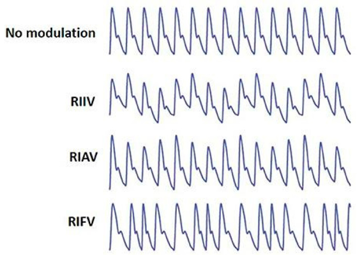
“RIAV causes significant changes in each peak amplitude.”
§ Sources of Inaccuracy in Photoplethysmography for Continuous Cardiovascular Monitoring > 3. Physiology > 3.1. Respiration
What the sources say
- Supportedcites [115] Dehkordi, 2018, Extracting instantaneous respiratory rate from multiple photoplethysmogram respiratory-induced variations, Front. Physiol. source
The Introduction (section 1.1, page unavailable) describes RIAV as a respiration-driven change in PPG pulse amplitude: amplitude decreases during inspiration and increases during expiration. This supports the citing sentence’s point about changes in peak amplitude; the source does not quantify their size. (LLM, conf=0.87)
- Supportedcites [121] Addison, 2012, Developing an algorithm for pulse oximetry derived respiratory rate (RR oxi): A healthy volunteer study, J. Clin. Monit. Comput. source
The respiratory-rate algorithm section describes pulse-amplitude modulation: respiration changes cardiac pulse amplitude, with amplitude decreasing during inspiration. Its Fig. 1 caption likewise describes cardiac pulse amplitudes varying over the respiratory cycle. The paper does not quantify the change in each individual peak, so “significant” is supported as a qualitative description, not a statistical result. (LLM, conf=0.88)
plaindirectn = 139- Asserted: passes — The authors put it forward as true.
- Checkable: passes — It can be checked against evidence.
- Load-bearing: unclear — It is unclear whether the paper’s conclusions depend on it.
- Based: passes — The paper gives a reason why it holds.
- Supported
The paper states this without a source or data behind it here, so there is nothing to check it against. The quote shows it is in the paper.
In the paper
“PPG-based RCIM devices that are U.S. Food and Drug Administration (FDA) cleared or approved and can accurately and consistently record clinical parameters in a sufficiently diverse population for true health monitoring are scarce.”
§ Sources of Inaccuracy in Photoplethysmography for Continuous Cardiovascular Monitoring > 1. Introduction
plain- Asserted: passes — The authors put it forward as true.
- Checkable: passes — It can be checked against evidence.
- Load-bearing: passes — The paper’s conclusions depend on it.
- Based: passes — The paper gives a reason why it holds.
None of the sources cited for this claim have been checked yet.
In the paper
“While, to the authors’ knowledge, publications that experimentally and explicitly demonstrate the effect of obesity or BMI on the PPG waveform are limited, we will explore works which suggest that there would be a substantial effect [56,59,60,61,62,63,64,65,66,67,68,69,70].”
§ Sources of Inaccuracy in Photoplethysmography for Continuous Cardiovascular Monitoring > 2. Individual Variations in the Human Population > 2.2. Obesity
What the sources say
- Not checked yetcites [56] Gibney, 2010, Skin and subcutaneous adipose layer thickness in adults with diabetes at sites used for insulin injections: Implications for needle length recommendations, Curr. Med. Res. Opin. source
- Not checked yetcites [59] Elgendi, 2012, On the analysis of fingertip photoplethysmogram signals, Curr. Cardiol. Rev. source
- Not checked yetcites [60] Iacopi, 2020, Using Skin Bioengineering to Highlight How Weight and Diabetes Mellitus Modify the Skin in the Lower Limbs of Super-Obese Patients, Diabetes Metab. Syndr. Obes. Targets Ther. source
- Not checked yetcites [61] Chin, 1999, Increased cutaneous blood flow but impaired post-ischemic response of nutritional flow in obese children, Atherosclerosis source
- Not checked yetcites [62] Francischetti, 2011, Skin capillary density and microvascular reactivity in obese subjects with and without metabolic syndrome, Microvasc. Res. source
- Not checked yetcites [63] Czernichow, 2010, Microvascular dysfunction in healthy insulin-sensitive overweight individuals, J. Hypertens. source
- Not checked yetcites [64] Debbabi, 2006, Increased skin capillary density in treated essential hypertensive patients, Am. J. Hypertens. source
- Not checked yetcites [65] Ijzerman, 2004, Impaired microvascular function in obesity: Implications for obesity-associated microangiopathy, hypertension, and insulin resistance, Circulation source
- Not checked yetcites [66] Kapur, 2013, Obesity is associated with a lower resting oxygen saturation in the ambulatory elderly: Results from the cardiovascular health study, Respir. Care source
- Not checked yetcites [67] Petrofsky, 2015, The effect of BMI on oxygen saturation at rest and during mild walking, J. Appl. Med. Sci.
hedged- Asserted: passes — The authors put it forward as true.
- Checkable: passes — It can be checked against evidence.
- Load-bearing: unclear — It is unclear whether the paper’s conclusions depend on it.
- Based: passes — The paper gives a reason why it holds.
The paper states this without a source or data behind it here, so there is nothing to check it against. The quote shows it is in the paper.
In the paper
“While many sources of noise had documented potential solutions, few had a comprehensive solution and even our understanding of how conditions such as obesity affect the skin and cardiovascular monitoring are still developing.”
§ Sources of Inaccuracy in Photoplethysmography for Continuous Cardiovascular Monitoring > 5. Summary
plain- Asserted: passes — The authors put it forward as true.
- Checkable: passes — It can be checked against evidence.
- Load-bearing: passes — The paper’s conclusions depend on it.
- Based: passes — The paper gives a reason why it holds.
The paper states this without a source or data behind it here, so there is nothing to check it against. The quote shows it is in the paper.
In the paper
“A review of the literature shows that the difficulty in creating such a device partially originates from the many sources of noise that researchers encounter.”
§ Sources of Inaccuracy in Photoplethysmography for Continuous Cardiovascular Monitoring > 5. Summary
plainconditional- Asserted: passes — The authors put it forward as true.
- Checkable: passes — It can be checked against evidence.
- Load-bearing: passes — The paper’s conclusions depend on it.
- Based: passes — The paper gives a reason why it holds.
A result the paper reports from its own data — it cites no other source for it, so there is none to check. The quote shows it is in the paper.
In the paper
“As shown in Table 7, we found that sources of noise can be divided primarily into three categories: individual variation, physiological processes, and external perturbation.”
§ Sources of Inaccuracy in Photoplethysmography for Continuous Cardiovascular Monitoring > 5. Summary
plain- Asserted: passes — The authors put it forward as true.
- Checkable: passes — It can be checked against evidence.
- Load-bearing: passes — The paper’s conclusions depend on it.
- Based: passes — The paper gives a reason why it holds.
The paper states this without a source or data behind it here, so there is nothing to check it against. The quote shows it is in the paper.
In the paper
“Systems aiming to derive such complex parameters do not always account for realistic sources of noise, as testing is performed within controlled parameter spaces.”
§ Sources of Inaccuracy in Photoplethysmography for Continuous Cardiovascular Monitoring > Abstract
plain- Asserted: passes — The authors put it forward as true.
- Checkable: passes — It can be checked against evidence.
- Load-bearing: passes — The paper’s conclusions depend on it.
- Based: passes — The paper gives a reason why it holds.
The paper states this without a source or data behind it here, so there is nothing to check it against. The quote shows it is in the paper.
In the paper
“The difficulty in determining features in PPG devices lies in the numerous sources of noise that can impede the output of the PPG.”
§ Sources of Inaccuracy in Photoplethysmography for Continuous Cardiovascular Monitoring > 1. Introduction
plaindirect- Asserted: passes — The authors put it forward as true.
- Checkable: passes — It can be checked against evidence.
- Load-bearing: passes — The paper’s conclusions depend on it.
- Based: passes — The paper gives a reason why it holds.
None of the sources cited for this claim have been checked yet.
In the paper
“The variation in the quasi-DC component is due to many factors: the optical properties of the tissue, average blood volume, respiration, vasomotor activity, vasoconstrictor waves, Traube Hering Meyer waves, and thermoregulation [4,5,6,7,8,9,10,11,12,13,14].”
§ Sources of Inaccuracy in Photoplethysmography for Continuous Cardiovascular Monitoring > 1. Introduction
What the sources say
- Not checked yetcites [4] Burton, 1940, A study of the adjustment of peripheral vascular tone to the requirements of the regulation of body temperature, Am. J. Physiol. Leg. Content source
- Not checked yetcites [5] Burton, 1939, The range and variability of the blood flow in the human fingers and the vasomotor regulation of body temperature, Am. J. Physiol. Leg. Content source
- Not checked yetcites [6] Hertzman, 1942, The vasomotor components in the vascular reactions in the finger to cold, Am. J. Physiol. Leg. Content source
- Not checked yetcites [7] Hertzman, 1942, The reactions of the digital artery and minute pad arteries to local cold, Am. J. Physiol. Leg. Content source
- Not checked yetcites [8] Hertzman, A.B., and Flath, F. (1963). Continuous Simultaneous Registration of Sweating and Blood Flow in a Small Skin Area, Saint Louis University. source
- Not checked yetcites [9] Hertzman, 1940, Applications of photoelectric plethysmography in peripheral vascular disease, Am. Heart J. source
- Not checked yetcites [10] Hertzman, 1942, The absence of vasoconstrictor reflexes in the forehead circulation. Effects of cold, Am. J. Physiol. Leg. Content source
- Not checked yetcites [11] Hyndman, 1971, Spontaneous rhythms in physiological control systems, Nature source
- Not checked yetcites [12] Fiser, 1978, Spectral analysis of resting variability of some circulatory parameters in man, Physiol. Bohemoslov.
- Not checked yetcites [13] Harness, 1989, Low-frequency photoplethysmograph signals, Clin. Phys. Physiol. Meas. source
plaindirect- Asserted: passes — The authors put it forward as true.
- Checkable: passes — It can be checked against evidence.
- Load-bearing: unclear — It is unclear whether the paper’s conclusions depend on it.
- Based: passes — The paper gives a reason why it holds.
- Probably not a substantial claim. 40
- It may state two separate claims as one. 102
- The quote only partly states this claim. 1
The paper states this without a source or data behind it here, so there is nothing to check it against. The quote shows it is in the paper.
In the paper
“All six identified devices with FDA status appear to be able to provide patients and providers with data for oxygen saturation (SpO2), respiration rate, and pulse rate.”
§ Sources of Inaccuracy in Photoplethysmography for Continuous Cardiovascular Monitoring > 1. Introduction
hedged- Asserted: passes — The authors put it forward as true.
- Checkable: passes — It can be checked against evidence.
- Load-bearing: fails — The paper’s conclusions may not depend on it.
- Based: passes — The paper gives a reason why it holds.
Probably not a substantial claim. The paper’s conclusions may not depend on it.
The paper states this without a source or data behind it here, so there is nothing to check it against. The quote shows it is in the paper.
In the paper
“In some cases, FDA approval is only for when the wearable is used within a software suite or healthcare framework. This is the case with the Samsung Gear S2, which is FDA cleared to monitor heart rate toward detecting atrial fibrillation when done with the LIVMOR Halo™ Detection System.”
§ Sources of Inaccuracy in Photoplethysmography for Continuous Cardiovascular Monitoring > 1. Introduction
plain- Asserted: passes — The authors put it forward as true.
- Checkable: passes — It can be checked against evidence.
- Load-bearing: fails — The paper’s conclusions may not depend on it.
- Based: passes — The paper gives a reason why it holds.
Probably not a substantial claim. The paper’s conclusions may not depend on it.
The paper states this without a source or data behind it here, so there is nothing to check it against. The quote shows it is in the paper.
In the paper
“To the authors’ knowledge, there is no work that comprehensively summarizes the literature surrounding these sources of noise and how they affect the waveform of the PPG and its derivatives.”
§ Sources of Inaccuracy in Photoplethysmography for Continuous Cardiovascular Monitoring > 1. Introduction
hedged- Asserted: passes — The authors put it forward as true.
- Checkable: passes — It can be checked against evidence.
- Load-bearing: passes — The paper’s conclusions depend on it.
- Based: fails — The paper may give no reason why it holds.
Probably not a substantial claim. The paper may give no reason why it holds.
The paper states this without a source or data behind it here, so there is nothing to check it against. The quote shows it is in the paper.
In the paper
“To the authors’ best knowledge, literature does not exist that demonstrates this relationship for the finger, a common location for pulse oximeters.”
§ Sources of Inaccuracy in Photoplethysmography for Continuous Cardiovascular Monitoring > 2. Individual Variations in the Human Population > 2.2. Obesity
hedged- Asserted: passes — The authors put it forward as true.
- Checkable: passes — It can be checked against evidence.
- Load-bearing: fails — The paper’s conclusions may not depend on it.
- Based: fails — The paper may give no reason why it holds.
Probably not a substantial claim. The paper’s conclusions may not depend on it. The paper may give no reason why it holds.
The paper states this without a source or data behind it here, so there is nothing to check it against. The quote shows it is in the paper.
In the paper
“It has been shown that the temperature of the body site where PPG data are being collected can influence the resultant waveform amplitude, and even the time between waveforms. This is summarized in Table 5.”
§ Sources of Inaccuracy in Photoplethysmography for Continuous Cardiovascular Monitoring > 3. Physiology > 3.4. Local Body Temperature
plaindirect- Asserted: passes — The authors put it forward as true.
- Checkable: passes — It can be checked against evidence.
- Load-bearing: passes — The paper’s conclusions depend on it.
- Based: passes — The paper gives a reason why it holds.
It may state two separate claims as one.
The paper states this without a source or data behind it here, so there is nothing to check it against. The quote shows it is in the paper.
In the paper
“While many sources of noise had documented potential solutions, few had a comprehensive solution and even our understanding of how conditions such as obesity affect the skin and cardiovascular monitoring are still developing.”
§ Sources of Inaccuracy in Photoplethysmography for Continuous Cardiovascular Monitoring > 5. Summary
plain- Asserted: passes — The authors put it forward as true.
- Checkable: fails — It may be too vague to check against evidence.
- Load-bearing: passes — The paper’s conclusions depend on it.
- Based: fails — The paper may give no reason why it holds.
Probably not a substantial claim. It may be too vague to check against evidence. The paper may give no reason why it holds.
A conclusion the paper draws from its own results — it cites no other source for it, so there is none to check. The quote shows it is in the paper.
In the paper
“PPG can fill this gap if a sufficiently accurate and precise device is developed.”
§ Sources of Inaccuracy in Photoplethysmography for Continuous Cardiovascular Monitoring > 1. Introduction
plainconditional- Asserted: passes — The authors put it forward as true.
- Checkable: passes — It can be checked against evidence.
- Load-bearing: passes — The paper’s conclusions depend on it.
- Based: passes — The paper gives a reason why it holds.
Probably not a substantial claim.
The paper states this without a source or data behind it here, so there is nothing to check it against. The quote shows it is in the paper.
In the paper
“PPG systems developed for remote and wearable use are typically for general wellness and fitness. This precludes it from being prescribed for medical use at home.”
§ Sources of Inaccuracy in Photoplethysmography for Continuous Cardiovascular Monitoring > 1. Introduction
plaindirect- Asserted: passes — The authors put it forward as true.
- Checkable: passes — It can be checked against evidence.
- Load-bearing: fails — The paper’s conclusions may not depend on it.
- Based: fails — The paper may give no reason why it holds.
Probably not a substantial claim. The paper’s conclusions may not depend on it. The paper may give no reason why it holds. It may state two separate claims as one.
A conclusion the paper draws from its own results — it cites no other source for it, so there is none to check. The quote shows it is in the paper.
In the paper
“The indications for use in these devices are very significant advancements in remote monitoring, but still lag in the potential prognostic capabilities of the PPG.”
§ Sources of Inaccuracy in Photoplethysmography for Continuous Cardiovascular Monitoring > 1. Introduction
plain- Asserted: passes — The authors put it forward as true.
- Checkable: fails — It may be too vague to check against evidence.
- Load-bearing: fails — The paper’s conclusions may not depend on it.
- Based: fails — The paper may give no reason why it holds.
Probably not a substantial claim. It may be too vague to check against evidence. The paper’s conclusions may not depend on it. The paper may give no reason why it holds. It may state two separate claims as one.
None of the sources cited for this claim have been checked yet.
In the paper
“Remote and continuous/intermittent monitoring (RCIM) has proven to be a promising route to deliver preventative care by reducing both the death rate and burdens placed on the healthcare system [1,2,3].”
§ Sources of Inaccuracy in Photoplethysmography for Continuous Cardiovascular Monitoring > 1. Introduction
What the sources say
- Not checked yetcites [1] Wilkoff, 2008, HRS/EHRA expert consensus on the monitoring of cardiovascular implantable electronic devices (CIEDs): Description of techniques, indications, personnel, frequency and ethical considerations: Developed in partnership with the Heart Rhythm Society (HRS) and the European Heart Rhythm Association (EHRA); and in collaboration with the American College of Cardiology (ACC), the American Heart Association (AHA), the European Society of Cardiology (ESC), the Heart Failure Association of ESC (HFA), and the Heart Failure Society of America (HFSA). Endorsed by the Heart Rhythm Society, the source
- Not checked yetcites [2] Roger, 2011, Heart disease and stroke statistics—2011 update: A report from the American Heart Association, Circulation source
- Not checked yetcites [3] Jencks, 2009, Rehospitalizations among patients in the Medicare fee-for-service program, N. Engl. J. Med. source
boosteddirect- Asserted: passes — The authors put it forward as true.
- Checkable: passes — It can be checked against evidence.
- Load-bearing: fails — The paper’s conclusions may not depend on it.
- Based: passes — The paper gives a reason why it holds.
Probably not a substantial claim. The paper’s conclusions may not depend on it. It may state two separate claims as one.
In the paper
“The blood pressure cuff, ECG patch, and Holter monitor are also not often used for long-term remote monitoring [24].”
§ Sources of Inaccuracy in Photoplethysmography for Continuous Cardiovascular Monitoring > 1. Introduction
What the sources say
- Not checked yetcites [24] Bansal, 2018, Portable out-of-hospital electrocardiography: A review of current technologies, J. Arrhythmia source
plain- Asserted: passes — The authors put it forward as true.
- Checkable: passes — It can be checked against evidence.
- Load-bearing: passes — The paper’s conclusions depend on it.
- Based: passes — The paper gives a reason why it holds.
It may state two separate claims as one.
The paper states this without a source or data behind it here, so there is nothing to check it against. The quote shows it is in the paper.
In the paper
“In addition, the hardware and software within the device itself can contribute to the noise.”
§ Sources of Inaccuracy in Photoplethysmography for Continuous Cardiovascular Monitoring > 1. Introduction
plaindirect- Asserted: passes — The authors put it forward as true.
- Checkable: passes — It can be checked against evidence.
- Load-bearing: fails — The paper’s conclusions may not depend on it.
- Based: fails — The paper may give no reason why it holds.
Probably not a substantial claim. The paper’s conclusions may not depend on it. The paper may give no reason why it holds.
In the paper
“Additionally, these tests are rarely administered preventatively, despite research which concludes that preventative testing could reduce deaths by up to 25% [1].”
§ Sources of Inaccuracy in Photoplethysmography for Continuous Cardiovascular Monitoring > 1. Introduction
What the sources say
- Not checked yetcites [1] Wilkoff, 2008, HRS/EHRA expert consensus on the monitoring of cardiovascular implantable electronic devices (CIEDs): Description of techniques, indications, personnel, frequency and ethical considerations: Developed in partnership with the Heart Rhythm Society (HRS) and the European Heart Rhythm Association (EHRA); and in collaboration with the American College of Cardiology (ACC), the American Heart Association (AHA), the European Society of Cardiology (ESC), the Heart Failure Association of ESC (HFA), and the Heart Failure Society of America (HFSA). Endorsed by the Heart Rhythm Society, the source
boosteddirect- Asserted: passes — The authors put it forward as true.
- Checkable: passes — It can be checked against evidence.
- Load-bearing: fails — The paper’s conclusions may not depend on it.
- Based: passes — The paper gives a reason why it holds.
Probably not a substantial claim. The paper’s conclusions may not depend on it. It may state two separate claims as one.
The paper states this without a source or data behind it here, so there is nothing to check it against. The quote shows it is in the paper.
In the paper
“These sources of error pertain to variation within and across individuals (e.g., skin tone, obesity, age, and gender), physiology (e.g., respiration, venous pulsation, body site of measurement, and body temperature), and external perturbations of the device itself (e.g., motion artifact, ambient light, and applied pressure to the skin).”
§ Sources of Inaccuracy in Photoplethysmography for Continuous Cardiovascular Monitoring > 1. Introduction
plain- Asserted: passes — The authors put it forward as true.
- Checkable: passes — It can be checked against evidence.
- Load-bearing: passes — The paper’s conclusions depend on it.
- Based: fails — The paper may give no reason why it holds.
Probably not a substantial claim. The paper may give no reason why it holds. It may state two separate claims as one.
None of the sources cited for this claim have been checked yet.
In the paper
“Additionally, some parameters in the literature such as pulse transit time (PTT), which is used to determine pulse wave velocity (PWV) and estimate blood pressure without a cuff, requires extraction of the time delay from two PPG waveforms or from an ECG and PPG waveform [31,32].”
§ Sources of Inaccuracy in Photoplethysmography for Continuous Cardiovascular Monitoring > 1. Introduction
What the sources say
- Not checked yetcites [31] Nabeel, 2018, Arterial blood pressure estimation from local pulse wave velocity using dual-element photoplethysmograph probe, IEEE Trans. Instrum. Meas. source
- Not checked yetcites [32] Nabeel, P., Karthik, S., Joseph, J., and Sivaprakasam, M. (2017, January 8–10). Experimental validation of dual PPG local pulse wave velocity probe. Proceedings of the 2017 IEEE International Symposium on Medical Measurements and Applications (MeMeA), Rochester, MN, USA. source
plain- Asserted: passes — The authors put it forward as true.
- Checkable: passes — It can be checked against evidence.
- Load-bearing: fails — The paper’s conclusions may not depend on it.
- Based: passes — The paper gives a reason why it holds.
Probably not a substantial claim. The paper’s conclusions may not depend on it.
The paper states this without a source or data behind it here, so there is nothing to check it against. The quote shows it is in the paper.
In the paper
“Numerous non-FDA cleared/approved fitness devices exist and can estimate heart rate by quantifying the number of systolic peaks in a period of time, but a single parameter limits the amount of extractable information.”
§ Sources of Inaccuracy in Photoplethysmography for Continuous Cardiovascular Monitoring > 1. Introduction
plaindirect- Asserted: passes — The authors put it forward as true.
- Checkable: passes — It can be checked against evidence.
- Load-bearing: fails — The paper’s conclusions may not depend on it.
- Based: passes — The paper gives a reason why it holds.
Probably not a substantial claim. The paper’s conclusions may not depend on it. It may state two separate claims as one.
The paper states this without a source or data behind it here, so there is nothing to check it against. The quote shows it is in the paper.
In the paper
“However, complex analyses of the PPG waveform yield valuable clinical information, such as: blood pressure, respiratory information, sympathetic nervous system activity, and heart rate variability.”
§ Sources of Inaccuracy in Photoplethysmography for Continuous Cardiovascular Monitoring > Abstract
plaindirect- Asserted: passes — The authors put it forward as true.
- Checkable: passes — It can be checked against evidence.
- Load-bearing: passes — The paper’s conclusions depend on it.
- Based: fails — The paper may give no reason why it holds.
Probably not a substantial claim. The paper may give no reason why it holds.
The paper states this without a source or data behind it here, so there is nothing to check it against. The quote shows it is in the paper.
In the paper
“Overall, the literature has demonstrated the potential diagnostic and prognostic strength for the PPG; however, the PPG features can only be utilized if the waveform is of a high quality with high signal-to-noise ratio (SNR).”
§ Sources of Inaccuracy in Photoplethysmography for Continuous Cardiovascular Monitoring > 1. Introduction
boostedconditional- Asserted: passes — The authors put it forward as true.
- Checkable: passes — It can be checked against evidence.
- Load-bearing: passes — The paper’s conclusions depend on it.
- Based: passes — The paper gives a reason why it holds.
It may state two separate claims as one.
None of the sources cited for this claim have been checked yet.
In the paper
“Beyond fitness and heart rate monitoring, the primary medical use of the PPG has been focused on obtaining information about the cardiovascular system towards cardiovascular disease (CVD) diagnosis and treatment [17,18].”
§ Sources of Inaccuracy in Photoplethysmography for Continuous Cardiovascular Monitoring > 1. Introduction
What the sources say
- Not checked yetcites [17] Elgendi, 2019, The use of photoplethysmography for assessing hypertension, NPJ Digit. Med. source
- Not checked yetcites [18] Vakily, 2017, A system for continuous estimating and monitoring cardiac output via arterial waveform analysis, J. Biomed. Phys. Eng.
plain- Asserted: passes — The authors put it forward as true.
- Checkable: passes — It can be checked against evidence.
- Load-bearing: fails — The paper’s conclusions may not depend on it.
- Based: passes — The paper gives a reason why it holds.
Probably not a substantial claim. The paper’s conclusions may not depend on it.
A conclusion the paper draws from its own results — it cites no other source for it, so there is none to check. The quote shows it is in the paper.
In the paper
“A wearable monitoring tool to be used beyond fitness and heart rate must account for noise sources originating from individual patient variations (e.g., skin tone, obesity, age, and gender), physiology (e.g., respiration, venous pulsation, body site of measurement, and body temperature), and external perturbations of the device itself (e.g., motion artifact, ambient light, and applied pressure to”
§ Sources of Inaccuracy in Photoplethysmography for Continuous Cardiovascular Monitoring > Abstract
plain- Asserted: passes — The authors put it forward as true.
- Checkable: passes — It can be checked against evidence.
- Load-bearing: passes — The paper’s conclusions depend on it.
- Based: passes — The paper gives a reason why it holds.
Probably not a substantial claim.
A conclusion the paper draws from its own results — it cites no other source for it, so there is none to check. The quote shows it is in the paper.
In the paper
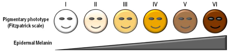
“These results indicate that overall, the mean modulation goes down with increasing Fitzpatrick scale, and with exercise one would expect greater blood volume changes causing a higher signal for the more absorbing blue/green wavelengths, at least for the source to detector distances used in the study.”
§ Sources of Inaccuracy in Photoplethysmography for Continuous Cardiovascular Monitoring > 2. Individual Variations in the Human Population > 2.1. Skin Tone
hedgedconditional- Asserted: passes — The authors put it forward as true.
- Checkable: passes — It can be checked against evidence.
- Load-bearing: passes — The paper’s conclusions depend on it.
- Based: passes — The paper gives a reason why it holds.
It may state two separate claims as one.
In the paper
“With 20 subjects ranging from Fitzpatrick II to Fitzpatrick IV, the perfusion index (AC/DC), pulsatile strength, and SNR were all found to increase when data collected with the 590 nm LED were analyzed.”
§ Sources of Inaccuracy in Photoplethysmography for Continuous Cardiovascular Monitoring > 2. Individual Variations in the Human Population > 2.1. Skin Tone
What the sources say
- Not checked yetcites [51] Mohapatra, P., Preejith, S., and Sivaprakasam, M. (2017, January 22–25). A novel sensor for wrist based optical heart rate monitor. Proceedings of the 2017 IEEE International Instrumentation and Measurement Technology Conference (I2MTC), Torino, Italy. source
plaindirectn = 20- Asserted: passes — The authors put it forward as true.
- Checkable: passes — It can be checked against evidence.
- Load-bearing: unclear — It is unclear whether the paper’s conclusions depend on it.
- Based: passes — The paper gives a reason why it holds.
It may state two separate claims as one.
In the paper
“Obesity, determined by having a body mass index greater than 30, affects 40% of the United States and leads to cardiovascular disease, hypertension, and type 2 diabetes [57].”
§ Sources of Inaccuracy in Photoplethysmography for Continuous Cardiovascular Monitoring > 2. Individual Variations in the Human Population > 2.2. Obesity
What the sources say
- Not checked yetcites [57] Poirier, 2006, Obesity and cardiovascular disease: Pathophysiology, evaluation, and effect of weight loss: An update of the 1997 American Heart Association Scientific Statement on Obesity and Heart Disease from the Obesity Committee of the Council on Nutrition, Physical Activity, and Metabolism, Circulation source
plaindirect- Asserted: passes — The authors put it forward as true.
- Checkable: passes — It can be checked against evidence.
- Load-bearing: fails — The paper’s conclusions may not depend on it.
- Based: passes — The paper gives a reason why it holds.
Probably not a substantial claim. The paper’s conclusions may not depend on it. It may state two separate claims as one.
A conclusion the paper draws from its own results — it cites no other source for it, so there is none to check. The quote shows it is in the paper.
In the paper
“However, the wavelength range needs to be optimized for both skin tone and blood absorption, particularly as more advanced parameters are derived from PPG signals, as the absorption of green light by melanin in individuals with a darker skin tone limits the light penetration to the subcutaneous tissue where the blood is located.”
§ Sources of Inaccuracy in Photoplethysmography for Continuous Cardiovascular Monitoring > 2. Individual Variations in the Human Population > 2.1. Skin Tone
plaindirect- Asserted: passes — The authors put it forward as true.
- Checkable: passes — It can be checked against evidence.
- Load-bearing: passes — The paper’s conclusions depend on it.
- Based: passes — The paper gives a reason why it holds.
It may state two separate claims as one.
In the paper
“It was found that relative amplitude of the AC component to the entire signal (AC + DC) increased as LED/photodiode spacing increased from 4 to 11 mm, although no statistical analyses were presented.”
§ Sources of Inaccuracy in Photoplethysmography for Continuous Cardiovascular Monitoring > 2. Individual Variations in the Human Population > 2.1. Skin Tone
What the sources say
- Not checked yetcites [53] Mendelson, 1988, Noninvasive pulse oximetry utilizing skin reflectance photoplethysmography, IEEE Trans. Biomed. Eng. source
plaindirect- Asserted: passes — The authors put it forward as true.
- Checkable: passes — It can be checked against evidence.
- Load-bearing: fails — The paper’s conclusions may not depend on it.
- Based: passes — The paper gives a reason why it holds.
Probably not a substantial claim. The paper’s conclusions may not depend on it. It may state two separate claims as one.
In the paper
“There was an error of 1.04 beats per minute (BPM) for “fair” skinned individuals and an error of 10.90 BPM for “dark” skinned participants, citing the lack of usability of their device for dark skinned individuals.”
§ Sources of Inaccuracy in Photoplethysmography for Continuous Cardiovascular Monitoring > 2. Individual Variations in the Human Population > 2.1. Skin Tone
What the sources say
- Not checked yetcites [40] Preejith, S., Alex, A., Joseph, J., and Sivaprakasam, M. (2016, January 12–14). Design, development and clinical validation of a wrist-based optical heart rate monitor. Proceedings of the 2016 IEEE International Symposium on Medical Measurements and Applications (MeMeA), Benevento, Italy. source
plaindirectn = 256- Asserted: passes — The authors put it forward as true.
- Checkable: passes — It can be checked against evidence.
- Load-bearing: passes — The paper’s conclusions depend on it.
- Based: passes — The paper gives a reason why it holds.
It may state two separate claims as one.
In the paper
“Blood flow regulation and oxygen saturation are both known to deviate with respect to obesity and BMI [71].”
§ Sources of Inaccuracy in Photoplethysmography for Continuous Cardiovascular Monitoring > 2. Individual Variations in the Human Population > 2.2. Obesity
What the sources say
- Not checked yetcites [71] Limberg, 2016, Peripheral Blood Flow Regulation in Human Obesity and Metabolic Syndrome, Exerc. Sport Sci. Rev. source
boostedcorrelational- Asserted: passes — The authors put it forward as true.
- Checkable: passes — It can be checked against evidence.
- Load-bearing: passes — The paper’s conclusions depend on it.
- Based: passes — The paper gives a reason why it holds.
It may state two separate claims as one.
In the paper
“Preejith et al. confirmed that skin tone matters when using green light only. The analysis of their PPG biosensor (single source 535 nm LED) showed a significant direct correlation SNR of the heart rate with skin tone, indicating that a darker skin tone yields higher error in the measurement [40].”
§ Sources of Inaccuracy in Photoplethysmography for Continuous Cardiovascular Monitoring > 2. Individual Variations in the Human Population > 2.1. Skin Tone
What the sources say
- Not checked yetcites [40] Preejith, S., Alex, A., Joseph, J., and Sivaprakasam, M. (2016, January 12–14). Design, development and clinical validation of a wrist-based optical heart rate monitor. Proceedings of the 2016 IEEE International Symposium on Medical Measurements and Applications (MeMeA), Benevento, Italy. source
boostedcorrelationaln = 256- Asserted: passes — The authors put it forward as true.
- Checkable: passes — It can be checked against evidence.
- Load-bearing: passes — The paper’s conclusions depend on it.
- Based: passes — The paper gives a reason why it holds.
It may state two separate claims as one.
The paper states this without a source or data behind it here, so there is nothing to check it against. The quote shows it is in the paper.
In the paper
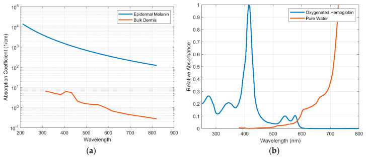
“However, many PPG devices use green light (~550 nm). The decision to use green light in most PPG systems is primarily driven by the relatively high absorption spectrum of hemoglobin in this range (Figure 3b), which is the main absorber in blood and thus can potentially give a strong pulsatile signal with changes in blood volume.”
§ Sources of Inaccuracy in Photoplethysmography for Continuous Cardiovascular Monitoring > 2. Individual Variations in the Human Population > 2.1. Skin Tone
plaindirect- Asserted: passes — The authors put it forward as true.
- Checkable: passes — It can be checked against evidence.
- Load-bearing: unclear — It is unclear whether the paper’s conclusions depend on it.
- Based: passes — The paper gives a reason why it holds.
It may state two separate claims as one.
None of the sources cited for this claim have been checked yet.
In the paper
“In particular, obesity can lead to changes in skin thickness, blood flow, and oxygen saturation. This affects the optical properties of skin in addition to the distance light has to travel to reach a target vessel or vessels [48,59,60].”
§ Sources of Inaccuracy in Photoplethysmography for Continuous Cardiovascular Monitoring > 2. Individual Variations in the Human Population > 2.2. Obesity
What the sources say
- Not checked yetcites [48] Jacques, 2013, Optical properties of biological tissues: A review, Phys. Med. Biol. source
- Not checked yetcites [59] Elgendi, 2012, On the analysis of fingertip photoplethysmogram signals, Curr. Cardiol. Rev. source
- Not checked yetcites [60] Iacopi, 2020, Using Skin Bioengineering to Highlight How Weight and Diabetes Mellitus Modify the Skin in the Lower Limbs of Super-Obese Patients, Diabetes Metab. Syndr. Obes. Targets Ther. source
plaindirect- Asserted: passes — The authors put it forward as true.
- Checkable: passes — It can be checked against evidence.
- Load-bearing: passes — The paper’s conclusions depend on it.
- Based: passes — The paper gives a reason why it holds.
It may state two separate claims as one.
None of the sources cited for this claim have been checked yet.
In the paper
“Individuals with obesity experience increased cutaneous blood flow to meet the oxygenation needs of tissue. However, the blood flow of adipose tissue generally decreases with obesity both after a meal and during a fasting state [61,72].”
§ Sources of Inaccuracy in Photoplethysmography for Continuous Cardiovascular Monitoring > 2. Individual Variations in the Human Population > 2.2. Obesity
What the sources say
- Not checked yetcites [61] Chin, 1999, Increased cutaneous blood flow but impaired post-ischemic response of nutritional flow in obese children, Atherosclerosis source
- Not checked yetcites [72] Altintas, 2016, In vivo reflectance-mode confocal microscopy assessments: Impact of overweight on human skin microcirculation and histomorphology, J. Biomed. Opt. source
plaindirect- Asserted: passes — The authors put it forward as true.
- Checkable: passes — It can be checked against evidence.
- Load-bearing: passes — The paper’s conclusions depend on it.
- Based: passes — The paper gives a reason why it holds.
It may state two separate claims as one.
None of the sources cited for this claim have been checked yet.
In the paper
“These often-overlooked disparities with diversity (e.g., skin tone and obesity) are now becoming more documented in the literature [34,35,36].”
§ Sources of Inaccuracy in Photoplethysmography for Continuous Cardiovascular Monitoring > 1. Introduction
What the sources say
- Not checked yetcites [34] Colvonen, P.J., DeYoung, P.N., Bosompra, N.-O.A., and Owens, R.L. (2020). Limiting Racial Disparities and Bias for Wearable Devices in Health Science Research, Oxford University Press US. source
- Not checked yetcites [35] Hidalgo, 2021, Critical care trainees call for pulse oximetry reform, Lancet Respir. Med. source
- Not checked yetcites [36] Rodriguez, 2021, Synthetic Photoplethysmography (PPG) of the radial artery through parallelized Monte Carlo and its correlation to Body Mass Index (BMI), Sci. Rep. source
plain- Asserted: passes — The authors put it forward as true.
- Checkable: passes — It can be checked against evidence.
- Load-bearing: fails — The paper’s conclusions may not depend on it.
- Based: passes — The paper gives a reason why it holds.
Probably not a substantial claim. The paper’s conclusions may not depend on it.
The paper states this without a source or data behind it here, so there is nothing to check it against. The quote shows it is in the paper.
In the paper
“This is due to the high absorbance of eumelanin with a peak in the ultraviolet wavelength (220 nm) and a steady decay through the visible wavelength region.”
§ Sources of Inaccuracy in Photoplethysmography for Continuous Cardiovascular Monitoring > 2. Individual Variations in the Human Population > 2.1. Skin Tone
plain- Asserted: passes — The authors put it forward as true.
- Checkable: passes — It can be checked against evidence.
- Load-bearing: passes — The paper’s conclusions depend on it.
- Based: passes — The paper gives a reason why it holds.
It may state two separate claims as one.
The paper states this without a source or data behind it here, so there is nothing to check it against. The quote shows it is in the paper.
In the paper
“Since the absorption of epidermal melanin is much higher in the visible region of light and much lower in the near infrared region (NIR), the NIR range of light will travel further through pigmented skin.”
§ Sources of Inaccuracy in Photoplethysmography for Continuous Cardiovascular Monitoring > 2. Individual Variations in the Human Population > 2.1. Skin Tone
plaindirect- Asserted: passes — The authors put it forward as true.
- Checkable: passes — It can be checked against evidence.
- Load-bearing: passes — The paper’s conclusions depend on it.
- Based: passes — The paper gives a reason why it holds.
It may state two separate claims as one.
In the paper
“In determining heart rate, it was found that bias (defined as the mean difference between the OH1 and ground truth) increased with darker skin (p < 0.001), and heart rate accuracy was positively correlated to skin tone (p < 0.05).”
§ Sources of Inaccuracy in Photoplethysmography for Continuous Cardiovascular Monitoring > 2. Individual Variations in the Human Population > 2.1. Skin Tone
What the sources say
- Not checked yetcites [41] Hermand, 2019, Validation of a photoplethysmographic heart rate monitor: Polar OH1, Int. J. Sports Med. source
plaincorrelationaln = 70 ·- Asserted: passes — The authors put it forward as true.
- Checkable: passes — It can be checked against evidence.
- Load-bearing: passes — The paper’s conclusions depend on it.
- Based: passes — The paper gives a reason why it holds.
It may state two separate claims as one.
In the paper
“Additionally, the wearable devices used in this study utilized red and near infrared light, which can more easily penetrate the epidermis.”
§ Sources of Inaccuracy in Photoplethysmography for Continuous Cardiovascular Monitoring > 2. Individual Variations in the Human Population > 2.1. Skin Tone
What the sources say
- Not checked yetcites [46] Bent, 2020, Investigating sources of inaccuracy in wearable optical heart rate sensors, NPJ Digit. Med. source
plaindirectn = 56- Asserted: passes — The authors put it forward as true.
- Checkable: passes — It can be checked against evidence.
- Load-bearing: fails — The paper’s conclusions may not depend on it.
- Based: passes — The paper gives a reason why it holds.
Probably not a substantial claim. The paper’s conclusions may not depend on it. It may state two separate claims as one.
In the paper
“For Fitzpatrick IV individuals, the perfusion index increased between 1.2 and 7.1 times with the use of the 590 light, while pulsatile strength increased by 1.1 to 3.1 times and SNR increased by 1.3 to 2.6 times, although no statistical significance analyses were performed [51].”
§ Sources of Inaccuracy in Photoplethysmography for Continuous Cardiovascular Monitoring > 2. Individual Variations in the Human Population > 2.1. Skin Tone
What the sources say
- Not checked yetcites [51] Mohapatra, P., Preejith, S., and Sivaprakasam, M. (2017, January 22–25). A novel sensor for wrist based optical heart rate monitor. Proceedings of the 2017 IEEE International Instrumentation and Measurement Technology Conference (I2MTC), Torino, Italy. source
plaindirectn = 20- Asserted: passes — The authors put it forward as true.
- Checkable: passes — It can be checked against evidence.
- Load-bearing: unclear — It is unclear whether the paper’s conclusions depend on it.
- Based: passes — The paper gives a reason why it holds.
It may state two separate claims as one.
In the paper
“In the resting condition, green light had larger mean modulation (p < 0.001) than other wavelengths, and after exercise, blue and green had greater SNR ratios than red or infrared (p < 0.001).”
§ Sources of Inaccuracy in Photoplethysmography for Continuous Cardiovascular Monitoring > 2. Individual Variations in the Human Population > 2.1. Skin Tone
What the sources say
- Not checked yetcites [52] Fallow, 2013, Influence of skin type and wavelength on light wave reflectance, J. Clin. Monit. Comput. source
plaincorrelational- Asserted: passes — The authors put it forward as true.
- Checkable: passes — It can be checked against evidence.
- Load-bearing: passes — The paper’s conclusions depend on it.
- Based: passes — The paper gives a reason why it holds.
It may state two separate claims as one.
A conclusion the paper draws from its own results — it cites no other source for it, so there is none to check. The quote shows it is in the paper.
In the paper
“It is possible that the lack of increase in error observed is due to the already large error present in the results reported—the mean absolute error is approximately 9 BPM across all skin tones.”
§ Sources of Inaccuracy in Photoplethysmography for Continuous Cardiovascular Monitoring > 2. Individual Variations in the Human Population > 2.1. Skin Tone
hedgedconditionaln = 56- Asserted: passes — The authors put it forward as true.
- Checkable: passes — It can be checked against evidence.
- Load-bearing: fails — The paper’s conclusions may not depend on it.
- Based: passes — The paper gives a reason why it holds.
Probably not a substantial claim. The paper’s conclusions may not depend on it.
A conclusion the paper draws from its own results — it cites no other source for it, so there is none to check. The quote shows it is in the paper.
In the paper
“Most of the listed changes are due to the cardiovascular system, as arteries change how they respond to a cardiac pulse as the vessel ages.”
§ Sources of Inaccuracy in Photoplethysmography for Continuous Cardiovascular Monitoring > 2. Individual Variations in the Human Population > 2.3. Age
plaindirect- Asserted: passes — The authors put it forward as true.
- Checkable: passes — It can be checked against evidence.
- Load-bearing: unclear — It is unclear whether the paper’s conclusions depend on it.
- Based: passes — The paper gives a reason why it holds.
It may state two separate claims as one.
None of the sources cited for this claim have been checked yet.
In the paper
“Perhaps most detrimental to the PPG waveform is skin thickness, as it is directly correlated to BMI and can dampen PPG signal amplitude [56,70].”
§ Sources of Inaccuracy in Photoplethysmography for Continuous Cardiovascular Monitoring > 2. Individual Variations in the Human Population > 2.2. Obesity
What the sources say
- Not checked yetcites [56] Gibney, 2010, Skin and subcutaneous adipose layer thickness in adults with diabetes at sites used for insulin injections: Implications for needle length recommendations, Curr. Med. Res. Opin. source
- Not checked yetcites [70] Derraik, J.G., Rademaker, M., Cutfield, W.S., Pinto, T.E., Tregurtha, S., Faherty, A., Peart, J.M., Drury, P.L., and Hofman, P.L. (2014). Effects of age, gender, BMI, and anatomical site on skin thickness in children and adults with diabetes. PLoS ONE, 9. source
plaindirect- Asserted: passes — The authors put it forward as true.
- Checkable: passes — It can be checked against evidence.
- Load-bearing: passes — The paper’s conclusions depend on it.
- Based: passes — The paper gives a reason why it holds.
It may state two separate claims as one.
None of the sources cited for this claim have been checked yet.
In the paper
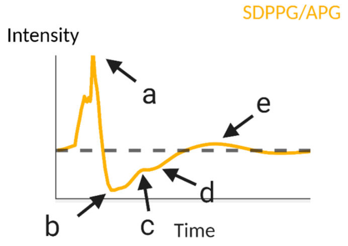
“The ratio b/a has been found to correlate positively to age, while c/a, d/a and e/a have all been correlated negatively to age [29,59].”
§ Sources of Inaccuracy in Photoplethysmography for Continuous Cardiovascular Monitoring > 2. Individual Variations in the Human Population > 2.3. Age
What the sources say
plaincorrelational- Asserted: passes — The authors put it forward as true.
- Checkable: passes — It can be checked against evidence.
- Load-bearing: unclear — It is unclear whether the paper’s conclusions depend on it.
- Based: passes — The paper gives a reason why it holds.
It may state two separate claims as one.
In the paper
“Keratinocytes become shorter, water content decreases, lipid content decreases, and most notably, collagen synthesis and turnover decrease [93].”
§ Sources of Inaccuracy in Photoplethysmography for Continuous Cardiovascular Monitoring > 2. Individual Variations in the Human Population > 2.3. Age
What the sources say
- Not checked yetcites [93] Farage, 2013, Characteristics of the aging skin, Adv. Wound Care source
plaindirect- Asserted: passes — The authors put it forward as true.
- Checkable: passes — It can be checked against evidence.
- Load-bearing: passes — The paper’s conclusions depend on it.
- Based: passes — The paper gives a reason why it holds.
It may state two separate claims as one.
A conclusion the paper draws from its own results — it cites no other source for it, so there is none to check. The quote shows it is in the paper.
In the paper
“This change in skin thickness decreases the necessary propagation of light to obtain a PPG signal, which can contribute to an increase in PPG signal strength, along with capillary depletion.”
§ Sources of Inaccuracy in Photoplethysmography for Continuous Cardiovascular Monitoring > 2. Individual Variations in the Human Population > 2.3. Age
plainconditional- Asserted: passes — The authors put it forward as true.
- Checkable: passes — It can be checked against evidence.
- Load-bearing: unclear — It is unclear whether the paper’s conclusions depend on it.
- Based: passes — The paper gives a reason why it holds.
It may state two separate claims as one.
A conclusion the paper draws from its own results — it cites no other source for it, so there is none to check. The quote shows it is in the paper.
In the paper
“The increased absorption of deoxygenated hemoglobin compared to that of oxygenated hemoglobin in the far-red region could decrease SNR of PPG measurements that use those wavelengths of light.”
§ Sources of Inaccuracy in Photoplethysmography for Continuous Cardiovascular Monitoring > 2. Individual Variations in the Human Population > 2.2. Obesity
hedgedconditional- Asserted: passes — The authors put it forward as true.
- Checkable: passes — It can be checked against evidence.
- Load-bearing: fails — The paper’s conclusions may not depend on it.
- Based: fails — The paper may give no reason why it holds.
Probably not a substantial claim. The paper’s conclusions may not depend on it. The paper may give no reason why it holds.
The paper states this without a source or data behind it here, so there is nothing to check it against. The quote shows it is in the paper.
In the paper
“PPG principally measures the response/elasticity of arteries to blood flow, and thus will change if the properties governing artery compliance changes.”
§ Sources of Inaccuracy in Photoplethysmography for Continuous Cardiovascular Monitoring > 2. Individual Variations in the Human Population > 2.3. Age
plaindirect- Asserted: passes — The authors put it forward as true.
- Checkable: passes — It can be checked against evidence.
- Load-bearing: passes — The paper’s conclusions depend on it.
- Based: passes — The paper gives a reason why it holds.
It may state two separate claims as one.
In the paper
“A Holtain skin caliper applied to the dorsal forearm found a decrease in skin thickness, and an infrared PPG was used to determine PPG amplitude. The authors attribute the positive, direct relationship between PPG amplitude and age to capillary depletion, due to similar skin thicknesses observed between older participants and children, despite an increased PPG amplitude in the older participants.”
§ Sources of Inaccuracy in Photoplethysmography for Continuous Cardiovascular Monitoring > 2. Individual Variations in the Human Population > 2.3. Age
What the sources say
- Not checked yetcites [94] Leveque, 1984, In vivo studies of the evolution of physical properties of the human skin with age, Int. J. Dermatol. source
plaindirectn = 69- Asserted: passes — The authors put it forward as true.
- Checkable: passes — It can be checked against evidence.
- Load-bearing: fails — The paper’s conclusions may not depend on it.
- Based: passes — The paper gives a reason why it holds.
Probably not a substantial claim. The paper’s conclusions may not depend on it. It may state two separate claims as one.
In the paper
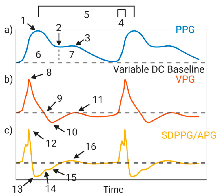
“Using data from 93 individuals of various ages, Ahn et al. extracted features from fingertip-based PPG and the second derivative PPG, acceleration plethysmography (APG, Figure 4), and correlated them to chronological age in order to determine what they defined as the vascular age index [83].”
§ Sources of Inaccuracy in Photoplethysmography for Continuous Cardiovascular Monitoring > 2. Individual Variations in the Human Population > 2.3. Age
What the sources say
- Not checked yetcites [83] Ahn, 2017, New aging index using signal features of both photoplethysmograms and acceleration plethysmograms, Healthc. Inform. Res. source
plaincorrelationaln = 93- Asserted: passes — The authors put it forward as true.
- Checkable: passes — It can be checked against evidence.
- Load-bearing: fails — The paper’s conclusions may not depend on it.
- Based: passes — The paper gives a reason why it holds.
Probably not a substantial claim. The paper’s conclusions may not depend on it.
In the paper
“Another study by Hartmann et al. determined that there is no statistically significant change in PPG amplitude as a function of age; however, this work featured 36 individuals ranging in age from only 33 to 58.”
§ Sources of Inaccuracy in Photoplethysmography for Continuous Cardiovascular Monitoring > 2. Individual Variations in the Human Population > 2.3. Age
What the sources say
- Not checked yetcites [95] Hartmann, 2019, Quantitative comparison of photoplethysmographic waveform characteristics: Effect of measurement site, Front. Physiol. source
plainn = 36- Asserted: passes — The authors put it forward as true.
- Checkable: passes — It can be checked against evidence.
- Load-bearing: fails — The paper’s conclusions may not depend on it.
- Based: passes — The paper gives a reason why it holds.
Probably not a substantial claim. The paper’s conclusions may not depend on it.
The paper states this without a source or data behind it here, so there is nothing to check it against. The quote shows it is in the paper.
In the paper
“However, the integumentary system also responds to aging.”
§ Sources of Inaccuracy in Photoplethysmography for Continuous Cardiovascular Monitoring > 2. Individual Variations in the Human Population > 2.3. Age
plaincorrelational- Asserted: passes — The authors put it forward as true.
- Checkable: passes — It can be checked against evidence.
- Load-bearing: fails — The paper’s conclusions may not depend on it.
- Based: fails — The paper may give no reason why it holds.
Probably not a substantial claim. The paper’s conclusions may not depend on it. The paper may give no reason why it holds.
In the paper
“Suzuki and Oguri similarly used artificial intelligence to determine blood pressure from a cuffless monitor and incorporated age into their algorithm [96].”
§ Sources of Inaccuracy in Photoplethysmography for Continuous Cardiovascular Monitoring > 2. Individual Variations in the Human Population > 2.3. Age
What the sources say
- Not checked yetcites [96] Suzuki, S., and Oguri, K. (2009, January 2–6). Cuffless blood pressure estimation by error-correcting output coding method based on an aggregation of adaboost with a photoplethysmograph sensor. Proceedings of the 2009 Annual International Conference of the IEEE Engineering in Medicine and Biology Society, Hilton Minneapolis, MI, USA. source
plain- Asserted: passes — The authors put it forward as true.
- Checkable: passes — It can be checked against evidence.
- Load-bearing: unclear — It is unclear whether the paper’s conclusions depend on it.
- Based: passes — The paper gives a reason why it holds.
It may state two separate claims as one.
In the paper
“Phuong and Maibach noted that gender differentiation in average blood pressure and average heart rate indicate that baseline differences in vascular and cardiovascular parameters must be considered to determine cardiovascular health between males and females, which also extends into analyses of the PPG waveform [98].”
§ Sources of Inaccuracy in Photoplethysmography for Continuous Cardiovascular Monitoring > 2. Individual Variations in the Human Population > 2.4. Gender
What the sources say
- Not checked yetcites [98] Phuong, C., and Maibach, H.I. (2016). Gender differences in skin. J. Textb. Aging Skin, 1729–1755. source
hedgeddirect- Asserted: passes — The authors put it forward as true.
- Checkable: passes — It can be checked against evidence.
- Load-bearing: passes — The paper’s conclusions depend on it.
- Based: passes — The paper gives a reason why it holds.
It may state two separate claims as one.
A conclusion the paper draws from its own results — it cites no other source for it, so there is none to check. The quote shows it is in the paper.
In the paper
“This leads to dramatic changes in PWV and subsequently PTT.”
§ Sources of Inaccuracy in Photoplethysmography for Continuous Cardiovascular Monitoring > 2. Individual Variations in the Human Population > 2.4. Gender
boosteddirect- Asserted: passes — The authors put it forward as true.
- Checkable: passes — It can be checked against evidence.
- Load-bearing: fails — The paper’s conclusions may not depend on it.
- Based: fails — The paper may give no reason why it holds.
Probably not a substantial claim. The paper’s conclusions may not depend on it. The paper may give no reason why it holds. It may state two separate claims as one.
In the paper
“Proctor et al. reported that the average heart rate for males was 70–72 BPM and the average for females was 78–85 BPM in 16 endurance-trained men and 14 endurance-trained women [99].”
§ Sources of Inaccuracy in Photoplethysmography for Continuous Cardiovascular Monitoring > 2. Individual Variations in the Human Population > 2.4. Gender
What the sources say
- Not checked yetcites [99] Proctor, D.N., Beck, K.C., Shen, P.H., Eickhoff, T.J., Halliwill, J.R., and Joyner, M.J. (1998). Influence of age and gender on cardiac output-V o 2 relationships during submaximal cycle ergometry. J. Appl. Physiol. source
plainn = 16- Asserted: passes — The authors put it forward as true.
- Checkable: passes — It can be checked against evidence.
- Load-bearing: unclear — It is unclear whether the paper’s conclusions depend on it.
- Based: passes — The paper gives a reason why it holds.
It may state two separate claims as one.
None of the sources cited for this claim have been checked yet.
In the paper
“Artificial intelligence and machine learning have been used to determine blood pressure from PPG and mitigate the effects age have on the estimate.”
§ Sources of Inaccuracy in Photoplethysmography for Continuous Cardiovascular Monitoring > 2. Individual Variations in the Human Population > 2.3. Age
What the sources say
- Not checked yetcites [92] 2011, Non-invasive estimate of blood glucose and blood pressure from a photoplethysmograph by means of machine learning techniques, Artif. Intell. Med. source
- Not checked yetcites [96] Suzuki, S., and Oguri, K. (2009, January 2–6). Cuffless blood pressure estimation by error-correcting output coding method based on an aggregation of adaboost with a photoplethysmograph sensor. Proceedings of the 2009 Annual International Conference of the IEEE Engineering in Medicine and Biology Society, Hilton Minneapolis, MI, USA. source
plaindirect- Asserted: passes — The authors put it forward as true.
- Checkable: passes — It can be checked against evidence.
- Load-bearing: unclear — It is unclear whether the paper’s conclusions depend on it.
- Based: passes — The paper gives a reason why it holds.
It may state two separate claims as one.
In the paper
“An algorithm that incorporates age into systolic and diastolic blood pressure determination was created.”
§ Sources of Inaccuracy in Photoplethysmography for Continuous Cardiovascular Monitoring > 2. Individual Variations in the Human Population > 2.3. Age
What the sources say
- Not checked yetcites [92] 2011, Non-invasive estimate of blood glucose and blood pressure from a photoplethysmograph by means of machine learning techniques, Artif. Intell. Med. source
plain- Asserted: passes — The authors put it forward as true.
- Checkable: passes — It can be checked against evidence.
- Load-bearing: fails — The paper’s conclusions may not depend on it.
- Based: passes — The paper gives a reason why it holds.
Probably not a substantial claim. The paper’s conclusions may not depend on it.
None of the sources cited for this claim have been checked yet.
In the paper
“Skin thickness is another gender-dependent variable known to affect signal strength (as mentioned previously), and is observed to be higher in men than women across all age ranges [97,103].”
§ Sources of Inaccuracy in Photoplethysmography for Continuous Cardiovascular Monitoring > 2. Individual Variations in the Human Population > 2.4. Gender
What the sources say
boostedcorrelational- Asserted: passes — The authors put it forward as true.
- Checkable: passes — It can be checked against evidence.
- Load-bearing: unclear — It is unclear whether the paper’s conclusions depend on it.
- Based: passes — The paper gives a reason why it holds.
It may state two separate claims as one.
In the paper
“These significant differences in artery diameter lead to significantly different radial artery flow rates as well, with a flow rate of 21 ± 4 and 10 ± 1 mL/min for males and females, respectively [102].”
§ Sources of Inaccuracy in Photoplethysmography for Continuous Cardiovascular Monitoring > 2. Individual Variations in the Human Population > 2.4. Gender
What the sources say
- Not checked yetcites [102] Joannides, 2002, Influence of vascular dimension on gender difference in flow-dependent dilatation of peripheral conduit arteries, Am. J. Physiol. Heart Circ. Physiol. source
plaindirect- Asserted: passes — The authors put it forward as true.
- Checkable: passes — It can be checked against evidence.
- Load-bearing: unclear — It is unclear whether the paper’s conclusions depend on it.
- Based: passes — The paper gives a reason why it holds.
It may state two separate claims as one.
In the paper
“However, post-menopausal females have higher blood pressure than males, which can be attributed to arterial stiffness [101].”
§ Sources of Inaccuracy in Photoplethysmography for Continuous Cardiovascular Monitoring > 2. Individual Variations in the Human Population > 2.4. Gender
What the sources say
- Not checked yetcites [101] Reckelhoff, 2001, Gender differences in the regulation of blood pressure, Hypertension source
plaindirect- Asserted: passes — The authors put it forward as true.
- Checkable: passes — It can be checked against evidence.
- Load-bearing: unclear — It is unclear whether the paper’s conclusions depend on it.
- Based: passes — The paper gives a reason why it holds.
It may state two separate claims as one.
In the paper
“Males have a 15–30% increase in heart mass compared to females resulting in females’ hearts having to beat faster to maintain the same output [100].”
§ Sources of Inaccuracy in Photoplethysmography for Continuous Cardiovascular Monitoring > 2. Individual Variations in the Human Population > 2.4. Gender
What the sources say
- Not checked yetcites [100] Prabhavathi, 2014, Role of biological sex in normal cardiac function and in its disease outcome—A review, J. Clin. Diagn. Res. JCDR
plaindirect- Asserted: passes — The authors put it forward as true.
- Checkable: passes — It can be checked against evidence.
- Load-bearing: unclear — It is unclear whether the paper’s conclusions depend on it.
- Based: passes — The paper gives a reason why it holds.
It may state two separate claims as one.
None of the sources cited for this claim have been checked yet.
In the paper
“The physiological differences between men and women extend into cardiovascular health and are thus noticeable in the PPG waveform [97,98].”
§ Sources of Inaccuracy in Photoplethysmography for Continuous Cardiovascular Monitoring > 2. Individual Variations in the Human Population > 2.4. Gender
What the sources say
plaincorrelational- Asserted: passes — The authors put it forward as true.
- Checkable: passes — It can be checked against evidence.
- Load-bearing: passes — The paper’s conclusions depend on it.
- Based: passes — The paper gives a reason why it holds.
It may state two separate claims as one.
In the paper
“Shuster et al. measured skin thickness in 90 Caucasian men and 107 women and found that forearm skin thickness is greater in men than women, but no statistics were presented to explore statistical significance [103].”
§ Sources of Inaccuracy in Photoplethysmography for Continuous Cardiovascular Monitoring > 2. Individual Variations in the Human Population > 2.4. Gender
What the sources say
- Not checked yetcites [103] Shuster, 1975, The influence of age and sex on skin thickness, skin collagen and density, Br. J. Dermatol. source
plaincorrelationaln = 90- Asserted: passes — The authors put it forward as true.
- Checkable: passes — It can be checked against evidence.
- Load-bearing: fails — The paper’s conclusions may not depend on it.
- Based: passes — The paper gives a reason why it holds.
Probably not a substantial claim. The paper’s conclusions may not depend on it. It may state two separate claims as one.
In the paper
“Post-puberty, both males and females have an increase in arterial compliance, determining no gender-dependent differences between the measurement.”
§ Sources of Inaccuracy in Photoplethysmography for Continuous Cardiovascular Monitoring > 2. Individual Variations in the Human Population > 2.4. Gender
What the sources say
- Not checked yetcites [104] Ahimastos, 2003, Gender differences in large artery stiffness pre-and post puberty, J. Clin. Endocrinol. Metab. source
plaindirect- Asserted: passes — The authors put it forward as true.
- Checkable: passes — It can be checked against evidence.
- Load-bearing: unclear — It is unclear whether the paper’s conclusions depend on it.
- Based: passes — The paper gives a reason why it holds.
It may state two separate claims as one.
In the paper
“For respiratory measurements, Nilsson et al. have reported that respiratory synchronous variation in the PPG signal is irrespective of gender, and thus no action is required [111].”
§ Sources of Inaccuracy in Photoplethysmography for Continuous Cardiovascular Monitoring > 2. Individual Variations in the Human Population > 2.4. Gender
What the sources say
- Not checked yetcites [111] Nilsson, 2006, Age and gender do not influence the ability to detect respiration by photoplethysmography, J. Clin. Monit. Comput. source
plain- Asserted: passes — The authors put it forward as true.
- Checkable: passes — It can be checked against evidence.
- Load-bearing: fails — The paper’s conclusions may not depend on it.
- Based: passes — The paper gives a reason why it holds.
Probably not a substantial claim. The paper’s conclusions may not depend on it. It may state two separate claims as one.
In the paper
“Dehghanojamahalleh et al. demonstrated variations in PPG morphology attributed to gender differences [105]. Interestingly, direct influence of gender variation was only significantly different at the upper peripheral measurement site such as the hand and fingers as opposed to lower extremities such as the ankle and feet.”
§ Sources of Inaccuracy in Photoplethysmography for Continuous Cardiovascular Monitoring > 2. Individual Variations in the Human Population > 2.4. Gender
What the sources say
- Not checked yetcites [105] Dehghanojamahalleh, 2019, Sex-Related Differences in Photoplethysmography Signals Measured From Finger and Toe, IEEE J. Transl. Eng. Health Med. source
boosteddirect- Asserted: passes — The authors put it forward as true.
- Checkable: passes — It can be checked against evidence.
- Load-bearing: unclear — It is unclear whether the paper’s conclusions depend on it.
- Based: passes — The paper gives a reason why it holds.
It may state two separate claims as one.
In the paper
“However, for females, PWV stayed roughly the same, while there was an increase in PWV for males.”
§ Sources of Inaccuracy in Photoplethysmography for Continuous Cardiovascular Monitoring > 2. Individual Variations in the Human Population > 2.4. Gender
What the sources say
- Not checked yetcites [104] Ahimastos, 2003, Gender differences in large artery stiffness pre-and post puberty, J. Clin. Endocrinol. Metab. source
plaindirect- Asserted: passes — The authors put it forward as true.
- Checkable: passes — It can be checked against evidence.
- Load-bearing: unclear — It is unclear whether the paper’s conclusions depend on it.
- Based: passes — The paper gives a reason why it holds.
It may state two separate claims as one.
In the paper
“It was discovered during prepuberty, females have lower arterial compliance and higher central and peripheral PWV yielding stiffer arteries compared to males.”
§ Sources of Inaccuracy in Photoplethysmography for Continuous Cardiovascular Monitoring > 2. Individual Variations in the Human Population > 2.4. Gender
What the sources say
- Not checked yetcites [104] Ahimastos, 2003, Gender differences in large artery stiffness pre-and post puberty, J. Clin. Endocrinol. Metab. source
plaincorrelational- Asserted: passes — The authors put it forward as true.
- Checkable: passes — It can be checked against evidence.
- Load-bearing: unclear — It is unclear whether the paper’s conclusions depend on it.
- Based: passes — The paper gives a reason why it holds.
It may state two separate claims as one.
In the paper
“The finger is arterially dominated while the forearm has a larger venous component [120].”
§ Sources of Inaccuracy in Photoplethysmography for Continuous Cardiovascular Monitoring > 3. Physiology > 3.2. Venous Pulsations
What the sources say
- Not checked yetcites [120] Ysehak Abay, T., Shafqat, K., and Kyriacou, P.A. (2019). Perfusion Changes at the Forehead Measured by Photoplethysmography during a Head-Down Tilt Protocol. Biosensors, 9. source
plaincorrelational- Asserted: passes — The authors put it forward as true.
- Checkable: passes — It can be checked against evidence.
- Load-bearing: passes — The paper’s conclusions depend on it.
- Based: passes — The paper gives a reason why it holds.
It may state two separate claims as one.
None of the sources cited for this claim have been checked yet.
In the paper
“Pulse wave propagation delay between the genders indicates baseline differences in arterial stiffness with women displaying lower degrees of pulse arrival latency delay indicating higher vascular stiffness [106,107].”
§ Sources of Inaccuracy in Photoplethysmography for Continuous Cardiovascular Monitoring > 2. Individual Variations in the Human Population > 2.4. Gender
What the sources say
- Not checked yetcites [106] Mukkamala, 2015, Toward ubiquitous blood pressure monitoring via pulse transit time: Theory and practice, IEEE Trans. Biomed. Eng. source
- Not checked yetcites [107] Guajardo, 2018, Sex differences in vascular dysfunction and cardiovascular outcomes: The cardiac, endothelial function, and arterial stiffness in ESRD (CERES) study, Hemodial. Int. source
hedgeddirect- Asserted: passes — The authors put it forward as true.
- Checkable: passes — It can be checked against evidence.
- Load-bearing: unclear — It is unclear whether the paper’s conclusions depend on it.
- Based: passes — The paper gives a reason why it holds.
It may state two separate claims as one.
In the paper
“The venous system similarly contributes periodicity to the PPG signal, but this is often considered noise.”
§ Sources of Inaccuracy in Photoplethysmography for Continuous Cardiovascular Monitoring > 3. Physiology > 3.2. Venous Pulsations
What the sources say
- Not checked yetcites [116] Shelley, 1993, The detection of peripheral venous pulsation using the pulse oximeter as a plethysmograph, J. Clin. Monit. source
plaincorrelational- Asserted: passes — The authors put it forward as true.
- Checkable: passes — It can be checked against evidence.
- Load-bearing: unclear — It is unclear whether the paper’s conclusions depend on it.
- Based: passes — The paper gives a reason why it holds.
It may state two separate claims as one.
None of the sources cited for this claim have been checked yet.
In the paper
“Shelley et al. and Sami et al. both demonstrated the influence of the venous signal on SpO2, showing that forehead and earlobe pulse oximeters (respectively) produced a significantly biased variable component of the PPG determined to originate from central venous pressure (CVP) [125,127].”
§ Sources of Inaccuracy in Photoplethysmography for Continuous Cardiovascular Monitoring > 3. Physiology > 3.2. Venous Pulsations
What the sources say
- Not checked yetcites [125] Shelley, 2005, The effect of venous pulsation on the forehead pulse oximeter wave form as a possible source of error in Spo2 calculation, Anesth. Analg. source
- Not checked yetcites [127] Sami, 1991, Central venous pulsations associated with a falsely low oxygen saturation measured by pulse oximetry, J. Clin. Monit. source
boosteddirectn = 5- Asserted: passes — The authors put it forward as true.
- Checkable: passes — It can be checked against evidence.
- Load-bearing: passes — The paper’s conclusions depend on it.
- Based: passes — The paper gives a reason why it holds.
It may state two separate claims as one.
In the paper
“They have been shown to artificially inflate the systolic peak amplitude and interfere with the rising systolic edge of the typical PPG waveform; an important parameter used in cardiovascular health determination [116].”
§ Sources of Inaccuracy in Photoplethysmography for Continuous Cardiovascular Monitoring > 3. Physiology > 3.2. Venous Pulsations
What the sources say
- Not checked yetcites [116] Shelley, 1993, The detection of peripheral venous pulsation using the pulse oximeter as a plethysmograph, J. Clin. Monit. source
boosteddirect- Asserted: passes — The authors put it forward as true.
- Checkable: passes — It can be checked against evidence.
- Load-bearing: unclear — It is unclear whether the paper’s conclusions depend on it.
- Based: passes — The paper gives a reason why it holds.
It may state two separate claims as one.
In the paper
“Nijland et al. also confirmed these results in fetal lambs, where reflectance pulse oximetry SpO2 readings were significantly lower than fiberoptic SaO2 values. However, when the vessel was coagulated, this difference became negligible [128].”
§ Sources of Inaccuracy in Photoplethysmography for Continuous Cardiovascular Monitoring > 3. Physiology > 3.2. Venous Pulsations
What the sources say
- Not checked yetcites [128] Nijland, 1996, Reflectance pulse oximetry in fetal lambs: Subcutaneous vessels and vasoconstriction affect its reliability, J. Clin. Monit. source
plaindirect- Asserted: passes — The authors put it forward as true.
- Checkable: passes — It can be checked against evidence.
- Load-bearing: passes — The paper’s conclusions depend on it.
- Based: passes — The paper gives a reason why it holds.
It may state two separate claims as one.
In the paper
“Dicrotic notch duration at the finger was not significantly different from measurements under the wrist and forehead, but was different from the earlobe, arm, and wrist.”
§ Sources of Inaccuracy in Photoplethysmography for Continuous Cardiovascular Monitoring > 3. Physiology > 3.3. Body Site of Measurement
What the sources say
- Not checked yetcites [95] Hartmann, 2019, Quantitative comparison of photoplethysmographic waveform characteristics: Effect of measurement site, Front. Physiol. source
plainn = 36 ·- Asserted: passes — The authors put it forward as true.
- Checkable: passes — It can be checked against evidence.
- Load-bearing: unclear — It is unclear whether the paper’s conclusions depend on it.
- Based: passes — The paper gives a reason why it holds.
It may state two separate claims as one.
In the paper
“However, it is possible that this will eliminate the arterial pulsation in hypotensive patients as well as increase pressure-induced injury risk to the measurement site [125].”
§ Sources of Inaccuracy in Photoplethysmography for Continuous Cardiovascular Monitoring > 3. Physiology > 3.2. Venous Pulsations
What the sources say
- Not checked yetcites [125] Shelley, 2005, The effect of venous pulsation on the forehead pulse oximeter wave form as a possible source of error in Spo2 calculation, Anesth. Analg. source
hedgedconditional- Asserted: passes — The authors put it forward as true.
- Checkable: passes — It can be checked against evidence.
- Load-bearing: unclear — It is unclear whether the paper’s conclusions depend on it.
- Based: passes — The paper gives a reason why it holds.
It may state two separate claims as one.
In the paper
“For silicon photodiodes, the absorption coefficient increases with temperature, and thus the detectors will absorb less light at higher temperatures, inducing an apparent shift in PPG amplitude [161].”
§ Sources of Inaccuracy in Photoplethysmography for Continuous Cardiovascular Monitoring > 3. Physiology > 3.4. Local Body Temperature
What the sources say
- Not checked yetcites [161] Optoelectronics, O. (2020, August 06). Temperature and Bias Effects. Available online: http://www.osioptoelectronics.com/technology-corner/frequently-asked-questions/temperature-bias-effects.aspx.
plaindirect- Asserted: passes — The authors put it forward as true.
- Checkable: passes — It can be checked against evidence.
- Load-bearing: unclear — It is unclear whether the paper’s conclusions depend on it.
- Based: passes — The paper gives a reason why it holds.
It may state two separate claims as one.
In the paper
“The back of the ear and earlobe yielded significantly higher PPG signal amplitude than the hand and postauricular region (i.e., neck behind the ear), but LDV values and comparisons to the fingertips and the rest of the face were not significant.”
§ Sources of Inaccuracy in Photoplethysmography for Continuous Cardiovascular Monitoring > 3. Physiology > 3.3. Body Site of Measurement
What the sources say
- Not checked yetcites [144] Tur, 1983, Basal perfusion of the cutaneous microcirculation: Measurements as a function of anatomic position, J. Investig. Dermatol. source
plainn = 10- Asserted: passes — The authors put it forward as true.
- Checkable: passes — It can be checked against evidence.
- Load-bearing: passes — The paper’s conclusions depend on it.
- Based: passes — The paper gives a reason why it holds.
It may state two separate claims as one.
In the paper
“Overall, the literature has shown that the fingertips have the thinnest skin, followed by the forearm, dorsal hand, cheek, and forehead [139].”
§ Sources of Inaccuracy in Photoplethysmography for Continuous Cardiovascular Monitoring > 3. Physiology > 3.3. Body Site of Measurement
What the sources say
- Not checked yetcites [139] Dahlqvist, 2018, Vibrotactile and thermal perception and its relation to finger skin thickness, Clin. Neurophysiol. Pract. source
boostedcorrelational- Asserted: passes — The authors put it forward as true.
- Checkable: passes — It can be checked against evidence.
- Load-bearing: unclear — It is unclear whether the paper’s conclusions depend on it.
- Based: passes — The paper gives a reason why it holds.
It may state two separate claims as one.
In the paper
“They reported a 2.6-times increase in PPG amplitude, 93% decrease in LDF flux, and median decrease of 0.089 degrees Celsius [150].”
§ Sources of Inaccuracy in Photoplethysmography for Continuous Cardiovascular Monitoring > 3. Physiology > 3.4. Local Body Temperature
What the sources say
- Not checked yetcites [150] Allen, 2002, Microvascular blood flow and skin temperature changes in the fingers following a deep inspiratory gasp, Physiol. Meas. source
plaindirect- Asserted: passes — The authors put it forward as true.
- Checkable: passes — It can be checked against evidence.
- Load-bearing: unclear — It is unclear whether the paper’s conclusions depend on it.
- Based: passes — The paper gives a reason why it holds.
It may state two separate claims as one.
In the paper
“All these motions range between 0.1 and 20 Hz, which is within the frequency range for heart rate (1–4 Hz) [163].”
§ Sources of Inaccuracy in Photoplethysmography for Continuous Cardiovascular Monitoring > 4. External Factors > 4.1. Motion Artifacts
What the sources say
- Not checked yetcites [163] Zong, C., and Jafari, R. (2015, January 25–29). Robust heart rate estimation using wrist-based PPG signals in the presence of intense physical activities. Proceedings of the 2015 37th Annual International Conference of the IEEE Engineering in Medicine and Biology Society (EMBC), Milan, Italy. source
plain- Asserted: passes — The authors put it forward as true.
- Checkable: passes — It can be checked against evidence.
- Load-bearing: passes — The paper’s conclusions depend on it.
- Based: passes — The paper gives a reason why it holds.
It may state two separate claims as one.
In the paper
“Fallet et al. found via iPPG that for all wavelengths of light, the forehead is superior to the cheeks and the whole face for determining heart rate.”
§ Sources of Inaccuracy in Photoplethysmography for Continuous Cardiovascular Monitoring > 3. Physiology > 3.3. Body Site of Measurement
What the sources say
- Not checked yetcites [146] Fallet, S., Moser, V., Braun, F., and Vesin, J.-M. (2016, January 11–14). Imaging photoplethysmography: What are the best locations on the face to estimate heart rate?. Proceedings of the 2016 Computing in Cardiology Conference (CinC), Vancouver, BC, Canada. source
plaincorrelational- Asserted: passes — The authors put it forward as true.
- Checkable: passes — It can be checked against evidence.
- Load-bearing: fails — The paper’s conclusions may not depend on it.
- Based: passes — The paper gives a reason why it holds.
Probably not a substantial claim. The paper’s conclusions may not depend on it.
A conclusion the paper draws from its own results — it cites no other source for it, so there is none to check. The quote shows it is in the paper.
In the paper
“Adjusting positions or tapping a finger can be considered a micro-motion, while a macro-motion is performing an exercise such as walking or jogging—both types of motion cause more significant fluctuations to the signal than sedentary motions.”
§ Sources of Inaccuracy in Photoplethysmography for Continuous Cardiovascular Monitoring > 4. External Factors > 4.1. Motion Artifacts
plaindirect- Asserted: passes — The authors put it forward as true.
- Checkable: passes — It can be checked against evidence.
- Load-bearing: unclear — It is unclear whether the paper’s conclusions depend on it.
- Based: passes — The paper gives a reason why it holds.
It may state two separate claims as one.
- Rests on an assumption: Motion artifacts are picked up by PPG sensors and cause fluctuations within the collected signal.
In the paper
“Grabovskis et al. noted that the amount of pressure applied is often underreported and can manipulate the resultant PPG waveform [132].”
§ Sources of Inaccuracy in Photoplethysmography for Continuous Cardiovascular Monitoring > 3. Physiology > 3.2. Venous Pulsations
What the sources say
- Not checked yetcites [132] Grabovskis, 2013, Effect of probe contact pressure on the photoplethysmographic assessment of conduit artery stiffness, J. Biomed. Opt. source
plaindirect- Asserted: passes — The authors put it forward as true.
- Checkable: passes — It can be checked against evidence.
- Load-bearing: unclear — It is unclear whether the paper’s conclusions depend on it.
- Based: passes — The paper gives a reason why it holds.
It may state two separate claims as one.
In the paper
“Additionally, PTT is also similar bilaterally at the ears and finger, while a small difference exists at the toes [91].”
§ Sources of Inaccuracy in Photoplethysmography for Continuous Cardiovascular Monitoring > 3. Physiology > 3.3. Body Site of Measurement
What the sources say
- Not checked yetcites [91] Allen, 2002, Age-related changes in peripheral pulse timing characteristics at the ears, fingers and toes, J. Hum. Hypertens. source
plain- Asserted: passes — The authors put it forward as true.
- Checkable: passes — It can be checked against evidence.
- Load-bearing: unclear — It is unclear whether the paper’s conclusions depend on it.
- Based: passes — The paper gives a reason why it holds.
It may state two separate claims as one.
A conclusion the paper draws from its own results — it cites no other source for it, so there is none to check. The quote shows it is in the paper.
In the paper
“Thus, while physiological temperature changes due to thermoregulation may impact a signal, studies detailing changes caused by external temperatures also illustrate significant PPG impact.”
§ Sources of Inaccuracy in Photoplethysmography for Continuous Cardiovascular Monitoring > 3. Physiology > 3.4. Local Body Temperature
hedgedconditionaln = 16- Asserted: passes — The authors put it forward as true.
- Checkable: passes — It can be checked against evidence.
- Load-bearing: passes — The paper’s conclusions depend on it.
- Based: passes — The paper gives a reason why it holds.
It may state two separate claims as one.
In the paper
“The authors conclude that PPG quality is improved with warm temperatures, yet do not mention the effect of thermoregulatory responses [155].”
§ Sources of Inaccuracy in Photoplethysmography for Continuous Cardiovascular Monitoring > 3. Physiology > 3.4. Local Body Temperature
What the sources say
- Not checked yetcites [154] Khan, 2015, Investigating the effects of temperature on photoplethysmography, IFAC Pap. Online source
plaindirectn = 20- Asserted: passes — The authors put it forward as true.
- Checkable: passes — It can be checked against evidence.
- Load-bearing: passes — The paper’s conclusions depend on it.
- Based: passes — The paper gives a reason why it holds.
Probably not a substantial claim.
A conclusion the paper draws from its own results — it cites no other source for it, so there is none to check. The quote shows it is in the paper.
In the paper
“With the addition of motion artifacts within a PPG signal, results for SpO2, heart rate, and other PPG dependent measurements can be skewed. This can create false alarms or inaccurate readings.”
§ Sources of Inaccuracy in Photoplethysmography for Continuous Cardiovascular Monitoring > 4. External Factors > 4.1. Motion Artifacts
plaindirect- Asserted: passes — The authors put it forward as true.
- Checkable: passes — It can be checked against evidence.
- Load-bearing: passes — The paper’s conclusions depend on it.
- Based: passes — The paper gives a reason why it holds.
It may state two separate claims as one.
- Rests on an assumption: Motion artifacts are picked up by PPG sensors and cause fluctuations within the collected signal.
In the paper
“Additionally, the authors noted a consistent increase in signal amplitude in the wrist PPG compared to the finger PPG.”
§ Sources of Inaccuracy in Photoplethysmography for Continuous Cardiovascular Monitoring > 3. Physiology > 3.3. Body Site of Measurement
What the sources say
- Not checked yetcites [26] Rajala, 2018, Comparison of photoplethysmogram measured from wrist and finger and the effect of measurement location on pulse arrival time, Physiol. Meas. source
plaincorrelationaln = 30- Asserted: passes — The authors put it forward as true.
- Checkable: passes — It can be checked against evidence.
- Load-bearing: fails — The paper’s conclusions may not depend on it.
- Based: passes — The paper gives a reason why it holds.
Probably not a substantial claim. The paper’s conclusions may not depend on it.
In the paper
“They determine that the appropriate amount of pressure is such that the b/a ratio of the SDPPG is equal to 0.70. This means that the external pressure on the arterial wall is equal to the mean arterial pressure [132].”
§ Sources of Inaccuracy in Photoplethysmography for Continuous Cardiovascular Monitoring > 3. Physiology > 3.2. Venous Pulsations
What the sources say
- Not checked yetcites [132] Grabovskis, 2013, Effect of probe contact pressure on the photoplethysmographic assessment of conduit artery stiffness, J. Biomed. Opt. source
plaindirect- Asserted: passes — The authors put it forward as true.
- Checkable: passes — It can be checked against evidence.
- Load-bearing: fails — The paper’s conclusions may not depend on it.
- Based: passes — The paper gives a reason why it holds.
Probably not a substantial claim. The paper’s conclusions may not depend on it. It may state two separate claims as one.
In the paper
“It was determined that green was the best option due to its higher correlation coefficient, but blue is an alternative option due to its significantly similar ΔSNR as green.”
§ Sources of Inaccuracy in Photoplethysmography for Continuous Cardiovascular Monitoring > 4. External Factors > 4.1. Motion Artifacts
What the sources say
- Not checked yetcites [164] Lee, J., Matsumura, K., Yamakoshi, K.-I., Rolfe, P., Tanaka, S., and Yamakoshi, T. (2013, January 3–7). Comparison between red, green and blue light reflection photoplethysmography for heart rate monitoring during motion. Proceedings of the 2013 35th Annual International Conference of the IEEE Engineering in Medicine and Biology Society (EMBC), Osaka, Japan.
plaincorrelational- Asserted: passes — The authors put it forward as true.
- Checkable: passes — It can be checked against evidence.
- Load-bearing: unclear — It is unclear whether the paper’s conclusions depend on it.
- Based: passes — The paper gives a reason why it holds.
It may state two separate claims as one.
None of the sources cited for this claim have been checked yet.
In the paper
“With their longer wavelengths, red and IR have deeper penetration depths compared to blue and green, making them prone to more motion artifacts such as the factors that directly affect the DC baseline [162,164].”
§ Sources of Inaccuracy in Photoplethysmography for Continuous Cardiovascular Monitoring > 4. External Factors > 4.1. Motion Artifacts
What the sources say
- Not checked yetcites [162] Maeda, 2011, Relationship between measurement site and motion artifacts in wearable reflected photoplethysmography, J. Med. Syst. source
- Not checked yetcites [164] Lee, J., Matsumura, K., Yamakoshi, K.-I., Rolfe, P., Tanaka, S., and Yamakoshi, T. (2013, January 3–7). Comparison between red, green and blue light reflection photoplethysmography for heart rate monitoring during motion. Proceedings of the 2013 35th Annual International Conference of the IEEE Engineering in Medicine and Biology Society (EMBC), Osaka, Japan.
plaindirect- Asserted: passes — The authors put it forward as true.
- Checkable: passes — It can be checked against evidence.
- Load-bearing: unclear — It is unclear whether the paper’s conclusions depend on it.
- Based: passes — The paper gives a reason why it holds.
It may state two separate claims as one.
In the paper
“In another study, performed by Dooley et al., the comparison was made between the Apple Watch (AP), FB, and Garmin Forerunner 225 (GF), where the AP was considered the best in terms of MAPE (1.14–6.70%), while the FB and the GF had a MAPE of 2.38–16.99% and 7.87–24.38%, respectively [167].”
§ Sources of Inaccuracy in Photoplethysmography for Continuous Cardiovascular Monitoring > 4. External Factors > 4.1. Motion Artifacts
What the sources say
- Not checked yetcites [167] Dooley, 2017, Estimating accuracy at exercise intensities: A comparative study of self-monitoring heart rate and physical activity wearable devices, JMIR mHealth uHealth source
plain- Asserted: passes — The authors put it forward as true.
- Checkable: passes — It can be checked against evidence.
- Load-bearing: unclear — It is unclear whether the paper’s conclusions depend on it.
- Based: passes — The paper gives a reason why it holds.
It may state two separate claims as one.
In the paper
“The AP (p = 0.84) and GF (p = 0.35) had no significant difference in HR readings from the ECG during vigorous intensity.”
§ Sources of Inaccuracy in Photoplethysmography for Continuous Cardiovascular Monitoring > 4. External Factors > 4.1. Motion Artifacts
What the sources say
- Not checked yetcites [167] Dooley, 2017, Estimating accuracy at exercise intensities: A comparative study of self-monitoring heart rate and physical activity wearable devices, JMIR mHealth uHealth source
plain- Asserted: passes — The authors put it forward as true.
- Checkable: passes — It can be checked against evidence.
- Load-bearing: unclear — It is unclear whether the paper’s conclusions depend on it.
- Based: passes — The paper gives a reason why it holds.
It may state two separate claims as one.
In the paper
“They found their algorithm to have an accuracy of 83 ± 11%, a sensitivity of 89 ± 10 %, and a specificity of 77 ± 19%.”
§ Sources of Inaccuracy in Photoplethysmography for Continuous Cardiovascular Monitoring > 4. External Factors > 4.1. Motion Artifacts
What the sources say
- Not checked yetcites [169] Sukor, 2011, Signal quality measures for pulse oximetry through waveform morphology analysis, Physiol. Meas. source
plain- Asserted: passes — The authors put it forward as true.
- Checkable: passes — It can be checked against evidence.
- Load-bearing: unclear — It is unclear whether the paper’s conclusions depend on it.
- Based: passes — The paper gives a reason why it holds.
It may state two separate claims as one.
In the paper
“When comparing their algorithm to an ECG, they were able to lower the error in heart rate readings to 0.49 ± 0.66 bpm, whereas without the algorithm, the error was 7.23 ± 5.78 bpm.”
§ Sources of Inaccuracy in Photoplethysmography for Continuous Cardiovascular Monitoring > 4. External Factors > 4.1. Motion Artifacts
What the sources say
- Not checked yetcites [169] Sukor, 2011, Signal quality measures for pulse oximetry through waveform morphology analysis, Physiol. Meas. source
plaindirect- Asserted: passes — The authors put it forward as true.
- Checkable: passes — It can be checked against evidence.
- Load-bearing: unclear — It is unclear whether the paper’s conclusions depend on it.
- Based: passes — The paper gives a reason why it holds.
It may state two separate claims as one.
A conclusion the paper draws from its own results — it cites no other source for it, so there is none to check. The quote shows it is in the paper.
In the paper
“This shows that, depending on the exercise, each tracker will have a different accuracy, which could be attributed to the different noise-canceling algorithms used on board.”
§ Sources of Inaccuracy in Photoplethysmography for Continuous Cardiovascular Monitoring > 4. External Factors > 4.1. Motion Artifacts
hedgedconditional- Asserted: passes — The authors put it forward as true.
- Checkable: passes — It can be checked against evidence.
- Load-bearing: fails — The paper’s conclusions may not depend on it.
- Based: fails — The paper may give no reason why it holds.
Probably not a substantial claim. The paper’s conclusions may not depend on it. The paper may give no reason why it holds. It may state two separate claims as one.
In the paper
“This study also showed that the Apple Watch 4 had the highest accuracy with an MAE of 2.7 BPM at rest and 4.6 BPM during the walking phase,”
§ Sources of Inaccuracy in Photoplethysmography for Continuous Cardiovascular Monitoring > 4. External Factors > 4.1. Motion Artifacts
What the sources say
- Not checked yetcites [46] Bent, 2020, Investigating sources of inaccuracy in wearable optical heart rate sensors, NPJ Digit. Med. source
plain- Asserted: passes — The authors put it forward as true.
- Checkable: passes — It can be checked against evidence.
- Load-bearing: unclear — It is unclear whether the paper’s conclusions depend on it.
- Based: passes — The paper gives a reason why it holds.
It may state two separate claims as one.
In the paper
“During the low-intensity events, the BP provided accurate heart rate readings (r = 0.84, p < 0.05), while the FB had a moderate correlation (r = 0.73, p < 0.05).”
§ Sources of Inaccuracy in Photoplethysmography for Continuous Cardiovascular Monitoring > 4. External Factors > 4.1. Motion Artifacts
What the sources say
- Not checked yetcites [165] Jo, 2016, Validation of biofeedback wearables for photoplethysmographic heart rate tracking, J. Sports Sci. Med.
plaincorrelational- Asserted: passes — The authors put it forward as true.
- Checkable: passes — It can be checked against evidence.
- Load-bearing: unclear — It is unclear whether the paper’s conclusions depend on it.
- Based: passes — The paper gives a reason why it holds.
It may state two separate claims as one.
In the paper
“When going from a resting phase to a low-intensity walking phase, they found that the correlation coefficient went down but had slowly rebounded when moved into a more intense walking phase.”
§ Sources of Inaccuracy in Photoplethysmography for Continuous Cardiovascular Monitoring > 4. External Factors > 4.1. Motion Artifacts
What the sources say
- Not checked yetcites [166] Stahl, 2016, How accurate are the wrist-based heart rate monitors during walking and running activities? Are they accurate enough?, BMJ Open Sport Exerc. Med. source
plaindirect- Asserted: passes — The authors put it forward as true.
- Checkable: passes — It can be checked against evidence.
- Load-bearing: unclear — It is unclear whether the paper’s conclusions depend on it.
- Based: passes — The paper gives a reason why it holds.
It may state two separate claims as one.
In the paper
“High-intensity events caused the BP to have a weaker correlation (r = 0.77, p < 0.05), whereas the FB had the weakest correlation (r = 0.58, p < 0.05).”
§ Sources of Inaccuracy in Photoplethysmography for Continuous Cardiovascular Monitoring > 4. External Factors > 4.1. Motion Artifacts
What the sources say
- Not checked yetcites [165] Jo, 2016, Validation of biofeedback wearables for photoplethysmographic heart rate tracking, J. Sports Sci. Med.
plaindirect- Asserted: passes — The authors put it forward as true.
- Checkable: passes — It can be checked against evidence.
- Load-bearing: passes — The paper’s conclusions depend on it.
- Based: passes — The paper gives a reason why it holds.
It may state two separate claims as one.
A conclusion the paper draws from its own results — it cites no other source for it, so there is none to check. The quote shows it is in the paper.
In the paper
“Aside from location and wavelength, the intensity of motion also plays a role in how accurate heart rate monitoring is.”
§ Sources of Inaccuracy in Photoplethysmography for Continuous Cardiovascular Monitoring > 4. External Factors > 4.1. Motion Artifacts
plaincorrelational- Asserted: passes — The authors put it forward as true.
- Checkable: passes — It can be checked against evidence.
- Load-bearing: fails — The paper’s conclusions may not depend on it.
- Based: fails — The paper may give no reason why it holds.
Probably not a substantial claim. The paper’s conclusions may not depend on it. The paper may give no reason why it holds.
In the paper
“These results align with an assessment done by Stahl et al., where they measured the mean absolute percentage error values (MAPE) of BP and the FB to be 3.6% and 6.2%, respectively [166].”
§ Sources of Inaccuracy in Photoplethysmography for Continuous Cardiovascular Monitoring > 4. External Factors > 4.1. Motion Artifacts
What the sources say
- Not checked yetcites [166] Stahl, 2016, How accurate are the wrist-based heart rate monitors during walking and running activities? Are they accurate enough?, BMJ Open Sport Exerc. Med. source
plain- Asserted: passes — The authors put it forward as true.
- Checkable: passes — It can be checked against evidence.
- Load-bearing: unclear — It is unclear whether the paper’s conclusions depend on it.
- Based: passes — The paper gives a reason why it holds.
It may state two separate claims as one.
None of the sources cited for this claim have been checked yet.
In the paper
“Accelerometers and gyroscopes are the standards, but when trying to measure micro motions such as finger tapping, accelerometers and gyroscopes do not always pick up on the motion because the wrist remains still.”
§ Sources of Inaccuracy in Photoplethysmography for Continuous Cardiovascular Monitoring > 4. External Factors > 4.1. Motion Artifacts
What the sources say
plaindirect- Asserted: passes — The authors put it forward as true.
- Checkable: passes — It can be checked against evidence.
- Load-bearing: unclear — It is unclear whether the paper’s conclusions depend on it.
- Based: passes — The paper gives a reason why it holds.
It may state two separate claims as one.
In the paper
“They were able to achieve an accuracy of 93.7% during a daily motion experiment, and they saw a significant reduction in error for SpO2 and heart rate measurements.”
§ Sources of Inaccuracy in Photoplethysmography for Continuous Cardiovascular Monitoring > 4. External Factors > 4.1. Motion Artifacts
What the sources say
- Not checked yetcites [170] Chong, 2014, Photoplethysmograph signal reconstruction based on a novel hybrid motion artifact detection–reduction approach. Part I: Motion and noise artifact detection, Ann. Biomed. Eng. source
plaindirect- Asserted: passes — The authors put it forward as true.
- Checkable: passes — It can be checked against evidence.
- Load-bearing: unclear — It is unclear whether the paper’s conclusions depend on it.
- Based: passes — The paper gives a reason why it holds.
It may state two separate claims as one.
A conclusion the paper draws from its own results — it cites no other source for it, so there is none to check. The quote shows it is in the paper.
In the paper
“The discussed detection methods are all real-time ways to detect and eliminate sections of acquired PPG signals that contain corrupted areas due to motion artifacts.”
§ Sources of Inaccuracy in Photoplethysmography for Continuous Cardiovascular Monitoring > 4. External Factors > 4.1. Motion Artifacts
plaindirect- Asserted: passes — The authors put it forward as true.
- Checkable: passes — It can be checked against evidence.
- Load-bearing: fails — The paper’s conclusions may not depend on it.
- Based: fails — The paper may give no reason why it holds.
Probably not a substantial claim. The paper’s conclusions may not depend on it. The paper may give no reason why it holds.
In the paper
“Kurtosis and Shannon Entropy were the measurements used by Selvaraj et al. to determine motion artifacts, and they were able to perform an accuracy of 88.8% in a laboratory setting [174].”
§ Sources of Inaccuracy in Photoplethysmography for Continuous Cardiovascular Monitoring > 4. External Factors > 4.1. Motion Artifacts
What the sources say
- Not checked yetcites [174] Selvaraj, N., Mendelson, Y., Shelley, K.H., Silverman, D.G., and Chon, K.H. (September, January 30). Statistical approach for the detection of motion/noise artifacts in Photoplethysmogram. Proceedings of the 2011 Annual International Conference of the IEEE Engineering in Medicine and Biology Society, Boston, MA, USA. source
plain- Asserted: passes — The authors put it forward as true.
- Checkable: passes — It can be checked against evidence.
- Load-bearing: unclear — It is unclear whether the paper’s conclusions depend on it.
- Based: passes — The paper gives a reason why it holds.
It may state two separate claims as one.
A conclusion the paper draws from its own results — it cites no other source for it, so there is none to check. The quote shows it is in the paper.
In the paper
“With the use of adaptive filters and secondary sensors, motion artifacts can be identified and eliminated, or they can be identified and reconstructed to restore the original PPG signal, void of all motion artifacts.”
§ Sources of Inaccuracy in Photoplethysmography for Continuous Cardiovascular Monitoring > 4. External Factors > 4.1. Motion Artifacts
plaindirect- Asserted: passes — The authors put it forward as true.
- Checkable: passes — It can be checked against evidence.
- Load-bearing: passes — The paper’s conclusions depend on it.
- Based: passes — The paper gives a reason why it holds.
It may state two separate claims as one.
In the paper
“They discovered skew and kurtosis measurements in the time domain to be higher when there was corruption, and the frequency-domain kurtosis was smaller when there was corruption.”
§ Sources of Inaccuracy in Photoplethysmography for Continuous Cardiovascular Monitoring > 4. External Factors > 4.1. Motion Artifacts
What the sources say
- Not checked yetcites [173] Krishnan, R., Natarajan, B., and Warren, S. (April, January 31). Analysis and detection of motion artifact in photoplethysmographic data using higher order statistics. Proceedings of the 2008 IEEE International Conference on Acoustics, Speech and Signal Processing, Las Vegas, NV, USA. source
plaincorrelational- Asserted: passes — The authors put it forward as true.
- Checkable: passes — It can be checked against evidence.
- Load-bearing: passes — The paper’s conclusions depend on it.
- Based: passes — The paper gives a reason why it holds.
It may state two separate claims as one.
In the paper
“They were able to achieve a specificity of 92.7% and a sensitivity of 82.7%.”
§ Sources of Inaccuracy in Photoplethysmography for Continuous Cardiovascular Monitoring > 4. External Factors > 4.1. Motion Artifacts
What the sources say
- Not checked yetcites [172] Couceiro, R., Carvalho, P., Paiva, R.P., Henriques, J., and Muehlsteff, J. (September, January 28). Detection of motion artifacts in photoplethysmographic signals based on time and period domain analysis. Proceedings of the 2012 Annual International Conference of the IEEE Engineering in Medicine and Biology Society, San Diego, CA, USA. source
plain- Asserted: passes — The authors put it forward as true.
- Checkable: passes — It can be checked against evidence.
- Load-bearing: unclear — It is unclear whether the paper’s conclusions depend on it.
- Based: passes — The paper gives a reason why it holds.
It may state two separate claims as one. The quote only partly states this claim.
In the paper
“They were able to achieve a positive predictive value of 96.68% and a sensitivity of 98.93%.”
§ Sources of Inaccuracy in Photoplethysmography for Continuous Cardiovascular Monitoring > 4. External Factors > 4.1. Motion Artifacts
What the sources say
- Not checked yetcites [171] Karlen, W., Ansermino, J.M., and Dumont, G. (September, January 28). Adaptive pulse segmentation and artifact detection in photoplethysmography for mobile applications. Proceedings of the 2012 Annual International Conference of the IEEE Engineering in Medicine and Biology Society, San Diego, CA, USA. source
plain- Asserted: passes — The authors put it forward as true.
- Checkable: passes — It can be checked against evidence.
- Load-bearing: unclear — It is unclear whether the paper’s conclusions depend on it.
- Based: passes — The paper gives a reason why it holds.
It may state two separate claims as one.
None of the sources cited for this claim have been checked yet.
In the paper
“Both the piezoelectric transducer and another wavelength of light can pick up these finer motions [181,182].”
§ Sources of Inaccuracy in Photoplethysmography for Continuous Cardiovascular Monitoring > 4. External Factors > 4.1. Motion Artifacts
What the sources say
- Not checked yetcites [181] Lee, 2019, Motion artifact identification and removal from wearable reflectance photoplethysmography using piezoelectric transducer, IEEE Sens. J. source
- Not checked yetcites [182] Zhang, Y., Song, S., Vullings, R., Biswas, D., Simões-Capela, N., van Helleputte, N., van Hoof, C., and Groenendaal, W. (2019). Motion artifact reduction for wrist-worn photoplethysmograph sensors based on different wavelengths. Sensors, 19. source
plaindirect- Asserted: passes — The authors put it forward as true.
- Checkable: passes — It can be checked against evidence.
- Load-bearing: unclear — It is unclear whether the paper’s conclusions depend on it.
- Based: passes — The paper gives a reason why it holds.
It may state two separate claims as one.
In the paper
“Without force regulation, the coefficient of variation between five posture stages was calculated at 50.9%. The addition of the force regulator dropped the coefficient of variation to 1.8%.”
§ Sources of Inaccuracy in Photoplethysmography for Continuous Cardiovascular Monitoring > 4. External Factors > 4.3. Applied Pressure to Measurement Site
What the sources say
- Not checked yetcites [212] Sim, 2018, A contact-force regulated photoplethysmography (PPG) platform, AIP Adv. source
plaindirect- Asserted: passes — The authors put it forward as true.
- Checkable: passes — It can be checked against evidence.
- Load-bearing: passes — The paper’s conclusions depend on it.
- Based: passes — The paper gives a reason why it holds.
It may state two separate claims as one.
In the paper
“Grabovskis et al. reported with an applied pressure range of 0–15 kPa, there was a variation of over 300 percent in the b/a ratio [209]. When an optimal pressure was reached, the variation coefficient dropped to less than 5 percent.”
§ Sources of Inaccuracy in Photoplethysmography for Continuous Cardiovascular Monitoring > 4. External Factors > 4.3. Applied Pressure to Measurement Site
What the sources say
- Not checked yetcites [209] Grabovskis, A., Marcinkevics, Z., Rubenis, O., Rubins, U., and Lusa, V. (2012, January 16–19). Photoplethysmography system for blood pulsation detection in unloaded artery conditions. Proceedings of the Biophotonics: Photonic Solutions for Better Health Care III, Brussels, Belgium. source
plaindirect- Asserted: passes — The authors put it forward as true.
- Checkable: passes — It can be checked against evidence.
- Load-bearing: passes — The paper’s conclusions depend on it.
- Based: passes — The paper gives a reason why it holds.
It may state two separate claims as one.
In the paper
“For shorter wavelengths, the occlusion pressure was lower, as more superficial arterioles with low MAP were in the light path at these wavelengths.”
§ Sources of Inaccuracy in Photoplethysmography for Continuous Cardiovascular Monitoring > 4. External Factors > 4.3. Applied Pressure to Measurement Site
What the sources say
- Not checked yetcites [210] Spigulis, J., Gailite, L., Erts, R., and Lihachev, A. (2007, January 17–19). Contact probe pressure effects in skin multi-spectral photoplethysmography. Proceedings of the European Conference on Biomedical Optics, Munich, Germany. source
plaindirect- Asserted: passes — The authors put it forward as true.
- Checkable: passes — It can be checked against evidence.
- Load-bearing: passes — The paper’s conclusions depend on it.
- Based: passes — The paper gives a reason why it holds.
It may state two separate claims as one.
None of the sources cited for this claim have been checked yet.
In the paper
“The change in these fundamental features can affect other measurements such as the b/a ratio, derived from the a and b peaks of the second derivative of a PPG signal, used to characterize arterial stiffness [132], the relationship between the frequency response of a PPG signal and a blood pressure signal [207], or the pulse transit time (PTT) which is used as an indicator for fluctuating stiffness”
§ Sources of Inaccuracy in Photoplethysmography for Continuous Cardiovascular Monitoring > 4. External Factors > 4.3. Applied Pressure to Measurement Site
What the sources say
- Not checked yetcites [132] Grabovskis, 2013, Effect of probe contact pressure on the photoplethysmographic assessment of conduit artery stiffness, J. Biomed. Opt. source
- Not checked yetcites [207] Hsiu, 2011, Effects of different contacting pressure on the transfer function between finger photoplethysmographic and radial blood pressure waveforms, Proc. Inst. Mech. Eng. Part H J. Eng. Med. source
plainconditional- Asserted: passes — The authors put it forward as true.
- Checkable: passes — It can be checked against evidence.
- Load-bearing: passes — The paper’s conclusions depend on it.
- Based: passes — The paper gives a reason why it holds.
It may state two separate claims as one.
A conclusion the paper draws from its own results — it cites no other source for it, so there is none to check. The quote shows it is in the paper.
In the paper
“As the external pressure exceeds the point where the transmural pressure is no longer zero, the vessel begins to occlude, and the AC amplitude begins to decrease until there is no longer a signal where the vessel is occluded.”
§ Sources of Inaccuracy in Photoplethysmography for Continuous Cardiovascular Monitoring > 4. External Factors > 4.3. Applied Pressure to Measurement Site
plaindirect- Asserted: passes — The authors put it forward as true.
- Checkable: passes — It can be checked against evidence.
- Load-bearing: passes — The paper’s conclusions depend on it.
- Based: passes — The paper gives a reason why it holds.
It may state two separate claims as one.
In the paper
“At 60 mmHg, there were the greatest R2 values for four out of five harmonics, and with the different applied pressures, the R2 values ranged from 0.13 to 0.77 for the first harmonic.”
§ Sources of Inaccuracy in Photoplethysmography for Continuous Cardiovascular Monitoring > 4. External Factors > 4.3. Applied Pressure to Measurement Site
What the sources say
- Not checked yetcites [207] Hsiu, 2011, Effects of different contacting pressure on the transfer function between finger photoplethysmographic and radial blood pressure waveforms, Proc. Inst. Mech. Eng. Part H J. Eng. Med. source
plaincorrelational- Asserted: passes — The authors put it forward as true.
- Checkable: passes — It can be checked against evidence.
- Load-bearing: unclear — It is unclear whether the paper’s conclusions depend on it.
- Based: passes — The paper gives a reason why it holds.
It may state two separate claims as one.
In the paper
“As reported by Teng and Zhang, with an applied force at the finger of 0.2–1.8 N, there was an increase in AC amplitude and AC/DC ratio until the parameters reached a peak and then decreased, while the pulse area decreased, and the DC offset increased [206].”
§ Sources of Inaccuracy in Photoplethysmography for Continuous Cardiovascular Monitoring > 4. External Factors > 4.3. Applied Pressure to Measurement Site
What the sources say
- Not checked yetcites [206] Teng, 2004, The effect of contacting force on photoplethysmographic signals, Physiol. Meas. source
plaindirect- Asserted: passes — The authors put it forward as true.
- Checkable: passes — It can be checked against evidence.
- Load-bearing: passes — The paper’s conclusions depend on it.
- Based: passes — The paper gives a reason why it holds.
It may state two separate claims as one.
In the paper
“Lastly, for the PTT calculated at 90% of the pulse amplitude, there was a similar increasing trend for the PTT but without a significant difference (p = 0.107). The PTT leveled out to a constant value for a transmural pressure of around 0.1 N.”
§ Sources of Inaccuracy in Photoplethysmography for Continuous Cardiovascular Monitoring > 4. External Factors > 4.3. Applied Pressure to Measurement Site
What the sources say
- Not checked yetcites [208] Teng, 2006, The effect of applied sensor contact force on pulse transit time, Physiol. Meas. source
plain- Asserted: passes — The authors put it forward as true.
- Checkable: passes — It can be checked against evidence.
- Load-bearing: unclear — It is unclear whether the paper’s conclusions depend on it.
- Based: passes — The paper gives a reason why it holds.
It may state two separate claims as one.
In the paper
“This band would apply a skin pressure of about 75 mmHg and, due to its compliance, it would hold steady the applied pressure while the finger was in different positions.”
§ Sources of Inaccuracy in Photoplethysmography for Continuous Cardiovascular Monitoring > 4. External Factors > 4.3. Applied Pressure to Measurement Site
What the sources say
- Not checked yetcites [213] Rhee, 2001, Artifact-resistant power-efficient design of finger-ring plethysmographic sensors, IEEE Trans. Biomed. Eng. source
hedgeddirect- Asserted: passes — The authors put it forward as true.
- Checkable: passes — It can be checked against evidence.
- Load-bearing: unclear — It is unclear whether the paper’s conclusions depend on it.
- Based: passes — The paper gives a reason why it holds.
It may state two separate claims as one.
A conclusion the paper draws from its own results — it cites no other source for it, so there is none to check. The quote shows it is in the paper.
In the paper
“Various attempts are also made in the PPG sensing circuitry level to effectively reject DC photocurrent.”
§ Sources of Inaccuracy in Photoplethysmography for Continuous Cardiovascular Monitoring > 4. External Factors > 4.2. Ambient Light
plain- Asserted: passes — The authors put it forward as true.
- Checkable: passes — It can be checked against evidence.
- Load-bearing: fails — The paper’s conclusions may not depend on it.
- Based: passes — The paper gives a reason why it holds.
Probably not a substantial claim. The paper’s conclusions may not depend on it.
None of the sources cited for this claim have been checked yet.
In the paper
“PPG signals are low-amplitude signals and have a normal pulse frequency within the range of 0.5 to 4 Hz [195,196].”
§ Sources of Inaccuracy in Photoplethysmography for Continuous Cardiovascular Monitoring > 4. External Factors > 4.2. Ambient Light
What the sources say
- Not checked yetcites [195] Wong, 2005, A near-infrared heart rate measurement IC with very low cutoff frequency using current steering technique, IEEE Trans. Circuits Syst. I Regul. Pap. source
- Not checked yetcites [196] Moraes, J.L., Rocha, M.X., Vasconcelos, G.G., Vasconcelos Filho, J.E., de Albuquerque, V.H.C., and Alexandria, A.R. (2018). Advances in photopletysmography signal analysis for biomedical applications. Sensors, 18. source
plain- Asserted: passes — The authors put it forward as true.
- Checkable: passes — It can be checked against evidence.
- Load-bearing: unclear — It is unclear whether the paper’s conclusions depend on it.
- Based: passes — The paper gives a reason why it holds.
It may state two separate claims as one.
In the paper
“With the change in transmural pressure, there is also a related compliance change in the affected vessel. The vessel reaches maximum compliance when the transmural pressure is zero [205].”
§ Sources of Inaccuracy in Photoplethysmography for Continuous Cardiovascular Monitoring > 4. External Factors > 4.3. Applied Pressure to Measurement Site
What the sources say
- Not checked yetcites [205] Forster, 1986, Oscillometric determination of diastolic, mean and systolic blood pressure—A numerical model, J. Biomech. Eng. source
plaincorrelational- Asserted: passes — The authors put it forward as true.
- Checkable: passes — It can be checked against evidence.
- Load-bearing: unclear — It is unclear whether the paper’s conclusions depend on it.
- Based: passes — The paper gives a reason why it holds.
It may state two separate claims as one.
A conclusion the paper draws from its own results — it cites no other source for it, so there is none to check. The quote shows it is in the paper.
In the paper
“With a low bandwidth of around 5 Hz, PPG signals can be extracted out from high frequency noise using a low-pass filter and from low frequency noise using a high-pass filter (e.g., to reject 60 Hz room lights and to reject DC sunlight).”
§ Sources of Inaccuracy in Photoplethysmography for Continuous Cardiovascular Monitoring > 4. External Factors > 4.2. Ambient Light
plaindirect- Asserted: passes — The authors put it forward as true.
- Checkable: passes — It can be checked against evidence.
- Load-bearing: fails — The paper’s conclusions may not depend on it.
- Based: passes — The paper gives a reason why it holds.
Probably not a substantial claim. The paper’s conclusions may not depend on it. It may state two separate claims as one.
In the paper
“This method was found to be effective in shielding from noise due to ambient light, as the DC level of the PPG signal was considerably low (measured to be less than 2 nA) when the LEDs were switched off.”
§ Sources of Inaccuracy in Photoplethysmography for Continuous Cardiovascular Monitoring > 4. External Factors > 4.2. Ambient Light
What the sources say
- Not checked yetcites [200] Wang, 2007, Multichannel reflective PPG earpiece sensor with passive motion cancellation, IEEE Trans. Biomed. Circuits Syst. source
plaindirect- Asserted: passes — The authors put it forward as true.
- Checkable: passes — It can be checked against evidence.
- Load-bearing: unclear — It is unclear whether the paper’s conclusions depend on it.
- Based: passes — The paper gives a reason why it holds.
It may state two separate claims as one.
A conclusion the paper draws from its own results — it cites no other source for it, so there is none to check. The quote shows it is in the paper.
In the paper
“Several characteristics of the PPG waveform are affected by changes in contact force, including; the AC amplitude, the DC offset amplitude, the ratio of AC/DC amplitudes, and the normalized pulse area.”
§ Sources of Inaccuracy in Photoplethysmography for Continuous Cardiovascular Monitoring > 4. External Factors > 4.3. Applied Pressure to Measurement Site
plaindirect- Asserted: passes — The authors put it forward as true.
- Checkable: passes — It can be checked against evidence.
- Load-bearing: passes — The paper’s conclusions depend on it.
- Based: passes — The paper gives a reason why it holds.
It may state two separate claims as one.
The paper relies on this without support.
The findings of the cited studies on specific PPG devices and populations are applicable to PPG-based continuous cardiovascular monitoring devices in general.
“Systems aiming to derive such complex parameters do not always account for realistic sources of noise, as testing is performed within controlled parameter spaces.”
The paper relies on this without support.
The Fitzpatrick scale is a valid proxy for skin pigmentation relevant to PPG light-tissue interactions.
“The most common way to characterize skin tone is via the Fitzpatrick Scale [37].”
The paper relies on this without support.
The physiological changes associated with obesity measured in the cited studies are present in the target population of PPG users.
“The variation of BMI across individuals is thus a potential source of variation for PPG measurements.”
The paper relies on this without support.
Gender is binary.
“While the wide spectrum of ages can make it a difficult parameter to incorporate into models, gender is binary, making it easier to incorporate.”
The paper relies on this without support.
PWV is highly dependent on vascular stiffness and is observable through PPG.
“PWV is highly dependent on vascular stiffness and is observable through PPG.”
The paper relies on this without support.
Pulse pressure is dependent on both arterial stiffness and cardiac output.
“Pulse pressure is dependent on both arterial stiffness and cardiac output.”
The paper relies on this without support.
Motion artifacts are picked up by PPG sensors and cause fluctuations within the collected signal.
“Within all settings, motion artifacts are picked up by the sensor and cause fluctuations within the collected signal.”
The paper relies on this without support.
Ambient light intensity is generally multiple orders of magnitude larger than the pulsatile (AC) component of the PPG, which can lead to signal saturation.
“The ambient light intensity can be a zero frequency (e.g., DC), such as sunlight, or at a variable frequency, as from room lights (e.g., 60 Hz in the US) and is generally multiple orders of magnitude larger than the pulsatile (AC) component of the PPG, which can lead to signal saturation.”
The paper relies on this without support.
Only a limited number of RCIM PPG devices have FDA approval.
“However, only a limited number of RCIM PPG devices have FDA approval.”
Claims other papers make about this one
Each is checked against this paper’s own text: does it say what the citing paper says it does?
The source backs part of the claim, not all of it — or backs it for a different population, condition or comparison.
In the citing paper
“Technological limitations are primarily sensor-related - such as LED wavelength, light intensity, and placement - while algorithmic limitations stem from biased datasets, poor calibration, and underrepresentation of darker skin in training cohorts.”
From Photoplethysmography in Diverse Skin Tones: Evaluating Bias in Smartwatch Health Monitoring (2025) · cites this paper as [28]
Experimental — accuracy not yet measuredThe cited source says what the claim attributes to it.
In the citing paper
“Nonetheless, statistically reliable patterns tied to phenotype, particularly adiposity, and, to a lesser extent, skin pigmentation, were evident and align with the optical and physiological mechanisms described in prior work [13,43].”
From PPG-Based Heart Rate Accuracy in Hispanic Adults with Fitzpatrick III–V Skin Tones: An Evaluation of Body Composition and Skin-Tone Effects (2026) · cites this paper as [43]
In the citing paper
“Melanin attenuates the wavelength of the incident light and limits the penetration to the subcutaneous tissue because is high light-absorbing [80,101].”
From Skin pigmentation influence on pulse oximetry accuracy: a systematic review and bibliometric analysis (2022) · cites this paper as [101]
Self-check
Percentages, sums and reported differences re-computed from the paper’s own numbers.
Each contradiction between two passages listed here was confirmed by an independent check.
Rated minor: it would not touch a result — a value no result rests on, a p-value that stays on the same side of the significance level, or a conflict that is clerical, resolved elsewhere in the paper, or outside its own findings.
In the paper
§ Sources of Inaccuracy in Photoplethysmography for Continuous Cardiovascular Monitoring > 2. Individual Variations in the Human Population > 2.3. Age
“PWV, which has been shown to decrease as a function of age.”
§ Sources of Inaccuracy in Photoplethysmography for Continuous Cardiovascular Monitoring > 2. Individual Variations in the Human Population > 2.3. Age
“an increase in pulse wave velocity with age is related to a diminished dicrotic notch.”
Rated minor: it would not touch a result — a value no result rests on, a p-value that stays on the same side of the significance level, or a conflict that is clerical, resolved elsewhere in the paper, or outside its own findings.
In the paper
§ Sources of Inaccuracy in Photoplethysmography for Continuous Cardiovascular Monitoring > 2. Individual Variations in the Human Population > 2.3. Age
“this work featured 36 individuals ranging in age from only 33 to 58.”
§ Sources of Inaccuracy in Photoplethysmography for Continuous Cardiovascular Monitoring > 3. Physiology > 3.3. Body Site of Measurement
“Hartmann et al. performed a similar study in 36 healthy subjects (12 male, 24 female, mean age of 33)”
Rated minor: it would not touch a result — a value no result rests on, a p-value that stays on the same side of the significance level, or a conflict that is clerical, resolved elsewhere in the paper, or outside its own findings.
In the paper
§ Sources of Inaccuracy in Photoplethysmography for Continuous Cardiovascular Monitoring > 3. Physiology > 3.4. Local Body Temperature
“For silicon photodiodes, the absorption coefficient increases with temperature”
§ Sources of Inaccuracy in Photoplethysmography for Continuous Cardiovascular Monitoring > 3. Physiology > 3.4. Local Body Temperature
“the detectors will absorb less light at higher temperatures”
Citations
Cites
1 retracted
Uçar, M. K., Bozkurt, M. R., Bilgin, C., & Polat, K. (2018) · Neural Computing and Applications · doi:10.1007/s00521-016-2365-x · Cited by 106
RetractedHeart Disease and Stroke Statistics—2023 Update: A Report From the American Heart Association
Tsao, C. W., Aday, A. W., Almarzooq, Z. I., Anderson, C. A. M., Arora, P., Avery, C. L., Baker-Smith, C. M., Beaton, A. Z., Boehme, A. K., Buxton, A. E., et al. (2023) · Circulation · doi:10.1161/cir.0000000000001123 · Cited by 8,300
General Cardiovascular Risk Profile for Use in Primary Care: The Framingham Heart Study
D’Agostino, R. B., Vasan, R. S., Pencina, M. J., Wolf, P. A., Cobain, M., Massaro, J. M., & Kannel, W. B. (2008) · Circulation · doi:10.1161/circulationaha.107.699579 · Cited by 7,491 · cited 1×
“more closely resembles a chronological age index, as they correlated PPG features to chronological age and not CVD risk [85].”
BackgroundHeart Disease and Stroke Statistics—2011 Update: A Report From the American Heart Association
Roger, V. L., Go, A. S., Lloyd-Jones, D. M., Adams, R. J., Berry, J. D., Brown, T. M., Carnethon, M. R., Dai, S., de Simone, G., Ford, E. S., et al. (2011) · Circulation · doi:10.1161/cir.0b013e3182009701 · Cited by 6,149
Rehospitalizations among Patients in the Medicare Fee-for-Service Program
Jencks, S. F., Williams, M. V., & Coleman, E. A. (2009) · New England Journal of Medicine · doi:10.1056/nejmsa0803563 · Cited by 5,164
Impact of Atrial Fibrillation on the Risk of Death: The Framingham Heart Study
Benjamin, E. J., Wolf, P. A., D’Agostino, R. B., Silbershatz, H., Kannel, W. B., & Levy, D. (1998) · Circulation · doi:10.1161/01.cir.98.10.946 · Cited by 4,575 · cited 2×
“’s work, often referred to as the “Framingham study”[84].”
The validity and practicality of sun-reactive skin types I through VI
Fitzpatrick, T. B. (1988) · Archives of Dermatology · doi:10.1001/archderm.1988.01670060015008 · Cited by 4,339 · cited 3×
“Shown in Figure 2, the Fitzpatrick Scale ranges from 1 to 6, where 1 is near-albino and 6 is highly pigmented skin.”
MethodInfluentialOptical properties of biological tissues: A review
Jacques, S. L. (2013) · Physics in Medicine and Biology · doi:10.1088/0031-9155/58/11/r37 · Cited by 4,008 · cited 2×
“It is caused by a combination of physical, behavioral, environmental, and genetic factors that lead to the accumulation of body fat [48,58].”
BackgroundPhotoplethysmography and its application in clinical physiological measurement
Allen, J. (2007) · Physiological Measurement · doi:10.1088/0967-3334/28/3/r01 · Cited by 3,967
Poirier, P., Giles, T. D., Bray, G. A., Hong, Y., Stern, J. S., Pi-Sunyer, F. X., & Eckel, R. H. (2006) · Circulation · doi:10.1161/circulationaha.106.171016 · Cited by 3,165 · cited 2×
“In this study, it was acknowledged that capillary recruitment was negatively correlated with blood pressure but positively correlated with insulin sensitivity [57].”
Background
Research works printed but not linked: 3
- Hasan, 2015, A New First-Derivative Related Index to Assess Pulse Wave Transit Time from a Photoplethysmographic Waveform: Age Dependence and Agreement with Normative Data, Int. J. Bioinform. Biomed. Eng., 1, 276
- Goepfert, L., Petersen, C.L., Ansermino, J.M., and Gorges, M. (2018, January 10–13). Evaluation of a Combined PPG/MPG Sensor in Healthy Volunteers. Proceedings of the Anesthesia and Analgesia, Miami, FL, USA.
- Sazonov, E. (2014). Wearable Sensors: Fundamentals, Implementation and Applications, Elsevier.
Cited by
Sorted by citations
Wearable Pressure Sensors for Pulse Wave Monitoring
Meng, K., Xiao, X., Wei, W., Chen, G., Nashalian, A., Shen, S., Xiao, X., & Chen, J. (2022) · Advanced Materials · doi:10.1002/adma.202109357 · Cited by 900
Min, S., Kim, D. H., Joe, D. J., Kim, B. W., Jung, Y.-H., Lee, J. H., Lee, B., Doh, I., An, J., Youn, Y., et al. (2023) · Advanced Materials · doi:10.1002/adma.202301627 · Cited by 290
de Zambotti, M., Goldstein, C. A., Cook, J. D., Menghini, L., Altini, M., Cheng, P., & Robillard, R. (2023) · SLEEP · doi:10.1093/sleep/zsad325 · Cited by 276
“Additionally, increasing skin thickness, which is directly correlated with body mass index (BMI), also dampens PPG signal [96].”
Quigley, K. S., Gianaros, P. J., Norman, G. J., Jennings, J. R., Berntson, G. G., & de Geus, E. J. C. (2024) · Psychophysiology · doi:10.1111/psyp.14604 · Cited by 281
The 2023 wearable photoplethysmography roadmap
Charlton, P. H., Allen, J., Bailón, R., Baker, S., Behar, J. A., Chen, F., Clifford, G. D., Clifton, D. A., Davies, H. J., Ding, C., et al. (2023) · Physiological Measurement · doi:10.1088/1361-6579/acead2 · Cited by 233
Self-citation
Metadata
- Authors
- Fine, J., Branan, K. L., Rodriguez, A. J., Boonya-ananta, T., Ajmal, Ramella-Roman, J. C., McShane, M. J., & Coté, G. L.
- Affiliations
- Texas A&M University (US)
- Florida International University (US)
- Department of Biomedical Engineering, Florida International University, Miami, FL 33174, USA
- Texas A&M Engineering Experiment Station (US)
- Corresponding author
- McShane, Michael J.
- Coté, Gerard L.
- Venue
- Biosensors
- Publisher
- MDPI AG
- Published
- 2021-04-16
- Volume
- 11
- Issue
- 4
- Pages
- 126
- ISSN
- 2079-6374 (ISSN-L)
- PMC
- PMC8073123
- OpenAlex
- W3155015885
- Cited by
- 348
- References
- 212
- Citations by year
- 2026: 80 · 2025: 74 · 2024: 70 · 2023: 74 · 2022: 40 · 2021: 10
- FWCI
- 4.09
- Type
- journal article
- Language
- en
- Open access
- gold
- doi.org
- Licence
- Locations
- Biosensors (publishedVersion, cc-by)
- PubMed Central (publishedVersion, cc-by)
- PubMed (publishedVersion)
- Florida International University Digital Commons (Florida International University) (submittedVersion)
- DOAJ (DOAJ: Directory of Open Access Journals) (submittedVersion, cc-by-sa)
- Funders
- National Science Foundation (1648451)
- Topics
- Physical Sciences › Engineering › Biomedical Engineering › Non-Invasive Vital Sign Monitoring
- Health Sciences › Medicine › Cardiology and Cardiovascular Medicine › Heart Rate Variability and Autonomic Control
- Health Sciences › Medicine › Surgery › Hemodynamic Monitoring and Therapy
- Keywords
- photoplethysmography, wearable health monitoring, cardiovascular monitoring, motion artifacts, ambient light interference, skin tone, body temperature, heart rate variability, blood pressure estimation, respiration, venous pulsation
- MeSH terms
- Wearable Electronic Devices, Blood Pressure, Heart Rate, Humans, Monitoring, Physiologic, Respiration, Signal Processing, Computer-Assisted, Artifacts, Photoplethysmography
About these checks
Registry, retraction, Cites and statistics results are computed, not judged, with one exception: a printed reference that no index lists is read once by a model before it counts as a citation error. Claims are extracted by a model and kept only when their quote appears verbatim in the text; each cited source is then checked, with a model’s help, for whether it says what the claim relies on. Those verdicts can be wrong — read the claim’s quote and the cited source before relying on one.
A language model drew the claims from the paper on 2026-10-09, under version 3 of the claim definition (model deepseek-v4.1-flash, prompt extract-v3).
A second model labelled each claim on 2026-10-09 (model jev-1.13-20260917, definition revision gold-guideline-v0.2).
Cited sources are checked with a language model’s help (gpt-6-sol).
Statistics and arithmetic are recomputed, not judged.
Confidence bands are shown only for fields calibrated on our annotated test set. None is calibrated yet, so no band is shown.
What counts as a claim ›How we measure confidence ›How we check this ›
Are you an author, or do you see a problem with this page? Request a review
