

The DOI matches its Crossref record; 530 of the 531 references it cites were found in a registry or an index; 1 of the 562 sources its claims cite were checked for support.
Signalling cognition: the gut microbiota and hypothalamic-pituitary-adrenal axis
Open accessAbstract
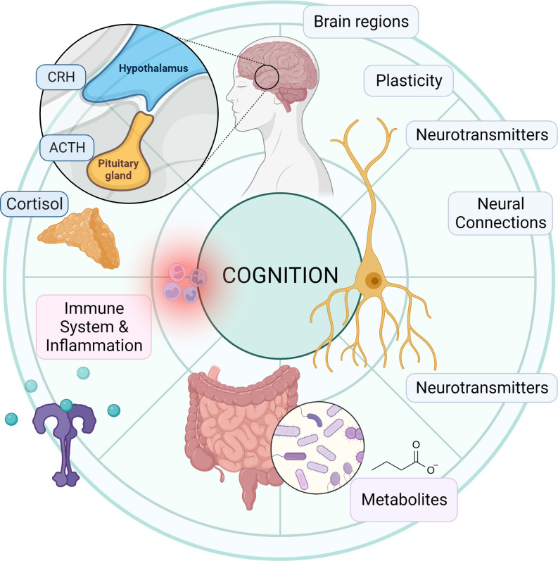
Cognitive function in humans depends on the complex and interplay between multiple body systems, including the hypothalamic-pituitary-adrenal (HPA) axis. The gut microbiota, which vastly outnumbers human cells and has a genetic potential that exceeds that of the human genome, plays a crucial role in this interplay. The microbiota-gut-brain (MGB) axis is a bidirectional signalling pathway that operates through neural, endocrine, immune, and metabolic pathways. One of the major neuroendocrine systems responding to stress is the HPA axis which produces glucocorticoids such as cortisol in humans and corticosterone in rodents. Appropriate concentrations of cortisol are essential for normal neurodevelopment and function, as well as cognitive processes such as learning and memory, and studies have shown that microbes modulate the HPA axis throughout life. Stress can significantly impact the MGB axis via the HPA axis and other pathways. Animal research has advanced our understanding of these mechanisms and pathways, leading to a paradigm shift in conceptual thinking about the influence of the microbiota on human health and disease. Preclinical and human trials are currently underway to determine how these animal models translate to humans. In this review article, we summarize the current knowledge of the relationship between the gut microbiota, HPA axis, and cognition, and provide an overview of the main findings and conclusions in this broad field.
Overview
AI snapshot
Written from the paper’s full text.
TL;DR
This narrative review summarizes bidirectional signaling between the gut microbiota, the hypothalamic-pituitary-adrenal (HPA) axis, and cognition via the microbiota-gut-brain axis. It concludes that microbial metabolites, neural pathways, immune signaling, and HPA-axis glucocorticoids are interlinked, but human causal evidence remains limited.
Key findings
- The gut microbiota and HPA axis are bidirectionally linked: germ-free mice show enhanced HPA-axis activity after acute stress, including increased ACTH and corticosterone responses, and altered cognition; microbial colonization can rescue some effects if given early in life.
- Human observational data link microbiota diversity to cognition: in 597 middle-aged adults, β-diversity was positively associated with all higher cognitive functions tested; in >4,000 healthy people aged 18–90, metagenomic profiles predicted chronological age within approximately six years.
- Antibiotic exposure is associated with cognitive risk: in a prospective cohort of 14,542 participants, chronic midlife antibiotic use was associated with later cognitive impairment, corresponding to an additional three years of aging compared with no antibiotic exposure.
- Mechanistic pathways include SCFAs, bile acids, tryptophan/kynurenine metabolites, serotonin, GABA, catecholamines, endocannabinoids, immune modulation, the vagus nerve, and the enteric nervous system; SCFA supplementation attenuated the cortisol response to psychosocial stress in healthy men.
Methods
- Design: narrative review article summarizing current knowledge of the gut microbiota, HPA axis, and cognition.
- Data/sample: no primary data; synthesizes preclinical animal studies, germ-free models, human observational studies, randomized controlled trials, and mechanistic literature.
- Main analysis: thematic synthesis of proposed microbiota-gut-brain mechanisms and evidence relating microbiota, HPA-axis function, and cognitive outcomes.
Limitations
- The paper states that mechanisms by which the gut microbiota impacts the human brain are not fully understood, human evidence remains sparse, and whether microbiota changes are causal, consequential, or coincidental is unresolved.
- The paper states that animal models, especially germ-free models, may not directly transfer to human function and calls for rigorous causal human studies and adequately powered, standardized clinical trials.
- Evident from the text: as a narrative review, it does not describe a systematic search strategy, formal quality appraisal, or meta-analytic synthesis, and it cites many small, short-term, or heterogeneous studies.
Model-written summary. Check it against the paper before relying on it.
Integrity signals
Registry
Cites
Statistics
Figures
Verification
Claims the paper makes
315 Not checked yet · 26 Own interpretation · 2 No basis given · 1 Supported
None of the sources cited for this claim have been checked yet.
In the paper
“In the gut, the GM and its metabolites have been shown to modulate gastrointestinal functions via their effects on intestinal permeability (32–35), mucosal immune function (36–38), intestinal motility and sensitivity (39, 40), and the enteric nervous system (ENS) (21, 24, 25, 41).”
§ Signalling cognition: the gut microbiota and hypothalamic-pituitary-adrenal axis > The microbiota-gut-brain axis
What the sources say
- Not checked yetcites [32] Cani, 2012, Involvement of gut microbiota in the development of low-grade inflammation and type 2 diabetes associated with obesity, Gut Microbes source
- Not checked yetcites [33] Rutsch, 2020, The gut-brain axis: how microbiota and host inflammasome influence brain physiology and pathology, Front Immunol source
- Not checked yetcites [34] Frazier, 2011, Gut microbiota, intestinal permeability, obesity-induced inflammation, and liver injury, JPEN J Parenter Enteral Nutr source
- Not checked yetcites [35] Fasano, 2017, Gut permeability, obesity, and metabolic disorders: who is the chicken and who is the egg, Am J Clin Nutr source
- Not checked yetcites [36] Round, 2009, The gut microbiota shapes intestinal immune responses during health and disease, Nat Rev Immunol source
- Not checked yetcites [37] Allaire, 2018, The intestinal epithelium: central coordinator of mucosal immunity, Trends Immunol source
- Not checked yetcites [38] Wlodarska, 2015, An integrative view of microbiome-host interactions in inflammatory bowel diseases, Cell Host Microbe source
- Not checked yetcites [39] Cani, 2013, Gut microbiota, enteroendocrine functions and metabolism, Curr Opin Pharmacol source
- Not checked yetcites [40] Valdez-Morales, 2013, Sensitization of peripheral sensory nerves by mediators from colonic biopsies of diarrhea-predominant irritable bowel syndrome patients: a role for PAR2, Am J Gastroenterol source
boosteddirect- Asserted: passes — The authors put it forward as true.
- Checkable: passes — It can be checked against evidence.
- Load-bearing: unclear — It is unclear whether the paper’s conclusions depend on it.
- Based: passes — The paper gives a reason why it holds.
A conclusion the paper draws from its own results — it cites no other source for it, so there is none to check. The quote shows it is in the paper.
In the paper
“Emerging evidence suggests that the GM, the hypothalamic-pituitary-adrenal (HPA) axis, and cognitive processes are linked bidirectionally via multiple pathways, including the vagus nerve, neurotransmitter and metabolite production, immune system and blood-brain barrier regulation, and hormone metabolism.”
§ Signalling cognition: the gut microbiota and hypothalamic-pituitary-adrenal axis > Introduction
hedgedcorrelational- Asserted: passes — The authors put it forward as true.
- Checkable: passes — It can be checked against evidence.
- Load-bearing: passes — The paper’s conclusions depend on it.
- Based: unclear — It is unclear why the paper says it holds.
- Rests on an assumption: Animal model findings about the GM-HPA-cognition relationship are informative about human biology.
A conclusion the paper draws from its own results — it cites no other source for it, so there is none to check. The quote shows it is in the paper.
In the paper
“Stress can significantly impact the MGB axis via the HPA axis and other pathways.”
§ Signalling cognition: the gut microbiota and hypothalamic-pituitary-adrenal axis > Abstract
plaindirect- Asserted: passes — The authors put it forward as true.
- Checkable: passes — It can be checked against evidence.
- Load-bearing: passes — The paper’s conclusions depend on it.
- Based: unclear — It is unclear why the paper says it holds.
- Rests on an assumption: Animal model findings about the GM-HPA-cognition relationship are informative about human biology.
None of the sources cited for this claim have been checked yet.
In the paper
“As a key regulator, the GM can modulate host physiological processes through several proposed mechanisms (25, 26).”
§ Signalling cognition: the gut microbiota and hypothalamic-pituitary-adrenal axis > The microbiota-gut-brain axis
What the sources say
plaindirect- Asserted: passes — The authors put it forward as true.
- Checkable: passes — It can be checked against evidence.
- Load-bearing: passes — The paper’s conclusions depend on it.
- Based: passes — The paper gives a reason why it holds.
In the paper
“Confirming the role of the GM in cognitive function in midlife, Meyer et al. explored the relationship between gut microbial community composition and cognitive function in 597 middle-aged adults, and found a significant positive association between β-diversity and all higher cognitive functions tested (89).”
§ Signalling cognition: the gut microbiota and hypothalamic-pituitary-adrenal axis > The microbiota-gut-brain axis > Development and life course of the MGB axis
What the sources say
- Not checked yetcites [89] Meyer, 2022, Association of the gut microbiota with cognitive function in midlife, JAMA Netw Open source
plaincorrelationaln = 597- Asserted: passes — The authors put it forward as true.
- Checkable: passes — It can be checked against evidence.
- Load-bearing: passes — The paper’s conclusions depend on it.
- Based: passes — The paper gives a reason why it holds.
None of the sources cited for this claim have been checked yet.
In the paper
“The GM can stimulate the release of peptides and hormones from enteroendocrine cells, which can have direct or indirect central effects (22, 39).”
§ Signalling cognition: the gut microbiota and hypothalamic-pituitary-adrenal axis > The microbiota-gut-brain axis
What the sources say
plaindirect- Asserted: passes — The authors put it forward as true.
- Checkable: passes — It can be checked against evidence.
- Load-bearing: passes — The paper’s conclusions depend on it.
- Based: passes — The paper gives a reason why it holds.
A conclusion the paper draws from its own results — it cites no other source for it, so there is none to check. The quote shows it is in the paper.
In the paper
“The first year of life encompasses a critical “window period” of development in which the GM may be more susceptible to environmental influences and highly influential with regards to the overall health of the host.”
§ Signalling cognition: the gut microbiota and hypothalamic-pituitary-adrenal axis > The microbiota-gut-brain axis > Development and life course of the MGB axis
hedgedconditional- Asserted: passes — The authors put it forward as true.
- Checkable: passes — It can be checked against evidence.
- Load-bearing: unclear — It is unclear whether the paper’s conclusions depend on it.
- Based: passes — The paper gives a reason why it holds.
- Rests on an assumption: Animal model findings about the GM-HPA-cognition relationship are informative about human biology.
None of the sources cited for this claim have been checked yet.
In the paper
“Furthermore, recent studies have shown dramatic changes in the GM of patients with Alzheimer’s disease, Parkinson’s disease, multiple sclerosis, and schizophrenia (73–77).”
§ Signalling cognition: the gut microbiota and hypothalamic-pituitary-adrenal axis > The microbiota-gut-brain axis > The role of the HPA axis in the MGB axis
What the sources say
- Not checked yetcites [73] Haran, 2019, Alzheimer’s disease microbiome is associated with dysregulation of the anti-inflammatory p-glycoprotein pathway, MBio source
- Not checked yetcites [74] Romano, 2021, Meta-analysis of the parkinson’s disease gut microbiome suggests alterations linked to intestinal inflammation, NPJ Park Dis source
- Not checked yetcites [75] Sampson, 2016, Gut microbiota regulate motor deficits and neuroinflammation in a model of parkinson’s disease, Cell source
- Not checked yetcites [76] Correale, 2022, The role of the gut microbiota in multiple sclerosis, Nat Rev Neurol source
- Not checked yetcites [77] Zheng, 2019, The gut microbiome from patients with schizophrenia modulates the glutamate-glutamine-GABA cycle and schizophrenia-relevant behaviors in mice, Sci Adv source
boostedcorrelational- Asserted: passes — The authors put it forward as true.
- Checkable: passes — It can be checked against evidence.
- Load-bearing: unclear — It is unclear whether the paper’s conclusions depend on it.
- Based: passes — The paper gives a reason why it holds.
None of the sources cited for this claim have been checked yet.
In the paper
“Preclinical evidence suggests that GM-mediated mechanisms are likely involved in modulating brain processes, including brain biochemistry, response to stress, pain interpretation, feeding, emotional behaviors, and cognition (57–64).”
§ Signalling cognition: the gut microbiota and hypothalamic-pituitary-adrenal axis > The microbiota-gut-brain axis > The role of the HPA axis in the MGB axis
What the sources say
- Not checked yetcites [57] Stilling, 2014, Microbial genes, brain & behaviour - epigenetic regulation of the gut-brain axis, Genes Brain Behav source
- Not checked yetcites [58] Foster, 2017, Stress & the gut-brain axis: regulation by the microbiome, Neurobiol Stress source
- Not checked yetcites [59] Cryan, 2012, Mind-altering microorganisms: the impact of the gut microbiota on brain and behaviour, Nat Rev Neurosci source
- Not checked yetcites [60] Bercik, 2012, Microbes and the gut-brain axis, Neurogastroenterol Motil Off J Eur Gastrointest Motil Soc source
- Not checked yetcites [61] Alcock, 2014, Is eating behavior manipulated by the gastrointestinal microbiota? evolutionary pressures and potential mechanisms, Bioessays source
- Not checked yetcites [62] Lam, 2017, Are the gut bacteria telling us to eat or not to eat? reviewing the role of gut microbiota in the etiology, disease progression and treatment of eating disorders, Nutrients source
- Not checked yetcites [63] Foster, 2013, Gut-brain axis: how the microbiome influences anxiety and depression, Trends Neurosci source
- Not checked yetcites [64] Humann, 2016, Bacterial peptidoglycan traverses the placenta to induce fetal neuroproliferation and aberrant postnatal behavior, Cell Host Microbe source
hedgedconditional- Asserted: passes — The authors put it forward as true.
- Checkable: passes — It can be checked against evidence.
- Load-bearing: passes — The paper’s conclusions depend on it.
- Based: passes — The paper gives a reason why it holds.
None of the sources cited for this claim have been checked yet.
In the paper
“Initial clinical evidence of MGB interactions primarily stems from associations between dysbiosis and CNS disorders, such as autism, anxiety-depressive behaviors, and functional gastrointestinal disorders (63, 65–72).”
§ Signalling cognition: the gut microbiota and hypothalamic-pituitary-adrenal axis > The microbiota-gut-brain axis > The role of the HPA axis in the MGB axis
What the sources say
- Not checked yetcites [63] Foster, 2013, Gut-brain axis: how the microbiome influences anxiety and depression, Trends Neurosci source
- Not checked yetcites [65] Mayer, 2014, Altered brain-gut axis in autism: comorbidity or causative mechanisms, Bioessays source
- Not checked yetcites [66] Song, 2004, Real-time PCR quantitation of clostridia in feces of autistic children, Appl Environ Microbiol source
- Not checked yetcites [67] Hsiao, 2013, Microbiota modulate behavioral and physiological abnormalities associated with neurodevelopmental disorders, Cell source
- Not checked yetcites [68] Naseribafrouei, 2014, Correlation between the human fecal microbiota and depression, Neurogastroenterol Motil Off J Eur Gastrointest Motil Soc source
- Not checked yetcites [69] Du, 2020, Crosstalk between the microbiota-gut-brain axis and depression, Heliyon source
- Not checked yetcites [70] Mayer, 2011, The brain-gut axis in abdominal pain syndromes, Annu Rev Med source
- Not checked yetcites [71] Berrill, 2013, An observational study of cognitive function in patients with irritable bowel syndrome and inflammatory bowel disease, Neurogastroenterol Motil Off J Eur Gastrointest Motil Soc source
- Not checked yetcites [72] Simrén, 2013, Intestinal microbiota in functional bowel disorders: a Rome foundation report, Gut source
plaincorrelational- Asserted: passes — The authors put it forward as true.
- Checkable: passes — It can be checked against evidence.
- Load-bearing: unclear — It is unclear whether the paper’s conclusions depend on it.
- Based: passes — The paper gives a reason why it holds.
- It may state two separate claims as one. 144
- Probably not a substantial claim. 11
- The quote only partly states this claim. 2
Every source the paper cites for this claim was checked and supports it.
In the paper
“In another study, B-GOS supplementation was compared to placebo in healthy participants and found to decrease waking salivary cortisol concentrations and increase the positive processing of information in a dot-probe task (485).”
§ Signalling cognition: the gut microbiota and hypothalamic-pituitary-adrenal axis > The relationship between the microbiota, HPA axis, and cognition > HPA axis-related interactions between the gut microbiota and cognition
What the sources say
- Supportedcites [485] Schmidt, 2015, Prebiotic intake reduces the waking cortisol response and alters emotional bias in healthy volunteers, Psychopharmacology source
The Discussion describes a study in healthy volunteers and reports decreased waking salivary cortisol reactivity after B-GOS compared with placebo. The Results (Cortisol; Emotional Test Battery) confirm a lower cortisol awakening response and increased vigilance to positive versus negative information in the unmasked dot-probe condition. (LLM, conf=0.94)
plaindirect- Asserted: passes — The authors put it forward as true.
- Checkable: passes — It can be checked against evidence.
- Load-bearing: passes — The paper’s conclusions depend on it.
- Based: passes — The paper gives a reason why it holds.
It may state two separate claims as one.
- Supported
None of the sources cited for this claim have been checked yet.
In the paper
“Studies in animals have demonstrated the effect that the GM has on neural development and neurochemistry in the host, influencing the stress system, behavior, and cognition (42–47).”
§ Signalling cognition: the gut microbiota and hypothalamic-pituitary-adrenal axis > The microbiota-gut-brain axis
What the sources say
- Not checked yetcites [42] Dinan, 2017, Gut instincts: microbiota as a key regulator of brain development, ageing and neurodegeneration, J Physiol source
- Not checked yetcites [43] Moloney, 2014, The microbiome: stress, health and disease, Mamm Genome source
- Not checked yetcites [44] Salami, 2021, Interplay of good bacteria and central nervous system: cognitive aspects and mechanistic considerations, Front Neurosci source
- Not checked yetcites [45] Sampson, 2015, Control of brain development, function, and behavior by the microbiome, Cell Host Microbe source
- Not checked yetcites [46] Vuong, 2017, The microbiome and host behavior, Annu Rev Neurosci source
- Not checked yetcites [47] Connell, 2022, Microbial-derived metabolites as a risk factor of age-related cognitive decline and dementia, Mol Neurodegener source
boosteddirect- Asserted: passes — The authors put it forward as true.
- Checkable: passes — It can be checked against evidence.
- Load-bearing: passes — The paper’s conclusions depend on it.
- Based: passes — The paper gives a reason why it holds.
It may state two separate claims as one.
A conclusion the paper draws from its own results — it cites no other source for it, so there is none to check. The quote shows it is in the paper.
In the paper
“Alterations in the GM, whether due to diet, antibiotics, or other factors, can impact the stress response, HPA axis activity, and overall cognitive health.”
§ Signalling cognition: the gut microbiota and hypothalamic-pituitary-adrenal axis > Introduction
plainconditional- Asserted: passes — The authors put it forward as true.
- Checkable: passes — It can be checked against evidence.
- Load-bearing: passes — The paper’s conclusions depend on it.
- Based: fails — The paper may give no reason why it holds.
Probably not a substantial claim. The paper may give no reason why it holds.
- Rests on an assumption: Animal model findings about the GM-HPA-cognition relationship are informative about human biology.
In the paper
“Appropriate concentrations of cortisol are essential for normal neurodevelopment and function, as well as cognitive processes such as learning and memory, and studies have shown that microbes modulate the HPA axis throughout life.”
- Also stated in: § The HPA axis, glucocorticoids, and the stress response · plain
“Glucocorticoids also play a vital role in central nervous system (CNS) development and are required for normal maturation (139).”
§ Signalling cognition: the gut microbiota and hypothalamic-pituitary-adrenal axis > Abstract
What the sources say
- Not checked yetcites [139] Strehl, 2019, Glucocorticoids–All-Rounders tackling the versatile players of the immune system, Front Immunol source
plaindirect- Asserted: passes — The authors put it forward as true.
- Checkable: passes — It can be checked against evidence.
- Load-bearing: passes — The paper’s conclusions depend on it.
- Based: passes — The paper gives a reason why it holds.
It may state two separate claims as one.
- Also stated in: § The HPA axis, glucocorticoids, and the stress response · plain
In the paper
“However, the acquisition of microbes occurs primarily at birth, with delivery through the birth canal exposing the infant to its mother’s microbiota, resulting in vertical transmission of an initial maternal signature (81).”
§ Signalling cognition: the gut microbiota and hypothalamic-pituitary-adrenal axis > The microbiota-gut-brain axis > Development and life course of the MGB axis
What the sources say
- Not checked yetcites [81] Frese, 2015, Birth of the infant gut microbiome: moms deliver twice, Cell Host Microbe source
plaindirect- Asserted: passes — The authors put it forward as true.
- Checkable: passes — It can be checked against evidence.
- Load-bearing: unclear — It is unclear whether the paper’s conclusions depend on it.
- Based: passes — The paper gives a reason why it holds.
It may state two separate claims as one.
In the paper
“Elevated cortisol levels have been associated with alterations in GM composition and increased gut permeability, which can lead to inflammation and contribute to brain dysfunction and various CNS disorders (56).”
§ Signalling cognition: the gut microbiota and hypothalamic-pituitary-adrenal axis > The microbiota-gut-brain axis > The role of the HPA axis in the MGB axis
What the sources say
- Not checked yetcites [56] Mangiola, 2016, Gut microbiota in autism and mood disorders, World J Gastroenterol source
plaindirect- Asserted: passes — The authors put it forward as true.
- Checkable: passes — It can be checked against evidence.
- Load-bearing: passes — The paper’s conclusions depend on it.
- Based: passes — The paper gives a reason why it holds.
It may state two separate claims as one.
In the paper
“Chronic or prolonged stress can lead to dysregulation of the HPA axis, which can have negative effects on various bodily systems, including the MGB axis (55).”
§ Signalling cognition: the gut microbiota and hypothalamic-pituitary-adrenal axis > The microbiota-gut-brain axis > The role of the HPA axis in the MGB axis
What the sources say
- Not checked yetcites [55] Kelly, 2016, Transferring the blues: depression-associated gut microbiota induces neurobehavioural changes in the rat, J Psychiatr Res source
plaindirect- Asserted: passes — The authors put it forward as true.
- Checkable: passes — It can be checked against evidence.
- Load-bearing: unclear — It is unclear whether the paper’s conclusions depend on it.
- Based: passes — The paper gives a reason why it holds.
It may state two separate claims as one.
None of the sources cited for this claim have been checked yet.
In the paper
“Cortisol receptors are expressed on various cells of the gut, including epithelial cells, immune cells, and enteroendocrine cells, indicating a direct effect of cortisol on gut function (21, 29, 48).”
§ Signalling cognition: the gut microbiota and hypothalamic-pituitary-adrenal axis > The microbiota-gut-brain axis > The role of the HPA axis in the MGB axis
What the sources say
- Not checked yetcites [21] Carabotti, 2015, The gut-brain axis: interactions between enteric microbiota, central and enteric nervous systems, Ann Gastroenterol
- Not checked yetcites [29] Tetel, 2018, Steroids, stress and the gut microbiome-brain axis, J Neuroendocrinol source
- Not checked yetcites [48] Mayer, 2000, The neurobiology of stress and gastrointestinal disease, Gut source
hedgeddirect- Asserted: passes — The authors put it forward as true.
- Checkable: passes — It can be checked against evidence.
- Load-bearing: passes — The paper’s conclusions depend on it.
- Based: passes — The paper gives a reason why it holds.
It may state two separate claims as one.
In the paper
“Cortisol can also affect the gut microbiota by altering gut transit time, intestinal permeability, and nutrient availability, which can in turn impact the composition and diversity of the GM (48).”
§ Signalling cognition: the gut microbiota and hypothalamic-pituitary-adrenal axis > The microbiota-gut-brain axis > The role of the HPA axis in the MGB axis
What the sources say
- Not checked yetcites [48] Mayer, 2000, The neurobiology of stress and gastrointestinal disease, Gut source
plaindirect- Asserted: passes — The authors put it forward as true.
- Checkable: passes — It can be checked against evidence.
- Load-bearing: passes — The paper’s conclusions depend on it.
- Based: passes — The paper gives a reason why it holds.
It may state two separate claims as one.
None of the sources cited for this claim have been checked yet.
In the paper
“A more stable and complex, adult-like microbiota starts to emerge as early as one to three years of age, although this development may continue as late as pre-adolescence (85, 86).”
§ Signalling cognition: the gut microbiota and hypothalamic-pituitary-adrenal axis > The microbiota-gut-brain axis > Development and life course of the MGB axis
What the sources say
hedged- Asserted: passes — The authors put it forward as true.
- Checkable: passes — It can be checked against evidence.
- Load-bearing: unclear — It is unclear whether the paper’s conclusions depend on it.
- Based: passes — The paper gives a reason why it holds.
It may state two separate claims as one.
In the paper
“The HPA axis plays a central role in mediating the stress response and regulating the interaction between the GM, gut, and brain (42).”
§ Signalling cognition: the gut microbiota and hypothalamic-pituitary-adrenal axis > The microbiota-gut-brain axis > The role of the HPA axis in the MGB axis
What the sources say
- Not checked yetcites [42] Dinan, 2017, Gut instincts: microbiota as a key regulator of brain development, ageing and neurodegeneration, J Physiol source
plaindirect- Asserted: passes — The authors put it forward as true.
- Checkable: passes — It can be checked against evidence.
- Load-bearing: passes — The paper’s conclusions depend on it.
- Based: passes — The paper gives a reason why it holds.
It may state two separate claims as one.
In the paper
“In adulthood, the core GM is relatively stable, and the main factors influencing its composition are diet, exercise, stress, disease, and medications (88).”
§ Signalling cognition: the gut microbiota and hypothalamic-pituitary-adrenal axis > The microbiota-gut-brain axis > Development and life course of the MGB axis
What the sources say
- Not checked yetcites [88] Martino, 2022, Microbiota succession throughout life from the cradle to the grave, Nat Rev Microbiol source
plaindirect- Asserted: passes — The authors put it forward as true.
- Checkable: passes — It can be checked against evidence.
- Load-bearing: fails — The paper’s conclusions may not depend on it.
- Based: passes — The paper gives a reason why it holds.
Probably not a substantial claim. The paper’s conclusions may not depend on it. It may state two separate claims as one.
In the paper
“Finally, germ-free animal studies demonstrate that the GM is necessary for normal stress responsivity, anxiety-like behaviors, sociability, and cognition (123).”
§ Signalling cognition: the gut microbiota and hypothalamic-pituitary-adrenal axis > The microbiota-gut-brain axis > The influence of the gut microbiota on neurodevelopment
What the sources say
- Not checked yetcites [123] Luczynski, 2016, Growing up in a bubble: using germ-free animals to assess the influence of the gut microbiota on brain and behavior, Int J Neuropsychopharmacol source
boosteddirect- Asserted: passes — The authors put it forward as true.
- Checkable: passes — It can be checked against evidence.
- Load-bearing: passes — The paper’s conclusions depend on it.
- Based: passes — The paper gives a reason why it holds.
It may state two separate claims as one.
None of the sources cited for this claim have been checked yet.
In the paper
“There is also evidence that the diversity levels of the GM differ with aging, with a reduction in the number of different bacterial species present and changes in the relative abundance of different bacterial phyla, with a decrease in beneficial bacteria such as Bifidobacteria and an increase in potentially harmful bacteria such as Proteobacteria (93–96).”
§ Signalling cognition: the gut microbiota and hypothalamic-pituitary-adrenal axis > The microbiota-gut-brain axis > Development and life course of the MGB axis
What the sources say
- Not checked yetcites [93] Claesson, 2011, Composition, variability, and temporal stability of the intestinal microbiota of the elderly, Proc Natl Acad Sci U.S.A. source
- Not checked yetcites [94] O’Toole, 2015, Gut microbiota and aging, Science source
- Not checked yetcites [95] Odamaki, 2016, Age-related changes in gut microbiota composition from newborn to centenarian: a cross-sectional study, BMC Microbiol source
- Not checked yetcites [96] Biagi, 2010, Through ageing, and beyond: gut microbiota and inflammatory status in seniors and centenarians, PloS One source
hedgedcorrelational- Asserted: passes — The authors put it forward as true.
- Checkable: passes — It can be checked against evidence.
- Load-bearing: unclear — It is unclear whether the paper’s conclusions depend on it.
- Based: passes — The paper gives a reason why it holds.
It may state two separate claims as one.
None of the sources cited for this claim have been checked yet.
In the paper
“Human studies comparing the composition of the GM of elderly participants suffering from cognitive impairment with healthy individuals indicate significant changes in GM composition. Specifically, there is an increase in pro-inflammatory taxa and a reduction in anti-inflammatory taxa (104–109).”
§ Signalling cognition: the gut microbiota and hypothalamic-pituitary-adrenal axis > The microbiota-gut-brain axis > Development and life course of the MGB axis
What the sources say
- Not checked yetcites [104] Cattaneo, 2017, Association of brain amyloidosis with pro-inflammatory gut bacterial taxa and peripheral inflammation markers in cognitively impaired elderly, Neurobiol Aging source
- Not checked yetcites [105] Vogt, 2017, Gut microbiome alterations in alzheimer’s disease, Sci Rep source
- Not checked yetcites [106] Manderino, 2017, Preliminary evidence for an association between the composition of the gut microbiome and cognitive function in neurologically healthy older adults, J Int Neuropsychol Soc source
- Not checked yetcites [107] Zhuang, 2018, Gut microbiota is altered in patients with alzheimer’s disease, J Alzheimers Dis source
- Not checked yetcites [108] Saji, 2019, Analysis of the relationship between the gut microbiome and dementia: a cross-sectional study conducted in Japan, Sci Rep source
- Not checked yetcites [109] Sheng, 2021, Altered gut microbiota in adults with subjective cognitive decline: the SILCODE study, J Alzheimers Dis source
hedgedcorrelational- Asserted: passes — The authors put it forward as true.
- Checkable: passes — It can be checked against evidence.
- Load-bearing: passes — The paper’s conclusions depend on it.
- Based: passes — The paper gives a reason why it holds.
It may state two separate claims as one.
In the paper
“Similar to aging, dysbiosis has been associated with increased cortisol secretion, decreased negative feedback at the level of the hypothalamus, and changes in the circadian rhythm of cortisol release (113).”
§ Signalling cognition: the gut microbiota and hypothalamic-pituitary-adrenal axis > The microbiota-gut-brain axis > Development and life course of the MGB axis
What the sources say
- Not checked yetcites [113] Farzi, 2018, Gut microbiota and the neuroendocrine system, Neurotherapeutics source
plaincorrelational- Asserted: passes — The authors put it forward as true.
- Checkable: passes — It can be checked against evidence.
- Load-bearing: unclear — It is unclear whether the paper’s conclusions depend on it.
- Based: passes — The paper gives a reason why it holds.
It may state two separate claims as one.
None of the sources cited for this claim have been checked yet.
In the paper
“Essentially, the HPA axis appears to become less responsive with aging, contributing to the dysregulation of stress responses and the resultant development of cognitive decline (111, 112).”
§ Signalling cognition: the gut microbiota and hypothalamic-pituitary-adrenal axis > The microbiota-gut-brain axis > Development and life course of the MGB axis
What the sources say
- Not checked yetcites [111] Lupien, 1998, Cortisol levels during human aging predict hippocampal atrophy and memory deficits, Nat Neurosci source
- Not checked yetcites [112] Hinkelmann, 2012, Changes in cortisol secretion during antidepressive treatment and cognitive improvement in patients with major depression: a longitudinal study, Psychoneuroendocrinology source
hedgeddirect- Asserted: passes — The authors put it forward as true.
- Checkable: passes — It can be checked against evidence.
- Load-bearing: unclear — It is unclear whether the paper’s conclusions depend on it.
- Based: passes — The paper gives a reason why it holds.
It may state two separate claims as one.
- Rests on an assumption: Animal model findings about the GM-HPA-cognition relationship are informative about human biology.
None of the sources cited for this claim have been checked yet.
In the paper
“These peptides stimulate the release of adrenocorticotropic hormone (ACTH) from corticotropic cells of the anterior pituitary gland, which in turn targets the zona fasciculata of the adrenal cortex to produce glucocorticoids, such as cortisol in humans and corticosterone in rodents (133–135).”
§ Signalling cognition: the gut microbiota and hypothalamic-pituitary-adrenal axis > Mechanisms and pathways of the MGB axis > The HPA axis, glucocorticoids, and the stress response
What the sources say
- Not checked yetcites [133] Rifai, 2018
- Not checked yetcites [134] Buckingham, 1996, The physiology of immunity, Progress in Brain Research
- Not checked yetcites [135] Buckingham, 2006, Glucocorticoids: exemplars of multi-tasking, Br J Pharmacol source
plaindirect- Asserted: passes — The authors put it forward as true.
- Checkable: passes — It can be checked against evidence.
- Load-bearing: unclear — It is unclear whether the paper’s conclusions depend on it.
- Based: passes — The paper gives a reason why it holds.
It may state two separate claims as one.
In the paper
“The HPA axis is a major stress response system in the body, with neurons in the hypothalamic paraventricular nucleus (PVN) synthesizing and secreting corticotropin-releasing hormone (CRH) and antidiuretic hormone (ADH) in response to stress (133).”
§ Signalling cognition: the gut microbiota and hypothalamic-pituitary-adrenal axis > Mechanisms and pathways of the MGB axis > The HPA axis, glucocorticoids, and the stress response
What the sources say
- Not checked yetcites [133] Rifai, 2018
plaindirect- Asserted: passes — The authors put it forward as true.
- Checkable: passes — It can be checked against evidence.
- Load-bearing: fails — The paper’s conclusions may not depend on it.
- Based: passes — The paper gives a reason why it holds.
Probably not a substantial claim. The paper’s conclusions may not depend on it. It may state two separate claims as one.
In the paper
“Cortisol acts on the hypothalamus and pituitary in a negative feedback loop to regulate the response (133).”
§ Signalling cognition: the gut microbiota and hypothalamic-pituitary-adrenal axis > Mechanisms and pathways of the MGB axis > The HPA axis, glucocorticoids, and the stress response
What the sources say
- Not checked yetcites [133] Rifai, 2018
plaindirect- Asserted: passes — The authors put it forward as true.
- Checkable: passes — It can be checked against evidence.
- Load-bearing: fails — The paper’s conclusions may not depend on it.
- Based: passes — The paper gives a reason why it holds.
Probably not a substantial claim. The paper’s conclusions may not depend on it.
In the paper
“HPA axis dysfunction is linked to a decline in cognitive function, aging, immune system dysfunction, and systemic inflammation (135).”
§ Signalling cognition: the gut microbiota and hypothalamic-pituitary-adrenal axis > Mechanisms and pathways of the MGB axis > The HPA axis, glucocorticoids, and the stress response
What the sources say
- Not checked yetcites [135] Buckingham, 2006, Glucocorticoids: exemplars of multi-tasking, Br J Pharmacol source
plaincorrelational- Asserted: passes — The authors put it forward as true.
- Checkable: passes — It can be checked against evidence.
- Load-bearing: unclear — It is unclear whether the paper’s conclusions depend on it.
- Based: passes — The paper gives a reason why it holds.
It may state two separate claims as one.
None of the sources cited for this claim have been checked yet.
In the paper
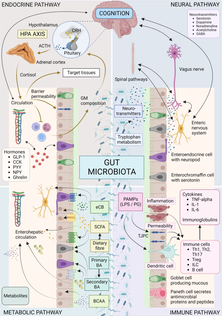
“Microbes influence CNS processes via modulation of the nervous system (52), endocrine system (51, 126), and immune system (120), together with their ability to synthesize neurotransmitters (127–129) and produce metabolites (127, 130–132) ( Figure 1 ).”
§ Signalling cognition: the gut microbiota and hypothalamic-pituitary-adrenal axis > Mechanisms and pathways of the MGB axis
What the sources say
- Not checked yetcites [52] Forsythe, 2014
- Not checked yetcites [51] Sudo, 2004, Postnatal microbial colonization programs the hypothalamic-pituitary-adrenal system for stress response in mice, J Physiol source
- Not checked yetcites [126] Mudd, 2017, Serum cortisol mediates the relationship between fecal ruminococcus and brain n-acetylaspartate in the young pig, Gut Microbes source
- Not checked yetcites [120] Erny, 2015, Host microbiota constantly control maturation and function of microglia in the CNS, Nat Neurosci source
plaindirect- Asserted: passes — The authors put it forward as true.
- Checkable: passes — It can be checked against evidence.
- Load-bearing: passes — The paper’s conclusions depend on it.
- Based: passes — The paper gives a reason why it holds.
It may state two separate claims as one.
In the paper
“male germ-free mice have increased hippocampal serotonin levels, which colonization immediately post-weaning does not reverse (124).”
§ Signalling cognition: the gut microbiota and hypothalamic-pituitary-adrenal axis > Mechanisms and pathways of the MGB axis > The nervous system
What the sources say
- Not checked yetcites [124] Heijtz, 2011, Normal gut microbiota modulates brain development and behavior, Proc Natl Acad Sci source
plaindirect- Asserted: passes — The authors put it forward as true.
- Checkable: passes — It can be checked against evidence.
- Load-bearing: unclear — It is unclear whether the paper’s conclusions depend on it.
- Based: passes — The paper gives a reason why it holds.
It may state two separate claims as one.
A conclusion the paper draws from its own results — it cites no other source for it, so there is none to check. The quote shows it is in the paper.
In the paper
“GABA does not cross the BBB and so microbial-derived GABA would need to act locally on the ENS or vagus nerve to influence the CNS.”
§ Signalling cognition: the gut microbiota and hypothalamic-pituitary-adrenal axis > Mechanisms and pathways of the MGB axis > The nervous system
hedgedconditional- Asserted: passes — The authors put it forward as true.
- Checkable: passes — It can be checked against evidence.
- Load-bearing: passes — The paper’s conclusions depend on it.
- Based: unclear — It is unclear why the paper says it holds.
It may state two separate claims as one.
- Rests on an assumption: GABA does not cross the blood-brain barrier.
A conclusion the paper draws from its own results — it cites no other source for it, so there is none to check. The quote shows it is in the paper.
In the paper
“the development of the serotonergic system within the MGB axis depends on a low-stress environment and early life events may be critical.”
§ Signalling cognition: the gut microbiota and hypothalamic-pituitary-adrenal axis > Mechanisms and pathways of the MGB axis > The nervous system
hedgeddirect- Asserted: passes — The authors put it forward as true.
- Checkable: passes — It can be checked against evidence.
- Load-bearing: unclear — It is unclear whether the paper’s conclusions depend on it.
- Based: passes — The paper gives a reason why it holds.
It may state two separate claims as one.
In the paper
“When administered to rats, B. infantis results in reduced 5-HIAA (serotonin metabolite) concentrations in the frontal cortex, and a marked increase in blood concentrations of tryptophan and kynurenic acid (224).”
§ Signalling cognition: the gut microbiota and hypothalamic-pituitary-adrenal axis > Mechanisms and pathways of the MGB axis > The nervous system
What the sources say
- Not checked yetcites [224] Desbonnet, 2008, The probiotic bifidobacteria infantis: an assessment of potential antidepressant properties in the rat, J Psychiatr Res source
plaindirect- Asserted: passes — The authors put it forward as true.
- Checkable: passes — It can be checked against evidence.
- Load-bearing: passes — The paper’s conclusions depend on it.
- Based: passes — The paper gives a reason why it holds.
It may state two separate claims as one.
In the paper
“brainstem catecholaminergic centers play an important role in the regulation of the HPA axis (227) and noradrenergic neurons are required for normal activation in response to a variety of stressors.”
§ Signalling cognition: the gut microbiota and hypothalamic-pituitary-adrenal axis > Mechanisms and pathways of the MGB axis > The nervous system
What the sources say
- Not checked yetcites [227] Herman, 2016, Regulation of the hypothalamic-Pituitary-Adrenocortical stress response, Compr Physiol source
boosteddirect- Asserted: passes — The authors put it forward as true.
- Checkable: passes — It can be checked against evidence.
- Load-bearing: unclear — It is unclear whether the paper’s conclusions depend on it.
- Based: passes — The paper gives a reason why it holds.
It may state two separate claims as one.
In the paper
“in a study of healthy women, levels of Bacteroides, identified as the major microbial producers of GABA, were associated with increased grey matter in the cerebellum, hippocampus, and frontal regions of the brain, as well as reduced levels of anxiety, distress, and irritability (236).”
§ Signalling cognition: the gut microbiota and hypothalamic-pituitary-adrenal axis > Mechanisms and pathways of the MGB axis > The nervous system
What the sources say
- Not checked yetcites [236] Tillisch, 2017, Brain structure and response to emotional stimuli as related to gut microbial profiles in healthy women, Psychosom Med source
plaincorrelational- Asserted: passes — The authors put it forward as true.
- Checkable: passes — It can be checked against evidence.
- Load-bearing: unclear — It is unclear whether the paper’s conclusions depend on it.
- Based: passes — The paper gives a reason why it holds.
It may state two separate claims as one.
None of the sources cited for this claim have been checked yet.
In the paper
“The GM can further regulate serotonin availability by signaling enterochromaffin cells to produce serotonin via expression of tryptophan hydroxylase, and by altering levels of SCFAs and BAs which can influence serotonin production (46, 164, 221–223).”
§ Signalling cognition: the gut microbiota and hypothalamic-pituitary-adrenal axis > Mechanisms and pathways of the MGB axis > The nervous system
What the sources say
- Not checked yetcites [46] Vuong, 2017, The microbiome and host behavior, Annu Rev Neurosci source
- Not checked yetcites [164] Yano, 2015, Indigenous bacteria from the gut microbiota regulate host serotonin biosynthesis, Cell source
- Not checked yetcites [221] Gershon, 2007, The serotonin signaling system: from basic understanding to drug development for functional GI disorders, Gastroenterology source
- Not checked yetcites [222] Reigstad, 2015, Gut microbes promote colonic serotonin production through an effect of short-chain fatty acids on enterochromaffin cells, FASEB J Off Publ Fed Am Soc Exp Biol source
- Not checked yetcites [223] Wikoff, 2009, Metabolomics analysis reveals large effects of gut microflora on mammalian blood metabolites, Proc Natl Acad Sci source
plaindirect- Asserted: passes — The authors put it forward as true.
- Checkable: passes — It can be checked against evidence.
- Load-bearing: passes — The paper’s conclusions depend on it.
- Based: passes — The paper gives a reason why it holds.
It may state two separate claims as one.
In the paper
“The GM can metabolize GABA and recent research has shown that certain microbes can produce GABA, required for their growth (235).”
§ Signalling cognition: the gut microbiota and hypothalamic-pituitary-adrenal axis > Mechanisms and pathways of the MGB axis > The nervous system
What the sources say
- Not checked yetcites [235] Strandwitz, 2019, GABA-modulating bacteria of the human gut microbiota, Nat Microbiol source
plaindirect- Asserted: passes — The authors put it forward as true.
- Checkable: passes — It can be checked against evidence.
- Load-bearing: unclear — It is unclear whether the paper’s conclusions depend on it.
- Based: passes — The paper gives a reason why it holds.
It may state two separate claims as one.
A conclusion the paper draws from its own results — it cites no other source for it, so there is none to check. The quote shows it is in the paper.
In the paper
“the GM can indirectly influence the activity of the serotonergic system, which in turn can affect the HPA axis and stress response.”
§ Signalling cognition: the gut microbiota and hypothalamic-pituitary-adrenal axis > Mechanisms and pathways of the MGB axis > The nervous system
plainconditional- Asserted: passes — The authors put it forward as true.
- Checkable: passes — It can be checked against evidence.
- Load-bearing: passes — The paper’s conclusions depend on it.
- Based: passes — The paper gives a reason why it holds.
It may state two separate claims as one.
In the paper
“SCFAs such as acetate can cross the BBB and be incorporated into the GABA metabolic cycle (237).”
§ Signalling cognition: the gut microbiota and hypothalamic-pituitary-adrenal axis > Mechanisms and pathways of the MGB axis > The nervous system
What the sources say
- Not checked yetcites [237] Frost, 2014, The short-chain fatty acid acetate reduces appetite via a central homeostatic mechanism, Nat Commun source
plaindirect- Asserted: passes — The authors put it forward as true.
- Checkable: passes — It can be checked against evidence.
- Load-bearing: passes — The paper’s conclusions depend on it.
- Based: passes — The paper gives a reason why it holds.
It may state two separate claims as one.
None of the sources cited for this claim have been checked yet.
In the paper
“bacteria produce noradrenaline as a quorum sensing molecule, and noradrenaline and adrenaline can promote pathogenesis and growth (229, 230)”
§ Signalling cognition: the gut microbiota and hypothalamic-pituitary-adrenal axis > Mechanisms and pathways of the MGB axis > The nervous system
What the sources say
plaindirect- Asserted: passes — The authors put it forward as true.
- Checkable: passes — It can be checked against evidence.
- Load-bearing: unclear — It is unclear whether the paper’s conclusions depend on it.
- Based: passes — The paper gives a reason why it holds.
It may state two separate claims as one.
In the paper
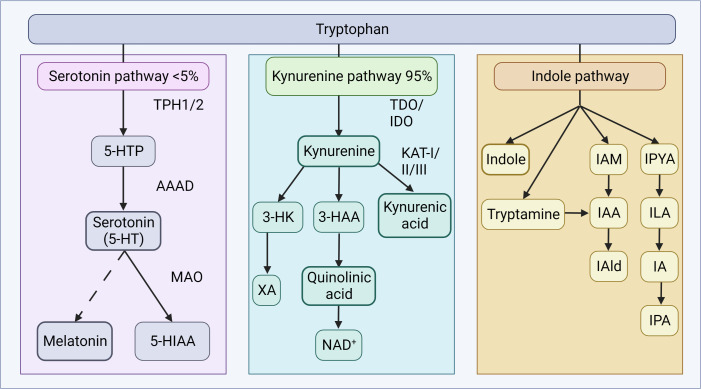
“Activation of stress-responsive TDO or immune-responsive IDO can limit the availability of tryptophan for serotonin synthesis and increase the downstream production of neurotoxic or neuroprotective metabolites (31).”
§ Signalling cognition: the gut microbiota and hypothalamic-pituitary-adrenal axis > Mechanisms and pathways of the MGB axis > The nervous system
What the sources say
- Not checked yetcites [31] O’Mahony, 2015, Serotonin, tryptophan metabolism and the brain-gut-microbiome axis, Behav Brain Res source
plaindirect- Asserted: passes — The authors put it forward as true.
- Checkable: passes — It can be checked against evidence.
- Load-bearing: passes — The paper’s conclusions depend on it.
- Based: passes — The paper gives a reason why it holds.
It may state two separate claims as one.
In the paper
“Glucocorticoids and tryptophan induce TDO, while cytokines induce IDO (200).”
§ Signalling cognition: the gut microbiota and hypothalamic-pituitary-adrenal axis > Mechanisms and pathways of the MGB axis > The nervous system
What the sources say
- Not checked yetcites [200] Ruddick, 2006, Tryptophan metabolism in the central nervous system: medical implications, Expert Rev Mol Med source
plaindirect- Asserted: passes — The authors put it forward as true.
- Checkable: passes — It can be checked against evidence.
- Load-bearing: passes — The paper’s conclusions depend on it.
- Based: passes — The paper gives a reason why it holds.
It may state two separate claims as one.
None of the sources cited for this claim have been checked yet.
In the paper
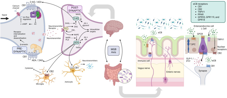
“The ECS modulates a multitude of physiological processes, including the HPA axis (241), cognition, learning and memory (242), intestinal-barrier function (243), inflammation (244), energy metabolism (245), among others (reviewed recently (239)).”
§ Signalling cognition: the gut microbiota and hypothalamic-pituitary-adrenal axis > Mechanisms and pathways of the MGB axis > The nervous system
What the sources say
- Not checked yetcites [241] Micale, 2018, Endocannabinoid system, stress and HPA axis, Eur J Pharmacol source
- Not checked yetcites [242] Zanettini, 2011, Effects of endocannabinoid system modulation on cognitive and emotional behavior, Front Behav Neurosci source
- Not checked yetcites [243] Muccioli, 2010, The endocannabinoid system links gut microbiota to adipogenesis, Mol Syst Biol source
- Not checked yetcites [244] Donvito, 2018, The endogenous cannabinoid system: a budding source of targets for treating inflammatory and neuropathic pain, Neuropsychopharmacol Off Publ Am Coll Neuropsychopharmacol source
- Not checked yetcites [245] Wang, 2008, Role of the endocannabinoid system in metabolic control, Curr Opin Nephrol Hypertens source
- Not checked yetcites [239] Cristino, 2020, Cannabinoids and the expanded endocannabinoid system in neurological disorders, Nat Rev Neurol source
plaindirect- Asserted: passes — The authors put it forward as true.
- Checkable: passes — It can be checked against evidence.
- Load-bearing: unclear — It is unclear whether the paper’s conclusions depend on it.
- Based: passes — The paper gives a reason why it holds.
It may state two separate claims as one.
In the paper
“The HPA axis is also regulated by GABAergic signaling at the level of CRH, and nearly 50% of all synapses in the PVN are GABAergic (233).”
§ Signalling cognition: the gut microbiota and hypothalamic-pituitary-adrenal axis > Mechanisms and pathways of the MGB axis > The nervous system
What the sources say
- Not checked yetcites [233] Miklós, 2002, GABAergic innervation of corticotropin-releasing hormone (CRH)-secreting parvocellular neurons and its plasticity as demonstrated by quantitative immunoelectron microscopy, Neuroscience source
plaindirect- Asserted: passes — The authors put it forward as true.
- Checkable: passes — It can be checked against evidence.
- Load-bearing: unclear — It is unclear whether the paper’s conclusions depend on it.
- Based: passes — The paper gives a reason why it holds.
It may state two separate claims as one.
In the paper
“Serotonergic neurons have a significant influence on neuroendocrine function, and there is a dynamic interplay and extensive crosstalk between the serotonergic system and HPA axis (215).”
§ Signalling cognition: the gut microbiota and hypothalamic-pituitary-adrenal axis > Mechanisms and pathways of the MGB axis > The nervous system
What the sources say
- Not checked yetcites [215] Berger, 2009, The expanded biology of serotonin, Annu Rev Med source
plaindirect- Asserted: passes — The authors put it forward as true.
- Checkable: passes — It can be checked against evidence.
- Load-bearing: unclear — It is unclear whether the paper’s conclusions depend on it.
- Based: passes — The paper gives a reason why it holds.
It may state two separate claims as one.
None of the sources cited for this claim have been checked yet.
In the paper
“Mounting evidence suggests that the GM modulates the tryptophan and kynurenine pathways and is a humoral route through which the GM may influence cognition at the level of the CNS (206–210).”
§ Signalling cognition: the gut microbiota and hypothalamic-pituitary-adrenal axis > Mechanisms and pathways of the MGB axis > The nervous system
What the sources say
- Not checked yetcites [206] Bosi, 2020, Tryptophan metabolites along the microbiota-Gut-Brain axis: an interkingdom communication system influencing the gut in health and disease, Int J Tryptophan Res source
- Not checked yetcites [207] Kiser, 2012, The reciprocal interaction between serotonin and social behaviour, Neurosci Biobehav Rev source
- Not checked yetcites [208] Homberg, 2012, Serotonin and decision making processes, Neurosci Biobehav Rev source
- Not checked yetcites [209] Schwarcz, 2012, Kynurenines in the mammalian brain: when physiology meets pathology, Nat Rev Neurosci source
- Not checked yetcites [210] Szalardy, 2012, Manipulating kynurenic acid levels in the brain - on the edge between neuroprotection and cognitive dysfunction, Curr Top Med Chem source
hedgedconditional- Asserted: passes — The authors put it forward as true.
- Checkable: passes — It can be checked against evidence.
- Load-bearing: passes — The paper’s conclusions depend on it.
- Based: passes — The paper gives a reason why it holds.
It may state two separate claims as one.
A conclusion the paper draws from its own results — it cites no other source for it, so there is none to check. The quote shows it is in the paper.
In the paper
“there is a growing body of evidence suggesting that the GM can ultimately influence the levels of neurotransmitters in the brain and alter brain function and cognition.”
§ Signalling cognition: the gut microbiota and hypothalamic-pituitary-adrenal axis > Mechanisms and pathways of the MGB axis > The nervous system
hedgeddirect- Asserted: passes — The authors put it forward as true.
- Checkable: passes — It can be checked against evidence.
- Load-bearing: passes — The paper’s conclusions depend on it.
- Based: passes — The paper gives a reason why it holds.
It may state two separate claims as one.
None of the sources cited for this claim have been checked yet.
In the paper
“Germ-free mice exhibit reduced colonic serotonin production and decreased levels in the blood, which normalize with microbial colonization (164, 223).”
§ Signalling cognition: the gut microbiota and hypothalamic-pituitary-adrenal axis > Mechanisms and pathways of the MGB axis > The nervous system
What the sources say
plaindirect- Asserted: passes — The authors put it forward as true.
- Checkable: passes — It can be checked against evidence.
- Load-bearing: passes — The paper’s conclusions depend on it.
- Based: passes — The paper gives a reason why it holds.
It may state two separate claims as one.
In the paper
“Healthy mice colonized with Candida albicans showed marked anxiety-like behavior and increased corticosterone concentrations that were inversely correlated with forebrain AEA, demonstrating disruption of the HPA axis through dysregulation of the ECS (253).”
§ Signalling cognition: the gut microbiota and hypothalamic-pituitary-adrenal axis > Mechanisms and pathways of the MGB axis > The nervous system
What the sources say
- Not checked yetcites [253] Markey, 2020, Colonization with the commensal fungus candida albicans perturbs the gut-brain axis through dysregulation of endocannabinoid signaling, Psychoneuroendocrinology source
boosteddirect- Asserted: passes — The authors put it forward as true.
- Checkable: passes — It can be checked against evidence.
- Load-bearing: passes — The paper’s conclusions depend on it.
- Based: passes — The paper gives a reason why it holds.
It may state two separate claims as one.
In the paper
“Microbes and microbial products then gain access to the circulation and may gain easier access to the CNS (262).”
§ Signalling cognition: the gut microbiota and hypothalamic-pituitary-adrenal axis > Mechanisms and pathways of the MGB axis > The immune system
What the sources say
- Not checked yetcites [262] Logsdon, 2018, Gut reactions: how the blood-brain barrier connects the microbiome and the brain, Exp Biol Med (Maywood) source
hedgedconditional- Asserted: passes — The authors put it forward as true.
- Checkable: passes — It can be checked against evidence.
- Load-bearing: passes — The paper’s conclusions depend on it.
- Based: passes — The paper gives a reason why it holds.
It may state two separate claims as one.
In the paper
“Dysbiosis affects eCB signaling, and vice versa (243).”
§ Signalling cognition: the gut microbiota and hypothalamic-pituitary-adrenal axis > Mechanisms and pathways of the MGB axis > The nervous system
What the sources say
- Not checked yetcites [243] Muccioli, 2010, The endocannabinoid system links gut microbiota to adipogenesis, Mol Syst Biol source
plaindirect- Asserted: passes — The authors put it forward as true.
- Checkable: passes — It can be checked against evidence.
- Load-bearing: passes — The paper’s conclusions depend on it.
- Based: passes — The paper gives a reason why it holds.
It may state two separate claims as one.
In the paper
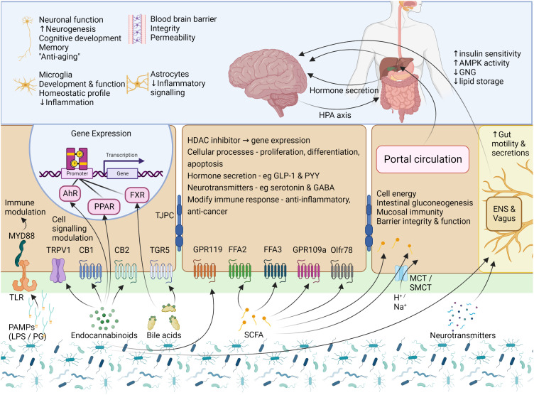
“SCFAs are necessary for the maintenance of CNS homeostasis, learning and cognition, and reward-associated behaviors (25).”
§ Signalling cognition: the gut microbiota and hypothalamic-pituitary-adrenal axis > Mechanisms and pathways of the MGB axis > Microbial metabolites
What the sources say
- Not checked yetcites [25] Cryan, 2019, The microbiota-gut-brain axis, Physiol Rev source
plaindirect- Asserted: passes — The authors put it forward as true.
- Checkable: passes — It can be checked against evidence.
- Load-bearing: unclear — It is unclear whether the paper’s conclusions depend on it.
- Based: passes — The paper gives a reason why it holds.
It may state two separate claims as one.
None of the sources cited for this claim have been checked yet.
In the paper
“SCFAs can modulate the levels of neurotransmitters and neurotrophic factors and regulate the expression of tryptophan 5-hydroxylase, the enzyme involved in the synthesis of serotonin, and tyrosine hydroxylase, the enzyme involved in the rate-limiting step in dopamine, noradrenaline, and adrenaline synthesis (18, 132, 164, 222).”
§ Signalling cognition: the gut microbiota and hypothalamic-pituitary-adrenal axis > Mechanisms and pathways of the MGB axis > Microbial metabolites
What the sources say
- Not checked yetcites [18] Clarke, 2014, Minireview: gut microbiota: the neglected endocrine organ, Mol Endocrinol source
- Not checked yetcites [132] Dalile, 2019, The role of short-chain fatty acids in microbiota–gut–brain communication, Nat Rev Gastroenterol Hepatol source
- Not checked yetcites [164] Yano, 2015, Indigenous bacteria from the gut microbiota regulate host serotonin biosynthesis, Cell source
- Not checked yetcites [222] Reigstad, 2015, Gut microbes promote colonic serotonin production through an effect of short-chain fatty acids on enterochromaffin cells, FASEB J Off Publ Fed Am Soc Exp Biol source
plaindirect- Asserted: passes — The authors put it forward as true.
- Checkable: passes — It can be checked against evidence.
- Load-bearing: passes — The paper’s conclusions depend on it.
- Based: passes — The paper gives a reason why it holds.
It may state two separate claims as one.
In the paper
“microbial products, cytokines, and other immune molecules released under the influence of the GM may further influence the BBB’s integrity, alter BBB transport rates, and promote the release of neuroimmune molecules from the cells of the BBB (262).”
§ Signalling cognition: the gut microbiota and hypothalamic-pituitary-adrenal axis > Mechanisms and pathways of the MGB axis > The immune system
What the sources say
- Not checked yetcites [262] Logsdon, 2018, Gut reactions: how the blood-brain barrier connects the microbiome and the brain, Exp Biol Med (Maywood) source
hedgedconditional- Asserted: passes — The authors put it forward as true.
- Checkable: passes — It can be checked against evidence.
- Load-bearing: passes — The paper’s conclusions depend on it.
- Based: passes — The paper gives a reason why it holds.
It may state two separate claims as one.
A conclusion the paper draws from its own results — it cites no other source for it, so there is none to check. The quote shows it is in the paper.
In the paper
“These factors may lead to neuroinflammation, which is an important process shaping brain function.”
§ Signalling cognition: the gut microbiota and hypothalamic-pituitary-adrenal axis > Mechanisms and pathways of the MGB axis > The immune system
hedgedconditional- Asserted: passes — The authors put it forward as true.
- Checkable: passes — It can be checked against evidence.
- Load-bearing: unclear — It is unclear whether the paper’s conclusions depend on it.
- Based: passes — The paper gives a reason why it holds.
It may state two separate claims as one.
None of the sources cited for this claim have been checked yet.
In the paper
“Perturbations in microbial diversity, secondary to antimicrobials, have been shown to influence pro-inflammatory cytokine secretion in the CNS and alter microglial morphology (268–270).”
§ Signalling cognition: the gut microbiota and hypothalamic-pituitary-adrenal axis > Mechanisms and pathways of the MGB axis > The immune system
What the sources say
- Not checked yetcites [268] Minter, 2016, Antibiotic-induced perturbations in gut microbial diversity influences neuro-inflammation and amyloidosis in a murine model of alzheimer’s disease, Sci Rep source
- Not checked yetcites [269] Minter, 2017, Antibiotic-induced perturbations in microbial diversity during post-natal development alters amyloid pathology in an aged APP SWE /PS1 ΔE9 murine model of alzheimer’s disease OPEN, Sci Rep source
- Not checked yetcites [270] Jang, 2018, Evidence for interplay among antibacterial-induced gut microbiota disturbance, neuro-inflammation, and anxiety in mice, Mucosal Immunol source
plaindirect- Asserted: passes — The authors put it forward as true.
- Checkable: passes — It can be checked against evidence.
- Load-bearing: unclear — It is unclear whether the paper’s conclusions depend on it.
- Based: passes — The paper gives a reason why it holds.
It may state two separate claims as one.
None of the sources cited for this claim have been checked yet.
In the paper
“The GM is therefore closely linked to the immune system, and they interact in several ways [reviewed recently (256, 257)].”
§ Signalling cognition: the gut microbiota and hypothalamic-pituitary-adrenal axis > Mechanisms and pathways of the MGB axis > The immune system
What the sources say
plaincorrelational- Asserted: passes — The authors put it forward as true.
- Checkable: passes — It can be checked against evidence.
- Load-bearing: unclear — It is unclear whether the paper’s conclusions depend on it.
- Based: passes — The paper gives a reason why it holds.
It may state two separate claims as one.
None of the sources cited for this claim have been checked yet.
In the paper
“In response to stress, eCB signaling modulates glucocorticoid and CRH signaling in the brain and is crucial in recovering homeostasis (241, 246, 247).”
§ Signalling cognition: the gut microbiota and hypothalamic-pituitary-adrenal axis > Mechanisms and pathways of the MGB axis > The nervous system
What the sources say
- Not checked yetcites [241] Micale, 2018, Endocannabinoid system, stress and HPA axis, Eur J Pharmacol source
- Not checked yetcites [246] Srivastava, 2022, The microbiome and gut endocannabinoid system in the regulation of stress responses and metabolism, Front Cell Neurosci source
- Not checked yetcites [247] Hillard, 2016, Endocannabinoid signaling and the hypothalamic-Pituitary-Adrenal axis, Compr Physiol source
plaindirect- Asserted: passes — The authors put it forward as true.
- Checkable: passes — It can be checked against evidence.
- Load-bearing: unclear — It is unclear whether the paper’s conclusions depend on it.
- Based: passes — The paper gives a reason why it holds.
It may state two separate claims as one.
In the paper
“Some BAs serve as substrates for gut microbes, while others exhibit antimicrobial properties, actively shaping the GM at the highest taxonomic levels (318).”
§ Signalling cognition: the gut microbiota and hypothalamic-pituitary-adrenal axis > Mechanisms and pathways of the MGB axis > Microbial metabolites
What the sources say
- Not checked yetcites [318] Peter, 2006, Mechanism of growth inhibition by free bile acids in lactobacilli and bifidobacteria, J Bacteriol source
plaindirect- Asserted: passes — The authors put it forward as true.
- Checkable: passes — It can be checked against evidence.
- Load-bearing: unclear — It is unclear whether the paper’s conclusions depend on it.
- Based: passes — The paper gives a reason why it holds.
It may state two separate claims as one.
In the paper
“The GM influences BA metabolism, and BAs affect the GM’s composition (296).”
§ Signalling cognition: the gut microbiota and hypothalamic-pituitary-adrenal axis > Mechanisms and pathways of the MGB axis > Microbial metabolites
What the sources say
- Not checked yetcites [296] Ramírez-Pérez, 2017, The role of the gut microbiota in bile acid metabolism, Ann Hepatol source
plaindirect- Asserted: passes — The authors put it forward as true.
- Checkable: passes — It can be checked against evidence.
- Load-bearing: passes — The paper’s conclusions depend on it.
- Based: passes — The paper gives a reason why it holds.
It may state two separate claims as one.
A conclusion the paper draws from its own results — it cites no other source for it, so there is none to check. The quote shows it is in the paper.
In the paper
“These findings suggest that SCFAs regulate CNS processes through both direct and indirect mechanisms and may ultimately affect host cognition and response to stress.”
§ Signalling cognition: the gut microbiota and hypothalamic-pituitary-adrenal axis > Mechanisms and pathways of the MGB axis > Microbial metabolites
hedgedconditional- Asserted: passes — The authors put it forward as true.
- Checkable: passes — It can be checked against evidence.
- Load-bearing: passes — The paper’s conclusions depend on it.
- Based: passes — The paper gives a reason why it holds.
It may state two separate claims as one.
In the paper
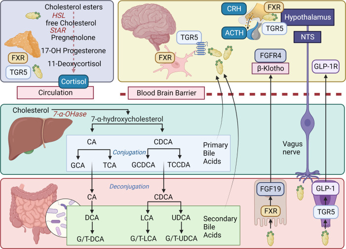
“TGR5, expressed in brain and peripheral neurons, as well as glial and microglial cells, can be activated by several neurosteroids (305).”
§ Signalling cognition: the gut microbiota and hypothalamic-pituitary-adrenal axis > Mechanisms and pathways of the MGB axis > Microbial metabolites
What the sources say
- Not checked yetcites [305] Keitel, 2010, The bile acid receptor TGR5 (Gpbar-1) acts as a neurosteroid receptor in brain, Glia source
plaindirect- Asserted: passes — The authors put it forward as true.
- Checkable: passes — It can be checked against evidence.
- Load-bearing: unclear — It is unclear whether the paper’s conclusions depend on it.
- Based: passes — The paper gives a reason why it holds.
It may state two separate claims as one.
None of the sources cited for this claim have been checked yet.
In the paper
“Changes in the GM’s composition correspond to changes in blood and brain BA profiles, which are essential because specific BA ligands’ distinct physicochemical properties determine the potency of BA receptor activation (320–323).”
§ Signalling cognition: the gut microbiota and hypothalamic-pituitary-adrenal axis > Mechanisms and pathways of the MGB axis > Microbial metabolites
What the sources say
- Not checked yetcites [320] Grant, 2020, Bile acid signaling in neurodegenerative and neurological disorders, Int J Mol Sci source
- Not checked yetcites [321] Jia, 2020, Expert insights: the potential role of the gut microbiome-bile acid-brain axis in the development and progression of alzheimer’s disease and hepatic encephalopathy, Med Res Rev source
- Not checked yetcites [322] Makishima, 1999, Identification of a nuclear receptor for bile acids, Science source
- Not checked yetcites [323] Sayin, 2013, Gut microbiota regulates bile acid metabolism by reducing the levels of tauro-beta-muricholic acid, a naturally occurring FXR antagonist, Cell Metab source
plaincorrelational- Asserted: passes — The authors put it forward as true.
- Checkable: passes — It can be checked against evidence.
- Load-bearing: passes — The paper’s conclusions depend on it.
- Based: passes — The paper gives a reason why it holds.
It may state two separate claims as one.
In the paper
“In stressed mice, SCFA administration reduced HPA axis hyperactivity and intestinal permeability (291).”
§ Signalling cognition: the gut microbiota and hypothalamic-pituitary-adrenal axis > Mechanisms and pathways of the MGB axis > Microbial metabolites
What the sources say
- Not checked yetcites [291] van de Wouw, 2018, Short-chain fatty acids: microbial metabolites that alleviate stress-induced brain–gut axis alterations, J Physiol source
plaindirect- Asserted: passes — The authors put it forward as true.
- Checkable: passes — It can be checked against evidence.
- Load-bearing: passes — The paper’s conclusions depend on it.
- Based: passes — The paper gives a reason why it holds.
It may state two separate claims as one.
In the paper
“FXR knockout mice exhibit abnormal BA and neurotransmitter concentrations, resulting in impaired cognition and motor coordination (304).”
§ Signalling cognition: the gut microbiota and hypothalamic-pituitary-adrenal axis > Mechanisms and pathways of the MGB axis > Microbial metabolites
What the sources say
- Not checked yetcites [304] Huang, 2015, Deletion of mouse FXR gene disturbs multiple neurotransmitter systems and alters neurobehavior, Front Behav Neurosci source
plaindirect- Asserted: passes — The authors put it forward as true.
- Checkable: passes — It can be checked against evidence.
- Load-bearing: unclear — It is unclear whether the paper’s conclusions depend on it.
- Based: passes — The paper gives a reason why it holds.
It may state two separate claims as one.
None of the sources cited for this claim have been checked yet.
In the paper
“these metabolites have emerged as important modulators of the MGB axis, functioning directly via BA receptors in the ENS and brain or indirectly via GLP-1 or the FXR-FGF15/19 axis (297, 319).”
§ Signalling cognition: the gut microbiota and hypothalamic-pituitary-adrenal axis > Mechanisms and pathways of the MGB axis > Microbial metabolites
What the sources say
plaindirect- Asserted: passes — The authors put it forward as true.
- Checkable: passes — It can be checked against evidence.
- Load-bearing: passes — The paper’s conclusions depend on it.
- Based: passes — The paper gives a reason why it holds.
It may state two separate claims as one.
None of the sources cited for this claim have been checked yet.
In the paper
“BA receptors are present in the brain, and BAs can either be synthesized locally or actively transported across the BBB by BA transporters from circulation (297, 301, 302).”
§ Signalling cognition: the gut microbiota and hypothalamic-pituitary-adrenal axis > Mechanisms and pathways of the MGB axis > Microbial metabolites
What the sources say
- Not checked yetcites [297] Mertens, 2017, Bile acid signaling pathways from the enterohepatic circulation to the central nervous system, Front Neurosci source
- Not checked yetcites [301] Trauner, 2003, Bile salt transporters: molecular characterization, function, and regulation, Physiol Rev source
- Not checked yetcites [302] Baloni, 2020, Metabolic network analysis reveals altered bile acid synthesis and metabolism in alzheimer’s disease, Cell Rep Med source
plaindirect- Asserted: passes — The authors put it forward as true.
- Checkable: passes — It can be checked against evidence.
- Load-bearing: passes — The paper’s conclusions depend on it.
- Based: passes — The paper gives a reason why it holds.
It may state two separate claims as one.
None of the sources cited for this claim have been checked yet.
In the paper
“Cholestasis, associated with suppression of the HPA axis, likely due to BA interactions with glucocorticoid receptors in the brain (299, 312).”
§ Signalling cognition: the gut microbiota and hypothalamic-pituitary-adrenal axis > Mechanisms and pathways of the MGB axis > Microbial metabolites
What the sources say
- Not checked yetcites [299] McMillin, 2015, Suppression of the HPA axis during cholestasis can be attributed to hypothalamic bile acid signaling, Mol Endocrinol source
- Not checked yetcites [312] Perry, 2015, FGF1 and FGF19 reverse diabetes by suppression of the hypothalamic–pituitary–adrenal axis, Nat Commun source
hedgedcorrelational- Asserted: passes — The authors put it forward as true.
- Checkable: passes — It can be checked against evidence.
- Load-bearing: unclear — It is unclear whether the paper’s conclusions depend on it.
- Based: passes — The paper gives a reason why it holds.
It may state two separate claims as one.
In the paper
“FXR activation is known to regulate lipoprotein receptors and transporters, as well as enzymes in the steroidogenic pathway, and has been shown to increase corticosterone levels in mice (311).”
§ Signalling cognition: the gut microbiota and hypothalamic-pituitary-adrenal axis > Mechanisms and pathways of the MGB axis > Microbial metabolites
What the sources say
- Not checked yetcites [311] Theiler-Schwetz, 2019, Bile acids and glucocorticoid metabolism in health and disease, Biochim Biophys Acta - Mol Basis Dis source
boosteddirect- Asserted: passes — The authors put it forward as true.
- Checkable: passes — It can be checked against evidence.
- Load-bearing: unclear — It is unclear whether the paper’s conclusions depend on it.
- Based: passes — The paper gives a reason why it holds.
It may state two separate claims as one.
In the paper
“Mice supplemented with a BCAA-enriched cocktail exhibited improved physical endurance and an extended lifespan (335).”
§ Signalling cognition: the gut microbiota and hypothalamic-pituitary-adrenal axis > Mechanisms and pathways of the MGB axis > Microbial metabolites
What the sources say
- Not checked yetcites [335] D’Antona, 2010, Branched-chain amino acid supplementation promotes survival and supports cardiac and skeletal muscle mitochondrial biogenesis in middle-aged mice, Cell Metab source
plaindirect- Asserted: passes — The authors put it forward as true.
- Checkable: passes — It can be checked against evidence.
- Load-bearing: unclear — It is unclear whether the paper’s conclusions depend on it.
- Based: passes — The paper gives a reason why it holds.
It may state two separate claims as one.
In the paper
“Stress during pregnancy disrupts the vertical transmission of microbes from mother to offspring, leading to alterations in the maternal microbiota, which are then transferred to the offspring (342).”
§ Signalling cognition: the gut microbiota and hypothalamic-pituitary-adrenal axis > The relationship between the microbiota, HPA axis, and cognition > Stress-related interactions between the HPA axis and gut microbiota
What the sources say
- Not checked yetcites [342] Dinan, 2012, Regulation of the stress response by the gut microbiota: implications for psychoneuroendocrinology, Psychoneuroendocrinology source
plaindirect- Asserted: passes — The authors put it forward as true.
- Checkable: passes — It can be checked against evidence.
- Load-bearing: passes — The paper’s conclusions depend on it.
- Based: passes — The paper gives a reason why it holds.
It may state two separate claims as one.
None of the sources cited for this claim have been checked yet.
In the paper
“The microbiota and HPA axis develop rapidly and profoundly in the first years of life, and environmental stressors can affect both (146–148).”
§ Signalling cognition: the gut microbiota and hypothalamic-pituitary-adrenal axis > The relationship between the microbiota, HPA axis, and cognition > Stress-related interactions between the HPA axis and gut microbiota
What the sources say
- Not checked yetcites [146] Gur, 2019, Prenatal stress disrupts social behavior, cortical neurobiology and commensal microbes in adult male offspring, Behav Brain Res source
- Not checked yetcites [147] Zijlmans, 2015, Maternal prenatal stress is associated with the infant intestinal microbiota, Psychoneuroendocrinology source
- Not checked yetcites [148] Jang, 2018, Immobilization stress-induced escherichia coli causes anxiety by inducing NF-κB activation through gut microbiota disturbance, Sci Rep source
plain- Asserted: passes — The authors put it forward as true.
- Checkable: passes — It can be checked against evidence.
- Load-bearing: passes — The paper’s conclusions depend on it.
- Based: passes — The paper gives a reason why it holds.
It may state two separate claims as one.
In the paper
“In response to early life stress, younger rats had increased neurogenesis, decreased BDNF IV promoter histone methylation, with a complementary increase in hippocampal BDNF concentration, and associated improvements in spatial and non-spatial learning (348).”
§ Signalling cognition: the gut microbiota and hypothalamic-pituitary-adrenal axis > The relationship between the microbiota, HPA axis, and cognition > Stress-related interactions between the HPA axis and gut microbiota
What the sources say
- Not checked yetcites [348] Suri, 2013, Early stress evokes age-dependent biphasic changes in hippocampal neurogenesis, BDNF expression, and cognition, Biol Psychiatry source
plaindirect- Asserted: passes — The authors put it forward as true.
- Checkable: passes — It can be checked against evidence.
- Load-bearing: passes — The paper’s conclusions depend on it.
- Based: passes — The paper gives a reason why it holds.
It may state two separate claims as one.
In the paper
“In contrast, middle-aged rats demonstrated opposing changes, concomitant with impairments in hippocampal-dependent cognitive tasks.”
§ Signalling cognition: the gut microbiota and hypothalamic-pituitary-adrenal axis > The relationship between the microbiota, HPA axis, and cognition > Stress-related interactions between the HPA axis and gut microbiota
What the sources say
- Not checked yetcites [348] Suri, 2013, Early stress evokes age-dependent biphasic changes in hippocampal neurogenesis, BDNF expression, and cognition, Biol Psychiatry source
plaincorrelational- Asserted: passes — The authors put it forward as true.
- Checkable: passes — It can be checked against evidence.
- Load-bearing: passes — The paper’s conclusions depend on it.
- Based: passes — The paper gives a reason why it holds.
It may state two separate claims as one.
In the paper
“In the CNS, BCAAs play roles in protein synthesis, food intake regulation, and serve as nitrogen donors involved in intercellular shuttling and the synthesis of the neurotransmitters glutamate and GABA (both modulate the HPA axis) (336).”
§ Signalling cognition: the gut microbiota and hypothalamic-pituitary-adrenal axis > Mechanisms and pathways of the MGB axis > Microbial metabolites
What the sources say
- Not checked yetcites [336] Sperringer, 2017, Branched-chain amino acids and brain metabolism, Neurochem Res source
plaindirect- Asserted: passes — The authors put it forward as true.
- Checkable: passes — It can be checked against evidence.
- Load-bearing: unclear — It is unclear whether the paper’s conclusions depend on it.
- Based: passes — The paper gives a reason why it holds.
It may state two separate claims as one.
In the paper
“Early life stressors, and in utero stressors, can impact the development and function of the HPA axis (149).”
§ Signalling cognition: the gut microbiota and hypothalamic-pituitary-adrenal axis > The relationship between the microbiota, HPA axis, and cognition > Stress-related interactions between the HPA axis and gut microbiota
What the sources say
- Not checked yetcites [149] Maniam, 2014, Early-life stress, HPA axis adaptation, and mechanisms contributing to later health outcomes, Front Endocrinol source
plaindirect- Asserted: passes — The authors put it forward as true.
- Checkable: passes — It can be checked against evidence.
- Load-bearing: passes — The paper’s conclusions depend on it.
- Based: passes — The paper gives a reason why it holds.
It may state two separate claims as one.
In the paper
“Early life stress, such as maternal separation, activates the HPA axis with associated changes to the developing microbiota, ultimately leading to an imbalance in the GM and an inappropriate stress response (18).”
§ Signalling cognition: the gut microbiota and hypothalamic-pituitary-adrenal axis > The relationship between the microbiota, HPA axis, and cognition > Stress-related interactions between the HPA axis and gut microbiota
What the sources say
- Not checked yetcites [18] Clarke, 2014, Minireview: gut microbiota: the neglected endocrine organ, Mol Endocrinol source
plaindirect- Asserted: passes — The authors put it forward as true.
- Checkable: passes — It can be checked against evidence.
- Load-bearing: passes — The paper’s conclusions depend on it.
- Based: passes — The paper gives a reason why it holds.
It may state two separate claims as one.
None of the sources cited for this claim have been checked yet.
In the paper
“The microbiota and HPA axis develop rapidly and profoundly in the first years of life, and environmental stressors can affect both (146–148).”
§ Signalling cognition: the gut microbiota and hypothalamic-pituitary-adrenal axis > The relationship between the microbiota, HPA axis, and cognition > Stress-related interactions between the HPA axis and gut microbiota
What the sources say
- Not checked yetcites [146] Gur, 2019, Prenatal stress disrupts social behavior, cortical neurobiology and commensal microbes in adult male offspring, Behav Brain Res source
- Not checked yetcites [147] Zijlmans, 2015, Maternal prenatal stress is associated with the infant intestinal microbiota, Psychoneuroendocrinology source
- Not checked yetcites [148] Jang, 2018, Immobilization stress-induced escherichia coli causes anxiety by inducing NF-κB activation through gut microbiota disturbance, Sci Rep source
plaindirect- Asserted: passes — The authors put it forward as true.
- Checkable: passes — It can be checked against evidence.
- Load-bearing: passes — The paper’s conclusions depend on it.
- Based: passes — The paper gives a reason why it holds.
It may state two separate claims as one.
A conclusion the paper draws from its own results — it cites no other source for it, so there is none to check. The quote shows it is in the paper.
In the paper
“These discordant results illustrate the biphasic consequences of early life stress and indicate a role for epigenetic modification of BDNF expression.”
§ Signalling cognition: the gut microbiota and hypothalamic-pituitary-adrenal axis > The relationship between the microbiota, HPA axis, and cognition > Stress-related interactions between the HPA axis and gut microbiota
hedgedcorrelational- Asserted: passes — The authors put it forward as true.
- Checkable: passes — It can be checked against evidence.
- Load-bearing: passes — The paper’s conclusions depend on it.
- Based: passes — The paper gives a reason why it holds.
It may state two separate claims as one.
In the paper
“Jašarević et al. demonstrated changes in the microbiome of these offspring, as well as alterations in the metabolome of the gut and brain (343).”
§ Signalling cognition: the gut microbiota and hypothalamic-pituitary-adrenal axis > The relationship between the microbiota, HPA axis, and cognition > Stress-related interactions between the HPA axis and gut microbiota
What the sources say
- Not checked yetcites [343] Jašarević, 2015, Alterations in the vaginal microbiome by maternal stress are associated with metabolic reprogramming of the offspring gut and brain, Endocrinology source
boostedcorrelational- Asserted: passes — The authors put it forward as true.
- Checkable: passes — It can be checked against evidence.
- Load-bearing: passes — The paper’s conclusions depend on it.
- Based: passes — The paper gives a reason why it holds.
It may state two separate claims as one.
In the paper
“In the CNS, BCAAs play roles in protein synthesis, food intake regulation, and serve as nitrogen donors involved in intercellular shuttling and the synthesis of the neurotransmitters glutamate and GABA (both modulate the HPA axis) (336).”
§ Signalling cognition: the gut microbiota and hypothalamic-pituitary-adrenal axis > Mechanisms and pathways of the MGB axis > Microbial metabolites
What the sources say
- Not checked yetcites [336] Sperringer, 2017, Branched-chain amino acids and brain metabolism, Neurochem Res source
plaindirect- Asserted: passes — The authors put it forward as true.
- Checkable: passes — It can be checked against evidence.
- Load-bearing: unclear — It is unclear whether the paper’s conclusions depend on it.
- Based: passes — The paper gives a reason why it holds.
It may state two separate claims as one.
In the paper
“Following chronic restraint stress, germ-free mice showed significantly greater HPA axis activity, whereas the SPF mice exhibited more anxiety-like behaviors under the same stress (349).”
§ Signalling cognition: the gut microbiota and hypothalamic-pituitary-adrenal axis > The relationship between the microbiota, HPA axis, and cognition > Stress-related interactions between the HPA axis and gut microbiota
What the sources say
- Not checked yetcites [349] Huo, 2017, Microbiota modulate anxiety-like behavior and endocrine abnormalities in hypothalamic-pituitary-adrenal axis, Front Cell Infect Microbiol source
plaincorrelational- Asserted: passes — The authors put it forward as true.
- Checkable: passes — It can be checked against evidence.
- Load-bearing: passes — The paper’s conclusions depend on it.
- Based: passes — The paper gives a reason why it holds.
It may state two separate claims as one.
In the paper
“A study by Allen et al. demonstrated that the GM is necessary for stress-induced immunomodulation, with enhancement of splenic macrophage reactivity occurring in colonized controls but not in germ-free mice, in response to social disruption stress (360).”
§ Signalling cognition: the gut microbiota and hypothalamic-pituitary-adrenal axis > The relationship between the microbiota, HPA axis, and cognition > Stress-related interactions between the HPA axis and gut microbiota
What the sources say
- Not checked yetcites [360] Allen, 2012, The intestinal microbiota are necessary for stressor-induced enhancement of splenic macrophage microbicidal activity, Brain Behav Immun source
boosteddirect- Asserted: passes — The authors put it forward as true.
- Checkable: passes — It can be checked against evidence.
- Load-bearing: passes — The paper’s conclusions depend on it.
- Based: passes — The paper gives a reason why it holds.
It may state two separate claims as one.
In the paper
“Participants reported reductions in perceived stress, but these were not significantly different to controls, and markers of stress were unaffected.”
§ Signalling cognition: the gut microbiota and hypothalamic-pituitary-adrenal axis > The relationship between the microbiota, HPA axis, and cognition > Stress-related interactions between the HPA axis and gut microbiota
What the sources say
- Not checked yetcites [374] Berding, 2022, Feed your microbes to deal with stress: a psychobiotic diet impacts microbial stability and perceived stress in a healthy adult population, Mol Psychiatry source
plain- Asserted: passes — The authors put it forward as true.
- Checkable: passes — It can be checked against evidence.
- Load-bearing: unclear — It is unclear whether the paper’s conclusions depend on it.
- Based: passes — The paper gives a reason why it holds.
It may state two separate claims as one.
In the paper
“In another RCT, a combination probiotic (L. helveticus (R0052) and B. longum (R0175)) administered to healthy volunteers was associated with beneficial psychological effects in participants and a decrease in 24-hour urinary cortisol, suggesting attenuation of the HPA axis in response to stressors (372).”
§ Signalling cognition: the gut microbiota and hypothalamic-pituitary-adrenal axis > The relationship between the microbiota, HPA axis, and cognition > Stress-related interactions between the HPA axis and gut microbiota
What the sources say
- Not checked yetcites [372] Messaoudi, 2011, Assessment of psychotropic-like properties of a probiotic formulation (Lactobacillus helveticus R0052 and bifidobacterium longum R0175) in rats and human subjects, Br J Nutr source
hedgedcorrelational- Asserted: passes — The authors put it forward as true.
- Checkable: passes — It can be checked against evidence.
- Load-bearing: passes — The paper’s conclusions depend on it.
- Based: passes — The paper gives a reason why it holds.
It may state two separate claims as one.
None of the sources cited for this claim have been checked yet.
In the paper
“The introduction of pathogenic bacteria reduced cognitive abilities and heightened anxiety-like behaviors (362, 363).”
§ Signalling cognition: the gut microbiota and hypothalamic-pituitary-adrenal axis > The relationship between the microbiota, HPA axis, and cognition > Stress-related interactions between the HPA axis and gut microbiota
What the sources say
- Not checked yetcites [362] Gareau, 2011, Bacterial infection causes stress-induced memory dysfunction in mice, Gut source
- Not checked yetcites [363] Jiang, 2013, The effect of maternal infection on cognitive development and hippocampus neuronal apoptosis, proliferation and differentiation in the neonatal rats, Neuroscience source
plaindirect- Asserted: passes — The authors put it forward as true.
- Checkable: passes — It can be checked against evidence.
- Load-bearing: unclear — It is unclear whether the paper’s conclusions depend on it.
- Based: passes — The paper gives a reason why it holds.
It may state two separate claims as one.
In the paper
“Similarly, pre-treatment with L. farciminis reduced HPA hyper-reactivity, intestinal permeability, and neuroinflammation resulting from restraint stress (367).”
§ Signalling cognition: the gut microbiota and hypothalamic-pituitary-adrenal axis > The relationship between the microbiota, HPA axis, and cognition > Stress-related interactions between the HPA axis and gut microbiota
What the sources say
- Not checked yetcites [367] Ait-Belgnaoui, 2012, Prevention of gut leakiness by a probiotic treatment leads to attenuated HPA response to an acute psychological stress in rats, Psychoneuroendocrinology source
plaindirect- Asserted: passes — The authors put it forward as true.
- Checkable: passes — It can be checked against evidence.
- Load-bearing: unclear — It is unclear whether the paper’s conclusions depend on it.
- Based: passes — The paper gives a reason why it holds.
It may state two separate claims as one.
None of the sources cited for this claim have been checked yet.
In the paper
“Animal models of chronic stress demonstrate altered intestinal physiology and GM composition, with an increased secretory state and permeability (352–355).”
§ Signalling cognition: the gut microbiota and hypothalamic-pituitary-adrenal axis > The relationship between the microbiota, HPA axis, and cognition > Stress-related interactions between the HPA axis and gut microbiota
What the sources say
- Not checked yetcites [352] Söderholm, 2002, Chronic stress induces mast cell-dependent bacterial adherence and initiates mucosal inflammation in rat intestine, Gastroenterology source
- Not checked yetcites [353] Cameron, 2005, Stress impairs murine intestinal barrier function: improvement by glucagon-like peptide-2, J Pharmacol Exp Ther source
- Not checked yetcites [354] Sun, 2013, Stress-induced corticotropin-releasing hormone-mediated NLRP6 inflammasome inhibition and transmissible enteritis in mice, Gastroenterology source
- Not checked yetcites [355] Lutgendorff, 2008, The role of microbiota and probiotics in stress-induced gastro-intestinal damage, Curr Mol Med source
boosteddirect- Asserted: passes — The authors put it forward as true.
- Checkable: passes — It can be checked against evidence.
- Load-bearing: passes — The paper’s conclusions depend on it.
- Based: passes — The paper gives a reason why it holds.
It may state two separate claims as one.
In the paper
“Alterations in cognition occurred both in the short- and long-term, confirming mechanisms for both the promotion and inhibition of cognitive performance during acute inflammatory stress.”
§ Signalling cognition: the gut microbiota and hypothalamic-pituitary-adrenal axis > The relationship between the microbiota, HPA axis, and cognition > Stress-related interactions between the HPA axis and gut microbiota
What the sources say
- Not checked yetcites [371] Grigoleit, 2011, Dose-dependent effects of endotoxin on neurobehavioral functions in humans, PloS One source
boosteddirect- Asserted: passes — The authors put it forward as true.
- Checkable: passes — It can be checked against evidence.
- Load-bearing: passes — The paper’s conclusions depend on it.
- Based: passes — The paper gives a reason why it holds.
It may state two separate claims as one. The quote only partly states this claim.
In the paper
“When challenged orally with the enteric murine pathogen C. rodentium, chronically stressed mice had an increased pathogen load and increased colonic TNF-α expression.”
§ Signalling cognition: the gut microbiota and hypothalamic-pituitary-adrenal axis > The relationship between the microbiota, HPA axis, and cognition > Stress-related interactions between the HPA axis and gut microbiota
What the sources say
- Not checked yetcites [359] Bailey, 2010, Stressor exposure disrupts commensal microbial populations in the intestines and leads to increased colonization by citrobacter rodentium, Infect Immun source
plaindirect- Asserted: passes — The authors put it forward as true.
- Checkable: passes — It can be checked against evidence.
- Load-bearing: passes — The paper’s conclusions depend on it.
- Based: passes — The paper gives a reason why it holds.
It may state two separate claims as one.
In the paper
“Notably, the neurochemical effects were not found in vagotomised mice, implicating the vagal pathway.”
§ Signalling cognition: the gut microbiota and hypothalamic-pituitary-adrenal axis > The relationship between the microbiota, HPA axis, and cognition > Stress-related interactions between the HPA axis and gut microbiota
What the sources say
- Not checked yetcites [191] Bravo, 2011, Ingestion of lactobacillus strain regulates emotional behavior and central GABA receptor expression in a mouse via the vagus nerve, Proc Natl Acad Sci source
plaindirect- Asserted: passes — The authors put it forward as true.
- Checkable: passes — It can be checked against evidence.
- Load-bearing: passes — The paper’s conclusions depend on it.
- Based: passes — The paper gives a reason why it holds.
It may state two separate claims as one.
In the paper
“Moreover, these animals showed decreased expression of NMDA receptor subunits (NR-1) in the cortex, and of NR-2a in the cortex and hippocampus, while BDNF levels were lower in the cortex and hippocampus.”
§ Signalling cognition: the gut microbiota and hypothalamic-pituitary-adrenal axis > The relationship between the microbiota, HPA axis, and cognition > Stress-related interactions between the HPA axis and gut microbiota
What the sources say
- Not checked yetcites [51] Sudo, 2004, Postnatal microbial colonization programs the hypothalamic-pituitary-adrenal system for stress response in mice, J Physiol source
plaincorrelational- Asserted: passes — The authors put it forward as true.
- Checkable: passes — It can be checked against evidence.
- Load-bearing: passes — The paper’s conclusions depend on it.
- Based: passes — The paper gives a reason why it holds.
It may state two separate claims as one.
In the paper
“A recent meta-analysis of RCTs focusing on the efficacy of probiotics on stress in healthy individuals showed that probiotic use generally reduced subjective stress levels and appeared to alleviate stress-related sub-threshold anxiety and depression (373).”
§ Signalling cognition: the gut microbiota and hypothalamic-pituitary-adrenal axis > The relationship between the microbiota, HPA axis, and cognition > Stress-related interactions between the HPA axis and gut microbiota
What the sources say
- Not checked yetcites [373] Zhang, 2020, Efficacy of probiotics on stress in healthy volunteers: a systematic review and meta-analysis based on randomized controlled trials, Brain Behav source
hedgeddirect- Asserted: passes — The authors put it forward as true.
- Checkable: passes — It can be checked against evidence.
- Load-bearing: unclear — It is unclear whether the paper’s conclusions depend on it.
- Based: passes — The paper gives a reason why it holds.
It may state two separate claims as one.
In the paper
“Without a complex microbiota, both types of mice exhibited altered fear-associated behavior, changes in gene expression in brain cells, and alterations in the firing patterns and rewiring ability of neurons.”
§ Signalling cognition: the gut microbiota and hypothalamic-pituitary-adrenal axis > The relationship between the microbiota, HPA axis, and cognition > HPA axis-related interactions between the gut microbiota and cognition
What the sources say
- Not checked yetcites [385] Chu, 2019, The microbiota regulate neuronal function and fear extinction learning, Nature source
plaindirect- Asserted: passes — The authors put it forward as true.
- Checkable: passes — It can be checked against evidence.
- Load-bearing: passes — The paper’s conclusions depend on it.
- Based: passes — The paper gives a reason why it holds.
It may state two separate claims as one.
None of the sources cited for this claim have been checked yet.
In the paper
“Supplementation with various Lactobacillus species resulted in improvements in cognitive abilities and social deficits and these changes were shown to occur alongside altered GABA expression in the brain, as well as significant changes in the oxytocin and vagus nerve pathways (191, 391).”
§ Signalling cognition: the gut microbiota and hypothalamic-pituitary-adrenal axis > The relationship between the microbiota, HPA axis, and cognition > HPA axis-related interactions between the gut microbiota and cognition
What the sources say
- Not checked yetcites [191] Bravo, 2011, Ingestion of lactobacillus strain regulates emotional behavior and central GABA receptor expression in a mouse via the vagus nerve, Proc Natl Acad Sci source
- Not checked yetcites [391] Buffington, 2016, Microbial reconstitution reverses maternal diet-induced social and synaptic deficits in offspring, Cell source
plaindirect- Asserted: passes — The authors put it forward as true.
- Checkable: passes — It can be checked against evidence.
- Load-bearing: unclear — It is unclear whether the paper’s conclusions depend on it.
- Based: passes — The paper gives a reason why it holds.
It may state two separate claims as one.
None of the sources cited for this claim have been checked yet.
In the paper
“The probiotic B. longum (NCC3001) demonstrated vagal pathway-mediated anxiolytic effects in mice and B. longum (1714) showed improved learning and memory after 11 weeks of supplementation (192, 390).”
§ Signalling cognition: the gut microbiota and hypothalamic-pituitary-adrenal axis > The relationship between the microbiota, HPA axis, and cognition > HPA axis-related interactions between the gut microbiota and cognition
What the sources say
- Not checked yetcites [192] Bercik, 2011, The anxiolytic effect of bifidobacterium longum NCC3001 involves vagal pathways for gut–brain communication, Neurogastroenterol Motil source
- Not checked yetcites [390] Savignac, 2015, Bifidobacteria modulate cognitive processes in an anxious mouse strain, Behav Brain Res source
boosteddirect- Asserted: passes — The authors put it forward as true.
- Checkable: passes — It can be checked against evidence.
- Load-bearing: unclear — It is unclear whether the paper’s conclusions depend on it.
- Based: passes — The paper gives a reason why it holds.
It may state two separate claims as one.
None of the sources cited for this claim have been checked yet.
In the paper
“Behavioral differences in germ-free mice include their anxiolytic-like manner when compared to SPF controls, although some studies report increased anxiety-like behavior in different species (124, 189, 351, 376).”
§ Signalling cognition: the gut microbiota and hypothalamic-pituitary-adrenal axis > The relationship between the microbiota, HPA axis, and cognition > HPA axis-related interactions between the gut microbiota and cognition
What the sources say
- Not checked yetcites [124] Heijtz, 2011, Normal gut microbiota modulates brain development and behavior, Proc Natl Acad Sci source
- Not checked yetcites [189] Neufeld, 2011, Reduced anxiety-like behavior and central neurochemical change in germ-free mice, Neurogastroenterol Motil Off J Eur Gastrointest Motil Soc source
- Not checked yetcites [351] Crumeyrolle-Arias, 2014, Absence of the gut microbiota enhances anxiety-like behavior and neuroendocrine response to acute stress in rats, Psychoneuroendocrinology source
- Not checked yetcites [376] Bercik, 2011, The intestinal microbiota affect central levels of brain-derived neurotropic factor and behavior in mice, Gastroenterology source
plaincorrelational- Asserted: passes — The authors put it forward as true.
- Checkable: passes — It can be checked against evidence.
- Load-bearing: unclear — It is unclear whether the paper’s conclusions depend on it.
- Based: passes — The paper gives a reason why it holds.
It may state two separate claims as one.
None of the sources cited for this claim have been checked yet.
In the paper
“Germ-free mice present with social cognitive deficits, which may be associated with biochemical changes, such as decreased hippocampal BDNF and c-FOS expression, important in memory (124, 362, 376, 377).”
§ Signalling cognition: the gut microbiota and hypothalamic-pituitary-adrenal axis > The relationship between the microbiota, HPA axis, and cognition > HPA axis-related interactions between the gut microbiota and cognition
What the sources say
- Not checked yetcites [124] Heijtz, 2011, Normal gut microbiota modulates brain development and behavior, Proc Natl Acad Sci source
- Not checked yetcites [362] Gareau, 2011, Bacterial infection causes stress-induced memory dysfunction in mice, Gut source
- Not checked yetcites [376] Bercik, 2011, The intestinal microbiota affect central levels of brain-derived neurotropic factor and behavior in mice, Gastroenterology source
- Not checked yetcites [377] Desbonnet, 2014, Microbiota is essential for social development in the mouse, Mol Psychiatry source
hedgedcorrelational- Asserted: passes — The authors put it forward as true.
- Checkable: passes — It can be checked against evidence.
- Load-bearing: passes — The paper’s conclusions depend on it.
- Based: passes — The paper gives a reason why it holds.
It may state two separate claims as one.
In the paper
“Six weeks of administration with the probiotic Clostridium butyricum restored cognitive function in a mouse model for vascular dementia, which was associated with increased butyrate levels in fecal and brain samples, and activation of the hippocampal BDNF-PI3K/Akt pathway (392).”
§ Signalling cognition: the gut microbiota and hypothalamic-pituitary-adrenal axis > The relationship between the microbiota, HPA axis, and cognition > HPA axis-related interactions between the gut microbiota and cognition
What the sources say
- Not checked yetcites [392] Liu, 2015, Neuroprotective effects of clostridium butyricum against vascular dementia in mice via metabolic butyrate, BioMed Res Int source
plaincorrelational- Asserted: passes — The authors put it forward as true.
- Checkable: passes — It can be checked against evidence.
- Load-bearing: unclear — It is unclear whether the paper’s conclusions depend on it.
- Based: passes — The paper gives a reason why it holds.
It may state two separate claims as one.
In the paper
“Female offspring displayed increased cognitive deficits and anxiety-like behavior following prenatal stress, which were associated with GM alterations in the pregnant females and increased IL-1β and decreased BDNF levels in utero (381).”
§ Signalling cognition: the gut microbiota and hypothalamic-pituitary-adrenal axis > The relationship between the microbiota, HPA axis, and cognition > HPA axis-related interactions between the gut microbiota and cognition
What the sources say
- Not checked yetcites [381] Gur, 2017, Prenatal stress affects placental cytokines and neurotrophins, commensal microbes, and anxiety-like behavior in adult female offspring, Brain Behav Immun source
plaincorrelational- Asserted: passes — The authors put it forward as true.
- Checkable: passes — It can be checked against evidence.
- Load-bearing: passes — The paper’s conclusions depend on it.
- Based: passes — The paper gives a reason why it holds.
It may state two separate claims as one.
In the paper
“Selective colonization revealed a critical developmental period, indicating that microbiota-derived signals were required during early post-natal and adult life for normal learning.”
§ Signalling cognition: the gut microbiota and hypothalamic-pituitary-adrenal axis > The relationship between the microbiota, HPA axis, and cognition > HPA axis-related interactions between the gut microbiota and cognition
What the sources say
- Not checked yetcites [385] Chu, 2019, The microbiota regulate neuronal function and fear extinction learning, Nature source
hedgeddirect- Asserted: passes — The authors put it forward as true.
- Checkable: passes — It can be checked against evidence.
- Load-bearing: passes — The paper’s conclusions depend on it.
- Based: passes — The paper gives a reason why it holds.
It may state two separate claims as one.
None of the sources cited for this claim have been checked yet.
In the paper
“These changes include increased hippocampal dopamine D1 receptor mRNA levels and decreased dopamine D1 receptor in the striatum and nucleus accumbens, as well as increased serotonin in the blood and hippocampus, with decreased serotonin receptor expression, respectively.”
§ Signalling cognition: the gut microbiota and hypothalamic-pituitary-adrenal axis > The relationship between the microbiota, HPA axis, and cognition > HPA axis-related interactions between the gut microbiota and cognition
What the sources say
- Not checked yetcites [124] Heijtz, 2011, Normal gut microbiota modulates brain development and behavior, Proc Natl Acad Sci source
- Not checked yetcites [189] Neufeld, 2011, Reduced anxiety-like behavior and central neurochemical change in germ-free mice, Neurogastroenterol Motil Off J Eur Gastrointest Motil Soc source
- Not checked yetcites [350] Clarke, 2013, The microbiome-gut-brain axis during early life regulates the hippocampal serotonergic system in a sex-dependent manner, Mol Psychiatry source
- Not checked yetcites [378] Lukić, 2019, Role of tryptophan in microbiota-induced depressive-like behavior: evidence from tryptophan depletion study, Front Behav Neurosci source
- Not checked yetcites [379] Lyte, 2020, Gut-brain axis serotonergic responses to acute stress exposure are microbiome-dependent, Neurogastroenterol Motil Off J Eur Gastrointest Motil Soc source
- Not checked yetcites [380] Matsumoto, 2013, Cerebral low-molecular metabolites influenced by intestinal microbiota: a pilot study, Front Syst Neurosci source
plaindirect- Asserted: passes — The authors put it forward as true.
- Checkable: passes — It can be checked against evidence.
- Load-bearing: unclear — It is unclear whether the paper’s conclusions depend on it.
- Based: passes — The paper gives a reason why it holds.
It may state two separate claims as one.
None of the sources cited for this claim have been checked yet.
In the paper
“Administering Mycobacterium vaccae, a transient commensal microbe, as a probiotic or vaccine to young adult mice improved behavior, learning, and memory during cognitive testing, confirming the role of the immune and serotonergic systems in MGB pathways (388, 389).”
§ Signalling cognition: the gut microbiota and hypothalamic-pituitary-adrenal axis > The relationship between the microbiota, HPA axis, and cognition > HPA axis-related interactions between the gut microbiota and cognition
What the sources say
- Not checked yetcites [388] Matthews, 2013, Ingestion of mycobacterium vaccae decreases anxiety-related behavior and improves learning in mice, Behav Processes source
- Not checked yetcites [389] Lowry, 2007, Identification of an immune-responsive mesolimbocortical serotonergic system: potential role in regulation of emotional behavior, Neuroscience source
boosteddirect- Asserted: passes — The authors put it forward as true.
- Checkable: passes — It can be checked against evidence.
- Load-bearing: passes — The paper’s conclusions depend on it.
- Based: passes — The paper gives a reason why it holds.
It may state two separate claims as one.
In the paper
“Humann et al. showed that microbial PGs can traverse the mouse placenta, such that maternal administration of PGs could be detected in the fetal brain (64).”
§ Signalling cognition: the gut microbiota and hypothalamic-pituitary-adrenal axis > The relationship between the microbiota, HPA axis, and cognition > HPA axis-related interactions between the gut microbiota and cognition
What the sources say
- Not checked yetcites [64] Humann, 2016, Bacterial peptidoglycan traverses the placenta to induce fetal neuroproliferation and aberrant postnatal behavior, Cell Host Microbe source
plaindirect- Asserted: passes — The authors put it forward as true.
- Checkable: passes — It can be checked against evidence.
- Load-bearing: passes — The paper’s conclusions depend on it.
- Based: passes — The paper gives a reason why it holds.
It may state two separate claims as one.
None of the sources cited for this claim have been checked yet.
In the paper
“Studies in germ-free mice have shown that, in the absence of microbes, the brain is markedly affected, exhibiting deficits in learning, memory formation and recognition, and social and emotional behaviors (58, 123, 124, 362).”
§ Signalling cognition: the gut microbiota and hypothalamic-pituitary-adrenal axis > The relationship between the microbiota, HPA axis, and cognition > HPA axis-related interactions between the gut microbiota and cognition
What the sources say
- Not checked yetcites [58] Foster, 2017, Stress & the gut-brain axis: regulation by the microbiome, Neurobiol Stress source
- Not checked yetcites [123] Luczynski, 2016, Growing up in a bubble: using germ-free animals to assess the influence of the gut microbiota on brain and behavior, Int J Neuropsychopharmacol source
- Not checked yetcites [124] Heijtz, 2011, Normal gut microbiota modulates brain development and behavior, Proc Natl Acad Sci source
- Not checked yetcites [362] Gareau, 2011, Bacterial infection causes stress-induced memory dysfunction in mice, Gut source
boosteddirect- Asserted: passes — The authors put it forward as true.
- Checkable: passes — It can be checked against evidence.
- Load-bearing: passes — The paper’s conclusions depend on it.
- Based: passes — The paper gives a reason why it holds.
It may state two separate claims as one.
In the paper
“The offspring exhibited decreased cognitive function related to TLR-2-mediated neuroproliferation via FoxG1 induction in the cortex.”
§ Signalling cognition: the gut microbiota and hypothalamic-pituitary-adrenal axis > The relationship between the microbiota, HPA axis, and cognition > HPA axis-related interactions between the gut microbiota and cognition
What the sources say
- Not checked yetcites [64] Humann, 2016, Bacterial peptidoglycan traverses the placenta to induce fetal neuroproliferation and aberrant postnatal behavior, Cell Host Microbe source
plaindirect- Asserted: passes — The authors put it forward as true.
- Checkable: passes — It can be checked against evidence.
- Load-bearing: passes — The paper’s conclusions depend on it.
- Based: passes — The paper gives a reason why it holds.
The quote only partly states this claim.
None of the sources cited for this claim have been checked yet.
In the paper
“Furthermore, the use of exogenous corticosteroids is not always associated with deleterious effects on cognition, and some studies in animals and humans have failed to demonstrate an association, while some provide evidence of corticosteroid-induced cognitive enhancement (438–443).”
§ Signalling cognition: the gut microbiota and hypothalamic-pituitary-adrenal axis > The relationship between the microbiota, HPA axis, and cognition > HPA axis-related interactions between the gut microbiota and cognition
What the sources say
- Not checked yetcites [438] Lupien, 1997, The acute effects of corticosteroids on cognition: integration of animal and human model studies, Brain Res Rev source
- Not checked yetcites [439] Cottrell, 1977, Effect of corticosteroids in the hippocampus on passive avoidance behavior in the rat, Pharmacol Biochem Behav source
- Not checked yetcites [440] Sandi, 1997, Experience-dependent facilitating effect of corticosterone on spatial memory formation in the water maze, Eur J Neurosci source
- Not checked yetcites [441] Sandi, 1994, Corticosterone enhances long-term retention in one-day-old chicks trained in a weak passive avoidance learning paradigm, Brain Res source
- Not checked yetcites [442] Buchanan, 2001, Enhanced memory for emotional material following stress-level cortisol treatment in humans, Psychoneuroendocrinology source
- Not checked yetcites [443] Lupien, 2002, The modulatory effects of corticosteroids on cognition: studies in young human populations, Psychoneuroendocrinology source
plaincorrelational- Asserted: passes — The authors put it forward as true.
- Checkable: passes — It can be checked against evidence.
- Load-bearing: unclear — It is unclear whether the paper’s conclusions depend on it.
- Based: passes — The paper gives a reason why it holds.
It may state two separate claims as one.
In the paper
“In a longitudinal study of 136 infants, those delivered by C-section had lower cortisol concentrations at baseline and in response to a painful stress test at six-month follow-up, signifying an altered HPA axis (402).”
§ Signalling cognition: the gut microbiota and hypothalamic-pituitary-adrenal axis > The relationship between the microbiota, HPA axis, and cognition > HPA axis-related interactions between the gut microbiota and cognition
What the sources say
- Not checked yetcites [402] Martinez, 2020, Cesarean delivery and infant cortisol regulation, Psychoneuroendocrinology source
plaincorrelationaln = 136- Asserted: passes — The authors put it forward as true.
- Checkable: passes — It can be checked against evidence.
- Load-bearing: passes — The paper’s conclusions depend on it.
- Based: passes — The paper gives a reason why it holds.
It may state two separate claims as one.
None of the sources cited for this claim have been checked yet.
In the paper
“Other studies have also linked delivery mode with neurocognitive development and long-term poor immune and metabolic health outcomes, highlighting the importance of microbial colonization at birth (400, 401).”
§ Signalling cognition: the gut microbiota and hypothalamic-pituitary-adrenal axis > The relationship between the microbiota, HPA axis, and cognition > HPA axis-related interactions between the gut microbiota and cognition
What the sources say
plaincorrelational- Asserted: passes — The authors put it forward as true.
- Checkable: passes — It can be checked against evidence.
- Load-bearing: passes — The paper’s conclusions depend on it.
- Based: passes — The paper gives a reason why it holds.
It may state two separate claims as one.
In the paper
“Indeed, it has been postulated that the development of higher cognition in humans may not have evolved in the absence of microbes (398).”
§ Signalling cognition: the gut microbiota and hypothalamic-pituitary-adrenal axis > The relationship between the microbiota, HPA axis, and cognition > HPA axis-related interactions between the gut microbiota and cognition
What the sources say
- Not checked yetcites [398] Montiel-Castro, 2013, The microbiota-gut-brain axis: neurobehavioral correlates, health and sociality, Front Integr Neurosci source
hedgedconditional- Asserted: passes — The authors put it forward as true.
- Checkable: passes — It can be checked against evidence.
- Load-bearing: fails — The paper’s conclusions may not depend on it.
- Based: passes — The paper gives a reason why it holds.
Probably not a substantial claim. The paper’s conclusions may not depend on it.
In the paper
“In a large prospective cohort of 14,542 participants, chronic antibiotic use in midlife was associated with cognitive impairment later in life, such that antibiotic use was associated with an additional three years of aging, compared to those with no antibiotic exposure (407).”
§ Signalling cognition: the gut microbiota and hypothalamic-pituitary-adrenal axis > The relationship between the microbiota, HPA axis, and cognition > HPA axis-related interactions between the gut microbiota and cognition
What the sources say
- Not checked yetcites [407] Mehta, 2022, Association of midlife antibiotic use with subsequent cognitive function in women, PloS One source
plaincorrelationaln = 14,542- Asserted: passes — The authors put it forward as true.
- Checkable: passes — It can be checked against evidence.
- Load-bearing: unclear — It is unclear whether the paper’s conclusions depend on it.
- Based: passes — The paper gives a reason why it holds.
It may state two separate claims as one.
In the paper
“Interestingly, these differences were not significant if C-section was performed during labor, suggesting that the labor process itself initiates important endocrine, physiological, and biochemical processes relevant to the neonatal immune system and stress response.”
§ Signalling cognition: the gut microbiota and hypothalamic-pituitary-adrenal axis > The relationship between the microbiota, HPA axis, and cognition > HPA axis-related interactions between the gut microbiota and cognition
What the sources say
- Not checked yetcites [403] Kiilerich, 2021, Delivery modality affect neonatal levels of inflammation, stress, and growth factors, Front Pediatr source
hedgeddirect- Asserted: passes — The authors put it forward as true.
- Checkable: passes — It can be checked against evidence.
- Load-bearing: passes — The paper’s conclusions depend on it.
- Based: passes — The paper gives a reason why it holds.
It may state two separate claims as one.
None of the sources cited for this claim have been checked yet.
In the paper
“For example, it is well established that the cognitive impairment arising due to hepatic encephalopathy is mediated by microbial disturbances and can be reversed with oral antibiotic treatment (414–417).”
§ Signalling cognition: the gut microbiota and hypothalamic-pituitary-adrenal axis > The relationship between the microbiota, HPA axis, and cognition > HPA axis-related interactions between the gut microbiota and cognition
What the sources say
- Not checked yetcites [414] Bajaj, 2015, Decompensated cirrhosis and microbiome interpretation, Nature source
- Not checked yetcites [415] Patidar, 2013, Antibiotics for the treatment of hepatic encephalopathy, Metab Brain Dis source
- Not checked yetcites [416] Sung, 2019, Predicting clinical outcomes of cirrhosis patients with hepatic encephalopathy from the fecal microbiome, Cell Mol Gastroenterol Hepatol source
- Not checked yetcites [417] Festi, 1992, Treatment of hepatic encephalopathy with non-absorbable antibiotics, Ital J Gastroenterol
boosteddirect- Asserted: passes — The authors put it forward as true.
- Checkable: passes — It can be checked against evidence.
- Load-bearing: unclear — It is unclear whether the paper’s conclusions depend on it.
- Based: passes — The paper gives a reason why it holds.
It may state two separate claims as one.
None of the sources cited for this claim have been checked yet.
In the paper
“Administration of non-absorbable antibiotics in patients with hepatic encephalopathy and mild cognitive impairment improves cognitive performance, subcortical brain activity, fronto-parietal connectivity, and serum metabolomic profile (418, 419).”
§ Signalling cognition: the gut microbiota and hypothalamic-pituitary-adrenal axis > The relationship between the microbiota, HPA axis, and cognition > HPA axis-related interactions between the gut microbiota and cognition
What the sources say
- Not checked yetcites [418] Ahluwalia, 2014, Enhancement of functional connectivity, working memory and inhibitory control on multi-modal brain MR imaging with rifaximin in cirrhosis: implications for the gut-liver-brain axis, Metab Brain Dis source
- Not checked yetcites [419] Bajaj, 2013, Modulation of the metabiome by rifaximin in patients with cirrhosis and minimal hepatic encephalopathy, PloS One source
plaindirect- Asserted: passes — The authors put it forward as true.
- Checkable: passes — It can be checked against evidence.
- Load-bearing: unclear — It is unclear whether the paper’s conclusions depend on it.
- Based: passes — The paper gives a reason why it holds.
It may state two separate claims as one.
In the paper
“Intriguingly, the deficit was only observed if an LPS challenge was administered at the time of learning and could be prevented by daily handling of the neonatal rats, which significantly altered their basal HPA axis activity (395).”
§ Signalling cognition: the gut microbiota and hypothalamic-pituitary-adrenal axis > The relationship between the microbiota, HPA axis, and cognition > HPA axis-related interactions between the gut microbiota and cognition
What the sources say
- Not checked yetcites [395] Bilbo, 2007, Differential effects of neonatal handling on early life infection-induced alterations in cognition in adulthood, Brain Behav Immun source
plaindirect- Asserted: passes — The authors put it forward as true.
- Checkable: passes — It can be checked against evidence.
- Load-bearing: passes — The paper’s conclusions depend on it.
- Based: passes — The paper gives a reason why it holds.
It may state two separate claims as one.
None of the sources cited for this claim have been checked yet.
In the paper
“These studies have identified compositional and functional changes in the intestine and brain, along with distinct changes in brain morphology, resting-state brain function, brain activity and functional connectivity in regions known to regulate stress (472, 473).”
§ Signalling cognition: the gut microbiota and hypothalamic-pituitary-adrenal axis > The relationship between the microbiota, HPA axis, and cognition > HPA axis-related interactions between the gut microbiota and cognition
What the sources say
- Not checked yetcites [472] Rode, 2022, Multi-strain probiotic mixture affects brain morphology and resting state brain function in healthy subjects: an RCT, Cells source
- Not checked yetcites [473] Edebol Carlman, 2022, Probiotic mixture containing lactobacillus helveticus, bifidobacterium longum and lactiplantibacillus plantarum affects brain responses to an arithmetic stress task in healthy subjects: a randomised clinical trial and proof-of-Concept study, Nutrients source
plaincorrelational- Asserted: passes — The authors put it forward as true.
- Checkable: passes — It can be checked against evidence.
- Load-bearing: passes — The paper’s conclusions depend on it.
- Based: passes — The paper gives a reason why it holds.
It may state two separate claims as one.
None of the sources cited for this claim have been checked yet.
In the paper
“Several studies in healthy participants have demonstrated reduced stress-induced cortisol levels and pro-inflammatory cytokines, along with improved cognitive test performance, perceptions of stress, anxiety, and mood (465–469).”
§ Signalling cognition: the gut microbiota and hypothalamic-pituitary-adrenal axis > The relationship between the microbiota, HPA axis, and cognition > HPA axis-related interactions between the gut microbiota and cognition
What the sources say
- Not checked yetcites [465] Allen, 2016, Bifidobacterium longum 1714 as a translational psychobiotic: modulation of stress, electrophysiology and neurocognition in healthy volunteers, Transl Psychiatry source
- Not checked yetcites [466] Chung, 2014, Fermented milk of lactobacillus helveticus IDCC3801 improves cognitive functioning during cognitive fatigue tests in healthy older adults, J Funct Foods source
- Not checked yetcites [467] Lew, 2019, Probiotic lactobacillus plantarum P8 alleviated stress and anxiety while enhancing memory and cognition in stressed adults: a randomised, double-blind, placebo-controlled study, Clin Nutr source
- Not checked yetcites [468] Steenbergen, 2015, A randomized controlled trial to test the effect of multispecies probiotics on cognitive reactivity to sad mood, Brain Behav Immun source
- Not checked yetcites [469] Dahiya, 2022, Clinical potential of microbial strains, used in fermentation for probiotic food, beverages and in synbiotic supplements, as psychobiotics for cognitive treatment through gut-brain signaling, Microorganisms source
boosteddirect- Asserted: passes — The authors put it forward as true.
- Checkable: passes — It can be checked against evidence.
- Load-bearing: passes — The paper’s conclusions depend on it.
- Based: passes — The paper gives a reason why it holds.
It may state two separate claims as one.
In the paper
“C. difficile has been shown to alter the metabolism of dopamine and interfere with cognitive functions that involve the neuromodulatory action of dopamine, such as motivation and memory consolidation (456).”
§ Signalling cognition: the gut microbiota and hypothalamic-pituitary-adrenal axis > The relationship between the microbiota, HPA axis, and cognition > HPA axis-related interactions between the gut microbiota and cognition
What the sources say
- Not checked yetcites [456] Vinithakumari, 2022, Clostridioides difficile infection dysregulates brain dopamine metabolism, Microbiol Spectr source
plaindirect- Asserted: passes — The authors put it forward as true.
- Checkable: passes — It can be checked against evidence.
- Load-bearing: unclear — It is unclear whether the paper’s conclusions depend on it.
- Based: passes — The paper gives a reason why it holds.
It may state two separate claims as one.
None of the sources cited for this claim have been checked yet.
In the paper
“Mechanistic studies have further linked the stress response, cognition, and MGB axis, demonstrating a relative abundance of fibre-degrading bacteria that produce SCFAs, as well as changes in the serotonin and dopamine-norepinephrine pathways (474, 475).”
§ Signalling cognition: the gut microbiota and hypothalamic-pituitary-adrenal axis > The relationship between the microbiota, HPA axis, and cognition > HPA axis-related interactions between the gut microbiota and cognition
What the sources say
- Not checked yetcites [474] Bloemendaal, 2021, Probiotics-induced changes in gut microbial composition and its effects on cognitive performance after stress: exploratory analyses, Transl Psychiatry source
- Not checked yetcites [475] Liu, 2020, Lactobacillus plantarum DR7 modulated bowel movement and gut microbiota associated with dopamine and serotonin pathways in stressed adults, Int J Mol Sci source
boostedcorrelational- Asserted: passes — The authors put it forward as true.
- Checkable: passes — It can be checked against evidence.
- Load-bearing: passes — The paper’s conclusions depend on it.
- Based: passes — The paper gives a reason why it holds.
It may state two separate claims as one.
In the paper
“The gut is often an unwarranted target of chemotherapeutic agents, with mucositis being a common complication (449).”
§ Signalling cognition: the gut microbiota and hypothalamic-pituitary-adrenal axis > The relationship between the microbiota, HPA axis, and cognition > HPA axis-related interactions between the gut microbiota and cognition
What the sources say
- Not checked yetcites [449] Bajic, 2018, From the bottom-up: chemotherapy and gut-brain axis dysregulation, Front Behav Neurosci source
plaindirect- Asserted: passes — The authors put it forward as true.
- Checkable: passes — It can be checked against evidence.
- Load-bearing: fails — The paper’s conclusions may not depend on it.
- Based: passes — The paper gives a reason why it holds.
Probably not a substantial claim. The paper’s conclusions may not depend on it. It may state two separate claims as one.
None of the sources cited for this claim have been checked yet.
In the paper
“Another group of drugs that has been shown to cause dysbiosis and is associated with cognitive decline is the proton pump inhibitors (PPI) (446, 447).”
§ Signalling cognition: the gut microbiota and hypothalamic-pituitary-adrenal axis > The relationship between the microbiota, HPA axis, and cognition > HPA axis-related interactions between the gut microbiota and cognition
What the sources say
plaincorrelational- Asserted: passes — The authors put it forward as true.
- Checkable: passes — It can be checked against evidence.
- Load-bearing: unclear — It is unclear whether the paper’s conclusions depend on it.
- Based: passes — The paper gives a reason why it holds.
It may state two separate claims as one.
In the paper
“In a study of non-obese and obese individuals, GM composition was found to be associated with scores of cognitive speed, attention, and flexibility, along with significant changes in neural activity in specific brain regions (thalamus, hypothalamus, and amygdala) (481).”
§ Signalling cognition: the gut microbiota and hypothalamic-pituitary-adrenal axis > The relationship between the microbiota, HPA axis, and cognition > HPA axis-related interactions between the gut microbiota and cognition
What the sources say
- Not checked yetcites [481] Fernandez-Real, 2015, Gut microbiota interacts with brain microstructure and function, J Clin Endocrinol Metab source
plaincorrelational- Asserted: passes — The authors put it forward as true.
- Checkable: passes — It can be checked against evidence.
- Load-bearing: passes — The paper’s conclusions depend on it.
- Based: passes — The paper gives a reason why it holds.
It may state two separate claims as one.
In the paper
“Co-administration of a probiotic containing L. rhamnosus, B. animalis, and magnesium chloride for nine weeks to obese male and female participants with depression did not rescue their altered cognition, mood, and intestinal integrity, but did decrease CRP levels (482).”
§ Signalling cognition: the gut microbiota and hypothalamic-pituitary-adrenal axis > The relationship between the microbiota, HPA axis, and cognition > HPA axis-related interactions between the gut microbiota and cognition
What the sources say
- Not checked yetcites [482] Mahboobi, 2022, Effects of probiotic and magnesium co-supplementation on mood, cognition, intestinal barrier function and inflammation in individuals with obesity and depressed mood: a randomized, double-blind placebo-controlled clinical trial, Front Nutr source
plaindirect- Asserted: passes — The authors put it forward as true.
- Checkable: passes — It can be checked against evidence.
- Load-bearing: unclear — It is unclear whether the paper’s conclusions depend on it.
- Based: passes — The paper gives a reason why it holds.
It may state two separate claims as one.
In the paper
“Women diagnosed with breast cancer undergoing chemotherapy experience disruption in GM diversity and composition, cognitive impairment, and symptoms of stress, such as anxiety and depression (450).”
§ Signalling cognition: the gut microbiota and hypothalamic-pituitary-adrenal axis > The relationship between the microbiota, HPA axis, and cognition > HPA axis-related interactions between the gut microbiota and cognition
What the sources say
- Not checked yetcites [450] Bilenduke, 2022, Impacts of breast cancer and chemotherapy on gut microbiome, cognitive functioning, and mood relative to healthy controls, Sci Rep source
plaincorrelational- Asserted: passes — The authors put it forward as true.
- Checkable: passes — It can be checked against evidence.
- Load-bearing: unclear — It is unclear whether the paper’s conclusions depend on it.
- Based: passes — The paper gives a reason why it holds.
It may state two separate claims as one.
None of the sources cited for this claim have been checked yet.
In the paper
“Evidence suggests that the cumulative effect of exposure to multiple infectious pathogens, both bacterial and viral, over several years is associated with changes in cognition, as well as multi-system biological dysfunction before clinical disease is apparent (459, 460).”
§ Signalling cognition: the gut microbiota and hypothalamic-pituitary-adrenal axis > The relationship between the microbiota, HPA axis, and cognition > HPA axis-related interactions between the gut microbiota and cognition
What the sources say
- Not checked yetcites [459] Tarter, 2014, Persistent viral pathogens and cognitive impairment across the life course in the third national health and nutrition examination survey, J Infect Dis source
- Not checked yetcites [460] Noppert, 2018, Investigating pathogen burden in relation to a cumulative deficits index in a representative sample of US adults, Epidemiol Infect source
hedgedcorrelational- Asserted: passes — The authors put it forward as true.
- Checkable: passes — It can be checked against evidence.
- Load-bearing: unclear — It is unclear whether the paper’s conclusions depend on it.
- Based: passes — The paper gives a reason why it holds.
It may state two separate claims as one.
None of the sources cited for this claim have been checked yet.
In the paper
“Outcomes vary, with some studies showing no association.”
§ Signalling cognition: the gut microbiota and hypothalamic-pituitary-adrenal axis > The relationship between the microbiota, HPA axis, and cognition > HPA axis-related interactions between the gut microbiota and cognition
What the sources say
plain- Asserted: passes — The authors put it forward as true.
- Checkable: passes — It can be checked against evidence.
- Load-bearing: unclear — It is unclear whether the paper’s conclusions depend on it.
- Based: passes — The paper gives a reason why it holds.
It may state two separate claims as one.
None of the sources cited for this claim have been checked yet.
In the paper
“In two case reports in humans, FMT improved gastrointestinal symptoms, cognition, and mood, along with the eradication of C. difficile in patients with Alzheimer’s disease (457, 458).”
§ Signalling cognition: the gut microbiota and hypothalamic-pituitary-adrenal axis > The relationship between the microbiota, HPA axis, and cognition > HPA axis-related interactions between the gut microbiota and cognition
What the sources say
- Not checked yetcites [457] Hazan, 2020, Rapid improvement in alzheimer’s disease symptoms following fecal microbiota transplantation: a case report, J Int Med Res source
- Not checked yetcites [458] Park, 2021, Cognitive function improvement after fecal microbiota transplantation in alzheimer’s dementia patient: a case report, Curr Med Res Opin source
plaindirectn = 2- Asserted: passes — The authors put it forward as true.
- Checkable: passes — It can be checked against evidence.
- Load-bearing: unclear — It is unclear whether the paper’s conclusions depend on it.
- Based: passes — The paper gives a reason why it holds.
It may state two separate claims as one.
None of the sources cited for this claim have been checked yet.
In the paper
“Chronic stress is associated with various cognitive deficits, systemic inflammation, premature aging, immune system dysfunction, and a higher likelihood of suffering from metabolic disorders such as diabetes, cardiovascular disease, and IBD (490–492).”
§ Signalling cognition: the gut microbiota and hypothalamic-pituitary-adrenal axis > The relationship between the microbiota, HPA axis, and cognition > HPA axis-related interactions between the gut microbiota and cognition
What the sources say
- Not checked yetcites [490] Sweis, 2013, Individual differences in the effects of chronic stress on memory: behavioral and neurochemical correlates of resiliency, Neuroscience source
- Not checked yetcites [491] Roos, 2019, Higher trait reappraisal predicts stronger HPA axis habituation to repeated stress, Psychoneuroendocrinology source
- Not checked yetcites [492] Frankiensztajn, 2020, The microbiota and the hypothalamus-pituitary-adrenocortical (HPA) axis, implications for anxiety and stress disorders, Curr Opin Neurobiol source
plaincorrelational- Asserted: passes — The authors put it forward as true.
- Checkable: passes — It can be checked against evidence.
- Load-bearing: unclear — It is unclear whether the paper’s conclusions depend on it.
- Based: passes — The paper gives a reason why it holds.
It may state two separate claims as one.
In the paper
“Notably, the participants were healthy adult males and not patients with stress-related disorders.”
§ Signalling cognition: the gut microbiota and hypothalamic-pituitary-adrenal axis > The relationship between the microbiota, HPA axis, and cognition > HPA axis-related interactions between the gut microbiota and cognition
What the sources say
- Not checked yetcites [487] Kelly, 2017, Lost in translation? the potential psychobiotic lactobacillus rhamnosus (JB-1) fails to modulate stress or cognitive performance in healthy male subjects, Brain Behav Immun source
plain- Asserted: passes — The authors put it forward as true.
- Checkable: passes — It can be checked against evidence.
- Load-bearing: passes — The paper’s conclusions depend on it.
- Based: passes — The paper gives a reason why it holds.
It may state two separate claims as one.
In the paper
“In a D-galactose/aluminum chloride-induced model of Alzheimer’s disease, selenium nanoparticles-enriched L. casei (ATCC 393) administered for thirteen weeks significantly improved cognitive dysfunction and minimized β-amyloid aggregation, hyperphosphorylation of Tau protein, and prevented neuronal death by modulating the Akt/cAMP-response element binding protein/BDNF signaling pathway (502).”
§ Signalling cognition: the gut microbiota and hypothalamic-pituitary-adrenal axis > The relationship between the microbiota, HPA axis, and cognition > The relationship between the gut microbiota and cognition, independent of the HPA axis
What the sources say
- Not checked yetcites [502] Li, 2009, Memory and learning behavior in mice is temporally associated with diet-induced alterations in gut bacteria, Physiol Behav source
plaindirect- Asserted: passes — The authors put it forward as true.
- Checkable: passes — It can be checked against evidence.
- Load-bearing: passes — The paper’s conclusions depend on it.
- Based: passes — The paper gives a reason why it holds.
It may state two separate claims as one.
In the paper
“A recent systematic review of interventions in children and adolescents to enhance cognitive functioning and emotional behavior found limited consistent effects in these developing populations, although study heterogeneity was seen as a major factor (488).”
§ Signalling cognition: the gut microbiota and hypothalamic-pituitary-adrenal axis > The relationship between the microbiota, HPA axis, and cognition > HPA axis-related interactions between the gut microbiota and cognition
What the sources say
- Not checked yetcites [488] Basso, 2022, A systematic review of psychobiotic interventions in children and adolescents to enhance cognitive functioning and emotional behavior, Nutrients source
plain- Asserted: passes — The authors put it forward as true.
- Checkable: passes — It can be checked against evidence.
- Load-bearing: unclear — It is unclear whether the paper’s conclusions depend on it.
- Based: passes — The paper gives a reason why it holds.
It may state two separate claims as one.
In the paper
“Stress is considered a trigger in patients with functional gastrointestinal disorders, such as IBS and functional dyspepsia, as well as IBDs like ulcerative colitis and Crohn’s disease (493).”
§ Signalling cognition: the gut microbiota and hypothalamic-pituitary-adrenal axis > The relationship between the microbiota, HPA axis, and cognition > HPA axis-related interactions between the gut microbiota and cognition
What the sources say
- Not checked yetcites [493] Ge, 2022, Psychological stress in inflammatory bowel disease: psychoneuroimmunological insights into bidirectional gut–brain communications, Front Immunol source
plaindirect- Asserted: passes — The authors put it forward as true.
- Checkable: passes — It can be checked against evidence.
- Load-bearing: unclear — It is unclear whether the paper’s conclusions depend on it.
- Based: passes — The paper gives a reason why it holds.
It may state two separate claims as one.
In the paper
“Additionally, while GM signaling pathways and mechanisms involving the vagus nerve, immune system, microbial metabolites, hormones, and neurotransmitters may impact cognition either directly or indirectly through the HPA axis, their involvement might not always require the HPA axis (223).”
§ Signalling cognition: the gut microbiota and hypothalamic-pituitary-adrenal axis > The relationship between the microbiota, HPA axis, and cognition > The relationship between the gut microbiota and cognition, independent of the HPA axis
What the sources say
- Not checked yetcites [223] Wikoff, 2009, Metabolomics analysis reveals large effects of gut microflora on mammalian blood metabolites, Proc Natl Acad Sci source
hedgedconditional- Asserted: passes — The authors put it forward as true.
- Checkable: passes — It can be checked against evidence.
- Load-bearing: passes — The paper’s conclusions depend on it.
- Based: passes — The paper gives a reason why it holds.
It may state two separate claims as one.
In the paper
“Barrio et al. reviewed the association between the GM and conditions with neurocognitive impairment, finding that dysbiosis can predict the development of these disorders and influence their pathogenesis (486).”
§ Signalling cognition: the gut microbiota and hypothalamic-pituitary-adrenal axis > The relationship between the microbiota, HPA axis, and cognition > HPA axis-related interactions between the gut microbiota and cognition
What the sources say
- Not checked yetcites [486] Barrio, 2022, The gut microbiota-brain axis, psychobiotics and its influence on brain and behaviour: a systematic review, Psychoneuroendocrinology source
plaindirect- Asserted: passes — The authors put it forward as true.
- Checkable: passes — It can be checked against evidence.
- Load-bearing: passes — The paper’s conclusions depend on it.
- Based: passes — The paper gives a reason why it holds.
It may state two separate claims as one.
In the paper
“In a study where healthy individuals were given the prebiotic inulin, subjective improvements in mood were noted, along with improved scores on a set of memory tasks (484).”
§ Signalling cognition: the gut microbiota and hypothalamic-pituitary-adrenal axis > The relationship between the microbiota, HPA axis, and cognition > HPA axis-related interactions between the gut microbiota and cognition
What the sources say
- Not checked yetcites [484] Smith, 2015, An investigation of the acute effects of oligofructose-enriched inulin on subjective wellbeing, mood and cognitive performance, Nutrients source
plaincorrelational- Asserted: passes — The authors put it forward as true.
- Checkable: passes — It can be checked against evidence.
- Load-bearing: passes — The paper’s conclusions depend on it.
- Based: passes — The paper gives a reason why it holds.
It may state two separate claims as one.
In the paper
“The intervention was shown to mitigate intestinal barrier dysfunction, inhibit the activation of microglia, and protect brain neurons from oxidative stress and neuroinflammation.”
§ Signalling cognition: the gut microbiota and hypothalamic-pituitary-adrenal axis > The relationship between the microbiota, HPA axis, and cognition > The relationship between the gut microbiota and cognition, independent of the HPA axis
What the sources say
- Not checked yetcites [502] Li, 2009, Memory and learning behavior in mice is temporally associated with diet-induced alterations in gut bacteria, Physiol Behav source
plaindirect- Asserted: passes — The authors put it forward as true.
- Checkable: passes — It can be checked against evidence.
- Load-bearing: passes — The paper’s conclusions depend on it.
- Based: passes — The paper gives a reason why it holds.
It may state two separate claims as one.
In the paper
“Administration of heat-killed M. vaccae to terminal lung cancer patients was shown to improve emotional health and cognitive function, thought to be as a result of the enhanced release of neurotransmitters such as serotonin, as part of the immune response to the microbial products (483).”
§ Signalling cognition: the gut microbiota and hypothalamic-pituitary-adrenal axis > The relationship between the microbiota, HPA axis, and cognition > HPA axis-related interactions between the gut microbiota and cognition
What the sources say
- Not checked yetcites [483] O’Brien, 2004, SRL172 (killed mycobacterium vaccae) in addition to standard chemotherapy improves quality of life without affecting survival, in patients with advanced non-small-cell lung cancer: phase III results, Ann Oncol Off J Eur Soc Med Oncol source
hedgedconditional- Asserted: passes — The authors put it forward as true.
- Checkable: passes — It can be checked against evidence.
- Load-bearing: unclear — It is unclear whether the paper’s conclusions depend on it.
- Based: passes — The paper gives a reason why it holds.
It may state two separate claims as one.
A conclusion the paper draws from its own results — it cites no other source for it, so there is none to check. The quote shows it is in the paper.
In the paper
“Therefore, prebiotics may modulate the HPA axis and benefit cognition.”
§ Signalling cognition: the gut microbiota and hypothalamic-pituitary-adrenal axis > The relationship between the microbiota, HPA axis, and cognition > HPA axis-related interactions between the gut microbiota and cognition
hedgedconditional- Asserted: passes — The authors put it forward as true.
- Checkable: passes — It can be checked against evidence.
- Load-bearing: passes — The paper’s conclusions depend on it.
- Based: passes — The paper gives a reason why it holds.
It may state two separate claims as one.
In the paper
“In a mouse model that overexpresses the protein α-synuclein, resulting in Parkinsonism, Sampson et al. were able to show that MGB axis signaling is required for motor deficits, microglia activation, and synucleinopathies (75).”
§ Signalling cognition: the gut microbiota and hypothalamic-pituitary-adrenal axis > The relationship between the microbiota, HPA axis, and cognition > The relationship between the gut microbiota and cognition, independent of the HPA axis
What the sources say
- Not checked yetcites [75] Sampson, 2016, Gut microbiota regulate motor deficits and neuroinflammation in a model of parkinson’s disease, Cell source
boosteddirect- Asserted: passes — The authors put it forward as true.
- Checkable: passes — It can be checked against evidence.
- Load-bearing: unclear — It is unclear whether the paper’s conclusions depend on it.
- Based: passes — The paper gives a reason why it holds.
It may state two separate claims as one.
In the paper
“Additionally, interventions such as dietary fiber supplementation or probiotics (Lactobacillus sp.) have been shown to improve cognitive function and modulate HPA axis activity (reducing cortisol response).”
§ Signalling cognition: the gut microbiota and hypothalamic-pituitary-adrenal axis > The relationship between the microbiota, HPA axis, and cognition > HPA axis-related interactions between the gut microbiota and cognition
What the sources say
- Not checked yetcites [486] Barrio, 2022, The gut microbiota-brain axis, psychobiotics and its influence on brain and behaviour: a systematic review, Psychoneuroendocrinology source
plaindirect- Asserted: passes — The authors put it forward as true.
- Checkable: passes — It can be checked against evidence.
- Load-bearing: passes — The paper’s conclusions depend on it.
- Based: passes — The paper gives a reason why it holds.
It may state two separate claims as one.
In the paper
“Chronic antibiotic treatment subdued C/EBPβ/AEP signaling and diminished amyloidogenic processes, rescuing cognitive functions.”
§ Signalling cognition: the gut microbiota and hypothalamic-pituitary-adrenal axis > The relationship between the microbiota, HPA axis, and cognition > The relationship between the gut microbiota and cognition, independent of the HPA axis
What the sources say
- Not checked yetcites [501] Chen, 2022, Gut dysbiosis contributes to amyloid pathology, associated with C/EBPβ/AEP signaling activation in alzheimer’s disease mouse model, Sci Adv source
plaindirect- Asserted: passes — The authors put it forward as true.
- Checkable: passes — It can be checked against evidence.
- Load-bearing: passes — The paper’s conclusions depend on it.
- Based: passes — The paper gives a reason why it holds.
It may state two separate claims as one.
- Rests on an assumption: In Alzheimer’s disease, C/EBPβ/AEP signaling mediates disease progression by cleaving both β-amyloid precursor protein and Tau.
In the paper
“Vagus nerve stimulation has been shown to improve cognitive processes and has been attributed to modulation of central noradrenergic and GABA systems, as well as neuronal adaptations within the amygdala, hippocampus, and prefrontal cortex (500).”
§ Signalling cognition: the gut microbiota and hypothalamic-pituitary-adrenal axis > The relationship between the microbiota, HPA axis, and cognition > The relationship between the gut microbiota and cognition, independent of the HPA axis
What the sources say
- Not checked yetcites [500] Klarer, 2017, Cognitive effects of subdiaphragmatic vagal deafferentation in rats, Neurobiol Learn Mem source
plaindirect- Asserted: passes — The authors put it forward as true.
- Checkable: passes — It can be checked against evidence.
- Load-bearing: unclear — It is unclear whether the paper’s conclusions depend on it.
- Based: passes — The paper gives a reason why it holds.
It may state two separate claims as one.
In the paper
“Another systematic review evaluating the utility of prebiotics, probiotics, and fermented food interventions on cognitive performance had negative findings, possibly due to the limited number of small and short-term studies, as well as clinical heterogeneity relating to the population, cognitive tests, and interventions (489).”
§ Signalling cognition: the gut microbiota and hypothalamic-pituitary-adrenal axis > The relationship between the microbiota, HPA axis, and cognition > HPA axis-related interactions between the gut microbiota and cognition
What the sources say
- Not checked yetcites [489] Marx, 2020, Prebiotics, probiotics, fermented foods and cognitive outcomes: a meta-analysis of randomized controlled trials, Neurosci Biobehav Rev source
hedged- Asserted: passes — The authors put it forward as true.
- Checkable: passes — It can be checked against evidence.
- Load-bearing: passes — The paper’s conclusions depend on it.
- Based: passes — The paper gives a reason why it holds.
It may state two separate claims as one.
In the paper
“IBS patients also show impairments in tests of cognitive flexibility and have abnormal brain activity in frontal regions of the brain (499).”
§ Signalling cognition: the gut microbiota and hypothalamic-pituitary-adrenal axis > The relationship between the microbiota, HPA axis, and cognition > HPA axis-related interactions between the gut microbiota and cognition
What the sources say
- Not checked yetcites [499] Aizawa, 2012, Altered cognitive function of prefrontal cortex during error feedback in patients with irritable bowel syndrome, based on FMRI and dynamic causal modeling, Gastroenterology source
plaincorrelational- Asserted: passes — The authors put it forward as true.
- Checkable: passes — It can be checked against evidence.
- Load-bearing: unclear — It is unclear whether the paper’s conclusions depend on it.
- Based: passes — The paper gives a reason why it holds.
It may state two separate claims as one.
The paper states this without a source or data behind it here, so there is nothing to check it against. The quote shows it is in the paper.
In the paper
“While the GM plays an important role in shaping brain function, including the activity of the HPA axis, its role with regards to the distinct structures of the HPA axis remains understudied.”
§ Signalling cognition: the gut microbiota and hypothalamic-pituitary-adrenal axis > Future perspectives
plain- Asserted: passes — The authors put it forward as true.
- Checkable: passes — It can be checked against evidence.
- Load-bearing: fails — The paper’s conclusions may not depend on it.
- Based: passes — The paper gives a reason why it holds.
Probably not a substantial claim. The paper’s conclusions may not depend on it.
None of the sources cited for this claim have been checked yet.
In the paper
“Proposed mechanisms include changes in GM composition and function, alterations in microbial metabolites, compromised barrier integrity, and peripheral and central inflammatory processes, as well as hormonal, glucoregulatory, and cardiovascular changes (502, 511, 512).”
§ Signalling cognition: the gut microbiota and hypothalamic-pituitary-adrenal axis > The relationship between the microbiota, HPA axis, and cognition > The relationship between the gut microbiota and cognition, independent of the HPA axis
What the sources say
- Not checked yetcites [502] Li, 2009, Memory and learning behavior in mice is temporally associated with diet-induced alterations in gut bacteria, Physiol Behav source
- Not checked yetcites [511] Leigh, 2020, Diet, inflammation and the gut microbiome: mechanisms for obesity-associated cognitive impairment, Biochim Biophys Acta - Mol Basis Dis source
- Not checked yetcites [512] Ohland, 2013, Effects of lactobacillus helveticus on murine behavior are dependent on diet and genotype and correlate with alterations in the gut microbiome, Psychoneuroendocrinology source
plaincorrelational- Asserted: passes — The authors put it forward as true.
- Checkable: passes — It can be checked against evidence.
- Load-bearing: passes — The paper’s conclusions depend on it.
- Based: passes — The paper gives a reason why it holds.
It may state two separate claims as one.
A conclusion the paper draws from its own results — it cites no other source for it, so there is none to check. The quote shows it is in the paper.
In the paper
“However, whether these changes are causal, consequential, or co-incidental remain unresolved.”
§ Signalling cognition: the gut microbiota and hypothalamic-pituitary-adrenal axis > Future perspectives
plain- Asserted: passes — The authors put it forward as true.
- Checkable: passes — It can be checked against evidence.
- Load-bearing: passes — The paper’s conclusions depend on it.
- Based: passes — The paper gives a reason why it holds.
Probably not a substantial claim.
A conclusion the paper draws from its own results — it cites no other source for it, so there is none to check. The quote shows it is in the paper.
In the paper
“While animal models have been critical to our understanding of how microbes influence host development and function, and germ-free models are a cornerstone for studying alterations that arise from microbial perturbations, they may not be directly transferable to models of human function.”
§ Signalling cognition: the gut microbiota and hypothalamic-pituitary-adrenal axis > Future perspectives
hedged- Asserted: passes — The authors put it forward as true.
- Checkable: passes — It can be checked against evidence.
- Load-bearing: passes — The paper’s conclusions depend on it.
- Based: fails — The paper may give no reason why it holds.
Probably not a substantial claim. The paper may give no reason why it holds.
In the paper
“A study of chronic adrenal insufficiency patients highlighted that 30% of the participants suffered frequent and incapacitating gut symptoms, consistent with the Rome IV criteria for IBS, and that they had a decreased quality of life (530).”
§ Signalling cognition: the gut microbiota and hypothalamic-pituitary-adrenal axis > Future perspectives
What the sources say
- Not checked yetcites [530] Quénéhervé, 2021, Digestive symptoms in daily life of chronic adrenal insufficiency patients are similar to irritable bowel syndrome symptoms, Sci Rep source
plaincorrelational- Asserted: passes — The authors put it forward as true.
- Checkable: passes — It can be checked against evidence.
- Load-bearing: unclear — It is unclear whether the paper’s conclusions depend on it.
- Based: passes — The paper gives a reason why it holds.
It may state two separate claims as one.
None of the sources cited for this claim have been checked yet.
In the paper
“Extra-adrenal glucocorticoid production is known to occur in the gut and other tissues, and these glucocorticoids play a highly specific role in regulating local homeostasis, cell development, and immune activation (522–526).”
§ Signalling cognition: the gut microbiota and hypothalamic-pituitary-adrenal axis > Future perspectives
What the sources say
- Not checked yetcites [522] Kostadinova, 2014, Why does the gut synthesize glucocorticoids, Ann Med source
- Not checked yetcites [523] Noti, 2009, Extra-adrenal glucocorticoid synthesis in the intestinal epithelium: more than a drop in the ocean, Semin Immunopathol source
- Not checked yetcites [524] Huang, 2018, Local synthesis of immunosuppressive glucocorticoids in the intestinal epithelium regulates anti-viral immune responses, Cell Immunol source
- Not checked yetcites [525] Merk, 2021, Regulation of tissue immune responses by local glucocorticoids at epithelial barriers and their impact on interorgan crosstalk, Front Immunol source
- Not checked yetcites [526] Talabér, 2013, Extra-adrenal glucocorticoid synthesis: immune regulation and aspects on local organ homeostasis, Mol Cell Endocrinol source
boosteddirect- Asserted: passes — The authors put it forward as true.
- Checkable: passes — It can be checked against evidence.
- Load-bearing: unclear — It is unclear whether the paper’s conclusions depend on it.
- Based: passes — The paper gives a reason why it holds.
It may state two separate claims as one.
In the paper
“Mice colonized with GM from donors fed a high-fat diet demonstrated compositionally distinct microbiota, altered biochemical markers, and developed impairments in exploratory, cognitive, and stereotypical behaviors, in the absence of significant differences in body weight (516).”
§ Signalling cognition: the gut microbiota and hypothalamic-pituitary-adrenal axis > The relationship between the microbiota, HPA axis, and cognition > The relationship between the gut microbiota and cognition, independent of the HPA axis
What the sources say
- Not checked yetcites [516] Bruce-Keller, 2015, Obese-type gut microbiota induce neurobehavioral changes in the absence of obesity, Biol Psychiatry source
boosteddirect- Asserted: passes — The authors put it forward as true.
- Checkable: passes — It can be checked against evidence.
- Load-bearing: passes — The paper’s conclusions depend on it.
- Based: passes — The paper gives a reason why it holds.
It may state two separate claims as one.
None of the sources cited for this claim have been checked yet.
In the paper
“Notably, weight gain and obesity do not appear to be necessary for diet-induced cognitive impairment and age-related cognitive decline (514, 515).”
§ Signalling cognition: the gut microbiota and hypothalamic-pituitary-adrenal axis > The relationship between the microbiota, HPA axis, and cognition > The relationship between the gut microbiota and cognition, independent of the HPA axis
What the sources say
hedged- Asserted: passes — The authors put it forward as true.
- Checkable: passes — It can be checked against evidence.
- Load-bearing: passes — The paper’s conclusions depend on it.
- Based: passes — The paper gives a reason why it holds.
It may state two separate claims as one.
In the paper
“Conversely, a poor diet and obesity are associated with GM alterations, cognitive dysfunction, and an increased risk for dementia [reviewed in (511)].”
§ Signalling cognition: the gut microbiota and hypothalamic-pituitary-adrenal axis > The relationship between the microbiota, HPA axis, and cognition > The relationship between the gut microbiota and cognition, independent of the HPA axis
What the sources say
- Not checked yetcites [511] Leigh, 2020, Diet, inflammation and the gut microbiome: mechanisms for obesity-associated cognitive impairment, Biochim Biophys Acta - Mol Basis Dis source
plaincorrelational- Asserted: passes — The authors put it forward as true.
- Checkable: passes — It can be checked against evidence.
- Load-bearing: unclear — It is unclear whether the paper’s conclusions depend on it.
- Based: passes — The paper gives a reason why it holds.
It may state two separate claims as one.
In the paper
“Xiang et al. showed that nucleotide-binding oligomerization domain 1 (Nod1) ligands, derived from the GM, modulate catecholamine storage and secretion, and therefore intestinal bacteria modulate the adreno-medullary response through Nod1 sensing in chromaffin cells (521).”
§ Signalling cognition: the gut microbiota and hypothalamic-pituitary-adrenal axis > Future perspectives
What the sources say
- Not checked yetcites [521] Xiang, 2021, Intestinal microbiota modulates adrenomedullary response through Nod1 sensing in chromaffin cells, iScience source
plaindirect- Asserted: passes — The authors put it forward as true.
- Checkable: passes — It can be checked against evidence.
- Load-bearing: unclear — It is unclear whether the paper’s conclusions depend on it.
- Based: passes — The paper gives a reason why it holds.
It may state two separate claims as one.
The paper states this without a source or data behind it here, so there is nothing to check it against. The quote shows it is in the paper.
In the paper
“Furthermore, GM-based studies of adrenal insufficiency and Cushing’s syndrome are also severely lacking, although, as has been demonstrated throughout this review, there are mechanistic reasons to believe that important associations may be uncovered.”
§ Signalling cognition: the gut microbiota and hypothalamic-pituitary-adrenal axis > Future perspectives
plain- Asserted: passes — The authors put it forward as true.
- Checkable: passes — It can be checked against evidence.
- Load-bearing: passes — The paper’s conclusions depend on it.
- Based: passes — The paper gives a reason why it holds.
Probably not a substantial claim.
A conclusion the paper draws from its own results — it cites no other source for it, so there is none to check. The quote shows it is in the paper.
In the paper
“It is clear that the development and maintenance of higher cognitive function in humans are influenced and modulated by bidirectional HPA axis and GM mechanisms in the complex interplay of the MGB axis.”
§ Signalling cognition: the gut microbiota and hypothalamic-pituitary-adrenal axis > Future perspectives
boosteddirect- Asserted: passes — The authors put it forward as true.
- Checkable: passes — It can be checked against evidence.
- Load-bearing: passes — The paper’s conclusions depend on it.
- Based: fails — The paper may give no reason why it holds.
Probably not a substantial claim. The paper may give no reason why it holds.
In the paper
“In mice with diet-induced obesity, antibacterial treatment improved metabolic parameters, insulin signaling in the brain, and neurobehavioral changes indicative of anxiety and depression (517).”
§ Signalling cognition: the gut microbiota and hypothalamic-pituitary-adrenal axis > The relationship between the microbiota, HPA axis, and cognition > The relationship between the gut microbiota and cognition, independent of the HPA axis
What the sources say
- Not checked yetcites [517] Soto, 2018, Gut microbiota modulate neurobehavior through changes in brain insulin sensitivity and metabolism, Mol Psychiatry source
plaindirect- Asserted: passes — The authors put it forward as true.
- Checkable: passes — It can be checked against evidence.
- Load-bearing: passes — The paper’s conclusions depend on it.
- Based: passes — The paper gives a reason why it holds.
It may state two separate claims as one.
In the paper
“These changes were associated with altered levels of tryptophan, GABA, BDNF, amino acids, and acylcarnitines, while these effects were transferable to germ-free mice by FMT.”
§ Signalling cognition: the gut microbiota and hypothalamic-pituitary-adrenal axis > The relationship between the microbiota, HPA axis, and cognition > The relationship between the gut microbiota and cognition, independent of the HPA axis
What the sources say
- Not checked yetcites [517] Soto, 2018, Gut microbiota modulate neurobehavior through changes in brain insulin sensitivity and metabolism, Mol Psychiatry source
plaincorrelational- Asserted: passes — The authors put it forward as true.
- Checkable: passes — It can be checked against evidence.
- Load-bearing: passes — The paper’s conclusions depend on it.
- Based: passes — The paper gives a reason why it holds.
It may state two separate claims as one.
None of the sources cited for this claim have been checked yet.
In the paper
“A comprehensive review of this topic is beyond the scope of this review, however, a significant body of evidence demonstrates that consuming a GM-favorable diet can positively influence the GM, improve health status, and enhance cognitive function (506–510).”
§ Signalling cognition: the gut microbiota and hypothalamic-pituitary-adrenal axis > The relationship between the microbiota, HPA axis, and cognition > The relationship between the gut microbiota and cognition, independent of the HPA axis
What the sources say
- Not checked yetcites [506] Tilg, 2015, Food, immunity, and the microbiome, Gastroenterology source
- Not checked yetcites [507] Luciano, 2017, Mediterranean-Type diet and brain structural change from 73 to 76 years in a Scottish cohort, Neurology source
- Not checked yetcites [508] Ghosh, 2020, Mediterranean Diet intervention alters the gut microbiome in older people reducing frailty and improving health status: the NU-AGE 1-year dietary intervention across five European countries, Gut source
- Not checked yetcites [509] Horn, 2022, Role of diet and its effects on the gut microbiome in the pathophysiology of mental disorders, Transl Psychiatry source
- Not checked yetcites [510] Romanenko, 2021, Nutrition, gut microbiota, and alzheimer’s disease, Front Psychiatry source
boosteddirect- Asserted: passes — The authors put it forward as true.
- Checkable: passes — It can be checked against evidence.
- Load-bearing: unclear — It is unclear whether the paper’s conclusions depend on it.
- Based: passes — The paper gives a reason why it holds.
It may state two separate claims as one.
In the paper
“Guo et al. showed that mice exposed to environmental low-dose radiation demonstrated long-term impairments in cognitive function, hippocampal synaptic ultrastructure, and signaling through the Akt/mTOR pathway (519).”
§ Signalling cognition: the gut microbiota and hypothalamic-pituitary-adrenal axis > The relationship between the microbiota, HPA axis, and cognition > The relationship between the gut microbiota and cognition, independent of the HPA axis
What the sources say
- Not checked yetcites [519] Guo, 2023, Long-term LDR exposure may induce cognitive impairments: a possible association through targeting gut microbiota-gut-brain axis, Ecotoxicol Environ Saf source
boosteddirect- Asserted: passes — The authors put it forward as true.
- Checkable: passes — It can be checked against evidence.
- Load-bearing: unclear — It is unclear whether the paper’s conclusions depend on it.
- Based: passes — The paper gives a reason why it holds.
It may state two separate claims as one.
The paper relies on this without support.
Animal model findings about the GM-HPA-cognition relationship are informative about human biology.
“Animal research has advanced our understanding of these mechanisms and pathways, leading to a paradigm shift in conceptual thinking about the influence of the microbiota on human health and disease.”
“Preclinical and human trials are currently underway to determine how these animal models translate to humans.”
The paper relies on this without support.
GABA does not cross the blood-brain barrier.
“GABA does not cross the BBB”
The paper relies on this without support.
The GM synthesizes and responds to catecholamines.
“The GM also synthesize and respond to catecholamines.”
The paper relies on this without support.
Findings from animal models of the microbiota-HPA axis-cognition relationship can be extrapolated to humans.
“Although there remains a dearth of evidence from human studies, a recent pilot study of 34 healthy infants found that GM composition at one month (measured as alpha diversity) was positively associated with HPA axis reactivity following a painful stressor (369).”
“Animal and human studies indicate an association between prenatal bacterial or viral exposure and the subsequent development of various disorders.”
The paper relies on this without support.
Functional gastrointestinal disorders and IBDs exhibit cognitive and behavioral alterations along with dysbiotic GM profiles.
“These conditions exhibit cognitive and behavioral alterations, along with dysbiotic GM profiles.”
The paper relies on this without support.
In Alzheimer’s disease, C/EBPβ/AEP signaling mediates disease progression by cleaving both β-amyloid precursor protein and Tau.
“In Alzheimer’s disease, CCAAT/enhancer binding protein β/asparagine endopeptidase (C/EBPβ/AEP) signaling mediates disease progression by cleaving both β-amyloid precursor protein and Tau.”
Claims other papers make about this one
Each is checked against this paper’s own text: does it say what the citing paper says it does?
In the citing paper
“Gut maturation, energy metabolism, nutrition uptake, and the immune system are processes influenced by the gut microbiota and also by our microbiome (billions of microorganisms that form a complex ecology in our gastrointestinal tract) [2].”
From The Potential Impact of Probiotics on Human Health: An Update on Their Health-Promoting Properties (2024) · cites this paper as [2]
Self-check
Percentages, sums and reported differences re-computed from the paper’s own numbers.
Each contradiction between two passages listed here was confirmed by an independent check.
Rated minor: it would not touch a result — a value no result rests on, a p-value that stays on the same side of the significance level, or a conflict that is clerical, resolved elsewhere in the paper, or outside its own findings.
In the paper
§ Signalling cognition: the gut microbiota and hypothalamic-pituitary-adrenal axis > Mechanisms and pathways of the MGB axis > The nervous system
“DAG and NAPE are produced from phospholipid precursors, and are converted to the endocannabinoids (eCB) 2-AE and AEA by DAGL and NAPE-PLD, respectively.”
§ Signalling cognition: the gut microbiota and hypothalamic-pituitary-adrenal axis > Mechanisms and pathways of the MGB axis > The nervous system
“The two primary endocannabinoids are anandamide (AEA) and 2-arachidonoylglycerol (2-AG) (238).”
Rated minor: it would not touch a result — a value no result rests on, a p-value that stays on the same side of the significance level, or a conflict that is clerical, resolved elsewhere in the paper, or outside its own findings.
In the paper
§ Signalling cognition: the gut microbiota and hypothalamic-pituitary-adrenal axis > Mechanisms and pathways of the MGB axis > Microbial metabolites
“This suggests that SCFAs play a key role in neuro-immuno-endocrine regulation (279–282) ( Figure 4 ).”
§ Signalling cognition: the gut microbiota and hypothalamic-pituitary-adrenal axis > Mechanisms and pathways of the MGB axis > The nervous system
“Figure: Figure 4 Endocannabinoid system.”
Rated minor: it would not touch a result — a value no result rests on, a p-value that stays on the same side of the significance level, or a conflict that is clerical, resolved elsewhere in the paper, or outside its own findings.
In the paper
§ Signalling cognition: the gut microbiota and hypothalamic-pituitary-adrenal axis > Mechanisms and pathways of the MGB axis
“HPA, hypothalamic-adrenal-pituitary;”
§ Signalling cognition: the gut microbiota and hypothalamic-pituitary-adrenal axis > Abstract
“the hypothalamic-pituitary-adrenal (HPA) axis”
Rated minor: it would not touch a result — a value no result rests on, a p-value that stays on the same side of the significance level, or a conflict that is clerical, resolved elsewhere in the paper, or outside its own findings.
In the paper
§ Signalling cognition: the gut microbiota and hypothalamic-pituitary-adrenal axis > Abstract
“The gut microbiota, which vastly outnumbers human cells and has a genetic potential that exceeds that of the human genome, plays a crucial role in this interplay.”
§ Signalling cognition: the gut microbiota and hypothalamic-pituitary-adrenal axis > Introduction
“While initially believed that microbial cells outnumbered human cells by a factor of 10 to 1, the current estimate is approximately 1.3 to 1 (10).”
Citations
Cites
2 retracted
Items 401–530 of 530
Hinkelmann, K., Moritz, S., Botzenhardt, J., Muhtz, C., Wiedemann, K., Kellner, M., & Otte, C. (2012) · Psychoneuroendocrinology · doi:10.1016/j.psyneuen.2011.08.012 · Cited by 103
Lukić, I., Getselter, D., Koren, O., & Elliott, E. (2019) · Frontiers in Behavioral Neuroscience · doi:10.3389/fnbeh.2019.00123 · Cited by 102
Humann, J., Mann, B., Gao, G., Moresco, P., Ramahi, J., Loh, L. N., Farr, A., Hu, Y., Durick-Eder, K., Fillon, S. A., et al. (2016) · Cell Host & Microbe · doi:10.1016/j.chom.2016.02.009 · Cited by 100
Decompensated cirrhosis and microbiome interpretation
Bajaj, J. S., Betrapally, N. S., & Gillevet, P. M. (2015) · Nature · doi:10.1038/nature14851 · Cited by 100
From the Bottom-Up: Chemotherapy and Gut-Brain Axis Dysregulation
Bajic, J. E., Johnston, I. N., Howarth, G. S., & Hutchinson, M. R. (2018) · Frontiers in Behavioral Neuroscience · doi:10.3389/fnbeh.2018.00104 · Cited by 100
Microbiota and Autoimmunity: Exploring New Avenues
Yurkovetskiy, L., Pickard, J. M., & Chervonsky, A. V. (2015) · Cell Host & Microbe · doi:10.1016/j.chom.2015.04.010 · Cited by 99
Vazquez, E., Barranco, A., Ramirez, M., Gruart, A., Delgado-Garcia, J. M., Jimenez, M. L., Buck, R., & Rueda, R. (2016) · PLOS ONE · doi:10.1371/journal.pone.0166070 · Cited by 98
Damiano, R. F., Guedes, B. F., de Rocca, C. C., de Pádua Serafim, A., Castro, L. H. M., Munhoz, C. D., Nitrini, R., Filho, G. B., Miguel, E. C., Lucchetti, G., et al. (2022) · European Archives of Psychiatry and Clinical Neuroscience · doi:10.1007/s00406-021-01286-4 · Cited by 98
Wang, P., Zhang, Y., Gong, Y., Yang, R., Chen, Z., Hu, W., Wu, Y., Gao, M., Xu, X., Qin, Y., et al. (2018) · Neurobiology of Disease · doi:10.1016/j.nbd.2017.12.006 · Cited by 97
Nonionic diffusion of short-chain fatty acids across rat colon
Charney, A. N., Micic, L., & Egnor, R. W. (1998) · American Journal of Physiology-Gastrointestinal and Liver Physiology · doi:10.1152/ajpgi.1998.274.3.g518 · Cited by 96
Gut microbiota: effect of pubertal status
Yuan, X., Chen, R., Zhang, Y., Lin, X., & Yang, X. (2020) · BMC Microbiology · doi:10.1186/s12866-020-02021-0 · Cited by 95
GABAergic regulation of the HPA and HPG axes and the impact of stress on reproductive function
Camille Melón, L., & Maguire, J. (2016) · The Journal of Steroid Biochemistry and Molecular Biology · doi:10.1016/j.jsbmb.2015.11.019 · Cited by 94
Chen, Y., Wang, H., Lu, W., Wu, T., Yuan, W., Zhu, J., Lee, Y. K., Zhao, J., Zhang, H., & Chen, W. (2022) · Gut Microbes · doi:10.1080/19490976.2021.2025016 · Cited by 94
Yang, Z., Huang, S., Zou, D., Dong, D., He, X., Liu., N., Liu, W., & Huang, L. (2016) · Amino Acids · doi:10.1007/s00726-016-2308-y · Cited by 93
Endocannabinoid signaling and the hypothalamic-Pituitary-Adrenal axis
Hillard, C. J., Beatka, M., & Sarvaideo, J. (2016) · Comprehensive physiology · doi:10.1002/cphy.c160005 · Cited by 93
Vijay, A., Kouraki, A., Gohir, S., Turnbull, J., Kelly, A., Chapman, V., Barrett, D. A., Bulsiewicz, W. J., & Valdes, A. M. (2021) · Gut Microbes · doi:10.1080/19490976.2021.1997559 · Cited by 93
Wang, K.-C., Fan, L.-W., Kaizaki, A., Pang, Y., Cai, Z., & Tien, L.-T. (2013) · Neuroscience · doi:10.1016/j.neuroscience.2012.12.049 · Cited by 92
Ahluwalia, V., Wade, J. B., Heuman, D. M., Hammeke, T. A., Sanyal, A. J., Sterling, R. K., Stravitz, R. T., Luketic, V., Siddiqui, M. S., Puri, P., et al. (2014) · Metabolic Brain Disease · doi:10.1007/s11011-014-9507-6 · Cited by 92
Of microbes and minds: A narrative review on the second brain aging
Calvani, R., Picca, A., Lo Monaco, M. R., Landi, F., Bernabei, R., & Marzetti, E. (2018) · Frontiers in Medicine · doi:10.3389/fmed.2018.00053 · Cited by 92
Cailotto, C., Costes, L. M. M., van der Vliet, J., van Bree, S. H. W., van Heerikhuize, J. J., Buijs, R. M., & Boeckxstaens, G. E. (2011) · Neurogastroenterology & Motility · doi:10.1111/j.1365-2982.2011.01824.x · Cited by 91
Paolo, W. F., & Nosanchuk, J. D. (2006) · International Journal of Infectious Diseases · doi:10.1016/j.ijid.2005.08.001 · Cited by 89
Bilbo, S. D., Newsum, N. J., Sprunger, D. B., Watkins, L. R., Rudy, J. W., & Maier, S. F. (2007) · Brain Behavior and Immunity · doi:10.1016/j.bbi.2006.10.005 · Cited by 88
Jia, W., Rajani, C., Kaddurah‐Daouk, R., & Li, H. (2020) · Medicinal Research Reviews · doi:10.1002/med.21653 · Cited by 88
Marx, W., Scholey, A., Firth, J., D’Cunha, N. M., Lane, M., Hockey, M., Ashton, M. M., Cryan, J. F., O’Neil, A., Naumovski, N., et al. (2020) · Neuroscience & Biobehavioral Reviews · doi:10.1016/j.neubiorev.2020.07.036 · Cited by 87
Lam, Y., Maguire, S., Palacios, T., & Caterson, I. (2017) · Nutrients · doi:10.3390/nu9060602 · Cited by 86
Agusti, A., Moya-Pérez, A., Campillo, I., Montserrat-de la Paz, S., Cerrudo, V., Perez-Villalba, A., & Sanz, Y. (2018) · Molecular Neurobiology · doi:10.1007/s12035-017-0768-z · Cited by 85
Association between proton pump inhibitor use and cognitive function in women
Lochhead, P., Hagan, K., Joshi, A. D., Khalili, H., Nguyen, L. H., Grodstein, F., & Chan, A. T. (2017) · Gastroenterology · doi:10.1053/j.gastro.2017.06.061 · Cited by 83
Liu, G., Chong, H.-X., Chung, F. Y.-L., Li, Y., & Liong, M.-T. (2020) · International Journal of Molecular Sciences · doi:10.3390/ijms21134608 · Cited by 83
Suppression of the HPA axis during cholestasis can be attributed to hypothalamic bile acid signaling
McMillin, M., Frampton, G., Quinn, M., Divan, A., Grant, S., Patel, N., Newell-Rogers, K., & DeMorrow, S. (2015) · Molecular Endocrinology · doi:10.1210/me.2015-1087 · Cited by 82
The bile steroid chenodeoxycholate is a potent antagonist at NMDA and GABAA receptors
Schubring, S. R., Fleischer, W., Lin, J. S., Haas, H. L., & Sergeeva, O. A. (2012) · Neuroscience Letters · doi:10.1016/j.neulet.2011.11.036 · Cited by 81
Free Fatty Acid Receptors in Enteroendocrine Cells
Lu, V. B., Gribble, F. M., & Reimann, F. M. (2018) · Endocrinology · doi:10.1210/en.2018-00261 · Cited by 81
Extra-adrenal glucocorticoid synthesis: immune regulation and aspects on local organ homeostasis
Talabér, G., Jondal, M., & Okret, S. (2013) · Molecular and Cellular Endocrinology · doi:10.1016/j.mce.2013.05.007 · Cited by 80
Role of the endocannabinoid system in the regulation of intestinal homeostasis
Cuddihey, H., MacNaughton, W. K., & Sharkey, K. A. (2022) · Cellular and Molecular Gastroenterology and Hepatology · doi:10.1016/j.jcmgh.2022.05.015 · Cited by 79
Tarter, K. D., Simanek, A. M., Dowd, J. B., & Aiello, A. E. (2014) · The Journal of Infectious Diseases · doi:10.1093/infdis/jit616 · Cited by 78
Altered gut microbiota in adults with subjective cognitive decline: the SILCODE study
Sheng, C., Lin, L., Lin, H., Wang, X., Han, Y., & Liu, S.-L. (2021) · Journal of Alzheimer s Disease · doi:10.3233/jad-210259 · Cited by 78
Gut-brain axis serotonergic responses to acute stress exposure are microbiome-dependent
Lyte, J. M., Gheorghe, C. E., Goodson, M. S., Kelley‐Loughnane, N., Dinan, T. G., Cryan, J. F., & Clarke, G. (2020) · Neurogastroenterology & Motility · doi:10.1111/nmo.13881 · Cited by 77
The resistance within: antibiotic disruption of the gut microbiome and resistome dynamics in infancy
Thänert, R., Sawhney, S. S., Schwartz, D. J., & Dantas, G. (2022) · Cell Host & Microbe · doi:10.1016/j.chom.2022.03.013 · Cited by 76
Ratnayake, U., Quinn, T. A., Castillo-Melendez, M., Dickinson, H., & Walker, D. W. (2012) · Brain Behavior and Immunity · doi:10.1016/j.bbi.2012.08.011 · Cited by 75
MahmoudianDehkordi, S., Bhattacharyya, S., Brydges, C. R., Jia, W., Fiehn, O., Rush, A. J., Dunlop, B. W., & Kaddurah-Daouk, R. (2022) · Frontiers in Neuroscience · doi:10.3389/fnins.2022.937906 · Cited by 75
A preliminary study of gut microbiome variation and HPA axis reactivity in healthy infants
Rosin, S., Xia, K., Azcarate-Peril, M. A., Carlson, A. L., Propper, C. B., Thompson, A. L., Grewen, K., & Knickmeyer, R. C. (2021) · Psychoneuroendocrinology · doi:10.1016/j.psyneuen.2020.105046 · Cited by 74
Gubert, C., Choo, J. M., Love, C. J., Kodikara, S., Masson, B. A., Liew, J. J. M., Wang, Y., Kong, G., Narayana, V. K., Renoir, T., et al. (2022) · Brain Communications · doi:10.1093/braincomms/fcac205 · Cited by 71
Marquette, C., Linard, C., Galonnier, M., Uye, A. V., Mathieu, J., Gourmelon, P., & Clarençon, D. (2003) · International Journal of Radiation Biology · doi:10.1080/09553000310001610998 · Cited by 70
Dose-dependent effects of hydrocortisone on memory in human males
Beckwith, B. E., Petros, T. V., Scaglione, C., & Nelson, J. (1986) · Physiology & Behavior · doi:10.1016/0031-9384(86)90017-x · Cited by 69
Antibiotics and mental health: the good, the bad and the ugly
Dinan, K., & Dinan, T. (2022) · Journal of Internal Medicine · doi:10.1111/joim.13543 · Cited by 69
Prolonged maternal separation disturbs the serotonergic system during early brain development
Ohta, K., Miki, T., Warita, K., Suzuki, S., Kusaka, T., Yakura, T., Liu, J., Tamai, M., & Takeuchi, Y. (2014) · International Journal of Developmental Neuroscience · doi:10.1016/j.ijdevneu.2013.10.007 · Cited by 68
Siddik, M. A. B., Mullins, C. A., Kramer, A., Shah, H., Gannaban, R. B., Zabet-Moghaddam, M., Huebinger, R. M., Hegde, V. K., MohanKumar, S. M. J., MohanKumar, P. S., et al. (2022) · Cells · doi:10.3390/cells11213523 · Cited by 68
Meijer, O. C., Buurstede, J. C., & Schaaf, M. J. M. (2018) · Cellular and Molecular Neurobiology · doi:10.1007/s10571-018-0625-2 · Cited by 67
Zhang, N., Zhang, Y., Li, M., Wang, W., Liu, Z., Xi, C., Huang, X., Liu, J., Huang, J., Tian, D., et al. (2020) · Brain and Behavior · doi:10.1002/brb3.1699 · Cited by 67
Salami, M. (2021) · Frontiers in Neuroscience · doi:10.3389/fnins.2021.613120 · Cited by 67
Szalardy, L., Zadori, D., Toldi, J., Fulop, F., Klivenyi, P., & Vecsei, L. (2012) · Current Topics in Medicinal Chemistry · doi:10.2174/1568026611209061797 · Cited by 67
Cheng, Y., Zhang, Q., Meng, Q., Xia, T., Huang, Z., Wang, C., Liu, B., Chen, S., Xiao, F., Du, Y., et al. (2011) · Molecular Endocrinology · doi:10.1210/me.2011-0028 · Cited by 65
Berrill, J. W., Gallacher, J., Hood, K., Green, J. T., Matthews, S. B., Campbell, A. K., & Smith, A. (2013) · Neurogastroenterology & Motility · doi:10.1111/nmo.12219 · Cited by 62
Cholestasis induced by bile duct ligation promotes changes in the intestinal microbiome in mice
Cabrera-Rubio, R., Patterson, A. M., Cotter, P. D., & Beraza, N. (2019) · Scientific Reports · doi:10.1038/s41598-019-48784-z · Cited by 61
Effect of corticosteroids in the hippocampus on passive avoidance behavior in the rat
Cottrell, G. A., & Nakajima, S. (1977) · Pharmacology Biochemistry and Behavior · doi:10.1016/0091-3057(77)90146-0 · Cited by 58
Corticosteroids and cognition: a meta-analysis
Prado, C. E., & Crowe, S. F. (2019) · Neuropsychology Review · doi:10.1007/s11065-019-09405-8 · Cited by 58
Nutrition, gut microbiota, and alzheimer’s disease
Romanenko, M., Kholin, V., Koliada, A., & Vaiserman, A. (2021) · Frontiers in Psychiatry · doi:10.3389/fpsyt.2021.712673 · Cited by 56
Long-term effects of glucocorticoid excess on the brain
Dekkers, A. J., Amaya, J. M., van der Meulen, M., Biermasz, N. R., Meijer, O. C., & Pereira, A. M. (2022) · Journal of Neuroendocrinology · doi:10.1111/jne.13142 · Cited by 56
The microbiome and gut endocannabinoid system in the regulation of stress responses and metabolism
Srivastava, R. K., Lutz, B., & Ruiz de Azua, I. (2022) · Frontiers in Cellular Neuroscience · doi:10.3389/fncel.2022.867267 · Cited by 56
Allen, R. G., Lafuse, W. P., Galley, J. D., Ali, M. M., Ahmer, B. M. M., & Bailey, M. T. (2012) · Brain Behavior and Immunity · doi:10.1016/j.bbi.2011.11.002 · Cited by 55
Huang, Y., Xu, H., Li, H., Yang, H., Chen, Y., & Shi, X. (2012) · BMC Neuroscience · doi:10.1186/1471-2202-13-22 · Cited by 55
Savas, M., Vinkers, C. H., Rosmalen, J. G. M., Hartman, C. A., Wester, V. L., van den Akker, E. L. T., Iyer, A. M., McEwen, B. S., & van Rossum, E. F. C. (2020) · Neuroendocrinology · doi:10.1159/000501617 · Cited by 55
Ou, Y., Belzer, C., Smidt, H., & de Weerth, C. (2022) · Gut Microbes · doi:10.1080/19490976.2022.2038853 · Cited by 54
Infectious burden and cognitive decline in the northern Manhattan study
Wright, C. B., Gardener, H., Dong, C., Yoshita, M., DeCarli, C., Sacco, R. L., Stern, Y., & Elkind, M. S. V. (2015) · Journal of the American Geriatrics Society · doi:10.1111/jgs.13557 · Cited by 52
Wang, M., Zhu, Z., Lin, X., Li, H., Wen, C., Bao, J., & He, Z. (2021) · Arthritis Research & Therapy · doi:10.1186/s13075-021-02620-w · Cited by 52
The association between infections and general cognitive ability in young men – a nationwide study
Benros, M. E., Sørensen, H. J., Nielsen, P. R., Nordentoft, M., Mortensen, P. B., & Petersen, L. (2015) · PLoS ONE · doi:10.1371/journal.pone.0124005 · Cited by 50
Rode, J., Edebol Carlman, H. M. T., König, J., Hutchinson, A. N., Thunberg, P., Persson, J., & Brummer, R. J. (2022) · Cells · doi:10.3390/cells11182922 · Cited by 50
Malatji, B. G., Mason, S., Mienie, L. J., Wevers, R. A., Meyer, H., van Reenen, M., & Reinecke, C. J. (2019) · Metabolomics · doi:10.1007/s11306-019-1513-6 · Cited by 48
Why does the gut synthesize glucocorticoids
Kostadinova, F., Schwaderer, J., Sebeo, V., & Brunner, T. (2014) · Annals of Medicine · doi:10.3109/07853890.2014.932920 · Cited by 46
Emerging role of intestinal microbiota and microbial metabolites in metabolic control
Herrema, H., IJzerman, R. G., & Nieuwdorp, M. (2016) · Diabetologia · doi:10.1007/s00125-016-4192-0 · Cited by 45
Gut permeability, obesity, and metabolic disorders: who is the chicken and who is the egg
Fasano, A. (2017) · The American Journal of Clinical Nutrition · doi:10.3945/ajcn.116.148338 · Cited by 45
Markey, L., Hooper, A., Melon, L. C., Baglot, S., Hill, M. N., Maguire, J., & Kumamoto, C. A. (2020) · Psychoneuroendocrinology · doi:10.1016/j.psyneuen.2020.104808 · Cited by 45
General anesthesia bullies the gut: a toxic relationship with dysbiosis and cognitive dysfunction
Liu, L., Shang, L., Jin, D., Wu, X., & Long, B. (2022) · Psychopharmacology · doi:10.1007/s00213-022-06096-7 · Cited by 45
The complex link and disease between the gut microbiome and the immune system in infants
Zhang, H., Zhang, Z., Liao, Y., Zhang, W., & Tang, D. (2022) · Frontiers in Cellular and Infection Microbiology · doi:10.3389/fcimb.2022.924119 · Cited by 45
Microbial–immune cross‐talk and regulation of the immune system
Cahenzli, J., Balmer, M. L., & McCoy, K. D. (2012) · Immunology · doi:10.1111/j.1365-2567.2012.03624.x · Cited by 44
Extra-adrenal glucocorticoid synthesis in the intestinal epithelium: more than a drop in the ocean
Noti, M., Sidler, D., & Brunner, T. (2009) · Seminars in Immunopathology · doi:10.1007/s00281-009-0159-2 · Cited by 44
Keskitalo, A., Aatsinki, A.-K., Kortesluoma, S., Pelto, J., Korhonen, L., Lahti, L., Lukkarinen, M., Munukka, E., Karlsson, H., & Karlsson, L. (2021) · Stress · doi:10.1080/10253890.2021.1895110 · Cited by 44
Gut Microbiota: FFAR Reaching Effects on Islets
Priyadarshini, M., Navarro, G., & Layden, B. T. (2018) · Endocrinology · doi:10.1210/en.2018-00296 · Cited by 43
Self-citationStanisavljević, S., Čepić, A., Bojić, S., Veljović, K., Mihajlović, S., Đedović, N., Jevtić, B., Momčilović, M., Lazarević, M., Mostarica Stojković, M., et al. (2019) · Scientific Reports · doi:10.1038/s41598-018-37505-7 · Cited by 43
Gut microbiota dysbiosis associated with bile acid metabolism in neonatal cholestasis disease
Li, M., Liu, S., Wang, M., Hu, H., Yin, J., Liu, C., & Huang, Y. (2020) · Scientific Reports · doi:10.1038/s41598-020-64728-4 · Cited by 41
Corticosteroid-induced delirium and dependency
Stoudemire, A., Anfinson, T., & Edwards, J. (1996) · General Hospital Psychiatry · doi:10.1016/0163-8343(96)00005-9 · Cited by 40
Dahiya, D., & Nigam, P. S. (2022) · Microorganisms · doi:10.3390/microorganisms10091687 · Cited by 40
Sweis, B. M., Veverka, K. K., Dhillon, E. S., Urban, J. H., & Lucas, L. R. (2013) · Neuroscience · doi:10.1016/j.neuroscience.2013.04.052 · Cited by 39
Bilenduke, E., Sterrett, J. D., Ranby, K. W., Borges, V. F., Grigsby, J., Carr, A. L., Kilbourn, K., & Lowry, C. A. (2022) · Scientific Reports · doi:10.1038/s41598-022-23793-7 · Cited by 39
Higher trait reappraisal predicts stronger HPA axis habituation to repeated stress
Roos, L. G., Janson, J., Sturmbauer, S. C., Bennett, J. M., & Rohleder, N. (2019) · Psychoneuroendocrinology · doi:10.1016/j.psyneuen.2018.10.018 · Cited by 37
The gut microbiota affects corticosterone production in the murine small intestine
Ergang, P., Vagnerová, K., Hermanová, P., Vodička, M., Jágr, M., Šrůtková, D., Dvořáček, V., Hudcovic, T., & Pácha, J. (2021) · International Journal of Molecular Sciences · doi:10.3390/ijms22084229 · Cited by 37
Edebol Carlman, H. M. T., Rode, J., König, J., Repsilber, D., Hutchinson, A. N., Thunberg, P., Persson, J., Kiselev, A., Pruessner, J. C., & Brummer, R. J. (2022) · Nutrients · doi:10.3390/nu14071329 · Cited by 37
Waking action of ursodeoxycholic acid (UDCA) involves histamine and GABAA receptor block
Yanovsky, Y., Schubring, S. R., Yao, Q., Zhao, Y., Li, S., May, A., Haas, H. L., Lin, J.-S., & Sergeeva, O. A. (2012) · PLoS ONE · doi:10.1371/journal.pone.0042512 · Cited by 34
Sleep, cognition and cortisol in addison’s disease: a mechanistic relationship
Henry, M., Thomas, K. G. F., & Ross, I. L. (2021) · Frontiers in Endocrinology · doi:10.3389/fendo.2021.694046 · Cited by 34
Farnesoid X receptor alpha: a molecular link between bile acids and steroid signaling
Baptissart, M., Vega, A., Martinot, E., Baron, S., Lobaccaro, J.-M. A., & Volle, D. H. (2013) · Cellular and Molecular Life Sciences · doi:10.1007/s00018-013-1387-0 · Cited by 32
Cesarean delivery and infant cortisol regulation
Martinez, L. D., Glynn, L. M., Sandman, C. A., Wing, D. A., & Davis, E. P. (2020) · Psychoneuroendocrinology · doi:10.1016/j.psyneuen.2020.104862 · Cited by 32
Jaric, I., Voelkl, B., Clerc, M., Schmid, M. W., Novak, J., Rosso, M., Rufener, R., von Kortzfleisch, V. T., Richter, S. H., Buettner, M., et al. (2022) · PLoS Biology · doi:10.1371/journal.pbio.3001837 · Cited by 32
Bile acids and glucocorticoid metabolism in health and disease
Theiler-Schwetz, V., Zaufel, A., Schlager, H., Obermayer-Pietsch, B., Fickert, P., & Zollner, G. (2019) · Biochimica et Biophysica Acta (BBA) - Molecular Basis of Disease · doi:10.1016/j.bbadis.2018.08.001 · Cited by 31
Clostridioides difficile infection dysregulates brain dopamine metabolism
Vinithakumari, A. A., Padhi, P., Hernandez, B., Lin, S. J.-H., Dunkerson-Kurzhumov, A., Showman, L., Breitzman, M., Stokes, C., Sulaiman, Y., Tangudu, C., et al. (2022) · Microbiology Spectrum · doi:10.1128/spectrum.00073-22 · Cited by 31
Maternal prenatal infection and anxiety predict neurodevelopmental outcomes in middle childhood
O'Connor, T. G., Ciesla, A. A., Sefair, A. V., Thornburg, L. L., Brown, A. S., Glover, V., & O'Donnell, K. J. (2022) · Journal of Psychopathology and Clinical Science · doi:10.1037/abn0000746 · Cited by 31
Mahboobi, S., Ghasvarian, M., Ghaem, H., Alipour, H., Alipour, S., & Eftekhari, M. H. (2022) · Frontiers in Nutrition · doi:10.3389/fnut.2022.1018357 · Cited by 31
The enteric nervous system: “A little brain in the gut”
Annaházi, A., & Schemann, M. (2020) · e-Neuroforum · doi:10.1515/nf-2019-0027 · Cited by 30
Basso, M., Johnstone, N., Knytl, P., Nauta, A., Groeneveld, A., & Cohen Kadosh, K. (2022) · Nutrients · doi:10.3390/nu14030614 · Cited by 28
Bossung, V., Lupatsii, M., Dashdorj, L., Tassiello, O., Jonassen, S., Pagel, J., Demmert, M., Wolf, E. A., Rody, A., Waschina, S., et al. (2022) · Gut Microbes · doi:10.1080/19490976.2022.2038855 · Cited by 28
Merk, V. M., Phan, T. S., & Brunner, T. (2021) · Frontiers in Immunology · doi:10.3389/fimmu.2021.672808 · Cited by 27
Association of midlife antibiotic use with subsequent cognitive function in women
Mehta, R. S., Lochhead, P., Wang, Y., Ma, W., Nguyen, L. H., Kochar, B., Huttenhower, C., Grodstein, F., & Chan, A. T. (2022) · PLoS ONE · doi:10.1371/journal.pone.0264649 · Cited by 26
Cognitive effects of subdiaphragmatic vagal deafferentation in rats
Klarer, M., Weber-Stadlbauer, U., Arnold, M., Langhans, W., & Meyer, U. (2017) · Neurobiology of Learning and Memory · doi:10.1016/j.nlm.2017.05.006 · Cited by 25
Sood, A., Pati, S., Bhattacharya, A., Chaudhari, K., & Vaidya, V. A. (2018) · International Journal of Developmental Neuroscience · doi:10.1016/j.ijdevneu.2017.10.005 · Cited by 25
Huang, J., Jia, R., & Brunner, T. (2018) · Cellular Immunology · doi:10.1016/j.cellimm.2018.08.009 · Cited by 24
O'Reilly, D., Crushell, E., Hughes, J., Ryan, S., Rogers, Y., Borovickova, I., Mayne, P., Riordan, M., Awan, A., Carson, K., et al. (2021) · Journal of Inherited Metabolic Disease · doi:10.1002/jimd.12337 · Cited by 24
Whole brain radiotherapy induces cognitive dysfunction in mice: key role of gut microbiota
Luo, X.-X., Yang, C., Zhan, G.-F., Li, S., Hua, D.-Y., Luo, A.-L., & Yuan, X.-L. (2020) · Psychopharmacology · doi:10.1007/s00213-020-05520-0 · Cited by 23
Elliott, J. E., Keil, A. T., Mithani, S., Gill, J. M., O’Neil, M. E., Cohen, A. S., & Lim, M. M. (2022) · Frontiers in Systems Neuroscience · doi:10.3389/fnsys.2022.854874 · Cited by 22
Effects of prenatal bacterial infection on cognitive performance in early childhood
Lee, Y. H., Papandonatos, G. D., Savitz, D. A., Heindel, W. C., & Buka, S. L. (2020) · Paediatric and Perinatal Epidemiology · doi:10.1111/ppe.12603 · Cited by 21
Delivery modality affect neonatal levels of inflammation, stress, and growth factors
Kiilerich, P., Cortes, R., Lausten-Thomsen, U., Borbye-Lorenzen, N., Holmgaard, S., & Skogstrand, K. (2021) · Frontiers in Pediatrics · doi:10.3389/fped.2021.709765 · Cited by 19
Birth of the infant gut microbiome: moms deliver twice
Frese, S. A., & Mills, D. A. (2015) · Cell Host & Microbe · doi:10.1016/j.chom.2015.04.014 · Cited by 18
FFAR from the Gut Microbiome Crowd: SCFA Receptors in T1D Pathology
Priyadarshini, M., Lednovich, K., Xu, K., Gough, S., Wicksteed, B. L., & Layden, B. T. (2021) · Metabolites · doi:10.3390/metabo11050302 · Cited by 18
Self-citationYamaoka, K., Uotsu, N., & Hoshino, E. (2022) · Neurobiology of Stress · doi:10.1016/j.ynstr.2022.100479 · Cited by 18
Role of the endocannabinoid system in metabolic control
Wang, J., & Ueda, N. (2008) · Current Opinion in Nephrology & Hypertension · doi:10.1097/mnh.0b013e3282f29071 · Cited by 17
The endocannabinoid system: a bridge between alzheimer’s disease and gut microbiota
Bisogno, T., Lauritano, A., & Piscitelli, F. (2021) · Life · doi:10.3390/life11090934 · Cited by 16
Intestinal microbiota modulates adrenomedullary response through Nod1 sensing in chromaffin cells
Xiang, C., Chen, P., Zhang, Q., Li, Y., Pan, Y., Xie, W., Sun, J., & Liu, Z. (2021) · iScience · doi:10.1016/j.isci.2021.102849 · Cited by 15
Treatment of hepatic encephalopathy with non-absorbable antibiotics.
Festi, D., Mazzella, G., Parini, P., Ronchi, M., Cipolla, A., Orsini, M., Sangermano, A., Bazzoli, F., Aldini, R., & Roda, E. (1992) · PubMed · Cited by 14
An overview of the microbiome and the effects of antibiotics
Taggart, H., & Bergstrom, L. (2014) · The Journal for Nurse Practitioners · doi:10.1016/j.nurpra.2014.05.005 · Cited by 14
Fernandez-Cotarelo, M.-J., Nagy-Agren, S. E., Smolkin, M. E., Jimenez-Diez-Canseco, L., Perez-Pomata, M.-T., Shenal, B. V., & Warren, C. A. (2019) · Journal of General Internal Medicine · doi:10.1007/s11606-019-04935-6 · Cited by 13
Gastric acid suppressants and cognitive decline in people with or without cognitive impairment
Wu, C., Xiong, L. Y., Ouk, M., Rabin, J. S., Herrmann, N., Lanctôt, K. L., Kapral, M. K., Law, M., Cogo‐Moreira, H., Edwards, J. D., et al. (2022) · Alzheimer's & Dementia: Translational Research & Clinical Interventions · doi:10.1002/trc2.12243 · Cited by 13
Noppert, G. A., Aiello, A. E., O'Rand, A. M., & Cohen, H. J. (2018) · Epidemiology and Infection · doi:10.1017/s095026881800153x · Cited by 11
Muzambi, R., Bhaskaran, K., Brayne, C., Smeeth, L., & Warren-Gash, C. (2019) · BMJ Open · doi:10.1136/bmjopen-2019-030874 · Cited by 11
Quénéhervé, L., Drui, D., Blin, J., Péré, M., Coron, E., Barbara, G., Barbaro, M. R., Cariou, B., Neunlist, M., Masson, D., et al. (2021) · Scientific Reports · doi:10.1038/s41598-021-87158-2 · Cited by 11
Clostridioides difficile and neurological disorders: new perspectives
Biazzo, M., Allegra, M., & Deidda, G. (2022) · Frontiers in Neuroscience · doi:10.3389/fnins.2022.946601 · Cited by 11
Liu, Z., Wei, S., Chen, X., Liu, L., Wei, Z., Liao, Z., Wu, J., Li, Z., Zhou, H., & Wang, D. (2022) · Frontiers in Aging Neuroscience · doi:10.3389/fnagi.2022.833365 · Cited by 10
Bauduin, S. E. E. C., den Rooijen, I. L. B., Meijer, M., van der Werff, S. J. A., Keo, A., Dzyubachyk, O., Pereira, A. M., Giltay, E. J., van der Wee, N. J. A., Meijer, O. C., et al. (2021) · Psychoneuroendocrinology · doi:10.1016/j.psyneuen.2021.105334 · Cited by 9
The road not taken: host genetics in shaping intergenerational microbiomes
Zeng, S., Wang, S., Ross, R. P., & Stanton, C. (2022) · Trends in Genetics · doi:10.1016/j.tig.2022.05.016 · Cited by 9
Guo, P., Lei, M., Hu, S., Xu, Z., Zhou, Y., Zhou, P., & Huang, R. (2023) · Ecotoxicology and Environmental Safety · doi:10.1016/j.ecoenv.2022.114351 · Cited by 8
Shida, Y., Endo, H., Owada, S., Inagaki, Y., Sumiyoshi, H., Kamiya, A., Eto, T., & Tatemichi, M. (2021) · Scientific Reports · doi:10.1038/s41598-021-02591-7 · Cited by 6
Evaluation of subcortical structure volumes in patients with non-specific digestive diseases
Skrobisz, K., Piotrowicz, G., Rudnik, A., Naumczyk, P., Sabisz, A., Markiet, K., & Szurowska, E. (2022) · Diagnostics · doi:10.3390/diagnostics12092199 · Cited by 4
Kennedy, P. J., Clarke, G., O'Neill, A., Groeger, J. A., Quigley, E. M., Shanahan, F., Cryan, J. F., & Dinan, T. G. (2012) · Gastroenterology · doi:10.1016/s0016-5085(12)62105-2 · Cited by 1
Stark, C. M., Susi, A., & Nylund, C. M. (2021) · PEDIATRICS · doi:10.1542/peds.147.3ma11.1036 · Cited by 1
Research works printed but not linked: 1
- Buckingham, 1996, The physiology of immunity, Progress in Brain Research
Cited by
Sorted by citations
The Potential Impact of Probiotics on Human Health: An Update on Their Health-Promoting Properties
Maftei, N.-M., Raileanu, C. R., Balta, A. A., Ambrose, L., Boev, M., Marin, D. B., & Lisa, E. L. (2024) · Microorganisms · doi:10.3390/microorganisms12020234 · Cited by 381
Bertollo, A. G., Santos, C. F., Bagatini, M. D., & Ignácio, Z. M. (2025) · Frontiers in Neuroscience · doi:10.3389/fnins.2025.1541075 · Cited by 183
Understanding the Gut–Brain Axis and Its Therapeutic Implications for Neurodegenerative Disorders
Zheng, Y., Bonfili, L., Wei, T., & Eleuteri, A. M. (2023) · Nutrients · doi:10.3390/nu15214631 · Cited by 175
Varghese, S., Rao, S., Khattak, A., Zamir, F., & Chaari, A. (2024) · Nutrients · doi:10.3390/nu16213663 · Cited by 152
Unraveling the Gut Microbiota: Implications for Precision Nutrition and Personalized Medicine
Abeltino, A., Hatem, D., Serantoni, C., Riente, A., Giulio, M. M. D., Spirito, M. D., Maio, F. D., & Maulucci, G. (2024) · Nutrients · doi:10.3390/nu16223806 · Cited by 121
Metadata
- Authors
- Rusch, J. A., Layden, B. T., & Dugas, L. R.
- Affiliations
- National Health Laboratory Service (ZA)
- University of Illinois Chicago (US)
- University of Cape Town (ZA)
- Corresponding author
- Rusch, Jody A.
- Venue
- Frontiers in Endocrinology
- Publisher
- Frontiers Media SA
- Published
- 2023-06-19
- Volume
- 14
- Pages
- 1130689-1130689
- ISSN
- 1664-2392 (ISSN-L)
- PMC
- PMC10316519
- OpenAlex
- W4381251484
- Semantic Scholar
- eacd78fdce11880adae9b837c05f11fef2ddd662
- Cited by
- 415
- References
- 531
- Citations by year
- 2026: 155 · 2025: 182 · 2024: 73 · 2023: 5
- FWCI
- 10.96
- Type
- journal article
- Language
- en
- Open access
- gold
- doi.org
- Licence
- Locations
- Frontiers in Endocrinology (publishedVersion, cc-by)
- PubMed Central (publishedVersion, cc-by)
- PubMed (publishedVersion)
- DOAJ (DOAJ: Directory of Open Access Journals) (submittedVersion, cc-by-sa)
- Funders
- AXA Research Fund
- National Institutes of Health
- National Heart, Lung, and Blood Institute (R01HL148271)
- National Institute of Diabetes and Digestive and Kidney Diseases (R01DK104927, R01DK111848, P30DK020595)
- Topics
- Life Sciences › Biochemistry, Genetics and Molecular Biology › Molecular Biology › Gut microbiota and health
- Life Sciences › Neuroscience › Behavioral Neuroscience › Stress Responses and Cortisol
- Health Sciences › Medicine › Gastroenterology › Gastrointestinal motility and disorders
- Keywords
- gut microbiota, hypothalamic-pituitary-adrenal axis, microbiota-gut-brain axis, cognition, stress, cortisol, glucocorticoids, learning and memory, neuroendocrine signalling, animal models
- MeSH terms
- Gastrointestinal Microbiome, Animals, Brain, Cognition, Humans, Hydrocortisone, Hypothalamo-Hypophyseal System, Pituitary-Adrenal System
About these checks
Registry, retraction, Cites and statistics results are computed, not judged, with one exception: a printed reference that no index lists is read once by a model before it counts as a citation error. Claims are extracted by a model and kept only when their quote appears verbatim in the text; each cited source is then checked, with a model’s help, for whether it says what the claim relies on. Those verdicts can be wrong — read the claim’s quote and the cited source before relying on one.
A language model drew the claims from the paper on 2026-10-09, under version 3 of the claim definition (model deepseek-v4.1-flash, prompt extract-v3).
A second model labelled each claim on 2026-10-09 (model jev-1.13-20260917, definition revision gold-guideline-v0.2).
Cited sources are checked with a language model’s help (gpt-6-sol).
Statistics and arithmetic are recomputed, not judged.
Confidence bands are shown only for fields calibrated on our annotated test set. None is calibrated yet, so no band is shown.
What counts as a claim ›How we measure confidence ›How we check this ›
Are you an author, or do you see a problem with this page? Request a review
